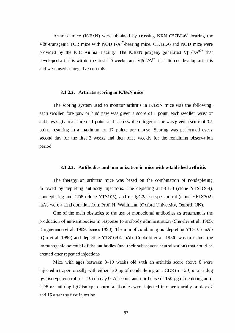
Helena Maria Lourenço Carvalheiro THE ROLE OF CD8 + T CELLS IN THE PATHOGENESIS OF RHEUMATOID ARTHRITIS Tese de Doutoramento em Ciências e Tecnologias da Saúde, especialidade de Biologia Celular e Molecular orientada pela Doutora Maria Margarida Souto Carneiro e pela Professora Doutora Maria Celeste Fernandes Lopes, apresentada à Faculdade de Farmácia da Universidade de Coimbra 2014
Transcript
Helena Maria Lourenço Carvalheiro
THE ROLE OF CD8+ T CELLS IN THE PATHOGENESIS
OF RHEUMATOID ARTHRITIS
Tese de Doutoramento em Ciências e Tecnologias da Saúde, especialidade de Biologia Celular e Molecular
orientada pela Doutora Maria Margarida Souto Carneiro e pela Professora Doutora Maria Celeste Fernandes Lopes,
apresentada à Faculdade de Farmácia da Universidade de Coimbra
2014
Imagem
i
Helena Maria Lourenço Carvalheiro
CD8+ T cells in the pathogenesis
of Rheumatoid Arthritis
Tese de Doutoramento em Ciências da Saúde, na especialidade de Biologia Celular e
Molecular, apresentada à Faculdade de Farmácia da Universidade de Coimbra para a
obtenção do grau de Doutor.
Orientadores: Doutora Maria Margarida Souto Carneiro e Professora Doutora Maria
Celeste Lopes.
Coimbra, 2014
iii
Front page art:
Reproduction of the painting “My Fear” by painter and RA patient Aleah Denton.
(reproduced with artist’s consent)
v
The research work presented in this thesis was performed at the Center for
Neuroscience and Cell Biology of Coimbra, University of Coimbra and at the Faculty of
Medicine of the University of Coimbra, Portugal, under supervision of Dr. Maria
Margarida Souto Carneiro and Prof. Dr. Maria Celeste Fernandes Lopes.
O trabalho experimental apresentado nesta tese foi elaborado no Centro de Neurociências e
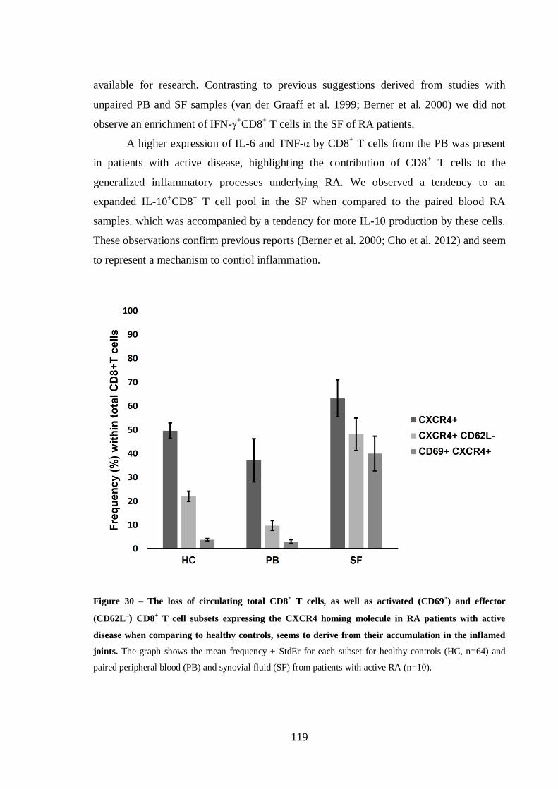
Biologia Celular de Coimbra e na Faculdade de Medicina da Universidade de Coimbra,
Portugal, sob supervisão da Doutora Maria Margarida Souto Carneiro e Professora Doutora
Maria Celeste Fernandes Lopes.
This work was funded by the Portuguese Foundation for Science and Technology, PhD
fellowship SFRH / BD / 60467 / 2009.
Este trabalho foi financiado pela Fundação Portuguesa para a Ciência e Tecnologia, bolsa
de doutoramento SFRH / BD / 60467 / 2009.
vii
Aos meus pais
A todos os doentes com Artrite Reumatóide
ix
The only real mistake is the one from which we learn nothing.
John Powell
Success is not final, failure is not fatal: it is the courage to continue that counts.
Winston Churchill
xi
Agradecimentos/Acknowledgements
Agradeço à Doutora Maria Margarida Souto Carneiro, por me ter acolhido no seu
laboratório no Centro de Neurociências e Biologia Celular em Coimbra. Agradeço toda a
confiança e apoio prestado em todas as etapas do meu doutoramento. Obrigada pela
disponibilidade que sempre demonstrou para discussões científicas, conselhos e sugestões
que permitiram a concretização deste trabalho, e me ajudaram a crescer como cientista.
Agradeço ao Professor Doutor José António Pereira da Silva, que sempre se
mostrou disponível para abrir novos caminhos científicos para este trabalho. Agradeço em
particular as discussões científicas assim como a sua disponibilidade e apoio ao longo
destes últimos anos.
Agradeço também à Professora Doutora Maria Celeste Fernandes Lopes, por ter
sido incansável durante este trabalho de doutoramento, em particular nesta última fase.
Gostaria também de agradecer à Professora Doutora Anabela Mota Pinto, pelo
apoio incondicional prestado em particular nesta última fase do doutoramento.
O meu muito obrigado à Doutora Cátia, pelo empenho nos estudos efectuados em
parceria com a Unidade de Reumatologia dos HUC, e por toda a ajuda prestada, em
particular na análise estatística.
Gostaria de agradecer aos meus colegas de laboratório, Tiago, David, Sandra,
The immune system comprises a complex array of molecules, cells and tissues
specialized in the discrimination between self and non-self molecules, leading to the
recognition and elimination of infectious agents, tumor and apoptotic cells among others.
In vertebrates, the immune system uses two different but integrated strategies to defend
itself from foreign elements: the innate and the adaptive immune responses.
1.1.1. The innate response
The innate response provides a first line of defense against pathogens. It is
characterized by a low degree of specificity and is classically defined as unable to generate
memory, however, this assumption has been reconsidered (Quintin et al. 2014). It includes
both physical barriers, such as the skin and mucosae, and chemical barriers, as the
complement system. The cells of the immune system responsible for the innate immune
response include macrophages, neutrophils, basophils, mast cells, eosinophils and a
specific subtype of lymphocytes: the natural killer (NK) cells (Parkin and Cohen 2001). T
lymphocytes are mostly involved in the adaptive immune response and only a small
subgroup of these cells, the NKT cells and γδ T cells (see below) are also members of the
innate response, behaving as a bridge between the two systems (Kabelitz 2011) and
expressing both T and NK cell surface markers (Chen and Freedman 2011). In fact, γδ T
cells are thought to play a role as antigen-presenting cells to adaptive immunity cells,
namely CD8+ T cells (Brandes et al. 2009), but also have a potent cytotoxic potential
(Chen and Freedman 2011). NKT cells, a separate lineage of T lymphocytes that express
surface markers that are typical of regular T and NK cells, can react with self and
microbial ligands and are thought to induce B cell activation (Galli et al. 2003; Van Kaer
2007). The lack of specificity classically attributed to innate immune responses can be
challenged, given that many of the above mentioned cells are equipped with Pattern
4
Recognition Receptors, such as Toll-like receptors (TLRs) or Killer-cell immunoglobulin-
like receptors (KIRs) capable of identifying a restricted variety of ligands. These receptors
include, for example, TLR4 which is capable of identifying gram-negative bacterial
structures, TLR9 which recognizes unmethylated CpG motifs present in bacterial DNA
(Janeway and Medzhitov 2002), and the KIRs, that interact with MHC class I molecules
(Vilches and Parham 2002). These receptors provide some level of specificity although not
as much as the T cell receptor (TCR), the B cell receptor (BCR) and immunoglobulins (Ig).
1.1.2. The adaptive response
The adaptive immune response is specific for a given antigen. It takes longer to
occur but it generates memory, so that a second exposure to the same antigen will trigger a
faster and more efficient response.
The adaptive response can be divided into two subtypes: the humoral and the cell-
based immune responses. The humoral response is characterized by the predominant
involvement of B lymphocytes, which produce specific antibodies against a given antigen.
The cell-based immune response is mediated by T lymphocytes, activated by the
recognition of peptides from foreign antigens presented by antigen-presenting cells
(APCs).
B lymphocytes can be distributed in different subsets according to their origin,
function, and localization. Different clones of B cells, all expressing the B cell receptor
(BCR) have a unique specificity. Each BCR, when in contact with their cognate antigen,
triggers a series of intracellular signals that lead to the activation, differentiation and
generation of plasma and memory B cells (Tobon et al. 2013).
The development of B cells starts in the bone marrow, where lymphoid progenitors,
with the help of stromal cells, further differentiate into pro-B cells, and undergo V(D)J
recombination1 to generate a functional BCR with IgM isotype, and undergo a negative
selection process, in order to eliminate autoreactive cells. After reaching the immature
stage, B cells leave the bone marrow and leave to secondary lymphoid tissues, where they
1 V(D)J recombination: also known as somatic recombination, it is the genetic recombination that occurs in
the primary lymphoid tissues (bone marrow for B cells and thymus for T cells). It leads to the production of
B and T cell receptors by primary B and T cells, by randomly combining genes of the Variable, Diverse and
Joining segments, thus forming proteins that are able to recognize a multitude of antigens.
5
develop into naïve and mature B cells, characterized by the expression of IgD in addition
to IgM (Tobon et al. 2013). Upon arriving in the spleen, B cells give rise to type-1 (T1)
and type-2 (T2) transitional B cells. T1 cells are short-lived and require BCR stimulation to
develop into T2 B cells (Sims et al. 2005). The latter can further differentiate into mature
circulating lymphocytes that will generate germinal centers, or non-circulating
lymphocytes that will settle in the marginal zone (Tobon et al. 2013). Upon encountering
their cognate antigen, activated B cells undergo proliferative expansion and differentiation
in the germinal center, where somatic hypermutation2 and immunoglobulin class switch
3
recombination take place, and further develop into either antibody producing plasmablasts
or memory B cells.
The T cell compartment comprises two major subtypes, which have been identified
for decades, the CD4+, classically designated Thelper/inducer (Th) cells and the CD8
+ also
named cytotoxic/suppressor T cells (Tc or CTLs).
The CD4+ T cell subtype includes Th1, Th2, Th9, Th17, Th22 and T regulatory
(Treg) subsets, which are mainly characterized on the basis of their cytokine production,
reflecting distinct functions in the course of an immune response. Th1 cells produce IFN-γ
and are responsible for phagocyte activation and for inducing the production of opsonizing
and complement-fixing antibodies. Accordingly, they play an important role in protection
against intracellular pathogens, but promote inflammation in autoimmune diseases. Th2
cells produce IL-4, IL-5, IL-9 and IL-13, thus playing a critical role in the immune
response against helminthes, invading cutaneous or mucosal sites, but can also be
responsible for the development of allergic disorders (Annunziato and Romagnani 2009).
Th17 cells produce IL-17, IL-22, and IL-26, and have been strongly implicated in the
pathogenesis of autoimmune diseases, such as rheumatoid arthritis (Lubberts 2010). Recent
studies have indicated that Th17 cells can convert into Th1 cells and acquire the ability to
produce IFN-γ. Both subsets, Th1 and Th17, are believed to exert decisive deleterious
effects in inflammatory disorders (Annunziato and Romagnani 2009). The Th9 and Th22
subsets are recent additions to the Th repertoire. Th9 cells produce high levels of IL-9,
while Th22 cells are potent producers of IL-22 and TNF-α. Both subsets appear to be
2 Somatic hypermutation: process occurring in activated B cells consisting in the introduction of mutations to
the variable region genes, leading to the production of high-affinity antigen receptors. 3 Immunoglobulin class switching: mechanism by which an activated B cell changes the class of antibodies it
produces (IgA, IgD, IgE, IgG or IgM) for another upon encountering their cognate antigen.
6
involved in the pathogenesis of autoimmune diseases (Kaplan 2013). Tregs are a subset of
T cells that facilitate peripheral immune tolerance. The most studied Tregs are the
CD4+CD127
-FoxP3
+CD25
+ population, and their main function is to suppress the immune
response either in a cytokine-independent manner, or through the production of IL-10 and
TGF-β (Anderson and Isaacs 2008).
The cell-based immune response involving CD8+ T cells will be discussed in detail
in the following chapters, as they are the main focus of this work.
1.1.3. CD8+ T cells
CD8+ T cells, or cytotoxic T lymphocytes (CTLs) or Tc, play a major role in the
protection against infectious agents and pathogens, and can also eradicate malignant cells.
An extensive array of molecular and cellular signals drive the development and
differentiation of naïve CD8+ T cells into effector and memory cells. These subsets are
especially known to induce and promote the inflammatory process and secrete
proinflammatory cytokines and proteolytic enzymes. However, CD8+ T cells can also
suppress immune responses through the production of anti-inflammatory cytokines.
Nevertheless, a predominance of proinflammatory over anti-inflammatory signals is
needed for an effective response against pathogens, while a predominance of inhibitory or
suppressive signals are required for the maintenance of tolerance against self-antigens, and
the altered CD8+ T cell response can lead to either the persistence of pathogens or
autoimmune disorders (Andersen et al. 2006).
1.1.3.1. CD8+ T cell development
Lymphocyte precursors arise from hematopoietic stem cells, in the bone marrow.
Their development can take two different pathways. While B cells finish their development
in the bone marrow, a subset of lymphoid progenitors leave the bone marrow and migrate
into the thymus, where they fully develop into the various subtypes of T cells. These cells
comprise the TCRαβ+ T cells which include the CD4
+ and CD8
+ T cells, and the TCRγδ
+ T
cells (Figure 1).
7
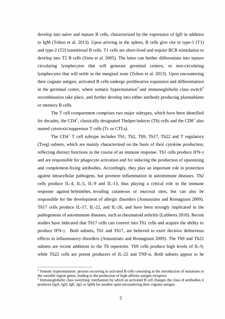
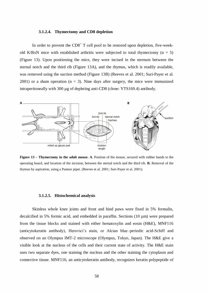
Figure 1 – CD8+ T cell development and differentiation. ① Medulla; ② Cortico-medullary junction; ③
Cortex; ④ Subcapsular zone. CD8+ T cell precursors develop from hematopoietic stem cells (HSC) in the
bone marrow, and migrate through the bloodstream as hematopoietic precursors (HP) into the thymus. The
HP cells enter the thymus in the cortico-medullary junction ② where they become committed to a T cell
lineage as lymphoid progenitors (LP). They then migrate to the cortex ③, where they become double
negative T cells (DN). As they further develop, DN cells migrate to the subcapsular zone ④ to form fully
functional TCRs. The αβ committed cells then migrate back into the cortex where they acquire both CD4 and
CD8 receptors, thus becoming double positive (DP) T cells. These cells then undergo a positive selection.
The selected DP cells that pull through selection become single positive T cells, committing to the CD4 or
CD8 lineage and then migrate into the medulla ①, enter the blood stream and migrate to lymphoid organs
where they will reside as naïve T cells. Upon priming with the right antigen, CD8+ T cells expand and
acquire an effector phenotype. Upon antigen clearance CD8+ T cells can undergo different fates: apoptosis,
the conversion into central memory CD8+ T cells, and the differentiation into effector memory cells. Upon
exposure to the antigen, effector CD8+ T cells can also differentiate into suppressor T cells, which down-
regulate the immune response. If the antigen persists, the CD8+ T cells suffer exhaustion, due to a continuous
activation. (Carvalheiro et al. 2012)
Differentiation and maturation of T cells occur within defined thymic areas: the
subcapsular region, the cortex, the cortico-medullary junction and the medulla (Petrie and
Zuniga-Pflucker 2007). The cortex comprises mainly immature thymocytes surrounded by
cortical epithelial cells and scattered macrophages, while the medulla consists of mature
thymocytes surrounded by medullary epithelial cells, macrophages and dendritic cells. The
lymphoid precursors arrive in the thymus through the bloodstream and seed into the
cortico-medullary junction. At this stage, the lymphoid progenitors are still uncommitted,
retaining myeloid, B and T cell potential (Luc et al. 2012). These lymphoid progenitors
8
then receive signals through the Notch1 receptor which activate specific genes, and induce
T cell lineage determination (Pui et al. 1999). They first evolve into double negative T
cells (CD4-CD8
-), which migrate into the cortical areas where they undergo further
differentiation steps. During their double-negative stage, T cells will also rearrange their β,
γ and δ genes to generate functional TCR chains and thus commit to the major aβ or γδ T
lineages (Burtrum et al. 1996). The main lineage, αβ TCR pathway, leads to the
differentiation into CD4+ or CD8
+ T cells. The γδ lineage leads to the γδ T cells which are
found in mucosae as part of the innate immune response, and may also function as APCs
(Brandes et al. 2009). Differentiation into the αβ or γδ T cells depends on the surface
expression or signaling potential of the γδ TCR complex. A strong signal favors the γδ
lineage development, while a weak γδ signal potentiates the αβ lineage (Hayes et al. 2005).
The αβ-committed lineage of double-negative thymocytes evolves into double positive
CD3+ T cells, as they express both the CD4 and the CD8 surface molecules. These cells are
produced in large numbers, but after positive selection their vast majority undergoes
apoptosis. Cells bearing an αβ TCR complex that recognizes the self-MHC complex with
an intermediate avidity will be positively selected to further differentiate, while their
counterparts will be eliminated (Klein et al. 2009). These selected double-positive
immature T cells then commit to the CD4+ or CD8
+ T cell lineages, and become single-
positive thymocytes. At this point, these semi-mature thymocytes migrate into the medulla
where they undergo negative selection: those harboring TCRs with a high affinity to self-
antigens are eliminated, thus reducing the risk of autoimmune disorders (Klein et al. 2009).
Once in the medulla, the single-positive thymocytes will upregulate the sphingosine-1
phosphate receptor (S1P1) that is required for T cells to leave the thymus (Weinreich and
Hogquist 2008), and further differentiate into other subtypes.
1.1.3.2. CD8+ T cell differentiation and subtypes
CD8+ T cells are currently classified into four subtypes, corresponding to different
levels of differentiation, activation status and cytokine production: Naïve, Effector, Central
memory and Effector memory (Figure 1).
9
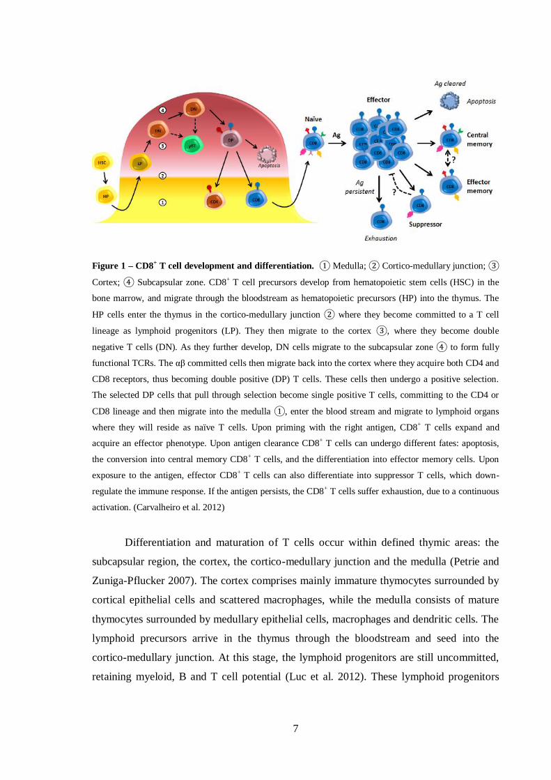
Table 1 - CD8+ T cell phenotypes
Naïve Effector Effector
memory
Central
memory
CCR7 +++ - +/- +/-
CD27 +++ - +++ +++
CD28 High Low Low High
CD45RA +++ -/+ - +/-
CD45RO - - +++ +++
CD62L +++ - - +++
Naïve CD8+ T cells still have not encountered their cognate antigen, and thus have
not been primed. They are usually found in the peripheral blood and lymphatic tissues
(Kaech and Ahmed 2001). The central memory subtype is already endowed to a specific
antigen whose presence will induce a strong proliferative response, as well as the
production of a variety of cytokines. Effector CD8+ T cells have proliferative and cytotoxic
properties. They can induce death of infected cells by cytolysis, through the secretion of
cytolytic proteins such as perforin and granzymes. Effector memory CD8+ T cells have
intermediate properties, presenting a lower ability to induce cytotoxic responses than
effector cells, and a much higher capacity to produce cytokines than the memory subtype
(Tomiyama et al. 2002).
Cell surface markers offer an expedite way to distinguish these CD8+ T cell
subtypes. This is based in the presence or absence of co-stimulatory (CD27, CD28,
CD45RA) and adhesion (CD62L) molecules and the chemokine receptor CCR7 (Kaech et
al. 2003). Naïve CD8+ T cells are characterized by the presence of CD27, CD28hi,
CD45RA, CD62L and CCR7. Effector cells express low levels of CD28 and are negative
for all other cell surface markers, while central memory cells can lose the expression of
CD45RA along with CCR7. The effector memory subtype is characterized by the absence
of CD62L and CCR7, the expression of CD28low, while the expression of CD45RA may
vary (Tomiyama et al. 2004) (Figure 1 and Table 1).
Our current understanding indicates that upon antigen encounter, naïve CD8+ T
cells differentiate into effector cells and undergo clonal expansion. Once the antigen is
cleared, 90-95% of all effector cells undergo apoptosis, while the remaining ones
differentiate into central memory CD8+ T cells, thus entering a resting (but vigilant) state.
The effector memory subtype is thought to represent an intermediate state occurring upon
10
the re-encounter of the antigen, when central memory CD8+ T cells gradually differentiate
towards an effector phenotype (Tomiyama et al. 2002).
CD8+ effector T cells are, therefore, characterized by their cytotoxic behavior (thus
the abbreviation Tc) through perforin, granzyme and Fas pathways. Several subtypes have
been identified based on cytokine production, these include the Tc1 subset (characterized
by the production of IFN-γ and not IL-4 and IL-5), and the Tc2 subset (secreting IL-4 and
IL-5 but not IFN-γ) (Mosmann et al. 1997). Both types can induce an inflammatory
response, with Tc1 and Tc2 inducing delayed-type hypersensitivity upon injection of Tc1
and Tc2 allospecific cells into mice bearing the target antigen (Li et al. 1997). Even though
both cell subtypes can induce inflammation, the Tc2-bearing mice had a higher eosinophil
infiltration, thus indicating that these may exert inflammation through a secondary pathway
by recruiting effector cells into the inflammatory site. The study of Tc1 and Tc2 functional
phenotypes also indicates that these cells can induce inflammation by activating CD4+
effector T cells, with Tc1 and Tc2 inducing a Th1 (cellular) and Th2 (humoral) response,
respectively (Vukmanovic-Stejic et al. 2000).
More recently, other functional subtypes have been identified. Special attention has
been devoted to the Tc17, characterized by the production of IL-17 and arising from the
same precursor as other functional subsets of CD8+ T cells (Kondo et al. 2009). Tc17 cells
are typically proinflammatory non-cytotoxic CD8+ T cells that express few or no cytotoxic
granules, and thus typically do not secrete granzyme B and perforin, although some subsets
can produce IFN-γ (Tajima et al. 2011). These cells seem to enhance inflammation in
various diseases, such as SLE (Henriques et al. 2010), immune thrombocytopenia (Hu et
al. 2011) and allergy-induced lung inflammation (Tang et al. 2012). Tc17 cells have also
been shown to promote immunity against infections, by Vaccinia (Yeh et al. 2010) and
Influenza viruses (Hamada et al. 2009), by promoting a proinflammatory response. A
subset of CD8+ T cells, is endowed with suppressor/regulatory capabilities, mediated by
IL-10 and TGF-β (Wang and Alexander 2009). These cells arise upon challenge by their
cognate antigen, and control inflammation by down-regulating the immune response by
effector T cells (Hu et al. 2004). These cells and their role in autoimmunity will be further
discussed.
11
1.1.3.3. Cytotoxic immune response
CD8+ T cells recognize pathogen peptides presented by MHC class I complexes on
the surface of APCs. During the first weeks after an acute infection with a pathogen both
the naïve and the central memory CD8+ T cells undergo activation and proliferation while
acquiring an effector phenotype. This is reflected by a down-regulation of the expression
of CD62L on the cell surface, accompanied by the production of granzymes and perforin,
as well as IFN-γ and TNF-α (Wherry and Ahmed 2004). Effector CD8+ T lymphocytes
cause the death of infected cells either by direct lysis, or by inducing apoptosis through the
activation of the Fas receptor (Barry and Bleackley 2002; Wong and Pamer 2003). After
the clearance of the infected cells, 90–95% of the effector cells undergo apoptosis, while
the surviving portion differentiates into a memory phenotype, regaining the CD62L
expression on their surface. This memory CD8+ T cell pool can later be reactivated,
proliferate and regain effector cytotoxic properties upon a re-encounter with the same
antigen.
Some infectious agents are readily eliminated, corresponding to acute self-limited
clinical manifestations. Chronic or latent infection-causing agents, such as viruses of the
herpes family, remain in the host indefinitely. In such cases, CD8+ T cells are permanently
stimulated and the cytotoxic response remains active, creating a persistent or even
expanding inflammatory response (Wong and Pamer 2003). In some patients, this chronic
state eventually leads to the exhaustion of CD8+ T cells: they gradually lose the ability to
produce cytolytic enzymes and even to proliferate, leading to a decline of the CD8+ T cell
population (Wherry et al. 2003). The exhaustion of CD8+ T cells is accelerated in the
presence of decreased numbers of CD4+ T cells, as they have an important role in
supporting the CD8+ T cell response (Matloubian et al. 1994).
CD8+ T cells exert important functions in the absence of infection: they are key
mediators in the clearance of some target cells, such as graft and tumor cells. In fact, CD8+
T cells have a crucial role in allograft rejection in mouse models (Tomita et al. 1990;
Yoshimura et al. 2000; Halamay et al. 2002), contributing to an accelerated immune
response (Yoshimura et al. 1998). Both Tc1 and Tc2 subsets can induce cardiac allograft
rejection by themselves without CD4+ T cell help. Tc1 cells are important in the early
rejection response, while the Tc2 subtype is involved in the recruitment of other effector
12
cells (Delfs et al. 2001). The cytotoxic behavior of CD8+ T cells is also involved in tumor
immunity, especially through the Tc1 subset (Kemp and Ronchese 2001).
1.1.3.4. Suppressor immune response
The suppressor T cells were initially described in the early 1970s, by Gershon and
colleagues (Gershon et al. 1972), along with classical cytotoxic T cells, as two cell subsets
with opposing roles in disease. Even though interest in CD8+ suppressor T cells faded with
time, they have regained attention in the last decade, in particular due to their possible role
in autoimmune disorders and antitumor activity (Niederkorn 2008).
As we have seen previously, the most widely known type of regulatory T cells is
CD4+CD25
+, commonly addressed as Tregs, and constitutes a distinct lineage of CD4
+ T
cells that arises in the thymus. They function as inflammatory response inhibitors and are
characterized by the production of IL-10 and TGF-β (Huang et al. 2005) or expression of
the transcription factor Foxp3 (Fontenot et al. 2003; Hori et al. 2003), and the loss of their
suppressive function is related to the onset of inflammatory diseases such as SLE (Sawla et
al. 2012). However, Kessel and colleagues have recently demonstrated that Bregs, that are
B cells that express high levels of CD25 on their surface and secrete IL-10 and TGF-β,
induce the production of Foxp3 by Tregs, thus contributing to the inhibition of
inflammatory responses (Kessel et al. 2012).
The CD8+ regulatory or suppressor T cells, commonly called Tcregs or Ts cells, are
less known, but behave in a similar manner to their CD4+CD25
+ counterparts (Cosmi et al.
2003). The most extensively analyzed Ts cells are the murine CD8+ expressing the β chain
of the IL-2/IL-15 receptor (CD122), which have a role in immunity through the production
and release of the anti-inflammatory cytokine IL-10 (Rifa'i et al. 2008). The adoptive
transfer of CD8+CD122
+ Ts cells into mice with established experimental autoimmune
encephalomyelitis (EAE) leads to an amelioration of the disease (Lee et al. 2008). CD122-
deficient mice are a model for autoimmune disease and are characterized by a high number
of abnormally activated T cells. The adoptive transfer of CD8+CD122
+ Ts cells into
CD122-deficient neonates fully prevents the development of these T cells, thus
maintaining T cell homeostasis (Rifa'i et al. 2004). Recently, the CD8+CXCR3
+ Ts cells
13
have been proposed as the human counterpart for the murine CD8+CD122
+ Ts cells, as
they have been shown to have a similar behavior in vivo and in vitro (Shi et al. 2009). CD8
suppressor T cells are thought to be involved in the onset of autoimmune disorders, such as
fibrotic disease, showing a lower suppressive activity (Fenoglio et al. 2012).
1.2. Autoimmune diseases
The immune system consists of an army of cellular and molecular elements whose
core function resides in protecting the body against harm induced by foreign elements. In
normal conditions, the immune system is “self-tolerant”, that is, it is unable to react against
“self” molecules, and thus does not react against endogenous components of the body.
However, when “self-tolerance” is lost, the immune system reacts against the body’s own
constituents, and this process may eventually result in autoimmune disease. Autoimmunity,
which was first described by Paul Ehrlich at the beginning of the 20th century as “horror
autotoxicus” (Murphy 2011), can, therefore, be defined as the result of a sustained immune
response directed against structures of the self, causing tissue damage (Bolon 2012).
Healthy individuals possess circulating, naturally occurring, auto-antibodies which
recognize self-antigens (Elkon and Casali 2008). Their presence indicates that under
normal physiological conditions these natural auto-antibodies act as house-keepers,
removing the debris resulting from natural cellular and tissue breakdown. Only when
autoimmune responses became uncontrolled and lead to exacerbated tissue damage or
symptoms are we in the presence of autoimmune disease.
Autoimmune diseases collectively affect 5% of the population in Western countries
(Jacobson et al. 1997) and they may affect virtually every organ and tissue in the human
body. Their etiology is essentially unknown, although it is believed to reside in the
interplay between both genetic and environmental factors. However, understanding what
triggers immune diseases has proven a difficult challenge, namely when it comes to
understand why so many healthy individuals present autoimmune processes but only a few
will develop clinically significant autoimmune disease (Sener and Afsar 2012).
14
1.2.1. Self-tolerance and its loss
Central tolerance is the process by which T and B cells are rendered unresponsive
to self-peptides during the maturation process in the thymus and bone marrow respectively.
This is the first checkpoint in the acquisition of tolerance to autoantigens.
As explained above, T cell development and maturation (CD4+ and CD8
+ T cells) is
based on a mechanism through which thymocytes are exposed to self-peptides bound to the
MHC complex. This process ultimately leads to the elimination of T cells that react to self-
antigens. However, some autoreactive T cells, with low affinity to these antigens, escape
the negative selection process and enter the blood stream (Klein et al. 2009).
The central tolerance to self-antigens during the maturation of B cells occurs in the
bone marrow. Immature B cells express a BCR molecule on their surface and will undergo
a negative selection process that determines whether the immature B cell will continue its
maturation. This mechanism can lead to the elimination of as much as 50 to 75% of
immature B cells at this stage. Again, some B cells with low autoreactivity levels escape
the negative selection and differentiate into mature B cells (Pelanda and Torres 2012).
In healthy individuals, other mechanisms in the periphery contribute to the active
removal of self-reactive T and B cells. This is done either by directly eliminating the
autoreactive T cells or through regulatory processes that render these cells inactive.
Peripheral tolerance can be obtained by three different processes: clonal ignorance, death
by deletion and induction of functional unresponsiveness (Srinivasan and Frauwirth 2009;
Mueller 2010). Self-reactive cells that escape the negative selection process but are
endowed with low affinity to self-antigens are the most likely to experience clonal
ignorance: because they have an avidity for the self-peptides that is generally lower than
that required to induce peripheral T cell activation, they are “ignored”. Clonal ignorance
may also be achieved when the cognate self-antigen is restricted to an immune privileged4
site. Under normal conditions, naïve T cells are presented their cognate antigen by
dendritic cells (DCs), in lymph nodes. In order to completely activate a naïve T cell, two
signals are required: the activation signal produced by the interaction of MHC-Ag (cognate
antigen within an MHC molecule) with the TCR, and the simultaneous costimulation
4 Immune privilege: Condition in which selected immune responses are suppressed or excluded in certain
organs. Certain sites in the human body, such as the cornea, tolerate the introduction of antigens without
triggering an immune response. The brain, the placenta and the cornea are all immune privileged sites.
15
signal sent by the DC’s molecules to the naïve T cells. Self-antigens are usually presented
by quiescent DCs, which have a reduced number of costimulatory molecules on their
surface, thus failing to produce the second stimulus required for a full T cell activation –
they are, thus, “ignored”. Partially activated naïve T cells are found to be tolerant. These
cells fail to differentiate into fully functional effector T cells, and will ultimately be
rendered unresponsive or eliminated from the T cell repertoire (Redmond and Sherman
2005; Srinivasan and Frauwirth 2009; Mueller 2010).
Functional unresponsiveness and deletion of autoreactive T cells occur upon their
partial activation due to the absence of costimulatory signals from APCs. Both confer
different forms of tolerance, but the mechanisms activating one pathway or the other are
still largely unknown. However, antigenic persistence has been shown to be an important
factor leading to tolerance by deletion (Redmond et al. 2003; Srinivasan and Frauwirth
2009; Nurieva et al. 2011), and is dose-dependent, with high doses of antigen leading to an
incomplete deletion, and low doses leading to complete deletion of the Ag-specific T cells
(Srinivasan and Frauwirth 2009).
Functional unresponsiveness, also called anergy, is a state in which a T cell that has
been exposed to an antigen becomes refractory to any further stimulatory signals. Anergic
cells are characterized by the lack of proliferation and IL-2 production, an irregular
effector function, a defective MAPK signaling pathway, a reduced intracellular calcium
mobilization and a decreased tyrosine phosphorylation. The exposure of T cells to high
doses of antigen can result in the functional unresponsiveness of these cells (Srinivasan
and Frauwirth 2009).
Tolerance breakdown occurs when mechanisms of central and/or peripheral
tolerance do not function properly, thus breaking the cellular homeostasis and triggering an
autoimmune disease.
1.2.1.1. Peripheral tolerance in CD8+ T cells
The establishment of peripheral tolerance in CD8+ T cells is particularly important,
as nearly every cell type can present these cells to their cognate antigen due to the presence
of MHC class I on all nucleated cells. Upon maturation and acquisition of cytotoxic
16
potential, CD8+ T cells will exert their cytotoxic function upon antigen presentation,
without requiring any additional stimuli. This stresses the need for peripheral tolerance
acting on these cells in order to prevent uncontrolled immune response (Redmond and
Sherman 2005; Srinivasan and Frauwirth 2009).
As seen previously, autoreactive naïve CD8+ T cells, which are only partially
activated by quiescent DCs upon recognition of a specific self-antigen, are deleted from the
repertoire. Exposure to persistent antigenic stimulation can also lead to tolerance, by
deletion of autoreactive CD8+ T cells or by induction of an anergic or unresponsive state.
Peripheral tolerance can also be induced in effector CD8+ T cells, and its main function is
to prevent naïve CD8+ T cells that escape the previous checkpoints of central and
peripheral tolerance from triggering an autoimmune response (Srinivasan and Frauwirth
2009). Fully activated CD8+ T cells undergo several rounds of proliferation and then
become quiescent. This state, known as activation-induced non-responsiveness (AINR), is
similar to the contraction phase occurring normally after intense CD8+ T cell responses
(Deeths et al. 1999). However, AINR can be reversed and from that point on, CD8+ T cells
can regain their proliferative potential and be activated without costimulatory signals
(Srinivasan and Frauwirth 2009). CD8+ T cells that are primed in the absence of CD4
+ T
cells, also called “helpless” T cells, also present a tolerant phenotype, and display a poor
recall response 5 (Kaech and Ahmed 2003), and undergo activation-induced cell death
(Janssen et al. 2005).
1.2.2. Role of CD8+ T cells in autoimmune diseases
CD8+ T cells have been implicated in the pathogenesis of autoimmune disorders
including diseases of the central nervous system (CNS) such as multiple sclerosis
(Annibali et al. 2011) or encephalomyelitis (York et al. 2010), diabetes mellitus (Wang et
al. 1996) and vitiligo (van den Boorn et al. 2009). The activation of CD8+ T cells that
recognize self-antigens, and are thus autoreactive, is mediated by the MHC: peptide
complex. The process through which these CD8+ T cells arise is still poorly understood,
5 Recall response: immune response elicited by memory lymphocytes to an antigen, which the immune
system has previously encountered.
17
even though these cells have been shown to have a preponderant role in autoimmune
disorders (Liblau et al. 2002).
In multiple sclerosis (MS) lesions in the brain, infiltrating CD8+ T cells were shown
to outnumber CD4+ T cells and to undergo clonal expansion locally (Babbe et al. 2000).
CD8+ T cells accumulation and clonal expansion has also been described in the
cerebrospinal fluid (CSF) and peripheral blood of these patients (Jacobsen et al. 2002). It
has also been demonstrated that T cells from MS patients frequently displayed resistance to
Fas-induced apoptosis, thus indicating that the cell death mechanism was altered in these
cells, making them prone to accumulation (Comi et al. 2012). These observations suggest
that CD8+ T cells are exposed to their cognate antigen in peripheral blood, CSF and MS
lesions in the brain. Recent data also indicate that MS patients have a higher number of
CNS-reactive CD8+ T cells in circulation than healthy individuals (Zang et al. 2004).
Studies with animal models of EAE have yielded controversial results, with CD8+ deficient
mice presenting a lower mortality but higher incidence of relapses (Jiang et al. 1992; Koh
et al. 1992; Kuchroo et al. 2002; Jiang et al. 2003; Montero et al. 2004; Lee et al. 2008;
York et al. 2010).
In the non-obese diabetic (NOD) mouse, an animal model for type I diabetes
mellitus, autoreactive CD8+ T cells are involved in the destruction of pancreatic β cells,
hence playing a key role in the pathogenesis of insulitis (Pang et al. 2009). Concurringly,
NOD mice treated with anti-CD8 antibody failed to initiate the disease (Wang et al. 1996).
Studies on a skin explant model of vitiligo demonstrated that perilesional CD8+ T
cells were capable of developing an autoimmune reaction against autologous skin explants,
efficiently lysing melanocytes, and inducing keratinocyte apoptosis (van den Boorn et al.
2009).
There is, therefore, a growing body of data suggesting that CD8+ T cells may be
involved in autoimmune diseases. This deleterious influence may be due to an excessive or
autoreactive cytotoxic activity, as suggested in the animal models of type 1 diabetes (Pang
et al. 2009) and EAE (Sun et al. 2001). Conversely, one may hypothesize that the disease
process may be enhanced by a reduced or deficient suppressor role by CD8+ T cells.
18
1.3. Rheumatoid arthritis
1.3.1. General perspective of the disease
Rheumatoid arthritis (RA) is a systemic and chronic autoimmune disease,
associated with a profound negative impact on quality of life, increased mortality and high
socioeconomic costs (McInnes and Schett 2011). RA is biologically mainly characterized
by synovial inflammation leading to chronic persistent pain, joint destruction and
associated deformity, systemic complications and progressive disability. Other organs and
tissues can also be affected by the inflammatory process. It affects around 1% of the
population in industrialized countries, being three times more frequent in women than in
men, with a peak incidence between 40 and 60 years of age (Scott and Steer 2007;
Klareskog et al. 2009).
The cause for RA is still unknown, but several factors (genetic and environmental)
play a role in the onset and course of the disease. A study in a cohort of twins estimated the
contribution of genetic factors to the disease to be about 50%, with the remainder
comprising environmental factors and chance (MacGregor et al. 2000; Klareskog et al.
2009). According to the current paradigm, in individuals that bear disease susceptibility
genes, specific environment factors may potentiate an immune reaction that will ultimately
lead to the production of autoantibodies. Later on in life, other events, such as infection or
trauma can contribute to further development of the disease pathogenesis, eventually
translating into joint inflammation. As the chronicity of the disease settles, patients will
display additional characteristics of the disease, such as joint deformity and systemic
manifestations associated with increased comorbidities (Klareskog et al. 2009).
The chronic inflammatory process is held as directly responsible for the destruction
of cartilage and bone However, the triggers and mechanisms involved in the origin of the
disease process remain vastly elusive (Williams et al. 2000; McInnes and Schett 2011).
Research over the past few decades has elucidated some of the mechanisms responsible for
the maintenance of the inflammatory process and its destructive ability. These efforts have
highlighted the extraordinary complexity of this disease. Although our current
understanding is far from complete, recent research has led to the development of
increasingly effective drugs that have gradually improved the outcome of the disease.
19
Among these new medications, biological agents targeting specific mediators of the
immune response are paramount.
1.3.2. Rheumatoid arthritis classification and clinical features
RA presents a broad spectrum of manifestations. The predominant symptoms are
pain, morning stiffness and swelling preferentially affecting the peripheral joints, in a
strikingly symmetrical fashion. The natural course of the disease is typically composed of
flares and partial remissions. Severity can be quite variable between individual patients,
ranging from mild symptoms without significant disability to a persistently active,
progressively crippling condition.
Table 2 - The 1987 revised classification criteria for Rheumatoid Arthritis (Arnett et al. 1988).
Criterion Definition
1. Morning stiffness Morning stiffness in and around the joints, lasting at least 1 hour before
maximal improvement
2. Arthritis of 3 or more joint areas
At least 3 joint areas simultaneously have had soft tissue swelling or fluid (not bony overgrowth alone) observed by a physician. The 14 possible areas
are right or left PIP, MCP, wrist, elbow, knee, ankle, and MTP joints
3. Arthritis of hand joints At least 1 area swollen (as defined above) in a wrist, MCP, or PIP joint
4. Symmetric arthritis Simultaneous involvement of the same joint areas (as defined in 2) on both
sides of the body (bilateral involvement of PIPs, MCPs, or MTPs is
acceptable without absolute symmetry)
5. Rheumatoid nodules Subcutaneous nodules, over bony prominences, or extensor surfaces, or in
juxtaarticular regions, observed by a physician
6. Serum rheumatoid
factor
Demonstration of abnormal amounts of serum rheumatoid factor by any
method for which the result has been positive in <5% of normal control
subjects
7. Radiographic changes Radiographic changes typical of rheumatoid arthritis on posteroanterior hand
and wrist radiographs, which must include erosions or unequivocal bone
decalcification localized in or most marked adjacent to the involved joints
(osteoarthritis changes alone do not qualify)
* For classification purposes, a patient shall be said to have rheumatoid arthritis if he/she has satisfied at
least 4 of these 7 criteria. Criteria 1 through 4 must have been present for at least 6 weeks. Patients with 2
clinical diagnoses are not excluded. Designation as classic, definite, or probable rheumatoid arthritis is
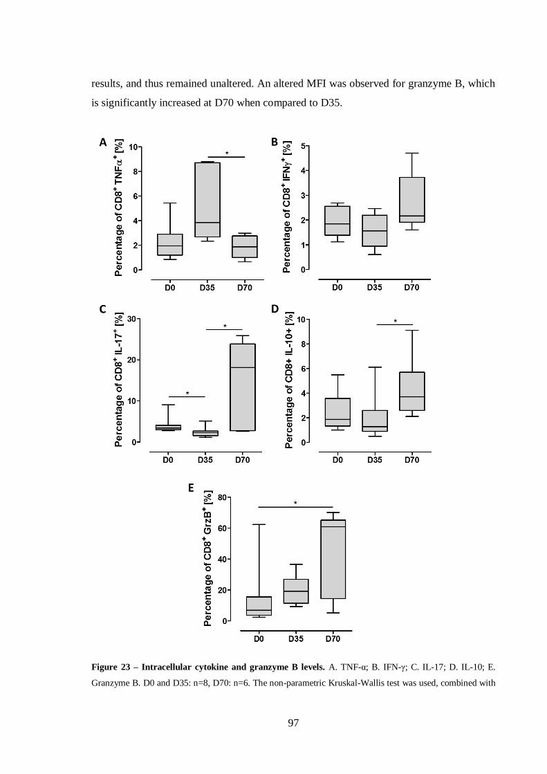
tissue (Boyce and Xing 2007). However, in RA there is an imbalance in favor of RANKL,
resulting in the overactivation of osteoclasts, which lead bone degradation. (Klareskog et
al. 2009).
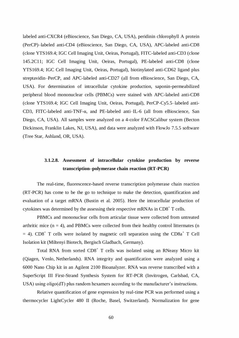
Figure 6 – miRNAs in the regulation of synovial fibroblasts in RA (FLS). MiR-155 has an increased
expression in FLS, and is further upregulated due to proinflammatory stimuli. The increased expression of
miR-155 suppresses stimulated expression of MMP-1/MMP-3, indicating that miR-155 regulates the
destructive properties of FLS. MiR-146 is also upregulated in RA, and inhibits the expression of TRAF6 and
IRAK1, both regulators of NF- κB, indicating that miRNAs have a role in the inflammatory process. Unlike
miR-155 and miR-146, the expression of miR-124a is downregulated in FLS. As miR-124a inhibits the
expression of monocyte chemoattractant protein (MCP-1), its decrease could leads inflammation and tissue
damage (Furer et al. 2010).
32
Recent studies have revealed that the expression of miRNA 7 in RA patients is
impaired, and may contribute to the development of the disease (Nakasa et al. 2011). The
expression profile of various miRNAs was analyzed in RA patients, with special attention
to the fibroblast-like synoviocytes (FLS) (Figure 6). The miRNA miR-124a proved to be
downregulated in FLS from RA patients. Additionally, it was demonstrated that the
overexpression of this miRNA led to the obliteration of FLS proliferation and subsequent
arrest of the cell cycle (Nakamachi et al. 2009). Other miRNAs such as miR-146a and
miR-155 were shown to be overexpressed in synovial tissue (Stanczyk et al. 2008), both
contributing to the local inflammation. MiR-146a is overexpressed in CD4+ T cells from
the SF and is closely correlated with TNF-α levels (Li et al. 2010), while miR-155 is up-
regulated in macrophages form SF and synovial membrane and its inhibition leads to a
decreased production of TNF-α (Kurowska-Stolarska et al. 2011).
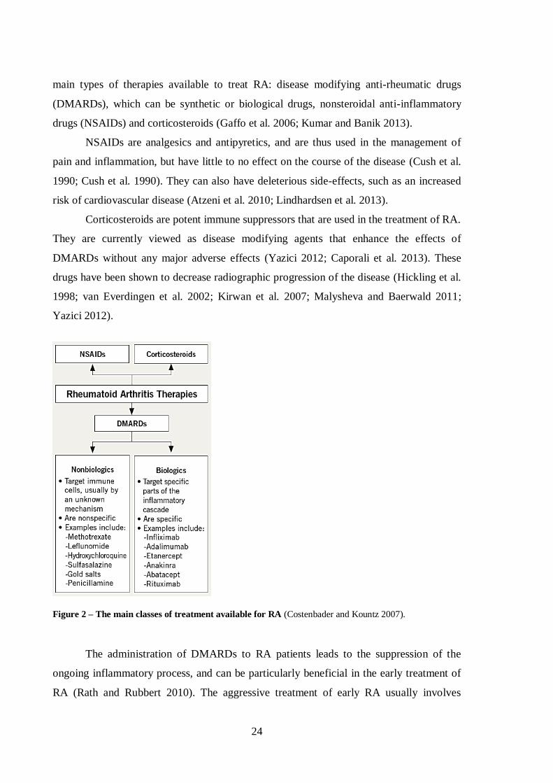
1.3.7. Biological agents currently used in RA
The knowledge revised above created the opportunity for the development of the
new biological agents that changed the clinical landscape of RA in this century.
Biologic DMARDs interfere directly with proinflammatory cytokines signaling
pathways, or cell to cell interactions (Figure 7). Biologic therapies currently available in
the clinic target TNF-α, IL-6 or IL-1, inhibit T cell co-stimulation or selectively deplete B
cells expressing CD20 on their surface (Scherer and Burmester 2009).
The first-line biologic therapy administered is TNF-α-inhibitory agents (Taylor and
Feldmann 2009). TNF-α is expressed at high levels in the inflamed joints of RA patients,
where they contribute considerably to the inflammatory process, therefore the use of anti-
TNF-α biologic agents tend to be highly beneficial (Navarro-Millan and Curtis 2013). The
combination of anti-TNF-α therapy with MTX has proven more effective than biologic
monotherapy (Choy et al. 2005; Soliman et al. 2011). However, as anticipated, anti-TNF-α
7 miRNA: Class of small endogenous non-coding RNAs of approximately 22 nucleotides that influence the
stability and translation of mRNA. miRNAs regulate gene expression by binding the 3’-untranslated region
of their target mRNAs leading to translational repression or mRNA degradation.
33
therapy significantly increases the risk of infection (about 2 fold) (Johnston et al. 2013).
No change has been documented in the risk of neoplasia.
Figure 7 – Overview of current and novel therapeutics used in the treatment of RA and their
mechanism of action (Scherer and Burmester 2009).
The IL-1 inhibitor, also called anakinra, has only a moderate therapeutic effect,
with the improvement conferred being markedly inferior when compared to studies using
other biologic agents(Mertens and Singh 2009). Conversely, the IL-6 inhibitor
(tocilizumab) was found very effective either in biologic therapy-naïve patients (Kawashiri
et al. 2013), or after a failed anti-TNF-α therapy (Tanaka et al. 2013), reaching remission
in a significant proportion of patients (Aguilar-Lozano et al. 2013).
Rituximab is a chimeric mouse/human monoclonal antibody that targets the CD20
molecule expressed on the surface of B cells, and further leads to the depletion of pre-B-
cell to memory B-cell stages (Nakou et al. 2009; Mok 2013). It is generally used in patients
who fail to respond to anti-TNF-α agents, (Finckh et al. 2007; Chatzidionysiou et al. 2011;
Soliman et al. 2012), and the concomitant administration of MTX leads to a better
34
outcome, with a significantly lower radiological progression of the disease when compared
to patients receiving monotherapy only (Cohen et al. 2006; Mok 2013).
Figure 8 – Mechanism of action of abatacept. Abatacept binds to CD80/86 on the surface of APCs and
blocks its interaction with CD28 on the surface of T cells, resulting in the inhibition of the co-stimulation of
T cells, thus preventing their activation. This mechanism further leads to the downregulation of the
inflammatory cascade and normalization of the levels cytokines and antibodies and inhibition of osteoclast
activity (von Kempis et al. 2012).
Abatacept is the only biologic DMARD currently in use that directly targets not
only CD8+ T cells, but total T cells by preventing their activation. It consists of the
extracellular domain of human cytotoxic T-lymphocyte-associated protein 4 (CTLA-4)
fused with the modified Fc portion of human immunoglobulin G1 (IgG1), and functions by
binding to the CD80 and CD86 molecules on the antigen-presenting cell surface, thus
inhibiting the binding of CD28 (Figure 8). It inhibits the co-stimulation of T cells, as
35
activated T cells have an important role in the pathogenesis of RA. Abatacept reduces T
cell proliferation and inhibits the production of proinflammatory cytokines, such as TNF-α,
IL-6 and IFN-γ, as well as MMPs (Weisman et al. 2006; Buch et al. 2009). The reduction
of proinflammatory cytokines leads to the inhibition of osteoclast activity, and the reduced
production of MMPs leads to a decreased cartilage degradation in the RA joint (von
Kempis et al. 2012). Abatacept is generally used when anti-TNF-α therapy is ineffective
(Gaffo et al. 2006; Nogid and Pham 2006; Buch et al. 2009; von Kempis et al. 2012).
The introduction of these biological therapies, together with new, targeted,
treatment strategies has operated a profound revolution in the treatment of rheumatoid
arthritis: disease remission, once seldom seen, has become the consensual objective of
therapy. It can be achieved in up to 60% of appropriately treated patients. Remission
provides the best assurance that bone erosion, loss of cartilage and functional deterioration
can he halted. This is achieved with manageable but not irrelevant toxicity.
Despite this, many patients still do not respond adequately to any of the
therapeutical agents available and there are no tools to predict response to individual
molecules. Further knowledge is dearly needed.
1.4. Mouse models of arthritis
Animal models have long had an important role in the study of the pathogenesis of
rheumatoid arthritis. These include induced-arthritis models and spontaneous arthritis
strains in rodents. In this section only mouse models of arthritis will be discussed.
1.4.1. Spontaneous arthritis models
1.4.1.1. K/BxN model
The K/BxN mouse model spontaneously develops an aggressive form of arthritis
and shares many features similar to those of human RA, including leukocyte invasion,
synoviocyte proliferation, pannus formation, synovitis, cartilage degradation and bone
erosion (Kouskoff et al. 1996; Korganow et al. 1999). This model also presents other
36
similarities with human RA, such as the polyclonal B cell activation with increased B cell
numbers, hypergammaglobulinemia8 and the production of autoantibodies. However, this
model lacks the production of RF, which is characteristic of RA (Ditzel 2004).
The K/BxN mice are originally originated from the crossing of KRN-C57BL/6
mice bearing a transgenic TCR (truncated Vβ6 TCR) with NOD (non-obese diabetic) mice,
which are known to be prone to autoimmune disorders.
The transgenic TCR Vβ6 from the KRN mice recognizes a bovine ribonuclease
peptide presented by I-Ak MHC class II molecule. Interestingly, the KRN transgenic TCR
in the context of the NOD-derived Ag7
MHC class II molecule also recognizes a peptide
(GPI 282–294) from the ubiquitous cytosolic enzyme glucose-6-phosphate isomerase
(GPI; EC 5.3.1.9), which catalyzes the interconversion of D-glucose 6-phosphate and D-
fructose-6-phosphate, an essential reaction of glycolysis and gluconeogenesis. This dual
specificity is responsible for inducing autoreactive T cells that cause severe arthritis with
an inset within the first 4-5 weeks of age (Ditzel 2004) (Figure 9). The autoreactive T cells
generated in the Vβ6-bearing K/BxN mice in the Ag7
background will help B cells by
presenting the autoantigen, and thus promote the production of anti-GPI autoantibodies.
Even though the arthritis developed in this model is due to the formation of
autoreactive T cells against a specific peptide in GPI, it was proven that the onset of
arthritis is triggered by autoantibodies. This was demonstrated by transferring serum or
purified immunoglobulin from TCR transgenic, I-Ag7
-positive K/BxN mice into wild-type,
B-cell-deficient and lymphocyte-deficient mice led to the rapid onset of arthritis, with
symptoms observed as early as 24 hours after the transfer, but unlike the arthritis
developed in K/BxN mice, this form of arthritis is transient, and is resolved in 15 to 30
days (Korganow et al. 1999).
GPI, which is known for being an isomerase that catalyzes an essential reaction in
gluconeogenesis. Nevertheless, multiple identities have been attributed to the secreted form
of this protein, such as neuroleukin (NLK) or autocrine motility factor (AMF). NLK was
found to be a lymphokine9 produced by activated T cells, and induced the differentiation of
B cells into antibody-secreting B cells (Gurney et al. 1986; Gurney et al. 1986). AMF was
8 Hypergammaglobulinemia: condition in which the patient has an abnormally high level of gamma
globulins, a class of plasma proteins which comprises antibodies. 9 Lymphokine: General term for any soluble protein mediators supposedly released by activated
lymphocytes, mainly T cells, on contact with an antigen. Lymphokines are believed to play a role in
macrophage activation, lymphocyte transformation, and cell-mediated immunity.
37
identified as a tumor product capable of inducing tumor cell migration, metastasis
formation and tissue invasion (Watanabe et al. 1996), and also promotes the maturation of
monocytes (Xu et al. 1996).
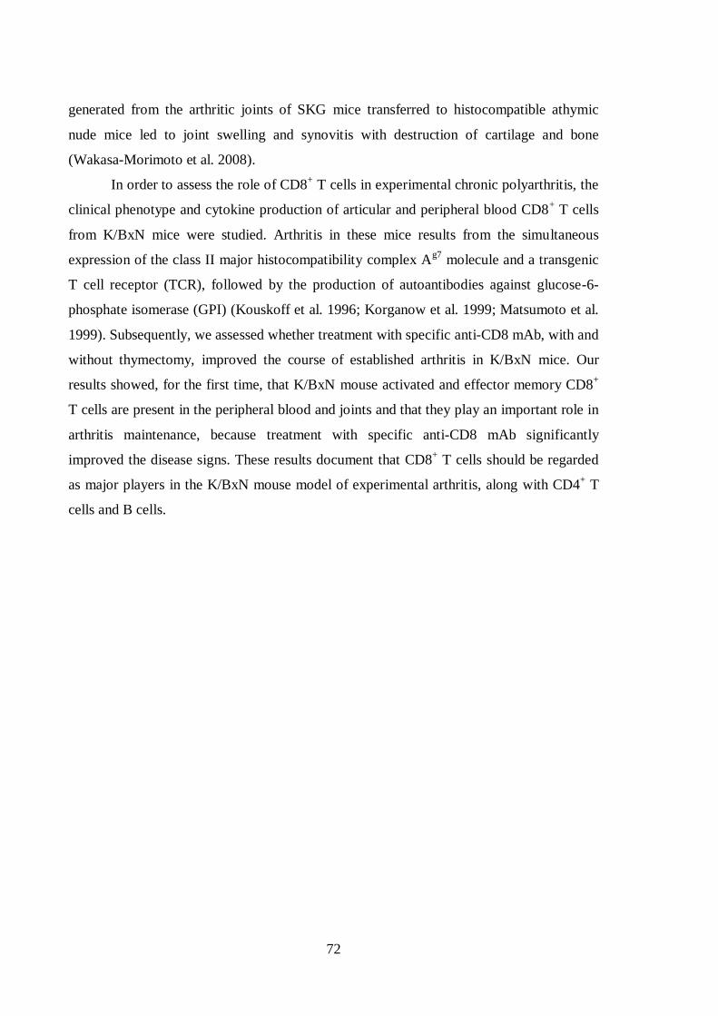
Figure 9 – Arthritis in K/BxN mice results from the dual specificity of the transgenic TCR. The KRN
TCR, which is specific for a peptide form bovine pancreatic ribonuclease (RNase 42-56) that is presented by
the MHC class II molecule I-Ak, also recognizes the self-antigen glucose-6-phosphate isomerase (GPI)
peptide (GPI 282–294) presented by the MHC class II molecule I-Ag7 from the NOD mice. In the NOD
background, autoreactive T cells help anti-GPI B cells and in turn produce anti-GPI antibodies (Ditzel 2004).
The K/BxN mouse model is thus relevant in the study of RA, as elevated levels of
GPI were found in the synovial fluid of RA patients (Cha et al. 2004; Schaller et al. 2005),
and the presence of these autoantibodies is associated with the HLA-DRB1 genotype in
Japanese patients (Furuya et al. 2008). However, the fact that other inflammatory arthritic
diseases present high levels of anti-GPI antibodies in the serum and synovial fluid
(Schaller et al. 2006), suggests that these antibodies may be involved in the perpetuation
rather than triggering the disease.
38
1.4.1.2. Other spontaneous arthritis models
Other transgenic spontaneous arthritis mouse models have been used in the study of
RA, such as the TNF-α transgenic mouse model, the SKG mouse strain or the
human/SCID chimeric mice.
The TNF-α transgenic mouse model was engineered to over-express the human
TNF-α, and was first described by Keffer et. al. (Keffer et al. 1991). This mouse model
develops a chronic inflammatory erosive polyarthritis, and the treatment with TNF-α
depleting antibodies completely prevents the disease (Keffer et al. 1991).
The SKG mouse strain is characterized by the presence of a point mutation in the
Zeta-chain-associated protein kinase 70 (ZAP-70), which is associated with thymic T-cell
selection defects, and leads to the onset of chronic arthritis at about 2 months of age
(Sakaguchi et al. 2003). However, they are influenced by their environment, and only
develop arthritis under conventional conditions, whereas they are healthy under specific
pathogen free (SPF) condition. In that case, arthritis can be induced by zymosan 10
(Kobayashi et al. 2006).
The human/SCID chimeric mice were initially originated by having SCID mice
implanted with human synovial tissue in the renal capsule (Geiler et al. 1994) and knee
joints (Sack et al. 1994), and both experiments indicated that the implants underwent
pannus formation and erosion of cartilage and bone, thus indicating that this model is
useful in studying pathogenetic aspects of joint destruction in RA.
1.4.2. Induced arthritis models
1.4.2.1. Collagen-induced arthritis
Collagen-induced arthritis (CIA) is widely used to study the pathogenesis of RA
and potential therapeutic targets, as it shares many similarities with human RA. It is
induced by immunization with emulsified autologous or heterologous type II collagen and
Freund’s adjuvant (Williams 2004), and develops through the generation of antibodies
10 Zymosan: polysaccharide from the cell wall of yeast, used to induce inflammation.
39
against type II collagen and self-peptides upon the breakdown of self-tolerance. CIA was
first studied in rats (Trentham et al. 1977; Trentham et al. 1978), and was subsequently
found to be also inducible in mouse strains (Courtenay et al. 1980; Wooley et al. 1981;
Stuart et al. 1982).
As in human RA, susceptibility to CIA is strongly associated with MHC class II
genes, developing mainly in strains containing the MHC class II H-2q haplotypes.
However, different strains display different degrees of susceptibility to the induction of
arthritis. The development of polyarthritis is accompanied by a T- and B-cell dependent
response to type II collagen (Holmdahl et al. 1985; Hom et al. 1986; Hom et al. 1986;
Zhang et al. 2002).
DBA/1 are the most frequently used mice in CIA studies. Clinical symptoms of
arthritis first appear 21-25 days after the first immunization, affecting preferentially the
joints of the limbs. Synovial inflammatory infiltration of polymorphonuclear and
mononuclear cells, pannus formation, eventually leading to cartilage degradation, bone
erosion and fibrosis are observed (Boissier et al. 1987). The peak of disease severity is
expected around day 35, after which DBA/1 mice enter remission. Similarly to human RA,
studies using homologous type II collagen have reported the occurrence of chronic
relapsing polyarthritis (Holmdahl et al. 1986; Malfait et al. 2001).
However, the induction of arthritis in DBA/1 mice has a major caveat: since the T
cell population peaks early and is in decline by the time of disease onset, the utility of this
model for studying T cell in the onset of the disease is limited. One alternative to DBA/1
mice are transgenic mice with C57BL/6 background. This strain was regarded as resistant
to CIA (Szeliga et al. 1996; Pan et al. 2004), but a new CIA protocol has successfully
managed to induce arthritis in these mice (Inglis et al. 2008). The C57BL/6 mice typically
develop arthritis 4-7 days later than DBA/1 mice, but with a comparable severity (Inglis et
al. 2007; Inglis et al. 2008). However, the incidence of the disease in the C57BL/6 mice is
lower than that of DBA/1 mice, and varies greatly among the different substrains with
C57BL/6 background.
CIA can also be successfully induced in the C57BL/10 (also called B10) strain.
These mice are very similar to the C57BL/6 strain, having been reported to differ only in 6
loci on chromosome 4 (McClive et al. 1994), and are often considered equivalent. Many
transgenic substrains of B10 mice that are commonly used in the induction of arthritis,
40
especially those bearing CIA susceptibility genes, such as the H-2q haplotype derived from
DBA/1 mice seen in the B10.Q strain (http://jaxmice.jax.org/strain/002024.html). The CIA
model is however known for having a variable incidence, severity and inconsistency
among different groups, which reflects the various strains sensitivity to environment,
maintenance conditions and stress.
1.4.2.2. Other forms of inducing arthritis
Collagen-antibody-induced arthritis (CAIA), an antibody-mediated model of
arthritis, is induced by using IgG antibodies against type II collagen. The disease onset
occurs within 48h of antibody administration, and develops in all strains, regardless of the
MHC class II haplotype. Even though the clinical development of the disease is similar to
that observed in CIA and RA, CAIA is characterized by the presence of macrophages and
polymorphonuclear cells in the inflamed joints (Santos et al. 1997), and is not driven by T-
or B-cells. Interestingly, the transfer of type II collagen reactive T cells was proven to
increase the disease severity (Nandakumar et al. 2004).
Other less known methods of induction of arthritis can also be used in mice, such as
the administration of zymosan and pristane. Zymosan, a polysaccharide found on the cell
wall of Saccharomyces cerevisae, can be injected into the joints of mice, resulting in the
local inflammation of the joint characterized by the infiltration of mononuclear cells,
synovial hypertrophy and pannus formation. Similarly, a single subcutaneous injection of
small amounts of pristane (2,6,10,14-tetramethylpentadecane), leads to a chronic relapsing
arthritis (Olofsson and Holmdahl 2007).
1.5. CD8+ T cells in the pathogenesis of Rheumatoid Arthritis –
Current knowledge
The role of CD8+ T cells in rheumatoid arthritis has attracted relatively little
attention. This is probably due to the remarkably conflicting results obtained with animal
41
models of polyarthritis, rendering researchers unable to discern if the global effect of CD8+
T cells in the disease process is protective or deleterious.
1.5.1. Lessons from animal models of arthritis
Mercuric chloride-induced arthritis in the Brown Norway rat is associated with
increased numbers of circulating CD4+ and CD8
+ T cells, and higher serum levels of IL-4
and IgE. The treatment of these animals with R73 (anti-aβ TCR monoclonal antibody
(mAb)) leads to a marked decrease in IgE and IgG levels as well as in B cell counts,
yielding an amelioration of the disease (Kiely et al. 1995; Prigent et al. 1995). In this
model, the depletion of CD8+ T cells with the OX8 depleting monoclonal antibody led to
reduced severity and incidence of the disease (Kiely et al. 1996). This was paralleled by an
increased production of IFN-γ, thus indicating a possible regulation of the disease through
a type I response (Kiely et al. 1996). These studies suggest an aggressive role for CD8+ T
cells in this disease model, presumably exerted through cytotoxicity. However, the
depletion of these cells with OX8 mAb in oil-induced arthritis in DA rats led to an earlier
onset of the disease, indicating a protective role, presumably mediated by their suppressor
functions (Jansson et al. 2000).
Studies using a depleting anti-CD3 antibody in collagen-induced arthritis in DBA/1
mice also argue for a protective role of CD8+ T cells in experimental arthritis. In the
repopulation of the T cell compartment after CD3-depletion, there was an enrichment of
CD4+ and CD8
+ T cells with regulatory/suppressor phenotype. Regulatory CD8
+ T cells
from treated mice were able to suppress IL-17 production, CD4+ T cell proliferation and
IFN-γ production. This suggests CD8+ T cells as responsible for maintaining the persistent
amelioration observed following anti-CD3 therapy (Notley et al. 2010). Taneja et al.
reported that transgenic CD8+ T cell deficient mice expressing the RA susceptibility gene
HLA-DQ8 have a higher incidence and severity of the disease than in the wild-type
counterparts. Conversely, the CD4+ T cell deficient mice failed to develop the disease.
These observations suggest that CD8+ T cells have a protective effect and CD4
+ T cells
have an initiator function in this model (Taneja et al. 2002). Studies with collagen-induced
arthritis (CIA) on B10.Q also suggest that CD4+ T cells have a globally deleterious
42
influence, mainly due to the IL-4 production, while CD8+ T cells appear to have little
effect on the disease. Moreover, CD8-deficient B10.Q mice show a tendency towards a
later onset of the disease, which might be related to the decreased production of
proinflammatory cytokines such as IFN-γ (Ehinger et al. 2001).
Conversely, CD8-/- DBA/1 mice are less susceptible to develop CIA on a first
collagen boost than their heterozygous counterparts, although the severity of the disease is
not significantly altered, thus indicating that CD8+ T cells may have a promoting role in
the initiation of the disease. After full recovery from the initial CIA, CD8-deficient mice
appear to be more susceptible to develop the disease than their heterozygous littermates,
thus indicating that CD8+ T cells may acquire a predominantly regulatory or suppressive
role (Tada et al. 1996).
The depletion of CD8+ T cells in BALB/c mice with proteoglycan aggrecan-
induced arthritis led to an aggravation of the disease, without affecting the amount of anti-
proteoglycan-antibodies at the peak of the disease (Banerjee et al. 1992).
The transfer of CD8+ T cells from thoracic duct lymph of adjuvant induced arthritic
DA rats into healthy normal syngeneic recipients failed to induce the disease (Spargo et al.
2001). However, the recipients had their normal CD8+ T cell population, which may have
eliminated the transferred CD8+ T cell population thus preventing the transference of the
disease by these cells. On the contrary, the transference of CD8+ T cell clones from SKG
mice, which develop a T cell-mediated autoimmune arthritis, to nude mice led to the
induction of arthritis and also pneumonitis, indicating that CD8+ T cells from this mouse
model are arthritogenic and have the ability to transfer the disease (Wakasa-Morimoto et
al. 2008).
Taken together, these studies suggest that CD8+ T cells have an important impact in
the pathogenesis of a variety of experimental models of arthritis, both in its initiation and
in the course of the disease. Additionally, they indicate that the global effect of eliminating
CD8+ T cells varies according to the disease model and the phase the disease. However, in
all those studies the total CD8+ T cell pool was manipulated, thus abrogating any insight
regarding the role of the different CD8+ T cell subsets. Since such subsets have distinct and
even opposing functions, it is plausible that the contradictions between studies might
derive, at least in part, from the importance of particular CD8+ T cell subsets in different
models and phases of the experimental disease. Hence, in our opinion, further studies
43
targeting particular CD8+ T cell subsets are indispensable to understand their role in
arthritis and explore their therapeutic potential.
1.5.2. Human studies
Several lines of indirect evidence suggest that CD8+ T cells are involved in the
pathogenesis of rheumatoid arthritis.
1.5.2.1. Circulating CD8+ T cells in patients and controls.
Several studies have looked for changes in the number and function of CD8+ T cells
in RA. Martinez-Taboada et al. compared the absolute numbers of circulating CD8+ T cells
in patients with active RA and healthy controls, concluding that RA patients tend to have
decreased numbers of circulating CD8+ T cells, though the differences failed to reach
statistical significance (Martinez-Taboada et al. 2001).
Peripheral blood CD8+ T cells from RA patients tend to have an increased
proportion of central memory phenotype (CD62L+CD45RA
-) while the proportion of the
effector memory subtype (CD62L-CD45RA
+) is decreased, in comparison with healthy
controls (Maldonado et al. 2003). Moreover, the levels of memory CD8+CD45RO
+ T cells
are correlated with the levels of IgM-rheumatoid factor (IgM-RF). It was also observed
that patients shifting from low to high levels of IgM-RF presented a decrease in naïve T
cells and an increase in the transient CD8+CD45RA
+CD45RO
+ T cell subset (Neidhart et
al. 1996).
A study of regulatory T cells in RA patients by Sempere-Ortells and colleagues
shows that increased numbers of regulatory CD8+CD28
- T cells correlated with the activity
of the disease, measured by the DAS28 (Disease Activity Score) (Sempere-Ortells et al.
2009). Little is known about changes in CD8+
T cell subpopulations in relation to disease
activity or effects of medications. Kao et al, reported that the regulatory CD8+CD11c
+
subpopulation, found to be highly expressed in an arthritic mouse model, is not correlated
with disease activity in RA patients (Kao et al. 2007).
44
1.5.2.2. CD8+ T cells in the synovial fluid
CD8+ T cells comprise approximately 40% of all T cells in the synovial fluid
(McInnes 2003). The analysis of serial synovial fluid samples obtained from different
arthritic joints in the same patient indicates that the CD8+ T cell accumulation in inflamed
joints is persistent (Masuko-Hongo et al. 1997). Furthermore, there is evidence that these
cells undergo clonal expansion in the synovial fluid, their TCR repertoire may be skewed,
they are genetically as well as environmentally determined, and can be induced by a
common antigen (DerSimonian et al. 1993; Fitzgerald et al. 1995; Hall et al. 1998).
CD8+T cells from synovial fluid of rheumatoid arthritis patients typically present
higher expression of both short-term and long-term activation markers (i.e. CD69 and
CD25) than observed in the peripheral blood (Afeltra et al. 1997). A study by Marrack and
colleagues has shown that type I interferons have the capability of keeping activated T
cells alive upon infection (Marrack et al. 1999), which can contribute to the high
percentage of persistently activated CD8+ T cells in RA joints. These cells (Tc1) are
characterized by the production of large amounts of IFN-γ, suggesting a potential to induce
local inflammatory responses, but also present an increased production of IL-10, which can
counteract the inflammatory process in the joint (Berner et al. 2000).
Autoreactive CD8+ T cells in rheumatoid inflamed joints have been characterized
as CD57+, oligoclonally expanded and in a terminal differentiation status. They are
functionally active but lack replicative capacity thus representing a state of “clonal
exhaustion” (Strioga et al. 2011). These cells are present in higher numbers in the synovial
fluid of RA patients than in matched peripheral blood (Arai et al. 1998).
The accumulating CD8+ T cells in the synovial fluid from RA patients are also
characterized by an oligoclonal TCR repertoire, i.e. different patients share the same TCR
sequence pattern. This is taken as a strong indicator of a common antigen-driven CD8+ T
cell response (Fitzgerald et al. 1995; Hingorani et al. 1996; Hall et al. 1998). It has been
suggested that the antigen driving this autoreactive CD8+ T cell response in RA may not be
related to the disease. The hypothesis was enunciated by Fazou et al. after observing that
the TCR repertoire of synovial fluid CD8+ T cells in RA patients was specific for several
types of virus, namely Epstein–Barr virus (EBV) (Klatt et al. 2005), cytomegalovirus and
influenza virus (Fazou et al. 2001). Another study reported that up to 15.5% of synovial
45
CD8+ T cells presented specificity for a single EBV epitope in a cohort of 15 EBV-
seropositive patients. These cells presented higher activation levels and increased secretion
of proinflammatory cytokines, suggesting that they could contribute to the maintenance of
the local inflammatory response (Tan et al. 2000). However, another study found little
correlation between disease progression and CD8+ T cell response to EBV in RA patients
(Berthelot et al. 2003).
Antibodies anti-BiP (immunoglobulin binding protein), can be found in the serum
of RA patients and in several mouse models of arthritis. CD8+ T cell clones responding to
BiP autoantigen are producers of IL-10, but also of other cytokines such as IFN-γ, IL-4 and
IL-5 (Bodman-Smith et al. 2003). This has been interpreted as an indication that CD8+ T
cells with a Tc2 phenotype can become regulatory upon BiP stimulation and undergo
clonal expansion locally, thus exerting a regulatory/suppressor function (Bodman-Smith et
al. 2000). In this line of thought, Davila and co-workers (Davila et al. 2005) demonstrated
that suppressor CD8+ T cells can be used as effective cell-based immunosuppressive
therapy. In fact, CD8+CD28
-CD56
+ T cell clones from synovial tissues of RA patients
displayed an anti-inflammatory immunosuppressive activity in NOD-SCID mice engrafted
with synovial tissue from RA patients. This was reflected by a decrease in the production
of proinflammatory cytokines and in the expression of activation markers by the engrafted
tissue. More recently, Cho et al. strengthened the hypothesis that CD8 exert a
predominantly suppressor effect in RA by showing that there is an accumulation of Ts cells
in the synovial fluid (Cho et al. 2012). However, a previous study observed a correlation of
CD8+ T cell numbers and proinflammatory cytokines in the synovial fluid of RA patients,
indicating that CD8+ T cells can produce high amounts of cytokines and thus contribute
actively to the inflammation and joint degradation in RA (Hussein et al. 2008).
1.5.2.3. CD8+ T cells in the synovial membrane.
Follicular structures – reminiscent of those found in secondary lymphoid organs –
can be found in the inflamed synovial membrane of approximately 50% of RA patients,
with a clearly organized ectopic germinal center present in approximately half of these
patients (Takemura et al. 2001). These structures are thought to contribute greatly to the
pathogenesis of RA due to their ability to produce autoantibodies, cytokines and
46
rheumatoid factor, which are known to contribute to tissue damage in this disease. Many
RA patients present T and B cell aggregates in the synovium that lack a typical germinal
center structure and have no follicular dendritic cells (FDCs). Along with these cells, the T
follicular helper cells, a subset of CD4+ T cells, is found in these follicular structures and
are thought to drive the B cell differentiation into plasma cells (Dong et al. 2011). This has
been interpreted as indicating that the formation of ectopic germinal centers in inflamed
joints depends solely on antigen recognition by TCRs and BCRs. The fact that T and B
cells can aggregate without the presence of FDCs can indicate that T and B cells may be
seeding in the synovial membrane prior to the FDCs, and may therefore be responsible for
their recruitment and maintenance in the synovial membrane (Takemura et al. 2001).
Indeed, the formation of ectopic germinal structures is associated with the local expression
of CXCL13, a strong B-cell chemoattractant that guides B cells into the synovium, thus
contributing to the formation of ectopic germinal structures and aggregates (Shi et al.
2001). Even though FDCs secrete large amounts of CXCL13, this chemokine can also be
produced by fibroblasts and endothelial cells (Weyand and Goronzy 2003).
The presence of ectopic germinal centers in the synovial membrane is associated
with a poorer disease prognosis (Wagner et al. 1998). CD8+ T cells are recognized as
essential for the formation of ectopic germinal centers in the synovial membrane of
inflamed RA joints. Indeed, after the engraftment with synovial membranes containing
ectopic germinal centers in NOD-SCID mice, they were treated with a depleting anti-CD8
antibody, which resulted in the disintegration of the synovial follicles, with a significant
decrease in the local production of TNF-α and IFN-γ (Wagner et al. 1998; Kang et al.
2002).
However, cytotoxic CD8+ T cells present in the synovial fluid contribute greatly to
the local increased production of proinflammatory cytokines, and may thus have a
predominantly deleterious effect in arthritis. Several studies have shown that CD8+ T cells
are as responsible as the CD4+ for type I proinflammatory cytokine secretion in the
synovial membrane (Berner et al. 2000).
47
CHAPTER 2
DRIVING HYPOTHESES
OBJECTIVES
49
2. Driving hypotheses and objectives
2.1. Driving Hypotheses
CD8+ T cells, formerly called killer T cells, have earned the reputation of being the
driving force behind proinflammatory processes, as they have the ability to induce cell
death in neighboring cells through the production of proteolytic enzymes, upon recognition
of a specific antigen. Concordantly, CD8+ T cells have been proven to play an important
role in the pathogenesis of several inflammatory disorders, such as multiple sclerosis
(Saxena et al. 2011) or allograft rejection (Halamay et al. 2002).
Research on the immune cells involved in the pathogenesis of rheumatoid arthritis -
regardless of using human samples or animal models- has mainly focused on the role of B
cells, CD4+ T cells and macrophages. Nevertheless, the few existing studies on CD8
+ T
cells present evidence that these cells are equally involved in the inflammatory process
underlying RA.
While it is unequivocal that CD8+ T cells have a role in the pathogenesis of RA, the
nature of that role, being it protective or deleterious, still remains to be elucidated. Indeed,
it is known that 40% of the T cells infiltrating the rheumatoid synovial membrane are
CD8-positive (McInnes 2003), however their importance in the pathogenesis and
maintenance of rheumatoid arthritis (RA) is still scarcely defined. Interestingly, many
studies have pointed towards a proinflammatory role of CD8+ T cells in RA (Fitzgerald et
al. 1995; Kang et al. 2002), while others defend that they have a protective role in RA
(Suzuki et al. 2008).
2.2. Objectives
In order to determine the role of CD8+ T cells in the pathogenesis of RA, the
following objectives were pursued:
50
• Understand the possible role played by the CD8+ T cells infiltrating the synovial
fluid in rheumatoid arthritis and the joint in animal models of experimental chronic
polyarthritis in initiating and maintaining disease chronicity;
• Phenotypic and functional characterization of CD8+ T cells isolated from the
synovial fluid and peripheral blood from RA patients comparing to healthy
controls, and from the articular infiltrate and peripheral blood of arthritic mice or
wild type controls;
• Define the similarities and differences in CD8+ T cell involvement in the
pathogenesis of RA and in the pathogenesis of experimental chronic polyarthritis,
to test the suitability of the animal models for in vivo studies of CD8+ T cell role in
chronic polyarthritis;
• Explore the therapeutic potential of manipulating CD8+ T cell function (through
blockade, or depletion) to ameliorate and/or reverse disease progression and signs
in the mouse model of chronic spontaneous polyarthritis K/BxN.
51
CHAPTER 3
MATERIALS AND METHODS
53
3. Materials and methods
3.1. Mice
3.1.1. Common procedures
3.1.1.1. Mouse breeding conditions
The KRN, NOD, K/BxN and B10.Q mice were group-housed in type III-H cages
(Tecniplast, Italy) and maintained in specific environmental conditions (22-24ºC, 45-65%
humidity, 15 changes/hour ventilation, 12 h artificial light/dark cycle) and free access to
irradiated standard rodent chow (4 RFN/I GLP certificate, Mucedola, Italy) and acidified
water (at pH 3.5 with HCl to avoid bacterial contamination). The research procedures were
carried out in accordance with the European directives (Directive 86/609/EEC and
Directive 2010/63/EU) on the protection of animals used for scientific purposes, and
according to the ethical standards for animal manipulation.
3.1.1.2. Blood collection
Blood collection from K/BxN and B10.Q mice was performed through the section
of the lateral caudal veins. The mice were heated under a heating lamp, and then
anesthetized with the volatile anesthetic isofluorane (IsoFlo®, Esteve Veterinaria,
Portugal). When the mouse reached unconsciousness, the lateral veins were incised with a
sterile surgical blade (Swann-Morton, Sheffield, UK), and the blood drops were collected
into blood collection tubes with either K2EDTA, or clot activator and gel for serum
separation (Microtainer™ Tubes, Becton Dickinson, New Jersey, USA). Blood samples
from K2EDTA were put in a blood tube rotator at room temperature to prevent blood clot
formation until they were processed.
54
3.1.1.3. Routes of administration
The antibodies and other treatments were administered to mice by intraperitoneal
injection in the left caudal abdomen, as it allows the administration of large quantities of
solution (Hirota and Shimizu 2012; Weiss and Bürge 2012). Every mouse was injected in
the left caudal abdomen (Figure 10), with up to 200 µl of solution, and using an insulin
syringe (Omnifix Duo, B. Braun, Germany).
Figure 10 – Intraperitoneal injection. Example of intraperitoneal injection in the left caudal abdomen of a
laboratory mouse with an insulin syringe (Hirota and Shimizu 2012; Weiss and Bürge 2012).
3.1.2. K/BxN poly-arthritis mouse model
The K/BxN spontaneous arthritis mouse model was first described by Kouskoff et
al. (Kouskoff et al. 1996). These mice were obtained by crossing the TCR transgenic KRN
strain with NOD mice expressing the MHC class II molecule I-Ag7
. The progeny bearing
both transgenic TCR and the Ag7
molecule spontaneously develop severe chronic and
destructive arthritis. They present high titers for antibodies recognizing glucose-6-
phosphate isomerase (GPI), and serum collected from these mice can induce arthritis in
other mouse strains (Kyburz and Corr 2003; Ditzel 2004). In this model the disease is
mainly mediated by TNF and IL-1, and involves the complement activation and mast cell
degranulation (Kyburz and Corr 2003; Ditzel 2004). The presence of anti-GPI antibodies
in this mouse model lead to the study of anti-GPI titers in RA patients, which has produced
conflicting results (Schaller et al. 2001; Matsumoto et al. 2003; Cha et al. 2004; Schaller et
al. 2005).
55
Figure 11 – K/BxN breeding. The K/BxN mice are generated from KRN+C57BL/6 mice possessing the Vβ6
transgenic TCR that are bread into NOD mice bearing the I-Ag7 MHC molecule.
The K/BxN mice are originated from the crossing of KRN-C57BL/6 mice bearing a
transgenic TCR with NOD mice (Figure 11). The mice are kept in a C57BL/6 background,
and the transmission of the Vβ6 transgenic TCR to the progeny is routinely assessed. The
expression of Vβ6 TCR is determined by flow cytometry, as seen in Figure 12, and KRN
mice expressing high levels of Vβ6 are selected for further crossing with the NOD breed.
The progeny expressing the transgenic TCR in the NOD background (Vβ6+
I-Ag7+
) will
develop arthritis within 4-5 weeks of age, while the littermates that have the KRN
background (Vβ6+ I-A
g7-) will be healthy and are used as controls.
56
Figure 12 – Selection for the Vβ6-bearing KRN-C57BL/6 mice for further crossing with NOD mice.
The expression of the Vβ6 transgenic TCR is determined in T cells, marked using the anti-CD3 anti-mouse
antibodies. The expression is considered positive in animals presenting a percentage above 20% of Vβ6-
expressing T cells.
3.1.2.1. K/BxN mouse breeding
The TCR-transgenic KRN mice were a kind gift from Dr. C. Benoist (Harvard
University, Boston, MA) and were maintained on a C57BL/6 background (K/B). The
KRN+C57BL/6
+ progeny bearing the Vβ6-transgenic TCR were identified at 3–4 weeks of
age by flow cytometry. The red blood cells were removed from the samples using red
blood cell (RBC) lysis buffer and were washed with phosphate buffered solution (PBS).
The peripheral blood cells were then stained using phycoerythrin (PE)–labeled anti-CD8
(clone YTS169.4; Instituto Gulbenkian de Ciência [IGC] Cell Imaging Unit, Oeiras,
Portugal) and fluorescein isothiocyanate (FITC)–labeled anti-Vβ6 (BD Pharmingen,
Becton Dickinson, Franklin Lakes, NJ, USA) antibodies. The samples were analyzed on a
4-color FACSCalibur system (Becton Dickinson, NJ, USA), and data were analyzed with
FlowJo 7.5.5 software (Tree Star, Ashland, OR, USA). The KRN+C57BL/6
+ mice with
over 20% of the CD8+ T cells expressing the Vβ6-transgenic TCR were selected for further
crossing with NOD mice while the Vβ6-negative mice were euthanized.
57
Arthritic mice (K/BxN) were obtained by crossing KRN+C57BL/6
+ bearing the
Vβ6-transgenic TCR mice with NOD I-Ag7
-bearing mice. C57BL/6 and NOD mice were
provided by the IGC Animal Facility. The K/BxN progeny generated Vβ6+/A
g7+ that
developed arthritis within the first 4-5 weeks, and Vβ6+/A
g7- that did not develop arthritis
and were used as negative controls.
3.1.2.2. Arthritis scoring in K/BxN mice
The scoring system used to monitor arthritis in K/BxN mice was the following:
each swollen fore paw or hind paw was given a score of 1 point, each swollen wrist or
ankle was given a score of 1 point, and each swollen finger or toe was given a score of 0.5
point, resulting in a maximum of 17 points per mouse. Scoring was performed every
second day for the first 3 weeks and then once weekly for the remaining observation
period.
3.1.2.3. Antibodies and immunization in mice with established arthritis
The therapy on arthritic mice was based on the combination of nondepleting
followed by depleting antibody injections. The depleting anti-CD8 (clone YTS169.4),
nondepleting anti-CD8 (clone YTS105), and rat IgG2a isotype control (clone YKIX302)
mAb were a kind donation from Prof. H. Waldmann (Oxford University, Oxford, UK).
One of the main obstacles to the use of monoclonal antibodies as treatment is the
production of anti-antibodies in response to antibody administration (Shawler et al. 1985;
Bruggemann et al. 1989; Isaacs 1990). The aim of combining nondepleting YTS105 mAb
(Qin et al. 1990) and depleting YTS169.4 mAb (Cobbold et al. 1986) was to reduce the
immunogenic potential of the antibodies (and their subsequent neutralization) that could be
created after repeated injections.
Mice with ages between 8–10 weeks old with an arthritis score above 8 were
injected intraperitoneally with either 150 µg of nondepleting anti-CD8 (n = 20) or anti-dog
IgG isotype control (n = 19) on day 0. A second and third dose of 150 µg of depleting anti-
CD8 or anti-dog IgG isotype control antibodies were injected intraperitoneally on days 7
and 16 after the first injection.
58
3.1.2.4. Thymectomy and CD8 depletion
In order to prevent the CD8+ T cell pool to be restored upon depletion, five-week-
old K/BxN mice with established arthritis were subjected to total thymectomy (n = 5)
(Figure 13). Upon positioning the mice, they were incised in the sternum between the
sternal notch and the third rib (Figure 13A), and the thymus, which is readily available,
was removed using the suction method (Figure 13B) (Reeves et al. 2001; Suri-Payer et al.
2001) or a sham operation (n = 3). Nine days after surgery, the mice were immunized
intraperitoneally with 300 µg of depleting anti-CD8 (clone: YTS169.4) antibody.
Figure 13 – Thymectomy in the adult mouse. A. Position of the mouse, secured with rubber bands to the
operating board, and location of the incision, between the sternal notch and the third rib. B. Removal of the
thymus by aspiration, using a Pasteur pipet. (Reeves et al. 2001; Suri-Payer et al. 2001).
3.1.2.5. Histochemical analysis
Skinless whole knee joints and front and hind paws were fixed in 5% formalin,
decalcified in 5% formic acid, and embedded in paraffin. Sections (10 µm) were prepared
from the tissue blocks and stained with either hematoxylin and eosin (H&E), MNF116
(anticytokeratin antibody), Herovici’s stain, or Alcian blue–periodic acid-Schiff and
observed on an Olympus IMT-2 microscope (Olympus, Tokyo, Japan). The H&E give a
visible look at the nucleus of the cells and their current state of activity. The H&E stain
uses two separate dyes, one staining the nucleus and the other staining the cytoplasm and
connective tissue. MNF116, an anticytokeratin antibody, recognizes keratin polypeptide of
59
45, 46 and 56.5 kDa, and has a broad pattern of reactivity with human epithelial tissues.
The Herovici’s stain is used to differentiate young and mature collagen , and the Alcian
blue–periodic acid-Schiff stain was used to mark glycoproteins (Yamabayashi 1987).
Images were analyzed with ImageJ 1.38x software (National Institutes of Health, Bethesda,
MD, USA).
3.1.2.6. Enzyme-linked immunosorbent assay (ELISA) for GPI