the synthesis, characterization anfor the next generation of bionicmoment is the design and fabricaforms for nely localized and targetmedical conditions, ranging from
Advanced Materials and Nanobiotechnology
University of Illinois, Chicago, IL 60607-7052
Cite this: RSC Adv., 2015, 5, 36614
Received 28th December 2014Accepted 27th March 2015
DOI: 10.1039/c4ra17180b
www.rsc.org/advances
36614 | RSC Adv., 2015, 5, 36614–3663
xyl channel in defining selectedphysicochemical peculiarities exhibited byhydroxyapatite
Vuk Uskokovic*
Mysteries surrounding the most important mineral for vertebrate biology, hydroxyapatite, are many.
Perhaps the Greek root of its name, apasao, meaning ‘to deceive’ and given to its mineral form by the
early gem collectors who confused it with more precious stones, is still applicable today, though in a
different connotation, descriptive of a number of physicochemical peculiarities exhibited by it.
Comparable to water as the epitome of peculiarities in the realm of liquids, hydroxyapatite can serve as a
paradigm of peculiarities in the world of solids. Ten of the peculiar properties of hydroxyapatite are
sketched in this study, ranging from (i) the crystal lattice flexibility to (ii) notorious surface layer instability,
(iii) finite piezoelectricity, pyroelectricity and conductivity to protons, (iv) accelerated growth and
improved osteoconductivity in electromagnetic fields, (v) a high nucleation rate at low supersaturation
and low crystal growth rate at high supersaturation, (vi) higher bioactivity and resorbability of biological
apatite compared to those of the synthetic ones, and beyond. An attempt has been made to explain this
array of curious characteristics by referring to a particular element of the crystal structure of
hydroxyapatite: the hydroxyl ion channel extending in the direction of the c-axis, through a
crystallographic column created by the overlapping calcium ion triangles.
1. Introduction
Figuration of structure–property relationships is engrained inthe heart of materials science, but it has suffered immensely in
uk Uskokovic is Director of thedvanced Materials and Nano-iotechnology Laboratory andssistant Professor in theepartment of Bioengineering athe University of Illinois at Chi-ago. His primary researchnterests include nanostructurediomaterials for controlled drugelivery and tissue engineeringpplications. Bridging the gapetween materials science andife science, his lab specializes ind biological testing of materialsimplants. His main goal at thetion of smart, theranostic plat-ed in situ remediation of variousbone infection to brain tumors.
the recent past. Demands for practicality and applicability innatural sciences have taken their toll. No longer is the main-stream materials scientist interested in paralleling practicalndings with fundamental insights. Rather, the former is givenpriority over the latter. As a result, methods for the synthesis ofmaterials with exciting properties multiply, though only rarelyfollowed up by their developers’ eagerness to comprehend thephysicochemical bases of the structural forms adopted by thematerials, and to link those with the characteristic types ofbehavior displayed by them. In this work, a major turnaroundhas been made with the attempt to look back at one of the keystructural features of a material that is of particular interest tobone biology, and to correlate it to some of the experimentallyobserved oddities displayed by this material and collected overthe past century.
The compound that is the subject of this review is hydroxy-apatite (HAp): Ca10(PO4)6(OH)2. It is a hydroxylated form ofapatite, Ca10(PO4)6(F,Cl,OH)2, the most abundant phosphorus-bearing mineral on Earth, ndable in igneous, sedimentaryand metamorphic rocks alike.1 In its calcium-decient andpartially carbonated form, representable as (Ca,Z)10(PO4,Y)6(OH,X)2 [Z ¼ Na+, Mg2+, K+, Sr2+, etc., Y ¼ CO3
2�, HPO42�, and
X ¼ Cl�, F�], it acts as the sole mineral component of bone. Itsrole in this complex and multifunctional organ is to impartsolid compressive properties to elastic but weak collagen bersand thus participate in a synergy thanks to which deciencies of
both the so and the hard components of bone alone becomeeffectively cancelled out.
HAp, interestingly, is a compound that appeals to the curi-osity of chemists owing to its broad range of peculiar properties,just about as much as water can. Like water, exhibiting so manyextraordinary properties in the realm of uids from (i) itshighest surface tension among all liquids except mercury to (ii)its density being highest in the liquid, not the solid state, at 4 �Cand 1 bar, (iii) its extremely low vapor pressure and an incred-ibly high boiling point compared to the neighboring hydrides inthe Periodic Table, (iv) its being one of the rare carbonlessliquids at room temperature, and (v) its being an almostuniversal solvent, HAp abounds with unexpected features thatreveal themselves to the studious and inquisitive eye of theresearcher. Perhaps due to Nature's nding utility in peculiarity,both of these compounds, water and HAp, have been chosen ascentral ingredients of the molecular machinery of life.
However, like most ceramics, HAp is difficult to work with. Itcannot be easily processed into any particle size or shape, asmetals can, nor can it be easily functionalized with therapeuticligands, as polymers, silica or gold can. It is also very unat-tractive to a casual observer – pale, fragile, not very conductiveto electricity or vibration as well as inexpensive, without reallydisplaying many appealing characteristics at the rst sight.Although in its mineral form it can be easily confused withmoreprecious stones, ranging from beryl to amethyst to olivine, adrop of lime juice is enough to start dissolving it, the reason forwhich A. G. Werner named it in 1786 using the Greek wordapasao, meaning ‘to deceive’ or ‘to mislead’.2 On top of this, itis highly defective in its most natural and functional, bioactiveand osteoconductive state. Yet, in spite of all this, it is thematerial selected by evolution to build the basis of ourbiophysical structures, yielding yet another glimpse into thewonders of natural creation whereby perfection is found almostexclusively in imperfection.
The central aim of this study, as elaborated in section 3, is toelucidate the idea that a particular structural feature of HAp,known as the hydroxyl ion channel, running straight throughthe center of the basal plane of its hexagonal lattice and parallelto the c-axis, is partially responsible for an array of its extraor-dinary characteristics. The preceding section 2, which followsnext, presents a compilation of unusual properties exhibited bythis strange member of the biomineral family: unappealing atrst, but to those who spend enough time examining it, it mayreveal semantic signs that extend across the world of atoms andmolecules and into the spheres of our psyches, our social livesand beyond.
2. Peculiarities of HAp2.1. Crystal lattice exibility
HAp has the capacity to undergo enormous lattice distortion asit accommodates cations and anions that differ in size fromCa2+ and PO4
3�, respectively. Approximately one-h of theCa2+ ions in biological HAp is substituted with cationic impu-rities and, for example, substitution of Ca2+ with Mg2+ reducesthe lattice constant c by 0.33% and increases the lattice constant
a by 0.1%,3 which itself is enough to produce signicant latticedisturbances and increase the solubility of the compound.4,5
The more frequent incorporation of CO32� ions in the hexag-
onal channel of the apatite structure (A-type HAp) leads to anincrease in lattice parameter a and amarkedly lesser decrease inparameter c in comparison with those of the stoichiometricHAp.6 In contrast, the parameter a decreases while c increaseswith a partial substitution of PO4
3� with the smaller CO32� ions
(B-type HAp).7
These changes in the crystal lattice parameters oen inducechanges in crystallinity, thermal stability, morphology, solu-bility and other physicochemical and biological properties ofthe material.8 The charge neutrality principle also implies theformation of structural defects in order to accommodate foreignions. The extraordinary stoichiometric exibility of thecompound manifests itself in accommodating such defects, asthe Ca/P molar ratio, equaling 1.67 for pure HAp, can dropdown to 1.3 without breaking of the crystallographic symmetry:P63/m. The crystal structure of stoichiometric HAp is, in fact,monoclinic (P21/b),9 but due to foreign ion inclusion, vacancyformation and Ca2+ depletion it becomes hexagonal (P63/m)10
for more disordered biological HAp. The same transition to acrystallographic state of lower symmetry is observed followingannealing at temperatures higher than 207 �C. This poly-morphic transition is reversible and the hexagonal symmetrytransforms back to the monoclinic one at temperatures lowerthan 204 �C in the cooling regime.11
Though the capacity to incorporate foreign ions may not beas large as that typifying silicate glasses, the lattice exibility ofHAp is large enough to accommodate one half of all theelements of the periodic table,12 in almost any valence state,from SiO4
4� to As5+. It is thanks to this effect of lattice exibilitythat bone can act as the mineral reservoir of the body and thestorage for toxic elements, thus fullling two of its essentialphysiological roles. Despite this exibility, the driving force forcrystallization is still sufficiently high to drive out most of theorganic molecules present at the moment of precipitation andlimit the loading capacity of the compound mostly to theamount that could be adsorbed on the surface. This surfacelayer of a drug bound by weak forces to a particle predisposesHAp to exhibit burst release, and prevents it from being used asa sustained release platform in its dispersed form. Powdercompaction and drug capturing within the pores is an approachthat overcomes this deciency of HAp,13–16 but limits the use ofsuch composites to their application as solid blocks. Althoughthe entrapment of uorophores has been reported for glassysilicate calcium phosphates prepared in an amorphous form inIgepal-based reverse micelles and stabilized with citric acid,17
the compound naturally present in bone coating HAp crystals at0.5 molecules per nm2 and preventing their coalescence in thecollagen matrix,18 it appears that loading HAp with organics inany amount greater than that present in nacre or tooth enamel(<3 wt%) via intercalation is not possible. The intra-crystallineloading also appears to be theoretically possible but only forsufficiently small molecules such as glycine, the smallest aminoacid, with amounts ranging from 1–3 wt%, depending on the
concentration of the paired cationic and OH� vacancies in thematerial.19
2.2. Surface layer instability
Surface layer instability, another one of the key properties ofHAp, is of vital importance in ensuring the timely resorption ofthis compound during in vivo remodeling in spite of its sparselysoluble nature. Dissolution of biological HAp, for example,yields twice higher Ca2+ concentrations in the supernatantcompared to that of the theoretically more soluble, commercialb-tricalcium phosphate (b-TCP),20 and this effect is predomi-nantly due to the highly mobile surface layer whose composi-tion and structure are constantly uctuating in response to theuctuant physicochemical conditions of the uid at the inter-face. As a result, the surface of HAp, particularly in dynamicenvironments, such as biological ones, can be imagined to be ina state of constant phase transition.
Although the exact mechanism of phase transformations incalcium phosphates has not been fully elucidated yet, it hasbeen traditionally held that the solution-mediated dissolution/reprecipitation mechanism constitutes the dominant one.21,22
This was elegantly proven by the noticed acceleration of phasetransformations by stirring and their slowing down bysubstituting water with nonpolar solvents,23 and later supportedby the ability of organic adsorbates, such as alendronatesodium, to extend the phase transformation timescale fromminutes to days.24 The complementary mechanism, on theother hand, which is similar in nature to the martensitic/austenitic transition involving bulk lattice rearrangements, isstill a subject of debate. Namely, although there are docu-mented cases of transitions that took place solely in the inte-riors of compact calcium phosphate blocks, it is usually thesurface that changes the phase composition to t the chemicaland energy demands of the local environment. Thus, forexample, the core of dicalcium phosphate monohydrate(DCPM, a.k.a. brushite) implants transformed to HAp evenwhile the surface retained the DCPM composition,25 thoughsuch reports are a rarity in the literature and a far more preva-lent scenario involves the surface transformation in the direc-tion of the most stable phase under the given chemicalconditions, which most oen serves to protect the bulk frompremature dissolution. DCP blocks implanted as bone substi-tutes would thus undergo restructuring of the surface, until themost stable phase under physiological conditions, HAp, ispresent on it. For this reason, it has been held that the solubilityof calcium phosphates does not conform to the solubilityproduct, just as much as experimentally determined solubilitiescannot be readily converted to bioresorption rates.26
Interestingly, this constant restructuring involving periodicdissolution and reprecipitation of surface ions could provide acounterforce against the low surface charge that facilitatesaggregation. Namely, the x-potential of HAp is almost alwaysbelow �15 mV, which is the lower threshold for colloidalinstability.27 On the other hand, the observed surface chargeuctuations during the aging of HAp precipitates in solution,28
along with frequent swings from the positive to the negative
36616 | RSC Adv., 2015, 5, 36614–36633
values and vice versa, have been suggestive of constantrestructuring of the surface, which may have the capacity topartially compensate for the low surface charge. This hasenabled HAp particles in suspension to be comparatively stablein phosphate buffer saline ([HxPO4
x�3] ¼ 11.8 mM, overall saltconcentration �150 mM), undergoing virtually no ‘salting out’effect.29 The central practical downside of this constantrestructuring of the surface, however, is the impossibility ofchemical functionalization of the surface since the continu-ously dissolving and reprecipitating surface ions would ensurethe rapid release of the ligand to the environment. HAp,otherwise, does have plenty of attributes that justify its use as adrug delivery vehicle, with the central one of them being thecapability of launching the endosomal escape of drugs, owing toits dissolution in the moderately acidic interior of the lateendosome (pH 4.5–5.5)30 before this intracellular compartmenttransitions into the enzymatically permeated lysosome whichwould degrade macromolecular drugs had they been retainedby the particle until then.31 This has been linked to the greateffectiveness of this compound in gene delivery,32,33 second tonone in the realm of non-viral transfection agents.34 Despite allof these pros, however, the instability of the surface, along withthe resistance to drug loading via trans-crystalline entrapment,has greatly limited the use of HAp in targeted and sustaineddrug delivery.
2.3. Multiphasic particle composition
So, we see that despite its sparse solubility in water, equalingonly 0.3 mg dm�3 at 25 �C for Ksp of 117.3, HAp has a veryunstable surface which undergoes constant restructuring andprevents stable chemical functionalization, achievable for someother ceramics, predominantly covalent ones such as alumina,35
titania36 or zirconia.37 On the other hand, a constant engage-ment in the exchange of ionic species across the interface withthe solution has allowed HAp to be used as an efficient indus-trial sorbent.38,39 This surface instability, along with the rela-tively high specic surface area of the compound, has alsocontributed to the facileness with which HAp accommodatesforeign ions and acts as a mineral reservoir for the body.
In spite of the long-time use of HAp as an adsorbent in wasteremoval industry40–42 and in chromatographic columns for theelectrophoretic separation of a variety of biological entities,including proteins,43,44 nucleic acids45 and even microorgan-isms,46 all thanks to the surface alternation of highly chargeddivalent calcium and trivalent phosphate ions capable ofinteracting with both positively and negatively charged func-tional groups in biomolecules and binding them in zwitterionicforms,47 constant dissolution and reprecipitation of surfaceions stands as an obstacle to their binding ligands stably. Suchswi surface rearrangements are, on one hand, responsible forthe fact that no grain boundary segregation of ionic species hasever been observed in HAp,48 suggesting that, perhaps with thehelp of structural defects, whose concentration is directlyproportional to the diffusivity of ions through the lattice,foreign ions comparatively easily diffuse across the particle–solution interface and into the bulk. On the other hand,
however, the constant exchange of ions with the solutioncreates conditions for the formation of surface calcium phos-phate phases different in composition and structure from thosecomprising the core of the particle. As a result, a HAp particle incontact with a pH 6 solution might form a protective DCP layeron its surface that would considerably slow down its dissolutionand the reverse effect might happen to a DCP particle in contactwith a pH 7 solution. A single-phase particle could thus undergoa partial internal phase transition upon immersion into solu-tion, yielding multiphasic and possibly even surface-gradientparticle compositions. This effect is the direct consequence ofthe unstable surface layers that undergo constant rearrange-ment in contact with the solution or body uids to nd themostenergetically favorable composition for the given chemicalconditions of the local environment.
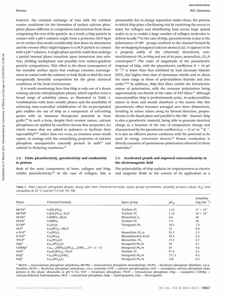
It is worth mentioning here that HAp is only one of a dozenexisting calcium orthophosphate phases, which together cover abroad range of solubility values, as illustrated in Table 1.Combinations with more soluble phases and the possibility ofachieving time-controlled solidication of the as-precipitatedgels enables the use of HAp as a component of self-settingpastes with an immense therapeutic potential as bonegras.49 In such a form, despite their ceramic nature, calciumphosphates are typied by excellent viscous ow properties, forwhich reason they are added to polymers to facilitate theirinjectability50,51 rather than vice versa, as common sense wouldexpect, in analogy with the emulsifying properties of calciumphosphate nanoparticles naturally present in milk52 andutilized in Pickering emulsions.53
2.4. Finite piezoelectricity, pyroelectricity and conductivityto protons
Both of the main components of bone, collagen and HAp,exhibit piezoelectricity.59 In the case of collagen, this is
Table 1 Main calcium phosphate phases, along with their chemical fosolubilities at 25 �C and pH 7.4 (ref. 54–58)
presumably due to charge separation under shear, the processin which HAp plays a facilitating role by restricting the access towater for collagen and distributing load across large spatialscales so as to enable a large number of collagen molecules todeform locally.60 In the case of HAp, piezoelectricity is due to thepolarization of OH� groups conned to the channel formed bythe overlapping hexagonal calcium atoms (Ca2). It appears to bea property solely of the inherently disordered, non-stoichiometric P63/mHAp and not of its pure, monoclinic, P21/bcounterpart.61 The order of magnitude of the piezoelectricresponse of HAp, with the piezoelectric coefficient D ¼ 16 pCN�1,62 is lower than that exhibited by lead zirconate titanate(PZT), but higher than that of aluminum nitride and in aboutthe same range as those of polyvinylidene uoride and zincoxide.63,64 In addition, HAp thin lms exhibit the ferroelectricnature of polarization, with the remnant polarization beingapproximately one-fourth of the value of PZT lms.65 Althoughnanocrystalline HAp is predominantly polar, its polycrystallinenature in bone and mostly elsewhere is the reason why thispiezoelectric effect becomes averaged over three dimensions,resulting in minor values along its favored direction, perpen-dicular to the basal plane and parallel to the OH� channel. HApis also a pyroelectric material, being able to generate electricalcharge as a function of the rate of temperature change andcharacterized by the pyroelectric coefficient pi ¼ 12 mCm�2 K�1.It is also an efficient proton conductor with the potential to beused in energy conversion devices.66 Proton conduction isdirectly causative of spontaneous polarization observed in thesematerials.67
2.5. Accelerated growth and improved osteoconductivity inthe electromagnetic eld
The polarizability of HAp explains its responsiveness to electricand magnetic elds in the context of its application as a
rmulas, space group symmetries, solubility product values (Ksp) and
biomaterial for bone regeneration. For example, sintered andelectrically polarized HAp with a total stored charge density of3.9 mC cm�2 proved to be able to enhance the osteobonding andosteoconducting abilities when implanted in the cortical boneof the femoral diaphysis in white rabbits.68 A metacarpal frac-ture in goats healed much faster in the presence of staticmagnetic eld stimulation (800 G cm�2) and the healingprocess was paralleled by an increase in preferential alignmentand crystallinity of the HAp crystals in bone.69 Cobalt-dopedHAp also facilitated the regeneration of mandibular osteopo-rotic bones in rats.70 Even outside the biomedical context, theeffects of external elds on the habit of crystal growth of HAphave been observed. Thus, the growth of HAp was acceleratedwhen it was grown on polarized HAp substrates and sloweddown when grown on non-polarized HAp.71 To verify that theeffect was due to the existence of lattice channels populated byOH� ions, annealing of the substrates was paired with dehy-dration under low water vapor content, the result of which wastricalcium phosphate, not HAp, but the difference between thegrowth on polarized and non-polarized substrates was expect-edly none. The presence of a static magnetic eld of 0.1 T wasalso shown to increase the precipitation rate of HAp by accel-erating phase transformations along the steps dened by theOstwald–Lussac rule, predominantly DCP / OCP / HAp.72
2.6. The fastest growing face in enamel HAp growsconsiderably slower in bone HAp
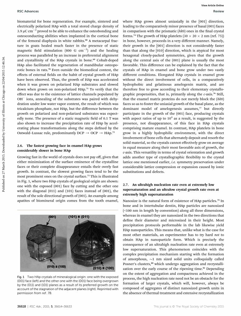
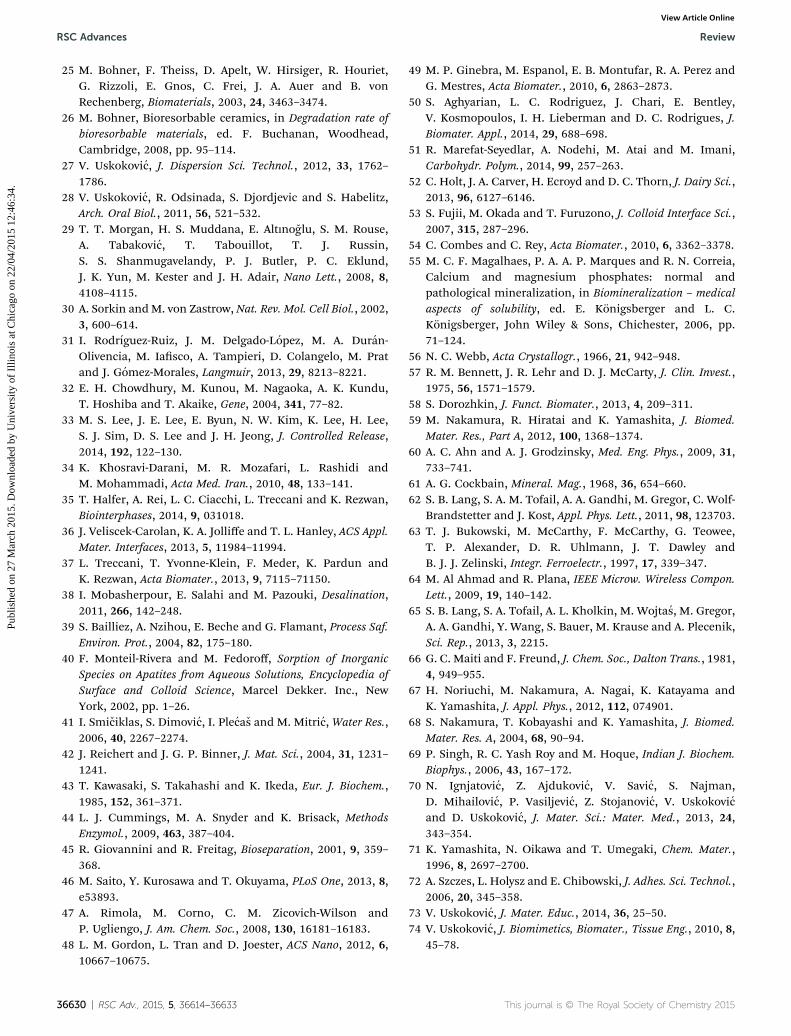
Growing fast in the world of crystals does not pay off, given thateither minimization of the surface eminence of the crystallinefaces or their complete disappearance entails their overly fastgrowth. In contrast, the slowest growing faces tend to be themost prominent ones on the crystal surface.73 This is illustratedin Fig. 1, where two HAp crystals of geological origin are shown:one with the exposed (001) face by cutting and the other onewith the diagonal (011) and (101) faces instead of (001), theresult of the sole directional growth of (001). An example amongapatites of biomineral origin comes from the tooth enamel,
Fig. 1 Two HAp crystals of mineralogical origin: one with the exposed(001) face (left) and the other one with the (001) face being overgrownby the (011) and (101) planes as a result of its preferred growth on theaccount of the stagnation of the adjacent planes (right). Reprinted withpermission from ref. 78.
36618 | RSC Adv., 2015, 5, 36614–36633
where HAp grows almost uniaxially in the [001] direction,leading to the comparatively minor presence of basal (001) facesin comparison with the prismatic (hk0) ones in the nal crystalforms.74 The growth of HAp platelets (30 � 20 � 2 nm (ref. 75))in bone, however, proceeds in a very different manner. Namely,their growth in the [001] direction is not considerably fasterthan that along the [010] direction, which is atypical for mosthexagonal closely-packed symmetries, given that the growthalong the central axis of the (001) plane is usually the mostfavorable. This difference can be explained by the fact that thecrystals of HAp in enamel and bone grow under two vastlydifferent conditions. Elongated HAp crystals in enamel growwithout the direct involvement of cells, in a comparativelyhydrophobic and gelatinous amelogenin matrix, and aretherefore free to grow according to their elementary crystallo-graphic propensities, that is, primarily along the c-axis.76 Still,that the enamel matrix proteins do not merely block the (hk0)faces so as to foster the uniaxial growth of the basal plane, as thedominant model of amelogenesis assumes,77 but directlyparticipate in the growth of the (001) face, producing crystalswith aspect ratios of up to 104 as a result, is suggested by thepresence, not disappearance, of this face in HAp crystalscomprising mature enamel. In contrast, HAp platelets in bonegrow in a highly hydrophilic environment, with the directinvolvement of bone cells that alternately deposit and resorb thesolid material, so the crystals cannot effectively grow on averagein equal measure along their most favorable axis of growth, thec-axis. This versatility in terms of crystal orientation and growthadds another type of crystallographic exibility to the crystallattice one mentioned earlier, i.e. symmetry preservation underconsiderable lattice compression or expansion caused by ionicsubstitutions and defects.
2.7. An ultrahigh nucleation rate even at extremely lowsupersaturation and an ultralow crystal growth rate even atextremely high supersaturation
Nanosize is the natural form of existence of HAp particles.79 Inbone and in intertubular dentin, HAp particles are nanosized(<100 nm in length by convention) along all three dimensions,whereas in enamel they are nanosized in the two directions thatdene their diameter and microsized in their height. Mostprecipitation protocols performable in the lab likewise yieldHAp nanoparticles. This means that, unlike what is the case formost other materials, an experimenter has to try hard not toobtain HAp in nanoparticle form. Which is precisely theconsequence of an ultrahigh nucleation rate even at extremelylow supersaturation. This phenomenon coincides with thecomplex precipitation mechanism starting with the formationof amorphous, �1 nm sized solid units colloquially calledPosner’s clusters,80 which undergo aggregation and recrystalli-zation over the early course of the ripening time.81 Dependingon the extent of aggregation and compactness achieved in theprocess, the high nucleation rate need not be an obstacle for theformation of larger crystals, which will, however, always becomposed of aggregates of distinct nanosized growth units inthe absence of thermal treatment and extensive recrystallization
that it brings about. HAp also has a relatively low interfacialenergy in aqueous environments,82 especially when the surfaceof the material is terminated by OH� groups,83 which addi-tionally contributes to its propensity to remain stable in theform of nanoparticles and resist uncontrolled growth.
Another remarkable feature of this material, following instep with the high nucleation rate in its favoring the adoption ofnanostructured forms, is a considerably low crystal growth rateeven under relatively high supersaturation.84–86 For example, thestep kinetic coefficient, a reciprocal measure of the resistance tocrystallization at the surface step,87 was measured to be around4� 10�5 cm s�1 which is by 3–4 orders of magnitude lower thanthe values attributed to most inorganic crystals and in about thesame range as those of protein crystals.88 The slow motion ofelementary steps on the crystal surface can be both a puzzlingand a logical observation in view of the rather high mobility ofionic species comprising the hydrated surface layers of HAp.For, while a high surface ionmobility can be translated to a highstep propagation rate, under the conditions of intense hydra-tion, as is the case with HAp, it can also provide conditions forthe constant dissolution of the steps, which as such propagateback and forth by advancing and retreating in an alternatingmanner. There are other reasons too, involving most impor-tantly the complex multistage growth mechanism with theinitial precipitation of Posner’s clusters and their subsequentaggregation and structural rearrangement followed by anincrease in size, compactness and crystallinity,81 all the whileobeying the Ostwald–Lussac principle which dictates that, inthe absence of kinetic factors, the least thermodynamicallystable phases (i.e. the most soluble ones having the lowestinterfacial energy) are the rst to form before transformingsequentially to the most stable phase,89 which is HAp in alkaline(>pH 6.8 at 37 �C) and DCP in acidic solutions (<pH 6.8 at 37 �C).By bearing resemblance to the growth of protein and viralcrystals through chirality selection and orientation tting, thelow crystal growth rate of HAp, involving not ions but amor-phous nanoparticle precursors, is sometimes compared to thatof these biologics.90 Despite the low crystal growth rate, thecrystallinity of HAp precipitated under physiological conditionsis remarkably low,91 the reason being the amorphous precursorsthat only partly crystallize as the growth proceeds. Slow crystalgrowth in ordinary crystals produces relatively smooth surfaces,but this is not so in HAp where both the leading edges andterraces in between the surface steps exhibit a highly irregularprole at the atomic scale.92 As a result, unlike DCP, whichdissolves via a combined volume diffusion and surface reactionmechanism,93 the dissolution of HAp follows the polynucleationmechanism94 and is greatly hindered for atomically smooth,step- and pit-free HAp crystals.95 The crystallization processmediated by amorphous precursors is responsible for yetanother peculiarity exhibited by HAp upon nucleation: theinversely proportional dependence of the nucleation rate onsupersaturation, as indicated by the linear increase in nucle-ation lag time with pH in the range of 6.8–7.6.96 Finally, in thepresence of kinetic factors which abound in biological milieusand a high content of impurities, this already low multistagegrowth rate can be prolonged indenitely and take unexpected
crystallographic turns, which is why it has been said that thereis no direct correlation between maturity and crystallinity inbiological HAp.97 This effect certainly contributes to the struc-tural versatility of this compound, a versatility we have barelybegun to translate into a functional one and harness as such.98
2.8. A higher bioactivity and resorbability of biological HApcompared to those of the synthetic one
As a result of the high carbonate content, calcium deciencyand topographic irregularities on the atomic scale, the bioac-tivity, osteoconductivity and resorbability of biological HAp arehigher than those of its synthetic analog. The carbonate contentof biological HAp, varying anywhere between 2 and 8 wt%, has aparticularly large effect on the higher solubility and bioresorb-ability compared to those of the synthetic one. Interestingly,whereas synthetic carbonated HAp is usually of A-type (CO3
2�
/ OH�) when prepared by annealing at high temperatures(owing to the high mobility of the OH� groups), and B-type(CO3
2� / PO43�) when it is precipitated at the room tempera-
ture,99 biological HAp is of a mixed A and B type.100,101 Thissuggests a vastly different formation mechanism of the bio-mineralized HAp compared to the synthetic processes usuallyrun in the lab. The surface energy and interaction potential ofbiological HAp were also measured to be 1.6 and 2.5 timeshigher, respectively, compared to those of pure synthetic HAp,indicating a considerably higher surface heterogeneity and agreater conduciveness to protein adhesion and cell binding.102
Multiple substitutions and defects in biological HAp arecertainly the key to explaining its superior biological activityover that of its synthetic, pure and defect-free HApcounterpart.103,104
2.9. Ultrahigh sensitivity to variations in the environmentalconditions
Precipitation of HAp, especially at low supersaturation when itbears resemblance to the biological formation pathway, isextraordinarily sensitive to the subtlest changes in the condi-tions. The reaction routes leading to HAp as the nal phase in along chain of transformations, frequently proceeding in concertwith the Ostwald–Lussac rule,105–107 are subject to changedepending on even the mildest modications to the initialexperimental conditions. For example, the formation pathwaysand properties of HAp are greatly sensitive to the amount andnature of impurities. Impurities create condensation centers forthe primary particle formation during crystallization ofsynthetic HAp, and calcium phosphates in general appear to besensitive to impurities with concentrations of less than 10�6%(0.01 ppm).108 The texture, porosity and chemical content of thereaction vessel also drastically affect the level of critical super-saturation.109 For example, nucleation of HAp will proceed at aconsiderably lower supersaturation level in borosilicate vesselsthan in plastic ones as a result of leaching of silicate ions and ofmore polar container walls which reduce the energy barrier fornucleation. Tadashi Kokubo, the inventor of the disputablythorough method for evaluating the bioactivity of a solidcompound by immersing it in simulated body uid (SBF) and
looking for the signs of HAp deposition on its surface,110
consequently suggested discarding any plastic bottles withvisible scratches and never reusing them for storing SBF,111 ametastable solution with a supersaturation per growth unitequaling 19.5 with respect to HAp. The kinetics of HAp disso-lution are also strongly inuenced by the presence of impuritiesreleased into the solution during the reaction.112 Solutionsparticularly prone to exhibit this sensitivity lie in the middle ofthe metastability zone, between the levels of saturation andcritical supersaturation. In this supersaturation window, kineticeffects such as the presence of an organic matrix exert the nestlevel of control over the microstructure and hierarchicalsuperstructure of HAp precipitates.113 As a result, there is amultitude of possible stoichiometries and phase combinationsthat could appear in the nal products of the precipitationreaction depending on the subtlest changes in the synthesisconditions,114,115 and some of them such as the acidic and bio-soluble transitory calcium phosphate phases, found exclusivelyin pathological calcication deposits, may be all but idealcomponents of HAp designed for specic biomedical applica-tions. This versatility of reaction outcomes depending on vari-ations in the experimental conditions becomes even moremagnied when various preparation methods are introduced inthe comparison. This is why different methods to synthesizeHAp have led tomaterials that vastly differed in crystalline orderand properties, with the most notable difference being found inthe diffusivity across the hydroxyl ion channel, which spannedtwo orders of magnitude, ranging from ultralow for the orderedmonoclinic samples prepared by solid state synthesis at 1100�C, to ultrahigh for the disordered hexagonal samples preparedby room temperature precipitation.116 Tracing this journey ofions across the channel running through the center of the triplehexagons that dene the crystal structure of HAp takes us to theelaboration of its properties, the subject of the next section.
3. The role of the hydroxyl ionchannel
The question framing this opinion piece is this: what if there isa single structural detail that could explain all these peculiarphenomena demonstrated by the protean nature of HAp? Ourimagination is limitless, we know, and, though unsupported bydirect computational analyses, it has been given free rein in thefollowing paragraphs. But let us look rst at the crystal structuremodel of HAp.
3.1. The place for hydroxyl in the crystal lattice of HAp
Although its rst chemical composition analyses of the mineralform date back to 1788 (ref. 117 and 118) and the rst synthesisto 1851,119 HAp was rst detected as a bone mineral phase in1926.120 In 1930, four years later, in two simultaneously pub-lished independent studies, its crystal structure was deter-mined.121,122 Pure HAp crystallizes in the monoclinic P21/b spacegroup symmetry with 88 atoms per unit cell and Ca20(PO4)12(-OH)4 stoichiometry, but presents a rarity in the lab, let alone inNature, where it virtually never occurs in such a form. For, any
36620 | RSC Adv., 2015, 5, 36614–36633
precipitation under atmospheric conditions leads to the inclu-sion of carbonate ions which necessitate charge compensationand, thus, lead to vacancies, deformations of the lattice andadoption of the hexagonal P63/m space group symmetry with 44atoms per unit cell and Ca10(PO4)6(OH)2 stoichiometry, whereassolid state processing at elevated temperatures, higher than 207�C, produces the same transition to the hexagonal symmetry.When it comes to pure HAp, the structural difference betweenthe two phases boils down to the ordered, head-to-tailarrangement of OH� groups located in the center of everyother Ca2 triangle in the monoclinic low-temperaturesymmetry, and the disordered arrangement of OH� groupswhere the head-to-tail and tail-to-head arrangements alternatethroughout the channel in the hexagonal high-temperaturesymmetry. The monoclinic-to-hexagonal transition at elevatedtemperatures is thus paralleled by a curious phenomenon:namely, thermal energy during annealing converts a moreordered OH� channel structure to a more disordered one. As aresult, the hexagonal phase is higher in energy than themonoclinic phase by a rather small amount of 22 meV per unitcell,123 and the transition between the two, typied by a rela-tively low transition enthalpy of 130 J mol�1,124 is initiated by therotation and reorientation of the OH� dipoles125,126 and mayproceed by the subsequent progressive twinning of the mono-clinic, pseudo-hexagonal lattice until the hexagonal symmetry isreached.127 Note that both the occupational and the orienta-tional order of OH� ions become perturbed by this transitionfrom the state of the higher, P21/b space group symmetry to thestate of the lower, P63/m one, leading to a greater diffusivity ofOH� species along the channel and a higher potential forrestructuring of the material under the demands of the localbiological environment. The hexagonal form is, thus, the onefavored by biological systems, in part because it allows for aneasier exchange of OH� groups78 with ions such as F�, Cl� andCO3
2�, which brings about an increased level of structuraldisorder, usable during the incessantly ongoing process of boneremodeling. The symmetry elements present in the ratherdisordered biological HAp include a six-fold rotation axisparallel to the c-axis, a 1/2 translation along the c-axis and amirror plane perpendicular to the screw axis and the c-axis. Thetheoretical lattice parameters are a¼ 9.418 A and c¼ 6.884 A forP63/m (b ¼ 2a, a ¼ 120� for P21/b). However, depending on thepreparation method, particularly when wet room temperaturesyntheses are involved, the lattice parameters a and c could befound anywhere in the ranges of 9.41–9.44 A and 6.84–6.94 A,respectively, for the P63/m space group.
The unit cell formula of hexagonal HAp, Ca10(PO4)6(OH)2,can be more precisely given as Ca14Ca26(PO4)6(OH)2, where Ca1denotes columnar calcium ions and Ca2 denotes the hexagonalones.128 Columnar calcium ions are named so because they arearranged in columns that uninterruptedly traverse the lattice,while the hexagonal ones outline hexagons when projected onthe basal plane. Such an etymology conceals a mild misnomerbecause the columnar Ca atoms also create a hexagon whenviewed down the c-axis, just as the hexagonal Ca atoms do. Thedifference between the two hexagons projected on the basalplane is that the one created by columnar Ca atoms is wider and
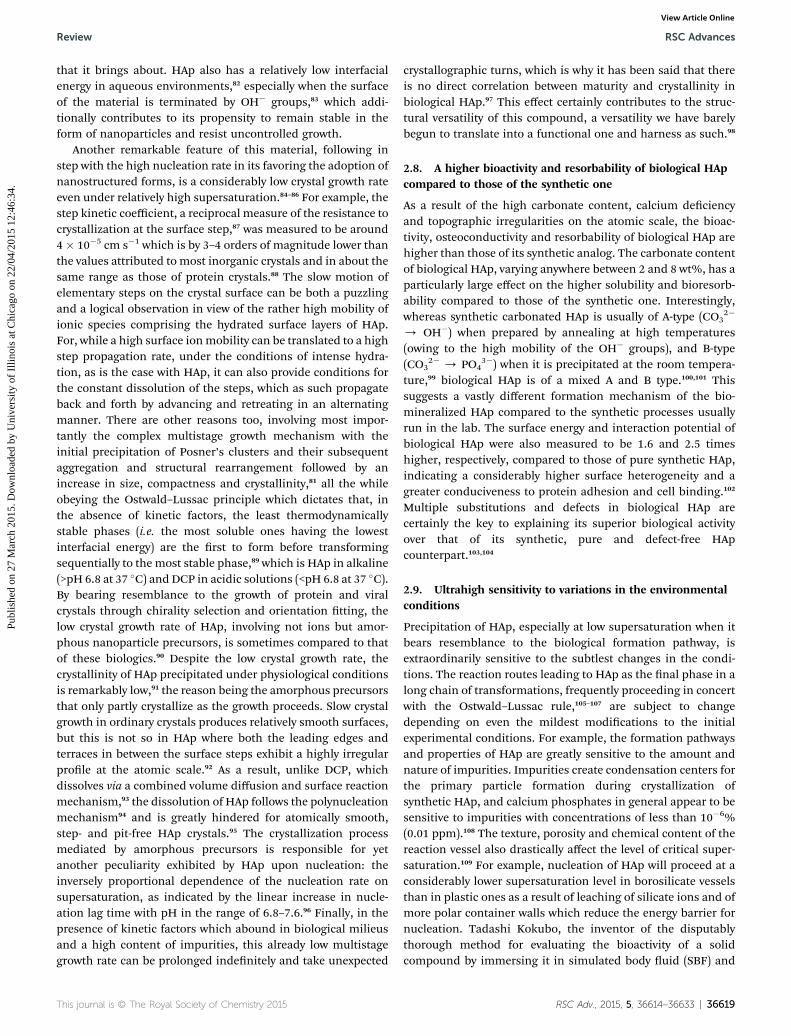
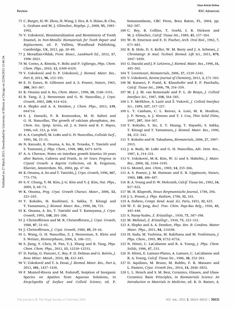
accommodates in its center the hexagon created by the hexag-onal ones. As seen from Fig. 2, the outer hexagon created by thecolumnar Ca atoms outlines the edges of the P63/m unit cell,while the six hexagonal Ca atoms form a series of overlappinginner hexagons. While all six hexagonal Ca atoms occupy theinterior of the P63/m unit cell, the columnar Ca atoms arepositioned in the six corners of the two basal planes and the sixcorners of the equatorial mirror plane in between them,yielding eighteen Ca1 atoms within a unit cell but only four perunit cell (each of the six basal plane atoms is shared with veneighboring unit cells, while each of the six atoms on themirrorplane is shared with two adjacent cells). Each inner hexagon isformed by two overlapping Ca2 triangles positioned parallel tothe basal plane at z ¼ 1/4 and z ¼ 3/4 and at a 60� angle withrespect to each other (Fig. 3a). Each of the six PO4
3� tetrahedrapopulating the interior of the hexagonal unit cell is positionedbetween a pair of Ca2+ ions in the outer hexagon when vieweddown the c-axis, with three of them lying on the Ca2 triangleplane at z ¼ 1/4 and the other three lying on the Ca2 triangleplane at z ¼ 3/4. Furthermore, one of the four oxygen atoms ofeach of the PO4
3� tetrahedra is found just outside the edge ofthe outer hexagon, which is compensated by the intrusion of anoxygen from the tetrahedra positioned on planes below andabove (z ¼ �1/2). The PO4
3� tetrahedra, 2.6 A in radius, arearranged so as to form hexagonal close packing (ABABA) and,given their size-wise dominance over the crystal structure, theCa2+ ions, 1.06 A in radius, could be visualized as merely llingthe holes between them. These holes come in two sizes andboth form long columns in the direction of the c-axis. While thesmaller holes can accommodate ions#0.225r in radius (r beingthe radius of a PO4
3� tetrahedron), which is too small to t Ca2+,the larger holes provide enough room for Ca2+, given that they
Fig. 2 View of a stoichiometric HAp crystal down the c [001] axis,showing an OH group (small green circle) in its center, surrounded byhexagonal (blue circles) and columnar (purple circles) Ca atoms andphosphate tetrahedra: each green pyramid is a P5+ ion and the four redcircles in its corners are four O2� ions. Notice the misrepresentationthough: the O2� ions are not the smallest ions in the lattice, as shownhere, but, in fact, the largest, occupyingmost space in it. As parts of thePO4 sublattice, they determine the stacking order of the unit cell. Allother ions could be imagined as merely occupying the intersticescreated by the oxygen atoms.
can accommodate ions #0.41r in radius (�1.1 A), and are lledwith Ca atoms.129 Each site at which a Ca1 atom is located lies inbetween two parallel PO4
3� sheets, A and B, and is coordinatedby nine oxygen atoms from three PO4
3� tetrahedra on top andthree more at the bottom. The Ca1 site is also called “octahe-dral” because the six PO4
3� groups it is coordinated to outlinethe corners of a regular octahedron together with Ca1. The Ca1atoms are thus found in columns perpendicular to the PO4
3�
sheets and parallel to the c-axis, with the Ca1/OH� ratio of 2 : 1.The Ca2 atoms, on the other hand, lie in the same plane as thePO4
3� tetrahedra and reside inside the holes formed by theoxygen atoms of the tetrahedra that line the c-axis channel. TheCa2 ions are engaged in a seven-fold coordination with sixoxygen ions belonging to ve different PO4
3� tetrahedra and anOH� group.130 Arranged in a series of triangles lying parallel tothe basal plane, the Ca2 atoms outline the boundaries of theOH� channel extending through the center of the crystalstructure, perpendicularly to the basal plane.
The OH� groups are thus lined up along the c-axis, beingpositioned inside the overlapping triangles formed by thehexagonal Ca2+ ions, with an average distance of 2.5 A betweenthem. The triangles are stacked on top of one another in thedirection of the c-axis, with each neighboring triangle beingrotated by 60� with respect to the triangles below and above it,and the line connecting all the OH� groups in a stacked array ofunit cells runs straight through their center, with both oxygenand hydrogen of the OH� lying on the c-axis in the higher,monoclinic symmetry (Fig. 3b). However, being inherentlydisordered as part of the P63/m symmetry, the oxygen atoms ofhydroxyl are equally likely to be found either slightly above orslightly below the plane of the calcium triangles, (Fig. 4a) andthe same applies to their halide ion substitutes. Moreover, thereis no energetic preference as to whether the hydrogen atom ofthe OH� group is to point upwards or downwards with respectto the c-axis and, as a result, each OH� ion has a 50% probabilityof being in one state or the other. Compared to other ionicgroups in the lattice, particularly PO4
3�, the OH� groups have aconsiderable freedom of movement along the Ca2 channel,which is circa 3 A in diameter. To be more precise, the diameterof the channel varies across the unit cell, being widest in itscenter and in the basal plane (z ¼ 0, z ¼ 1/2, 2.85 A) and nar-rowest at the two midpoints between the basal planes andthe center of the unit cell, lying in plane with the Ca2 triangles(z¼ 1/4, z¼ 3/4, 2.73 A).131 For this reason, the channel could bebetter visualized as a continuous array of ovoid cavities than as ahollow cylinder (Fig. 4b).
As can be seen from Fig. 4b, there is not enough room in thelattice to accommodate two OH� groups pointing towards eachother (�OH//HO�) in the adjacent calcium triangles withoutproducing signicant lattice distortion and the loss of thehigher, monoclinic symmetry. For this reason, the OH� groupsin the channel must either be arranged in an ‘ordered column’fashion, i.e. as OH� OH� OH� OH�. along the c-axis, or in a‘disordered column’ fashion, implying the reversal of thedirection from OH� OH� OH� OH�. to HO� HO� HO� HO�.and back at various places. While the former head-to-tailarrangement is seen in stoichiometrically pure, monoclinic
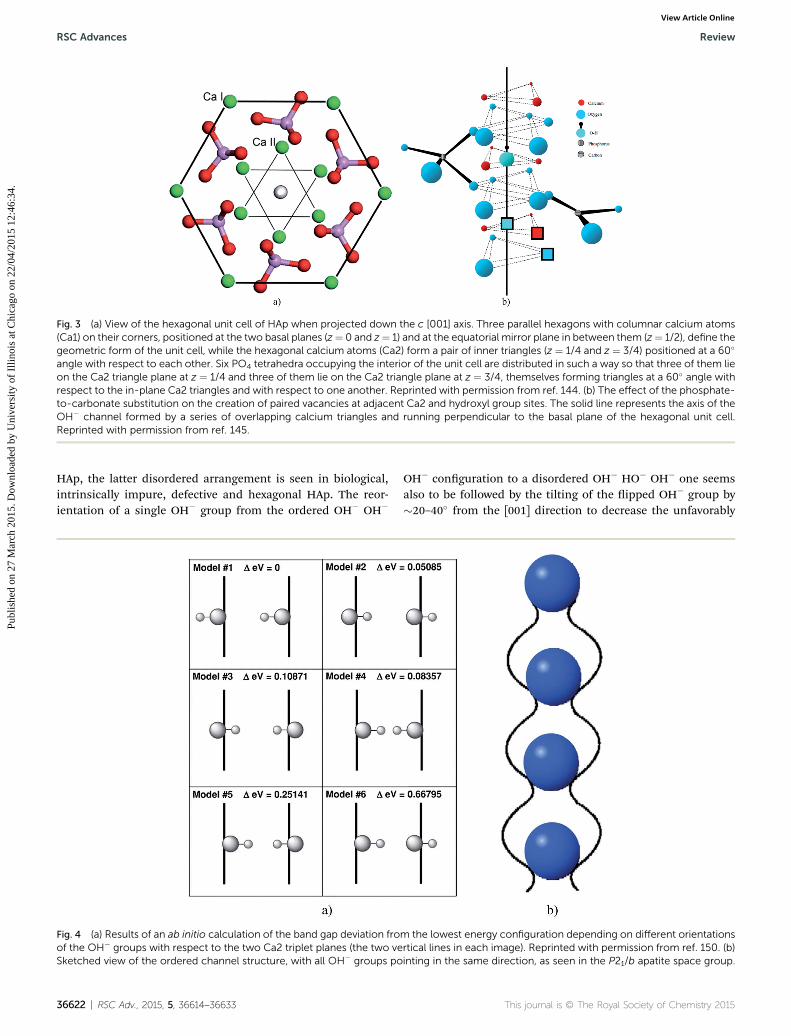
Fig. 3 (a) View of the hexagonal unit cell of HAp when projected down the c [001] axis. Three parallel hexagons with columnar calcium atoms(Ca1) on their corners, positioned at the two basal planes (z¼ 0 and z¼ 1) and at the equatorial mirror plane in between them (z¼ 1/2), define thegeometric form of the unit cell, while the hexagonal calcium atoms (Ca2) form a pair of inner triangles (z ¼ 1/4 and z ¼ 3/4) positioned at a 60�
angle with respect to each other. Six PO4 tetrahedra occupying the interior of the unit cell are distributed in such a way so that three of them lieon the Ca2 triangle plane at z ¼ 1/4 and three of them lie on the Ca2 triangle plane at z ¼ 3/4, themselves forming triangles at a 60� angle withrespect to the in-plane Ca2 triangles and with respect to one another. Reprinted with permission from ref. 144. (b) The effect of the phosphate-to-carbonate substitution on the creation of paired vacancies at adjacent Ca2 and hydroxyl group sites. The solid line represents the axis of theOH� channel formed by a series of overlapping calcium triangles and running perpendicular to the basal plane of the hexagonal unit cell.Reprinted with permission from ref. 145.
RSC Advances Review
Publ
ishe
d on
27
Mar
ch 2
015.
Dow
nloa
ded
by U
nive
rsity
of
Illin
ois
at C
hica
go o
n 22
/04/
2015
12:
46:3
4.
View Article Online
HAp, the latter disordered arrangement is seen in biological,intrinsically impure, defective and hexagonal HAp. The reor-ientation of a single OH� group from the ordered OH� OH�
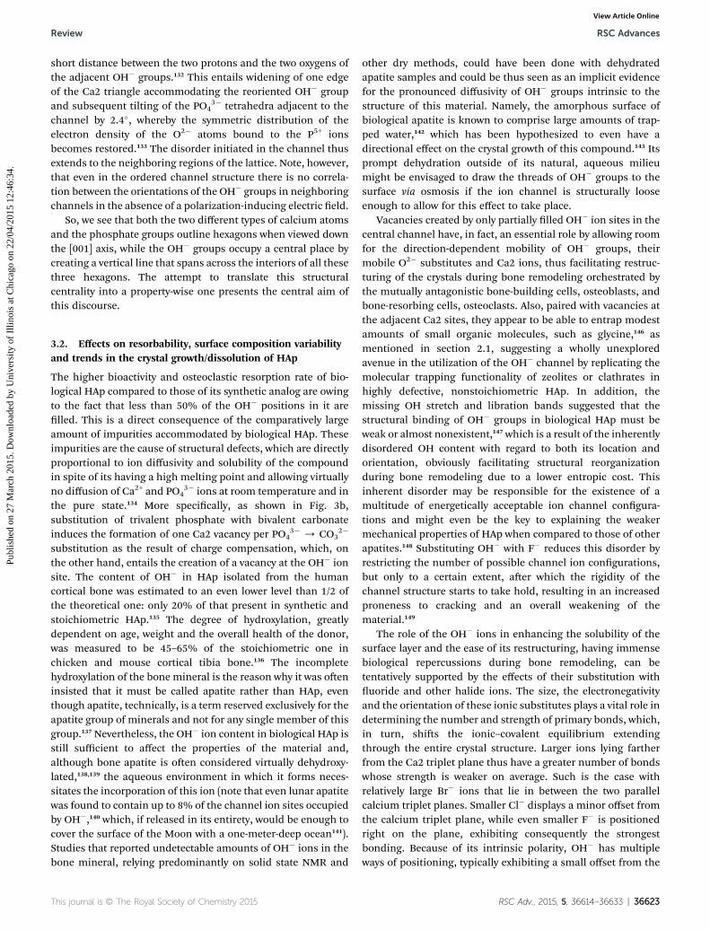
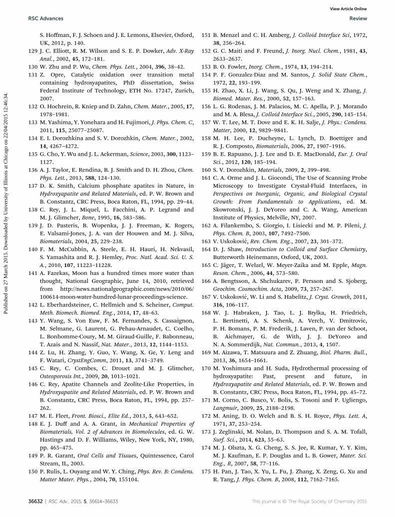
Fig. 4 (a) Results of an ab initio calculation of the band gap deviation froof the OH� groups with respect to the two Ca2 triplet planes (the two veSketched view of the ordered channel structure, with all OH� groups po
36622 | RSC Adv., 2015, 5, 36614–36633
OH� conguration to a disordered OH� HO� OH� one seemsalso to be followed by the tilting of the ipped OH� group by�20–40� from the [001] direction to decrease the unfavorably
m the lowest energy configuration depending on different orientationsrtical lines in each image). Reprinted with permission from ref. 150. (b)inting in the same direction, as seen in the P21/b apatite space group.
short distance between the two protons and the two oxygens ofthe adjacent OH� groups.132 This entails widening of one edgeof the Ca2 triangle accommodating the reoriented OH� groupand subsequent tilting of the PO4
3� tetrahedra adjacent to thechannel by 2.4�, whereby the symmetric distribution of theelectron density of the O2� atoms bound to the P5+ ionsbecomes restored.133 The disorder initiated in the channel thusextends to the neighboring regions of the lattice. Note, however,that even in the ordered channel structure there is no correla-tion between the orientations of the OH� groups in neighboringchannels in the absence of a polarization-inducing electric eld.
So, we see that both the two different types of calcium atomsand the phosphate groups outline hexagons when viewed downthe [001] axis, while the OH� groups occupy a central place bycreating a vertical line that spans across the interiors of all thesethree hexagons. The attempt to translate this structuralcentrality into a property-wise one presents the central aim ofthis discourse.
3.2. Effects on resorbability, surface composition variabilityand trends in the crystal growth/dissolution of HAp
The higher bioactivity and osteoclastic resorption rate of bio-logical HAp compared to those of its synthetic analog are owingto the fact that less than 50% of the OH� positions in it arelled. This is a direct consequence of the comparatively largeamount of impurities accommodated by biological HAp. Theseimpurities are the cause of structural defects, which are directlyproportional to ion diffusivity and solubility of the compoundin spite of its having a high melting point and allowing virtuallyno diffusion of Ca2+ and PO4
3� ions at room temperature and inthe pure state.134 More specically, as shown in Fig. 3b,substitution of trivalent phosphate with bivalent carbonateinduces the formation of one Ca2 vacancy per PO4
3� / CO32�
substitution as the result of charge compensation, which, onthe other hand, entails the creation of a vacancy at the OH� ionsite. The content of OH� in HAp isolated from the humancortical bone was estimated to an even lower level than 1/2 ofthe theoretical one: only 20% of that present in synthetic andstoichiometric HAp.135 The degree of hydroxylation, greatlydependent on age, weight and the overall health of the donor,was measured to be 45–65% of the stoichiometric one inchicken and mouse cortical tibia bone.136 The incompletehydroxylation of the bone mineral is the reason why it was oeninsisted that it must be called apatite rather than HAp, eventhough apatite, technically, is a term reserved exclusively for theapatite group of minerals and not for any single member of thisgroup.137 Nevertheless, the OH� ion content in biological HAp isstill sufficient to affect the properties of the material and,although bone apatite is oen considered virtually dehydroxy-lated,138,139 the aqueous environment in which it forms neces-sitates the incorporation of this ion (note that even lunar apatitewas found to contain up to 8% of the channel ion sites occupiedby OH�,140 which, if released in its entirety, would be enough tocover the surface of the Moon with a one-meter-deep ocean141).Studies that reported undetectable amounts of OH� ions in thebone mineral, relying predominantly on solid state NMR and
other dry methods, could have been done with dehydratedapatite samples and could be thus seen as an implicit evidencefor the pronounced diffusivity of OH� groups intrinsic to thestructure of this material. Namely, the amorphous surface ofbiological apatite is known to comprise large amounts of trap-ped water,142 which has been hypothesized to even have adirectional effect on the crystal growth of this compound.143 Itsprompt dehydration outside of its natural, aqueous milieumight be envisaged to draw the threads of OH� groups to thesurface via osmosis if the ion channel is structurally looseenough to allow for this effect to take place.
Vacancies created by only partially lled OH� ion sites in thecentral channel have, in fact, an essential role by allowing roomfor the direction-dependent mobility of OH� groups, theirmobile O2� substitutes and Ca2 ions, thus facilitating restruc-turing of the crystals during bone remodeling orchestrated bythe mutually antagonistic bone-building cells, osteoblasts, andbone-resorbing cells, osteoclasts. Also, paired with vacancies atthe adjacent Ca2 sites, they appear to be able to entrap modestamounts of small organic molecules, such as glycine,146 asmentioned in section 2.1, suggesting a wholly unexploredavenue in the utilization of the OH� channel by replicating themolecular trapping functionality of zeolites or clathrates inhighly defective, nonstoichiometric HAp. In addition, themissing OH stretch and libration bands suggested that thestructural binding of OH� groups in biological HAp must beweak or almost nonexistent,147 which is a result of the inherentlydisordered OH content with regard to both its location andorientation, obviously facilitating structural reorganizationduring bone remodeling due to a lower entropic cost. Thisinherent disorder may be responsible for the existence of amultitude of energetically acceptable ion channel congura-tions and might even be the key to explaining the weakermechanical properties of HAp when compared to those of otherapatites.148 Substituting OH� with F� reduces this disorder byrestricting the number of possible channel ion congurations,but only to a certain extent, aer which the rigidity of thechannel structure starts to take hold, resulting in an increasedproneness to cracking and an overall weakening of thematerial.149
The role of the OH� ions in enhancing the solubility of thesurface layer and the ease of its restructuring, having immensebiological repercussions during bone remodeling, can betentatively supported by the effects of their substitution withuoride and other halide ions. The size, the electronegativityand the orientation of these ionic substitutes plays a vital role indetermining the number and strength of primary bonds, which,in turn, shis the ionic–covalent equilibrium extendingthrough the entire crystal structure. Larger ions lying fartherfrom the Ca2 triplet plane thus have a greater number of bondswhose strength is weaker on average. Such is the case withrelatively large Br� ions that lie in between the two parallelcalcium triplet planes. Smaller Cl� displays a minor offset fromthe calcium triplet plane, while even smaller F� is positionedright on the plane, exhibiting consequently the strongestbonding. Because of its intrinsic polarity, OH� has multipleways of positioning, typically exhibiting a small offset from the
plane, like Cl�. Orientation of the OH� ions in the channel canalso have a profound effect on the bonding strength.150 This isillustrated in Fig. 4a, where the results of the theoretical bandgap prediction based on an ab initio density-functional-theory-based computational model are shown: depending on theorientation of the two OH� groups per unit cell with respect tothe Ca2 triplet plane, the deviation from the lowest energyconguration can vary from 0–15% (i.e., 0–0.66795 eV for theband gap estimated at 4.71 eV). Another way in which halidesubstitutes, such as F� and Cl�, modify the structure andproperties of HAp is by promoting hydrogen bonding betweenneighboring OH� dipoles in the channel.151,152 These bondshave otherwise not been observed in pure HAp due to the largedistance between adjacent OH� groups,153,154 limiting hydrogenbonding only to that between OH� groups and the nearestoxygen atoms belonging to the PO4
3� groups.155
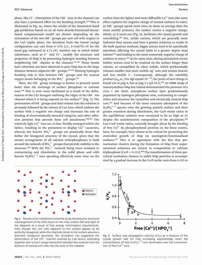
Then, the OH� group exchange is known to proceed muchfaster than the exchange of surface phosphate or calciumions.156 This is even more facilitated as a result of the defor-mation of the Ca2 hexagon outlining the edges of the OH� ionchannel when it is being exposed on the surface157 (Fig. 5). Theprotonation of OH� groups and their release into the solution ispromptly followed by the release of Ca2 ions, which endows thesurface with a negative net charge and increases the rate ofbinding of electrostatically attracted integrins and other adhe-sion proteins that precede bone cell attachment.158,159 Therelatively light Ca2+ ions are, in fact, exibly arranged within thelattice, resulting in the proneness to display Ca2+ vacancies,whereas the heavier PO4
3� groups are practically those thatdene the hexagonal structure of the crystal, given that theatomic arrangement of all calcium orthophosphates is builtaround the network of PO4
3� groups that provide stability to thestructure.160 With the PO4
3� network being more resistant torestructuring and dissolution in the solid phase and withheavier HxPO3
x�3 ions spending effectively more time on the
Fig. 5 Results of an interatomic potential study showing the structuralrearrangement of the (001) faces on the HAp surface (left and right ofthe diagram) as a result of free energy minimization requirements.Even though the unit cells adjacent to the surface appear to beperfectly hexagonal, while the channels closer to the surface assume adistorted hexagonal geometry, the simulation has suggested thedeformation of the OH� channel outlined by Ca2 atoms, extendingtogether with a short-range interaction between the surfaces over thedistance of several unit cells into the bulk of the material.
36624 | RSC Adv., 2015, 5, 36614–36633
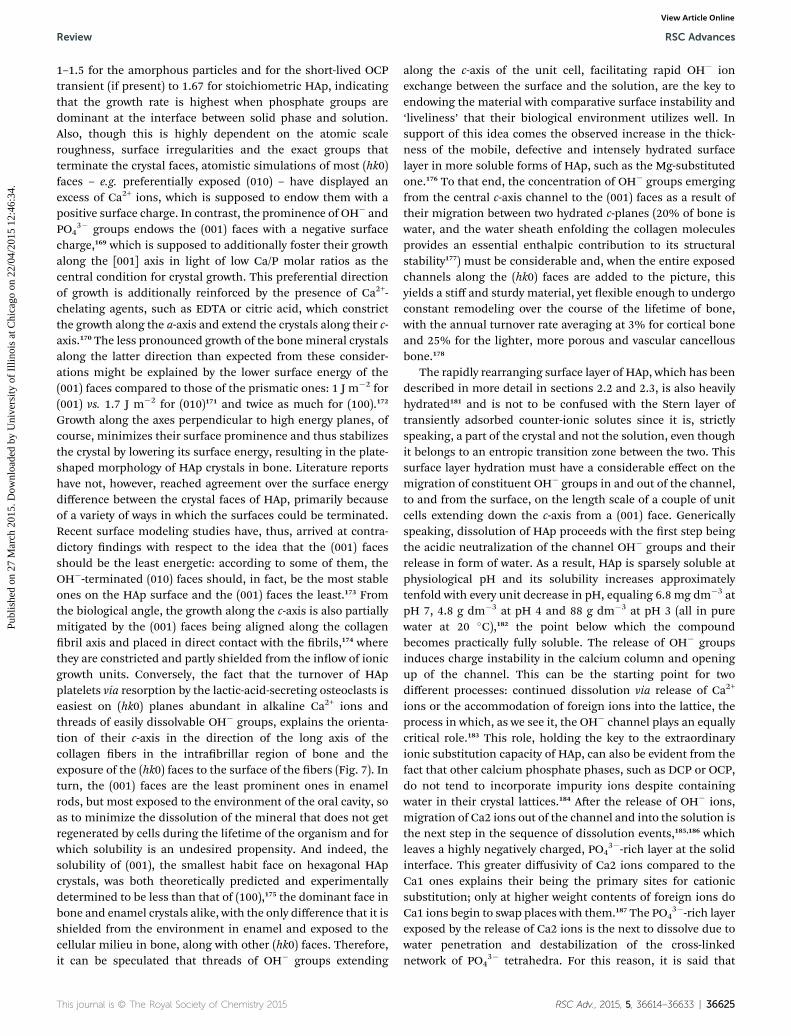
surface than the lighter and more diffusible Ca2+ ions (the sameeffect explains the negative charge of neutral surfaces in water,as OH� groups spend more time on them on average than themore mobile protons), the surface carries a negative charge,which, as it turns out (Fig. 6), facilitates the crystal growth andremodeling.161 For, unlike cations, which are generally morehydrated than anions and have a greater tendency to reside inthe bulk aqueous medium, bigger anions tend to be specicallyadsorbed, affecting the crystal habit to a greater degree thancations162 and leading to the most commonly negative charge ofsurfaces in water.163 At the same time, during dissolution eventsbulkier anions tend to be retained on the surface longer thancations, as exemplied by silver iodide, which preferentiallyreleases smaller and more mobile Ag+ ions and retains heavierand less mobile I�. Consequently, although the solubilityproduct (aAg+aI�) for AgI equals 10
�16, the point of zero charge isfound not at pAg 8, but at pAg 5.5 (pI 10.5).164 An NMR study ofnanocrystalline HAp has indeed demonstrated the presence of acirca 1 nm thick, amorphous surface layer predominantlypopulated by hydrogen phosphate ions, contrasting in compo-sition and structure the crystalline and electrically neutral HApcore.165 And because of the more extensive adsorption of theHxPO4
x�3 species onto the growing particle surface and theirgreater retention during dissolution, the Ca/P molar ratios inthe equilibrium solution were measured to be as high as 25despite the stoichiometric composition of the precipitate.166
Low Ca/P molar ratios, naturally brought about by the bindingof free Ca2+ by phosphorylated proteins in the bone matrix,have, for example, been shown to be critical for promoting thecontrolled growth of HAp on amelogenin-functionalizedsurfaces.167 This is in agreement with the fact that pre-nucleation clusters during the formation of HAp from super-saturated solutions are closest in composition to calciumtriphosphate (Ca/P ¼ 0.333).168 The transformation of these pre-critical nucleation clusters to stable HAp particles is accompa-nied by a gradual increase in the Ca/P molar ratio from 0.333 to
Fig. 6 Surface step propagation velocity (vi/v0) as a measure of thecrystal growth rate for HAp increasing exponentially when theconcentration of free HxPO4
x�3 ions dominates over the concentra-tion of free Ca2+ ions.
1–1.5 for the amorphous particles and for the short-lived OCPtransient (if present) to 1.67 for stoichiometric HAp, indicatingthat the growth rate is highest when phosphate groups aredominant at the interface between solid phase and solution.Also, though this is highly dependent on the atomic scaleroughness, surface irregularities and the exact groups thatterminate the crystal faces, atomistic simulations of most (hk0)faces – e.g. preferentially exposed (010) – have displayed anexcess of Ca2+ ions, which is supposed to endow them with apositive surface charge. In contrast, the prominence of OH� andPO4
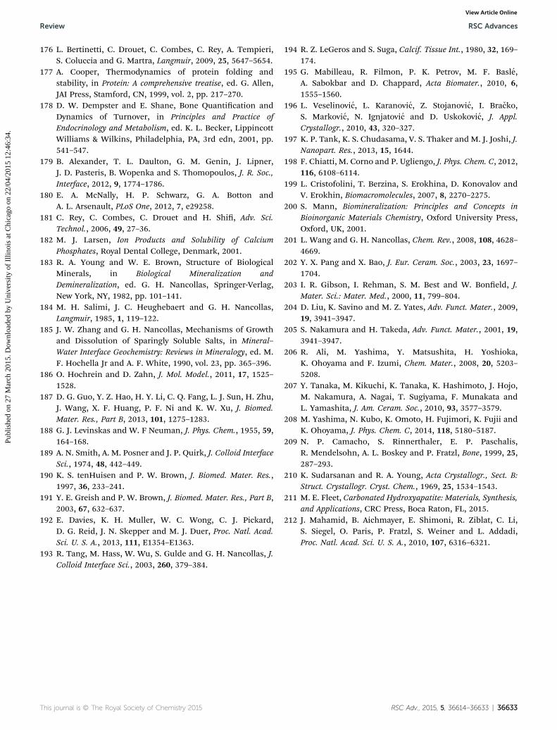
3� groups endows the (001) faces with a negative surfacecharge,169 which is supposed to additionally foster their growthalong the [001] axis in light of low Ca/P molar ratios as thecentral condition for crystal growth. This preferential directionof growth is additionally reinforced by the presence of Ca2+-chelating agents, such as EDTA or citric acid, which constrictthe growth along the a-axis and extend the crystals along their c-axis.170 The less pronounced growth of the bone mineral crystalsalong the latter direction than expected from these consider-ations might be explained by the lower surface energy of the(001) faces compared to those of the prismatic ones: 1 J m�2 for(001) vs. 1.7 J m�2 for (010)171 and twice as much for (100).172
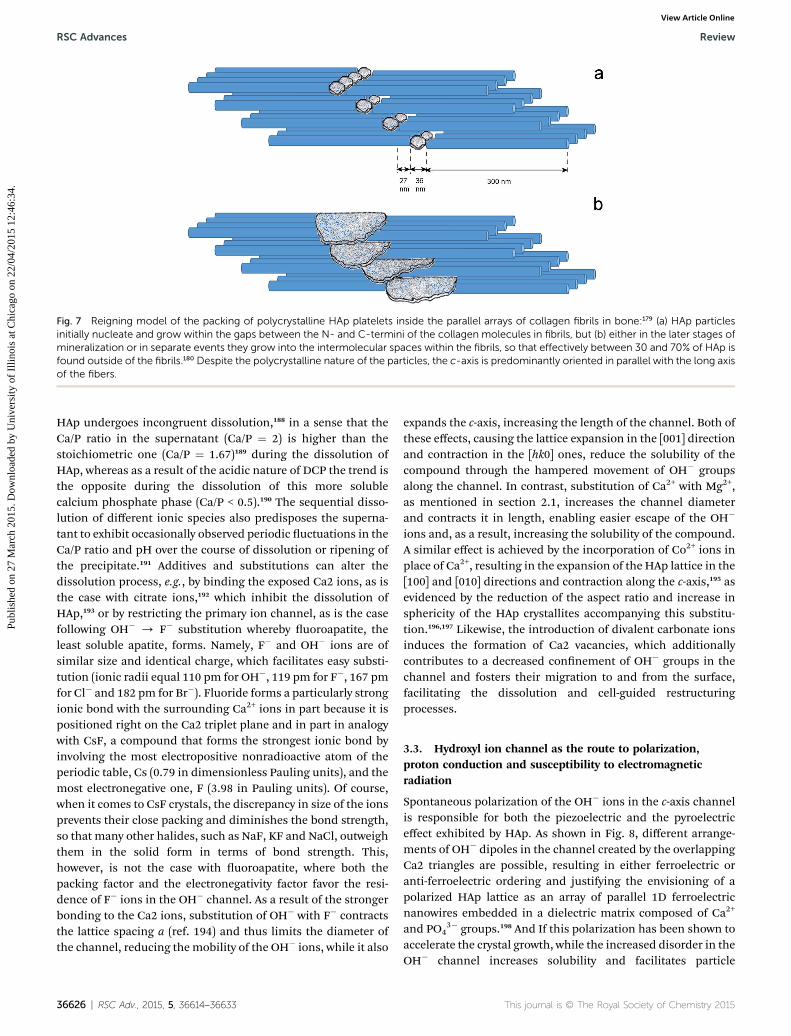
Growth along the axes perpendicular to high energy planes, ofcourse, minimizes their surface prominence and thus stabilizesthe crystal by lowering its surface energy, resulting in the plate-shaped morphology of HAp crystals in bone. Literature reportshave not, however, reached agreement over the surface energydifference between the crystal faces of HAp, primarily becauseof a variety of ways in which the surfaces could be terminated.Recent surface modeling studies have, thus, arrived at contra-dictory ndings with respect to the idea that the (001) facesshould be the least energetic: according to some of them, theOH�-terminated (010) faces should, in fact, be the most stableones on the HAp surface and the (001) faces the least.173 Fromthe biological angle, the growth along the c-axis is also partiallymitigated by the (001) faces being aligned along the collagenbril axis and placed in direct contact with the brils,174 wherethey are constricted and partly shielded from the inow of ionicgrowth units. Conversely, the fact that the turnover of HApplatelets via resorption by the lactic-acid-secreting osteoclasts iseasiest on (hk0) planes abundant in alkaline Ca2+ ions andthreads of easily dissolvable OH� groups, explains the orienta-tion of their c-axis in the direction of the long axis of thecollagen bers in the intrabrillar region of bone and theexposure of the (hk0) faces to the surface of the bers (Fig. 7). Inturn, the (001) faces are the least prominent ones in enamelrods, but most exposed to the environment of the oral cavity, soas to minimize the dissolution of the mineral that does not getregenerated by cells during the lifetime of the organism and forwhich solubility is an undesired propensity. And indeed, thesolubility of (001), the smallest habit face on hexagonal HApcrystals, was both theoretically predicted and experimentallydetermined to be less than that of (100),175 the dominant face inbone and enamel crystals alike, with the only difference that it isshielded from the environment in enamel and exposed to thecellular milieu in bone, along with other (hk0) faces. Therefore,it can be speculated that threads of OH� groups extending
along the c-axis of the unit cell, facilitating rapid OH� ionexchange between the surface and the solution, are the key toendowing the material with comparative surface instability and‘liveliness’ that their biological environment utilizes well. Insupport of this idea comes the observed increase in the thick-ness of the mobile, defective and intensely hydrated surfacelayer in more soluble forms of HAp, such as the Mg-substitutedone.176 To that end, the concentration of OH� groups emergingfrom the central c-axis channel to the (001) faces as a result oftheir migration between two hydrated c-planes (20% of bone iswater, and the water sheath enfolding the collagen moleculesprovides an essential enthalpic contribution to its structuralstability177) must be considerable and, when the entire exposedchannels along the (hk0) faces are added to the picture, thisyields a stiff and sturdy material, yet exible enough to undergoconstant remodeling over the course of the lifetime of bone,with the annual turnover rate averaging at 3% for cortical boneand 25% for the lighter, more porous and vascular cancellousbone.178
The rapidly rearranging surface layer of HAp, which has beendescribed in more detail in sections 2.2 and 2.3, is also heavilyhydrated181 and is not to be confused with the Stern layer oftransiently adsorbed counter-ionic solutes since it is, strictlyspeaking, a part of the crystal and not the solution, even thoughit belongs to an entropic transition zone between the two. Thissurface layer hydration must have a considerable effect on themigration of constituent OH� groups in and out of the channel,to and from the surface, on the length scale of a couple of unitcells extending down the c-axis from a (001) face. Genericallyspeaking, dissolution of HAp proceeds with the rst step beingthe acidic neutralization of the channel OH� groups and theirrelease in form of water. As a result, HAp is sparsely soluble atphysiological pH and its solubility increases approximatelytenfold with every unit decrease in pH, equaling 6.8 mg dm�3 atpH 7, 4.8 g dm�3 at pH 4 and 88 g dm�3 at pH 3 (all in purewater at 20 �C),182 the point below which the compoundbecomes practically fully soluble. The release of OH� groupsinduces charge instability in the calcium column and openingup of the channel. This can be the starting point for twodifferent processes: continued dissolution via release of Ca2+
ions or the accommodation of foreign ions into the lattice, theprocess in which, as we see it, the OH� channel plays an equallycritical role.183 This role, holding the key to the extraordinaryionic substitution capacity of HAp, can also be evident from thefact that other calcium phosphate phases, such as DCP or OCP,do not tend to incorporate impurity ions despite containingwater in their crystal lattices.184 Aer the release of OH� ions,migration of Ca2 ions out of the channel and into the solution isthe next step in the sequence of dissolution events,185,186 whichleaves a highly negatively charged, PO4
3�-rich layer at the solidinterface. This greater diffusivity of Ca2 ions compared to theCa1 ones explains their being the primary sites for cationicsubstitution; only at higher weight contents of foreign ions doCa1 ions begin to swap places with them.187 The PO4
3�-rich layerexposed by the release of Ca2 ions is the next to dissolve due towater penetration and destabilization of the cross-linkednetwork of PO4
Fig. 7 Reigning model of the packing of polycrystalline HAp platelets inside the parallel arrays of collagen fibrils in bone:179 (a) HAp particlesinitially nucleate and grow within the gaps between the N- and C-termini of the collagen molecules in fibrils, but (b) either in the later stages ofmineralization or in separate events they grow into the intermolecular spaces within the fibrils, so that effectively between 30 and 70% of HAp isfound outside of the fibrils.180 Despite the polycrystalline nature of the particles, the c-axis is predominantly oriented in parallel with the long axisof the fibers.
RSC Advances Review
Publ
ishe
d on
27
Mar
ch 2
015.
Dow
nloa
ded
by U
nive
rsity
of
Illin
ois
at C
hica
go o
n 22
/04/
2015
12:
46:3
4.
View Article Online
HAp undergoes incongruent dissolution,188 in a sense that theCa/P ratio in the supernatant (Ca/P ¼ 2) is higher than thestoichiometric one (Ca/P ¼ 1.67)189 during the dissolution ofHAp, whereas as a result of the acidic nature of DCP the trend isthe opposite during the dissolution of this more solublecalcium phosphate phase (Ca/P < 0.5).190 The sequential disso-lution of different ionic species also predisposes the superna-tant to exhibit occasionally observed periodic uctuations in theCa/P ratio and pH over the course of dissolution or ripening ofthe precipitate.191 Additives and substitutions can alter thedissolution process, e.g., by binding the exposed Ca2 ions, as isthe case with citrate ions,192 which inhibit the dissolution ofHAp,193 or by restricting the primary ion channel, as is the casefollowing OH� / F� substitution whereby uoroapatite, theleast soluble apatite, forms. Namely, F� and OH� ions are ofsimilar size and identical charge, which facilitates easy substi-tution (ionic radii equal 110 pm for OH�, 119 pm for F�, 167 pmfor Cl� and 182 pm for Br�). Fluoride forms a particularly strongionic bond with the surrounding Ca2+ ions in part because it ispositioned right on the Ca2 triplet plane and in part in analogywith CsF, a compound that forms the strongest ionic bond byinvolving the most electropositive nonradioactive atom of theperiodic table, Cs (0.79 in dimensionless Pauling units), and themost electronegative one, F (3.98 in Pauling units). Of course,when it comes to CsF crystals, the discrepancy in size of the ionsprevents their close packing and diminishes the bond strength,so that many other halides, such as NaF, KF and NaCl, outweighthem in the solid form in terms of bond strength. This,however, is not the case with uoroapatite, where both thepacking factor and the electronegativity factor favor the resi-dence of F� ions in the OH� channel. As a result of the strongerbonding to the Ca2 ions, substitution of OH� with F� contractsthe lattice spacing a (ref. 194) and thus limits the diameter ofthe channel, reducing the mobility of the OH� ions, while it also
36626 | RSC Adv., 2015, 5, 36614–36633
expands the c-axis, increasing the length of the channel. Both ofthese effects, causing the lattice expansion in the [001] directionand contraction in the [hk0] ones, reduce the solubility of thecompound through the hampered movement of OH� groupsalong the channel. In contrast, substitution of Ca2+ with Mg2+,as mentioned in section 2.1, increases the channel diameterand contracts it in length, enabling easier escape of the OH�
ions and, as a result, increasing the solubility of the compound.A similar effect is achieved by the incorporation of Co2+ ions inplace of Ca2+, resulting in the expansion of the HAp lattice in the[100] and [010] directions and contraction along the c-axis,195 asevidenced by the reduction of the aspect ratio and increase insphericity of the HAp crystallites accompanying this substitu-tion.196,197 Likewise, the introduction of divalent carbonate ionsinduces the formation of Ca2 vacancies, which additionallycontributes to a decreased connement of OH� groups in thechannel and fosters their migration to and from the surface,facilitating the dissolution and cell-guided restructuringprocesses.
3.3. Hydroxyl ion channel as the route to polarization,proton conduction and susceptibility to electromagneticradiation
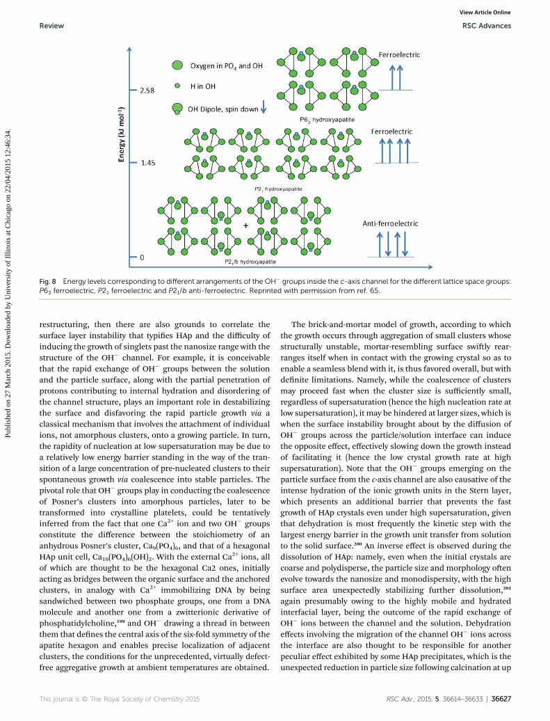
Spontaneous polarization of the OH� ions in the c-axis channelis responsible for both the piezoelectric and the pyroelectriceffect exhibited by HAp. As shown in Fig. 8, different arrange-ments of OH� dipoles in the channel created by the overlappingCa2 triangles are possible, resulting in either ferroelectric oranti-ferroelectric ordering and justifying the envisioning of apolarized HAp lattice as an array of parallel 1D ferroelectricnanowires embedded in a dielectric matrix composed of Ca2+
and PO43� groups.198 And If this polarization has been shown to
accelerate the crystal growth, while the increased disorder in theOH� channel increases solubility and facilitates particle
Fig. 8 Energy levels corresponding to different arrangements of the OH� groups inside the c-axis channel for the different lattice space groups:P63 ferroelectric, P21 ferroelectric and P21/b anti-ferroelectric. Reprinted with permission from ref. 65.
Review RSC Advances
Publ
ishe
d on
27
Mar
ch 2
015.
Dow
nloa
ded
by U
nive
rsity
of
Illin
ois
at C
hica
go o
n 22
/04/
2015
12:
46:3
4.
View Article Online
restructuring, then there are also grounds to correlate thesurface layer instability that typies HAp and the difficulty ofinducing the growth of singlets past the nanosize range with thestructure of the OH� channel. For example, it is conceivablethat the rapid exchange of OH� groups between the solutionand the particle surface, along with the partial penetration ofprotons contributing to internal hydration and disordering ofthe channel structure, plays an important role in destabilizingthe surface and disfavoring the rapid particle growth via aclassical mechanism that involves the attachment of individualions, not amorphous clusters, onto a growing particle. In turn,the rapidity of nucleation at low supersaturation may be due toa relatively low energy barrier standing in the way of the tran-sition of a large concentration of pre-nucleated clusters to theirspontaneous growth via coalescence into stable particles. Thepivotal role that OH� groups play in conducting the coalescenceof Posner’s clusters into amorphous particles, later to betransformed into crystalline platelets, could be tentativelyinferred from the fact that one Ca2+ ion and two OH� groupsconstitute the difference between the stoichiometry of ananhydrous Posner’s cluster, Ca9(PO4)6, and that of a hexagonalHAp unit cell, Ca10(PO4)6(OH)2. With the external Ca2+ ions, allof which are thought to be the hexagonal Ca2 ones, initiallyacting as bridges between the organic surface and the anchoredclusters, in analogy with Ca2+ immobilizing DNA by beingsandwiched between two phosphate groups, one from a DNAmolecule and another one from a zwitterionic derivative ofphosphatidylcholine,199 and OH� drawing a thread in betweenthem that denes the central axis of the six-fold symmetry of theapatite hexagon and enables precise localization of adjacentclusters, the conditions for the unprecedented, virtually defect-free aggregative growth at ambient temperatures are obtained.
The brick-and-mortar model of growth, according to whichthe growth occurs through aggregation of small clusters whosestructurally unstable, mortar-resembling surface swily rear-ranges itself when in contact with the growing crystal so as toenable a seamless blend with it, is thus favored overall, but withdenite limitations. Namely, while the coalescence of clustersmay proceed fast when the cluster size is sufficiently small,regardless of supersaturation (hence the high nucleation rate atlow supersaturation), it may be hindered at larger sizes, which iswhen the surface instability brought about by the diffusion ofOH� groups across the particle/solution interface can inducethe opposite effect, effectively slowing down the growth insteadof facilitating it (hence the low crystal growth rate at highsupersaturation). Note that the OH� groups emerging on theparticle surface from the c-axis channel are also causative of theintense hydration of the ionic growth units in the Stern layer,which presents an additional barrier that prevents the fastgrowth of HAp crystals even under high supersaturation, giventhat dehydration is most frequently the kinetic step with thelargest energy barrier in the growth unit transfer from solutionto the solid surface.200 An inverse effect is observed during thedissolution of HAp: namely, even when the initial crystals arecoarse and polydisperse, the particle size and morphology oenevolve towards the nanosize and monodispersity, with the highsurface area unexpectedly stabilizing further dissolution,201
again presumably owing to the highly mobile and hydratedinterfacial layer, being the outcome of the rapid exchange ofOH� ions between the channel and the solution. Dehydrationeffects involving the migration of the channel OH� ions acrossthe interface are also thought to be responsible for anotherpeculiar effect exhibited by some HAp precipitates, which is theunexpected reduction in particle size following calcination at up
to 750 �C, in spite of the evident increase in crystallinity.202,203 Inany case, the exposure of OH� groups necessitates majorsurface reconstructions in search of stability83 and theirdynamic interaction with the aqueous environment makes thesurface reconstruction processes perpetually uctuant.
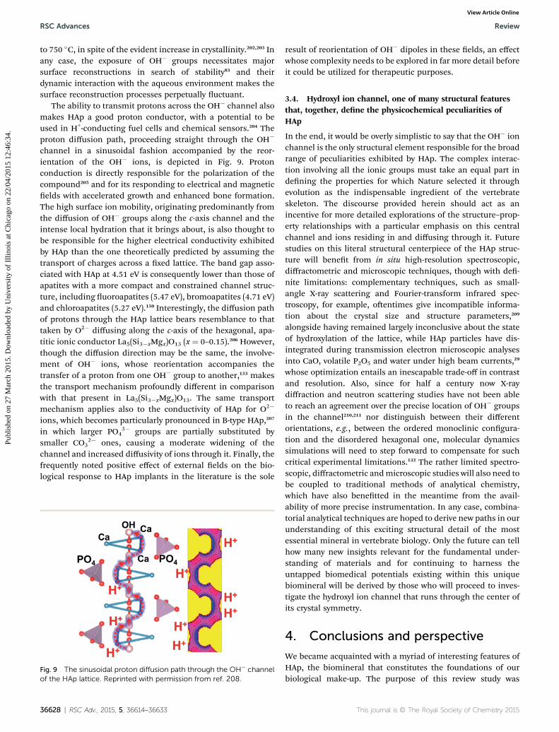
The ability to transmit protons across the OH� channel alsomakes HAp a good proton conductor, with a potential to beused in H+-conducting fuel cells and chemical sensors.204 Theproton diffusion path, proceeding straight through the OH�
channel in a sinusoidal fashion accompanied by the reor-ientation of the OH� ions, is depicted in Fig. 9. Protonconduction is directly responsible for the polarization of thecompound205 and for its responding to electrical and magneticelds with accelerated growth and enhanced bone formation.The high surface ion mobility, originating predominantly fromthe diffusion of OH� groups along the c-axis channel and theintense local hydration that it brings about, is also thought tobe responsible for the higher electrical conductivity exhibitedby HAp than the one theoretically predicted by assuming thetransport of charges across a xed lattice. The band gap asso-ciated with HAp at 4.51 eV is consequently lower than those ofapatites with a more compact and constrained channel struc-ture, including uoroapatites (5.47 eV), bromoapatites (4.71 eV)and chloroapatites (5.27 eV).150 Interestingly, the diffusion pathof protons through the HAp lattice bears resemblance to thattaken by O2� diffusing along the c-axis of the hexagonal, apa-titic ionic conductor La5(Si3�xMgx)O13 (x ¼ 0–0.15).206 However,though the diffusion direction may be the same, the involve-ment of OH� ions, whose reorientation accompanies thetransfer of a proton from one OH� group to another,133 makesthe transport mechanism profoundly different in comparisonwith that present in La5(Si3�xMgx)O13. The same transportmechanism applies also to the conductivity of HAp for O2�
ions, which becomes particularly pronounced in B-type HAp,207
in which larger PO43� groups are partially substituted by
smaller CO32� ones, causing a moderate widening of the
channel and increased diffusivity of ions through it. Finally, thefrequently noted positive effect of external elds on the bio-logical response to HAp implants in the literature is the sole
Fig. 9 The sinusoidal proton diffusion path through the OH� channelof the HAp lattice. Reprinted with permission from ref. 208.
36628 | RSC Adv., 2015, 5, 36614–36633
result of reorientation of OH� dipoles in these elds, an effectwhose complexity needs to be explored in far more detail beforeit could be utilized for therapeutic purposes.
3.4. Hydroxyl ion channel, one of many structural featuresthat, together, dene the physicochemical peculiarities ofHAp
In the end, it would be overly simplistic to say that the OH� ionchannel is the only structural element responsible for the broadrange of peculiarities exhibited by HAp. The complex interac-tion involving all the ionic groups must take an equal part indening the properties for which Nature selected it throughevolution as the indispensable ingredient of the vertebrateskeleton. The discourse provided herein should act as anincentive for more detailed explorations of the structure–prop-erty relationships with a particular emphasis on this centralchannel and ions residing in and diffusing through it. Futurestudies on this literal structural centerpiece of the HAp struc-ture will benet from in situ high-resolution spectroscopic,diffractometric and microscopic techniques, though with de-nite limitations: complementary techniques, such as small-angle X-ray scattering and Fourier-transform infrared spec-troscopy, for example, oentimes give incompatible informa-tion about the crystal size and structure parameters,209
alongside having remained largely inconclusive about the stateof hydroxylation of the lattice, while HAp particles have dis-integrated during transmission electron microscopic analysesinto CaO, volatile P2O5 and water under high beam currents,29
whose optimization entails an inescapable trade-off in contrastand resolution. Also, since for half a century now X-raydiffraction and neutron scattering studies have not been ableto reach an agreement over the precise location of OH� groupsin the channel210,211 nor distinguish between their differentorientations, e.g., between the ordered monoclinic congura-tion and the disordered hexagonal one, molecular dynamicssimulations will need to step forward to compensate for suchcritical experimental limitations.132 The rather limited spectro-scopic, diffractometric andmicroscopic studies will also need tobe coupled to traditional methods of analytical chemistry,which have also benetted in the meantime from the avail-ability of more precise instrumentation. In any case, combina-torial analytical techniques are hoped to derive new paths in ourunderstanding of this exciting structural detail of the mostessential mineral in vertebrate biology. Only the future can tellhow many new insights relevant for the fundamental under-standing of materials and for continuing to harness theuntapped biomedical potentials existing within this uniquebiomineral will be derived by those who will proceed to inves-tigate the hydroxyl ion channel that runs through the center ofits crystal symmetry.
4. Conclusions and perspective
We became acquainted with a myriad of interesting features ofHAp, the biomineral that constitutes the foundations of ourbiological make-up. The purpose of this review study was
twofold: rstly, to compile physicochemical peculiaritiesexhibited by HAp, and, secondly, to engage in an effort toexplain them by referring to the hydroxyl ion channel as anessential crystallographic element of this compound. We haveseen that the spontaneous polarization of HAp, the increasedsolubility, resorption rate and bioactivity of biological apatite aswell as the precise crystallographic orientation of apatiteplatelets in bone may all have their roots in the crystallographiccolumn of hydroxyl ions extending along the 63 screw axis thatruns through the center of a series of overlapping calcium iontriangles. Still, what this review has aimed at is to provide only aglimpse at a remarkable structural feature of a crystallinestructure that is, inexplicably, evolutionarily favored overothers, and assuring the readers that there are reasons forwhich it should be studied in more detail.
Ideally, therefore, the impact of this study is to be thestimulation of a greater interest in examining the relationshipsbetween the hydroxyl ion channel extending across the longestaxis of the crystal structure of HAp and the properties of thiscompound in both simple chemical environments and morecomplex, biological niches. Unfortunately, as already insinu-ated in section 3.4, the level of sophistication of the currentstate-of-the-art microscopic and spectrometric techniques is notadequate for visualizing this channel and quantifying inter-atomic potentials in and around it, let alone solving the nestsymmetry perturbations on its scale under dynamic conditions.We have just begun to visualize the ripening of nanoclustersduring crystal growth in real time and there is a long way beforethe dynamic surface restructuring that distinguishes the ionchannel from the rest of the lattice will be observable usingatomic-resolution imaging techniques. Although combinatorialtechniques – such as microbeam X-ray diffraction and small-angle scattering paired with electron microscopy212 – arecapable of discerning the deposition of Posner’s clusters ontogrowing HAp crystals in real time, any ideas about the precisestructural transformations taking place on the atomic scaleupon the coalescence of amorphous precursors and theirrecrystallization, have so far emerged strictly from theorizing.To compensate for these denitive deciencies of experimentalmethods, computational analyses have justiably stepped in,yet their shortcomings, stemming from a range of approxima-tions applied to enable mathematical solvability, are equallysubstantial. Continued examination of the effects of thehydroxyl ion channel on the properties of HAp will undoubtedlyresemble the digging of a tunnel from two opposite sides,hoping that the hollows will eventually meet in the middle. Andeach time they do, a new line, like that running through thecenter of the crystal that was the subject of this study, will bedrawn, perhaps to remind us, on a grander scale of things, ofthe innitude of applicative and inspirational analogies latentin the structure of this magnicent material.
Acknowledgements
Writing of this review article was supported by the NationalInstitutes of Health grant R00-DE021416: osteogenic calciumphosphate nanoparticles with designable drug release kinetics.
1 N. H. de Leeuw, J. R. Bowe and J. A. L. Rabone, FaradayDiscuss., 2007, 134, 195–214.
2 A. G. Werner, Gerhards Grundr (as Apatit), 1786, 281.3 A. Farzadi, F. Bakhshi, M. Solati-Hashjin, M. Asadi-Eydivand and N. A. A. Osman, Ceram. Int., 2014, 40, 6021–6029.
4 M. Okazaki, Biomaterials, 1991, 12, 831–835.5 E. Landi, G. Logroscino, L. Proietti, A. Tampieri, M. Sandriand S. Sprio, J. Mater. Sci.: Mater. Med., 2008, 19, 239–247.
6 S. Markovic, L. Veselinovic, M. Lukic, L. Karanovic,I. Bracko, N. Ignjatovic and D. Uskokovic, Biomed. Mater.,2011, 6, 045005.
7 F. Ren, X. Lu and Y. Leng, J. Mech. Behav. Biomed. Mater.,2013, 26, 59–67.
8 D. Shi, Introduction to Biomaterials, World Scientic Press,Singapore, 2006.
9 J. C. Elliot, P. E. Mackie and R. A. Young, Science, 1973, 180,1055–1057.
10 M. I. Kay, R. A. Young and A. S. Posner, Nature, 1964, 204,1050–1052.
11 T. Ikoma, A. Yamazaki, S. Nakamura and M. Akao, NetsuSokutei, 1998, 25, 141–149.
12 B. Wopenka and J. D. Pasteris, Mater. Sci. Eng., C, 2005, 25,131–143.
13 V. Uskokovic and T. A. Desai, J. Biomed. Mater. Res., Part A,2013, 101, 1416–1426.
14 M. Itokazu, W. Yang, T. Aoki, A. Ohara and N. Kato,Biomaterials, 1998, 19, 817–819.
15 C. Petrone, G. Hall, M. Langman and M. J. Filiaggi, ActaBiomater., 2008, 4, 403–413.
16 M. A. Rauschmann, T. A. Wichelhaus, V. Stirnal,E. Dingeldein, L. Zichner, R. Schnettler and V. Alt,Biomaterials, 2005, 26, 2677–2684.
17 B. M. Barth, E. Altinoglu, S. S. Shanmugavelandy,J. M. Kaiser, D. Crespo-Gonzalez, N. A. DiVittore,C. McGovern, T. M. Goff, N. R. Keasey, J. H. Adair,T. P. Loughran Jr, D. F. Claxton and M. Kester, ACS Nano,2011, 5, 5325–5337.
18 Y. Y. Hu, A. Rawal and K. Schmidt-Rohr, Proc. Natl. Acad.Sci. U. S. A., 2010, 107, 22425–22429.
19 C. Rey, J. C. Trombe and G. Montel, J. Chem. Res., Synop.,1978, 188, M 2401.
20 R. Z. LeGeros, Biological and Synthetic Apatites, inHydroxyapatite and Related Materials, ed. P. W. Brown andB. Constantz, CRC Press, Boca Raton, FL, 1994, pp. 3–28.
21 M. S.-A. Johnsson and G. H. Nancollas, Crit. Rev. Oral Biol.Med., 1992, 3, 61–82.
22 R. Z. LeGeros, G. Daculsi and I. Orly, Scanning ElectronMicrosc., 1989, 3, 129–138.
23 A. L. Boskey and A. S. Posner, J. Phys. Chem., 1973, 77, 2313–2317.
24 G. Zhang, R. Huang, Z. Li, X. Yang, X. Chen, W. Xia, X. Sun,G. Yang, C. Gao and Z. Gou, J. Inorg. Biochem., 2012, 113, 1–8.
25 M. Bohner, F. Theiss, D. Apelt, W. Hirsiger, R. Houriet,G. Rizzoli, E. Gnos, C. Frei, J. A. Auer and B. vonRechenberg, Biomaterials, 2003, 24, 3463–3474.
26 M. Bohner, Bioresorbable ceramics, in Degradation rate ofbioresorbable materials, ed. F. Buchanan, Woodhead,Cambridge, 2008, pp. 95–114.
27 V. Uskokovic, J. Dispersion Sci. Technol., 2012, 33, 1762–1786.
28 V. Uskokovic, R. Odsinada, S. Djordjevic and S. Habelitz,Arch. Oral Biol., 2011, 56, 521–532.
29 T. T. Morgan, H. S. Muddana, E. Altınoglu, S. M. Rouse,A. Tabakovic, T. Tabouillot, T. J. Russin,S. S. Shanmugavelandy, P. J. Butler, P. C. Eklund,J. K. Yun, M. Kester and J. H. Adair, Nano Lett., 2008, 8,4108–4115.
30 A. Sorkin andM. von Zastrow, Nat. Rev. Mol. Cell Biol., 2002,3, 600–614.
31 I. Rodrıguez-Ruiz, J. M. Delgado-Lopez, M. A. Duran-Olivencia, M. Iasco, A. Tampieri, D. Colangelo, M. Pratand J. Gomez-Morales, Langmuir, 2013, 29, 8213–8221.
32 E. H. Chowdhury, M. Kunou, M. Nagaoka, A. K. Kundu,T. Hoshiba and T. Akaike, Gene, 2004, 341, 77–82.
33 M. S. Lee, J. E. Lee, E. Byun, N. W. Kim, K. Lee, H. Lee,S. J. Sim, D. S. Lee and J. H. Jeong, J. Controlled Release,2014, 192, 122–130.
34 K. Khosravi-Darani, M. R. Mozafari, L. Rashidi andM. Mohammadi, Acta Med. Iran., 2010, 48, 133–141.
35 T. Halfer, A. Rei, L. C. Ciacchi, L. Treccani and K. Rezwan,Biointerphases, 2014, 9, 031018.
36 J. Veliscek-Carolan, K. A. Jolliffe and T. L. Hanley, ACS Appl.Mater. Interfaces, 2013, 5, 11984–11994.
37 L. Treccani, T. Yvonne-Klein, F. Meder, K. Pardun andK. Rezwan, Acta Biomater., 2013, 9, 7115–71150.
38 I. Mobasherpour, E. Salahi and M. Pazouki, Desalination,2011, 266, 142–248.
39 S. Bailliez, A. Nzihou, E. Beche and G. Flamant, Process Saf.Environ. Prot., 2004, 82, 175–180.
40 F. Monteil-Rivera and M. Fedoroff, Sorption of InorganicSpecies on Apatites from Aqueous Solutions, Encyclopedia ofSurface and Colloid Science, Marcel Dekker. Inc., NewYork, 2002, pp. 1–26.
41 I. Smiciklas, S. Dimovic, I. Plecas and M. Mitric, Water Res.,2006, 40, 2267–2274.
42 J. Reichert and J. G. P. Binner, J. Mat. Sci., 2004, 31, 1231–1241.
43 T. Kawasaki, S. Takahashi and K. Ikeda, Eur. J. Biochem.,1985, 152, 361–371.
44 L. J. Cummings, M. A. Snyder and K. Brisack, MethodsEnzymol., 2009, 463, 387–404.
45 R. Giovannini and R. Freitag, Bioseparation, 2001, 9, 359–368.
46 M. Saito, Y. Kurosawa and T. Okuyama, PLoS One, 2013, 8,e53893.
47 A. Rimola, M. Corno, C. M. Zicovich-Wilson andP. Ugliengo, J. Am. Chem. Soc., 2008, 130, 16181–16183.
48 L. M. Gordon, L. Tran and D. Joester, ACS Nano, 2012, 6,10667–10675.
36630 | RSC Adv., 2015, 5, 36614–36633
49 M. P. Ginebra, M. Espanol, E. B. Montufar, R. A. Perez andG. Mestres, Acta Biomater., 2010, 6, 2863–2873.
50 S. Aghyarian, L. C. Rodriguez, J. Chari, E. Bentley,V. Kosmopoulos, I. H. Lieberman and D. C. Rodrigues, J.Biomater. Appl., 2014, 29, 688–698.
51 R. Marefat-Seyedlar, A. Nodehi, M. Atai and M. Imani,Carbohydr. Polym., 2014, 99, 257–263.
52 C. Holt, J. A. Carver, H. Ecroyd and D. C. Thorn, J. Dairy Sci.,2013, 96, 6127–6146.
53 S. Fujii, M. Okada and T. Furuzono, J. Colloid Interface Sci.,2007, 315, 287–296.
54 C. Combes and C. Rey, Acta Biomater., 2010, 6, 3362–3378.55 M. C. F. Magalhaes, P. A. A. P. Marques and R. N. Correia,
Calcium and magnesium phosphates: normal andpathological mineralization, in Biomineralization – medicalaspects of solubility, ed. E. Konigsberger and L. C.Konigsberger, John Wiley & Sons, Chichester, 2006, pp.71–124.
56 N. C. Webb, Acta Crystallogr., 1966, 21, 942–948.57 R. M. Bennett, J. R. Lehr and D. J. McCarty, J. Clin. Invest.,
1975, 56, 1571–1579.58 S. Dorozhkin, J. Funct. Biomater., 2013, 4, 209–311.59 M. Nakamura, R. Hiratai and K. Yamashita, J. Biomed.
Mater. Res., Part A, 2012, 100, 1368–1374.60 A. C. Ahn and A. J. Grodzinsky, Med. Eng. Phys., 2009, 31,
733–741.61 A. G. Cockbain, Mineral. Mag., 1968, 36, 654–660.62 S. B. Lang, S. A. M. Tofail, A. A. Gandhi, M. Gregor, C. Wolf-
Brandstetter and J. Kost, Appl. Phys. Lett., 2011, 98, 123703.63 T. J. Bukowski, M. McCarthy, F. McCarthy, G. Teowee,
T. P. Alexander, D. R. Uhlmann, J. T. Dawley andB. J. J. Zelinski, Integr. Ferroelectr., 1997, 17, 339–347.
64 M. Al Ahmad and R. Plana, IEEE Microw. Wireless Compon.Lett., 2009, 19, 140–142.
65 S. B. Lang, S. A. Tofail, A. L. Kholkin, M. Wojtas, M. Gregor,A. A. Gandhi, Y. Wang, S. Bauer, M. Krause and A. Plecenik,Sci. Rep., 2013, 3, 2215.
66 G. C. Maiti and F. Freund, J. Chem. Soc., Dalton Trans., 1981,4, 949–955.
67 H. Noriuchi, M. Nakamura, A. Nagai, K. Katayama andK. Yamashita, J. Appl. Phys., 2012, 112, 074901.
68 S. Nakamura, T. Kobayashi and K. Yamashita, J. Biomed.Mater. Res. A, 2004, 68, 90–94.
69 P. Singh, R. C. Yash Roy and M. Hoque, Indian J. Biochem.Biophys., 2006, 43, 167–172.
70 N. Ignjatovic, Z. Ajdukovic, V. Savic, S. Najman,D. Mihailovic, P. Vasiljevic, Z. Stojanovic, V. Uskokovicand D. Uskokovic, J. Mater. Sci.: Mater. Med., 2013, 24,343–354.
71 K. Yamashita, N. Oikawa and T. Umegaki, Chem. Mater.,1996, 8, 2697–2700.
72 A. Szczes, L. Holysz and E. Chibowski, J. Adhes. Sci. Technol.,2006, 20, 345–358.
73 V. Uskokovic, J. Mater. Educ., 2014, 36, 25–50.74 V. Uskokovic, J. Biomimetics, Biomater., Tissue Eng., 2010, 8,
75 C. Burger, H.-W. Zhou, H. Wang, I. Sics, B. S. Hsiao, B. Chu,L. Graham and M. J. Glimcher, Biophys. J., 2008, 95, 1985–1992.
76 V. Uskokovic, Biomineralization and Biomimicry of ToothEnamel, in Non-Metallic Biomaterials for Tooth Repair andReplacement, ed. P. Vallittu, Woodhead Publishing,Cambridge, UK, 2013, pp. 20–89.
77 J. Moradian-Oldak, Front. Biosci., Landmark Ed., 2012, 17,1996–2023.
78 M. Corno, A. Rimola, V. Bolis and P. Ugliengo, Phys. Chem.Chem. Phys., 2010, 12, 6309–6329.
79 V. Uskokovic and D. P. Uskokovic, J. Biomed. Mater. Res.,Part B, 2011, 96, 152–191.
80 E. D. Eanes, H. Gillessen and A. S. Posner, Nature, 1965,208, 365–367.
81 K. Onuma and A. Ito, Chem. Mater., 1998, 10, 3346–3351.82 R. Tang, Z. J. Henneman and G. H. Nancollas, J. Cryst.
Growth, 2003, 249, 614–624.83 A. Slepko and A. A. Demkov, J. Chem. Phys., 2013, 139,
044714.84 S. J. Zawacki, P. B. Koutsoukos, M. H. Salimi and
G. H. Nancollas, The growth of calcium phosphates, Am.Chem. Soc. Symp. Series, ed. J. A. Davis and K. F. Hayes,1986, vol. 323, p. 650.
85 A. A. Campbell, M. LoRe and G. H. Nancollas, Colloids Surf.,1991, 54, 25–31.
86 N. Kanzaki, K. Onuma, A. Ito, K. Teraoka, T. Tateishi andS. Tsutsumi, J. Phys. Chem., 1998, 102, 6471–6476.
87 A. A. Chernov, Notes on interface growth kinetics 50 yearsaer Barton, Cabrera and Frank, in 50 Years Progress inCrystal Growth: A Reprint Collection, ed. R. Feigeison,Elsevier, Amsterdam. NL, 2004, pp. 47–66.
88 K. Onuma, A. Ito and T. Tateishi, J. Cryst. Growth, 1996, 167,773–776.
89 S.-Y. Chung, Y.-M. Kim, J.-G. Kim and Y.-J. Kim, Nat. Phys.,2009, 5, 68–73.
90 K. Onuma, Prog. Cryst. Growth Charact. Mater., 2006, 52,223–245.
91 T. Kokubo, H. Kushitani, S. Sakka, T. Kitsugi andT. Yamamuro, J. Biomed. Mater. Res., 1990, 24, 721.
92 K. Onuma, A. Ito, T. Tateishi and T. Kameyama, J. Cryst.Growth, 1995, 148, 201–206.
93 J. Christoffersen and M. R. Christoffersen, J. Cryst. Growth,1988, 87, 51–61.
94 J. Christoffersen, J. Cryst. Growth, 1980, 49, 29–44.95 L. Wang, G. H. Nancollas, Z. J. Henneman, E. Klein and
S. Weiner, Biointerphases, 2006, 1, 106–111.96 S. Jiang, Y. Chen, H. Pan, Y.-J. Zhang and R. Tang, Phys.
Chem. Chem. Phys., 2013, 15, 12530–12533.97 D. Farlay, G. Panczer, C. Rey, P. D. Delmas and G. Boivin, J.
Bone Miner. Metab., 2010, 28, 433–445.98 V. Uskokovic and T. A. Desai, J. Biomed. Mater. Res., Part A,
2013, 101, 1427–1436.99 F. Monteil-Rivera and M. Fedoroff, Sorption of Inorganic
Species on Apatites from Aqueous Solutions, inEncyclopedia of Surface and Colloid Science, ed. P.
100 C. Rey, B. Collins, T. Goehl, I. R. Dickson andM. J. Glimcher, Calcif. Tissue Int., 1989, 45, 157–164.
101 W. H. Emerson and E. D. Fischer, Arch. Oral Biol., 1962, 7,671–683.
102 B. B. Hole, D. S. Keller, W. M. Burry and J. A. Schwarz, J.Chromatogr. B: Anal. Technol. Biomed. Life Sci., 2011, 879,1847–1850.
103 G. Daculsi and J. P. LeGeros, J. Biomed. Mater. Res., 1996, 31,495–501.
104 T. Leventouri, Biomaterials, 2006, 27, 3339–3342.105 V. Uskokovic, Review Journal of Chemistry, 2013, 3, 271–303.106 M. Kazanci, P. Fratzl, K. Klaushofer and E. P. Paschalis,
Calcif. Tissue Int., 2006, 79, 354–359.107 M. J. J. M. van Kemenade and P. L. de Bruyn, J. Colloid
Interface Sci., 1987, 118, 564–585.108 I. V. Melikhov, S. Lazic and Z. Vukovic, J. Colloid Interface
Sci., 1989, 127, 317–327.109 L. T. Canham, C. L. Reeves, A. Loni, M. R. Houlton,
J. P. Newey, A. J. Simons and T. I. Cox, Thin Solid Films,1997, 297, 304–307.
110 T. Kokubo, S. Ito, Z. T. Huang, T. Hayashi, S. Sakka,T. Kitsugi and T. Yamamuro, J. Biomed. Mater. Res., 1990,24, 331–343.
111 T. Kokubo and H. Takadama, Biomaterials, 2006, 27, 2907–2915.
112 J. A. Budz, M. LoRe and G. H. Nancollas, Adv. Dent. Res.,1987, 1, 314–321.
113 V. Uskokovic, M.-K. Kim, W. Li and S. Habelitz, J. Mater.Res., 2008, 32, 3184–3195.
114 G. Montel, Ann. Chim., 1969, 14, 255–266.115 A. S. Posner, J. M. Stutman and E. R. Lippincott, Nature,
1960, 188, 486–487.116 R. A. Young and D.W. Holcomb, Calcif. Tissue Int., 1982, 34,
S17–S32.117 M. H. Klaproth, Neues Bergmannische Journal, 1788, 294.118 J. L. Proust, J. Phys. Radium, 1788, 32, 241.119 A. Dubree, Compt. Rend. Acad. Sci. Paris, 1851, 32, 625.120 W. F. de Jong, Recl. Trav. Chim. Pays-Bas Belg., 1926, 45,
445–448.121 S. Naray-Szabo, Z. Kristallogr., 1930, 75, 387–398.122 M. Mehmel, Z. Kristallogr., 1930, 75, 323–331.123 A. Slepko and A. A. Demkov, Phys. Rev. B: Condens. Matter
Mater. Phys., 2011, 84, 134108.124 H. Suda, M. Yashima, M. Kakihana and M. Yoshimura, J.
Phys. Chem., 1995, 99, 6752–6754.125 N. Hitmi, C. LaCabanne and R. A. Young, J. Phys. Chem.
Solids, 1986, 47, 533.126 N. Hitmi, E. Lamure-Plaino, A. Lamure, C. LaCabanne and
R. A. Young, Calcif. Tissue Int., 1986, 38, 252–261.127 D. Aquilano, M. Bruno, M. Rubbo, F. R. Massaro and
L. Pastero, Cryst. Growth Des., 2014, 14, 2846–2852.128 L. L. Hench and S. M. Best, Ceramics, Glasses, and Glass-
Ceramics: Basic Principles, in Biomaterials Science: AnIntroduction to Materials in Medicine, ed. B. D. Ratner, A.
S. Hoffman, F. J. Schoen and J. E. Lemons, Elsevier, Oxford,UK, 2012, p. 140.
129 J. C. Elliott, R. M. Wilson and S. E. P. Dowker, Adv. X-RayAnal., 2002, 45, 172–181.
130 W. Zhu and P. Wu, Chem. Phys. Lett., 2004, 396, 38–42.131 Z. Opre, Catalytic oxidation over transition metal
containing hydroxyapatites, PhD dissertation, SwissFederal Institute of Technology, ETH No. 17247, Zurich,2007.
132 O. Hochrein, R. Kniep and D. Zahn, Chem. Mater., 2005, 17,1978–1981.
133 M. Yashima, Y. Yonehara and H. Fujimori, J. Phys. Chem. C,2011, 115, 25077–25087.
134 E. I. Dorozhkina and S. V. Dorozhkin, Chem. Mater., 2002,14, 4267–4272.
135 G. Cho, Y. Wu and J. L. Ackerman, Science, 2003, 300, 1123–1127.
136 A. J. Taylor, E. Rendina, B. J. Smith and D. H. Zhou, Chem.Phys. Lett., 2013, 588, 124–130.
137 D. K. Smith, Calcium phosphate apatites in Nature, inHydroxyapatite and Related Materials, ed. P. W. Brown andB. Constantz, CRC Press, Boca Raton, FL, 1994, pp. 29–44.
138 C. Rey, J. L. Miquel, L. Facchini, A. P. Legrand andM. J. Glimcher, Bone, 1995, 16, 583–586.
139 J. D. Pasteris, B. Wopenka, J. J. Freeman, K. Rogers,E. Valsami-Jones, J. A. van der Houwen and M. J. Silva,Biomaterials, 2004, 25, 229–238.
140 F. M. McCubbin, A. Steele, E. H. Hauri, H. Nekvasil,S. Yamashita and R. J. Hemley, Proc. Natl. Acad. Sci. U. S.A., 2010, 107, 11223–11228.
141 A. Fazekas, Moon has a hundred times more water thanthought, National Geographic, June 14, 2010, retrievedfrom http://news.nationalgeographic.com/news/2010/06/100614-moon-water-hundred-lunar-proceedings-science.
142 L. Eberhardsteiner, C. Hellmich and S. Scheiner, Comput.Meth. Biomech. Biomed. Eng., 2014, 17, 48–63.
143 Y. Wang, S. Von Euw, F. M. Fernandes, S. Cassaignon,M. Selmane, G. Laurent, G. Pehau-Arnaudet, C. Coelho,L. Bonhomme-Coury, M. M. Giraud-Guille, F. Babonneau,T. Azais and N. Nassif, Nat. Mater., 2013, 12, 1144–1153.
144 Z. Lu, H. Zhang, Y. Guo, Y. Wang, X. Ge, Y. Leng andF. Watari, CrystEngComm, 2011, 13, 3741–3749.
145 C. Rey, C. Combes, C. Drouet and M. J. Glimcher,Osteoporosis Int., 2009, 20, 1013–1021.
146 C. Rey, Apatite Channels and Zeolite-Like Properties, inHydroxyapatite and Related Materials, ed. P. W. Brown andB. Constantz, CRC Press, Boca Raton, FL, 1994, pp. 257–262.
147 M. E. Fleet, Front. Biosci., Elite Ed., 2013, 5, 643–652.148 E. J. Duff and A. A. Grant, in Mechanical Properties of
Biomaterials, Vol. 2 of Advances in Biomolecules, ed. G. W.Hastings and D. F. Williams, Wiley, New York, NY, 1980,pp. 465–475.
149 P. R. Garant, Oral Cells and Tissues, Quintessence, CarolStream, IL, 2003.
150 P. Rulis, L. Ouyang and W. Y. Ching, Phys. Rev. B: Condens.Matter Mater. Phys., 2004, 70, 155104.
36632 | RSC Adv., 2015, 5, 36614–36633
151 B. Menzel and C. H. Amberg, J. Colloid Interface Sci, 1972,38, 256–264.
152 G. C. Maiti and F. Freund, J. Inorg. Nucl. Chem., 1981, 43,2633–2637.
153 B. O. Fowler, Inorg. Chem., 1974, 13, 194–214.154 P. F. Gonzalez-Diaz and M. Santos, J. Solid State Chem.,
1972, 22, 193–199.155 H. Zhao, X. Li, J. Wang, S. Qu, J. Weng and X. Zhang, J.
Biomed. Mater. Res., 2000, 52, 157–163.156 L. G. Rodenas, J. M. Palacios, M. C. Apella, P. J. Morando
and M. A. Blesa, J. Colloid Interface Sci., 2005, 290, 145–154.157 W. T. Lee, M. T. Dove and E. K. H. Salje, J. Phys.: Condens.
Matter, 2000, 12, 9829–9841.158 M. H. Lee, P. Ducheyne, L. Lynch, D. Boettiger and
R. J. Composto, Biomaterials, 2006, 27, 1907–1916.159 B. E. Rapuano, J. J. Lee and D. E. MacDonald, Eur. J. Oral
Sci., 2012, 120, 185–194.160 S. V. Dorozhkin, Materials, 2009, 2, 399–498.161 C. A. Orme and J. L. Giocondi, The Use of Scanning Probe
Microscopy to Investigate Crystal-Fluid Interfaces, inPerspectives on Inorganic, Organic, and Biological CrystalGrowth: From Fundamentals to Applications, ed. M.Skowronski, J. J. DeYoreo and C. A. Wang, AmericanInstitute of Physics, Melville, NY, 2007.
162 A. Filankembo, S. Giorgio, I. Lisiecki and M. P. Pileni, J.Phys. Chem. B, 2003, 107, 7492–7500.
163 V. Uskokovic, Rev. Chem. Eng., 2007, 23, 301–372.164 D. J. Shaw, Introduction to Colloid and Surface Chemistry,
Butterworth Heinemann, Oxford, UK, 2003.165 C. Jager, T. Welzel, W. Meyer-Zaika and M. Epple, Magn.
Reson. Chem., 2006, 44, 573–580.166 A. Bengtsson, A. Shchukarev, P. Persson and S. Sjoberg,
Geochim. Cosmochim. Acta, 2009, 73, 257–267.167 V. Uskokovic, W. Li and S. Habelitz, J. Cryst. Growth, 2011,
316, 106–117.168 W. J. Habraken, J. Tao, L. J. Brylka, H. Friedrich,
L. Bertinetti, A. S. Schenk, A. Verch, V. Dmitrovic,P. H. Bomans, P. M. Frederik, J. Laven, P. van der Schoot,B. Aichmayer, G. de With, J. J. DeYoreo andN. A. Sommerdijk, Nat. Commun., 2013, 4, 1507.
169 M. Aizawa, T. Matsuura and Z. Zhuang, Biol. Pharm. Bull.,2013, 36, 1654–1661.
170 M. Yoshimura and H. Suda, Hydrothermal processing ofhydroxyapatite: Past, present and future, inHydroxyapatite and Related Materials, ed. P. W. Brown andB. Constantz, CRC Press, Boca Raton, FL, 1994, pp. 45–72.
171 M. Corno, C. Busco, V. Bolis, S. Tosoni and P. Ugliengo,Langmuir, 2009, 25, 2188–2198.
172 M. Aning, D. O. Welch and B. S. H. Royce, Phys. Lett. A,1971, 37, 253–254.
173 J. Zeglinski, M. Nolan, D. Thompson and S. A. M. Tofall,Surf. Sci., 2014, 623, 55–63.
174 M. J. Olszta, X. G. Cheng, S. S. Jee, R. Kumar, Y. Y. Kim,M. J. Kaufman, E. P. Douglas and L. B. Gower, Mater. Sci.Eng., R, 2007, 58, 77–116.
175 H. Pan, J. Tao, X. Yu, L. Fu, J. Zhang, X. Zeng, G. Xu andR. Tang, J. Phys. Chem. B, 2008, 112, 7162–7165.
176 L. Bertinetti, C. Drouet, C. Combes, C. Rey, A. Tempieri,S. Coluccia and G. Martra, Langmuir, 2009, 25, 5647–5654.
177 A. Cooper, Thermodynamics of protein folding andstability, in Protein: A comprehensive treatise, ed. G. Allen,JAI Press, Stamford, CN, 1999, vol. 2, pp. 217–270.
178 D. W. Dempster and E. Shane, Bone Quantication andDynamics of Turnover, in Principles and Practice ofEndocrinology and Metabolism, ed. K. L. Becker, LippincottWilliams & Wilkins, Philadelphia, PA, 3rd edn, 2001, pp.541–547.
179 B. Alexander, T. L. Daulton, G. M. Genin, J. Lipner,J. D. Pasteris, B. Wopenka and S. Thomopoulos, J. R. Soc.,Interface, 2012, 9, 1774–1786.
180 E. A. McNally, H. P. Schwarz, G. A. Botton andA. L. Arsenault, PLoS One, 2012, 7, e29258.
181 C. Rey, C. Combes, C. Drouet and H. Shi, Adv. Sci.Technol., 2006, 49, 27–36.
182 M. J. Larsen, Ion Products and Solubility of CalciumPhosphates, Royal Dental College, Denmark, 2001.
183 R. A. Young and W. E. Brown, Structure of BiologicalMinerals, in Biological Mineralization andDemineralization, ed. G. H. Nancollas, Springer-Verlag,New York, NY, 1982, pp. 101–141.
184 M. H. Salimi, J. C. Heughebaert and G. H. Nancollas,Langmuir, 1985, 1, 119–122.
185 J. W. Zhang and G. H. Nancollas, Mechanisms of Growthand Dissolution of Sparingly Soluble Salts, in Mineral–Water Interface Geochemistry: Reviews in Mineralogy, ed. M.F. Hochella Jr and A. F. White, 1990, vol. 23, pp. 365–396.
186 O. Hochrein and D. Zahn, J. Mol. Model., 2011, 17, 1525–1528.
187 D. G. Guo, Y. Z. Hao, H. Y. Li, C. Q. Fang, L. J. Sun, H. Zhu,J. Wang, X. F. Huang, P. F. Ni and K. W. Xu, J. Biomed.Mater. Res., Part B, 2013, 101, 1275–1283.
188 G. J. Levinskas and W. F Neuman, J. Phys. Chem., 1955, 59,164–168.
189 A. N. Smith, A. M. Posner and J. P. Quirk, J. Colloid InterfaceSci., 1974, 48, 442–449.
190 K. S. tenHuisen and P. W. Brown, J. Biomed. Mater. Res.,1997, 36, 233–241.
191 Y. E. Greish and P. W. Brown, J. Biomed. Mater. Res., Part B,2003, 67, 632–637.
192 E. Davies, K. H. Muller, W. C. Wong, C. J. Pickard,D. G. Reid, J. N. Skepper and M. J. Duer, Proc. Natl. Acad.Sci. U. S. A., 2013, 111, E1354–E1363.
193 R. Tang, M. Hass, W. Wu, S. Gulde and G. H. Nancollas, J.Colloid Interface Sci., 2003, 260, 379–384.
194 R. Z. LeGeros and S. Suga, Calcif. Tissue Int., 1980, 32, 169–174.
195 G. Mabilleau, R. Filmon, P. K. Petrov, M. F. Basle,A. Sabokbar and D. Chappard, Acta Biomater., 2010, 6,1555–1560.
196 L. Veselinovic, L. Karanovic, Z. Stojanovic, I. Bracko,S. Markovic, N. Ignjatovic and D. Uskokovic, J. Appl.Crystallogr., 2010, 43, 320–327.
197 K. P. Tank, K. S. Chudasama, V. S. Thaker and M. J. Joshi, J.Nanopart. Res., 2013, 15, 1644.
198 F. Chiatti, M. Corno and P. Ugliengo, J. Phys. Chem. C, 2012,116, 6108–6114.
199 L. Cristofolini, T. Berzina, S. Erokhina, D. Konovalov andV. Erokhin, Biomacromolecules, 2007, 8, 2270–2275.
200 S. Mann, Biomineralization: Principles and Concepts inBioinorganic Materials Chemistry, Oxford University Press,Oxford, UK, 2001.
201 L. Wang and G. H. Nancollas, Chem. Rev., 2008, 108, 4628–4669.
202 Y. X. Pang and X. Bao, J. Eur. Ceram. Soc., 2003, 23, 1697–1704.
203 I. R. Gibson, I. Rehman, S. M. Best and W. Boneld, J.Mater. Sci.: Mater. Med., 2000, 11, 799–804.
204 D. Liu, K. Savino and M. Z. Yates, Adv. Funct. Mater., 2009,19, 3941–3947.
205 S. Nakamura and H. Takeda, Adv. Funct. Mater., 2001, 19,3941–3947.
206 R. Ali, M. Yashima, Y. Matsushita, H. Yoshioka,K. Ohoyama and F. Izumi, Chem. Mater., 2008, 20, 5203–5208.
207 Y. Tanaka, M. Kikuchi, K. Tanaka, K. Hashimoto, J. Hojo,M. Nakamura, A. Nagai, T. Sugiyama, F. Munakata andL. Yamashita, J. Am. Ceram. Soc., 2010, 93, 3577–3579.
208 M. Yashima, N. Kubo, K. Omoto, H. Fujimori, K. Fujii andK. Ohoyama, J. Phys. Chem. C, 2014, 118, 5180–5187.
209 N. P. Camacho, S. Rinnerthaler, E. P. Paschalis,R. Mendelsohn, A. L. Boskey and P. Fratzl, Bone, 1999, 25,287–293.
210 K. Sudarsanan and R. A. Young, Acta Crystallogr., Sect. B:Struct. Crystallogr. Cryst. Chem., 1969, 25, 1534–1543.
211 M. E. Fleet, Carbonated Hydroxyapatite: Materials, Synthesis,and Applications, CRC Press, Boca Raton, FL, 2015.
212 J. Mahamid, B. Aichmayer, E. Shimoni, R. Ziblat, C. Li,S. Siegel, O. Paris, P. Fratzl, S. Weiner and L. Addadi,Proc. Natl. Acad. Sci. U. S. A., 2010, 107, 6316–6321.