1 Department of Pharmaceutical Technology and Chemistry, School of Pharmacy and Nutrition, University ofNavarra, 31009 Pamplona, Spain; [email protected] (A.S.-A.); [email protected] (I.F.T.)
2 Navarra Institute for Health Research (IdiSNA), 31009 Pamplona, Spain3 Department of Pharmacy and Pharmaceutical Technology and Parasitology, University of Valencia,
46100 Valencia, Spain4 Interuniversity Research Institute for Molecular Recognition and Technological Development,
Abstract: Immuno-oncology (IO) focuses on the ability of the immune system to detect and eliminatecancer cells. Since the approval of the first immune checkpoint inhibitor, immunotherapies havebecome a major player in oncology treatment and, in 2021, represented the highest number ofapproved drugs in the field. In spite of this, there is still a fraction of patients that do not respondto these therapies and develop resistance mechanisms. In this sense, mathematical models offer anopportunity to identify predictive biomarkers, optimal dosing schedules and rational combinationsto maximize clinical response. This work aims to outline the main therapeutic targets in IO and toprovide a description of the different mathematical approaches (top-down, middle-out, and bottom-up) integrating the cancer immunity cycle with immunotherapeutic agents in clinical scenarios.Among the different strategies, middle-out models, which combine both theoretical and evidence-based description of tumor growth and immunological cell-type dynamics, represent an optimalframework to evaluate new IO strategies.
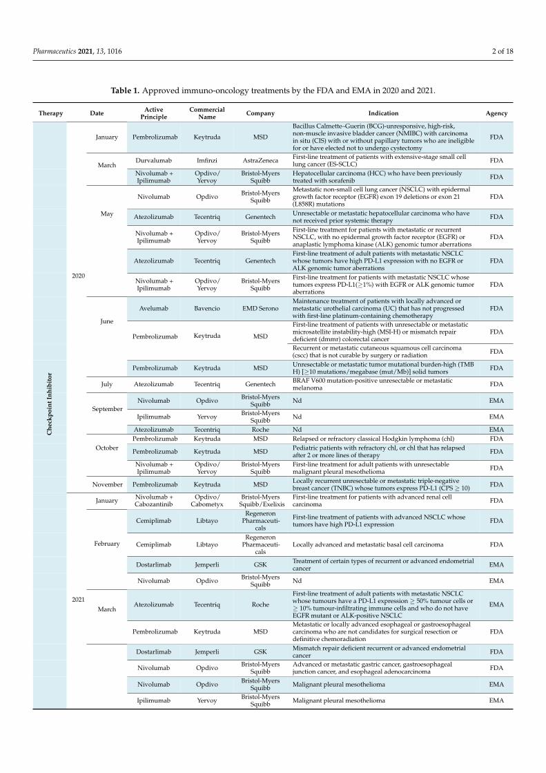
Cancer is one of the leading causes of death worldwide with a growing incidence due,in part, to increased life expectancy and diagnosis. Research advances in molecular biologyhave led to an expansion in the knowledge about the etiology of cancer, means of increasingthe number of targets, as well as the therapeutic strategies available. Immuno-oncology (IO)focuses on stimulating the patient’s own immune system to act selectively against tumor cellstreatments through the production of sustainable T cell responses and, thereby, diminishing thetoxicity linked with traditional treatments [1–3]. In this sense, IO has revolutionized the cancertherapeutic paradigm, especially in non-solid hematological tumors and metastatic cancer,with an exponential growth in the number of scientific publications since 2016 and becoming,in 2021, the therapeutic oncology strategy with the highest number of approved drugs by theFood and Drug Administration (FDA) and the European Medicines Agency (EMA) (Table 1).
The tumor microenvironment (TME) comprises a heterogeneous population of cancercells, as well as a variety of resident and infiltrating host cells, secreted factors and extra-cellular matrix proteins. The study of TME has provided insight on the possible factorscontrolling tumor progression and determining if the primary tumor eradicates, metas-tasizes or establishes dormant micrometastases [2]. Factors such as transforming growthfactor-β (TGF-β), interleukin (IL)-4, programmed cell death 1 (PD-1), and programmeddeath ligand 1 (PD-L1) have been identified as fundamental elements developed by thetumor itself to escape the immune response [3].
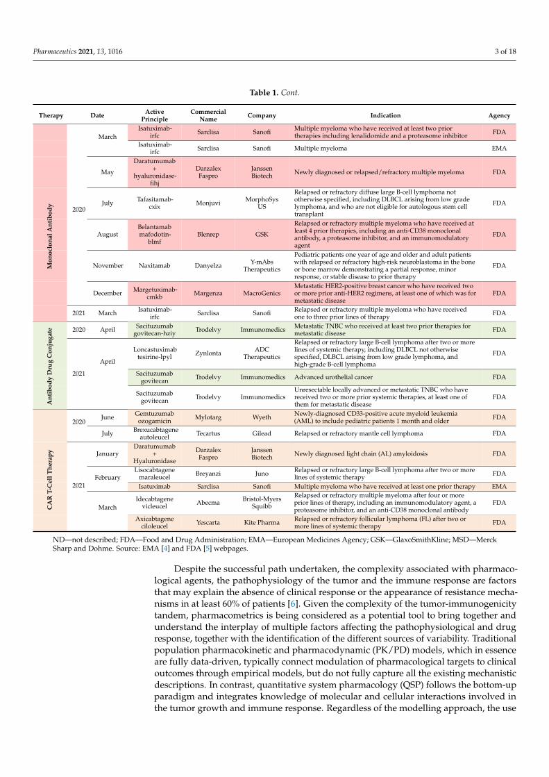
Table 1. Approved immuno-oncology treatments by the FDA and EMA in 2020 and 2021.
Therapy Date ActivePrinciple
CommercialName Company Indication Agency
2020
January Pembrolizumab Keytruda MSD
Bacillus Calmette–Guerin (BCG)-unresponsive, high-risk,non-muscle invasive bladder cancer (NMIBC) with carcinomain situ (CIS) with or without papillary tumors who are ineligiblefor or have elected not to undergo cystectomy
FDA
MarchDurvalumab Imfinzi AstraZeneca First-line treatment of patients with extensive-stage small cell
lung cancer (ES-SCLC) FDA
Nivolumab +Ipilimumab
Opdivo/Yervoy
Bristol-MyersSquibb
Hepatocellular carcinoma (HCC) who have been previouslytreated with sorafenib FDA
May
Nivolumab Opdivo Bristol-MyersSquibb
Metastatic non-small cell lung cancer (NSCLC) with epidermalgrowth factor receptor (EGFR) exon 19 deletions or exon 21(L858R) mutations
FDA
Atezolizumab Tecentriq Genentech Unresectable or metastatic hepatocellular carcinoma who havenot received prior systemic therapy FDA
Nivolumab +Ipilimumab
Opdivo/Yervoy
Bristol-MyersSquibb
First-line treatment for patients with metastatic or recurrentNSCLC, with no epidermal growth factor receptor (EGFR) oranaplastic lymphoma kinase (ALK) genomic tumor aberrations
FDA
Atezolizumab Tecentriq GenentechFirst-line treatment of adult patients with metastatic NSCLCwhose tumors have high PD-L1 expression with no EGFR orALK genomic tumor aberrations
FDA
Nivolumab +Ipilimumab
Opdivo/Yervoy
Bristol-MyersSquibb
First-line treatment for patients with metastatic NSCLC whosetumors express PD-L1(≥1%) with EGFR or ALK genomic tumoraberrations
FDA
June
Avelumab Bavencio EMD SeronoMaintenance treatment of patients with locally advanced ormetastatic urothelial carcinoma (UC) that has not progressedwith first-line platinum-containing chemotherapy
FDA
Pembrolizumab Keytruda MSD
First-line treatment of patients with unresectable or metastaticmicrosatellite instability-high (MSI-H) or mismatch repairdeficient (dmmr) colorectal cancer
FDA
Recurrent or metastatic cutaneous squamous cell carcinoma(cscc) that is not curable by surgery or radiation FDA
Pembrolizumab Keytruda MSD Unresectable or metastatic tumor mutational burden-high (TMBH) [≥10 mutations/megabase (mut/Mb)] solid tumors FDA
July Atezolizumab Tecentriq Genentech BRAF V600 mutation-positive unresectable or metastaticmelanoma FDA
SeptemberNivolumab Opdivo Bristol-Myers
Squibb Nd EMA
Ipilimumab Yervoy Bristol-MyersSquibb Nd EMA
Atezolizumab Tecentriq Roche Nd EMA
OctoberPembrolizumab Keytruda MSD Relapsed or refractory classical Hodgkin lymphoma (chl) FDA
Pembrolizumab Keytruda MSD Pediatric patients with refractory chl, or chl that has relapsedafter 2 or more lines of therapy FDA
Nivolumab +Ipilimumab
Opdivo/Yervoy
Bristol-MyersSquibb
First-line treatment for adult patients with unresectablemalignant pleural mesothelioma FDA
November Pembrolizumab Keytruda MSD Locally recurrent unresectable or metastatic triple-negativebreast cancer (TNBC) whose tumors express PD-L1 (CPS ≥ 10) FDA
2021
January Nivolumab +Cabozantinib
Opdivo/Cabometyx
Bristol-MyersSquibb/Exelixis
First-line treatment for patients with advanced renal cellcarcinoma FDA
February
Cemiplimab LibtayoRegeneron
Pharmaceuti-cals
First-line treatment of patients with advanced NSCLC whosetumors have high PD-L1 expression FDA
Cemiplimab LibtayoRegeneron
Pharmaceuti-cals
Locally advanced and metastatic basal cell carcinoma FDA
Dostarlimab Jemperli GSK Treatment of certain types of recurrent or advanced endometrialcancer EMA
Nivolumab Opdivo Bristol-MyersSquibb Nd EMA
MarchAtezolizumab Tecentriq Roche
First-line treatment of adult patients with metastatic NSCLCwhose tumours have a PD-L1 expression ≥ 50% tumour cells or≥ 10% tumour-infiltrating immune cells and who do not haveEGFR mutant or ALK-positive NSCLC
EMA
Pembrolizumab Keytruda MSDMetastatic or locally advanced esophageal or gastroesophagealcarcinoma who are not candidates for surgical resection ordefinitive chemoradiation
FDA
Dostarlimab Jemperli GSK Mismatch repair deficient recurrent or advanced endometrialcancer FDA
Nivolumab Opdivo Bristol-MyersSquibb
Advanced or metastatic gastric cancer, gastroesophagealjunction cancer, and esophageal adenocarcinoma FDA
Nivolumab Opdivo Bristol-MyersSquibb Malignant pleural mesothelioma EMA
Che
ckpo
intI
nhib
itor
Ipilimumab Yervoy Bristol-MyersSquibb Malignant pleural mesothelioma EMA
Pharmaceutics 2021, 13, 1016 3 of 18
Table 1. Cont.
Therapy Date ActivePrinciple
CommercialName Company Indication Agency
2020
MarchIsatuximab-
irfc Sarclisa Sanofi Multiple myeloma who have received at least two priortherapies including lenalidomide and a proteasome inhibitor FDA
Isatuximab-irfc Sarclisa Sanofi Multiple myeloma EMA
May
Daratumumab+
hyaluronidase-fihj
DarzalexFaspro
JanssenBiotech Newly diagnosed or relapsed/refractory multiple myeloma FDA
July Tafasitamab-cxix Monjuvi MorphoSys
US
Relapsed or refractory diffuse large B-cell lymphoma nototherwise specified, including DLBCL arising from low gradelymphoma, and who are not eligible for autologous stem celltransplant
FDA
AugustBelantamabmafodotin-
blmfBlenrep GSK
Relapsed or refractory multiple myeloma who have received atleast 4 prior therapies, including an anti-CD38 monoclonalantibody, a proteasome inhibitor, and an immunomodulatoryagent
FDA
November Naxitamab Danyelza Y-mAbsTherapeutics
Pediatric patients one year of age and older and adult patientswith relapsed or refractory high-risk neuroblastoma in the boneor bone marrow demonstrating a partial response, minorresponse, or stable disease to prior therapy
FDA
December Margetuximab-cmkb Margenza MacroGenics
Metastatic HER2-positive breast cancer who have received twoor more prior anti-HER2 regimens, at least one of which was formetastatic disease
FDA
Mon
oclo
nalA
ntib
ody
2021 March Isatuximab-irfc Sarclisa Sanofi Relapsed or refractory multiple myeloma who have received
one to three prior lines of therapy FDA
2020 April Sacituzumabgovitecan-hziy Trodelvy Immunomedics Metastatic TNBC who received at least two prior therapies for
metastatic disease FDA
2021
April
Loncastuximabtesirine-lpyl Zynlonta ADC
Therapeutics
Relapsed or refractory large B-cell lymphoma after two or morelines of systemic therapy, including DLBCL not otherwisespecified, DLBCL arising from low grade lymphoma, andhigh-grade B-cell lymphoma
FDA
Sacituzumabgovitecan Trodelvy Immunomedics Advanced urothelial cancer FDA
Ant
ibod
yD
rug
Con
juga
te
Sacituzumabgovitecan Trodelvy Immunomedics
Unresectable locally advanced or metastatic TNBC who havereceived two or more prior systemic therapies, at least one ofthem for metastatic disease
FDA
2020June Gemtuzumab
ozogamicin Mylotarg Wyeth Newly-diagnosed CD33-positive acute myeloid leukemia(AML) to include pediatric patients 1 month and older FDA
July Brexucabtageneautoleucel Tecartus Gilead Relapsed or refractory mantle cell lymphoma FDA
2021
JanuaryDaratumumab
+Hyaluronidase
DarzalexFaspro
JanssenBiotech Newly diagnosed light chain (AL) amyloidosis FDA
FebruaryLisocabtagene
maraleucel Breyanzi Juno Relapsed or refractory large B-cell lymphoma after two or morelines of systemic therapy FDA
Isatuximab Sarclisa Sanofi Multiple myeloma who have received at least one prior therapy EMA
MarchIdecabtagene
vicleucel Abecma Bristol-MyersSquibb
Relapsed or refractory multiple myeloma after four or moreprior lines of therapy, including an immunomodulatory agent, aproteasome inhibitor, and an anti-CD38 monoclonal antibody
FDA
CA
RT-
Cel
lThe
rapy
Axicabtageneciloleucel Yescarta Kite Pharma Relapsed or refractory follicular lymphoma (FL) after two or
more lines of systemic therapy FDA
ND—not described; FDA—Food and Drug Administration; EMA—European Medicines Agency; GSK—GlaxoSmithKline; MSD—MerckSharp and Dohme. Source: EMA [4] and FDA [5] webpages.
Despite the successful path undertaken, the complexity associated with pharmaco-logical agents, the pathophysiology of the tumor and the immune response are factorsthat may explain the absence of clinical response or the appearance of resistance mecha-nisms in at least 60% of patients [6]. Given the complexity of the tumor-immunogenicitytandem, pharmacometrics is being considered as a potential tool to bring together andunderstand the interplay of multiple factors affecting the pathophysiological and drugresponse, together with the identification of the different sources of variability. Traditionalpopulation pharmacokinetic and pharmacodynamic (PK/PD) models, which in essenceare fully data-driven, typically connect modulation of pharmacological targets to clinicaloutcomes through empirical models, but do not fully capture all the existing mechanisticdescriptions. In contrast, quantitative system pharmacology (QSP) follows the bottom-upparadigm and integrates knowledge of molecular and cellular interactions involved inthe tumor growth and immune response. Regardless of the modelling approach, the use
Pharmaceutics 2021, 13, 1016 4 of 18
of model-based strategies has become an essential tool in the decision-making process toefficiently guide the selection of therapeutic agents, dosing regimens, biomarkers and/orclinical outcomes during the drug discovery and development process [7–9].
Several articles have already reviewed the different QSP models developed in theIO area and their role in drug development [10–14]. Those types of models are hard toapply to in vivo data with the aim of estimating individual parameters and correlatingthem with patients’ specific characteristics. The current review is intended to highlightmodelling efforts that stretch the granularity of the in vivo longitudinal data, and can serveas a template for semi-mechanistic PKPD modelling in clinical trials. Prior to discussingmodelling cases, we provide a comprehensive summary of different pharmacologicaltargets for a better understanding of the models’ structures.
2. Current and Emerging Targets in Immuno-Oncology
Immune checkpoint inhibitors (ICIs) currently represent the most promising cancertherapeutics, producing durable responses in 40–50% of the patients administered themas monotherapies [15–20]. Among the different checkpoints expressed by cancer cells,cytotoxic T-lymphocyte-associated protein 4 (CTLA-4) and PD-1 are the most exploredcheckpoints for ICI-based therapeutics. Nevertheless, single drug checkpoint inhibitorsdid not achieve adequate response rates or prolonged disease control for ovarian [21–23],prostate and pancreatic cancers [22,24,25]. In this sense, current efforts are focused on devel-oping predictors of response to immunotherapy and rational therapeutic combinations ofcurrent immune checkpoint inhibitors (PD-1 and CTLA-4) with novel checkpoints, cellularimmunotherapies and delivery strategies, to improve the success rates in oncology [15].
2.1. Current Immune Checkpoint Inhibitors
CTLA-4 is a checkpoint of the immune system involved in the negative regulationof T cells at early immune response and is upregulated in activated T cells and expressedon regulatory T cells (Figure 1). The interaction between CTLA-4 and B7 molecules leadsto an inhibitory signal to T cells and prevents the co-stimulatory signal transduction [26].Anti-CTLA-4 agents are able to decrease regulatory T cells (Tregs) in the TME [27,28] andpromote the activation of effector cells by blocking the inhibitory axis. Following CTLA-4,the PD-1/PD-L1/PD-L2 axis was the next prime target that received more attention forimmune checkpoint therapies. These receptors are expressed on the cell surface of immunecells, dendritic cells and cancer cells. Similarly to CTLA-4, PD-1 ligation inhibits signalingdownstream of the T cell receptor [29] (TCR) (Figure 1). Therefore, the developmentof antagonist against PD-1 or PD-L1 has emerged as a valuable therapeutic strategy toenhance the activity of T cells against tumoral cells, especially for melanoma, non-smallcell lung cancer, renal cell carcinoma or Hodgkin lymphoma, among others, where durableresponses were observed in 20–40% of patients [30,31].
Pharmaceutics 2021, 13, 1016 5 of 18
Pharmaceutics 2021, 13, x 5 of 19
promote the activation of effector cells by blocking the inhibitory axis. Following CTLA-
4, the PD-1/PD-L1/PD-L2 axis was the next prime target that received more attention for
immune checkpoint therapies. These receptors are expressed on the cell surface of im-
mune cells, dendritic cells and cancer cells. Similarly to CTLA-4, PD-1 ligation inhibits
signaling downstream of the T cell receptor [29] (TCR) (Figure 1). Therefore, the develop-
ment of antagonist against PD-1 or PD-L1 has emerged as a valuable therapeutic strategy
to enhance the activity of T cells against tumoral cells, especially for melanoma, non-small
cell lung cancer, renal cell carcinoma or Hodgkin lymphoma, among others, where dura-
ble responses were observed in 20–40% of patients [30,31].
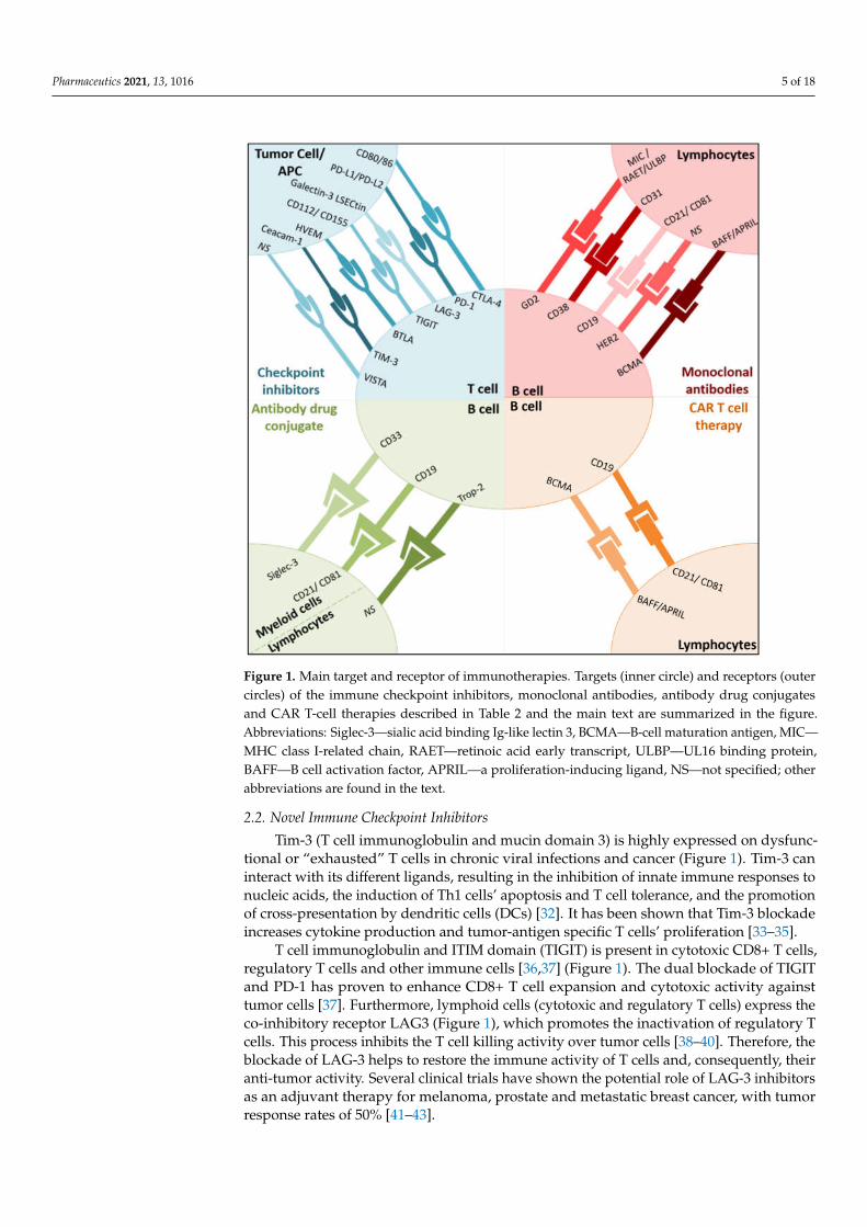
Figure 1. Main target and receptor of immunotherapies. Targets (inner circle) and receptors (outer
circles) of the immune checkpoint inhibitors, monoclonal antibodies, antibody drug conjugates and
CAR T-cell therapies described in Table 2 and the main text are summarized in the figure. Abbrevi-
class I-related chain, RAET—retinoic acid early transcript, ULBP—UL16 binding protein, BAFF—B
cell activation factor, APRIL—a proliferation-inducing ligand, NS—not specified; other abbrevia-
tions are found in the text.
2.2. Novel Immune Checkpoint Inhibitors
Tim-3 (T cell immunoglobulin and mucin domain 3) is highly expressed on dysfunc-
tional or “exhausted” T cells in chronic viral infections and cancer (Figure 1). Tim-3 can in-
teract with its different ligands, resulting in the inhibition of innate immune responses to
nucleic acids, the induction of Th1 cells’ apoptosis and T cell tolerance, and the promotion
of cross-presentation by dendritic cells (DCs) [32]. It has been shown that Tim-3 blockade
increases cytokine production and tumor-antigen specific T cells’ proliferation [33–35].
Figure 1. Main target and receptor of immunotherapies. Targets (inner circle) and receptors (outercircles) of the immune checkpoint inhibitors, monoclonal antibodies, antibody drug conjugatesand CAR T-cell therapies described in Table 2 and the main text are summarized in the figure.Abbreviations: Siglec-3—sialic acid binding Ig-like lectin 3, BCMA—B-cell maturation antigen, MIC—MHC class I-related chain, RAET—retinoic acid early transcript, ULBP—UL16 binding protein,BAFF—B cell activation factor, APRIL—a proliferation-inducing ligand, NS—not specified; otherabbreviations are found in the text.
2.2. Novel Immune Checkpoint Inhibitors
Tim-3 (T cell immunoglobulin and mucin domain 3) is highly expressed on dysfunc-tional or “exhausted” T cells in chronic viral infections and cancer (Figure 1). Tim-3 caninteract with its different ligands, resulting in the inhibition of innate immune responses tonucleic acids, the induction of Th1 cells’ apoptosis and T cell tolerance, and the promotionof cross-presentation by dendritic cells (DCs) [32]. It has been shown that Tim-3 blockadeincreases cytokine production and tumor-antigen specific T cells’ proliferation [33–35].
T cell immunoglobulin and ITIM domain (TIGIT) is present in cytotoxic CD8+ T cells,regulatory T cells and other immune cells [36,37] (Figure 1). The dual blockade of TIGITand PD-1 has proven to enhance CD8+ T cell expansion and cytotoxic activity againsttumor cells [37]. Furthermore, lymphoid cells (cytotoxic and regulatory T cells) express theco-inhibitory receptor LAG3 (Figure 1), which promotes the inactivation of regulatory Tcells. This process inhibits the T cell killing activity over tumor cells [38–40]. Therefore, theblockade of LAG-3 helps to restore the immune activity of T cells and, consequently, theiranti-tumor activity. Several clinical trials have shown the potential role of LAG-3 inhibitorsas an adjuvant therapy for melanoma, prostate and metastatic breast cancer, with tumorresponse rates of 50% [41–43].
Pharmaceutics 2021, 13, 1016 6 of 18
V-domain Ig suppressor of T cell activation (VISTA) is a transmembrane protein in-volved in antitumor immunity through the negative regulation of T cells [44] (Figure 1).VISTA is an immune checkpoint gene that is structurally similar to PD-L1 and PD-L2 [45],and its over-expression in tumor cells inhibits T cell proliferation and cytokine production,resulting in tumor evasion [46–48]. High levels of VISTA have been identified in several ma-lignant tumors, including oral squamous cell carcinoma, gastric carcinoma, hepatocellularcarcinoma, prostate carcinoma, and melanoma [49,50].
B and T lymphocyte attenuator (BTLA) is a recently investigated inhibitory receptor,present in lymphoid cells, with promising preclinical results [51] (Figure 1). BTLA hasstructural and functional similarities with CTLA-4 and PD-1, and it has been found tobe highly expressed in tumor antigen-specific CD8+ T cells of melanoma patients afterpeptide vaccinations. Preclinical evaluations in melanoma evidence the promising ac-tivity of monoclonal anti-BTLA antibodies by leading the promotion of T cell immuneresponse [52,53].
2.3. Adoptive Cellular Immunotherapy
Adoptive cellular immunotherapy is an innovative and recently developed treatmentstrategy of promising success in the treatment of cancer patients [54], which aims tostimulate durable anti-tumor immune activity. These strategies include tumor-infiltratinglymphocytes (TIL), gene modified T cells expressing novel T cell receptors (TCR) andchimeric antigen receptors (CAR) [55,56].
CAR T cell therapies on the cell surface transmembrane protein of B cells’ CD19, themost studied target [57], have emerged as a promising tool in the management of hemato-logic malignancies [57,58] (acute lymphoblastic leukemia, diffuse large B cell lymphoma,chronic lymphocytic leukemia, and B cell non-Hodgkin lymphomas) (Figure 1). Despite theefficacy rates (50–90%) [59] of CAR T cell therapies on hematological malignancies observedin clinical trials [59–61], safety concerns (neurotoxicity, hepatotoxicity and multi-organfailure [58,62–64]) and tumor relapses have been identified [65,66]. In this sense, antigenescape resistance mechanisms have been recognized as a relevant factor to explain tumorrelapse. This mechanism is enhanced in solid tumors, which might explain the reducedefficacy observed.
The use of TIL therapy has also emerged as an alternative tool to increase responserates and/or to reduce relapse rates, especially in metastatic melanoma and testis cancerpatients [67–74]. Autologous lymphocytes are removed from the patient, externally ex-panded and re-infused to the patients to promote the immune response against tumor cells.Investigations are currently ongoing to evaluate the efficacy and safety of TIL administeredas a monotherapy or in combination for different cancer treatments [75–77].
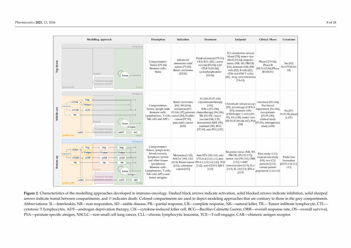
3. Mathematical Approaches Integrating Cancer Immunity Cycle withImmuno-Oncology Therapies
Mathematical modelling has broadly been used in support of preclinical and clinicalresearch, as well as in decision-making in the oncology field [12]. This review will focuson those mathematical models that use ordinary differential equations to describe the IOsystem dynamics and that have been applied to clinical data. Additionally, we will dividethe different works according to the modelling approach used: (i) top-down data-drivenmodels built predominantly on the observed clinical data, and with a reduced numberof parameters and equations leading to an empirical description of the biological system;(ii) bottom-up models based on knowledge about the human body and that are, therefore,as mechanistic as possible, utilizing in vitro as well as preclinical and clinical informationas input data; and (iii) models that utilize a middle-out approach, combining bottom-up(model) and top-down (data) systems and applying different modeling strategies (Figure 2).
3.1. Top-Down Modelling and Simulation Approaches
Pharmacokinetic/pharmacodynamic (PK/PD) modeling has been used in the devel-opment of IO agents such as immune checkpoint inhibitors [78], monoclonal antibodies,
Pharmaceutics 2021, 13, 1016 7 of 18
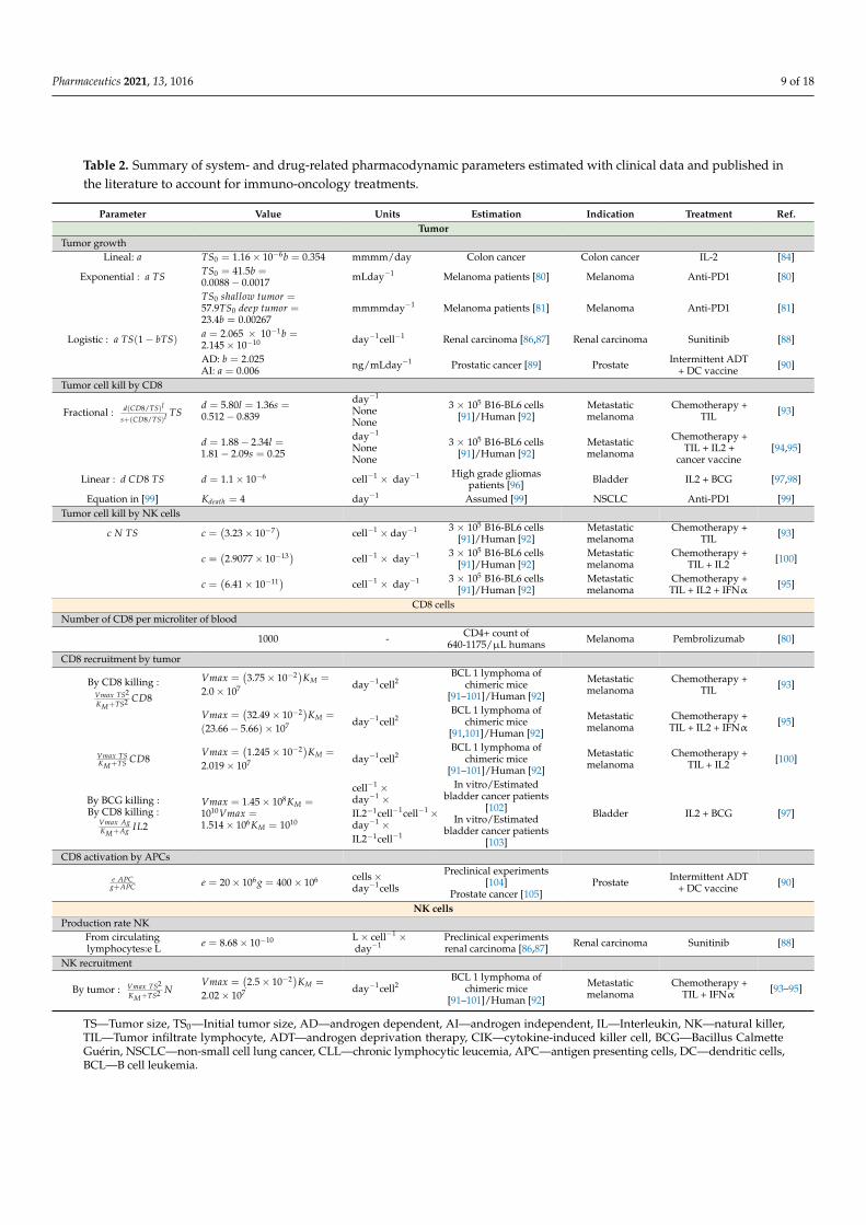
toll-like receptor (TLR) agonists or cancer vaccines. As an example, PK/PD models haveplayed an important role in supporting the dose setting and characterization of pem-brolizumabs’ clinical pharmacology, a monoclonal antibody directed to PD-1 receptors.In particular, Elassaiss-Schaap et al. [79] developed a PK and a PK/PD model describingthe pembrolizumab concentrations and doses at which maximal target engagement wasachieved (Figure 2). As a major assumption, the IL-2 stimulation ratio in blood was con-sidered as a surrogate for target engagement in the tumor, and, thus, a potential markerfor antitumor efficacy. Similarly, Lindauer et al. [80] built a translational PKPD modelthat aimed to optimize dose setting in early clinical development in oncology, integratingin vitro, preclinical and clinical (Phase I, Phase II and Phase III clinical trial) data (Figure 2).The components of the final structure are: an empirical model for pembrolizumab PK inplasma, a mechanistic tissue compartment representing the site of drug action, a mecha-nistic binding model for drug–receptor interaction, and a tumor growth model. Finally,Chatterjee et at. [81] developed two top-down models describing the tumor size changesin melanoma patients (Phase I, Phase II and Phase III). Both models show the lack ofclinically relevant impact of pembrolizumab exposure on response rate. Contrary, baselinedisease, BRAF mutation, the degree of PD-1 receptor and ipilimumab treatment historywere identified as possible predictors of individual variability. One of the limitations of thisanalysis for clinical extrapolation of the simulations is that the model does not considerdropouts (patients that discontinued the clinical trial), which could potentially impact theresults. In spite of the different modeling approaches, equations and assumptions, all thesemodels were developed for the same therapeutic agent and the same study population,and, thus, we can also find some similarities. In particular, these works described tumorgrowth following a simple exponential model
(dTSdt = a TS
)with a constant growth rate
(a) ranging from 0.0017 to 0.0088 1/day and an initial tumor size (TS0) estimated to bebetween 23.4 and 41.5 mm3 (Table 2).
A different PKPD model was developed by Ribba et al. [82] to guide a dose esca-lation study design of cergutuzumab amunaleukin (CEA-IL2v), a monoclonal antibodydirected against carcinoembryonic antigens. In this study, a relation between drug plasmaconcentration (PK) and immune cell count (PD), including NK cells, CD4, CD8 T cells,was established using data from 74 patients with advanced and/or metastatic solid CEA+tumors. Therefore, limited validation with clinical data was performed during model de-velopment. Besides, as the model equations describe drug, target (immune cells expressingIL2 receptor in blood) and complex drug–receptor concentrations, instead of tumor growthdynamics, the schematic representation does not fit the structure of Figure 2.
The mathematical models described until this point are focused on dealing with aspecific pharmacological question such as the lowest effective dose to be used in a clinicaltrial [80], the optimal dosing schedule [82] or the quantification of the exposure–responserelationships determining the efficacy of a certain therapeutic agent [81]. However, they donot consider the interactions between the immunological system and cancer cells. In thissense, more mechanistic models that aim to incorporate more components of the biologicalsystem can help to understand the mechanisms of actions of immunotherapies (Figure 2).
Parra-Guillen et al. [83,84] developed a model combining mechanistic features, specif-ically tumor resistance mechanisms, and mixed effects to describe tumor growth dynamicsafter the administration of different combinations of an antitumor vaccine, a TLR-9 agonist(CpG), chemotherapy (cyclophosphamide) and IL-2. This approach is also an example ofhow the kinetics of the therapeutic agents can be analyzed and simulated in the absenceof PK information. Plasma concentration–time profiles of a drug are usually necessary toestablish a relationship between the administered dose and the kinetics of drug action [85].However, is not always possible to collect all the required PK data and several models havebeen proposed. Even so, despite the fact that this model is based on preclinical data only, itwas successfully applied to reproduce clinical outcomes from three different studies (PhaseI and Phase II data) (Figure 2). Still, the simplistic description of the tumor and immunesystem interactions is a handicap to be generalized to other mechanisms of actions.
Pharmaceutics 2021, 13, 1016 8 of 18Pharmaceutics 2021, 13, x 8 of 19
Figure 2. Characteristics of the modelling approaches developed in immuno-oncology. Dashed black arrows indicate activation, solid blocked arrows indicate inhibition, solid sharped
arrows indicate transit between compartments, and ∅ indicates death. Colored compartments are used to depict modeling approaches that are contrary to those in the grey Figure 2. Characteristics of the modelling approaches developed in immuno-oncology. Dashed black arrows indicate activation, solid blocked arrows indicate inhibition, solid sharpedarrows indicate transit between compartments, and ∅ indicates death. Colored compartments are used to depict modeling approaches that are contrary to those in the grey compartments.Abbreviations: IL—Interleukin, NR—non-responders, SD—stable disease, PR—partial response, CR—complete response, NK—natural killer, TIL—Tumor infiltrate lymphocyte, CTL—cytotoxic T lymphocytes, ADT—androgen deprivation therapy, CIK—cytokine-induced killer cell, BCG—Bacillus Calmette Guérin, ORR—overall response rate, OS—overall survival,PSA—prostate specific atnigen, NSCLC—non-small cell lung cancer, CLL—chronic lymphocytic leucemia, TCE—T-cell-engager, CAR—chimeric antigen receptor.
Pharmaceutics 2021, 13, 1016 9 of 18
Table 2. Summary of system- and drug-related pharmacodynamic parameters estimated with clinical data and published inthe literature to account for immuno-oncology treatments.
Parameter Value Units Estimation Indication Treatment Ref.Tumor
Tumor growthLineal: a TS0 = 1.16 × 10⁻⁶b = 0.354 mmmm/day Colon cancer Colon cancer IL-2 [84]
3.2. Middle-Out Modelling and Simulation Approaches
Middle-out approaches aim to incorporate the main biological and pharmacodynamicmechanisms of the system while maintaining a simplified model structure. However,keeping the principle of parsimony represents a challenge since it is probable that somemodel parameters will be hard to estimate precisely. As a consequence, the integration ofdifferent pharmacometric techniques, such as the use of Bayesian priors based on previousknowledge to inform poorly estimated parameters, is warranted [106].
This strategy provides a quantitative platform for model development in clinicalscenarios (Figure 2). The first attempt to describe the interactions between the immunesystem and cancer cells [101] was a model of two ordinary differential equations (ODEs)describing the dynamics of CD8 T cells and their effect on tumor cell killing. On this basis,and with the increasing experimental data available, models were expanded to includedifferent entities, including immune cells and cytokines, and mechanisms such as receptorexpression dynamics.
At first, aiming to incorporate cytokines’ function into the models, Kirschner andPanetta [104] built a three-ODE system addressing the potential of IL-2 and its effects ontumor relapse. Similarly, in a more recent work carried out by Isaeva et al. [107], the effectsof chemotherapy and immunotherapy (IL-2 and IFN-α) were studied using a middle-outapproach. A limitation of this analysis is that in order to reflect the different clinicaloutcomes, patients were conditionally divided into three groups characterized by tumorantigen expression, the strength of the immune response and the reaction to vaccination(Figure 2 covariates).
On the other hand, with the goal of adding new entities, de Pillis et al. [93] developeda very simple model that included three cell populations: tumor cells, natural killer (NK)cells and CD8 T cells. This work supported the relevance of considering multiple cell typesin the overall anti-tumor immune activity. In spite of the model’s simplicity, it is able to fitdata from two metastatic melanoma patients treated with tumor-infiltrating lymphocytes(TIL) after chemotherapy. An essential feature of this model is the cell killing term definingthe interaction of tumor cells with either NK or CD8 T cells. Although for NK cells, alinear product (Table 2; tumor cells killed by NK cells) was sufficient to reproduce theexperimental data, for CD8 T cells, a rational form was needed. In Table 2 (fractional tumorcell kill by CD8), the parameter d gives the maximum lysis rate, the exponent l representshow the lysis rate depends on the effector/target ratio, and s is the parameter affectingthe steepness of the curve. This new term and the parameter values have been usedin previous works [94,95,100]. Although the model fits the empirical data, its structureis still very simple and does not incorporate self-regulatory terms or down-regulationof the activated immune response, among other mechanisms. In a different approach,Perlstein et al. [108] incorporated memory T cells and the senescence and exhaustionmechanisms of PD-1/PD-L1 checkpoint blockade immunotherapy in their model. Oneof their limitations is that the model was adjusted to fit the clinically measured dynamicsof just one reference patient from a hospital cohort suffering from metastatic melanoma.Furthermore, Xuefang et al. [109] developed a mathematical prognosis model for pancreaticcancer patients including not only cancer cells and CD8 T cells, but also pancreatic stellatecells, other immune cells (NK cells and helper T cells) and cytokines (IL-2, IFN-α andTGF-β). They assumed that survival time is the time taken for the cancer cell density toreach a certain threshold (500 cells per µL).
Some of the common biological assumptions of the previously described modelsinclude: (i) cancer cells grow logistically in the absence of an immune response; (ii) bothNK cells and CD8 T cells are capable of killing cancer cells; (iii) both NK cells and CD8T cells are activated by cancer cells; (iv) both NK cells and CD8 cells eventually becomeinactivated after some number of interactions with tumor cells; (v) as part of the innatesystem, NK cells are always present, but CD8 T cells are only present when tumor is present.
The aforementioned models incorporate an increasing number of immune cells. How-ever, they do not include immune-suppressive components, which have been demonstrated
Pharmaceutics 2021, 13, 1016 11 of 18
to play a critical role in tumor evasion mechanisms. Tumors escape the immune-mediatedelimination by producing substances, such as TGF-β and IL-10, that stimulate the expan-sion of immunosuppressive cells, particularly regulatory T cells (Tregs), MDSCs, and M2macrophages. With the aim of including these mechanisms, de Pillis et al. [88] expandedtheir previous model and incorporated Tregs as the main immunosuppressive component.This analysis studies the anti-angiogenic effect of sunitinib as well as its ability to directlyinhibit the immunosuppressive environment by reducing the number of Tregs.
Whereas, until this point, the new entities incorporated into the models includedimmune players or cytokines, other authors have focused on including different tumorcell clones. Mahasa et al. [110] used a middle-out approach, with a model structure able tofit the representation of Figure 2, to study the immune surveillance of tumors includingimmune cells, different tumor cell populations (naïve and resistant), and the complexesformed among these. The model describes how tumor cell populations escape and acquireresistance after the interaction with the immune system mediated by NK and CD8 Tcells. In a different work, Portz et al. [90] extended the model proposed by Kirschnerand Panetta [104] and developed a system of six ODEs in which tumor mass was dividedinto androgen dependent and independent cells. This approach was driven by the factthat patients were treated with androgen deprivation therapy, which prevents growthand induces apoptosis only of androgen dependent cells. On a similar basis, Bunimovich-Mendrazitzky et al. [98] developed a model considering two different populations of tumorcells—infected and uninfected tumor cells. They studied Bacillus Calmette–Guerin (BCG)immunotherapy for superficial bladder cancer patients. This work was later expanded [97]by adding IL-2. In both situations, they modeled the encounter of effector cells and cancercells with proportional rate constant (d CD8 TS). Despite the fact that more mechanismsare incorporated, the limited validation with clinical data together with the remainingsimple description of the tumor and immune system interactions makes the extrapolationof this model to other immunotherapies challenging.
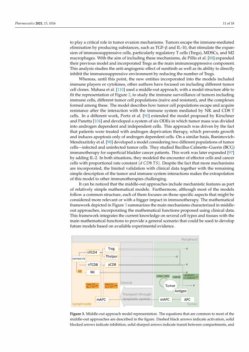
It can be noticed that the middle-out approaches include mechanistic features as partof relatively simple mathematical models. Furthermore, although most of the modelsfollow a common structure, each of them focuses on those specific aspects that might beconsidered more relevant or with a bigger impact in immunotherapy. The mathematicalframework depicted in Figure 3 summarizes the main mechanisms characterized in middle-out approaches, incorporating the mathematical functions proposed using clinical data.This framework integrates the current knowledge on several cell types and tissues with themain mathematical functions to provide a general scenario that could be used to developfuture models based on available experimental evidence.
Pharmaceutics 2021, 13, x FOR PEER REVIEW 13 of 19
considered more relevant or with a bigger impact in immunotherapy. The mathematical
framework depicted in Figure 3 summarizes the main mechanisms characterized in mid-
dle-out approaches, incorporating the mathematical functions proposed using clinical
data. This framework integrates the current knowledge on several cell types and tissues
with the main mathematical functions to provide a general scenario that could be used to
develop future models based on available experimental evidence.
Figure 3. Middle-out approach model representation. The equations that are common to most of the middle-out ap-
proaches are described in the figure. Dashed black arrows indicate activation, solid blocked arrows indicate inhibition,
solid sharped arrows indicate transit between compartments, and ∅ indicates death. Colored compartments are used to
depict modeling approaches that are contrary those in the grey compartments. Abbreviations: NK—natural killer,
nTCD4—naïve CD4 cells, nTCD8—naïve CD8 cells, Treg—regulatory T cells, Thelper—Helper T cells, aCD8—activated
phocytes. Other parameters are presented in Table 2.
3.3. Bottom-Up Modelling and Simulation Approaches
Large-scale QSP platform models are able to integrate the current knowledge of the
cancer immunity-cycle (Figure 2) by incorporating more mechanisms, different cell-types
(tumor cells, innate and adaptive immune cells, and stromal cells) and diverse molecular
components (cytokines, cell-surface receptors, etc.). However, these bottom-up modelling
approaches are built up with a very large number of parameters and equations, and model
calibration is usually challenging.
Popel’s group developed one of the first full-scale QSP platforms in IO, describing
cancer cells, the dynamics and development of antigen presenting cells (APCs), T cells
(naïve, primed, effector and regulatory) and myeloid derived suppressor cells (MDSCs)
in the tumor, the tumor draining lymph node, the blood and other tissues. This framework
has been validated for melanoma, breast cancer, and NSCLC patients treated with anti-
PD1, anti-PD-L1, anti-CTLA4 and epigenetic inhibitors [99,111–113]. Moreover, the cellu-
lar interactions of TCR, MHC I, CD28, CD80, CD86, PD1, PD-L1, PD-L2, and CTLA4 sur-
face receptor were considered. In these works, rather than developing a model from
scratch in each particular situation, a platform model was created and then used to se-
quentially add components and/or adapt it to other tumor types. Wang et al. [47] studied
the combination of entinostat (histone deacetylase inhibitor) with ICIs in breast cancer
Figure 3. Middle-out approach model representation. The equations that are common to most of themiddle-out approaches are described in the figure. Dashed black arrows indicate activation, solidblocked arrows indicate inhibition, solid sharped arrows indicate transit between compartments, and
Pharmaceutics 2021, 13, 1016 12 of 18
∅ indicates death. Colored compartments are used to depict modeling approaches that are contrarythose in the grey compartments. Abbreviations: NK—natural killer, nTCD4—naïve CD4 cells,nTCD8—naïve CD8 cells, Treg—regulatory T cells, Thelper—Helper T cells, aCD8—activated CD8 Tcells, mAPC—mature antigen presenting cells, APC—immature antigen presenting cells, TS—tumorsize, L—lymphocytes. Other parameters are presented in Table 2.
3.3. Bottom-Up Modelling and Simulation Approaches
Large-scale QSP platform models are able to integrate the current knowledge of thecancer immunity-cycle (Figure 2) by incorporating more mechanisms, different cell-types(tumor cells, innate and adaptive immune cells, and stromal cells) and diverse molecularcomponents (cytokines, cell-surface receptors, etc.). However, these bottom-up modellingapproaches are built up with a very large number of parameters and equations, and modelcalibration is usually challenging.
Popel’s group developed one of the first full-scale QSP platforms in IO, describingcancer cells, the dynamics and development of antigen presenting cells (APCs), T cells(naïve, primed, effector and regulatory) and myeloid derived suppressor cells (MDSCs) inthe tumor, the tumor draining lymph node, the blood and other tissues. This frameworkhas been validated for melanoma, breast cancer, and NSCLC patients treated with anti-PD1,anti-PD-L1, anti-CTLA4 and epigenetic inhibitors [99,111–113]. Moreover, the cellularinteractions of TCR, MHC I, CD28, CD80, CD86, PD1, PD-L1, PD-L2, and CTLA4 surfacereceptor were considered. In these works, rather than developing a model from scratchin each particular situation, a platform model was created and then used to sequentiallyadd components and/or adapt it to other tumor types. Wang et al. [47] studied thecombination of entinostat (histone deacetylase inhibitor) with ICIs in breast cancer patientsincorporating PD-L1 expression and tumor mutational burden (TMB). Then, Jafarnejadet al. [99] included a detailed model for APC antigen presentation, and Milberg et al. [111]proved that this platform was able to predict longitudinal tumor size profiles and thenumber of patients showing partial or complete response for anti-PD1 and anti-CTLA4combinations in melanoma.
A different QSP model for anti-CD19 chimeric antigen receptor T cells (CAR T) in a pa-tient with chronic lymphocytic leukemia (CLL) was developed by Hardiansyah et al. [114].Since we are dealing with a non-solid tumor, the model-based platform developed bythe authors does not fit the bottom-up structure shown in Figure 2. In this case, theauthors have included the dynamics of B-cells, effector and memory CAR T cells, andinflammatory cytokines (interleukin-6, Interleukin-10, and interferon gamma) in peripheralblood and tissue. The proposed model is able to describe the observed CART kinetic andpro-inflammatory cytokine profiles in a clinical scenario.
In bottom-up approaches, although the majority of model parameters are literature-based (Table 2), a selected set of parameters are adjusted using clinical data. Nevertheless,one of their main limitations in drug development is building confidence intervals for avery large number of parameters. In spite of this, QSP models have been demonstratedto be a valuable tool for deepening our understanding on how the mechanism of actionconnects to the clinical outcomes and, therefore, may serve as important model-basedplatforms to guide the development of, and personalize, treatment therapy.
4. Conclusions
The number of approved immunotherapies has grown exponentially in the past twoyears, particularly immune checkpoint inhibitors, monoclonal antibodies, antibody drugconjugates and CAR T-cell therapies. Despite the improvement in response rates, there isstill a high percentage of patients that do not respond to these treatments. For this reason,current efforts are focused on finding new therapeutic targets and different combinationstrategies. In this sense, mathematical strategies have proven to be an efficient tool tocharacterize, select and predict optimal therapeutic alternatives in the field of IO. However,
Pharmaceutics 2021, 13, 1016 13 of 18
it is still necessary to develop a quantitative framework that allows the evaluation of twoor more agents administered in combination and to identify their possible interactions.Among the different mathematical models proposed to describe the tumor and immunecells’ interactions, in this review, we have focused on those that use ordinary differentialequations and have been applied to clinical settings. Additionally, models have beenclassified into top-down, middle-out or bottom-up approaches, according to the modellingstrategy applied.
On the one hand, data-driven top-down models have been demonstrated to be asuccessful tool in clinical trials, for example, to predict the minimum efficacious dose ofICIs. However, one of the limitations is that due to the simplicity of such models, someassumptions have to be considered, for example, the simplification of the drug deliveryprocess or the extrapolation of parameters from animals to humans. Furthermore, sincethe interactions of the tumor and the immune system are not considered, they cannot begeneralized to therapeutic agents with other mechanisms of action or their combinations.On the other hand, QSP models simulate various biological processes and interactionson different tissues and, thus, can help to overcome the challenges of understanding theimmune response dynamics and the interplay of tumor infiltration processes and tumorcell growth. Nevertheless, the large number of parameters, and the relatively small amountof observed data usually available, makes the development of these models very complex.
In between these two approaches, middle-out strategies offer theoretical and evidence-based description, representing an optimal framework for the evaluation of new strategiesin IO. These models are based on experimental and/or clinical data while constraining themodel structure to the current knowledge of the system. Therefore, this modeling strategyneeds either data from specific biomarkers that allow the identification of immune cells’dynamics, or an experimental design in which immune-modulators acting on different stepsof the cancer-immunity cycle are studied. Moreover, a relevant aspect to be incorporatedinto mathematical models is the development of biomarkers capable of predicting degreesof response in cancer patients.
In this regard, the design of studies that allow the collection of informative longitudinaldata, together with the integration of pharmacogenetics, can contribute to establishing earlyresponse indicators. On the whole, this work provides a schematic representation (Figure 2),including the description of tumor growth and immunological cell-type dynamics, aswell as a range of model equations and parameters, with the aim of establishing anoptimal theoretical framework for middle-out approaches, which may help to evaluatenew IO strategies.
Author Contributions: Conceptualization, A.S.-A., V.M.-S. and I.F.T.; investigation, A.S.-A. and V.M.-S.; writing—original draft preparation, A.S.-A. and V.M.-S.; writing—review and editing, A.S.-A.,V.M.-S. and I.F.T.; visualization, A.S.-A.; supervision, I.F.T. All authors have read and agreed to thepublished version of the manuscript.
Funding: This research received no external funding.
Institutional Review Board Statement: Not applicable.
Informed Consent Statement: Not applicable.
Conflicts of Interest: The authors declare no conflict of interest.
References1. Chen, D.S.; Mellman, I. Oncology meets immunology: The cancer-immunity cycle. Immunity 2013, 39, 1–10. [CrossRef] [PubMed]2. How Immunotherapy Is Used to Treat Cancer. Available online: https://www.cancer.org/content/dam/CRC/PDF/Public/6678
.00.pdf (accessed on 19 April 2021).3. Jiang, X.; Wang, J.; Deng, X.; Xiong, F.; Ge, J.; Xiang, B.; Wu, X.; Ma, J.; Zhou, M.; Li, X.; et al. Role of the tumor microenvironment
in PD-L1/PD-1-mediated tumor immune escape. Mol. Cancer 2019, 18, 1–17. [CrossRef]4. European Medicines Agency. Available online: https://www.ema.eu,ropa.eu/en (accessed on 8 June 2021).5. Drugs@FDA: FDA-Approved Drugs. Available online: https://www.accessdata.fda.gov/scripts/cder/daf/ (accessed on 8 June 2021).
7. Netterberg, I.; Li, C.C.; Molinero, L.; Budha, N.; Sukumaran, S.; Stroh, M.; Jonsson, E.N.; Friberg, L.E. A PK/PD Analysis ofCirculating Biomarkers and Their Relationship to Tumor Response in Atezolizumab-Treated non-small Cell Lung Cancer Patients.Clin. Pharmacol. Ther. 2019, 105, 486–495. [CrossRef]
8. Bradshaw, E.L.; Spilker, M.E.; Zang, R.; Bansal, L.; He, H.; Jones, R.D.O.; Le, K.; Penney, M.; Schuck, E.; Topp, B.; et al. Applicationsof Quantitative Systems Pharmacology in Model-Informed Drug Discovery: Perspective on Impact and Opportunities. CPTPharmacometrics Syst. Pharmacol. 2019, 8, 777–791. [CrossRef]
9. Bender, B.C.; Schindler, E.; Friberg, L.E. Population pharmacokinetic-pharmacodynamic modelling in oncology: A tool forpredicting clinical response. Br. J. Clin. Pharmacol. 2015, 79, 56–71. [CrossRef] [PubMed]
10. Valentinuzzi, D.; Jeraj, R. Computational modelling of modern cancer immunotherapy. Phys. Med. Biol. 2020, 65, 24TR01.[CrossRef]
11. Bekisz, S.; Geris, L. Cancer modeling: From mechanistic to data-driven approaches, and from fundamental insights to clinicalapplications. J. Comput. Sci. 2020, 46, 101198. [CrossRef]
12. Peskov, K.; Azarov, I.; Chu, L.; Voronova, V.; Kosinsky, Y.; Helmlinger, G. Quantitative mechanistic modeling in support ofpharmacological therapeutics development in immuno-oncology. Front. Immunol. 2019, 10, 924. [CrossRef]
13. Sové, R.J.; Jafarnejad, M.; Zhao, C.; Wang, H.; Ma, H.; Popel, A.S. QSP-IO: A Quantitative Systems Pharmacology Toolbox forMechanistic Multiscale Modeling for Immuno-Oncology Applications. CPT Pharmacometrics Syst. Pharmacol. 2020, 9, 484–497.[CrossRef]
14. Chelliah, V.; Lazarou, G.; Bhatnagar, S.; Gibbs, J.P.; Nijsen, M.; Ray, A.; Stoll, B.; Thompson, R.A.; Gulati, A.; Soukharev, S.; et al.Quantitative Systems Pharmacology Approaches for Immuno-Oncology: Adding Virtual Patients to the Development Paradigm.Clin. Pharmacol. Ther. 2021, 109, 605–618. [CrossRef] [PubMed]
15. Della Gravara, L.; Battiloro, C.; Cantile, R.; Letizia, A.; Vitiello, F.; Montesarchio, V.; Rocco, D. Chemotherapy and/or immunecheckpoint inhibitors in NSCLC first-line setting: What is the best approach? Lung Cancer Manage. 2020, 9, LMT22. [CrossRef]
16. Quinn, C.; Garrison, L.P.; Pownell, A.K.; Atkins, M.B.; De Pouvourville, G.; Harrington, K.; Ascierto, P.A.; McEwan, P.; Wagner, S.;Borrill, J.; et al. Current challenges for assessing the long-term clinical benefit of cancer immunotherapy: A multi-stakeholderperspective. J. Immunother. Cancer 2020, 8, e000648. [CrossRef] [PubMed]
17. Gevaert, T.; Van Eycke, Y.R.; Broeck, T.V.; Van Poppel, H.; Salmon, I.; Rorive, S.; Muilwijk, T.; Claessens, F.; De Ridder, D.; Joniau,S.; et al. The potential of tumour microenvironment markers to stratify the risk of recurrence in prostate cancer patients. PLoSONE 2020, 15, e0244663. [CrossRef] [PubMed]
18. Lee, H.; Na, K.J.; Choi, H. Differences in Tumor Immune Microenvironment in Metastatic Sites of Breast Cancer. Front. Oncol.2021, 11, 722. [CrossRef] [PubMed]
19. Cocco, C.; Morandi, F.; Airoldi, I. Immune Checkpoints in Pediatric Solid Tumors: Targetable Pathways for Advanced TherapeuticPurposes. Cells 2021, 10, 927. [CrossRef] [PubMed]
21. Cai, D.L.; Jin, L.P. Immune cell population in ovarian tumor microenvironment. J. Cancer 2017, 8, 2915–2923. [CrossRef]22. Alexandrov, L.B.; Nik-Zainal, S.; Wedge, D.C.; Aparicio, S.A.J.R.; Behjati, S.; Biankin, A.V.; Bignell, G.R.; Bolli, N.; Borg, A.;
Børresen-Dale, A.L.; et al. Signatures of mutational processes in human cancer. Nature 2013, 500, 415–421. [CrossRef]23. Heong, V.; Ngoi, N.; Peng Tan, D.S. Update on immune checkpoint inhibitors in gynecological cancers. J. Gynecol. Oncol. 2017, 28,
e20. [CrossRef]24. Martinez-Bosch, N.; Vinaixa, J.; Navarro, P. Immune evasion in pancreatic cancer: From mechanisms to therapy. Cancers 2018, 10,
6. [CrossRef]25. Strasner, A.; Karin, M. Immune infiltration and prostate cancer. Front. Oncol. 2015, 5, 128. [CrossRef] [PubMed]26. Buchbinder, E.I.; Desai, A. CTLA-4 and PD-1 pathways similarities, differences, and implications of their inhibition. Am. J. Clin.
J.D.; et al. Fc-dependent depletion of tumor-infiltrating regulatory t cells co-defines the efficacy of anti-CTLA-4 therapy againstmelanoma. J. Exp. Med. 2013, 210, 1695–1710. [CrossRef] [PubMed]
28. Pol, J.; Kroemer, G. Anti-CTLA-4 immunotherapy: Uncoupling toxicity and efficacy. Cell Res. 2018, 28, 501–502. [CrossRef][PubMed]
29. Yusa, S.; Campbell, K.S. Src Homology Region 2-Containing Protein Tyrosine Phosphatase-2 (SHP-2) Can Play a Direct Role inthe Inhibitory Function of Killer Cell Ig-Like Receptors in Human NK Cells. J. Immunol. 2003, 170, 4539–4547. [CrossRef]
30. Waldman, A.D.; Fritz, J.M.; Lenardo, M.J. A guide to cancer immunotherapy: From T cell basic science to clinical practice. Nat.Rev. Immunol. 2020, 20, 651–668. [CrossRef]
32. Du, W.; Yang, M.; Turner, A.; Xu, C.; Ferris, R.L.; Huang, J.; Kane, L.P.; Lu, B. Tim-3 as a target for cancer immunotherapy andmechanisms of action. Int. J. Mol. Sci. 2017, 18, 645. [CrossRef]
33. Fourcade, J.; Sun, Z.; Benallaoua, M.; Guillaume, P.; Luescher, I.F.; Sander, C.; Kirkwood, J.M.; Kuchroo, V.; Zarour, H.M.Upregulation of Tim-3 and PD-1 expression is associated with tumor antigen-specific CD8+ T cell dysfunction in melanomapatients. J. Exp. Med. 2010, 207, 2175–2186. [CrossRef]
34. Sakuishi, K.; Apetoh, L.; Sullivan, J.M.; Blazar, B.R.; Kuchroo, V.K.; Anderson, A.C. Targeting Tim-3 and PD-1 pathways to reverseT cell exhaustion and restore anti-tumor immunity. J. Exp. Med. 2010, 207, 2187–2194. [CrossRef]
35. Ngiow, S.F.; Von Scheidt, B.; Akiba, H.; Yagita, H.; Teng, M.W.L.; Smyth, M.J. Anti-TIM3 antibody promotes T cell IFN-γ-mediatedantitumor immunity and suppresses established tumors. Cancer Res. 2011, 71, 3540–3551. [CrossRef]
36. Kon, E.; Benhar, I. Immune checkpoint inhibitor combinations: Current efforts and important aspects for success. Drug Resist.Updat. 2019, 45, 13–29004. [CrossRef]
37. Dougall, W.C.; Kurtulus, S.; Smyth, M.J.; Anderson, A.C. TIGIT and CD96: New checkpoint receptor targets for cancerimmunotherapy. Immunol. Rev. 2017, 276, 112–120. [CrossRef]
39. Huang, C.T.; Workman, C.J.; Flies, D.; Pan, X.; Marson, A.L.; Zhou, G.; Hipkiss, E.L.; Ravi, S.; Kowalski, J.; Levitsky, H.I.; et al.Role of LAG-3 in regulatory T cells. Immunity 2004, 21, 503–513. [CrossRef] [PubMed]
40. Blackburn, S.D.; Shin, H.; Haining, W.N.; Zou, T.; Workman, C.J.; Polley, A.; Betts, M.R.; Freeman, G.J.; Vignali, D.A.A.; Wherry,E.J. Coregulation of CD8+ T cell exhaustion by multiple inhibitory receptors during chronic viral infection. Nat. Immunol. 2009,10, 29–37. [CrossRef]
41. Wang-Gillam, A.; Plambeck-Suess, S.; Goedegebuure, P.; Simon, P.O.; Mitchem, J.B.; Hornick, J.R.; Sorscher, S.; Picus, J.; Suresh, R.;Lockhart, A.C.; et al. A phase i study of IMP321 and gemcitabine as the front-line therapy in patients with advanced pancreaticadenocarcinoma. Invest. New Drugs 2013, 31, 707–713. [CrossRef] [PubMed]
42. Brignone, C.; Gutierrez, M.; Mefti, F.; Brain, E.; Jarcau, R.; Cvitkovic, F.; Bousetta, N.; Medioni, J.; Gligorov, J.; Grygar, C.; et al.First-line chemoimmunotherapy in metastatic breast carcinoma: Combination of paclitaxel and IMP321 (LAG-3Ig) enhancesimmune responses and antitumor activity. J. Transl. Med. 2010, 8, 1–11. [CrossRef] [PubMed]
43. Legat, A.; Maby-El Hajjami, H.; Baumgaertner, P.; Cagnon, L.; Maillard, S.A.; Geldhof, C.; Iancu, E.M.; Lebon, L.; Guillaume,P.; Dojcinovic, D.; et al. Vaccination with LAG-3Ig (IMP321) and peptides induces specific CD4 and CD8 T-cell responses inmetastatic melanoma patients-report of a phase I/IIa clinical trial. Clin. Cancer Res. 2016, 22, 1330–1340. [CrossRef] [PubMed]
44. Lines, J.L.; Sempere, L.F.; Broughton, T.; Wang, L.; Noelle, R. VISTA Is a novel broad-spectrum negative checkpoint regulator forcancer immunotherapy. Cancer Immunol. Res. 2014, 2, 510–517. [CrossRef]
46. Mulati, K.; Hamanishi, J.; Matsumura, N.; Chamoto, K.; Mise, N.; Abiko, K.; Baba, T.; Yamaguchi, K.; Horikawa, N.; Murakami,R.; et al. VISTA expressed in tumour cells regulates T cell function. Br. J. Cancer 2019, 120, 115–127. [CrossRef]
47. Wang, H.; Milberg, O.; Bartelink, I.H.; Vicini, P.; Wang, B.; Narwal, R.; Roskos, L.; Santa-Maria, C.A.; Popel, A.S. In silicosimulation of a clinical trial with anti-CTLA-4 and anti-PD-L1 immunotherapies in metastatic breast cancer using a systemspharmacology model. R. Soc. Open Sci. 2019, 6, 190366. [CrossRef]
48. Wang, L.; Jia, B.; Claxton, D.F.; Ehmann, W.C.; Rybka, W.B.; Mineishi, S.; Naik, S.; Khawaja, M.R.; Sivik, J.; Han, J.; et al. VISTAis highly expressed on MDSCs and mediates an inhibition of T cell response in patients with AML. Oncoimmunology 2018, 7,e1469594. [CrossRef] [PubMed]
49. Gao, J.; Ward, J.F.; Pettaway, C.A.; Shi, L.Z.; Subudhi, S.K.; Vence, L.M.; Zhao, H.; Chen, J.; Chen, H.; Efstathiou, E.; et al. VISTA isan inhibitory immune checkpoint that is increased after ipilimumab therapy in patients with prostate cancer. Nat. Med. 2017, 23,551–555. [CrossRef]
50. Wu, L.; Deng, W.-W.; Huang, C.-F.; Bu, L.-L.; Yu, G.-T.; Mao, L.; Zhang, W.-F.; Liu, B.; Sun, Z.-J. Expression of VISTA correlatedwith immunosuppression and synergized with CD8 to predict survival in human oral squamous cell carcinoma. Cancer Immunol.Immunother. 2017, 66, 627–636. [CrossRef] [PubMed]
51. Vendel, A.C.; Calemine-Fenaux, J.; Izrael-Tomasevic, A.; Chauhan, V.; Arnott, D.; Eaton, D.L. B and T Lymphocyte AttenuatorRegulates B Cell Receptor Signaling by Targeting Syk and BLNK. J. Immunol. 2009, 182, 1509–1517. [CrossRef]
52. Derré, L.; Rivals, J.P.; Jandus, C.; Pastor, S.; Rimoldi, D.; Romero, P.; Michielin, O.; Olive, D.; Speiser, D.E. BTLA mediatesinhibition of human tumor-specific CD8+ T cells that can be partially reversed by vaccination. J. Clin. Invest. 2010, 120, 157–167.[CrossRef]
53. Han, P.; Goularte, O.D.; Rufner, K.; Wilkinson, B.; Kaye, J. An Inhibitory Ig Superfamily Protein Expressed by Lymphocytes andAPCs Is Also an Early Marker of Thymocyte Positive Selection. J. Immunol. 2004, 172, 5931–5939. [CrossRef] [PubMed]
54. Figueroa, J.A.; Reidy, A.; Mirandola, L.; Trotter, K.; Suvorava, N.; Figueroa, A.; Konala, V.; Aulakh, A.; Littlefield, L.; Grizzi, F.;et al. Chimeric antigen receptor engineering: A right step in the evolution of adoptive cellular immunotherapy. Int. Rev. Immunol.2015, 34, 154–187. [CrossRef] [PubMed]
55. Rosenberg, S.A.; Restifo, N.P. Adoptive cell transfer as personalized immunotherapy for human cancer. Science 2015, 348, 62–68.[CrossRef] [PubMed]
56. Coulie, P.G.; Van Den Eynde, B.J.; Van Der Bruggen, P.; Boon, T. Tumour antigens recognized by T lymphocytes: At the core ofcancer immunotherapy. Nat. Rev. Cancer 2014, 14, 135–146. [CrossRef] [PubMed]
58. Knochelmann, H.M.; Smith, A.S.; Dwyer, C.J.; Wyatt, M.M.; Mehrotra, S.; Paulos, C.M. CAR T Cells in Solid Tumors: Blueprintsfor Building Effective Therapies. Front. Immunol. 2018, 9, 1740. [CrossRef]
59. Maude, S.L.; Laetsch, T.W.; Buechner, J.; Rives, S.; Boyer, M.; Bittencourt, H.; Bader, P.; Verneris, M.R.; Stefanski, H.E.; Myers, G.D.;et al. Tisagenlecleucel in Children and Young Adults with B-Cell Lymphoblastic Leukemia. N. Engl. J. Med. 2018, 378, 439–448.[CrossRef]
60. Maude, S.L.; Teachey, D.T.; Rheingold, S.R.; Shaw, P.A.; Aplenc, R.; Barrett, D.M.; Barker, C.S.; Callahan, C.; Frey, N.V.;Nazimuddin, F.; et al. Sustained remissions with CD19-specific chimeric antigen receptor (CAR)-modified T cells in children withrelapsed/refractory ALL. J. Clin. Oncol. 2016, 34, 3011. [CrossRef]
61. Davila, M.L.; Riviere, I.; Wang, X.; Bartido, S.; Park, J.; Curran, K.; Chung, S.S.; Stefanski, J.; Borquez-Ojeda, O.; Olszewska, M.;et al. Efficacy and toxicity management of 19-28z CAR T cell therapy in B cell acute lymphoblastic leukemia. Sci. Transl. Med.2014, 6, 224ra25. [CrossRef]
62. Bonifant, C.L.; Jackson, H.J.; Brentjens, R.J.; Curran, K.J. Toxicity and management in CAR T-cell therapy. Mol. Ther. Oncolytics2016, 3, 16011. [CrossRef]
63. Lamers, C.H.J.; Sleijfer, S.; Vulto, A.G.; Kruit, W.H.J.; Kliffen, M.; Debets, R.; Gratama, J.W.; Stoter, G.; Oosterwijk, E. Treatment ofmetastatic renal cell carcinoma with autologous T-lymphocytes genetically retargeted against carbonic anhydrase IX: First clinicalexperience. J. Clin. Oncol. 2006, 24, e20–e22. [CrossRef]
64. Morgan, R.A.; Yang, J.C.; Kitano, M.; Dudley, M.E.; Laurencot, C.M.; Rosenberg, S.A. Case report of a serious adverse eventfollowing the administration of t cells transduced with a chimeric antigen receptor recognizing ERBB2. Mol. Ther. 2010, 18,843–851. [CrossRef] [PubMed]
65. D’Aloia, M.M.; Zizzari, I.G.; Sacchetti, B.; Pierelli, L.; Alimandi, M. CAR-T cells: The long and winding road to solid tumorsreview-article. Cell Death Dis. 2018, 9, 282. [CrossRef]
66. Xu, X.; Sun, Q.; Liang, X.; Chen, Z.; Zhang, X.; Zhou, X.; Li, M.; Tu, H.; Liu, Y.; Tu, S.; et al. Mechanisms of Relapse After CD19CAR T-Cell Therapy for Acute Lymphoblastic Leukemia and Its Prevention and Treatment Strategies. Front. Immunol. 2019, 10,2664. [CrossRef] [PubMed]
67. Rosenberg, S.A.; Yang, J.C.; Sherry, R.M.; Kammula, U.S.; Hughes, M.S.; Phan, G.Q.; Citrin, D.E.; Restifo, N.P.; Robbins, P.F.;Wunderlich, J.R.; et al. Durable complete responses in heavily pretreated patients with metastatic melanoma using T-cell transferimmunotherapy. Clin. Cancer Res. 2011, 17, 4550–4557. [CrossRef]
68. Rosenberg, S.A.; Yannelli, J.R.; Yang, J.C.; Topalian, S.L.; Schwartzentruber, D.J.; Weber, J.S.; Parkinson, D.R.; Seipp, C.A.; Einhorn,J.H.; White, D.E. Treatment of patients with metastatic melanoma with autologous tumor-infiltrating lymphocytes and interleukin2. J. Natl. Cancer Inst. 1994, 86, 1159–1166. [CrossRef] [PubMed]
70. Dudley, M.E.; Wunderlich, J.R.; Yang, J.C.; Hwu, P.; Schwartzentruber, D.J.; Topalian, S.L.; Sherry, R.M.; Marincola, F.M.; Leitman,S.F.; Seipp, C.A.; et al. A Phase I Study of Nonmyeloablative Chemotherapy and Adoptive Transfer of Autologous TumorAntigen-Specific T Lymphocytes in Patients With Metastatic Melanoma. J. Immunother. 2002, 25, 243–251. [CrossRef]
71. Kvistborg, P.; Shu, C.J.; Heemskerk, B.; Fankhauser, M.; Thrue, C.A.; Toebes, M.; Van Rooij, N.; Linnemann, C.; Van Buuren, M.M.;Urbanus, J.H.M.; et al. TIL therapy broadens the tumor-reactive CD8+ T cell compartment in melanoma patients. Oncoimmunology2012, 1, 409–418851. [CrossRef]
72. Coulie, P.G.; Brichard, V.; Van Pel, A.; Wolfel, T.; Schneider, J.; Traversari, C.; Mattei, S.; De Plaen, E.; Lurquin, C.; Szikora, J.P.;et al. A new gene coding for a differentiation antigen recognized by autologous cytolytic T lymphocytes on HLA-A2 melanomas.J. Exp. Med. 1994, 180, 35–42. [CrossRef]
73. Kawakami, Y.; Eliyahu, S.; Delgado, C.H.; Robbins, P.F.; Sakaguchi, K.; Appella, E.; Yannelli, J.R.; Adema, G.J.; Miki, T.; Rosenberg,S.A. Identification of a human melanoma antigen recognized by tumor- infiltrating lymphocytes associated with in vivo tumorrejection. Proc. Natl. Acad. Sci. USA 1994, 91, 6458–6462. [CrossRef]
74. AACR Publications. An Overview of the MAGE Gene Family with the Identification of All Human Members of the Family | CancerResearch. Available online: https://cancerres.aacrjournals.org/content/61/14/5544.long (accessed on 19 April 2021).
75. US National Library of Medicine. Home—ClinicalTrials.gov. Available online: https://www.clinicaltrials.gov/ (accessed on 19April 2021).
76. Rohaan, M.W.; Van Den Berg, J.H.; Kvistborg, P.; Haanen, J.B.A.G. Adoptive transfer of tumor-infiltrating lymphocytes inmelanoma: A viable treatment option 11 Medical and Health Sciences 1107 Immunology 11 Medical and Health Sciences 1112Oncology and Carcinogenesis. J. Immunother. Cancer 2018, 6, 102. [CrossRef] [PubMed]
77. National Cancer Institute. Clinical Trials Using Tumor Infiltrating Lymphocyte Therapy. Available online: https://www.cancer.gov/about-cancer/treatment/clinical-trials/intervention/tumor-infiltrating-lymphocyte-therapy (accessed on 14 April 2021).
78. de Greef, R.; Elassaiss-Schaap, J.; Chatterjee, M.; Turner, D.; Ahamadi, M.; Forman, M.; Cutler, D.; de Alwis, D.; Kondic, A.; Stone,J. Pembrolizumab: Role of Modeling and Simulation in Bringing a Novel Immunotherapy to Patients With Melanoma. CPTPharmacometrics Syst. Pharmacol. 2017, 6, 5–7. [CrossRef]
79. Elassaiss-Schaap, J.; Rossenu, S.; Lindauer, A.; Kang, S.; de Greef, R.; Sachs, J.; de Alwis, D. Using Model-Based “Learn andConfirm” to Reveal the Pharmacokinetics-Pharmacodynamics Relationship of Pembrolizumab in the KEYNOTE-001 Trial. CPTPharmacometrics Syst. Pharmacol. 2017, 6, 21–28. [CrossRef] [PubMed]
80. Lindauer, A.; Valiathan, C.; Mehta, K.; Sriram, V.; De Greef, R.; Elassaiss-Schaap, J.; De Alwis, D.P. Translational Pharmacoki-netic/Pharmacodynamic Modeling of Tumor Growth Inhibition Supports Dose-Range Selection of the Anti–PD-1 AntibodyPembrolizumab. CPT Pharmacometrics Syst. Pharmacol. 2017, 6, 11–20. [CrossRef]
81. Chatterjee, M.; Elassaiss-Schaap, J.; Lindauer, A.; Turner, D.; Sostelly, A.; Freshwater, T.; Mayawala, K.; Ahamadi, M.; Stone, J.; DeGreef, R.; et al. Population Pharmacokinetic/Pharmacodynamic Modeling of Tumor Size Dynamics in Pembrolizumab-TreatedAdvanced Melanoma. CPT Pharmacometrics Syst. Pharmacol. 2017, 6, 29–39. [CrossRef] [PubMed]
82. Ribba, B.; Boetsch, C.; Nayak, T.; Grimm, H.P.; Charo, J.; Evers, S.; Klein, C.; Tessier, J.; Charoin, J.E.; Phipps, A.; et al. Predictionof the optimal dosing regimen using a mathematical model of tumor uptake for immunocytokine-based cancer immunotherapy.Clin. Cancer Res. 2018, 24, 3325–3333. [CrossRef]
83. Parra-Guillen, Z.P.; Berraondo, P.; Ribba, B.; Trocóniz, I.F. Modeling Tumor Response after Combined Administration of DifferentImmune-Stimulatory Agents s. J. Pharmacol. Exp. Ther. J Pharmacol Exp Ther 2013, 346, 432–442. [CrossRef] [PubMed]
84. Parra-Guillen, Z.P.; Berraondo, P.; Grenier, E.; Ribba, B.; Troconiz, I.F. Mathematical model approach to describe tumour responsein mice after vaccine administration and its applicability to immune-stimulatory cytokine-based strategies. AAPS J. 2013, 15,797–807. [CrossRef]
85. Betts, A.; Clark, T.; Jasper, P.; Tolsma, J.; van der Graaf, P.H.; Graziani, E.I.; Rosfjord, E.; Sung, M.; Ma, D.; Barletta, F. Useof translational modeling and simulation for quantitative comparison of PF-06804103, a new generation HER2 ADC, withTrastuzumab-DM1. J. Pharmacokinet. Pharmacodyn. 2020, 47, 513–526. [CrossRef]
86. Gao, P.; Ding, Q.; Wu, Z.; Jiang, H.; Fang, Z. Therapeutic potential of human mesenchymal stem cells producing IL-12 in a mousexenograft model of renal cell carcinoma. Cancer Lett. 2010, 290, 157–166. [CrossRef] [PubMed]
87. Doehn, C.; Esser, N.; Pauels, H.G.; Kießig, S.T.; Stelljes, M.; Grossmann, A.; Jocham, D.; Drevs, J. Mode-of-Action, Efficacy, andSafety of a Homologous Multi-Epitope Vaccine in a Murine Model for Adjuvant Treatment of Renal Cell Carcinoma. Eur. Urol.2009, 56, 123–133. [CrossRef]
88. De Pillis, L.; Caldwell, T.; Sarapata, E.; Williams, H. Mathematical modeling of regulatory T cell effects on renal cell carcinomatreatment. Discret. Contin. Dyn. Syst. B 2013, 18, 915–943.
89. Ideta, A.M.; Tanaka, G.; Takeuchi, T.; Aihara, K. A mathematical model of intermittent androgen suppression for prostate cancer.J. Nonlinear Sci. 2008, 18, 593–614. [CrossRef]
90. Portz, T.; Kuang, Y. A Mathematical Model for the Immunotherapy of Advanced Prostate Cancer; World Scientific Pub Co Pte Ltd.:Singapore, 2013; pp. 70–85.
91. Diefenbach, A.; Jensen, E.R.; Jamieson, A.M.; Raulet, D.H. Rae1 and H60 ligands of the NKG2D receptor stimulate tumourimmunity. Nature 2001, 413, 165–171. [CrossRef]
92. Dudley, M.E.; Wunderlich, J.R.; Robbins, P.F.; Yang, J.C.; Hwu, P.; Schwartzentruber, D.J.; Topalian, S.L.; Sherry, R.; Restifo, N.P.;Hubicki, A.M.; et al. Cancer regression and autoimmunity in patients after clonal repopulation with antitumor lymphocytes.Science 2002, 298, 850–854. [CrossRef] [PubMed]
93. De Pillis, L.G.; Radunskaya, A.E.; Wiseman, C.L. A validated mathematical model of cell-mediated immune response to tumorgrowth. Cancer Res. 2005, 65, 7950–7958. [CrossRef] [PubMed]
94. De Pillis, L.G.; Gu, W.; Radunskaya, A.E. Mixed immunotherapy and chemotherapy of tumors: Modeling, applications andbiological interpretations. J. Theor. Biol. 2006, 238, 841–862. [CrossRef]
95. Mamat, M.; Kartono, A. Mathematical Model of Cancer Treatments Using Immunotherapy, Chemotherapy and Biochemotherapy.Appl. Math. Sci. 2013, 7, 247–261. [CrossRef]
96. Kogan, Y.; Fory´s, U.; Fory´s, F.; Shukron, O.; Kronik, N. CELLULAR IMMUNOTHERAPY FOR HIGH GRADE GLIOMAS:MATHEMATICAL ANALYSIS DERIVING EFFICACIOUS INFUSION RATES BASED ON PATIENT REQUIREMENTS *. Soc.Ind. Appl. Math. 2010, 70, 1953–1976. [CrossRef]
97. Bunimovich-Mendrazitsky, S.; Halachmi, S.; Kronik, N. Improving Bacillus Calmette-Guérin (BCG) immunotherapy for bladdercancer by adding interleukin 2 (IL-2): A mathematical model. Math. Med. Biol. 2016, 33, 159–188. [CrossRef]
98. Bunimovich-Mendrazitsky, S.; Shochat, E.; Stone, L. Mathematical model of BCG immunotherapy in superficial bladder cancer.Bull. Math. Biol. 2007, 69, 1847–1870. [CrossRef]
99. Jafarnejad, M.; Gong, C.; Gabrielson, E.; Bartelink, I.H.; Vicini, P.; Wang, B.; Narwal, R.; Roskos, L.; Popel, A.S. A ComputationalModel of Neoadjuvant PD-1 Inhibition in Non-Small Cell Lung Cancer. AAPS J. 2019, 21, 79. [CrossRef] [PubMed]
100. de Pillis, L.; Fister, K.R.; Gu, W.; Collins, C.; Daub, M.; Gross, D.; Moore, J.; Preskill, B. Mathematical model creation for cancerchemo-immunotherapy. Comput. Math. Methods Med. 2009, 10, 165–184. [CrossRef]
101. KUZNETSOV, V.; MAKALKIN, I.; TAYLOR, M.; PERELSON, A. Nonlinear dynamics of immunogenic tumors: Parameterestimation and global bifurcation analysis. Bull. Math. Biol. 1994, 56, 295–321. [CrossRef]
102. Fikri, Y.; Pastoret, P.-P.; Nyabenda, J. Costimulatory Molecule Requirement for Bovine WC1 + γδ T Cells’ Proliferative Responseto Bacterial Superantigens. Scand. J. Immunol. 2002, 55, 373–381. [CrossRef] [PubMed]
103. Kronin, V.; Fitzmaurice, C.J.; Caminschi, I.; Shortman, K.; Jackson, D.C.; Brown, L.E. Differential effect of CD8+ and CD8-dendritic cells in the stimulation of secondary CD4+ T cells. Int. Immunol. 2001, 13, 465–473. [CrossRef] [PubMed]
104. Kirschner, D.; Panetta, J.C. Modeling immunotherapy of the tumor-immune interaction. J. Math. Biol. 1998, 37, 235–252. [CrossRef][PubMed]
105. Small, E.J.; Fratesi, P.; Reese, D.M.; Strang, G.; Laus, R.; Peshwa, M.V.; Valone, F.H. Immunotherapy of hormone-refractoryprostate cancer with antigen-loaded dendritic cells. J. Clin. Oncol. 2000, 18, 3894–3903. [CrossRef] [PubMed]
106. Chan Kwong, A.H.X.P.; Calvier, E.A.M.; Fabre, D.; Gattacceca, F.; Khier, S. Prior information for population pharmacokinetic andpharmacokinetic/pharmacodynamic analysis: Overview and guidance with a focus on the NONMEM PRIOR subroutine. J.Pharmacokinet. Pharmacodyn. 2020, 47, 431–446. [CrossRef] [PubMed]
107. Isaeva, O.G.; Osipov, V.A. Different strategies for cancer treatment: Mathematical modelling. Comput. Math. Methods Med. 2009,10, 253–272. [CrossRef]
108. Perlstein, D.; Shlagman, O.; Kogan, Y.; Halevi-Tobias, K.; Yakobson, A.; Lazarev, I.; Agur, Z. Personal response to immunecheckpoint inhibitors of patients with advanced melanoma explained by a computational model of cellular immunity, tumorgrowth, and drug. PLoS ONE 2019, 14, e0226869. [CrossRef] [PubMed]
109. Li, X.; Xu, J.X. A mathematical prognosis model for pancreatic cancer patients receiving immunotherapy. J. Theor. Biol. 2016, 406,42–51. [CrossRef] [PubMed]
110. Mahasa, K.J.; Ouifki, R.; Eladdadi, A.; de Pillis, L. Mathematical model of tumor–immune surveillance. J. Theor. Biol. 2016, 404,312–330. [CrossRef]
111. Milberg, O.; Gong, C.; Jafarnejad, M.; Bartelink, I.H.; Wang, B.; Vicini, P.; Narwal, R.; Roskos, L.; Popel, A.S. A QSP Modelfor Predicting Clinical Responses to Monotherapy, Combination and Sequential Therapy Following CTLA-4, PD-1, and PD-L1Checkpoint Blockade. Sci. Rep. 2019, 9, 1–17.
112. Wang, H.; Sové, R.J.; Jafarnejad, M.; Rahmeh, S.; Jaffee, E.M.; Stearns, V.; Torres, E.T.R.; Connolly, R.M.; Popel, A.S. Conducting aVirtual Clinical Trial in HER2-Negative Breast Cancer Using a Quantitative Systems Pharmacology Model with an EpigeneticModulator and Immune Checkpoint Inhibitors. Front. Bioeng. Biotechnol. 2020, 8, 141. [CrossRef] [PubMed]
113. Ma, H.; Wang, H.; Sové, R.J.; Wang, J.; Giragossian, C.; Popel, A.S. Combination therapy with T cell engager and PD-L1 blockadeenhances the antitumor potency of T cells as predicted by a QSP model. J. Immunother. Cancer 2020, 8, e001141. [CrossRef][PubMed]
114. Hardiansyah, D.; Ng, C.M. Quantitative Systems Pharmacology Model of Chimeric Antigen Receptor T-Cell Therapy. Clin. Transl.Sci. 2019, 12, 343–349. [CrossRef]


![Tumor Mutational Burden Immuno-Oncology Scientific Updates · Title: Tumor Mutational Burden Immuno-Oncology Scientific Updates Author: Ritterhouse, Lauren [BSD] - PTH Created Date:](https://static.documents.pub/doc/80x56/5fc25b7302cd4e75fc4563dd/tumor-mutational-burden-immuno-oncology-scientific-updates-title-tumor-mutational.jpg)


