Page 1
THE ROLE OF THE N-END RULE PATHWAY IN CARDIOVASCULAR
DEVELOPMENT, SIGNALING, AND HOMEOSTASIS
by
Dong Eun Kim
Bachelor of Science, Seoul National University, 2003
Master of Science, Seoul National University, 2006
Submitted to the Graduate Faculty of
the School of Pharmacy in partial fulfillment
of the requirements for the degree of
Master of Arts
University of Pittsburgh
2010
Page 2
ii
UNIVERSITY OF PITTSBURGH
SCHOOL OF PHARMACY
This thesis was presented
by
Dong Eun Kim
It was defended on
October 6, 2010
and approved by
Song Li, MD. PhD., Associate Professor, Pharmaceutical Sciences
Regis R. Vollmer, PhD., Professor, Pharmaceutical Sciences
Yong J. Lee, PhD., Professor, Pharmacology and Chemical Biology
Jeffrey S. Isenberg, MD. MPH, Associate Professor, Pulmonary, Allergy, and Critical Care
Medicine
Thesis Director: Yong Tae Kwon, PhD., Associate Professor, Pharmaceutical Sciences
Page 3
iii
Copyright © by Dong Eun Kim
2010
THE ROLE OF THE N-END RULE PATHWAY IN CARDIOVASCULAR
DEVELOPMENT, SIGNALING, AND HOMEOSTASIS
Dong Eun Kim, M.S.
University of Pittsburgh, 2010
Page 4
iv
ABSTRACT
The N-end rule pathway relates the in vivo half-life of a protein to the identity of its N-
terminal residue. The conjugation of arginine (Arg) from Arg-tRNAArg
to N-terminal Asp, Glu,
or Cys is a universal eukaryotic protein modification that can lead to ubiquitylation and
proteasomal degradation of the resulting Arg-conjugated proteins through the N-end rule
pathway. The mammalian ATE1 gene encodes Arg-transferase that mediates all known N-
terminal arginylation reactions. ATE1-/- embryos die owing to various cardiovascular defects
including ventricular hypoplasia, ventricular septal defect, and late angiogenesis. The
genomewide functional proteomics previously identified a set of RGS proteins (RGS4, RGS5,
and RGS16) as in vivo substrates of ATE1. These RGS proteins are important negative
regulators of Gαq-activated signaling for myocardial growth and vascular maturation/integrity.
In my first project, I attempted to determine the role of ATE1-dependent posttranslational
arginylation in Gαq-dependent cardiac signaling. I constructed and characterized ATE1-/-
GαqTg
compound mutant mice, where Gαq is exclusively overexpressed in the heart from αMHC
promoter. I found that while Gαq overexpression in the heart rescues significantly cardiac
defects in ATE1-/- embryonic hearts, it does not cause a noticeable change in vascular defects.
These results together suggest that ATE1 controls cardiac development and signaling in part
through Gq-activated signaling pathways. In the second project, I generated RGS5 transgenic
mouse (TG) strains overexpressing either MC-RGS5 (wild-type, short-lived) or MV-RGS5
(mutant, long-lived) from vascular smooth muscle-specific SM22α promoter to determine the
physiological importance of RGS5 proteolysis in Gq signaling of VSMC. Both MC-RGS5 and
MV-RGS5 mice were viable and fertile without any visible defects. However, MC-RGS5 female
Page 5
v
mice demonstrated impaired delivery in that newborn pups were often found dead associated
with an absence of milk in their stomachs. In contrast, MV-RGS5 mice did not show this
phenotype. The mis-regulated RGS5 proteolysis in MC-RGS5 mice may result in the failure in
oxytocin-induced uterine and mammary gland smooth muscle contraction. In summary, my
research provides an insight into the role of N-end rule pathway in cardiovascular Gq signaling.
Page 6
vi
TABLE OF CONTENTS
PREFACE ..................................................................................................................................... X
CHAPTER I
1.0 THE MAMMALIAN N-END RULE PATHWAY.................................................... 1
CHAPTER II
2.1 OVERVIEW ................................................................................................................. 7
2.2 BACKGROUND .......................................................................................................... 9
2.3 METHODS ................................................................................................................. 15
2.4 RESULTS ................................................................................................................... 17
2.5 DISCUSSIONS ........................................................................................................... 29
CHAPTER III
3.1 OVERVIEW ............................................................................................................... 32
3.2 BACKGROUND ........................................................................................................ 33
3.3 METHODS ................................................................................................................. 37
3.4 RESULTS ................................................................................................................... 40
3.5 DISCUSSIONS ........................................................................................................... 50
BIBLIOGRAPHY ....................................................................................................................... 53
Page 7
vii
LIST OF TABLES
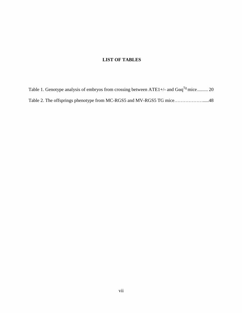
Table 1. Genotype analysis of embryos from crossing between ATE1+/- and GαqTg
mice ......... 20
Table 2. The offsprings phenotype from MC-RGS5 and MV-RGS5 TG mice……………….....48
Page 8
viii
LIST OF FIGURES
Chapter 1.
Figure 1. The ubiquitin system ....................................................................................................... 2
Figure 2. The hierarchical structure of the mammalian N-end rule pathway ................................. 3
Chapter 2.
Figure 3. The proteolysis of RGS proteins and its role in cardiovascular signaling and
homeostasis………………………………………………………………………………………12
Figure 4. The genotyping of embryos from intercross between ATE1+/- and GqTg
mice ........... 18
Figure 5. Immunoblotting of Gαq from intercross between ATE1+/- and GαqTg
mice ............... 19
Figure 6. Gross morphology of embryos at E14.5 through E16.5 ................................................ 22
Figure 7. Gross morphology of yolk sacs at E14.5 through E16.5, in which vascular defects are
obvious in ATE1-/-
and ATE1-/-
GqTg
............................................................................................. 23
Figure 8. Gross morphology of embryonic hearts at E14.5 through E16.5. ................................. 24
Figure 9. Internal morphology of embryonic hearts at E14.5 and E15.5...................................... 25
Figure 10. Internal morphology of embryonic hearts at E16.5. .................................................... 26
Figure 11. Histological analysis of left ventricle. ......................................................................... 27
Figure 12. Arterial phenotype of embryos at E16.5.. .................................................................... 28
Page 9
ix
Chapter 3.
Figure 13. Schematic diagram of RGS5 transgenic construct. ..................................................... 42
Figure 14. Southern blot analysis to identify founder transgenic mice. ....................................... 43
Figure 15. Genotyping of F0 generation of RGS5 TG mice. ....................................................... 44
Figure 16. Tissue distribution of RGS5 protein ............................................................................ 45
Figure 17. Establishing the expression of transient transfection of mouse RGS5 into A7r5 cells…
....................................................................................................................................................... 46
Figure 18. Tissue distribution of RGS5 protein ............................................................................ 48
Figure 19. Gross morphology of embryos and yolk sacs of MV-RGS5 at E14.5 and MC-RGS5 at
E16.5 ............................................................................................................................................. 49
Page 10
x
PREFACE
Dedicated to my family whom I love more than anyone else in the world
Page 11
1
1.0 THE MAMMALIAN N-END RULE PATHWAY
Ubiquitin (Ub) is a highly-conserved small protein which is only 76 amino acids, whose
conjugation to other proteins provides a signal for the proteasomal degradation by labeling
proteins (Hershko and Ciechanover 1998; Sriram, Banerjee et al. 2009). The ubiquitin (Ub)
system is composed of three enzymes (Figure 1). E1 Ub-activating enzyme activates ubiquitin
by forming a thioester linkage between the cysteine sulfhydryl group of E1 enzyme and the C-
terminal carboxyl group of ubiquitin. This activating process requires ATP as an energy source.
The activated ubiquitin is transferred to a cysteine residue of E2 Ub-conjugating enzyme via
trans-esterification reaction. In the last step, E3 Ub ligase enzyme recognizes the substrate
protein and interacts with both E2 and substrate. The E3 enzyme creates an amide isopeptide
bond between a lysine of the substrate protein and C-terminal of ubiquitin.
Page 12
2
Figure 1. The ubiquitin system A protein substrate is conjugated to Ub through the E1-E2-E3 enzymatic cascade
(Sriram, Banerjee et al. 2009).
The N-end rule pathway is a Ub-dependent protein degradation system where a class of
E3s called N-recognins recognize type 1 (basic) and type 2 (bulky hydrophobic) destabilizing N-
terminal residues of short-lived proteins as an essential determinant of N-terminal degradation
signals called N-degron (Tasaki and Kwon 2007). N-terminal degradation determinants of the N-
end rule pathway are divided into type 1 (basic; Arg, Lys, and His) and type 2 (bulky
hydrophobic; Phe, Tyr, Trp, Leu, and Ile) destabilizing residues. An N-degron can be created by
N-terminal modifications of a pre-N-degron. Since 1994, studies have identified approximately
10 genes involved in this process and characterized their gene products with respect to the
conversion of pre-N-degron to N-degron (Figure 2).
Page 13
3
Figure 2. The hierarchical structure of the mammalian N-end rule pathway (Sriram, Banerjee et al. 2009).
One way to create destabilizing N-degron is through the removal of the N-terminal Met
by Met aminopeptidases (MetAPs), which exposes the second residue at the N-terminus. Another
way to create an N-degron is through endoproteolytic cleavage of a long-lived polypeptide,
yielding a short-lived C-terminal fragment of the protein which consequently bears a
destabilizing N-terminal residue. Intracellular endopeptidases can create a C-terminal fragment
bearing a tertiary or secondary destabilizing residue or a primary destabilizing residue (de Groot,
Rumenapf et al. 1991; Rao, Uhlmann et al. 2001; Ditzel, Wilson et al. 2003; Tasaki, Mulder et
al. 2005; Tasaki and Kwon 2007). Specifically, exposed N-terminal Asn and Gln are tertiary
destabilizing residues that function through their deamidation by N-terminal amidohydrolases
(Nt-amidases), NTAN1 and NTAQ1, into the secondary destabilizing N-terminal residues Asp
and Glu, respectively. Considering that knockout of NTAN1 selectively abolished the
deamidation activity for N-terminal Asn, the hypothetical enzyme, termed NTAQ1, might be
responsible for deamidation of N-terminal Gln, which remains unknown (Stewart, Arfin et al.
Page 14
4
1995; Grigoryev, Stewart et al. 1996; Balogh, Kwon et al. 2000). NTAN1-deficient mice were
shown to have defects in spontaneous activity, spatial memory and a socially conditioned
exploratory behavior (Kwon, Balogh et al. 2000). N-terminal Asp and Glu are secondary
destabilizing residues that function through their arginylation by ATE1-encoded Arg-tRNA-
protein transferase (R-transferase), which creates the primary destabilizing residue Arg at the N-
terminus. ATE1 encodes at least six isoforms and they exhibit differential cellular localization,
tissue distribution and enzymatic activities for N-terminal Asp, Glu and Cys (Kwon, Kashina et
al. 1999; Rai and Kashina 2005; Hu, Brower et al. 2006). Blunted arginylation activities of all
known N-end rule substrates in ATE1-knockout mice suggested that ATE1 is the only enzyme
responsible for N-terminal arginylation (Kwon, Kashina et al. 2002). ATE1-/- mouse embryos
die at midgestation during embryogenesis primarily due to defects in cardiac development and
angiogenesis (Kwon, Kashina et al. 2002). N-terminal Cys can also function as a tertiary
destabilizing residue through its oxidation in a manner depending on nitric oxide (NO) and
oxygen (O2) or its derivatives; the oxidized Cys residue into Cys-sulfinic acid [CysO2(H)] or
Cys-sulfonic acid [CysO3(H)] is subsequently arginylated by ATE1 (Kwon, Kashina et al. 2002;
Hu, Sheng et al. 2005; Lee, Tasaki et al. 2005). Cys-sulfinic acid is structurally similar to Asp,
one of known arginylation-permissive N-terminal residues, suggesting that the oxidation of N-
terminal Cys is a specific modification to create a secondary destabilizing residue. The resulting
N-terminal Arg together with other type 1 and type 2 N-degrons are recognized by N-recognins
for protein ubiquitylation. Polyubiquitylated substrate with repeated conjugation of Ub is
recognized and degraded by the proteolytic machinery of the ubiquitin-proteasome system
(UPS), the 26S proteasome (Figure 2). Proteomic purification of endogenous E3s that bind to
synthetic degrons, recently revealed a novel Ub ligase family, termed UBR1 through UBR7,
Page 15
5
characterized by a ~70-residue domain, termed UBR box, that functions as a general substrate
recognition domain (Tasaki, Mulder et al. 2005). Amongst these E3s, UBR1, UBR2, UBR4 and
UBR5 were determined to have the ability to recognize N-degrons (Tasaki, Mulder et al. 2005).
UBR box proteins have generally heterogeneous size and sequence but contain specific
signatures as E3 Ub ligases or a substrate recognition subunit of the E3 complex: the RING
domain in UBR1, UBR2 and UBR3; the HECT domain in UBR5; the F-box in UBR6 and the
plant homeodomain (PHD) finger in UBR7. Characterization of knockout mice indicated that
mammalian UBR1 and UBR2 are functionally overlapping N-recognins whose biochemical
properties are similar to each other (Kwon, Reiss et al. 1998; Kwon, Levy et al. 1999; Kwon, Xia
et al. 2001). UBR1-deficient mice develop Johanson-Blizard syndrome (JBS)-like phenotypes,
including exocrine pancreatic abnormality, hypoglycemia and altered fat metabolism (Kwon, Xia
et al. 2001). UBR2-deficient mice exhibit male-specific infertility and female-specific lethality
that are distinguished from UBR1 null mice (Kwon, Xia et al. 2003). Mouse embryos lacking
both UBR1 and UBR2, which share 46% of similarity, die at midgestation from impaired
neurogenesis during development, indicating a functional interaction between these two E3s.
UBR3-deficient neonatal pups die associated with impaired olfactory system, and UBR3 is
prominently expressed in sensory cells for the five major senses (smell, touch, vision, hearing
and taste), suggesting that UBR3 controls a general circuit underlying different sensory nervous
systems. UBR4 with a size of 570 kDa binds to both type 1 and type 2 residues, interacts with
human paillomavirus type 16 (HPV-16) E7 oncoprotein and retinobalastoma tumor suppressor
protein (pRb) and has a role in anchorage-independent growth and cellular transformation in
cancer cells (DeMasi, Huh et al. 2005; Huh, DeMasi et al. 2005). UBR5 (also known as EDD)
with a size of 300 kDa, which preferentially bind to type 1 N-degron, has been implicated in
Page 16
6
progesterone-regulated cell proliferation, DNA damage responses and tumorigenesis (Callaghan,
Russell et al. 1998; Henderson, Munoz et al. 2006). The biochemical properties of UBR6 and
UBR7 as N-recognins remain unclear.
Page 17
7
2.0 POSTTRANSLATIONAL ARGINYLATION IN Gq-DEPENDENT CARDIAC
DEVELOPMENT
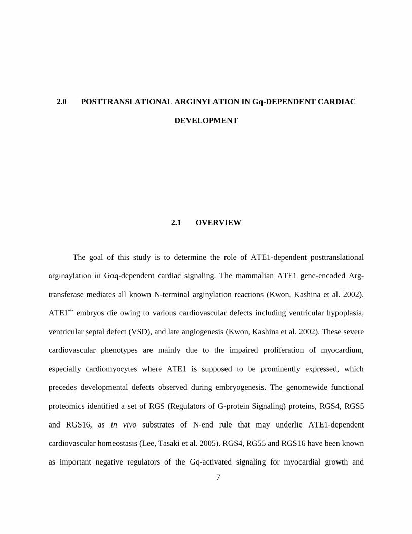
2.1 OVERVIEW
The goal of this study is to determine the role of ATE1-dependent posttranslational
arginaylation in Gαq-dependent cardiac signaling. The mammalian ATE1 gene-encoded Arg-
transferase mediates all known N-terminal arginylation reactions (Kwon, Kashina et al. 2002).
ATE1-/-
embryos die owing to various cardiovascular defects including ventricular hypoplasia,
ventricular septal defect (VSD), and late angiogenesis (Kwon, Kashina et al. 2002). These severe
cardiovascular phenotypes are mainly due to the impaired proliferation of myocardium,
especially cardiomyocytes where ATE1 is supposed to be prominently expressed, which
precedes developmental defects observed during embryogenesis. The genomewide functional
proteomics identified a set of RGS (Regulators of G-protein Signaling) proteins, RGS4, RGS5
and RGS16, as in vivo substrates of N-end rule that may underlie ATE1-dependent
cardiovascular homeostasis (Lee, Tasaki et al. 2005). RGS4, RG55 and RGS16 have been known
as important negative regulators of the Gq-activated signaling for myocardial growth and
Page 18
8
vascular maturation/integrity. RGS4, known to regulate Gq-activated signaling, was found to be
accumulated throughout the entire body of embryos lacking ATE1 (Lee, Kwon et al.
unpublished). It has been found that Gq-activated signaling components, including protein kinase
C (PKC) and its downstream component, MEK1 are downregulated in ATE1-deficient hearts
(Lee, Kwon et al. unpublished). There results suggest that developmental defects in ATE1-/-
hearts may be at least in part caused by dampened Gq-dependent proliferation of cardiac cells.
In this study, I attempted to determine whether cardiac–specific overexpression of Gαq
would rescue the cardiac defects in ATE1-deficient mice. To this end, I constructed ATE1-/-
GαqTg
compound mutant mice, where Gαq is exclusively overexpressed in the heart from αMHC
promoter. Morphological and histological analysis suggested that myocardial Gαq
overexpression rescues significantly cardiac defects of ATE1-/-
embryos in size and development
of ventricular myocardium, ventricular septum, trabeculae, and atrium. While Gαq
overexpression in the heart rescues significantly cardiac defects in ATE1-/- embryonic hearts, it
did not cause a noticeable change in vascular defects, indicating, for the first time, cell-
autonomous function of ATE1 in vascular development. These results together suggest that
ATE1 controls cardiac development and signaling in part through regulated proteolysis of
multiple regulators of Gq-activated signaling pathways.
Page 19
9
2.2 BACKGROUND
The N-end rule pathway is one of ubiquitin (Ub)-mediated proteolytic system that relates
the in vivo half-life of a protein to the identity of its N-terminal residue. Conjugation of arginine
(Arg) by ATE1-encoded Arg-transferase to N-terminal aspirate (Asp), glutamate (Glu), or
cysteine (Cys) is a posttranslational modification which creates the primary destabilizing residue
Arg. The resulting N-terminal Arg of an arginylated substrate is subsequently recognized for
protein ubiquitination by E3 Ub ligases, which produces a secondary degradation signal for
processive degradation by the 26S proteasome complex. This universal eukaryotic protein
modification, described 40 years ago (Kaji, Novelli et al. 1963), requires Arg from Arg-tRNAArg
of the protein synthesis machinery and, thereby defines a tRNA-dependent Ub proteolytic
system. In mammals ATE1 appears to be the only gene responsible for posttranslational
arginylation as ATE1 knockout disrupts all known N-terminal arginylation processes (Kwon,
Kashina et al. 2002). Characterization of ATE1-knockout mice demonstrated that they die at
midgestation during embryogenesis primarily due to defects in cardiac development and
angiogenesis. ATE1-deficient embryos at E12.5 started to show growth retardation associated
with local hemorrhage in the midbrain or heart, swollen pericardial sac and edema under the
dermal layer. Histological analysis of the hearts indicated thin ventricular myocardial hypoplasia
and disorganized ventricular trabeculation, resembling thin myocardium syndrome (Jaber, Koch
et al. 1996). The additional cardiac phenotypes of ATE1-/-
embryos include a ventricular septal
defect (VSD), a blood flow between the septum and endocardial cushion and also through
fenestrations in the septal tissue itself. ATE1-/- embryos also exhibit a persistent truncus
arteriosus, a common root of the aorta and pulmonary artery straddling a large VSD, with a
Page 20
10
prevalence of 4 per 10,000 births in humans (Creazzo, Godt et al. 1998). In addition to cardiac
defects, the majority of ATE1-/- embryos revealed vascular phenotypes including insufficiently
branched and thinner vitelline vessels and prematurely terminated primary-plexus capillaries
(Kwon, Kashina et al. 2002). These findings unveil unexpected physiological functions of the N-
end rule pathway in cardiac development and angiogenesis in mammals.
Substrates of ATE1-dependent arginylation have remained unknown for the last four
decades. In an attempt to identify the physiological substrates of ATE1 and other N-end rule
components, a genome-wide functional proteomics was performed. In this assay, 18,000 cDNAs
were individually expressed, labeled with biotin, and subjected to ubiquitination (Lee, Tasaki et
al. 2005). Screening of ubiquitination substrates that are also sensitive to dipeptide inhibitors of
the N-end rule pathway yielded ~35 candidate N-end rule substrates, including RGS4, RGS5 and
RGS16 (Lee, Tasaki et al. 2005). Biochemical characterization in mouse embryonic fibroblasts
(MEFs) indicated that these RGS proteins were ubiquitylated and degraded through ATE1-
dependent arginylation, which required N-terminal second cystein (Cys-2) as a degron. RGS4
and RGS5 were rapidly ubiquitylated and degraded in MEFs, and their degradation was
abolished in ATE1-/- MEFs (Lee, Tasaki et al. 2005). Mass spectrometric analysis of purified
RGS4, overexpressed in MEFs, revealed Arg conjugation of N-terminal Cys-2, which has been
exposed by Met aminopeptidases. Moreover, N-terminal Cys-2 itself was found to be conjugated
with a mass of 48 Da (Kwon, Kashina et al. 2002). The interpretation for an additional mass was
that 48-Da mass represents oxidation of Cys to Cys sulfonate (CysO3), which was verified by the
finding that depletion of oxygen in vitro and in mammalian cells disrupts degradation and
ubiquitylation of RGS4 and RGS5 (Lee, Tasaki et al. 2005). Notably, the oxidized Cys residue
(especially CysO2) becomes structurally similar to Asp, a genuine substrate of ATE1 R-
Page 21
11
transferase. N-terminally oxidized Cys should be recognized by UBR E3s for ubiquitylation and
subsequent degradation by the 26S proteasome complex, yet the E3s involved remain
incompletely understood. Recent studies demonstrated that UBR1-/-UBR2-/- mouse embryos die
at midgestation associated with defects in cardiovascular development (An, Seo et al. 2006) and
fail to degrade RGS4 and RGS5 (Lee, Tasaki et al. 2005). Although these results indicate that
RGS4, RGS5 and RGS16 are biochemical substrates of the N-end rule pathway, it is unknown
whether the O2-dependent pathway controlling RGS proteolysis, and thus cardiac GPCR
signaling, is present in cardiomyocytes.
Belonging to structurally related R4 subfamily of RGS proteins, RGS4, RGS5, and
RGS16 are considered prototypical among RGS proteins, which negatively regulate GPCR
signaling by converting the GPCR-activated, GTP-bound Gα subunit and Gβγ subunits into the
GDP-bound, inactive Gαβγ heterotrimeric complex. Among the entire RGS proteins, RGS4 has
received much attention as an important negative regulator of Gαq-mediated cardiovascular
system, in part because RGS4 is prominently expressed in the myocardium (Kardestuncer, Wu et
al. 1998; Adams, Pagel et al. 2000; Patten, Bunemann et al. 2002). RGS4 overexpression on the
mRNA and/or protein level was frequently observed in the terminally failing myocardium from
patients with dilated or ischemic cardiomyopathy with nonfailing controls or in some animal
models with heart dysfunction (Zhang, Watson et al. 1998; Owen, Burton et al. 2001; Mittmann,
Chung et al. 2002). RGS4 binding to Gαq inhibits Gαq-mediated PLCβ activation in the heart,
suggesting that RGS4 is a PLCβ antagonist (Tamirisa, Blumer et al. 1999; Mittmann, Chung et
al. 2002) (Figure 3). Stimulation of Gq-coupled receptors (e.g., by endothelin-1 receptor or α1-
adrenoreceptor agonists), is involved in the development of cardiac hypertrophy and failure
(D'Angelo, Sakata et al. 1997; Mende, Kagen et al. 1998). Recombinant RGS4 expression
Page 22
12
inhibited endothelin-1-stimulated PLC activity in left ventricular myocardium (Mittmann, Chung
et al. 2002). RGS4 overexpression in cardiomyocytes completely abolished the endothelin-1-
induced increase in fractional shortening (Mittmann, Chung et al. 2002), and inhibited the effects
of phenylephrine and endothelin-1 on ANF- and myosin light chain promoter activity and on
phenylephrine-stimulated myofilament organization and cell growth (Tamirisa, Blumer et al.
1999).
Figure 3. The proteolysis of RGS proteins and its role in cardiovascular signaling and homeostasis (Tasaki and Kwon
2007).
Page 23
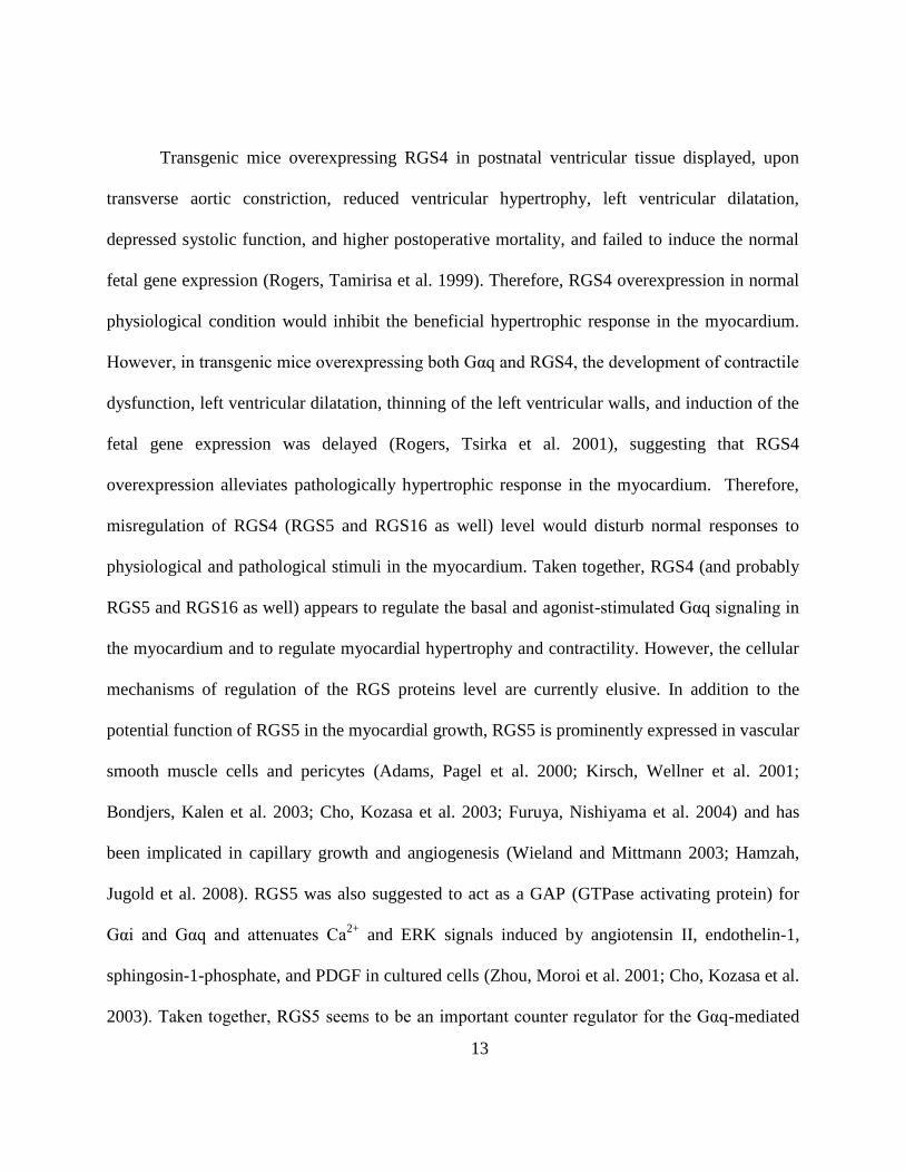
13
Transgenic mice overexpressing RGS4 in postnatal ventricular tissue displayed, upon
transverse aortic constriction, reduced ventricular hypertrophy, left ventricular dilatation,
depressed systolic function, and higher postoperative mortality, and failed to induce the normal
fetal gene expression (Rogers, Tamirisa et al. 1999). Therefore, RGS4 overexpression in normal
physiological condition would inhibit the beneficial hypertrophic response in the myocardium.
However, in transgenic mice overexpressing both Gαq and RGS4, the development of contractile
dysfunction, left ventricular dilatation, thinning of the left ventricular walls, and induction of the
fetal gene expression was delayed (Rogers, Tsirka et al. 2001), suggesting that RGS4
overexpression alleviates pathologically hypertrophic response in the myocardium. Therefore,
misregulation of RGS4 (RGS5 and RGS16 as well) level would disturb normal responses to
physiological and pathological stimuli in the myocardium. Taken together, RGS4 (and probably
RGS5 and RGS16 as well) appears to regulate the basal and agonist-stimulated Gαq signaling in
the myocardium and to regulate myocardial hypertrophy and contractility. However, the cellular
mechanisms of regulation of the RGS proteins level are currently elusive. In addition to the
potential function of RGS5 in the myocardial growth, RGS5 is prominently expressed in vascular
smooth muscle cells and pericytes (Adams, Pagel et al. 2000; Kirsch, Wellner et al. 2001;
Bondjers, Kalen et al. 2003; Cho, Kozasa et al. 2003; Furuya, Nishiyama et al. 2004) and has
been implicated in capillary growth and angiogenesis (Wieland and Mittmann 2003; Hamzah,
Jugold et al. 2008). RGS5 was also suggested to act as a GAP (GTPase activating protein) for
Gαi and Gαq and attenuates Ca2+
and ERK signals induced by angiotensin II, endothelin-1,
sphingosin-1-phosphate, and PDGF in cultured cells (Zhou, Moroi et al. 2001; Cho, Kozasa et al.
2003). Taken together, RGS5 seems to be an important counter regulator for the Gαq-mediated
Page 24
14
signaling in the vascular system as well. Although several studies have implicated RGS16 in the
endotoxin-induced heart failure and PLC inactivation (Patten, Bunemann et al. 2002), it appears
to play a role in lymphocyte migration as well (Lippert, Yowe et al. 2003).
The BrdU incorporation assay performed on developing hearts and cardiac cells of +/+
and ATE1-/-
embryos showed that ATE1 null cardiac defects are directly linked to cardiac
proliferation and signaling rather than a secondary effect to non-cardiac defects (Lee, Kwon et al.
unpublished). The levels of cells in the S-phase were significantly decreased in ventricular wall
and intraventricular septum, but not in trabeculae of ATE1 null embryos. The BrdU index was
profoundly reduced in troponin I (a marker for cardiomyocyte)-positive ATE1-/-
cardiomyocytes
(~9.4%) compared to those (~20%) of ATE1+/+
cardiomyocytes. Cardiomyocyte-specific
expression of NLS-lacZ (β-galactosidase N-terminally fused with a nuclear localization signal),
marked into the ATG codon of ATE1 gene, suggested that ATE1 has a cell-autonomous function
in cardiac proliferation. Whole-mount staining detected significantly stabilized endogeneous
RGS4 in ATE1-/-
embryos and sectional immunohistochemistry exhibited robust RGS4 signal in
ATE1-/-
embryonic hearts (Lee, Kwon et al. unpublished), indicating RGS4 functions are tightly
controlled by posttranslational arginylation. In an attempt to identify Gq agonists whose
functions are linked to ATE1, the proliferation of +/+ and ATE1-/- cardiac cells was monitored
after the treatment of various GPCR agonists, including PGF2α (prostraglandin F receptor
coupled with Gq), phenylephrine (α-adrenergic receptor coupled with Gq and Gi), FGFb (FGF
basic receptor which is a receptor tyrosine kinase), ISO (β-adrenergic receptor coupled with Gs),
and angiotensin II (AngII, AT1 receptor coupled with Gq and Gi). Amonst these GPCR agonists,
AngII treatment increased significantly cardiac proliferation in +/+ cardiomyocytes, whereas
AngII-induced proliferation was prominently reduced in ATE1-/- cardiomyocytes (Lee, Kwon et
Page 25
15
al. unpublished).
Based on these results, I employed genetic dissection to determine the role of ATE1 in
Gq-dependent cardiac signaling. To this end, I crossed ATE1+/- mice and Gαq transgenic mice
(D'Angelo, Sakata et al. 1997) and asked whether overexpression of Gαq in the heart would
significantly rescue cardiac null phenotypes in ATE1-/- embryos. Consistent with previous
observations (Kwon, Kashina et al. 2002), gross morphology of ATE1-/-
embryos at E14.5
through E16.5 exhibited growth retardation, local hemorrhages and swollen pericardial sac.
Gross morphology of hearts of ATE1-/-
and ATE1-/-
GαqTg
was largely comparable at E14.5 and
E15.5. However at E16.5, the development of hearts between ATE1-/- and ATE1-/-GαqTg
showed obvious difference. Whereas ATE1-/-
hearts at E16.5 are morphologically defective,
ATE1-/-GαqTg
hearts at same stage resembled, at least significantly and to a varying degree,
those of littermate wild-type and Gαq embryos. Cross section of embryonic hearts suggested that
ATE1 null cardiac defects in size, myocardium, ventricular septum, trabeculae and atrium were
significantly rescued by Gαq overexpression. These results together indicate that
posttranslational arginylation plays an important role in cardiac Gq-signaling pathways.
2.3 METHODS
Construction of ATE1-/-
GαqTg
mice. All animal studies were in accordance with
protocols approved by the Institutional Animal Care and Use Committee at University of
Pittsburgh. We obtained Gαq transgenic mice expressing 40 copies of Gαq transgene from α-
Page 26
16
myosin heavy chain (MHC) on FVB/N background in collaboration with Dr. Gerald Dorn II at
the University of Cincinnati (D'Angelo, Sakata et al. 1997). I constructed ATE1-/-
GαqTg
mice by
crossing ATE1+/-
mice (Kwon, Kashina et al. 2002) with Gαq transgenic mice. Mice lacking
ATE1 was as previously described (Kwon, Kashina et al. 2002). Embryos at E14.5 to E16.5 were
obtained from timed mating of ATE1+/- mice with Gαq transgenic mice. The presence of a
vaginal plug after overnight mating was regarded as E0.5.
Genotyping. Genotyping of the yolk sac or tail genomic DNA from each embryos or
mice was performed by standard PCR using the following condition: one cycle at 94°C 2min, 20
cycles of 94°C 20s, 56°C 20s, 65°C 30s, 10 cycles of 94°C 20s, 56°C 20s, 65°C 1min, 10 cycles
of 94°C 20s, 56°C 20s, 65°C 1min 30s and a final extension step at 65°C for 2min. The primers
used for detecting ATE1 were AK49 5’-GGTATTTGCTGCCGTCCTTTGGTGGT-3’ (forward),
AK83 5’-CTGGAGACAAAGCCCCAGCCAGAC-3’ (reverse), and YT655 5’-
CCAGCTCATTCCTCCCACTCATGATC-3’ (reverse), respectively amplifying 570-bp and
430-bp fragments for the wild type allele and knockout allele, respectively. The primers used for
detecting Gαq are MHC5A 5’-CAGGACTTCACATAGAAGCC-3’ (forward) and G4 5’-
CGTGAAGATGTTCTGATACAC-3’ (reverse), amplifying 500-bp of transgene.
Histology. Embryonic hearts were dissected in cold PBS and fixed overnight at 4°C in
4% paraformaldehyde (Fisher Scientific) in PBS. Samples were stored in 70% ethanol at 4°C
after several washing with PBS. For histological analysis, samples were dehydrated in serial
ethanol, cleared in histosol (National Diagnostics), paraffin-embedded, sectioned transversely at
5μm, and stained with hematoxylin and eosin (H&E).
Page 27
17
Western Blot. Embryonic hearts or other tissues (brain, liver, lung) at E15.5 were
dissected in cold PBS and homogenized in RIPA lysis buffer (150mM Sodium Chloride, 1% NP-
40, 0.5% Sodium deoxycholate, 0.1% SDS, 50mM Tris, pH 8.0) including protease inhibitor
cocktail (Sigma). Protein concentrations were determined using the BCA protein assay (Pierce,
Rockford, Ill.) with bovine serum albumin as a standard. 20ug of total protein extracts from each
tissue were subjected to electrophoresis and transfered to polybvinylidene fluoride (PVDF)
membrane, and stained with anti-Gαq antibody (Santa Cruz), followed by re-staining with anti-β-
actin antibody (Sigma) as a loading control. Gαq expression was quantitated by densitometry
(Image J software) followed by normalization with β-actin signal.
2.4 RESULTS
To genetically determine whether myocardial-specific Gαq overexpression would rescue
ATE1 null cardiac defects, I constructed ATE1-/-
GαqTg
mice by crossing ATE1+/- mice in the
C57BL/6J-129SvEv backgrounds with Gαq transgenic mice in the FVB/N background, where
Gαq is overexpressed from the α-myosin heavy chain (MHC) promoter.
Page 28
18
Figure 4. The genotyping of embryos from intercross between ATE1+/- and GqTg
mice. Yolk sac genomic
DNA was isolated from embryos and PCR was performed using two sets of primer. DNA bands corresponding to
the ATE1 alleles for wild-type (wt) and mutant (mut) and αMHC-Gαq transgene (Gαq) are indicated on the right.
I observed approximately 100 embryos at E14.5 through E16.5 from the timed-mating
and determined the gross morphology and the timing of cardiac defects. Genotyping was
performed by standard PCR using the yolk sac genomic DNA of embryos (Figure 4). Two
separate sets of primers to detect the wild-type and knock-out ATE1 alleles and the αMHC-Gαq
transgene are respectively AK49/AK83/YT655 and MHC5A/G4. Immunoblotting of compound
mutant embryos at E15.5 showed that Gαq was overexpressed only in the heart by ~5-fold, but
not in other tissues such as liver, lung and brain (Figure 5). It has been recently observed that
ATE1-/-
embryos at the stages up to E11.5 were recovered approximately at Mendelian ratios but
developmentally retarded from E12.5 (Lee, Kwon et al. unpublished). No embryos were
retrieved alive beyond E15.5. However, in the mixed background of C57BL6/129S and FVB/N,
I found that the onset of overall phenotype was delayed by 2~3 days and the resulting
phenotypes are somewhat moderate. At E15.5 and E16.5, some of ATE1-deficient embryos
(60% and 43% respectively) were still alive (Table 1).
Page 29
19
Figure 5. Immunoblotting of Gαq from intercross between ATE1+/- and GαqTg
mice. (A) Gαq protein expression in embryonic hearts at E14.5. Anti-β-actin
antibody was used to confirm an equal loading. (B) Quantitative Gαq protein expression normalized to β-actin (shown as fold induction over NTG animals) (n=2
in each group) (C) Gαq protein expression in liver, lung and brain of embryos at E14.5. Labeled bands correspond to the followings: ns, nonspecific; Gq, GαqTg
.
Page 30
20
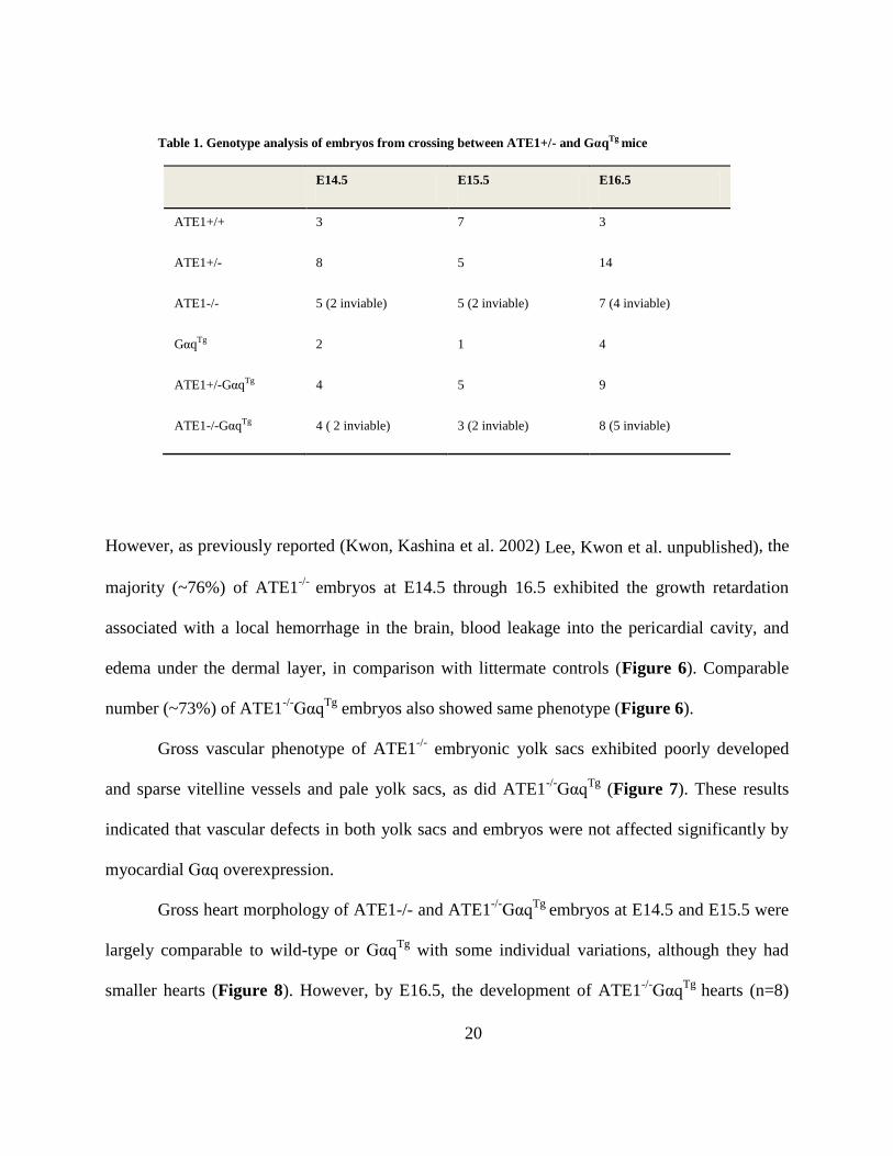
Table 1. Genotype analysis of embryos from crossing between ATE1+/- and GαqTg mice
E14.5 E15.5 E16.5
ATE1+/+ 3 7 3
ATE1+/- 8 5 14
ATE1-/- 5 (2 inviable) 5 (2 inviable) 7 (4 inviable)
GαqTg 2 1 4
ATE1+/-GαqTg 4 5 9
ATE1-/-GαqTg 4 ( 2 inviable) 3 (2 inviable) 8 (5 inviable)
However, as previously reported (Kwon, Kashina et al. 2002) Lee, Kwon et al. unpublished), the
majority (~76%) of ATE1-/-
embryos at E14.5 through 16.5 exhibited the growth retardation
associated with a local hemorrhage in the brain, blood leakage into the pericardial cavity, and
edema under the dermal layer, in comparison with littermate controls (Figure 6). Comparable
number (~73%) of ATE1-/-
GαqTg
embryos also showed same phenotype (Figure 6).
Gross vascular phenotype of ATE1-/-
embryonic yolk sacs exhibited poorly developed
and sparse vitelline vessels and pale yolk sacs, as did ATE1-/-
GαqTg
(Figure 7). These results
indicated that vascular defects in both yolk sacs and embryos were not affected significantly by
myocardial Gαq overexpression.
Gross heart morphology of ATE1-/- and ATE1-/-
GαqTg
embryos at E14.5 and E15.5 were
largely comparable to wild-type or GαqTg
with some individual variations, although they had
smaller hearts (Figure 8). However, by E16.5, the development of ATE1-/-
GαqTg
hearts (n=8)
Page 31
21
became apparently equivalent to littermate wild-type and GαqTg
embryos, whereas ATE1-/-
hearts at the same stage were morphologically defective and exhibited cone-like shape (Figure
8).
To confirm the above-mentioned observations, we examined internal morphology of
embryonic hearts at E14.5 through E16.5 by H&E staining. Transverse sections of embryonic
hearts demonstrated that both ATE1-/-
and ATE1-/-
GαqTg
at E14.5 have impaired cardiac
phenotype showing ventricular septal defect (VSD), ventricular hypoplasia and poor trabecular
formation (Figure 9A~D). By E15.5, however, interventricular septum in ATE1-/-
GαqTg
hearts
grew toward the atrioventricular cushion tissue (Figure 9E~G) and subsequently fused with the
myocardial tissue resulting in completely closed septum by E16.5 (Figure 10). In addition to
VSD, the cardiac defects including thin myocardium and poor trabeculae were rescued in 4 out
of 5 hearts when observed between E15.5 and E16.5. The thickness of compact zone of the left
ventricle in E16.5 ATE1-/-
GαqTg
hearts was 13~17-cells, which is comparable to ATE1+/+
(10~12-cells) and GαqTg
(10~14-cells), whereas ATE1-/-
hearts displayed 1~6-cells-thickness at
same developmental stage (Figure 11). The atria (data not shown) and major cardiac vessels
such as aorta and pulmonary artery between genotypes did not display any significant difference
(Figure 12).
Based on these results, I conclude that cardiac-specific Gαq overexpression rescues
significantly cardiac defects in ATE1-/-
embryos. In contrast to cardiac defects, Gαq
overexpression in the heart did not affect invariably vascular defects of ATE1-/-
at E14.5 through
E16.5, suggesting that ATE1 plays a cell-autonomous function in vascular signaling.
Page 32
22
Figure 6. Gross morphology of embryos at E14.5 through E16.5.
Page 33
23
Figure 7. Gross morphology of yolk sacs at E14.5 through E16.5, in which vascular defects are obvious in ATE1-/- and ATE1-/-GqTg.
Page 34
24
Figure 8. Gross morphology of embryonic hearts at E14.5 through E16.5. RV, right ventricle; LV, left ventricle; RA, right atrium; LA, left atrium.
Page 35
25
Figure 9. Internal morphology of embryonic hearts at E14.5 and E15.5. H&E-stained transverse sections of embryonic
hearts at different stages. L, liver; A, aorta; O, oesophagus; RA, right atrium; LA, left atrium; RV, right ventricle; LV, left
ventricle; VSD, ventricular septal defect. Magnification x40
Page 36
26
Figure 10. Internal morphology of embryonic hearts at E16.5. H&E-stained transverse sections of embryonic hearts. L, liver;
A, aorta; O, oesophagus; RA, right atrium; LA, left atrium; RV, right ventricle; LV, left ventricle. Magnification x40.
Page 37
27
Figure 11. Histological analysis of left ventricular myocardium. The thickness of compact zone in left ventricle myocardium
was compared between different genotypes. CZ, compact zone. Magnification x200
Page 38
28
Figure 12. Arterial phenotype of embryos at E16.5. H&E-stained transverse sections of embryonic hearts. Ao, aorta; PA,
pulmonary artery; LA, left atrium; O, oesophagus. Magnification x200
Page 39
29
2.5 DISCUSSION
ATE1 mediates arginylation, a universal eukaryotic protein modification, in N-terminal
Cys as well as Asp and Glu of proteins. ATE1-deficient mice die during embryogenesis
associated with ventricular myocardial hypoplasia, disorganized ventricular trabeculation, thin
myocardium syndrome, and angiogenetic defects (Kwon, Kashina et al. 2002). Recent data
suggested that significantly reduced proliferation of cardiomyocytes in ATE1-/-
hearts mainly
attributes to the cardiac defects in a manner depending on cardiac Gq signaling (Lee, Kwon et al.
unpublished). In this study, I determined the role of ATE1 at the molecular level in cardiac Gq
signaling and development. I found that transgenic overexpression of Gαq partially rescued
cardiac defects but not vascular defects of ATE1-deficient mice, as determined by overall size,
morphology and histological analysis of H&E stained cardiac sections. Although cardiac Gαq
overexpression didn’t influence the gross morphology of ATE1-/- embryos including growth
retardation or local hemorrhages, embryonic heart sections revealed that ATE1 null cardiac
defects such as VSD and thin myocardium were significantly rescued by Gαq overexpression.
Recent work has discovered new physiological functions of N-terminal arginylation in adult
mice (Brower and Varshavsky 2009). The postnatal deletion of mouse Ate1 using Cre/lox
technique induced growth retardation resulting in early death of ATE1-deficient mice. They
displayed a strikingly lower white adipose tissue (WAT) and they are resistant to high-fat diet-
induced obesity due to the ectopic induction of Ucp1 in WAT. In addition, they have a kyphotic
posture (a forward rounding of the upper back) and disproportionately large brains contributed
by cerebral edema, in comparison to those of control siblings. They exhibited the
neurological/behavioral abnormality including hyperactivity and higher liability to seizure.
Page 40
30
Moreover, postnatally ATE1-deficient mice show male-specific infertility with few sperm cells
in disorganized arrangement. Taken all together, arginylation by ATE1 involves in broad range
of physiological process including spermatogenesis, adipogenesis, and neurogenesis, as well as
cardiovascular development.
The genetic background of mutant animals influences the phenotypes observed to a
greater or lesser extent (Jackson and Abbott 2000). The issue of genetic background has
particularly been raised in neurosciences, regarding the complex behavioral phenotypes. Some
literatures have shown that genetic background markedly influences the severity of phenotypes
in broad range of biomedical fields including cardiovascular research (Schlager 1966; Blizard
and Welty 1971; Hoit, Kiatchoosakun et al. 2002; Tominaga, Matsuda et al. 2004; Heydemann,
Huber et al. 2005). One example is two distinct transgenic lines expressing hypertrophic
cardiomyopathy-linked mutant tropomyosin E108G showing drastically different phenotypes.
One created on the FVB/N background displayed relatively mild diastolic dysfunction (Michele,
Gomez et al. 2002) and the other on the C57BL/6J background developed obvious hypertrophic
cardiomyopathy and heart failure (Prabhakar, Boivin et al. 2001). As described in these finding,
C57BL/6J is more susceptible to revealing cardiac phenotype than FVB/N, which may explain
why the onset of phenotype in ATE1-/- embryos on mixed background between C57BL6/129S
and FVB/N was overall delayed and rather milder than on C57BL6/129S.
I observed that the internal cardiac morphology of ATE1-/-
GαqTg
mice exhibited dilated
ventricles compared to wild-type controls (Figure 10). It has been known that transgenic mice
overexpressing wild-type Gαq in the heart using the α-myosin heavy chain promoter
demonstrated cardiac hypertrophy, depending on the level of overexpression, with marked
increases in markers of hypertrophy and heart failure and revealed impaired intrinsic contractility
Page 41
31
(D'Angelo, Sakata et al. 1997). High levels (40-copies, approximately five-fold excess Gαq
protein over endogenous levels) of Gαq overexpression can lead to a dilated cardiomyopathy,
which is the consequence of lethal congestive heart failure (D'Angelo, Sakata et al. 1997). As
previously reported, in this study, ATE1-/-
GαqTg
and Gαq
Tg mice overexpressing 40 copies of
transgenes showed mortality on the mixed background (FVB/N X C57J/129S) as well. I
observed that ATE1-/-
GαqTg
hearts together with GαqTg
developed enlarged ventricles and
showed the hint of cardiomyocyte hypertrophy on left ventricles. The hypertrophy of ATE1-/-
GαqTg
hearts indicates that high copy number of Gαq in compound mice induced hypertrophy
exceeding the rescue of cardiac defect of ATE1-/-
embryos.
Page 42
32
3.0 THE N-END RULE PATHWAY IN RGS5 UBIQUITINATION AND
CARDIOVASCULAR SIGNALING
3.1 OVERVIEW
RGS5, which has been identified as N-end rule substrate through genomewide functional
proteomic screening, is known to play a role in negatively regulating G protein-coupled receptor
(GPCR) signaling in the hearts and blood vessels. As one of R4 family, RGS5 exhibits
prominent expression in vascular smooth muscle cells and pericyte and has been recently
identified as a key regulator for blood pressure regulation, vascular remodeling, and pericyte
maturation in tumors. The biochemical analyses in mouse embryonic fibroblasts (MEF) have
previously shown that ubiquitination of RGS5 involves Cys-2 as a degradation determinant and
is impaired by hypoxic treatment, indicating that RGS5 proteolysis may act as an O2 sensor
controlling cardiovascular homeostasis. In this study, I generated RGS5 transgenic mouse strains
overexpressing either MC-RGS5 (wild-type, short-lived) or MV-RGS5 (mutant, long-lived) from
vascular smooth muscle-specific SM22α promoter to determine the physiological importance of
RGS5 proteolysis in Gq signaling of VSMC. The results from transient transfection using
Page 43
33
transgenic constructs showed that MC-RGS5 (wild-type) expressed from the SM22α promoter is
rapidly degraded in rat aortic smooth muscle cells (A7r5), while the mutation of RGS5 Cys-2 to
Val results in an increased steady state level of RGS5. Both MC-RGS5 and MV-RGS5 mice
were viable and fertile without any visible defects. Gross morphology of embryos from both
transgenic mice did not exhibit any significant phenotype compared to non-transgenic siblings.
However, MC-RGS5 female transgenic mice demonstrated impaired delivery in that newborn
pups were often found dead associated with a reduced level or the total absence of milk in their
stomachs, whereas MV-RGS5 mice never showed this phenotype. This phenotype mimics that of
oxytocin null mice, indicating that RGS5 may negatively regulate oxytocin receptor which is one
of Gq-protein coupled receptor. The mis-regulation of this RGS5 function may result in the
failure in oxytocin-induced uterine and mammary gland smooth muscle contraction. This
transgenic model will be useful in testing a model where the steady state level of RGS5 in
vascular smooth muscle cells (VSMC) of blood vessels as well as uterine SMC is regulated by
the N-end rule pathway through its posttranslational modifications such as oxidation,
arginylation, ubiquitination and degradation and provide a molecular basis in homeostasis of
cardiovascular Gq signaling.
3.2 BACKGROUND
RGS5 (Regulators of G-protein Signaling 5) was identified, together with RGS4 and
RGS16, as a substrate of N-end rule pathway through genome-wide functional proteomics.
Page 44
34
RGS5 has been known to negatively regulate GPCR signaling as a GTPase activating protein
(GAP) for Gαi and Gαq by converting the GPCR-activated, GTP-bound Gα subunit and the Gβγ
subunits into the GDP-bound, inactive Gαβγ heterotrimeric complex. RGS5 attenuates
angiotensin II and endothelin-1-induced intracellular Ca2+
transients in 293T cells. The
translocation of RGS5 from cytosol to plasma membrane requires its N terminal extension (aa 1-
33), which is not essential for exerting inhibitory activities (Zhou, Moroi et al. 2001). In addition,
RGS5 suppresses ERK signals induced by angiotensin II, endothelin-1, sphigosin-1-phosphate,
and PDGFββ in NIH-3T3 cells, suggesting that RGS5 inhibits signaling through GPCR as well
as through PDGFR (Cho, Kozasa et al. 2003). Microarray analysis to identify markers in artery
revealed RGS5 as one of the most prominent markers in pericytes of capillary vessels and
VSMC of larger vessels (Adams, Pagel et al. 2000; Kirsch, Wellner et al. 2001; Bondjers, Kalen
et al. 2003; Cho, Kozasa et al. 2003; Furuya, Nishiyama et al. 2004). RGS5 is prominently
expressed in cells associated with the abluminal side of the endothelium rather than the
endothelial cells. In the brain and arterial blood vessels of embryos, RGS5 colocalizes with
pericyte markers, PDGFRβ and NG2 chondroitin sulfate proteoglycan. VSMC around arteries
and arterioles and kidney mesangial cells show prominent RGS5 expression, whereas veins show
limited or undetectable levels of RGS5 expression (Bondjers, Kalen et al. 2003). In adult mice,
RGS5 expression is induced in VSMC of arteries during neovascularization associated with skin
wound-healing, ovulation and tumor angiogenesis (Furuya, Nishiyama et al. 2004; Berger,
Bergers et al. 2005), indicating the role of RGS5 in active vessel remodeling.
As shown in structurally-related RGS4 proteolysis, RGS5, one of substrates of ATE1-
dependent arginylation, was rapidly degraded when expressed in reticulocyte lysates and
metabolically stabilized by the proteasome inhibitor MG132 or the type-1 dipeptide N-end rule
Page 45
35
inhibitor Arg-Ala (Lee, Tasaki et al. 2005). RGS5 was short-lived in mouse embryonic
fibroblasts (MEF) as determined by pulse chase analysis, and its degradation was disrupted by
the treatment of MG132. RGS5 was also metabolically stabilized in ATE1-/-
MEF or by
mutations of Cys-2 to stabilizing residues, Val or Ala, suggesting that in vivo RGS5 degradation
depends on ATE1-dependent arginylation. Biochemical analyses demonstrate that arginylated
RGS5 is subsequently recognized by N-recognins, including UBR1 and UBR2, for
ubiquitination and degradation by the 26S proteasome complex. Consistent with these results,
mouse embryos lacking UBR1 and UBR2, two functionally overlapping E3s, die at midgestation
associated with defects in cardiovascular development (An, Seo et al. 2006), and MEFs derived
from these embryos are impaired in the ability to degrade RGS4 (Lee, Tasaki et al. 2005). The
degradation and ubiquitination of normally short-lived RGS5 was significantly decreased upon
hypoxic condition, in contrast to the level of the long-lived C2A-RGS5 mutant, which was not
affected by O2 depletion (Lee, Tasaki et al. 2005). Mass spectrometry analysis found that the
molecular mass of the Cys-2 residue of arginylated RGS4 purified from mouse L cells was
increased by 48 Da (Kwon, Kashina et al. 2002), suggesting that the N-terminal Cys-2 residue of
RGS4 (exposed at the N-terminus by Met aminopeptidases) may be oxidized into CysO2 or
CysO3. Given that oxygen is essential for the proteolysis of RGS5 which is structurally related to
RGS4, RGS5 may undergo the same posttranslational modification including arginylation and
oxidation. These results suggest that ATE1-dependent pathway may be an O2 sensor that controls
cardiovascular homeostasis through oxidation, arginylation, ubiquitination, and subsequent
degradation of RGS5.
Recently RGS5-deficient mouse strains were created by two laboratories (Cho, Park et al.
2008; Nisancioglu, Mahoney et al. 2008). RGS5-deficient mice exhibit hypotension associated
Page 46
36
with slightly dilated aortas without detectible defects in vascular development (Cho, Park et al.
2008; Nisancioglu, Mahoney et al. 2008). To assess whether RGS5 is required for normal BP
homeostasis, the tail BP of awake +/+
and RGS5-/-
mice was monitored. Studies with RGS5-/-
mice
showed that female mutants have lower BP than male mutants. Specifically, the mean arterial
pressure (MAP) of five male RGS5-/- mice was 116 ± 6 mmHg which was significantly lower
than 145 ± 5 mmHg of wild-type littermate controls (P-value=0.005). The average MAP of three
female RGS5-/- mice was 84 ± 1 mmHg compared with 144 ± 9 mmHg for wild-type littermate
controls (P-value=0.003). The examination of the ascending, mid-thoracic aorta and proximal to
the diaphragm suggested that the low BP of RGS5-deficient mice can be attributed at least in part
to dilated aorta. Another group genetically induced pancreatic carcinoma in RGS5-deficient
mice, exhibiting reduction in tumor hypoxia and vessel leakiness associated with pericyte
maturation and vascular normalization (Hamzah, Jugold et al. 2008). These in vivo animal
models indicate that RGS5 plays a pivotal role in blood pressure regulation and vascular
remodeling.
In this study, to determine the physiological role of RGS5 proteolysis in vascular
signaling, I constructed RGS5 transgenic mouse strains, overexpressing either wild type RGS5
(MC-RGS5) or mutant RGS5 (MV-RGS5) from the vascular smooth muscle-specific SM22α
promoter. The results from transient transfection using the transgenic constructs showed that
MC-RGS5 (wild-type) expressed from the SM22α promoter is rapidly degraded in rat aortic
smooth muscle cells (A7r5), while the mutation of RGS5 Cys-2 to Val results in an increased
steady state level of RGS5. This suggests that Cys-2-dependent degradation of RGS5 is regulated
by N-end rule pathway. Genotyping and mating analysis suggested that both MC-RGS5 and
Page 47
37
MV-RGS5 mice were viable and fertile without visible developmental defects. Gross
morphology of embryos from both transgenic mice did not exhibit noticeable phenotypes such as
growth retardation and cardiovascular defect when compared with non-transgenic siblings.
However, the majority of offspring from mating of MC-RGS5 females and C57BL6 males died
within 1~2 days after birth, associated with a reduced level or the total absence of milk in their
stomachs, even though MC-RGS5 female transgenic mice demonstrated normal maternal
behavior. MC-RGS5 females built a typical nest and quickly retrieved offspring that moved
outside of the nest. In contrast, the postnatal lethality was not observed in pups from mating of
MC-RGS5 male and C57BL6 female. This phenotype mimics that of oxytocin null mice
(Nishimori, Young et al. 1996). Since it is known that RGS5 negatively regulates oxytocin
receptor, one of Gq-protein coupled receptors, the above-mentioned defect of MC-RGS5 may
result from the failure of oxytocin-induced mammary gland and uterine smooth muscle
contraction. Taken together, RGS5 may play a role in controlling contraction in various smooth
muscle-containing tissues. These transgenic models may be used as a valuable tool to
characterize the homeostasis of RGS5 turnover in contractile pathway and to determine in vivo
function of posttranslational modification of N-terminal degradation determinant, Cys-2, of
RGS5 in vascular signaling and development.
Page 48
38
3.3 METHODS
Construction of MC-RGS5 and MV-RGS5 Transgenic mice. The wild type (MC)
murine RGS5 cDNA (genebank accession no. NM_009063 ) and mutant (MV) RGS5 cDNA
were released with BamHI, and ligated into the BamHI site of a plasmid containing the 481-bp
murine SM22alpha (Transgelin, -441 to 41 relative to transcription start) promoter provided by
Dr. Andrea Ekhart (Thomas Jefferson University, Philadelphia, PA) (genebank accession no.
U36589) (Eckhart, Ozaki et al. 2002). The resulting recombinant plasmids, SM22α-MC-RGS5 or
SM22a-MV-RGS5, were confirmed by restriction enzyme mapping and nucleotide sequencing.
A linear 1.9 kb DNA fragment containing the SM22α promoter, the complete murine MC-RGS5
or MV-RGS5 cDNA ORF, and a simian virus 40 (SV40) intron and polyadenylylation signal
was released by digestion with XhoI and SacII (Fig.13). This fragment was microinjected into
male pronuclei of fertilized C57BL6 mouse oocytes and implanted into pseudopregnant females
by Transgenic and Gene Targeting Core Facility in University of Pittsburgh. Three-week-old
mouse pups were screened for presence of the transgene by Southern analysis. From second
generation, nested PCR was performed for genotyping.
Southern Blot. Random integration of transgene into mouse genome was accessed by
Southern blot analysis from tail genomic DNAs of three-week-old mice. Tail genomic DNAs
(10g) were digested with SalI, electrophoresed on a 1% agarose gel, transfered to Nytran
membrane (Wattman), and hybridized with 32
P-labeled 800-bp BamHI–SacII fragment of SV40
viral DNA. Probe was labeled using [32
P]dCTP by Ready-To-Go DNA Labelling Beads
(Amersham) and radiolabeled probe was purified by ProbeQuant G-50 Micro Columns
Page 49
39
(Amersham). The membrane was prehybridized in ExpressHyb Solution (Clontech) and
radiolabeled DNA probe with 100mg/ml salmon sperm DNA was added to the filter at 60°C for
4 hours. Membranes were washed several times and exposed to Kodak Biomax MS film with an
intensifying screen. Approximate copy numbers were estimated by comparison of signal
intensity with that of copy numbers of transgene.
Genotyping. Genomic DNA from embryo yolk sacs or mouse tails was analyzed by
nested PCR amplification using two primer pair sets (Fig.13). The first-round PCR primers
(outer primer pairs) were designed to have a forward primer on 5’ upstream of SM22α promoter
and a reverse primer on poly A signal, amplifying 1,295-bp and their sequences are SM5 (5’-
CAGTCAAGACTAGTTCCCACCAACTCG-3’) and Poly3 (5’-
CTAGATGGCATTTCTTCTGAGCAAAACAG-3’). The second-round PCR primers (inner
primer pairs) were designed to detect the inner sequence of the first-round PCR product,
amplifying 1,184-bp and their sequences are SM6 (5’-
CAGGTTCCTTTGTCGGGCCAAACTCTAG-3’) and Poly4 (5’-
GGCATTCCACCACTGCTCCCATTCATC-3’).
Western Blot. Different tissues including aorta, heart, uterus, liver, and brain from two-
month-old mice were dissected in cold PBS and homogenized in RIPA lysis buffer (150mM
Sodium Chloride, 1% NP-40, 0.5% Sodium deoxycholate, 0.1% SDS, 50mM Tris, pH 8.0)
containing a protease inhibitor cocktail (Sigma). Protein concentration was determined using the
BCA protein assay (Pierce, Rockford, Ill.)
with bovine serum albumin as a standard.
Approximately 30ug of total protein extract from each tissue was subjected to electrophoresis
Page 50
40
and transfered to polybvinylidene fluoride (PVDF) membrane, and stained with anti-RGS5
antibody (gift of Dr. Jian Li, Havard University), followed by re-staining with anti-actin antibody
(Sigma) as a loading control.
Cell Culture and Transfection. A7r5 cells, rat aortic smooth muscle cells (ATCC), were
maintained in DMEM supplemented with 10% FBS and 100U/ml penicillin/streptomycin. For
transfection, cells were plated at 3 x 10⁴ cells/well in 12-well plate and transfected with 1ug of
plasmid using Lipofectamine LTX by manufacturer’s protocol. Cells were maintained for 44
hours, treated with 5μM of MG132 for 4 hours, and harvested.
Immunohistochemistry. Thoracic aorta was dissected in cold PBS and fixed overnight
at 4°C in 4% paraformaldehyde (Fisher Scientific) in PBS. Samples were stored in 70% ethanol
at 4°C after several washing with PBS. For histological analysis, samples were dehydrated in
serial ethanol, cleared in histosol (National Diagnostics), paraffin-embedded, and sectioned
transversely at 5μm. Sections were antigen-retrieved by heating in Tris-EDTA (pH. 9.0) for 20
min using a microwave oven, and blocked in PBST (0.2% Triton X-100 in PBS) containing 1%
BSA for 1.5 hrs, and incubated with primary antibody (rabbit anti-RGS5 antibody (1:400), a gift
from Dr. Li in Harvard University; mouse anti-α smooth muscle actin (1:400), Sigma) in PBS
with 0.5% BSA overnight, followed by secondary antibody (Alexa 555-conjugated anti-rabbit
IgG; Alexa 48- conjugated anti-mouse IgG2a, Invitrogen) in PBS for 1 hr. Sections were
counterstained with 4’6’-diamidino-2-phenylindole (DAPI, Vector Laboratories) to visualize the
nuclei.
Page 51
41
3.4 RESULTS
To investigate the role of RGS5 proteolysis in vascular smooth muscle cells, MC-RGS5
and MV-RGS5 transgenic mice carrying the construct shown in Figure 13 were produced as
described in Methods. I used the mouse SM22alpha promoter, which has been well characterized
to target transgene expression to arterial but not venous or visceral smooth muscle cells (Eckhart,
Ozaki et al. 2002; Keys, Zhou et al. 2005). A schematic diagram of the transgene construct is
presented in Fig. 13. Briefly, the SM22α promoter (-441 to +41) was fused to the cDNA of
mouse RGS5 (MC-RGS5 or MV-RGS5) and the SV40 poly A signal sequence by conventional
cloning. MV-RGS5 cDNA was simply generated by replacing Cys-2 encoded-TGT with Val-
encoded GTG. Southern blot analysis of tail genomic DNA identified one potential MC-RGS5
and one potential MV-RGS5 transgenic founders (Figure 14). However, this analysis was not
sensitive enough to detect 1 and 5 copies of transgene. Further analysis using nested PCR primer
sets revealed two more MC-RGS5 male founders and another putative MV-RGS5 founder
(Figure 15). Five of the founder animals were viable and fertile, but only three of them (one
MC-RGS5 female, one MV-RGS5 female, and one MV-RGS5 male) exhibited germline
transmission of the transgene to subsequent progeny.
Page 52
42
Figure 13. Schematic diagram of RGS5 transgenic construct. Full-length cDNA of RGS5 (MC-RGS5 and MV-RGS5)
including kozac sequence and the SV40 large T antigen polyadenylation signal sequence (SV40 polyA) were inserted into a
modified pBluescript KS+ vector under the SM22alpha promoter (-441 to +41). To generate MV-RGS5 TG mice, TGT encoding
second Cys amino acid was replaced by GTG encoding Val. Restriction sites for excision of the construct from the cloning vector
(XhoI/SacII) and for generation of radiolabeled probe (BamHI/SacII) are indicated with solid line. The primers for nested PCR
are indicated as arrows labeled with S5 (SM5), S6 (SM6), P3 (poly3), and P4 (poly4).
Page 53
43
Figure 14. Southern blot analysis to identify founder transgenic mice. Tail DNA was digested with Sal I to generate a 800-bp fragment containing the SV40
polyadenylation sequence from the transgene (see Fig.13). 10 μg of digested genomic DNA was resolved by gel electrophoresis and transferred to Nytran, and
the membrane was incubated with 32
P-labeled probe. The copy number standards were produced from transgenic construct plasmids. M, DNA marker.
Page 54
44
Figure 15. Genotyping of F0 generation of RGS5 TG mice. Nested PCR was performed to detect RGS5 transgene using genomic DNA from mouse tails as a
template for PCR genotype screening. The transgenic plasmid was loaded to estimate approximate copy number of RGS5 transgene expression.
Page 55
45
To analyze RGS5 transgenic expression, transgenic tissues including aorta, heart, uterus,
liver, and brain from two-month-old MC-RGS5 and MV-RGS5 mice were dissected and lysed
followed by western blotting using anti-RGS5 antibody (Figure 16). Consistent with the
previous northern blot analysis showing that RGS5 mRNA is abundantly expressed in heart,
lung, and intestine and at low levels in brain, liver, and placenta (Seki, Sugano et al. 1998), high
levels of endogenous RGS proteins were detected in heart and aorta of NTG mice, whereas
relatively lower amounts of RGS proteins were detected in liver and brain and, more
significantly, uterus. This suggests that endogenous RGS5 is widely distributed in various
smooth muscle-containing tissues. Immunoblotting of transgenic tissues revealed that MC-RGS5
TG mice contain a higher level of RGS5 compared to that in control mice (Figure 16).
Consistent with the finding that RGS5 Cys-2 is a degradation determinant, MV-RGS5 tissues
showed an even higher level of RGS5 compared to MC-RGS5 mice (Figure 16). These results
together suggest that RGS5 in these transgenic mice undergoes at least in part N-end rule
degradation based on Cys-2 as a degron.
Figure 16. Tissue distribution of RGS5 protein. RGS5 protein levels in different tissues of adult RGS5 transgenic mice and
non-transgenic mice (NTG) were determined by Western blotting. L, Liver; B, brain; H, heart; A, aorta; U, uterus.
Page 56
46
To confirm VSMC-specific transgenic expression, thoracic aorta from two types of
transgenic mice were isolated and immunostained with anti-RGS5 antibody, followed by staining
of the cross sections for α-smooth muscle actin (αSMA), a marker of VSMC (Figure 17). NTG
aorta demonstrated that endogenous RGS5 is expressed at basal level, whereas the aorta of both
MC-RGS5 and MV-RGS5 exhibited higher level of RGS5 expression in VSMC, displaying co-
localized expression with αSMA.
Figure 17. RGS5 expression in the aorta of MC-RGS5, MV-RGS5, and NTG mice. Transverse section of thoracic aorta
were stained for RGS5 (red), α-SMA (green) and nucleus (blue) as indicated above. Lu, Lumen.
Page 57
47
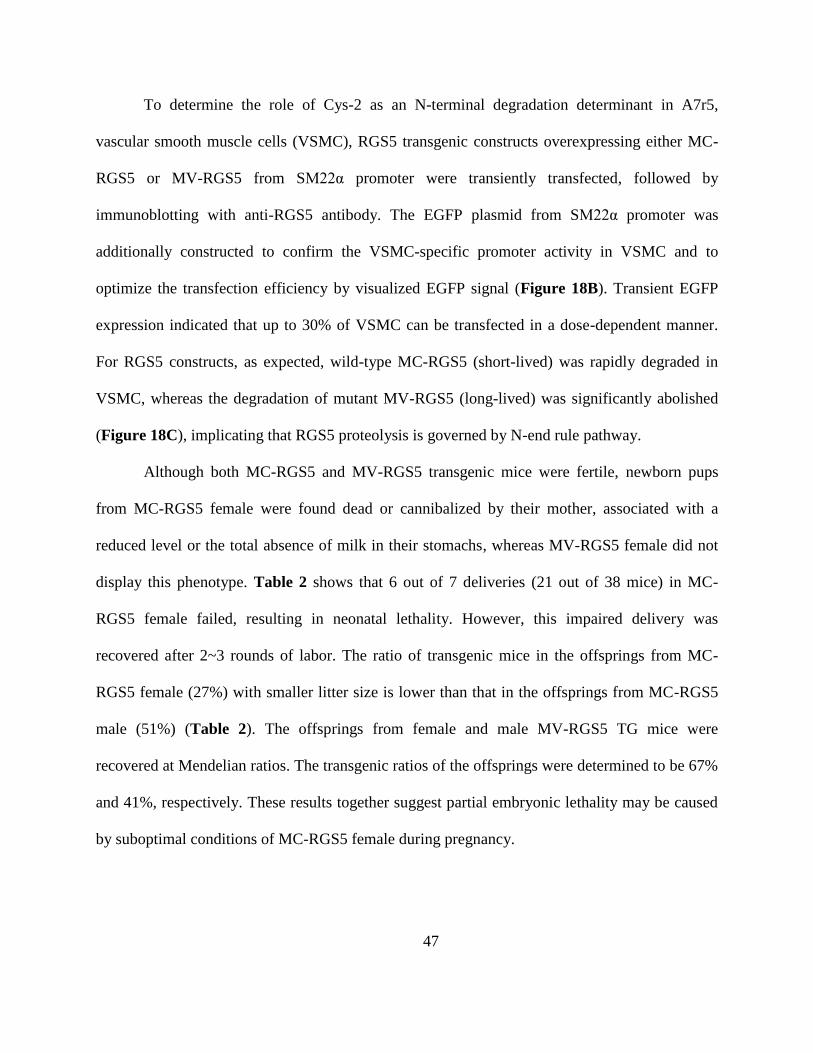
To determine the role of Cys-2 as an N-terminal degradation determinant in A7r5,
vascular smooth muscle cells (VSMC), RGS5 transgenic constructs overexpressing either MC-
RGS5 or MV-RGS5 from SM22α promoter were transiently transfected, followed by
immunoblotting with anti-RGS5 antibody. The EGFP plasmid from SM22α promoter was
additionally constructed to confirm the VSMC-specific promoter activity in VSMC and to
optimize the transfection efficiency by visualized EGFP signal (Figure 18B). Transient EGFP
expression indicated that up to 30% of VSMC can be transfected in a dose-dependent manner.
For RGS5 constructs, as expected, wild-type MC-RGS5 (short-lived) was rapidly degraded in
VSMC, whereas the degradation of mutant MV-RGS5 (long-lived) was significantly abolished
(Figure 18C), implicating that RGS5 proteolysis is governed by N-end rule pathway.
Although both MC-RGS5 and MV-RGS5 transgenic mice were fertile, newborn pups
from MC-RGS5 female were found dead or cannibalized by their mother, associated with a
reduced level or the total absence of milk in their stomachs, whereas MV-RGS5 female did not
display this phenotype. Table 2 shows that 6 out of 7 deliveries (21 out of 38 mice) in MC-
RGS5 female failed, resulting in neonatal lethality. However, this impaired delivery was
recovered after 2~3 rounds of labor. The ratio of transgenic mice in the offsprings from MC-
RGS5 female (27%) with smaller litter size is lower than that in the offsprings from MC-RGS5
male (51%) (Table 2). The offsprings from female and male MV-RGS5 TG mice were
recovered at Mendelian ratios. The transgenic ratios of the offsprings were determined to be 67%
and 41%, respectively. These results together suggest partial embryonic lethality may be caused
by suboptimal conditions of MC-RGS5 female during pregnancy.
Page 58
48
Figure 18. Establishing the expression of transient transfection of mouse RGS5 into A7r5 cells. (A) and (B) show
fluorescence microscopy images 48 h after transient transfection of rat aortic smooth muscle cells A7r5 with the EGFP construct
under SM22α promoter. Phase contrast (A) and the corresponding fluorescence (B) image show that GFP expression is seen
throughout the cell cytosol. Magnification ×100 (C) Western blotting of transfected cell lysates for MC-RGS5 and MV-RGS5
constructs. The non-specific band (ns) shows equal amount of loading in each sample.
Table 2. The offsprings phenotype from MC-RGS5 and MV-RGS5 TG mice.
MC-RGS5 offsprings
Paternal Maternal n # of lethality # of lethal mice Litter size % of TG
C57BL6 MC-RGS5 7 6 21/38 5.5 27
MC-RGS5 C57BL6 5 0 0/35 7 51
MV-RGS5 offsprings
C57BL6 MV-RGS5 3 0 0/24 8 67
MV-RGS5 C57BL6 4 0 0/27 6.8 41
Page 59
49
To examine the physiological function of RGS5, I harvested embryos from MC-RGS5
and MV-RGS5 TG mice at E16.5 and E14.5, respectively (Figure 19). Given that RGS5 is one
of ATE1 substrates and that ATE1 null mice revealed severe cardiovascular defect from E12.5, I
expected that RGS5 TG mice may have similar cardiovascular phenotype or at least milder
phenotype. However, both MC-RGS5 and MV-RGS5 TG embryos displayed no obvious
developmental defects and the vasculature appeared to develop normally in comparison to wild-
type siblings. Therefore, the overexpression of RGS5 in smooth muscle cells does not induce
noticeable developmental defects in embryos.
Figure 19. Gross morphology of embryos and yolk sacs of MV-RGS5 at E14.5 (A~D) and MC-RGS5 at E16.5 (E~H).
Page 60
50
3.5 DISCUSSION
The most obvious phenotype from RGS5 TG mice is the lethality of offspring during or
after labor from MC-RGS5 TG females. It is commonly observed that first time mothers and
very young females cannibalize their litters, but as they become experienced and mature, they
raise their litters successfully. However, this is not the case in RGS5 TG mice, because the same
phenotype was observed in 2~3 rounds of labor for each of MC-RGS5 TG females. Literature
search revealed that this phenotype is very similar to that observed with oxytocin null mice
(Nishimori, Young et al. 1996). It is notable that oxytocin receptor is one of Gq-coupled GPCRs.
During delivery, oxytocin binds Gq-coupled GPCR and induces the contraction of uterine
smooth muscle and mammary smooth muscle to help laboring and produce milking. Therefore,
the phenotype observed with MC-RGS5 TG mice appears to be related with the failure of
oxytocin-activated smooth muscle contraction due to inhibitory effect of RGS5 overexpression.
In addition, the fact that this phenotype is only found in MC-RGS5 female, not in MV-RGS5
female, suggests that MV-RGS5 may not be functional in vivo. A recent study has shown that
RGS2 and RGS5 were abundantly expressed at various stages of pregnancy (non-pregnant,
preterm, term non-labouring, and term laboring) in human (Ladds, Zervou et al. 2009). The co-
immunoprecipitation assay showed that RGS2 and RGS5 interact with the oxytocin receptor,
further suggesting that functional MC-RGS5 negatively regulates the oxytocin receptor
signaling.
RGS5-deficient mice generated by two independent groups have been shown to be
hypotensive relative to wild-type controls (Cho, Park et al. 2008; Nisancioglu, Mahoney et al.
2008). This result is apparently unexpected because, given the function of RGS5 as a GTPase
Page 61
51
activating protein (GAP) in Gαq- and Gαi-mediated cardiovascular signaling, it has been
expected that RGS5 inactivation would cause hypertension rather than hypotension. One feasible
explanation is that the functionally related RGS2 and RGS4 may compensate the loss of RGS5,
which lowers BP. Together with RGS5, RGS2, a member of RGS4 subfamily, is emerging as a
key regulator of the blood pressure (BP). RGS2 is a potent negative regulator of Gαq, which
mediates the action of most physiological vasoconstrictors, including norepinephrine,
angiotensin II, endothelin-1, and thrombin (Heximer, Knutsen et al. 2003). Mice lacking RGS2
are hypertensive due to the increased vascular tone in the BP homeostasis (Heximer, Knutsen et
al. 2003; Gu, Cifelli et al. 2009). These results suggest that RGS5 together with RGS2 and RGS4
participate in the regulation of BP homeostasis. The possible consequence of perturbation of
RGS5 turnover in RGS5 TG mice is dampened vascular Gq signaling involved in controlling BP.
I anticipate that the accumulation of RGS5 in VSMC results in hypotension.
Another interesting study using RGS5 TG mice is to characterize the physiological
importance of oxidation of Cys-2 of RGS5 in VSMC. Given that NADPH oxidase is a major
generator of ROS in VSMC among various ROS sources including NADPH oxidases, xanthine
oxidase, mitochondrial electron transport system, cytochrom p450, nitric oxide synthase (NOS)
(Landmesser, Cai et al. 2002; Clempus and Griendling 2006), the reactive oxygen species (ROS)
generated from NADPH oxidase may play a role in RGS5 oxidation in VSMC. It has been
shown that loss of p47phox, a critical subunit of NADPH oxidase, resulted in impaired ROS
production when cultured VSMC were stimulated by angiotensin II or PDGF-ββ. The loss of
p47phox resulted in lower BP in mice. These results suggest that NADPH oxidase-generated
ROS is involves in angiotensin II-induced BP homeostasis (Landmesser, Cai et al. 2002).
NADPH oxidase-produced ROS induces calcium-independent activation of the Rho-GTP
Page 62
52
pathway, in which Rho-GTP-activated kinase inhibits MLC phosphatase (MLCP) and, thus,
accelerates phosphorylation of myosin light chain (MLC), leading to smooth muscle contraction
(Jin, Ying et al. 2004). Taken together with these results, the oxidation of RGS5 Cys-2 via
NADPH oxidase-produced ROS may negatively regulate Gq agonists-induced vascular
signaling, which in turn modulates vascular smooth muscle contraction and BP controlling
(Clempus and Griendling 2006). This transgenic model will be a useful tool to determine
whether the steady state level of RGS5 in vascular smooth muscle cells (VSMC) of blood vessels
as well as uterine SMC is regulated by the N-end rule pathway through its posttranslational
modifications such as oxidation, arginylation, ubiquitination and degradation. Successful results
will provide a molecular basis in homeostasis of cardiovascular Gq signaling.
Page 63
53
BIBLIOGRAPHY
Adams, J. W., A. L. Pagel, et al. (2000). "Cardiomyocyte apoptosis induced by Galphaq
signaling is mediated by permeability transition pore formation and activation of the
mitochondrial death pathway." Circ Res 87(12): 1180-7.
An, J. Y., J. W. Seo, et al. (2006). "Impaired neurogenesis and cardiovascular development in
mice lacking the E3 ubiquitin ligases UBR1 and UBR2 of the N-end rule pathway." Proc
Natl Acad Sci U S A 103(16): 6212-7.
Balogh, S. A., Y. T. Kwon, et al. (2000). "Varying intertrial interval reveals temporally defined
memory deficits and enhancements in NTAN1-deficient mice." Learn Mem 7(5): 279-86.
Berger, M., G. Bergers, et al. (2005). "Regulator of G-protein signaling-5 induction in pericytes
coincides with active vessel remodeling during neovascularization." Blood 105(3): 1094-
101.
Blizard, D. A. and R. Welty (1971). "Cardiac activity in the mouse: strain differences." J Comp
Physiol Psychol 77(2): 337-44.
Bondjers, C., M. Kalen, et al. (2003). "Transcription profiling of platelet-derived growth factor-
B-deficient mouse embryos identifies RGS5 as a novel marker for pericytes and vascular
smooth muscle cells." Am J Pathol 162(3): 721-9.
Brower, C. S. and A. Varshavsky (2009). "Ablation of arginylation in the mouse N-end rule
pathway: loss of fat, higher metabolic rate, damaged spermatogenesis, and neurological
perturbations." PLoS One 4(11): e7757.
Callaghan, M. J., A. J. Russell, et al. (1998). "Identification of a human HECT family protein
with homology to the Drosophila tumor suppressor gene hyperplastic discs." Oncogene
17(26): 3479-91.
Page 64
54
Cho, H., T. Kozasa, et al. (2003). "Pericyte-specific expression of Rgs5: implications for PDGF
and EDG receptor signaling during vascular maturation." FASEB J 17(3): 440-2.
Cho, H., C. Park, et al. (2008). "Rgs5 targeting leads to chronic low blood pressure and a lean
body habitus." Mol Cell Biol 28(8): 2590-7.
Clempus, R. E. and K. K. Griendling (2006). "Reactive oxygen species signaling in vascular
smooth muscle cells." Cardiovasc Res 71(2): 216-25.
Creazzo, T. L., R. E. Godt, et al. (1998). "Role of cardiac neural crest cells in cardiovascular
development." Annu Rev Physiol 60: 267-86.
D'Angelo, D. D., Y. Sakata, et al. (1997). "Transgenic Galphaq overexpression induces cardiac
contractile failure in mice." Proc Natl Acad Sci U S A 94(15): 8121-6.
de Groot, R. J., T. Rumenapf, et al. (1991). "Sindbis virus RNA polymerase is degraded by the
N-end rule pathway." Proc Natl Acad Sci U S A 88(20): 8967-71.
DeMasi, J., K. W. Huh, et al. (2005). "Bovine papillomavirus E7 transformation function
correlates with cellular p600 protein binding." Proc Natl Acad Sci U S A 102(32): 11486-
91.
Ditzel, M., R. Wilson, et al. (2003). "Degradation of DIAP1 by the N-end rule pathway is
essential for regulating apoptosis." Nat Cell Biol 5(5): 467-73.
Eckhart, A. D., T. Ozaki, et al. (2002). "Vascular-targeted overexpression of G protein-coupled
receptor kinase-2 in transgenic mice attenuates beta-adrenergic receptor signaling and
increases resting blood pressure." Mol Pharmacol 61(4): 749-58.
Furuya, M., M. Nishiyama, et al. (2004). "Expression of regulator of G protein signalling protein
5 (RGS5) in the tumour vasculature of human renal cell carcinoma." J Pathol 203(1):
551-8.
Grigoryev, S., A. E. Stewart, et al. (1996). "A mouse amidase specific for N-terminal asparagine.
The gene, the enzyme, and their function in the N-end rule pathway." J Biol Chem
271(45): 28521-32.
Gu, S., C. Cifelli, et al. (2009). "RGS proteins: identifying new GAPs in the understanding of
blood pressure regulation and cardiovascular function." Clin Sci (Lond) 116(5): 391-9.
Hamzah, J., M. Jugold, et al. (2008). "Vascular normalization in Rgs5-deficient tumours
promotes immune destruction." Nature 453(7193): 410-4.
Page 65
55
Henderson, M. J., M. A. Munoz, et al. (2006). "EDD mediates DNA damage-induced activation
of CHK2." J Biol Chem 281(52): 39990-40000.
Hershko, A. and A. Ciechanover (1998). "The ubiquitin system." Annu Rev Biochem 67: 425-
79.
Heximer, S. P., R. H. Knutsen, et al. (2003). "Hypertension and prolonged vasoconstrictor
signaling in RGS2-deficient mice." J Clin Invest 111(8): 1259.
Heydemann, A., J. M. Huber, et al. (2005). "Genetic background influences muscular
dystrophy." Neuromuscul Disord 15(9-10): 601-9.
Hoit, B. D., S. Kiatchoosakun, et al. (2002). "Naturally occurring variation in cardiovascular
traits among inbred mouse strains." Genomics 79(5): 679-85.
Hu, R. G., C. S. Brower, et al. (2006). "Arginyltransferase, its specificity, putative substrates,
bidirectional promoter, and splicing-derived isoforms." J Biol Chem 281(43): 32559-73.
Hu, R. G., J. Sheng, et al. (2005). "The N-end rule pathway as a nitric oxide sensor controlling
the levels of multiple regulators." Nature 437(7061): 981-6.
Huh, K. W., J. DeMasi, et al. (2005). "Association of the human papillomavirus type 16 E7
oncoprotein with the 600-kDa retinoblastoma protein-associated factor, p600." Proc Natl
Acad Sci U S A 102(32): 11492-7.
Jaber, M., W. J. Koch, et al. (1996). "Essential role of beta-adrenergic receptor kinase 1 in
cardiac development and function." Proc Natl Acad Sci U S A 93(23): 12974-9.
Jackson, I. J. and C. M. Abbott (2000). Mouse genetics and transgenics : a practical approach.
Oxford ; New York, Oxford University Press.
Jin, L., Z. Ying, et al. (2004). "Activation of Rho/Rho kinase signaling pathway by reactive
oxygen species in rat aorta." Am J Physiol Heart Circ Physiol 287(4): H1495-500.
Kaji, H., G. D. Novelli, et al. (1963). "A Soluble Amino Acid-Incorporating System from Rat
Liver." Biochim Biophys Acta 76: 474-7.
Kardestuncer, T., H. Wu, et al. (1998). "Cardiac myocytes express mRNA for ten RGS proteins:
changes in RGS mRNA expression in ventricular myocytes and cultured atria." FEBS
Lett 438(3): 285-8.
Page 66
56
Keys, J. R., R. H. Zhou, et al. (2005). "Vascular smooth muscle overexpression of G protein-
coupled receptor kinase 5 elevates blood pressure, which segregates with sex and is
dependent on Gi-mediated signaling." Circulation 112(8): 1145-53.
Kirsch, T., M. Wellner, et al. (2001). "Altered gene expression in cerebral capillaries of stroke-
prone spontaneously hypertensive rats." Brain Res 910(1-2): 106-15.
Kwon, Y. T., S. A. Balogh, et al. (2000). "Altered activity, social behavior, and spatial memory
in mice lacking the NTAN1p amidase and the asparagine branch of the N-end rule
pathway." Mol Cell Biol 20(11): 4135-48.
Kwon, Y. T., A. S. Kashina, et al. (2002). "An essential role of N-terminal arginylation in
cardiovascular development." Science 297(5578): 96-9.
Kwon, Y. T., A. S. Kashina, et al. (1999). "Alternative splicing results in differential expression,
activity, and localization of the two forms of arginyl-tRNA-protein transferase, a
component of the N-end rule pathway." Mol Cell Biol 19(1): 182-93.
Kwon, Y. T., F. Levy, et al. (1999). "Bivalent inhibitor of the N-end rule pathway." J Biol Chem
274(25): 18135-9.
Kwon, Y. T., Y. Reiss, et al. (1998). "The mouse and human genes encoding the recognition
component of the N-end rule pathway." Proc Natl Acad Sci U S A 95(14): 7898-903.
Kwon, Y. T., Z. Xia, et al. (2003). "Female lethality and apoptosis of spermatocytes in mice
lacking the UBR2 ubiquitin ligase of the N-end rule pathway." Mol Cell Biol 23(22):
8255-71.
Kwon, Y. T., Z. Xia, et al. (2001). "Construction and analysis of mouse strains lacking the
ubiquitin ligase UBR1 (E3alpha) of the N-end rule pathway." Mol Cell Biol 21(23):
8007-21.
Ladds, G., S. Zervou, et al. (2009). "Regulators of G protein signalling proteins in the human
myometrium." Eur J Pharmacol 610(1-3): 23-8.
Landmesser, U., H. Cai, et al. (2002). "Role of p47(phox) in vascular oxidative stress and
hypertension caused by angiotensin II." Hypertension 40(4): 511-5.
Lee, M. J., T. Tasaki, et al. (2005). "RGS4 and RGS5 are in vivo substrates of the N-end rule
pathway." Proc Natl Acad Sci U S A 102(42): 15030-5.
Page 67
57
Lippert, E., D. L. Yowe, et al. (2003). "Role of regulator of G protein signaling 16 in
inflammation-induced T lymphocyte migration and activation." J Immunol 171(3): 1542-
55.
Mende, U., A. Kagen, et al. (1998). "Transient cardiac expression of constitutively active
Galphaq leads to hypertrophy and dilated cardiomyopathy by calcineurin-dependent and
independent pathways." Proc Natl Acad Sci U S A 95(23): 13893-8.
Michele, D. E., C. A. Gomez, et al. (2002). "Cardiac dysfunction in hypertrophic
cardiomyopathy mutant tropomyosin mice is transgene-dependent, hypertrophy-
independent, and improved by beta-blockade." Circ Res 91(3): 255-62.
Mittmann, C., C. H. Chung, et al. (2002). "Expression of ten RGS proteins in human
myocardium: functional characterization of an upregulation of RGS4 in heart failure."
Cardiovasc Res 55(4): 778-86.
Nisancioglu, M. H., W. M. Mahoney, Jr., et al. (2008). "Generation and characterization of rgs5
mutant mice." Mol Cell Biol 28(7): 2324-31.
Nishimori, K., L. J. Young, et al. (1996). "Oxytocin is required for nursing but is not essential for
parturition or reproductive behavior." Proc Natl Acad Sci U S A 93(21): 11699-704.
Owen, V. J., P. B. Burton, et al. (2001). "Expression of RGS3, RGS4 and Gi alpha 2 in acutely
failing donor hearts and end-stage heart failure." Eur Heart J 22(12): 1015-20.
Patten, M., J. Bunemann, et al. (2002). "Endotoxin induces desensitization of cardiac endothelin-
1 receptor signaling by increased expression of RGS4 and RGS16." Cardiovasc Res
53(1): 156-64.
Prabhakar, R., G. P. Boivin, et al. (2001). "A familial hypertrophic cardiomyopathy alpha-
tropomyosin mutation causes severe cardiac hypertrophy and death in mice." J Mol Cell
Cardiol 33(10): 1815-28.
Rai, R. and A. Kashina (2005). "Identification of mammalian arginyltransferases that modify a
specific subset of protein substrates." Proc Natl Acad Sci U S A 102(29): 10123-8.
Rao, H., F. Uhlmann, et al. (2001). "Degradation of a cohesin subunit by the N-end rule pathway
is essential for chromosome stability." Nature 410(6831): 955-9.
Rogers, J. H., P. Tamirisa, et al. (1999). "RGS4 causes increased mortality and reduced cardiac
hypertrophy in response to pressure overload." J Clin Invest 104(5): 567-76.
Page 68
58
Rogers, J. H., A. Tsirka, et al. (2001). "RGS4 reduces contractile dysfunction and hypertrophic
gene induction in Galpha q overexpressing mice." J Mol Cell Cardiol 33(2): 209-18.
Schlager, G. (1966). "Systolic blood pressure in eight inbred strains of mice." Nature 212(5061):
519-20.
Seki, N., S. Sugano, et al. (1998). "Isolation, tissue expression, and chromosomal assignment of
human RGS5, a novel G-protein signaling regulator gene." J Hum Genet 43(3): 202-5.
Sriram, S. M., R. Banerjee, et al. (2009). "Multivalency-assisted control of intracellular signaling
pathways: application for ubiquitin- dependent N-end rule pathway." Chem Biol 16(2):
121-31.
Stewart, A. E., S. M. Arfin, et al. (1995). "The sequence of porcine protein NH2-terminal
asparagine amidohydrolase. A new component of the N-end Rule pathway." J Biol Chem
270(1): 25-8.
Tamirisa, P., K. J. Blumer, et al. (1999). "RGS4 inhibits G-protein signaling in cardiomyocytes."
Circulation 99(3): 441-7.
Tasaki, T. and Y. T. Kwon (2007). "The mammalian N-end rule pathway: new insights into its
components and physiological roles." Trends Biochem Sci 32(11): 520-8.
Tasaki, T., L. C. Mulder, et al. (2005). "A family of mammalian E3 ubiquitin ligases that contain
the UBR box motif and recognize N-degrons." Mol Cell Biol 25(16): 7120-36.
Tominaga, K., J. Matsuda, et al. (2004). "Genetic background markedly influences vulnerability
of the hippocampal neuronal organization in the "twitcher" mouse model of globoid cell
leukodystrophy." J Neurosci Res 77(4): 507-16.
Wieland, T. and C. Mittmann (2003). "Regulators of G-protein signalling: multifunctional
proteins with impact on signalling in the cardiovascular system." Pharmacol Ther 97(2):
95-115.
Zhang, S., N. Watson, et al. (1998). "RGS3 and RGS4 are GTPase activating proteins in the
heart." J Mol Cell Cardiol 30(2): 269-76.
Zhou, J., K. Moroi, et al. (2001). "Characterization of RGS5 in regulation of G protein-coupled
receptor signaling." Life Sci 68(13): 1457-69.