46
The Role of Therapeutic Apheresis in Neurological Disorders Vishesh Chhibber, MD November 15, 2013
| Date post: | 25-Dec-2015 |
| Category: |
Documents |
| Upload: | eugene-lloyd |
| View: | 219 times |
| Download: | 0 times |

The Role of Therapeutic Apheresis in Neurological
Disorders
Vishesh Chhibber, MDNovember 15, 2013

Disclosure
• I have no actual or potential conflict of interest in relation to the content of this presentation

The Role of Apheresis
• Several autoimmune neurological disorders can be treated with TA
• Usually a protein in the plasma (often an autoantibody) is being removed
• Most common procedure performed is TPE– Immunoadsorption (IA) is also utilized
• In some disease indications, it is not clear why TPE is beneficial

Evidence Based Guidelines
• The American Academy of Neurology (AAN) published updated evidence-based guidelines in 2011
• ASFA published the 6th edition of their evidence based guidelines in 2013

AAN Guidelines
• Previous AAN assessment was in 1996• Controlled clinical trials (in humans)• Data from observational studies and case
reports was not included in the analysis as this was considered class IV evidence

Cortese I et al. Neurology 2011;76:294-300
°°
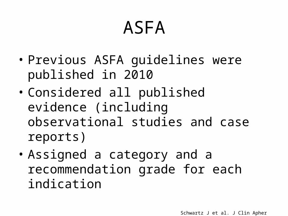
ASFA
• Previous ASFA guidelines were published in 2010
• Considered all published evidence (including observational studies and case reports)
• Assigned a category and a recommendation grade for each indication
Schwartz J et al. J Clin Apher 2013;28:145-284
°°

Schwartz J et al. J Clin Apher 2013;28:145-284

Schwartz J et al. J Clin Apher 2013;28:145-284

Disorders that will be discussed today
Schwartz J et al. J Clin Apher 2013;28:145-284

Technical Details of TPE
• 1-1.5 Plasma volumes• Albumin (5%) used as the replacement fluid• Donor plasma added as a portion of the
replacement fluid if patient at risk of coagulopathy/bleeding (e.g. daily TPE)
• Number and frequency of TA procedures will depend on the disease/indication

Acute Disseminated Encephalomyelitis (ADEM)
• Acute inflammatory demyelinating disease of the CNS
• Immune response to myelin (or other autoantigens)
• Presents with acute encephalopathy– altered mental status, hemiparesis, ataxia, motor
deficits, may progress to coma
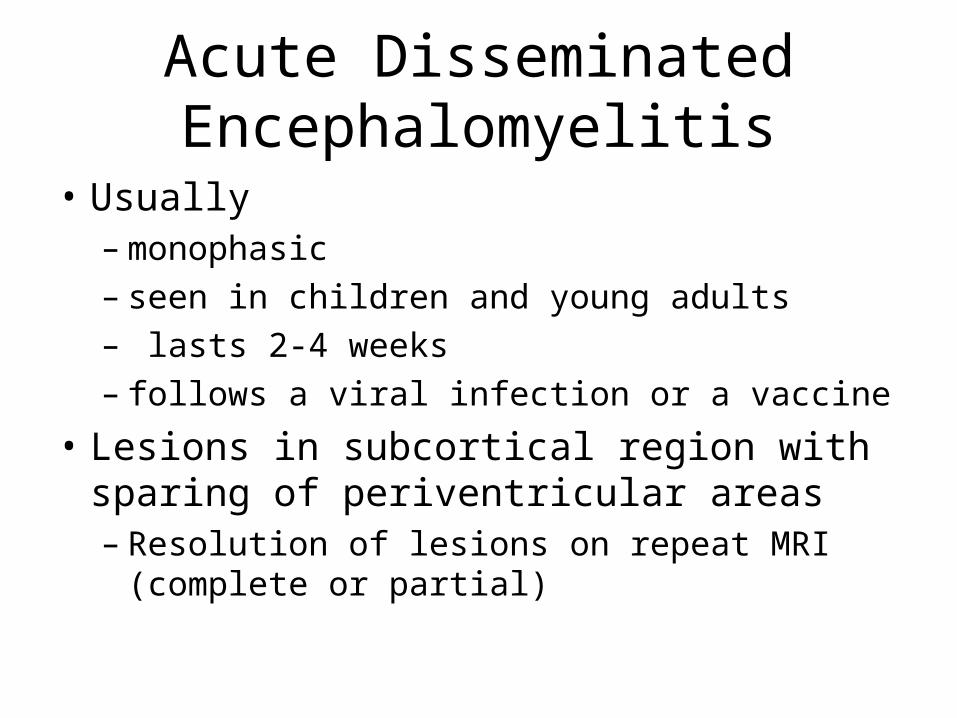
Acute Disseminated Encephalomyelitis
• Usually – monophasic– seen in children and young adults– lasts 2-4 weeks– follows a viral infection or a vaccine
• Lesions in subcortical region with sparing of periventricular areas– Resolution of lesions on repeat MRI (complete or
partial)

Therapy
• Corticosteroids (first line therapy)– 1/3 fail
• In patients that do not respond to steroids, TPE or IVIG can be used– 3 to 6 TPE with 1-1.5 volume replacement using
5% albumin performed daily or every other day
• In patients that respond to TPE, improvement is noted soon after initiation of TPE
Schwartz J et al. J Clin Apher 2013;28:145-284

Guidelines
• AAN: Possibly effective in acute CNS demyelination (includes MS, ADEM, and NMO) if steroid therapy fails
• ASFA: Category II with a recommendation of 2C
Cortese I et al. Neurology 2011;76:294-300
Schwartz J et al. J Clin Apher 2013;28:145-284

Neurological Disease ConditionsCNS PNS
Acute ADEM AIDP (Guillain-Barre)
Chronic MSNMO
CIDPMG

Acute Inflammatory Demyelinating Polyneuropathy (Guillain-Barre Syndrome)
• Most common general paralytic disorder• Acute motor and sensory deficiency involving
the peripheral nerves• Caused by a complement binding IgM
antibody to peripheral nerve myelin• RCT’s have shown
– Less time to recovery– Greater extent of neurological recovery
The GBS Study Group. Neurology 1985;35:1096 -1104
French Coop. Group on TPE in GBS. Ann Neurol 1987;22:753-761
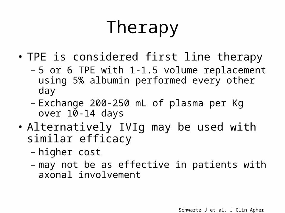
Therapy
• TPE is considered first line therapy– 5 or 6 TPE with 1-1.5 volume replacement using 5%
albumin performed every other day– Exchange 200-250 mL of plasma per Kg over 10-14
days• Alternatively IVIg may be used with similar
efficacy – higher cost– may not be as effective in patients with axonal
involvement
Schwartz J et al. J Clin Apher 2013;28:145-284

Guidelines
• AAN: TPE is effective in severe AIDP (impaired ability to walk, requirement of mechanical ventilation) and should be offered to these patients. In milder disease, it is probably effective and should be considered.
• ASFA: Category I with recommendation grade of 1A– Category III with a recommendation grade of 2C
has been assigned for TPE post IVIG
Cortese I et al. Neurology 2011;76:294-300
Schwartz J et al. J Clin Apher 2013;28:145-284

Neurological Disease ConditionsCNS PNS
Acute ADEM AIDP (Guillain-Barre)
Chronic MSNMO
CIDPMG

Chronic Inflammatory Demyelinating Polyradiculoneuropathy (CIDP)
• Chronic autoimmune demyelinating disease of the peripheral nervous system
• Symmetric sensory loss and muscle weakness• Progressive or Relapsing course • Believed to be an autoimmune disorder but
the pathogenesis is not fully understood

Therapy
• Improvement seen with immunomodulating and immunosuppressive therapy
• Corticosteroids, TPE and IVIg have been used• 60-80% respond to initial therapy• Response to therapy is short lived (improvement
often wanes after about 2 weeks)• Therapy should be initiated early to prevent
inflammatory demyelination and axonal degeneration

Therapy
• TPE performed 2-3 times per week until improvement is noted– 1-1.5 volumes using 5% albumin– Taper TPE as tolerated to a maintenance schedule– Maintenance frequency varies between patients
(weekly to monthly)• Alternatively IVIg may be used with similar
efficacy • Corticosteroids may be used for maintenance
(after initial therapy with TPE)
Schwartz J et al. J Clin Apher 2013;28:145-284

Guidelines
• AAN: TPE is effective in CIDP and should be offered as short-term therapy
• ASFA: Category I with recommendation grade of 1B
Cortese I et al. Neurology 2011;76:294-300
Schwartz J et al. J Clin Apher 2013;28:145-284

Multiple Sclerosis (MS)
• Relapsing/progressive CNS disorder that presents in early adulthood
• Pathogenesis not fully understood – believed to be an autoimmune disorder that
involves both the cellular and humoral immune systems and results in white matter demyelination
– oligoclonal bands in the CSF
• Heterogenous with variable prognosis

Multiple Sclerosis (MS)
• Presents with sensory deficits, diplopia, optic neuritis (unilateral), extremity weakness, bowel/bladder symptoms
• Several patterns of disease: relapsing-remitting, chronic progressive (primary-progressive, secondary-progressive, progressive-relapsing)

Therapy
• High dose corticosteroids• In patients with severe acute MS without
response to steroids, consider TPE– 5 to 7 TPE with 1-1.5 volume replacement using
5% albumin performed over 14 days
• Other disease modifying therapies include: interferon beta-1a, glatiramer acetate, IVIG, rituximab, cyclophosphamide, azathioprine, mitoxantrone, natalizumab
Schwartz J et al. J Clin Apher 2013;28:145-284

Guidelines
• AAN: Possibly effective in acute CNS demyelination (includes MS, ADEM, and NMO) if steroid therapy fails
• ASFA: Category II with a recommendation of 1B in acute CNS demyelination– Category III with a recommendation grade of 2B
has been assigned for chronic progressive MS
Cortese I et al. Neurology 2011;76:294-300
Schwartz J et al. J Clin Apher 2013;28:145-284

Neurological Disease ConditionsCNS PNS
Acute ADEM AIDP (Guillain-Barre)
Chronic MSNMO
CIDPMG

Neuromyelitis Optica (NMO)
• Aka Devic’s disease (Previously considered a form of MS)
• Women are affected more commonly• Typically, patients are in their 30’s but seen at
all ages• Autoimmune disorder that results in
inflammatory demyelination of the CNS• Primarily involves the spinal cord & optic nerve
Neuromyelitis Optica
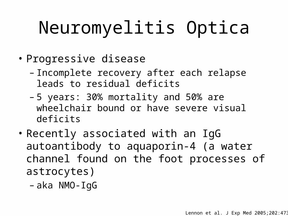
• Progressive disease – Incomplete recovery after each relapse leads to
residual deficits– 5 years: 30% mortality and 50% are wheelchair
bound or have severe visual deficits
• Recently associated with an IgG autoantibody to aquaporin-4 (a water channel found on the foot processes of astrocytes)– aka NMO-IgG
Lennon et al. J Exp Med 2005;202:473-477

NMO Diagnostic Criteria
Wingerchuk et al. Neurology 2006;66:1485-1489

Therapy
• High dose corticosteroids (first line therapy)• TPE is initiated if no improvement or
progression after steroid administration– 5 TPE with 1-1.5 volume replacement using 5%
albumin performed daily or every other day
• More likely to see benefit if TPE is initiated early in the course
Schwartz J et al. J Clin Apher 2013;28:145-284

Guidelines
• AAN: Possibly effective in acute CNS demyelination (includes MS, ADEM, and NMO) if steroid therapy fails
• ASFA: Category II with recommendation grade of 1B in acute NMO– Category III with a recommendation grade of 2C
has been assigned for maintenance therapy
Cortese I et al. Neurology 2011;76:294-300
Schwartz J et al. J Clin Apher 2013;28:145-284
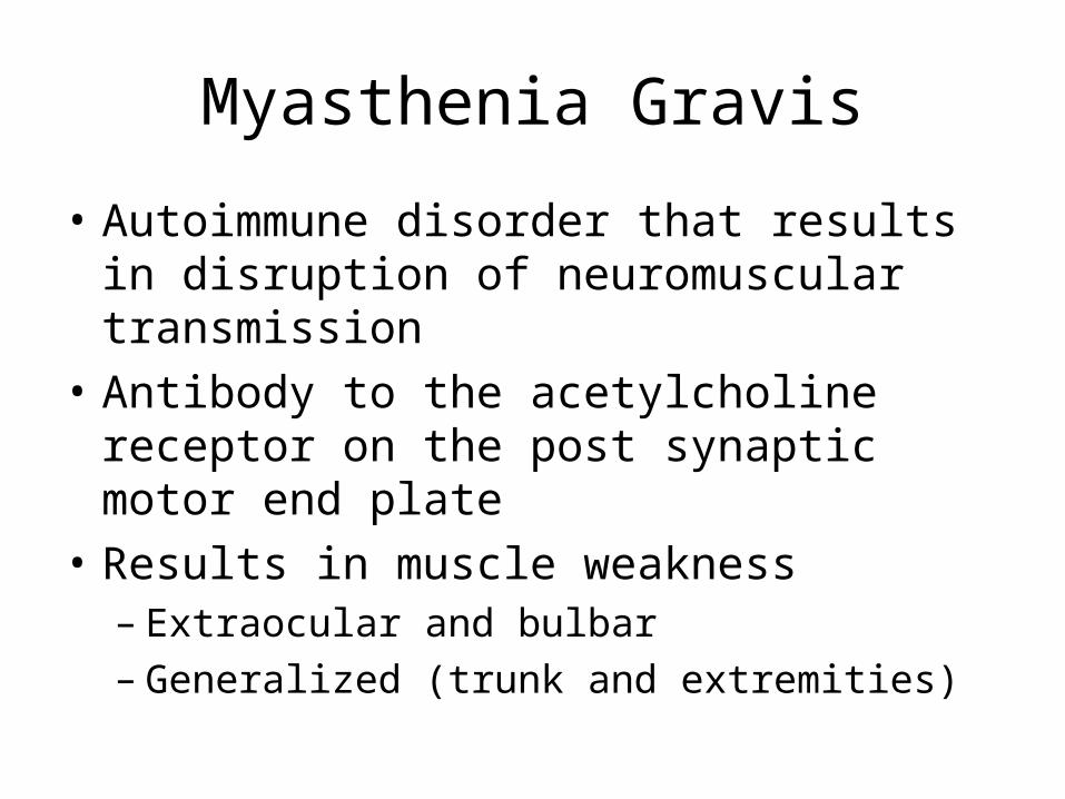
Myasthenia Gravis
• Autoimmune disorder that results in disruption of neuromuscular transmission
• Antibody to the acetylcholine receptor on the post synaptic motor end plate
• Results in muscle weakness– Extraocular and bulbar– Generalized (trunk and extremities)
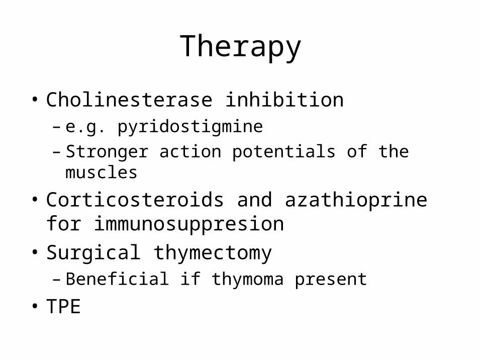
Therapy
• Cholinesterase inhibition– e.g. pyridostigmine– Stronger action potentials of the muscles
• Corticosteroids and azathioprine for immunosuppresion
• Surgical thymectomy– Beneficial if thymoma present
• TPE

TPE
• TPE used in moderate/severe cases or prior to thymectomy– 1-1.5 volumes with replacement using 5% albumin
• The number and frequency of TPE depends on the patient’s disease
• Induction with 225 mL/kg over 1-2 weeks• Long-term maintenance may be required
Schwartz J et al. J Clin Apher 2013;28:145-284

Guidelines
• AAN– “There is insufficient evidence to support or refute
the use of plasmapheresis for myasthenia gravis”
• ASFA: – Moderate-severe disease: Category I with a
recommendation grade of 1B– Prethymectomy: Category I with a
recommendation grade of 1C
Cortese I et al. Neurology 2011;76:294-300
Schwartz J et al. J Clin Apher 2013;28:145-284

Discrepancy between AAN and ASFA
• ASFA reviewed all of the published evidence• AAN did not consider evidence from
observational studies or case reports– “…reexamination of the methodologies may be warranted
due to the failure, in some cases, to account for overwhelming anecdotal evidence or evidence from uncontrolled studies. Thus it may be the case that Class IV evidence should be considered when it is both abundant and consistent, especially when higher standards of evidence cannot feasibly be obtained for either ethical or other reasons”
http://www.neurology.org/content/76/3/294/reply#neurology_el_42491

Natalizumab
• Used to treat relapsing-remitting MS• Monoclonal antibody against α4-integrin
– Entry of WBC into the CNS
• PML is a life threatening complication of natalizumab therapy– Opportunistic CNS infection by the JC virus

Natalizumab Clearance
• Natalizumab has a long half life & may be detectable >12 weeks after the last dose
• TPE has been used to hasten the removal • May provide significant benefit by restoring ability of
WBCs to enter the CNS– TPE decreases the drug levels and T ½ – Shorter time to immune reconstitution– Clinical improvement
Khatri et al. Neurology 2009;72:402-409

Guidelines
• AAN– Initial data suggests TPE may be useful in restoring
leukocyte function by removal of natalizumab– Additional studies are necessary to see whether
this results in any benefit in patients with infectious complications
• ASFA: Category III with a recommendation grade of 2C
Cortese I et al. Neurology 2011;76:294-300
Schwartz J et al. J Clin Apher 2013;28:145-284

Adverse Consequences of TPE
• Immune Reconstitution Inflammatory Syndrome (IRIS)– Inflammatory response – Symptom exacerbation– Enlargement of MS lesions
• IV Corticosteroid therapy may be of benefit
Schroder et al. Arch Neurol 2010;67:1391-1394

Additional Use of TPE in Natalizumab Clearance
• Natalizumab is usually used as monotherapy• Alternate therapy may be necessary if MS
patient is not responsive to natalizumab• Adding a second immunosuppressive therapy
may place patient at increased risk of infection
• Neurologists may request TPE to hasten clearance prior to initiating another drug

Summary
• TPE plays an important role in the treatment of several autoimmune neurological disorders
• Therapy should be based on evidence based review of the literature
• The AAN and ASFA have published evidence based guidelines regarding the use of TA
• Additional studies are necessary to determine the optimal role of TA in several neurological disease conditions

Questions?


