The Roles and Regulation of the Redundant Phenazine Biosynthetic Operons in Pseudomonas aeruginosa PA14 David Alfonso Recinos Submitted in partial fulfillment of the requirements for the degree of Doctor of Philosophy in the Graduate School of Arts and Sciences COLUMBIA UNIVERSITY 2012
Transcript
The Roles and Regulation of the Redundant Phenazine Biosynthetic Operons in Pseudomonas aeruginosa PA14
David Alfonso Recinos
Submitted in partial fulfillment of the requirements for the degree of
Doctor of Philosophy in the Graduate School of Arts and Sciences
Supplementary Figure 7 (S7). SoxR protein expression, soxS promoter binding, and in vitro
transcription of the soxS gene ......................................................................................................142
List of Tables
Table 1. General features of the completed Pseudomonas genomes ...............................................4
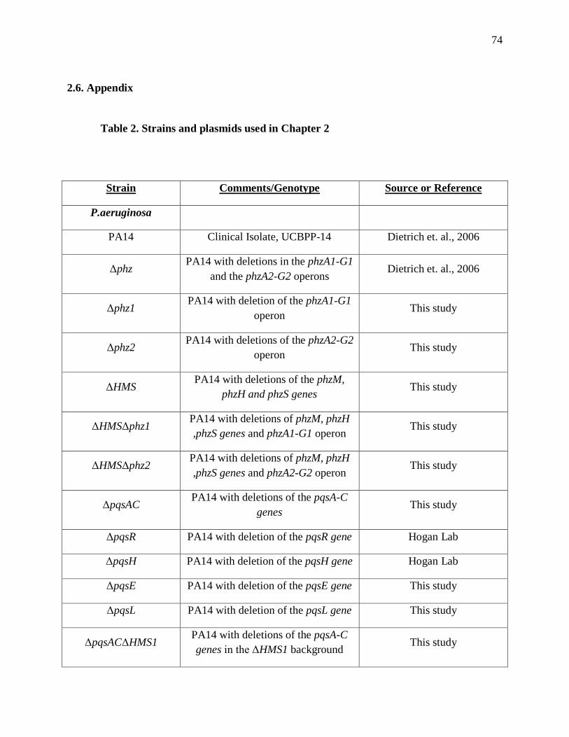
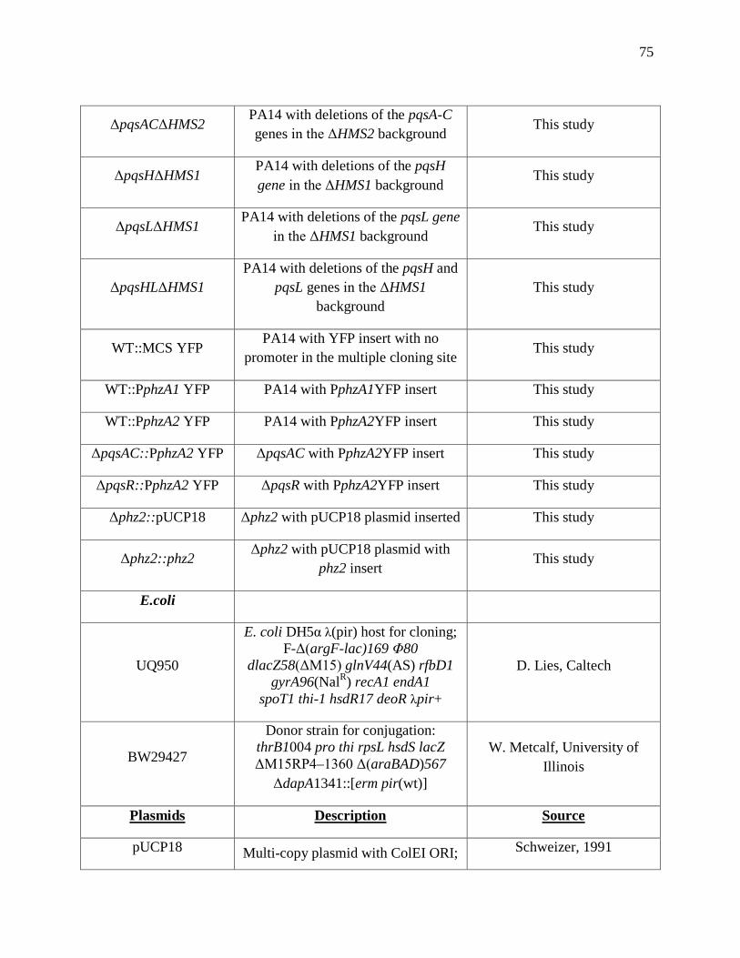
Table 2. Strains and plasmids used in Chapter 2 ...........................................................................74
Table 3. Primers used in Chapter 2 ................................................................................................77
Table 4. Phenazines produced by mutant strains .........................................................................108
Table 5. Strains used in Chapter 3 ...............................................................................................108
Table 6. Primers used in Chapter 3 ..............................................................................................110
ix
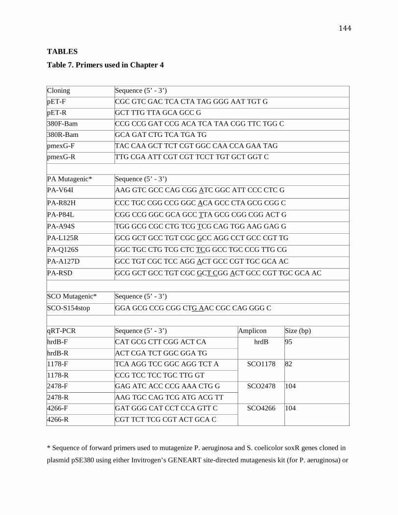
Table 7. Primers used in Chapter 4 ..............................................................................................144
Table 8. Redox drugs used in Chapter 4 ......................................................................................145
Table 9. Bacterial strains and plasmids used in Chapter 4 ..........................................................147
x
Acknowledgements
I would like to thank my advisor Dr. Lars Dietrich for giving me the opportunity to join his lab
and finish my Ph.D. there. These past two years in the lab have proved to be productive and I
have learned a lot. I have become a better scientist in the process. I would also like to thank the
members of the Dietrich lab for their help and support. Starting with the undergrads past and
present: Adriana, Georgia, Leslie, Diya, Ana and Maria. Specifically, I would like to thank
Adriana for her hard work and dedication. She worked with me for the better part of two years
and was a great help in my research. I am also grateful to Ana who helped me finish some of my
experiments while I was writing my thesis. I would like to thank a former post-doc Matthew
Sekedat for being the only other “old man” in the lab. We had many great conversations about
the 80s, 90s, sports and babies (sometimes even in the same conversation!). Oh, and he is also a
pretty good scientist. He was extremely helpful when answering my scientific questions and
always had good suggestions for my project. He also made many of the reporter constructs that
went into my paper. Thanks!
Many thanks to the graduate students in the lab, Hassan Sakhtah and Chinweike “Chinwookie”
Okegbe. Both are already very talented scientists and have helped me a lot in my projects. I
appreciated Hassan’s honesty, inquisitive nature and knowledge of metal music. Thanks to
Chinweike for his support, friendship and for letting me use his computer monitor for 2 years.
You will finally get it back! I will miss sitting behind you and having conversations about life
and science. You are a good labmate and friend.
I would like to thank the members of my thesis committee: Dr. Songtao Jia, Dr. Monica
Chander, Dr. Liz Miller and Dr. Brent Stockwell. Thank you for taking the time to be on my
xi
committee. Special thanks to Dr. Liz Miller for being one of my first committee members and
imparting guidance throughout my graduate career. I appreciated your support and keen insights
into my projects (no matter what lab they were from). Special thanks also to my “surrogate”
advisor Dr. Brent Stockwell. Thank you for letting me use your HPLC machine and for your
advice and help with my project. I appreciate how supportive you have been to both Reka and
myself. Thank you for taking an interest in my project and my career.
Many thanks to the members of the Stockwell lab for answering my questions about chemistry
and helping me find things in their lab. I am grateful for the help of Gisun Park who taught me
how to use the HPLC machine and for her synthesis of phenazines.
Throughout my time in graduate school I have been fortunate to meet and become friends with
many people who have made an impact on my life and career. Many of these people are no
longer in the department (I have been here for a long time!) and may be too numerous to name
here but I will thank some of them. Briefly, thanks to Ben Dubin-Thaler and Adam Meshel who
taught me how to balance lab work and life when I was new to the department. Thanks to all of
the “BioDorks” for befriending me during my first year of graduate school and continuing to be
my friends to this day. I would also like to thank my very good friends Bharat Reddy, Tony
Barsotti, Eric Henckels and Ragan Robertson. They were there for me during the good and bad
times in grad school. I can always count on them for encouragement, support and drinking.
xii
Dedication
I would like to dedicate my thesis to my wonderful wife Dr. Reka R. Recinos (R3). She is my
rock, my inspiration and my life. I met Reka in 2004 when we were both first years in this
department. We started dating several months after we met, fell in love and have been
inseparable ever since. Despite what I may have accomplished in graduate school, I believe my
greatest achievement was to get this intelligent, beautiful, caring woman to talk to me.
She is my staunchest supporter and believes in me more than I believe in myself. She supported
me when I was thinking of leaving graduate school and she was one of the reasons that I decided
to return to graduate school to finish my Ph.D. Without her I would not be writing this. Reka,
you are now responsible for the achievement of two PhDs! I thank you so much for your love
and support.
So in the words of Rocky Balboa, I say this to you Reka: “Yo Reka, I did it!!”
1
CHAPTER 1
1. Introduction and Background
1.1. The pathogenic bacterium Pseudomonas aeruginosa and its genus
Microbiologists have been studying pathogenic bacteria for almost two centuries. Robert Koch
and Louis Pasteur started their investigations into disease-causing bacteria in 1859 1. They
formulated the germ theory of disease, which states that microorganisms are the cause of
diseases such as cholera, tuberculosis, syphilis, and typhoid 2. One of the most studied bacteria in
the context of disease is Pseudomonas aeruginosa. It was first described in 1885 by Carle
Gessard in his paper “On the blue and green coloration that appears in bandages” 3 . He
characterized it as a rod-shaped, aerobic and very motile bacterium that secretes blue-green
pigments. Based on his observations, he named it Bacillus (meaning “rod”) pyocyaneus
(meaning “blue pus”). It has since been renamed as Pseudomonas aeruginosa. The colorful
pigments exuded by P. aeruginosa belong to a class of redox-active molecules known as
phenazines. These compounds have long been known to act as antibiotics4,5 and are required for
full virulence6,7. In recent years we have gained an appreciation for the beneficial roles of
phenazines for the producing organism in redox homeostasis, iron uptake and as signaling
molecules, which may give P. aeruginosa a competitive advantage at the site of infection.
2
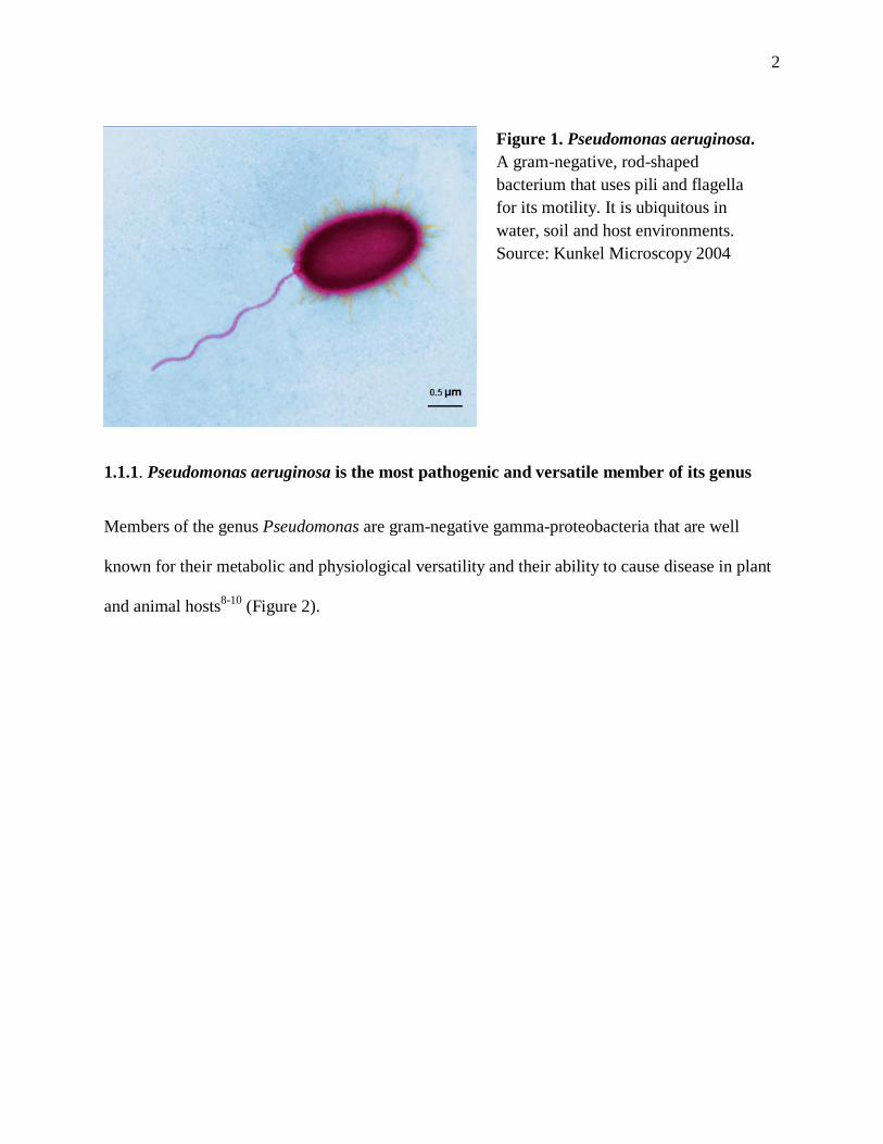
1.1.1. Pseudomonas aeruginosa is the most pathogenic and versatile member of its genus
Members of the genus Pseudomonas are gram-negative gamma-proteobacteria that are well
known for their metabolic and physiological versatility and their ability to cause disease in plant
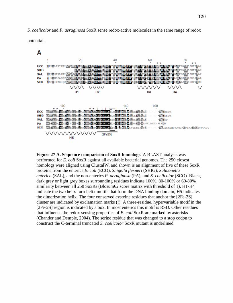
and animal hosts8-10 (Figure 2).
Figure 1. Pseudomonas aeruginosa. A gram-negative, rod-shaped bacterium that uses pili and flagella for its motility. It is ubiquitous in water, soil and host environments. Source: Kunkel Microscopy 2004
3
Currently, the NCBI database lists 18 complete pseudomonad genome sequences and 72 partial
sequences11. The complete genomes are available for strains from the plant and animal pathogen
P. aeruginosa12, the plant pathogens P. syringae13 and P. fluorescens14, as well as P. stutzeri15,
P. putida16 and P. entomophila17 (Table 1).
Figure 2. Pseudomonas aeruginosa is a versatile member of its genus. The pseudomonads inhabit diverse environments. This has led to the evolution of a wide-range of traits, many of which are shared among species. P. aeruginosa is one of the most versatile of the genus as it contains most of the shared traits of the genus. Source: Silby, et al., FEMS Microbiol Rev, 2011
4
The best-studied P. aeruginosa strains are PAO1 and PA14. PAO1 was isolated from a patient’s
wound in Melbourne, Australia in 195418. It became the standard for investigations of P.
aeruginosa’s metabolism and physiology. PA14 was isolated from a burn wound and generally
shows more virulent characteristics compared to PAO119. PA14 is the preferred strain for the
study of P. aeruginosa virulence and pathogenicity. The major virulence-related genomic
differences between PAO1 and PA14 are found in two large pathogenicity islands with PA14
Table 1. General features of the completed Pseudomonas genomes. The Pseudomonas genome is one of the largest in the bacterial domain (~6 Mb). Of note, the smallest genome of the pseudomonads belongs to the non-fluorescent, saprophyte P. stutzeri. Source: Silby, et al., FEMS Microbiol Rev, 2011
5
containing 322 more mobile coding sequences than PAO120. These sequences are grouped into
58 PA14-specific gene clusters, of which about half are of unknown function.
1.1.2. P. aeruginosa is an opportunistic pathogen that adapts to different environments
P. aeruginosa thrives in diverse environments, such as water, air, soil, animal and plant hosts. It
can infect a range of organisms including nematodes21, fruit flies22, waxmoths23, zebrafish24 and
mammals25,26. As an opportunistic pathogen it is capable of causing serious infections in a
variety of tissues and organs, predominantly in immunocompromised patients27. For example, it
has been associated with many hospital-acquired infections including burn wound infections,
chronic lung infections, pneumonia, respiratory tract and even infections of the eye associated
with contact lens use28 29. P. aeruginosa is also the major pathogen contributing to the morbidity
and mortality of patients with the genetic disorder cystic fibrosis (CF)30. One of the hallmarks of
P. aeruginosa infections in CF patients is the colonization of the lungs as sessile, antibiotic-
Transcriptomic and genetic studies revealed the importance of virulence factors in establishing
chronic P. aeruginosa infections. Many virulence genes are located in ‘conserved’ regions of the
genome and are required for the production of rhamnolipids, phenazines, exotoxins, and
proteases32. Mobile DNA elements, or the ‘accessory’ genome, have also been suggested to be
determinants of environmental adaptability in P. aeruginosa33. These include phage and plasmid
elements, genomic islands, transposons and repetitive extragenic palindromic elements11,17,34.
6
Accessory genes have been shown to contribute to increased virulence or competitiveness of
particular strains of P. aeruginosa32.
1.1.3. Physiological changes in response to the host environment
During chronic infections, P. aeruginosa populations change and diversify genetically. The
properties characterizing the bacterial population during the initial infection period (acute) are
different from those in later stages. P. aeruginosa isolates from acute infections are non-mucoid,
motile, and susceptible to antibiotics35. As the infection progresses, changes in colony
morphology, hypermutability, antibiotic resistance and loss of virulence traits manifest
themselves36. In fact, genomic analysis of sequential isolates has suggested that loss of virulence
may be beneficial for the persistence of infection37. However, conflicting studies have shown
that some members of the infecting population maintain their virulence capabilities even after
many years of infection38. The diversity in P. aeruginosa populations within chronic infections is
a striking feature that highlights its versatility in adapting to host environments at the population
level.
Acclimation to the CF lung by P. aeruginosa can also be accelerated by environmental factors
including host immune response, nutrient limitation, oxidative stress and iron availability 39.
Despite investigations into P. aeruginosa gene expression during infection, the molecular basis
for infection is currently unknown. Proteomic analyses have attempted to address infection by
comparing the proteome of AES-1 (an acute, transmissible CF strain) to that of proteomes from
common laboratory strains, such as PAO1 and the more virulent PA14 40. Hare et al. found that
7
of 1700 proteins identified, 183 were significantly altered between the strains. Many of these
proteins are involved in virulence and metabolism but demonstrated different expression patterns
between the strains. This suggests that P. aeruginosa alters its protein expression pattern
depending on its environment.
1.1.4. Environmental effects on P. aeruginosa’s transcriptome: planktonic vs. biofilms
Bacteria can take on dramatically different lifestyles: as free-living cells or as part of
multicellular communities (biofilms). The physical and chemical properties of biofilms
significantly alter gene expression patterns41. Transcriptional studies in PA14 have found key
differences in bacterial cells grown in planktonic cultures compared to cells grown in
biofilms42,43. Genes involved in the type III secretion system (T3SS), adaptation to anaerobic
growth, and production of the extracellular matrix were highly upregulated in biofilms 43. The
T3SS is utilized by many bacterial species to deliver over 100 effector proteins into the host 44.
These effector proteins are often multifunctional proteins that help coordinate bacterial responses
to the host. However, a subset of genes were similarly expressed in stationary phase planktonic
culture and biofilms. These included genes involved in metabolism, translation and motility (pili-
and flagella-mediated motility) and are likely linked to the nutrient depletion and slower growth
rate bacterial cells experience in both stationary phase planktonic cultures and biofilms45.
8
1.1.5. The metabolic versatility of P. aeruginosa: Use of different carbon sources and
electron acceptors
P. aeruginosa can utilize a variety of carbon sources and electron acceptors for energy
generation. This metabolic versatility is another characteristic that allows for its ability to survive
in diverse environmental niches. Unlike E. coli, P. aeruginosa does not use glucose as its
preferred carbon source46. Rather, it consumes organic acids and amino acids prior to
glucose47,48. The sequential metabolism of carbon sources is regulated by catabolite repression,
allowing for the utilization of preferred substrates in an ordered fashion49,50. Once preferred
substrates are depleted, sugars are degraded through the Entner-Doudoroff pathway instead of
Embden-Meyerhof glycolysis as P. aeruginosa lacks a key enzyme required for the latter39 51.
Figure 3. Denitrification in P. aeruginosa. P. aeruginosa is a denitrifying bacterium that can use nitrate as an electron acceptor to carry out anaerobic respiration. This is catalyzed by four enzyme complexes: nitrate reductase (NAR), nitrite reductase (NIR), nitric oxide reductase (NOR) and nitrous oxide reductase (N2OR). Source: Williams et al., Adv. Micro. Phys., 2007
9
Energy production in P. aeruginosa is mainly based on aerobic respiration. Its metabolic
versatility is highlighted by its ability to use a variety of electron acceptors. In low oxygen
environments, it can flourish by using the alternative external electron acceptors nitrate and
nitrite in a multi-step process called denitrification52 (Figure 3). The membrane-bound enzyme
NAR reduces nitrate to nitrite, which is further reduced to nitrite by NIR. Both reduction steps
are coupled to the generation of a proton-motive force53. The metabolic differences between P.
aeruginosa and other bacteria highlight different strategies to compete in various environments.
P. aeruginosa can thrive in any soil and host environments where it can take advantage of the
flux of organic acids, amino acids, sugars and nitrogenated bases.
1.2. Bacterial Communication
Members of all three domains of life use various modes of intercellular communication.
Historically, research into cell-cell signaling has centered on eukaryotes. However, discoveries
over the past 30 years have demonstrated that bacteria have an arsenal of signals that rival the
most complex eukaryotes. It is now known that bacteria engage in cooperative and social
behavior in order to perform a wide range of activities and developmental processes54. This
research has revealed a previously unimagined complexity of bacterial communication that
opens the door for further exploration into this exciting new realm.
Studies of several bacterial species known to form multicellular communities have found that
they are capable of concerted actions and use extracellular signals for cell-cell communication. It
is also clear that these extracellular signals can not only be detected by other bacterial cells, but
10
that the receiving cells can respond to these signals in a variety of ways. One such response is
referred to as “quorum sensing” (QS), which involves the regulation of gene expression
dependent on cell proximity and density55. There are many signaling pathways regulated by QS
and these have a myriad of different functions within the cell and the bacterial community. These
collections of signals within the community are used in a coordinated manner to benefit the
population as a whole and imbue them with characteristics for survival. This is evident in chronic
infections of mucoid bacterial communities and their resistance to antibiotics56. The QS system
allows bacterial communities as a whole to respond to extracellular signals and is a prominent
feature of bacterial survival mechanisms.
1.2.1. Quorum sensing in P. aeruginosa
Quorum sensing (QS) signaling is the best-studied communication system in bacteria. It is a
complex and extensive array of molecules that can detect and react to endogenous and
environmental signals. These signals trigger a response characterized by massive changes in
gene expression57. This happens in a cell-dependent manner as gene expression is only triggered
at a certain threshold concentration55. One of the first models used to study QS was the
luminescent bacterium Vibrio fischeri58. Genes involved in the regulation of light production
which activate the transcriptional regulator LuxR. (Figure 4). This QS system is conserved
across gram-negative bacteria.
11
1.2.1.1. The Las/Rhl system
In P. aeruginosa the two Lux-homologs LasI and RhlI catalyze the production of the N-
homoserine lactones (HSL) 3-oxo-C12-HSL and 3-oxo-C4-HSL, respectively59,60. These HSLs
activate two transcriptional regulators, LasR and RhlR, which bind to specific binding sites,
“lux-boxes”, in the promoter regions of their target genes61-63. The LasR and RhlR regulons show
significant overlap, both regulating dozens of virulence genes such as the ones responsible for
the production of rhamnolipids, elastases, exotoxins and proteases64-66.
1.2.1.2. Quinolones as signaling molecules
P. aeruginosa’s QS system is extended by another class of compounds, the alkyl quinolones
(AQ). P. aeruginosa produces over 50 AQs67,68 which vary in the lengths of their saturated or
unsaturated alkyl side chains. The main AQs produced by P. aeruginosa are the Pseudomonas
Figure 4. Simplified model of bacterial quorum sensing (QS). The QS system allows for a large-scale response to environmental factors. Bacteria exude signaling molecules that alter gene expression in a growth-dependent manner. Source: Bassler et al., Curr. Opin. Bio., 2000
12
quinolone signal (PQS), its precursor 2-heptyl-4-quinolone (HHQ), and N-oxide 2-heptyl-4-
hydroxyquinoline-N-oxide (HQNO) (Figure 5). Despite the great number of AQs produced, their
synthesis and transport are closely regulated69-71. PQS and its precursor HHQ are the best-studied
AQs. Together they control the production of many virulence factors produced by P. aeruginosa
including phenazines72,73.
Quinolones are the only known QS signals that are not members of the acylated-HSL family.
The quinolone HHQ is synthesized by the enzymes encoded within the five gene operon
pqsABCDE74. HHQ is then converted to PQS by the distally located monooxygenase PqsH
(Figure 6). PQS production starts in late exponential phase, reaching its maximum during early
stationary phase, and decreases subsequently75. The presence of PQS-producing P. aeruginosa
strains in the lungs of cystic fibrosis patients suggests PQS is important for infection76. The PQS
receptor PqsR (also known as MvfR, for multiple virulence factor regulator), is a membrane-
associated protein that induces synthesis of elastase, phospholipase, 3-oxo-C12-HSL, and
Figure 5. Structures of some common quorum sensing signals in P. aeruginosa. The three most abundant quinolones are the Pseudomonas quinolone signal (PQS), 2-heptyl-4-quinolone (HHQ) and N-oxide 2-heptyl-4-hydroxyquinoline-N-oxide (HQNO). C4-HSL and 3-oxo-C12-HSL are products of the Rhl and Las systems, respectively. Source: Williams et al., Curr. Opin. Micro., 2009
13
phenazines77-79. Like PQS production, pqsR expression reaches maximum levels at late
exponential phase. Although PqsR’s relevance in the pathogenesis of P. aeruginosa is well-
established, its precise binding motif and complete transcriptome have yet to be elucidated.
Figure 6. Synthesis, regulation and autoinduction of the quinolone signaling system in P. aeruginosa. Anthranilate is the substrate for quinolone biosynthesis. The quinolone PQS binds the transcription factor PqsR for its own autoinduction as well as the control of several virulence genes. PqsE is known as the “PQS response” protein, as it is needed for many PQS-dependent downstream effects. Its exact mechanism of action is unknown. Source: Jimenez et al., Micro. Mol. Bio. Rev., 2012.
14
1.2.2. The formation of multicellular communities
Most bacteria are able to aggregate into multicellular communities (biofilms)80-82. The formation
of biofilms is an active, concerted process that involves the coordinated action of billions of
bacterial cells. This mode of growth is a strategy that is employed by bacteria in response to
challenging environmental stimuli such as nutrient depletion and protects them from antibiotics,
detergents, and other potentially harmful foreign molecules83, allowing them to thrive in hostile
environments. Although the mechanisms that govern biofilm formation can differ between
species and even between strains of the same species, it seems to be an adaptation common to
most bacterial species84.
Biofilms can form on any type of surface (even on an air-liquid interface) in a wide variety of
environments. Of medical concern are biofilms that form in the host (in lungs, wounds, skin,
teeth, and the urinary tracts)25,85,86 or on equipment, such as catheters, medical implants and
inside water pipes84,87. This form of adaptation seems to be an evolutionarily conserved process
to insure species survival in environments rife with competing organisms. However, in nature,
biofilms tend not to consist of just one species but represent communities between multiple
microbial species. In some instances, biofilms can be beneficial to their eukaryotic hosts:
biofilms of Actinobacteria on the backs of ants provide protection from fungal and protozoan
pathogens88,89, while P. chlororaphis biofilms on roots protect plants from invaders90.
Biofilms are architecturally and chemically complex structures. They are composed of a matrix
made up of polysaccharides, proteins, and extracellular DNA91 that harbors a metabolically
heterogeneous population of cells. These give rise to chemical gradients across the biofilms92
consisting of metabolic products and signaling molecules that create unique environmental
15
niches92. The best-studied gradient is that of oxygen, characterized by high levels at the top of
the biofilm (which is exposed to oxygen) and low concentrations at the bottom of the biofilm
(where no oxygen can penetrate)93. The oxygen gradient in turn affects gene expression,
metabolism and redox balancing94.
In P. aeruginosa, the extracellular polymeric substance (EPS) is made up of three main
polysaccharides: alginate, Psl and Pel. Alginate is associated with a subset of P. aeruginosa
variants that form mucoid colonies on agar plates38. It is a high molecular weight acetylated
polymer composed of non-repetitive monomers of L-glucoronic and D-mannuronic acids.
Alginate production confers a selective advantage within the CF lung95, protecting P. aeruginosa
from the consequences of inflammation and phagocytic clearance. However, despite its
protective roles, alginate is not necessary for biofilm formation96. The common laboratory strains
PAO1 and PA14 do not produce much alginate, relying on PSL and PEL for matrix
construction97. PSL is necessary for biofilm formation in PAO1 and is also involved in cell-
surface and cell-cell interactions98. It is composed mainly of mannose and galactose but its
structure has yet to be solved99. In PA14, PEL is the main contributor to biofilm development
and morphology. This may be in part because PA14 has a partial deletion in the psl gene locus
100. The structure of PEL forms a glucose-rich polysaccharide polymer but its exact structure
remains to be elucidated.
Two main laboratory models are used for the study of P. aeruginosa biofilms. The most
prominent is the “flow cell” biofilm model: Nutritious medium with a low bacterial inoculum is
streamed over the surface of a glass slide. Individual cells attach to the slide, multiply and
eventually form a structured biofilm101. Biofilm development can be monitored by fluorescence
16
microscopy. The flow-cell model mimics environmental conditions found in aquatic
environments. Another model for the study of biofilms is the colony biofilm assay. This assay
involves spotting 10 µl of high-density cell suspension onto an agar plate. Once spotted, it is
possible to follow the development of the community of cells over time. This technique is ideal
for studying stages of biofilm development following initial attachment. The macroscopic
colonies are particularly amenable for high-throughput screens.
1.3. Phenazines
Phenazines are redox-active, heterocyclic compounds produced by several bacterial species.
Their discovery dates back to the late 19th century when doctors noticed blue-tinted pus secreted
from purulent wounds in patients102. They were able to isolate a blue compound, “pyocyanin”
that belongs to the class of phenazines. Additional phenazines were subsequently identified from
culture supernatants as well as chronic P. aeruginosa infections5. Phenazine are characterized by
a heterocyclic three-ring core that can be decorated with different functional groups, which
change the chemical properties of phenazines (redox activity, solubility, color) (Figure 7). The
colors range from the blue of pyocyanin (PYO), the lemon yellow of phenazine-1-carboxylic
acid (PCA), the orange hue of 1-hydroxyphenazine (1-OH-PHZ), to the green tint of phenazine-
1-carboxamide (Figure 8). P. aeruginosa contains a pair of redundant seven-gene operons (phzA-
G) that encode the enzymes responsible for the biosynthesis of the phenazine PCA from
chorismate102,103 (Figure 8). The core phenazine operons are often found next to phenazine-
modifying enzymes and other regulatory genes104,105. In P. aeruginosa, the phzA1-G1 phenazine
operon (phz1) is flanked by the methyl-transferase encoding gene phzM and the monooxygenase
17
gene phzS. PhzM and PhzS convert PCA to the blue phenazine pyocyanin (PYO) (Figure 8),
which is unique to P. aeruginosa106.
Researchers and clinicians alike have delved into the physiological effects of phenazines. They
found that phenazines are required for P. aeruginosa’s virulence and competitiveness, which is
mainly due to its superoxide-generating redox activity 107.
Phenazines were originally viewed as secondary metabolites that assert their deleterious effect on
other organisms via their ability to transfer electrons to oxygen. While phenazines have been
observed within other bacterial species and some archaea, most of the work on the physiological
role of phenazines has been done in the context of pseudomonad infections. Increased phenazine
concentrations within the lung, such as during chronic P. aeruginosa infection of a CF patient,
Figure 7. Some characteristics of phenazines produced by P. aeruginosa. The functional groups and the redox potentials at pH 7 are shown. Source: Price-Whelan et al., Nat. Chem. Bio., 2006
18
can impair epithelial cell function while also attenuating immunological responses108. PYO
reacts with oxygen to form superoxide radicals that can severely disrupt the host cells’ internal
redox balance 109. These reactive oxygen species can also act as antibiotics towards other
microbes competing for resources in human hosts, as well as in soil ecosystems. For example, P.
aeruginosa biofilms that form around the roots of plants can protect the plant from pathogenic
fungi via phenazine secretion. A large body of work has established the role of phenazines in
physiological effects on hosts and ecological competition during P.aeruginosa infections.
However, research within the last decade has elucidated a new role for phenazines as signaling
molecules that can affect gene expression.
19
1.3.1. Distribution of the phenazine operon across the bacterial domain
Phylogenetic analyses revealed that the phenazine operon (phzA-G) is highly conserved among
phenazine-producing bacteria, such as Gram-positive actinobacteria and Gram-negative beta-
and gamma-proteobacteria105 (Figure 9). Mavrodi et al. have suggested that the transfer of the
phz operon may have occurred via horizontal gene transfer in certain lineages, as the operon is
Figure 8. P. aeruginosa produces a variety of phenazines with colorful properties. The gene products encoded within the redundant 7-gene operons convert chorismate to phenazine-1-carboxilic acid (PCA). PCA can then be converted to several phenazines including pyocyanin (PYO), phenazine-1-carboxamide (PCN), and 1-hydroxyphenazine (1-OH-PHZ). These phenazines have different biochemical properties. Source: Adapted from Price-Whelan et al., Nat. Chem. Bio., 2006
20
found in diverse species, such as Streptomyces spp. (actinobacteria) and Pseudomonas spp.
(gamma-proteobacteria)110,111. The strongest evidence for horizontal gene transfer is found in
Burkholderia species where the phz operon is surrounded by conserved transposon elements105.
Additionally, in Burkholderia the phz operon has an unusually high degree of sequence
conservation and it is inconsistently distributed within the genomes. Transfer of the phz operon
between species that occupy diverse environments highlights the importance of this biosynthetic
pathway.
Figure 9. Distribution of phenazine producers based on phzF phylogeny analysis. Classification of bacterial species based on 16S sequencing (A) and phzF phylogeny. (B) Phenazine producing species were classified in three major clades that agree with 16S phylogeny of the taxa analyzed. Source: Mavrodi et al., App. Env. Micro., 2010
21
A large portion of phenazine producers are soil-dwellers and part of the rhizosphere11,112.
Amongst those, only P. aeruginosa and S. cinnamonensis contain two redundant phz operons113
(Figure 10). The fact that P. aeruginosa can thrive in both soil and host environments, and
contains a redundant set of phz operons may be of importance. Does having redundant phz
operons give P. aeruginosa an advantage in certain environments? Examination of the location
of the operons and their flanking regions may begin to answer this question. The phzA2-G2
operon (phz2) is found approximately 2 MB away from the phz1 operon and does not have any
phenazine-modifying enzymes flanking it. In addition, although the phz1 and phz2 operons are
nearly identical (~98%), they contain distinct regulatory elements114. The differences between
the flanking regions of phz1 and phz2 may point to different characteristics of each operon that
were first present at the time of the duplication event. While the regulation of phenazine
production through the phz1 operon has been investigated thoroughly6,115,116 the specific
regulation of the phz2 operon remains to be elucidated.
22
1.3.2. Some thoughts on genetic redundancy
Evolutionarily, the perpetuation of functionally redundant genes within a genome may at first
seem paradoxical. In theory, these genes should be selected against over time since at first glance
they provide no obvious beneficial advantage for the organism. However, investigations into
redundant genes in various organisms have found that there is a synergy between redundant
genes that may be beneficial to the organism 117. Genetic diversity through gene duplication
leads to organism-specific phenotypes and adaptive characteristics. The existence of multiple
gene copies in eukaryotes has been known for a long time and is considered an important
Figure 10. Organization of the phenazine biosynthetic operons in several bacterial species. A comparison of sequences of the phenazine operons across diverse bacteria. (A) The phenazine biosynthetic genes in the Burkholderia species. (B) In this species, they are surrounded by several mobile transposon elements. Source: Mavrodi et al., App. Env. Micro., 2010
23
element in their molecular evolution118,119. However, bacteria were considered to be “simple”
and were thought to carry very little, if any, redundant information in their genomes. It was
surprising when the genome of Escherichia coli K12 showed that nearly 30% of the coding
sequences could be grouped into gene families that were similar enough to be assigned similar
functions120. They were described as 'paralog' gene families, and it was thought that their
similarity reflected similar evolutionary descent, but actual or potential functional divergence.
Since then, the presence of gene families typically containing between two to thirty copies has
been described for nearly every prokaryotic genome sequenced. The number of paralogous genes
and families appears to correlate with an increase in genome size118,121.
Many redundant gene products are found in crucial cellular processes such as signal
transduction, development and metabolism122. Examples of genetic redundancy include the
myogenic development regulators of mammals123, cell surface receptors in Caenorhabditis
elegans124 and Ser/Thr kinases in Saccharomyces cerevisiae125. The function of genetic
redundancy within signaling networks has been studied thoroughly. In S. cerevisae, inspection of
the 239 redundant genes reveals that 29% of these are found in signaling networks125-127. The
redundant genes within signaling networks are also found to be differentially regulated compared
to redundant genes in other contexts125-128. Differential regulation of redundant genes may
facilitate signal transduction and modulate gene expression through collaboration between genes.
It is this collaboration between redundant genes that may help propagate specific responses to
numerous and diverse environmental stimuli.
24
1.3.3. Quinolone-dependent regulation of phenazine production
Quinolones, specifically PQS, have been shown to be necessary for the production of
phenazines23,129 (Figure 11). This is thought to require only one of the redundant phenazine
operons, phz1. Expression of the phz1 operon has been shown to be dependent upon the
quinolone signaling network, as deletions of genes encoding quinolone biosynthetic enzymes
(pqsA) or the PQS receptor (pqsR) correlate with reduced expression of the phz1 operon130,131.
However, a PqsR binding motif has not been identified upstream of phz1.
Interestingly, in P. aeruginosa strain PA14, deletion of the pqsA gene does not completely
abolish PYO production78,132. Furthermore, deletion of pqsH, which is specifically required for
the production of the quinolone PQS, only reduces PYO production by ~20%133. This is in
Figure 11. Model of P. aeruginosa’s quorum sensing network. PQS is produced in late exponential phase and regulates the production of phenazines. Source: Price-Whelan et al., Nat. Chem. Bio, 2006
25
contrast to investigations in strain PAO1 that found that a lack of PQS production reduces PYO
levels by ~90%130. The reason for these strain-specific differences remains to be elucidated.
1.3.4. Functions of phenazines
1.3.4.1. Phenazines as signaling molecules
Recent work has increased our understanding of the cell-cell signaling cascades present in P.
aeruginosa. Work from the Newman lab has established that phenazines extend the QS signaling
network in P. aeruginosa134 and regulate a specific set of genes. Subsequent studies showed that
phenazines modulate the maturation of colony biofilms135. Specifically, PYO and PCA altered
colony formation and structure with different potencies136, demonstrating that individual
phenazines contribute differently to biofilm development. These initial investigations into the
role of phenazines as signaling molecules laid the groundwork for further research into this
exciting topic.
An intriguing aspect of phenazine signaling is that it can activate the redox sensor SoxR, a
transcriptional activator that contains an iron sulfur cluster in its sensory domain. Redox active
agents, such as phenazines, are molecules that are easily reduced and re-oxidized under
physiological conditions. These molecules are secreted by bacteria, fungi and plants and can
impair cell function by the generation of reactive oxygen species7,137. SoxR, along with another
transcriptional activator, SoxS, is part of the oxidative stress response in enterobacteria, such as
E. coli and Salmonella enterica138,139. Together, these transcription factors regulate the
expression of over one hundred genes involved in the suppression of oxidative stress. However,
26
recent work indicates that the SoxR response pathway has different functions in non-enteric
bacteria140.
It had long been assumed that superoxide stress was the sole activator of SoxR, and that the
deleterious effect of redox-cycling agents was mediated through the creation of these toxic
species. However, Gu and Imlay found that the SoxR response could be activated by redox
active compounds and did not necessarily require superoxide141. Using the natural redox-active
antibiotic paraquat, they showed that SoxR could be activated under anaerobobic conditions,
suggesting that SoxR is sensing redox-cycling agents directly instead of superoxide. In support
of this hypothesis, studies on Streptomyces coelicolor and P. aeruginosa have found that the
endogenous redox-active compounds actinorhodin and phenazines elicit a SoxR-mediated
response135,142. Actinorhodin is a polyketide endogenously produced by S. coelicolor that induces
the expression of several SoxR target genes143. Similarly, phenazines produced by P. aeruginosa
activate several genes under the control of SoxR135. Interestingly, none of these genes are
involved in mediating a general stress response to superoxides. Rather, the gene products may
function in the export or modification of actinorhodin or phenazines in S. coelicolor or P.
aeruginosa, respectively (Figure 12).
27
A closer look at the SoxR regulon in non-enteric bacteria, such as P. aeruginosa and S.
coelicolor, reveals that it is very different than the SoxR regulon of enteric bacteria, such as E.
coli. The SoxR regulon from enteric bacteria is composed of only the transcription factor SoxS,
which regulates the expression of more than 100 genes gene, many of which are involved in a
general stress response to superoxide (e.g. superoxide dismutase)144,145. The SoxR regulon in
non-enterics differs in both number and types of genes affected. In P. aeruginosa, SoxR controls
the expression of a Resistance-Nodulation-cell Division (RND) family efflux pump MexGHI-
OpmD, a Major Facilitator Family (MFS) transporter, and a putative monooxygenase135. In S.
coelicolor, SoxR is responsible for genes encoding putative reductases, a monooxygenase and an
ABC transporter143. The differing regulons of P. aeruginosa and S. coelicolor indicate that the
SoxR response may be specific to endogenously produced redox-active signals. In support of
this, the growth of SoxR mutants in both of these non-enteric bacteria is unaffected by
Figure 12. Activation of SoxR-dependent gene expression by pyocyanin (PYO). The transcription factor SoxR forms a homodimer that binds to the “SoxRbox” of target genes. In its reduced form, SoxR prevents transcription. PYO then oxidizes SoxR through a one electron transfer. This causes the homodimer to undergo a conformational change that allows DNA polymerase to bind the DNA, resulting in transcription of the target genes.
28
endogenous or exogenous redox-active compounds suggesting that SoxR is not part of the
detoxification response in these organisms.
1.3.4.2. The role of phenazines in iron reduction
Iron is an essential element that is required for crucial metabolic processes such as respiration
(ferredoxins, cytochromes) and key enzymatic reactions (fumirase and aconitase of the TCA
cycle)146. However, under aerobic conditions iron is not readily usable, as it is commonly found
in the poorly soluble form Fe3+147,148. As such, host defense systems employ a series of
mechanisms to limit iron availability for the invading pathogens. These mechanisms include
proteins that use iron such as hemoglobins, cytochromes and ferritins or chelators of extracellular
iron such as glycoproteins, transferrins and lactoferrins149,150. The phenazine PYO may assist P.
aeruginosa in the acquisition of iron by reducing it and freeing it from transferrin, a protein that
normally sequesters iron so that it is available only to the human host151,152. Another
pseudomonad strain, P. chlororaphis, has been shown to reduce iron oxides through electron
transfer to the phenazine PCN, and it is thought that this ability may be important in the
rhizosphere, where iron is also present predominantly in an insoluble, oxidized form153. In
addition to phenazines, P. aeruginosa also uses strong extracellular iron chelators, termed
siderophores, for iron uptake154. Interestingly, transcriptomic studies of P. aeruginosa have
found that the biosynthetic genes that control phenazine and siderophore production are
upregulated during infection111.
29
1.3.4.3. The role of phenazines in redox homeostasis
Phenazines also act as substrates in intracellular redox transformations. The redox
transformations of phenazines can be observed as a color change in cultures that have become
limited for terminal electron acceptors. The phenazine PYO is blue in its oxidized state, but
colorless upon reduction. A shaking culture is blue because oxygen is continuously introduced
into the medium. If the culture is limited for oxygen the cells will rapidly reduce all phenazines
and the culture will lose its blue color155. This activity has also been demonstrated in oxygen-
limited cultures of the bacterium P. chlororaphis, which can use its phenazine product,
phenazine-1-carboxamide (PCN) to reduce extracellular iron oxides 153.
The redox potentials of phenazines are such that they can be easily reduced by the bacterial cell
and react extracellularly with higher potential oxidants such as ferric iron and oxygen, acting as
electron shuttles between the bacterium and an external substrate156,157. In homogeneous liquid
cultures of P. aeruginosa, phenazines affect gene expression and oxidize the intracellular redox
state158-160. Under conditions where no other oxidant is available, phenazine-dependent electron
transfer between cells and an oxidizing electrode supports survival9,161. Phenazines also help
maintain redox homeostasis by acting as electron acceptors for the re-oxidation of accumulating
NADH. Maintaining a proper redox balance in the pyridine nucleotide pool is essential for
metabolism162. This suggests that an important role for phenazines could be to serve as
intracellular redox buffers.
The building of cellular communities such as biofilms leads to the creation of gradients due to
limited diffusion and consumption of substrates by individual cells within a community. Cells
within biofilms use different strategies to ensure substrate acquisition and survival, depending on
30
the specific microenvironment they inhabit. Mechanisms that aid in redox homeostasis at the
cellular level have been characterized in diverse organisms. In mammals, redox-balancing
mechanisms are involved in the development of lung and blood vessel systems, which prevent
oxygen starvation of the developing embryo 163. In such large, multicellular species, cells must
cope with limited oxygen availability that leads to the formation of zones with varying
concentrations of oxygen. During processes such as tumor angiogenesis, relative oxygen
concentrations act as cues that determine adaptive morphological features, facilitating oxygen
delivery to cells within the macroscopic structure94.
In summary, Pseudomonas aeruginosa is a versatile bacterium that can inhabit diverse
environments such as water, air, soil and host organisms. Phenazine production and formation of
multi-cellular communities are two important aspects of its physiology that help this bacterium
adapt to different environments. How phenazines modulate biofilm development is poorly
understood..The second chapter will address how phenazine production is affected in biofilms.
More specifically, we addressed the role of the second phenazine operon in phenazine production
in the biofilm environment. In the third chapter, we investigated the roles of individual
phenazines on colony development. It has been established that phenazines are necessary for
colony development but exactly which phenazines are involved in this process has yet to be
elucidated. Finally, in chapter four we investigated the activation of the transcription factor SoxR
by phenazines. Specifically, we addressed the ability of SoxR to respond to specific redox
potentials. The work described below is aimed at furthering our understanding of the intimate
link between phenazines and biofilm development.
31
1.4. References
1. Koch, R. A Further Communication on a Remedy for Tuberculosis. British medical journal 1, 125-127 (1891).
2. Disease-Germs. Science 5, 158-159 (1885). 3. Gessard, C. On the Blue and Green Coloration That Appears on Bandages. Reviews of
infectious diseases 6, S775-S776 (1984). 4. Malik, V.S. Regulation of chorismate-derived antibiotic production. Advances in applied
microbiology 25, 75-93 (1979). 5. Leisinger, T. & Margraff, R. Secondary metabolites of the fluorescent pseudomonads.
Microbiological reviews 43, 422-442 (1979). 6. Liang, H., Duan, J., Sibley, C.D., Surette, M.G. & Duan, K. Identification of mutants
with altered phenazine production in Pseudomonas aeruginosa. Journal of medical microbiology 60, 22-34 (2011).
7. Look, D.C., et al. Pyocyanin and its precursor phenazine-1-carboxylic acid increase IL-8 and intercellular adhesion molecule-1 expression in human airway epithelial cells by oxidant-dependent mechanisms. Journal of immunology 175, 4017-4023 (2005).
8. D'Aes, J., et al. Biological control of Rhizoctonia root rot on bean by phenazine- and cyclic lipopeptide-producing Pseudomonas CMR12a. Phytopathology 101, 996-1004 (2011).
9. Pierson, L.S., 3rd & Pierson, E.A. Metabolism and function of phenazines in bacteria: impacts on the behavior of bacteria in the environment and biotechnological processes. Applied microbiology and biotechnology 86, 1659-1670 (2010).
10. Rahme, L.G., et al. Plants and animals share functionally common bacterial virulence factors. Proceedings of the National Academy of Sciences of the United States of America 97, 8815-8821 (2000).
12. Stover, C.K., et al. Complete genome sequence of Pseudomonas aeruginosa PAO1, an opportunistic pathogen. Nature 406, 959-964 (2000).
13. Buell, C.R., et al. The complete genome sequence of the Arabidopsis and tomato pathogen Pseudomonas syringae pv. tomato DC3000. Proceedings of the National Academy of Sciences of the United States of America 100, 10181-10186 (2003).
14. Paulsen, I.T., et al. Complete genome sequence of the plant commensal Pseudomonas fluorescens Pf-5. Nature biotechnology 23, 873-878 (2005).
15. Yan, Y., et al. Nitrogen fixation island and rhizosphere competence traits in the genome of root-associated Pseudomonas stutzeri A1501. Proceedings of the National Academy of Sciences of the United States of America 105, 7564-7569 (2008).
16. Nelson, K.E., et al. Complete genome sequence and comparative analysis of the metabolically versatile Pseudomonas putida KT2440. Environmental microbiology 4, 799-808 (2002).
17. Vodovar, N., et al. Complete genome sequence of the entomopathogenic and metabolically versatile soil bacterium Pseudomonas entomophila. Nature biotechnology 24, 673-679 (2006).
32
18. Holloway, B.W. Genetic recombination in Pseudomonas aeruginosa. Journal of general microbiology 13, 572-581 (1955).
19. Mikkelsen, H., McMullan, R. & Filloux, A. The Pseudomonas aeruginosa reference strain PA14 displays increased virulence due to a mutation in ladS. PloS one 6, e29113 (2011).
20. He, J., et al. The broad host range pathogen Pseudomonas aeruginosa strain PA14 carries two pathogenicity islands harboring plant and animal virulence genes. Proceedings of the National Academy of Sciences of the United States of America 101, 2530-2535 (2004).
21. Tan, M.W., Mahajan-Miklos, S. & Ausubel, F.M. Killing of Caenorhabditis elegans by Pseudomonas aeruginosa used to model mammalian bacterial pathogenesis. Proceedings of the National Academy of Sciences of the United States of America 96, 715-720 (1999).
22. Apidianakis, Y. & Rahme, L.G. Drosophila melanogaster as a model for human intestinal infection and pathology. Disease models & mechanisms 4, 21-30 (2011).
23. Lee, D.G., et al. Genomic analysis reveals that Pseudomonas aeruginosa virulence is combinatorial. Genome biology 7, R90 (2006).
24. Clatworthy, A.E., et al. Pseudomonas aeruginosa infection of zebrafish involves both host and pathogen determinants. Infection and immunity 77, 1293-1303 (2009).
25. Williams, B.J., Dehnbostel, J. & Blackwell, T.S. Pseudomonas aeruginosa: host defence in lung diseases. Respirology 15, 1037-1056 (2010).
26. Harji, D.P., Rastall, S., Catchpole, C., Bright-Thomas, R. & Thrush, S. Pseudomonal breast infection. Annals of the Royal College of Surgeons of England 92, W20-22 (2010).
27. Talbot, G.H., et al. Bad bugs need drugs: an update on the development pipeline from the Antimicrobial Availability Task Force of the Infectious Diseases Society of America. Clinical infectious diseases : an official publication of the Infectious Diseases Society of America 42, 657-668 (2006).
28. Wainwright, C.E., et al. Safety of bronchoalveolar lavage in young children with cystic fibrosis. Pediatric pulmonology 43, 965-972 (2008).
29. Lyczak, J.B., Cannon, C.L. & Pier, G.B. Establishment of Pseudomonas aeruginosa infection: lessons from a versatile opportunist. Microbes and infection / Institut Pasteur 2, 1051-1060 (2000).
30. Govan, J.R. & Deretic, V. Microbial pathogenesis in cystic fibrosis: mucoid Pseudomonas aeruginosa and Burkholderia cepacia. Microbiological reviews 60, 539-574 (1996).
31. Costerton, J.W., Stewart, P.S. & Greenberg, E.P. Bacterial biofilms: a common cause of persistent infections. Science 284, 1318-1322 (1999).
32. Mathee, K., et al. Dynamics of Pseudomonas aeruginosa genome evolution. Proceedings of the National Academy of Sciences of the United States of America 105, 3100-3105 (2008).
33. Winstanley, C., et al. Newly introduced genomic prophage islands are critical determinants of in vivo competitiveness in the Liverpool Epidemic Strain of Pseudomonas aeruginosa. Genome research 19, 12-23 (2009).
34. Tobes, R. & Pareja, E. Bacterial repetitive extragenic palindromic sequences are DNA targets for Insertion Sequence elements. BMC genomics 7, 62 (2006).
35. Hoiby, N., et al. The clinical impact of bacterial biofilms. International journal of oral science 3, 55-65 (2011).
33
36. Fothergill, J.L., Mowat, E., Ledson, M.J., Walshaw, M.J. & Winstanley, C. Fluctuations in phenotypes and genotypes within populations of Pseudomonas aeruginosa in the cystic fibrosis lung during pulmonary exacerbations. Journal of medical microbiology 59, 472-481 (2010).
37. Perez, L.R., de Freitas, A.L. & Barth, A.L. Cystic and non-cystic fibrosis Pseudomonas aeruginosa isolates are not differentiated by the quorum-sensing signaling and biofilm production. Current microbiology 64, 81-84 (2012).
38. Sarkisova, S., Patrauchan, M.A., Berglund, D., Nivens, D.E. & Franklin, M.J. Calcium-induced virulence factors associated with the extracellular matrix of mucoid Pseudomonas aeruginosa biofilms. Journal of bacteriology 187, 4327-4337 (2005).
39. Williams, H.D., Zlosnik, J.E. & Ryall, B. Oxygen, cyanide and energy generation in the cystic fibrosis pathogen Pseudomonas aeruginosa. Advances in microbial physiology 52, 1-71 (2007).
40. Hare, N.J., et al. Proteomics of Pseudomonas aeruginosa Australian epidemic strain 1 (AES-1) cultured under conditions mimicking the cystic fibrosis lung reveals increased iron acquisition via the siderophore pyochelin. Journal of proteome research 11, 776-795 (2012).
41. Manos, J., et al. Transcriptome analyses and biofilm-forming characteristics of a clonal Pseudomonas aeruginosa from the cystic fibrosis lung. Journal of medical microbiology 57, 1454-1465 (2008).
42. Manos, J., et al. Gene expression characteristics of a cystic fibrosis epidemic strain of Pseudomonas aeruginosa during biofilm and planktonic growth. FEMS microbiology letters 292, 107-114 (2009).
43. Dotsch, A., et al. The Pseudomonas aeruginosa transcriptome in planktonic cultures and static biofilms using RNA sequencing. PloS one 7, e31092 (2012).
44. Kenny, B. & Valdivia, R. Host-microbe interactions: bacteria. Current opinion in microbiology 12, 1-3 (2009).
45. Kuchma, S.L., Connolly, J.P. & O'Toole, G.A. A three-component regulatory system regulates biofilm maturation and type III secretion in Pseudomonas aeruginosa. Journal of bacteriology 187, 1441-1454 (2005).
46. Diab, F., et al. Succinate-mediated catabolite repression control on the production of glycine betaine catabolic enzymes in Pseudomonas aeruginosa PAO1 under low and elevated salinities. Microbiology 152, 1395-1406 (2006).
47. Collier, D.N., Hager, P.W. & Phibbs, P.V., Jr. Catabolite repression control in the Pseudomonads. Research in microbiology 147, 551-561 (1996).
48. O'Toole, G.A., Gibbs, K.A., Hager, P.W., Phibbs, P.V., Jr. & Kolter, R. The global carbon metabolism regulator Crc is a component of a signal transduction pathway required for biofilm development by Pseudomonas aeruginosa. Journal of bacteriology 182, 425-431 (2000).
49. Smyth, P.F. & Clarke, P.H. Catabolite repression of Pseudomonas aeruginosa amidase: the effect of carbon source on amidase synthesis. Journal of general microbiology 90, 81-90 (1975).
50. Huang, J., Sonnleitner, E., Ren, B., Xu, Y. & Haas, D. Catabolite Repression Control of Pyocyanin Biosynthesis at an Intersection of Primary and Secondary Metabolism in Pseudomonas aeruginosa. Applied and environmental microbiology (2012).
34
51. Fliege, R., Tong, S., Shibata, A., Nickerson, K.W. & Conway, T. The Entner-Doudoroff pathway in Escherichia coli is induced for oxidative glucose metabolism via pyrroloquinoline quinone-dependent glucose dehydrogenase. Applied and environmental microbiology 58, 3826-3829 (1992).
52. Zumft, W.G. & Korner, H. Enzyme diversity and mosaic gene organization in denitrification. Antonie van Leeuwenhoek 71, 43-58 (1997).
53. Zumft, W.G. Cell biology and molecular basis of denitrification. Microbiology and molecular biology reviews : MMBR 61, 533-616 (1997).
54. Dunny, G.M., Brickman, T.J. & Dworkin, M. Multicellular behavior in bacteria: communication, cooperation, competition and cheating. BioEssays : news and reviews in molecular, cellular and developmental biology 30, 296-298 (2008).
55. Fuqua, W.C., Winans, S.C. & Greenberg, E.P. Quorum sensing in bacteria: the LuxR-LuxI family of cell density-responsive transcriptional regulators. Journal of bacteriology 176, 269-275 (1994).
56. Tielen, P., et al. Genotypic and phenotypic characterization of Pseudomonas aeruginosa isolates from urinary tract infections. International journal of medical microbiology : IJMM 301, 282-292 (2011).
57. Jimenez, P.N., et al. The multiple signaling systems regulating virulence in Pseudomonas aeruginosa. Microbiology and molecular biology reviews : MMBR 76, 46-65 (2012).
58. James, S., Nilsson, P., James, G., Kjelleberg, S. & Fagerstrom, T. Luminescence control in the marine bacterium Vibrio fischeri: An analysis of the dynamics of lux regulation. Journal of molecular biology 296, 1127-1137 (2000).
59. Wilder, C.N., Diggle, S.P. & Schuster, M. Cooperation and cheating in Pseudomonas aeruginosa: the roles of the las, rhl and pqs quorum-sensing systems. The ISME journal 5, 1332-1343 (2011).
60. Williams, P. & Camara, M. Quorum sensing and environmental adaptation in Pseudomonas aeruginosa: a tale of regulatory networks and multifunctional signal molecules. Current opinion in microbiology 12, 182-191 (2009).
61. Whiteley, M. & Greenberg, E.P. Promoter specificity elements in Pseudomonas aeruginosa quorum-sensing-controlled genes. Journal of bacteriology 183, 5529-5534 (2001).
62. Hagen, S.J., Son, M., Weiss, J.T. & Young, J.H. Bacterium in a box: sensing of quorum and environment by the LuxI/LuxR gene regulatory circuit. Journal of biological physics 36, 317-327 (2010).
63. Stevens, A.M. & Greenberg, E.P. Quorum sensing in Vibrio fischeri: essential elements for activation of the luminescence genes. Journal of bacteriology 179, 557-562 (1997).
64. Reis, R.S., Pereira, A.G., Neves, B.C. & Freire, D.M. Gene regulation of rhamnolipid production in Pseudomonas aeruginosa--a review. Bioresource technology 102, 6377-6384 (2011).
65. Storey, D.G., Ujack, E.E., Rabin, H.R. & Mitchell, I. Pseudomonas aeruginosa lasR transcription correlates with the transcription of lasA, lasB, and toxA in chronic lung infections associated with cystic fibrosis. Infection and immunity 66, 2521-2528 (1998).
66. Latifi, A., et al. Multiple homologues of LuxR and LuxI control expression of virulence determinants and secondary metabolites through quorum sensing in Pseudomonas aeruginosa PAO1. Molecular microbiology 17, 333-343 (1995).
35
67. Lepine, F., Deziel, E., Milot, S. & Rahme, L.G. A stable isotope dilution assay for the quantification of the Pseudomonas quinolone signal in Pseudomonas aeruginosa cultures. Biochimica et biophysica acta 1622, 36-41 (2003).
68. Ortori, C.A., et al. Simultaneous quantitative profiling of N-acyl-L-homoserine lactone and 2-alkyl-4(1H)-quinolone families of quorum-sensing signaling molecules using LC-MS/MS. Analytical and bioanalytical chemistry 399, 839-850 (2011).
69. McGrath, S., Wade, D.S. & Pesci, E.C. Dueling quorum sensing systems in Pseudomonas aeruginosa control the production of the Pseudomonas quinolone signal (PQS). FEMS microbiology letters 230, 27-34 (2004).
70. Lamarche, M.G. & Deziel, E. MexEF-OprN efflux pump exports the Pseudomonas quinolone signal (PQS) precursor HHQ (4-hydroxy-2-heptylquinoline). PloS one 6, e24310 (2011).
71. Tian, Z.X., et al. MexT modulates virulence determinants in Pseudomonas aeruginosa independent of the MexEF-OprN efflux pump. Microbial pathogenesis 47, 237-241 (2009).
72. Diggle, S.P., Cornelis, P., Williams, P. & Camara, M. 4-quinolone signalling in Pseudomonas aeruginosa: old molecules, new perspectives. International journal of medical microbiology : IJMM 296, 83-91 (2006).
73. Dubern, J.F. & Diggle, S.P. Quorum sensing by 2-alkyl-4-quinolones in Pseudomonas aeruginosa and other bacterial species. Molecular bioSystems 4, 882-888 (2008).
74. Coleman, J.P., et al. Pseudomonas aeruginosa PqsA is an anthranilate-coenzyme A ligase. Journal of bacteriology 190, 1247-1255 (2008).
75. Choi, Y., et al. Growth phase-differential quorum sensing regulation of anthranilate metabolism in Pseudomonas aeruginosa. Molecules and cells 32, 57-65 (2011).
76. Kim, K., et al. HHQ and PQS, two Pseudomonas aeruginosa quorum-sensing molecules, down-regulate the innate immune responses through the nuclear factor-kappaB pathway. Immunology 129, 578-588 (2010).
77. Lu, J., et al. LysR family transcriptional regulator PqsR as repressor of pyoluteorin biosynthesis and activator of phenazine-1-carboxylic acid biosynthesis in Pseudomonas sp. M18. Journal of biotechnology 143, 1-9 (2009).
78. Deziel, E., et al. The contribution of MvfR to Pseudomonas aeruginosa pathogenesis and quorum sensing circuitry regulation: multiple quorum sensing-regulated genes are modulated without affecting lasRI, rhlRI or the production of N-acyl-L-homoserine lactones. Molecular microbiology 55, 998-1014 (2005).
79. Wade, D.S., et al. Regulation of Pseudomonas quinolone signal synthesis in Pseudomonas aeruginosa. Journal of bacteriology 187, 4372-4380 (2005).
80. Costerton, J.W. Introduction to biofilm. International journal of antimicrobial agents 11, 217-221; discussion 237-219 (1999).
82. Lopez, D., Vlamakis, H. & Kolter, R. Biofilms. Cold Spring Harbor perspectives in biology 2, a000398 (2010).
83. Haussler, S. Multicellular signalling and growth of Pseudomonas aeruginosa. International journal of medical microbiology : IJMM 300, 544-548 (2010).
36
84. Hu, J.Y., et al. Microbial diversity and prevalence of virulent pathogens in biofilms developed in a water reclamation system. Research in microbiology 154, 623-629 (2003).
85. Jackson, K., Keyser, R. & Wozniak, D.J. The role of biofilms in airway disease. Seminars in respiratory and critical care medicine 24, 663-670 (2003).
86. Zegans, M.E., Becker, H.I., Budzik, J. & O'Toole, G. The role of bacterial biofilms in ocular infections. DNA and cell biology 21, 415-420 (2002).
87. Stickler, D.J., King, J.B., Winters, C. & Morris, S.L. Blockage of urethral catheters by bacterial biofilms. The Journal of infection 27, 133-135 (1993).
88. Kaltenpoth, M. Actinobacteria as mutualists: general healthcare for insects? Trends in microbiology 17, 529-535 (2009).
89. Currie, C.R. A community of ants, fungi, and bacteria: a multilateral approach to studying symbiosis. Annual review of microbiology 55, 357-380 (2001).
90. Chin, A.W.T.F., Bloemberg, G.V., Mulders, I.H., Dekkers, L.C. & Lugtenberg, B.J. Root colonization by phenazine-1-carboxamide-producing bacterium Pseudomonas chlororaphis PCL1391 is essential for biocontrol of tomato foot and root rot. Molecular plant-microbe interactions : MPMI 13, 1340-1345 (2000).
91. Ryder, C., Byrd, M. & Wozniak, D.J. Role of polysaccharides in Pseudomonas aeruginosa biofilm development. Current opinion in microbiology 10, 644-648 (2007).
93. Schobert, M. & Tielen, P. Contribution of oxygen-limiting conditions to persistent infection of Pseudomonas aeruginosa. Future Microbiol 5, 603-621 (2010).
94. Giaccia, A.J., Simon, M.C. & Johnson, R. The biology of hypoxia: the role of oxygen sensing in development, normal function, and disease. Genes & development 18, 2183-2194 (2004).
95. Lee, B., et al. Heterogeneity of biofilms formed by nonmucoid Pseudomonas aeruginosa isolates from patients with cystic fibrosis. Journal of clinical microbiology 43, 5247-5255 (2005).
96. Wozniak, D.J., et al. Alginate is not a significant component of the extracellular polysaccharide matrix of PA14 and PAO1 Pseudomonas aeruginosa biofilms. Proceedings of the National Academy of Sciences of the United States of America 100, 7907-7912 (2003).
97. Vasseur, P., Vallet-Gely, I., Soscia, C., Genin, S. & Filloux, A. The pel genes of the Pseudomonas aeruginosa PAK strain are involved at early and late stages of biofilm formation. Microbiology 151, 985-997 (2005).
98. Overhage, J., Schemionek, M., Webb, J.S. & Rehm, B.H. Expression of the psl operon in Pseudomonas aeruginosa PAO1 biofilms: PslA performs an essential function in biofilm formation. Applied and environmental microbiology 71, 4407-4413 (2005).
99. Friedman, L. & Kolter, R. Two genetic loci produce distinct carbohydrate-rich structural components of the Pseudomonas aeruginosa biofilm matrix. Journal of bacteriology 186, 4457-4465 (2004).
100. Friedman, L. & Kolter, R. Genes involved in matrix formation in Pseudomonas aeruginosa PA14 biofilms. Molecular microbiology 51, 675-690 (2004).
101. O'Toole, G.A., et al. Genetic approaches to study of biofilms. Methods in enzymology 310, 91-109 (1999).
37
102. Mentel, M., et al. Of two make one: the biosynthesis of phenazines. Chembiochem : a European journal of chemical biology 10, 2295-2304 (2009).
103. McDonald, M., Mavrodi, D.V., Thomashow, L.S. & Floss, H.G. Phenazine biosynthesis in Pseudomonas fluorescens: branchpoint from the primary shikimate biosynthetic pathway and role of phenazine-1,6-dicarboxylic acid. Journal of the American Chemical Society 123, 9459-9460 (2001).
104. Mavrodi, D.V., Blankenfeldt, W. & Thomashow, L.S. Phenazine compounds in fluorescent Pseudomonas spp. biosynthesis and regulation. Annual review of phytopathology 44, 417-445 (2006).
105. Mavrodi, D.V., et al. Diversity and evolution of the phenazine biosynthesis pathway. Applied and environmental microbiology 76, 866-879 (2010).
106. Gohain, N., Thomashow, L.S., Mavrodi, D.V. & Blankenfeldt, W. The purification, crystallization and preliminary structural characterization of FAD-dependent monooxygenase PhzS, a phenazine-modifying enzyme from Pseudomonas aeruginosa. Acta crystallographica. Section F, Structural biology and crystallization communications 62, 989-992 (2006).
107. Winstanley, C. & Fothergill, J.L. The role of quorum sensing in chronic cystic fibrosis Pseudomonas aeruginosa infections. FEMS microbiology letters 290, 1-9 (2009).
108. Britigan, B.E., et al. Interaction of the Pseudomonas aeruginosa secretory products pyocyanin and pyochelin generates hydroxyl radical and causes synergistic damage to endothelial cells. Implications for Pseudomonas-associated tissue injury. The Journal of clinical investigation 90, 2187-2196 (1992).
109. Price-Whelan, A., Dietrich, L.E. & Newman, D.K. Rethinking 'secondary' metabolism: physiological roles for phenazine antibiotics. Nature chemical biology 2, 71-78 (2006).
110. Fitzpatrick, D.A. Lines of evidence for horizontal gene transfer of a phenazine producing operon into multiple bacterial species. Journal of molecular evolution 68, 171-185 (2009).
111. Finnan, S., Morrissey, J.P., O'Gara, F. & Boyd, E.F. Genome diversity of Pseudomonas aeruginosa isolates from cystic fibrosis patients and the hospital environment. Journal of clinical microbiology 42, 5783-5792 (2004).
112. Parejko, J.A., Mavrodi, D.V., Mavrodi, O.V., Weller, D.M. & Thomashow, L.S. Population Structure and Diversity of Phenazine-1-Carboxylic Acid Producing Fluorescent Pseudomonas spp. from Dryland Cereal Fields of Central Washington State (USA). Microbial ecology (2012).
113. Haagen, Y., et al. A gene cluster for prenylated naphthoquinone and prenylated phenazine biosynthesis in Streptomyces cinnamonensis DSM 1042. Chembiochem : a European journal of chemical biology 7, 2016-2027 (2006).
114. Mavrodi, D.V., et al. Functional analysis of genes for biosynthesis of pyocyanin and phenazine-1-carboxamide from Pseudomonas aeruginosa PAO1. Journal of bacteriology 183, 6454-6465 (2001).
115. Whiteley, M., Lee, K.M. & Greenberg, E.P. Identification of genes controlled by quorum sensing in Pseudomonas aeruginosa. Proceedings of the National Academy of Sciences of the United States of America 96, 13904-13909 (1999).
38
116. Liang, H., Li, L., Kong, W., Shen, L. & Duan, K. Identification of a novel regulator of the quorum-sensing systems in Pseudomonas aeruginosa. FEMS microbiology letters 293, 196-204 (2009).
117. Kafri, R., Springer, M. & Pilpel, Y. Genetic redundancy: new tricks for old genes. Cell 136, 389-392 (2009).
118. Lynch, M. & Conery, J.S. The evolutionary fate and consequences of duplicate genes. Science 290, 1151-1155 (2000).
119. Gogarten, J.P. & Olendzenski, L. Orthologs, paralogs and genome comparisons. Current opinion in genetics & development 9, 630-636 (1999).
120. Liang, P., Labedan, B. & Riley, M. Physiological genomics of Escherichia coli protein families. Physiological genomics 9, 15-26 (2002).
121. Hooper, S.D. & Berg, O.G. On the nature of gene innovation: duplication patterns in microbial genomes. Molecular biology and evolution 20, 945-954 (2003).
122. Kafri, R., Levy, M. & Pilpel, Y. The regulatory utilization of genetic redundancy through responsive backup circuits. Proceedings of the National Academy of Sciences of the United States of America 103, 11653-11658 (2006).
123. Haldar, M., Karan, G., Tvrdik, P. & Capecchi, M.R. Two cell lineages, myf5 and myf5-independent, participate in mouse skeletal myogenesis. Developmental cell 14, 437-445 (2008).
124. Wang, X., Greenberg, J.F. & Chamberlin, H.M. Evolution of regulatory elements producing a conserved gene expression pattern in Caenorhabditis. Evolution & development 6, 237-245 (2004).
125. Kafri, R., Dahan, O., Levy, J. & Pilpel, Y. Preferential protection of protein interaction network hubs in yeast: evolved functionality of genetic redundancy. Proceedings of the National Academy of Sciences of the United States of America 105, 1243-1248 (2008).
126. Dean, E.J., Davis, J.C., Davis, R.W. & Petrov, D.A. Pervasive and persistent redundancy among duplicated genes in yeast. PLoS genetics 4, e1000113 (2008).
127. DeLuna, A., et al. Exposing the fitness contribution of duplicated genes. Nature genetics 40, 676-681 (2008).
128. Musso, G., et al. The extensive and condition-dependent nature of epistasis among whole-genome duplicates in yeast. Genome research 18, 1092-1099 (2008).
129. Pesci, E.C., et al. Quinolone signaling in the cell-to-cell communication system of Pseudomonas aeruginosa. Proceedings of the National Academy of Sciences of the United States of America 96, 11229-11234 (1999).
130. Gallagher, L.A., McKnight, S.L., Kuznetsova, M.S., Pesci, E.C. & Manoil, C. Functions required for extracellular quinolone signaling by Pseudomonas aeruginosa. Journal of bacteriology 184, 6472-6480 (2002).
131. Diggle, S.P., et al. The Pseudomonas aeruginosa quinolone signal molecule overcomes the cell density-dependency of the quorum sensing hierarchy, regulates rhl-dependent genes at the onset of stationary phase and can be produced in the absence of LasR. Molecular microbiology 50, 29-43 (2003).
132. Deziel, E., et al. Analysis of Pseudomonas aeruginosa 4-hydroxy-2-alkylquinolines (HAQs) reveals a role for 4-hydroxy-2-heptylquinoline in cell-to-cell communication. Proceedings of the National Academy of Sciences of the United States of America 101, 1339-1344 (2004).
39
133. Xiao, G., He, J. & Rahme, L.G. Mutation analysis of the Pseudomonas aeruginosa mvfR and pqsABCDE gene promoters demonstrates complex quorum-sensing circuitry. Microbiology 152, 1679-1686 (2006).
134. Dietrich, L.E., Price-Whelan, A., Petersen, A., Whiteley, M. & Newman, D.K. The phenazine pyocyanin is a terminal signalling factor in the quorum sensing network of Pseudomonas aeruginosa. Molecular microbiology 61, 1308-1321 (2006).
135. Dietrich, L.E., Teal, T.K., Price-Whelan, A. & Newman, D.K. Redox-active antibiotics control gene expression and community behavior in divergent bacteria. Science 321, 1203-1206 (2008).
136. Ramos, I., Dietrich, L.E., Price-Whelan, A. & Newman, D.K. Phenazines affect biofilm formation by Pseudomonas aeruginosa in similar ways at various scales. Research in microbiology 161, 187-191 (2010).
137. Gibson, J., Sood, A. & Hogan, D.A. Pseudomonas aeruginosa-Candida albicans interactions: localization and fungal toxicity of a phenazine derivative. Applied and environmental microbiology 75, 504-513 (2009).
138. Chander, M., Raducha-Grace, L. & Demple, B. Transcription-defective soxR mutants of Escherichia coli: isolation and in vivo characterization. Journal of bacteriology 185, 2441-2450 (2003).
139. Gort, A.S. & Imlay, J.A. Balance between endogenous superoxide stress and antioxidant defenses. Journal of bacteriology 180, 1402-1410 (1998).
140. Palma, M., et al. Pseudomonas aeruginosa SoxR does not conform to the archetypal paradigm for SoxR-dependent regulation of the bacterial oxidative stress adaptive response. Infection and immunity 73, 2958-2966 (2005).
141. Gu, M. & Imlay, J.A. The SoxRS response of Escherichia coli is directly activated by redox-cycling drugs rather than by superoxide. Molecular microbiology 79, 1136-1150 (2011).
142. Shin, J.H., Singh, A.K., Cheon, D.J. & Roe, J.H. Activation of the SoxR regulon in Streptomyces coelicolor by the extracellular form of the pigmented antibiotic actinorhodin. Journal of bacteriology 193, 75-81 (2011).
143. Dela Cruz, R., et al. Expression of the Streptomyces coelicolor SoxR regulon is intimately linked with actinorhodin production. Journal of bacteriology 192, 6428-6438 (2010).
144. Ding, H. & Demple, B. In vivo kinetics of a redox-regulated transcriptional switch. Proceedings of the National Academy of Sciences of the United States of America 94, 8445-8449 (1997).
145. Chiang, S.M. & Schellhorn, H.E. Regulators of oxidative stress response genes in Escherichia coli and their functional conservation in bacteria. Archives of biochemistry and biophysics (2012).
146. Andrews, S.C., Robinson, A.K. & Rodriguez-Quinones, F. Bacterial iron homeostasis. FEMS microbiology reviews 27, 215-237 (2003).
147. Bollinger, N., Hassett, D.J., Iglewski, B.H., Costerton, J.W. & McDermott, T.R. Gene expression in Pseudomonas aeruginosa: evidence of iron override effects on quorum sensing and biofilm-specific gene regulation. Journal of bacteriology 183, 1990-1996 (2001).
40
148. Finkelstein, R.A., Sciortino, C.V. & McIntosh, M.A. Role of iron in microbe-host interactions. Reviews of infectious diseases 5 Suppl 4, S759-777 (1983).
149. Posen, Y., et al. Manipulation of redox signaling in mammalian cells enabled by controlled photogeneration of reactive oxygen species. Journal of cell science 118, 1957-1969 (2005).
150. Hassett, D.J., Schweizer, H.P. & Ohman, D.E. Pseudomonas aeruginosa sodA and sodB mutants defective in manganese- and iron-cofactored superoxide dismutase activity demonstrate the importance of the iron-cofactored form in aerobic metabolism. Journal of bacteriology 177, 6330-6337 (1995).
151. Britigan, B.E., Rasmussen, G.T., Olakanmi, O. & Cox, C.D. Iron acquisition from Pseudomonas aeruginosa siderophores by human phagocytes: an additional mechanism of host defense through iron sequestration? Infection and immunity 68, 1271-1275 (2000).
152. Cox, C.D. Role of pyocyanin in the acquisition of iron from transferrin. Infection and immunity 52, 263-270 (1986).
153. Hernandez, M.E., Kappler, A. & Newman, D.K. Phenazines and Other Redox-Active Antibiotics Promote Microbial Mineral Reduction. Applied and environmental microbiology 70, 921-928 (2004).
154. Coffman, T.J., Cox, C.D., Edeker, B.L. & Britigan, B.E. Possible role of bacterial siderophores in inflammation. Iron bound to the Pseudomonas siderophore pyochelin can function as a hydroxyl radical catalyst. The Journal of clinical investigation 86, 1030-1037 (1990).
155. Friedheim, E.A. Pyocyanine, an Accessory Respiratory Enzyme. The Journal of experimental medicine 54, 207-221 (1931).
156. Muller, M. Scavenging of neutrophil-derived superoxide anion by 1-hydroxyphenazine, a phenazine derivative associated with chronic Pseudomonas aeruginosa infection: relevance to cystic fibrosis. Biochimica et biophysica acta 1272, 185-189 (1995).
157. Wang, Y. & Newman, D.K. Redox reactions of phenazine antibiotics with ferric (hydr)oxides and molecular oxygen. Environmental science & technology 42, 2380-2386 (2008).
158. Dietrich, L.E.P., Price-Whelan, A., Petersen, A., Whiteley, M. & Newman, D.K. The phenazine pyocyanin is a terminal signalling factor in the quorum sensing network of Pseudomonas aeruginosa. Molecular Microbiology 61, 1308-1321 (2006).
159. Price-Whelan, A., Dietrich, L.E. & Newman, D.K. Pyocyanin alters redox homeostasis and carbon flux through central metabolic pathways in Pseudomonas aeruginosa PA14. Journal of Bacteriology 189, 6372-6381 (2007).
160. Sullivan, N.L., Tzeranis, D.S., Wang, Y., So, P.T. & Newman, D. Quantifying the dynamics of bacterial secondary metabolites by spectral multiphoton microscopy. ACS chemical biology 6, 893-899 (2011).
161. Traudt, M. & Kleinberg, I. Use of N-phenylmethazonium methosulphate oxidation of NADH in the quantification of oxygen uptake by oral bacteria under open-system conditions. Archives of oral biology 41, 959-964 (1996).
162. Price-Whelan, A., Dietrich, L.E. & Newman, D.K. Pyocyanin alters redox homeostasis and carbon flux through central metabolic pathways in Pseudomonas aeruginosa PA14. Journal of bacteriology 189, 6372-6381 (2007).
41
163. Simon, M.C. & Keith, B. The role of oxygen availability in embryonic development and stem cell function. Nat Rev Mol Cell Biol 9, 285-296 (2008).
42
Chapter 2. The redundant phenazine biosynthetic operons in Pseudomonas aeruginosa
exhibit environment-dependent expression and differential roles in pathogenicity
This chapter is adapted from a manuscript that has been submitted for publication (Recinos, et
al., 2012).
2.1. Introduction
Gene duplications give rise to genetic redundancy, an unstable condition that would not be
expected to persist over evolutionary time. Despite this, genomes from diverse organisms
maintain redundant genes1,2. Redundancy may be favored for a variety of reasons. For example,
differential expression of redundant genes may allow an organism to thrive under varying
environmental conditions3,4. Redundant genes are found in diverse organisms, and their products
are involved in crucial cellular processes such as signal transduction, development and
metabolism5. Examples of systems utilizing genetic redundancy include regulators of myogenic
development in mammals6, cell surface receptors in Caenorhabditis elegans 7 and Ser/Thr
kinases in Saccharomyces cerevisiae8.
An excellent example of maintained genetic redundancy is the versatile bacterium Pseudomonas
aeruginosa, an opportunistic pathogen that can thrive in both soil and host environments. The P.
aeruginosa genome contains a set of redundant seven-gene operons, each encoding the
biosynthetic enzymes for phenazine-1-carboxylic acid (PCA)9. Additional genes encode
43
decorating enzymes that derivatize this precursor, generating other phenazines (Fig. 13A). The
pseudomonad phenazines are toxic to many other organisms and cell types due to their inherent
redox activity10,11. Studies conducted in various plant and animal models of infection have
implicated phenazines in colonization and pathogenicity12-14. For example, the most familiar
biological phenazine, pyocyanin (PYO), has been shown to be necessary for pseudomonad
infection of the lungs of mice15. Some of the deleterious effects of pseudomonad phenazines
leading to toxicity in host cells include the generation of reactive oxygen species, inhibition of
ciliary beating, and reduction of the macrophage response 11,16,17.
In addition to the effects that secreted phenazines can exert on hosts, recent studies have
elucidated roles for phenazines in P. aeruginosa physiology. Phenazines can act as intercellular
signals, altering gene expression within a population of pseudomonads18. Additionally, P.
aeruginosa is not negatively affected by the redox toxicity of phenazines19; rather, phenazines
balance the intracellular redox state and may support survival when other electron acceptors are
not available20,21. The signaling and redox balancing effects of phenazines are thought to
contribute to their dramatic influences on P. aeruginosa colony biofilm development, where
phenazine production induces a drastic morphotypic switch between wrinkled (rugose) and
smooth phenotypes22.
The various advantages conferred by phenazines may support conservation of the phz
biosynthetic operon in the more than 57 phenazine-producing species currently identified23. phz
operons have been discovered in Gram-positive Actinobacteria and Gram-negative Beta- and
Gamma-proteobacteria and are often found next to phenazine-modifying enzymes and regulatory
44
genes23. However, although the phz operon displays a broad phylogenetic distribution, bacterial
genomes containing more than one phz operon are rare: of the bacterial genomes sequenced to
date, only those belonging to P. aeruginosa, the closely related Pseudomonas sp M18, and
Streptomyces cinnamonensis contain a second, redundant phz operon23. In P. aeruginosa, the
regions surrounding each of the phz operons are highly divergent. The phzA1-G1 operon (phz1)
is flanked by phzM, encoding a methyltransferase and phzS, encoding a monooxygenase, which
convert PCA to the well-studied phenazine pyocyanin (PYO) (Fig. 13A). The phzA2-G2 operon
(phz2) is found approximately 2 MB away from the phz1 operon and is not flanked by
phenazine-modifying enzymes. A third phenazine-modifying enzyme, PhzH, is encoded at a
distinct site in the genome and is responsible for the conversion of PCA to phenazine-1-
carboxamide (PCN). Additional phenazine derivatives that have been detected in P. aeruginosa
cultures are either (1) intermediates and by-products that arise from PhzM and PhzS activity
and/or (2) produced by enzymes for which the coding genes are not known24-26.
The phz1 and phz2 operons are nearly identical (~98% similarity at the DNA level), yet they are
preceded by distinct promoter regions9 . Although this suggests that different environmental cues
may be required to activate each operon, the differential regulation of the phz operons has not
been thoroughly explored. Studies examining phz gene expression and the relative contributions
of the phz operons to phenazine production have typically conflated phz1 and phz2 or measured
PYO as the representative product, discounting other phenazines. Despite the difficulties
associated with dissecting the regulation of the two phz operons, some advances have been made
on this front. Several lines of evidence suggest that phz1 is regulated by quorum sensing (QS).
The phz1 promoter contains a lux box 390 bp upstream of the phzA1 translational start site27. lux
45
box motifs are found upstream of many genes regulated by QS 28 and recruit the LuxR family
transcriptional regulators LasR and/or RhlR. The upstream lux box motif, LasR, and RhlR have
been shown to be necessary for the full induction of phzA128, indicating that expression of the
phz1 operon is strongly dependent on quorum sensing cues. It has also been shown that the phz1
operon is regulated by quinolones. Although it has been reported that binding of the
Pseudomonas quinolone signal (PQS) to its receptor protein PqsR (MvfR) is required for wild-
type PYO production29,30, a PqsR binding motif has not been identified in the promoter region of
phz129. On the other hand, simultaneous transcription of phz1 and phz2 has been observed9.
Furthermore, the PQS-dependence of phenazine production has been observed in several RNA
array studies and is often attributed to the QS control of phz1, but it is unlikely that microarray
probes can distinguish between phz1 and phz2 due to their high similarity. The quinolone
dependence of phz2 expression has therefore remained an open question.
To investigate the quinolone-dependent regulation of the phz operons, we generated a panel of
mutants lacking genes involved in the biosynthesis of phenazines and quinolones and evaluated
their effects on phenazine production, colony biofilm morphogenesis and pathogenicity.
Promoter-YFP fusions were used to examine the relative expression levels of phz1 and phz2
during growth in liquid batch cultures and during colony development. The relative contributions
of phz1 and phz2 to P. aeruginosa pathogenicity were tested using a murine model of lung
colonization. Our results have identified a previously underestimated role for phz2 under
planktonic growth conditions, and discovered a novel, near-exclusive dependence on phz2-
derived phenazines during P. aeruginosa biofilm development and host infection.
46
2.2. Results
2.2.1. Both phz operons contribute to phenazine production in planktonic cultures
P. aeruginosa planktonic cultures begin to produce phenazines in early stationary phase. To
investigate the relative contributions of the two phz operons to phenazine production in
planktonic cultures, we deleted each individual operon in P. aeruginosa PA14. Using high-
performance liquid chromatography (HPLC) analysis of culture supernatants, we compared the
production of phenazines from the phz1 deletion mutant (Δphz1), the phz2 deletion mutant
(Δphz2) and a mutant in which both phenazine operons had been deleted (Δphz)18 . We were able
to quantify the levels of PCA and PYO; however the levels of PCN and other phenazines were
below our detection limit. The PCA levels produced by Δphz1 and Δphz2 were significantly
lower than that produced by the wild type (Fig. 13B, bottom). Δphz1 produced more PCA than
Δphz2, but the sum of PCA produced in the individual mutants (22 µM and 6 µM, respectively)
did not reach wild type levels (95 µM). Downstream phenazine modifications may have
prevented us from accurately quantifying the total PCA produced by each operon (Fig. 13A).
The Δphz1 mutant produced PYO at a level near that of the wild type, while the Δphz2 mutant
was severely defective in PYO production (Fig. 13B, top). The PYO production phenotypes of
the individual mutants are intriguing in light of their chromosomal environments: phz1, which
does not contribute significantly to PYO production, is the operon that is flanked by the genes
for the PYO-producing enzymes PhzM and PhzS.
47
To address the possibility that the phz operons are expressed at different levels, we created
fluorescent reporter constructs containing the 500 bp promoter regions upstream of each operon
fused to the gene encoding Venus, a YFP derivative. These reporters, PphzA1YFP (A1YFP) and
PphzA2YFP (A2YFP) were integrated into the chromosome at a neutral site in the wild type and
the ∆phz mutant. We observed that the expression of PphzA1YFP was higher than that of
PphzA2YFP in the wild type and ∆phz backgrounds (Fig. 13C, top). This concurs with
Figure 13A-B. PA14 expresses a redundant set of phenazine biosynthetic operons that contribute to phenazine production in planktonic culture. (A) Diagram showing differing flanking regions and regulatory elements of phz1 and phz2. The operons are composed of 7 genes that catalyze the synthesis of phenazine-1-carboxylic acid (PCA) which is the precursor to pyocyanin (PYO), phenazine-1-carboxamide (PCN) and 1-hydroxyphenazine (1-OH-PHZ). (B) Growth curve of WT, Δphz, Δphz1 and Δphz2 strains with PYO (top panel) and PCA (bottom panel) quantification. Error bars indicate standard deviation of three independent experiments.
48
previously published work reporting that phz1 is expressed at higher levels than phz2 in
planktonic cultures31-33. It is noteworthy that expression of phz1 and phz2 were lower in the Δphz
mutant background than in the wild type (Fig.13C, bottom).
Figure 13C. The phz1 operon is expressed at higher levels than phz2 in planktonic culture. (C) Expression levels of YFP reporter constructs for phz1 and phz2 operons in WT (top panel) and Δphz (bottom panel) backgrounds. Error bars indicate standard deviation of one experiment performed in biological triplicates.Experiment was repeated four times with similar results.
49
Overall, these data demonstrate that both phz1 and phz2 make significant contributions to
phenazine production during growth in planktonic cultures. However, the observed difference in
PCA production between the Δphz1 and Δphz2 mutants was not consistent with the expression
patterns of the phz1 and phz2 operons: phz1 was the higher-expressed operon, but Δphz2 showed
more severe defects in PCA and PYO production. However, PCA measurements for the Δphz1
and Δphz2 strains may not accurately represent total PCA production due to the various
phenazine-modifying enzymes (PhzH, PhzM and PhzS) that use PCA as a precursor (Fig. 13A).
2.2.2. The phz2 operon is responsible for producing the majority of the PCA pool in
planktonic cultures
We made a triple deletion strain (ΔphzHMS) that lacks the phzH, phzM and phzS genes and in
this background deleted either phz operon in order to assess their contributions to the total PCA
pool. The ΔphzHMS mutant produced more PCA than the wild type (Fig. 14B); this represents
the total PCA production from both operons. That this amount was higher than the combined
total PCA produced by the phz1 and phz2 mutants confirms that conversion of PCA to other
phenazines prevented us from accurately quantifying the total PCA pool. Deleting phz1 in this
background (ΔphzHMSΔphz1) decreased PCA production slightly, while deleting phz2
drastically reduced PCA production (ΔphzHMSΔphz2), indicating that phz2 is responsible for the
majority of the PCA production in planktonic cultures.
50
2.2.3. The phz2 operon is sufficient for wild-type phenazine production in colony
biofilms
Growth-dependent control of phenazine production has been characterized most extensively
using planktonic cultures. However, phenazines have also been shown to affect the morphology
of different types of biofilms22,34. We evaluated the importance of the individual phz operons in
P. aeruginosa biofilm morphogenesis. Using a colony morphology assay, we compared the
development of Δphz, Δphz1 and Δphz2 to that of the wild type. As has been previously shown,
Figure 14. The phz2 operon contributes the majority of the PCA pool in planktonic culture. (A) Growth curves and (B) PCA quantification of ΔphzHMS, ΔphzHMSΔphz1 and ΔphzHMSΔphz2 deletion strains. PCA quantification was performed after 16 hours of growth for B. Error bars indicate standard deviation of three independent experiments.
51
the phenazine-null mutant (Δphz) exhibited a wrinkled morphology18. Strikingly, the presence of
the phz2 operon alone was sufficient for maintenance of the wild type (smooth) phenotype. In
contrast, deleting the phz2 operon led to a wrinkled morphology much like that of the Δphz strain
(Fig.15A). Biofilms formed by the Δphz and the Δphz2 strains exhibited a two-fold increase in
surface coverage compared to those formed by the wild type (Fig. 15B), while loss of the phz1
operon had no effect on colony morphology. This suggests that phz2, but not phz1, is important
for phenazine production in biofilms.
52
To confirm that the mutant biofilm phenotypes were consistent with their phenazine production
profiles, we extracted and quantified the phenazines from the agar on which the biofilms were
grown. The Δphz1 strain produced ~60% of the amount of PCA produced by the wild type, but
generated ~60% more PYO than the wild type (Fig. 16A). This shift toward PYO production is
consistent with the increased PYO/PCA ratio we observed for the Δphz1 mutant grown
planktonically. The combined total of PYO and PCA produced by the Δphz1 biofilm (201 µM)
Figure 15. The phz2 operon is necessary and sufficient for maintaining WT colony morphology. (A) Growth of WT, Δphz, Δphz1 and Δphz2 colonies on agar plates and (B) surface area quantification over the course of 6 days. Error bars indicate standard deviation of three independent experiments. Scale bar = 1cm.
53
was comparable to that produced by the wild type biofilm (225 µM) (Fig. S1). We were not able
to detect any PCA or PYO peaks from the Δphz2 strain in our HPLC analysis (Fig.S2). Removal
of phzH, phzM, and phzS, from the phz1 and phz2 backgrounds confirmed that all of the
detectable phenazines produced by the wild-type colony could be produced by phz2 alone (Fig.
16B). Complementation with the phz2 operon restored phenazine production and rescued the
wrinkled colony morphology (Fig. S3).
54
We next evaluated the expression levels of the phz operons in biofilms. We quantified
fluorescence across the midsection of colonies grown from strains containing our A1YFP and
A2YFP reporter constructs. Fluorescence levels for the A2YFP reporter were significantly higher
Figure 16. The total PCA pool in biofilms is produced by phz2. (A) Quantification of PYO and PCA produced by WT, Δphz, Δphz1 and Δphz2 colonies grown on agar plates. (B) Quantification of PCA produced by ΔphzHMS, ΔphzHMSΔphz1 and ΔphzHMSΔphz2 deletion strains grown on agar plates. Error bars indicate standard deviation of three independent experiments.
55
than background in a colony containing the YFP-encoding gene cloned without a promoter
(Pmcs) (Fig.17). The fluorescence levels for the A1YFP colony were indistinguishable from
background. The midsection fluorescence quantification gave rise to a “Batman”-shaped plot for
all colony types. This is likely due to an increased cell concentration at the colony perimeter,
which results from the coffee ring effect 35 when a cell suspension is first spotted onto an agar
surface for the colony morphology assay.
Figure 17. phz2 is expressed at higher levels than phz1 in biofilms. (A) 3D surface fluorescence intensity plot and (B) quantification of fluorescence of YFP reporter constructs for phz1 and phz2 operons. Fluorescence quantification was performed by using the surface plot analysis across the middle of the colony (Image J). Data represents one experiment performed in biological triplicates. Experiment was repeated three additional times with similar results. Shading indicates standard deviation.
56
The exclusive expression of phz2 in biofilms contrasts with the observation made for liquid batch
cultures, where phz1 was expressed at higher levels than phz2. In biofilms, the production of
PCA could be fully attributed to phz2. In planktonic cultures, most, but not all, of the PCA
production was phz2-dependent. Although the phz2 operon is consistently the primary
contributor to phenazine production, additional regulatory differences between planktonic and
biofilm cells likely tune the extent of the phz1 contribution.
2.2.4. Quinolone-dependent regulation of the phz2 operon
Although the P. aeruginosa phz operons are almost identical, their upstream promoter regions
differ significantly. Previous studies have demonstrated that phz1 expression (1) is upregulated
by Las and Rhl 28 and (2) depends on the quinolone PQS and its receptor PqsR (MvfR)36. The
mechanism whereby PQS controls phz1 is unknown, as direct binding of PqsR to the phz1
promoter has not been observed. Even less is known about the regulation of phz2, except for its
apparent repression by QscR33. We sought to identify signals required for the activation of the
phz2 operon. We tested whether quinolones were required for induction of phz2 in biofilms.
PA14 produces three major types of alkyl quinolones: PQS, 2-heptyl-4(1H)-quinolone (HHQ)
and 2-heptyl-4-hydroxyquinoline-N-oxide (HQNO) 37. We generated a mutant lacking the genes
pqsABC (ΔpqsAC), which is unable to produce any quinolones. We also created individual
mutants with deletions in the genes encoding the monooxygenases PqsL and PqsH, which
catalyze the formation of HQNO and PQS respectively38,39. The ΔpqsAC strain showed the most
significant defect in PCA production while the ΔpqsL deletion seemed to have no effect (Fig.
18A). The double mutant ΔpqsHL showed PCA production similar to that of the ΔpqsH strain
57
suggesting that HHQ and not NQNO is responsible for PCA production. To investigate whether
quinolones affect PCA production directly from the phz2 operon we deleted the pqsABC genes in
the ΔHMSΔphz1 and ΔHMSΔphz2 backgrounds and assayed for phenazine production. The
ΔpqsACΔHMSΔphz1 mutant showed a drastic reduction in PCA production compared to the
ΔHMSΔphz1 strain (Fig.18B), illustrating quinolone-dependent regulation of phz2. Removing
quinolones also abolished PCA production from phz1 (ΔpqsACΔHMS2 strain). Finally, to
evaluate whether PQS specifically is the quinolone responsible for phz2 induction (as is the case
for phz1), we generated the mutants ΔpqsHΔHMSΔphz1 and ΔpqsLΔHMSΔphz1. PCA
production in these mutants was identical to that of the ΔHMSΔphz1 mutant, suggesting that the
PQS/HQNO precursor HHQ positively regulates phenazine production from the phz2 operon
(Fig.18C).
To further verify that quinolone signaling affects phz2 expression, we inserted the PphzA2YFP
reporter construct into the ΔpqsAC, ΔpqsR and ΔpqsHL mutants and compared fluorescence
between the strains. As shown in figure 18D, phz2 expression levels were significantly reduced
in the ΔpqsAC::A2YFP and the ΔpqsR::A2YFP strains in biofilm environments. However, phz2
expression levels in the ΔpqsHL::A2YFP strain were similar to that of the wild type (Fig.18E).
These observations were recapitulated in the planktonic environment (Fig.S4). These results
indicate that quinolones, specifically HHQ, can positively regulate transcription of the phz2
operon.
58
Figure 18A-C. Quinolones regulate the phz2 operon in biofilms. Quantification of PCA from strains containing deletions in various biosynthetic genes involved in H-alkyl-quinolone (HAQ) production (A) in the ΔphzHMSΔphz1 and ΔphzHMSΔphz2 backgrounds (B). PCA production of ΔphzHMSΔphz1, ΔpqsHΔHMS1 and ΔpqsLΔHMS1 strains (C). Error bars indicate standard deviation of three independent experiments
59
Figure 18D-E. HHQ positively regulates phz2 expression in biofilms. Expression levels of the phz2 operon in the ΔpqsAC, ΔpqsR (D) and in the ΔpqsHL strain (E). Representative experiment performed in biological triplicates is shown (for D and E). Experiment was repeated several times with similar results. Shading indicates standard deviation.
60
2.2.5. The phz2 operon is required for lung colonization in a murine model of
infection
Phenazine production contributes to virulence in diverse infection models (flies, worm, mice and
lettuce leaves14,15,40,41). Characterizations of the bacterial populations associated with infections
have suggested that P. aeruginosa assumes a biofilm-like lifestyle during host colonization. We
observed that the phz2 operon was required for phenazine production in biofilms; we therefore
set out to test whether the phz2 operon is required for infection in a mouse lung colonization
model15. Mice were inoculated with 0.3-1 x 10⁵ colony-forming units (cfu) of P. aeruginosa
PA14 wild-type, Δphz1, or Δphz2 and euthanized 18 h after infection. Cfu counts were
performed by dilution and plating of whole lung homogenates. In this model, the Δphz1 strain
had the capacity to colonize the mouse lung to the same extent as the wild type (Fig.19). In
comparison, the Δphz2 and the Δphz strains showed a significantly decreased rate of infection.
The pathogenicity of the panel of mutants was also evaluated in the lettuce leaf assay (data not
shown). These results demonstrate that the phz2 operon is required for P. aeruginosa virulence in
divergent host systems.
61
2.3. Discussion
Surveys of redundancy across bacterial phylogeny have revealed that gene duplication gives rise
to organism-specific phenotypes and adaptive traits 42. Many of these duplicated genes products
have redundant functions. P. aeruginosa contains a redundant phenazine biosynthetic operon
(phz2) whose regulation was previously underappreciated. We hypothesized that the
Figure 19. The phz2 operon is necessary for lung colonization in the murine infection model. CFU counts of the WT, Δphz, Δphz1 and Δphz2 strains from mouse lungs. Mice were inoculated with 0.3-1 x 10⁵ colony-forming units (cfu) of P. aeruginosa PA14 wild-type, Δphz1, or Δphz2 and euthanized 18 h after infection. Cfu counts were performed by dilution and plating of whole lung homogenates. Straight lines within the data points indicate average of CFU/ml. P-value between data is shown.
62
nonhomologous promoter regions of the redundant P. aeruginosa phz1 and phz2 operons allow
for condition-dependent regulation of PCA biosynthesis in diverse environments. We generated
mutants with deletions in each of these operons, and created fluorescent reporter constructs to
monitor operon expression. We also deleted genes required for downstream conversion of PCA
to the phenazines PYO and PCN. For planktonic cultures we found that phz1 was expressed at
higher levels than phz2 (Fig. 13C), and that both contributed significantly to PCA production,
with the phz2 making a greater contribution (Fig. 14B). This result suggests that factors other
than transcriptional regulation are controlling the amount of PCA produced in such cultures.
In contrast to planktonic cultures, phz expression in colony biofilms correlates well with PCA
production. During colony development, phz1 expression was undetectable, while fluorescence
from the phz2 yfp reporter was detectable throughout the structure (Fig. 17). Biofilm
morphogenesis (a process dependent on phenazine production) and PCA synthesis of the phz1
deletion mutant phenocopied the wild type. With respect to biofilm morphology and PCA
production, the phz2 mutant was indistinguishable from a mutant lacking both phz operons,
suggesting that phz2 is sufficient for the wild-type phenotype during multicellular growth and
survival.
Our phz expression data for planktonically grown P. aeruginosa PA14 are consistent with
published reports comparing relative expression of phz1 and phz2 in P. aeruginosa PAO1 and
Pseudomonas sp. M1843. This difference in expression has been attributed to post-transcriptional
regulation at the RNA level as part of a feedback loop through PCA production from phz2 to
phz1. The orphan repressor QscR has also been shown to negatively affect the transcription of
63
both phenazine operons through an unknown mechanism33. Recently, the small RNA chaperone
protein Hfq was shown to positively affect phz2 transcription by inactivating QscR44.
It is well established that phenazine production is controlled by the quinolones. P. aeruginosa
produces at least 50 quinolone derivatives45. The best studied quinolones are HHQ, HQNO and
PQS. PqsA-D synthesizes HHQ, which can then be converted by the monooxygenases PqsH or
PqsL to PQS or HHQ, respectively46-48. Previous reports demonstrated that phz1 induction is
dependent on PQS though its transcriptional regulator PqsR30. Both PQS and its precursor
molecule HHQ activate PqsR although HHQ does this with lower efficiency49,50. The connection
between PQS and phz1 is in agreement with our findings that ΔpqsH and the Δphz1 mutants
produce similar amounts of PCA (compare figure 16B to figure 18A). We now show that phz2
expression is also controlled by quinolones and does not rely on PQS. The production of HHQ is
sufficient to allow full expression of phz2 and that this regulation is PqsR- dependent (Fig. 18).
HHQ has been previously shown to act as a signaling molecule responsible for cell-cell
communication50. It is worth noting that neither the phz1 nor phz2 promoters contain an
identifiable PqsR binding motif, suggesting that additional regulators may be required for phz
expression. Interestingly, we also detect some quinolone independent regulation of the phz2
operon as removing the quinolone biosynthetic genes does not completely abolish PCA
production (Fig. 18B).
An important distinction between the quinolone signals HHQ and PQS is that biosynthesis of the
latter requires oxygen as it is catalyzed by the oxygen-dependent monooxygenase PqsH51. An
intriguing idea therefore is that HHQ/phz2 ensures PCA production under anaerobic conditions
64
because it is oxygen-independent. In contrast PQS/phz1 may be exclusively tuned to aerobic
environments. This is further supported by the observation that another oxygen-dependent
monooxygenase gene phzS is found adjacent to phz1 and transcription of both is thought to be
controlled by the same promoter (unpublished observation). It is somewhat peculiar that we were
not able to phz1 expression in the colony biofilm environment, which is characterized by steep
oxygen gradients and therefore should allow the expression of both operons.
From an evolutionary perspective, the maintenance of redundant genes is paradoxical. One might
expect redundancy to disappear over time as redundant genes alone do not provide and
functional selective advantage52. However, subtle functional specialization and differential
regulation are features that can render duplicate genes and their products beneficial 53. The phz1
and phz2 operons exhibit environment-specific expression and individual characteristics that
account for distinguishing phenotypes. The increased expression and phenazine production from
the phz2 operon in biofilms may allow the bacteria to adapt to its environment as the phz2
operon is sufficient and necessary for colonization of mouse lungs.
James Thomas and others have put forth several mechanisms for the maintenance of redundant
genes3,54. Specifically, the divergent function mechanism states that two genes that have
overlapping activities are selected for properties that are unique to each. Our findings suggest
that the mechanism of maintenance for the redundant phenazine operons of P. aeruginosa agrees
with this hypothesis. Both the phz1 and phz2 operons have the shared function of producing PCA
but also have distinct regulatory mechanisms in different environments. It is tempting to
speculate that the PQS-dependent regulation of the phz1 operon is enhanced in the planktonic
65
environment while the HHQ-dependent regulation of phz2 operon is dependent on factors that
are prominent in the biofilm environment.
2.4. Materials and Methods
2.4.1. Bacterial Strains and Growth Conditions
All strains were grown at 37°C in Luria-Bertani (LB) broth or 1% tryptone as specified. Biofilms
were grown on 1% tryptone/1% agar plates. Coomasie Blue (20 µg/mL) and Congo Red (40
µg/mL) were added to plates used for morphology assays.
2.4.2. Construction of mutants
We generated unmarked deletions of the phenazine modifying enzymes phzH, phzM, phzS and of
the two redundant phenazine biosynthetic operons phzA1-G1 and phzA2-G2 in PA14. Deletions
of phzA1-G1 and phzA2-G2 genes have been described previously18. Here we describe the
protocol for generating the unmarked deletion of phzA2-G2: The 5′ region (~1 kb in length) of
the sequence flanking phzA2 was amplified using the primer pair #1 and the 3 ′ region (~1 kb in
length) of the sequence flanking phzG2 with primer pair #2 (Table 3). These flanking DNA
fragments were joined using overlap extension PCR. The resulting PCR product, containing a
deletion of phzA2-G2, was cloned into a unique SpeI site in the mobilizable plasmid pSMV10.
pSMV10 is a suicide plasmid for PA14 and contains an oriR6K origin of replication that does
not function in PA14 but replicates in E. coli strains containing the pir gene; a gentamicin
66
resistance gene (aacC1); an oriT from RP4 that allows for mobilization by E. coli strains
carrying RP4-derivatives on their chromosome (E. coli BW29427); and the counterselectable
sacB gene. The resulting deletion plasmid was transformed into E. coli BW29427 and mobilized
into PA14 using biparental conjugation28. PA14 single recombinants (merodiploid containing the
intact phzA2-G2 operon and the deleted operon) were selected on LB agar containing
gentamicin. Potential phzA2-G2 deletion mutants were generated by selecting for a resolved
merodiploid (double recombinant) by identifying strains that grew in the presence of 10%
sucrose (these strains lost the sacB containing plasmid because sacB is toxic in the presence of
sucrose). Strains with properties of a double recombination were further analyzed by PCR to
determine if phzA2-G2 has been deleted and one was selected. The deletions of all other strains
listed in Table 2 were made in a similar manner using primer pairs shown in Table 3.
2.4.3. Quantification of phenazines from biofilms and liquid cultures
For phenazine quantification from biofilms assay, starter cultures were grown for approximately
16 hours in LB. Ten microliters from these stationary-phase cultures were spotted on 1%
tryptone/1% agar plates. These were then grown for 3 and 6 days. On day 3 or 6, five of these
colonies were scraped from the plate and the agar was broken into pieces and put into 50ml
conical with 3ml of water to extract phenazines. The tubes were then placed on a rotator for
approximately 16 hours. Two hundred microliters from the overnight extraction was then
centrifuged twice at 13,000 x g for 5min to remove any debris. A final centrifugation of the
aliquots in spin columns (0.2 mm filter pore size) at 13 000 x g was done to remove any cells.
The cleared extract was then loaded directly onto a Waters Symmetry C18 reverse-phase column
67
(5 mm particle size; 4.6 x 250 mm) in a Beckman SystemGold set up with a photodiode array
detector. Phenazines were separated in a gradient of water-0.01% TFA (solvent A) to
acetonitrile-0.01% TFA (solvent B) at a flow rate of 0.6 ml/min in the following method: linear
gradient from 0 to 5% solvent B from 0 to 2 min, linear gradient to 83% solvent B from 2 to 22
min, then a linear gradient to 0% solvent B from 22 to 24 min. The total method time was 39
min. Retention times for PYO and PCA averaged 10.933 and 19.918 respectively. System Gold
32 Karat Software was used to calculate the area under each peak in absorbance units in the 366
nm channel. Phenazine standards at known concentrations were used to calculate conversion
factors for PYO and PCA and were 8 x 10⁻⁶ mM/AU and 9.5 x 10⁻⁶ mM/AU respectively.
For liquid cultures grown in 1% Tryptone medium, 200 microliter samples were taken after
being grown for approximately 16 hours. These were then prepared for HPLC analysis using the
method described above.
2.4.4. Construction of the YFP-reporter plasmids
To generate the yfp reporter construct, we amplified an optimized yfp gene (Venus) from an E.
coli strain in which the yfp gene is integrated within the chromosome (courtesy of Dr. John
Hunt) using primers 1 and 2. The PCR product was digested with KpnI and NcoI and ligated
with KpnI and NcoI digested miniTn7 (Gm) PA1/04/03 eyfp-a 55 to give pAKN69-venus, which
contains a SpeI site upstream of the engineered SphI site at the Venus start codon. The multiple
cloning site from pUCP18-mini Tn7T-Gm-lacZ 56 was amplified with primers 3 and 4. The PCR
product was digested with SpeI and SphI and ligated with SpeI and SphI digested pAKN69-
68
venus to give pAKN69-MCS-venus. The multiple cloning site from pAKN69-MCS-venus was
then amplified with primers 5 and 6. The PCR product was digested with SalI and MfeI and
ligated with XhoI and EcoRI digested pYL122 57. This step replaced the rhlA promoter in
pYL122 with a multiple cloning site to give pSEK101-yfp. The phzA1 promoter was amplified
from P. aeruginosa PA14 genomic DNA by PCR with primers 7 and 8. The phzA2 promoter was
amplified from P. aeruginosa PA14 genomic DNA by PCR with primers 9 and 10. These PCR
products were digested with SpeI and SphI and ligated with SpeI and SphI digested pSEK101-
yfp to give pSEK-PphzA1-yfp and pSEK-PphzA2-yfp. To integrate the reporter fusions into the
PA14 genome, the PphzA1-yfp and PphzA2-yfp fusions were inserted as single-copies into the
chromosomal attB site in P. aeruginosa PA14 using a modified version of a previously described
protocol57. Briefly, pSEK-PphzA1-yfp and pSEK-PphzA2-yfp were transformed into chemically
competent E. coli BW29427 cells for conjugation with P. aeruginosa. Merodiploids were
selected with 200 µg/mL tetracycline, and Flp-catalyzed excision of the integrase and TetR
cassette was carried out as previously described58,59.
2.4.5. Yfp fluorescence quantification
Yfp fluorescence was quantified for yfp-reporter strains described above in planktonic and
biofilm growth. For planktonic culture, strains were grown in biological triplicates in LB for 16
hours after which, cultures were diluted 1:100 and grown for another 3 hours to assure strains
were in logarithmic phase. After 3 hours of growth, strains were diluted to an OD of 0.05 into a
96-well plate (Costar). The OD500 and fluorescence was monitored for 20 hours using Synergy
69
4 plate reader from BioTek. The excitation wavelength was 488nm and emission was at 520nm.
Gen5 program was used to acquire the data.
For yfp-reporter strains grown on solid media, strains were spotted on morphology assay plates
(90mL of media) the same as above and followed for 3 days. High resolution images of the
plates were acquired using a Typhoon Trio variable mode scanner on Day 3. The excitation
wavelength was 488nm and emission was at 520nm. Fluorescent data was quantified using the
surface plot analysis in Image J.
2.4.6. Mouse lung colonization assay
P. aeruginosa strains were grown in LB broth or agar at 37°C with appropriate selection when
applicable. Lung infections of P. aeruginosa were performed using eight-week-old C57BL/6J
mice. Mice were anaesthetized with 100 mg/kg ketamine and 5 mg/kg xylazine and inoculated
with 0.3-1 x 10⁵ colony-forming units (cfu) of organism before euthanasia 18 h after infection.
Bacterial cfu were determined by homogenizing the whole lung and plating dilutions of the re-
suspended tissue on LB agar. All mouse infections were performed under the guidelines of the
Institutional Animal Care and Use Committee of Columbia University.
70
2.5. References
1. Riehle, M.M., Bennett, A.F., Lenski, R.E. & Long, A.D. Evolutionary changes in heat-inducible gene expression in lines of Escherichia coli adapted to high temperature. Physiological genomics 14, 47-58 (2003).
2. Krakauer, D.C. & Plotkin, J.B. Redundancy, antiredundancy, and the robustness of genomes. Proceedings of the National Academy of Sciences of the United States of America 99, 1405-1409 (2002).
3. Thomas, J.H. Thinking about genetic redundancy. Trends in genetics : TIG 9, 395-399 (1993).
4. Yamanaka, K., Fang, L. & Inouye, M. The CspA family in Escherichia coli: multiple gene duplication for stress adaptation. Molecular microbiology 27, 247-255 (1998).
5. Kafri, R., Levy, M. & Pilpel, Y. The regulatory utilization of genetic redundancy through responsive backup circuits. Proceedings of the National Academy of Sciences of the United States of America 103, 11653-11658 (2006).
6. Haldar, M., Karan, G., Tvrdik, P. & Capecchi, M.R. Two cell lineages, myf5 and myf5-independent, participate in mouse skeletal myogenesis. Developmental cell 14, 437-445 (2008).
7. Wang, X., Greenberg, J.F. & Chamberlin, H.M. Evolution of regulatory elements producing a conserved gene expression pattern in Caenorhabditis. Evolution & development 6, 237-245 (2004).
8. Kafri, R., Dahan, O., Levy, J. & Pilpel, Y. Preferential protection of protein interaction network hubs in yeast: evolved functionality of genetic redundancy. Proceedings of the National Academy of Sciences of the United States of America 105, 1243-1248 (2008).
9. Mavrodi, D.V., et al. Functional analysis of genes for biosynthesis of pyocyanin and phenazine-1-carboxamide from Pseudomonas aeruginosa PAO1. Journal of bacteriology 183, 6454-6465 (2001).
10. Caldwell, C.C., et al. Pseudomonas aeruginosa exotoxin pyocyanin causes cystic fibrosis airway pathogenesis. The American journal of pathology 175, 2473-2488 (2009).
11. Ran, H., Hassett, D.J. & Lau, G.W. Human targets of Pseudomonas aeruginosa pyocyanin. Proceedings of the National Academy of Sciences of the United States of America 100, 14315-14320 (2003).
12. Rahme, L.G., et al. Common virulence factors for bacterial pathogenicity in plants and animals. Science 268, 1899-1902 (1995).
13. Apidianakis, Y. & Rahme, L.G. Drosophila melanogaster as a model for human intestinal infection and pathology. Disease models & mechanisms 4, 21-30 (2011).
14. Starkey, M. & Rahme, L.G. Modeling Pseudomonas aeruginosa pathogenesis in plant hosts. Nature protocols 4, 117-124 (2009).
15. Lau, G.W., Ran, H., Kong, F., Hassett, D.J. & Mavrodi, D. Pseudomonas aeruginosa pyocyanin is critical for lung infection in mice. Infection and immunity 72, 4275-4278 (2004).
71
16. Bianchi, S.M., et al. Impairment of apoptotic cell engulfment by pyocyanin, a toxic metabolite of Pseudomonas aeruginosa. American journal of respiratory and critical care medicine 177, 35-43 (2008).
17. Prince, L.R., et al. Subversion of a lysosomal pathway regulating neutrophil apoptosis by a major bacterial toxin, pyocyanin. Journal of immunology 180, 3502-3511 (2008).
18. Dietrich, L.E., Price-Whelan, A., Petersen, A., Whiteley, M. & Newman, D.K. The phenazine pyocyanin is a terminal signalling factor in the quorum sensing network of Pseudomonas aeruginosa. Molecular microbiology 61, 1308-1321 (2006).
19. Hassett, D.J., Charniga, L., Bean, K., Ohman, D.E. & Cohen, M.S. Response of Pseudomonas aeruginosa to pyocyanin: mechanisms of resistance, antioxidant defenses, and demonstration of a manganese-cofactored superoxide dismutase. Infect. Immun. 60, 328-336 (1992).
20. Price-Whelan, A., Dietrich, L.E. & Newman, D.K. Pyocyanin alters redox homeostasis and carbon flux through central metabolic pathways in Pseudomonas aeruginosa PA14. J. Bacteriol. 189, 6372-6381 (2007).
21. Wang, Y., Kern, S.E. & Newman, D.K. Endogenous phenazine antibiotics promote anaerobic survival of Pseudomonas aeruginosa via extracellular electron transfer. J. Bacteriol. 192, 365-369 (2010).
22. Dietrich, L.E., Teal, T.K., Price-Whelan, A. & Newman, D.K. Redox-active antibiotics control gene expression and community behavior in divergent bacteria. Science 321, 1203-1206 (2008).
23. Mavrodi, D.V., et al. Diversity and evolution of the phenazine biosynthesis pathway. Applied and environmental microbiology 76, 866-879 (2010).
24. Mavrodi, D.V., Blankenfeldt, W. & Thomashow, L.S. Phenazine compounds in fluorescent Pseudomonas spp. biosynthesis and regulation. Annual review of phytopathology 44, 417-445 (2006).
25. Byng, G.S., Eustice, D.C. & Jensen, R.A. Biosynthesis of phenazine pigments in mutant and wild-type cultures of Pseudomonas aeruginosa. Journal of bacteriology 138, 846-852 (1979).
26. Hansford, G.S., Holliman, F.G. & Herbert, R.B. Pigments of Pseudomonas species. IV. In vitro and in vivo conversion of 5-methylphenazinium-1-carboxylate into aeruginosin A. Journal of the Chemical Society. Perkin transactions 1 1, 103-105 (1972).
27. Whiteley, M. & Greenberg, E.P. Promoter specificity elements in Pseudomonas aeruginosa quorum-sensing-controlled genes. Journal of bacteriology 183, 5529-5534 (2001).
28. Whiteley, M., Lee, K.M. & Greenberg, E.P. Identification of genes controlled by quorum sensing in Pseudomonas aeruginosa. Proceedings of the National Academy of Sciences of the United States of America 96, 13904-13909 (1999).
29. Deziel, E., et al. The contribution of MvfR to Pseudomonas aeruginosa pathogenesis and quorum sensing circuitry regulation: multiple quorum sensing-regulated genes are modulated without affecting lasRI, rhlRI or the production of N-acyl-L-homoserine lactones. Molecular microbiology 55, 998-1014 (2005).
30. Xiao, G., et al. MvfR, a key Pseudomonas aeruginosa pathogenicity LTTR-class regulatory protein, has dual ligands. Molecular microbiology 62, 1689-1699 (2006).
72
31. Lee, D.G., et al. Genomic analysis reveals that Pseudomonas aeruginosa virulence is combinatorial. Genome biology 7, R90 (2006).
32. Chugani, S.A., et al. QscR, a modulator of quorum-sensing signal synthesis and virulence in Pseudomonas aeruginosa. Proceedings of the National Academy of Sciences of the United States of America 98, 2752-2757 (2001).
33. Lequette, Y., Lee, J.H., Ledgham, F., Lazdunski, A. & Greenberg, E.P. A distinct QscR regulon in the Pseudomonas aeruginosa quorum-sensing circuit. Journal of bacteriology 188, 3365-3370 (2006).
34. Ramos, I., Dietrich, L.E., Price-Whelan, A. & Newman, D.K. Phenazines affect biofilm formation by Pseudomonas aeruginosa in similar ways at various scales. Research in microbiology 161, 187-191 (2010).
35. Deegan, R.D., et al. Contact line deposits in an evaporating drop. Phys Rev E Stat Phys Plasmas Fluids Relat Interdiscip Topics 62, 756-765 (2000).
36. Xiao, G., He, J. & Rahme, L.G. Mutation analysis of the Pseudomonas aeruginosa mvfR and pqsABCDE gene promoters demonstrates complex quorum-sensing circuitry. Microbiology 152, 1679-1686 (2006).
37. Lepine, F., Milot, S., Deziel, E., He, J. & Rahme, L.G. Electrospray/mass spectrometric identification and analysis of 4-hydroxy-2-alkylquinolines (HAQs) produced by Pseudomonas aeruginosa. Journal of the American Society for Mass Spectrometry 15, 862-869 (2004).
38. Gallagher, L.A., McKnight, S.L., Kuznetsova, M.S., Pesci, E.C. & Manoil, C. Functions required for extracellular quinolone signaling by Pseudomonas aeruginosa. Journal of bacteriology 184, 6472-6480 (2002).
39. D'Argenio, D.A., Calfee, M.W., Rainey, P.B. & Pesci, E.C. Autolysis and autoaggregation in Pseudomonas aeruginosa colony morphology mutants. Journal of bacteriology 184, 6481-6489 (2002).
40. Apidianakis, Y. & Rahme, L.G. Drosophila melanogaster as a model host for studying Pseudomonas aeruginosa infection. Nature protocols 4, 1285-1294 (2009).
41. Tan, M.W., Rahme, L.G., Sternberg, J.A., Tompkins, R.G. & Ausubel, F.M. Pseudomonas aeruginosa killing of Caenorhabditis elegans used to identify P. aeruginosa virulence factors. Proceedings of the National Academy of Sciences of the United States of America 96, 2408-2413 (1999).
42. Serres, M.H., Kerr, A.R., McCormack, T.J. & Riley, M. Evolution by leaps: gene duplication in bacteria. Biology direct 4, 46 (2009).
43. Li, Y., et al. Regulatory feedback loop of two phz gene clusters through 5'-untranslated regions in Pseudomonas sp. M18. PloS one 6, e19413 (2011).
44. Wang, G., et al. The RNA chaperone Hfq regulates antibiotic biosynthesis in the rhizobacterium Pseudomonas aeruginosa M18. Journal of bacteriology 194, 2443-2457 (2012).
45. Ortori, C.A., et al. Simultaneous quantitative profiling of N-acyl-L-homoserine lactone and 2-alkyl-4(1H)-quinolone families of quorum-sensing signaling molecules using LC-MS/MS. Analytical and bioanalytical chemistry 399, 839-850 (2011).
46. Coleman, J.P., et al. Pseudomonas aeruginosa PqsA is an anthranilate-coenzyme A ligase. Journal of bacteriology 190, 1247-1255 (2008).
73
47. Gallagher, L.A., McKnight, S.L., Kuznetsova, M.S., Pesci, E.C. & Manoil, C. Functions Required for Extracellular Quinolone Signaling by Pseudomonas aeruginosa. Journal of bacteriology 184, 6472-6480 (2002).
48. Jimenez, P.N., et al. The multiple signaling systems regulating virulence in Pseudomonas aeruginosa. Microbiology and molecular biology reviews : MMBR 76, 46-65 (2012).
49. Diggle, S.P., et al. The Pseudomonas aeruginosa quinolone signal molecule overcomes the cell density-dependency of the quorum sensing hierarchy, regulates rhl-dependent genes at the onset of stationary phase and can be produced in the absence of LasR. Molecular microbiology 50, 29-43 (2003).
50. Deziel, E., et al. Analysis of Pseudomonas aeruginosa 4-hydroxy-2-alkylquinolines (HAQs) reveals a role for 4-hydroxy-2-heptylquinoline in cell-to-cell communication. Proceedings of the National Academy of Sciences of the United States of America 101, 1339-1344 (2004).
51. Schertzer, J.W., Brown, S.A. & Whiteley, M. Oxygen levels rapidly modulate Pseudomonas aeruginosa social behaviours via substrate limitation of PqsH. Molecular microbiology 77, 1527-1538 (2010).
52. Mendonca, A.G., Alves, R.J. & Pereira-Leal, J.B. Loss of genetic redundancy in reductive genome evolution. PLoS computational biology 7, e1001082 (2011).
53. Caporale, L.H. Natural selection and the emergence of a mutation phenotype: an update of the evolutionary synthesis considering mechanisms that affect genome variation. Annual review of microbiology 57, 467-485 (2003).
54. Gevers, D., Vandepoele, K., Simillon, C. & Van de Peer, Y. Gene duplication and biased functional retention of paralogs in bacterial genomes. Trends in microbiology 12, 148-154 (2004).
55. Lambertsen, L., Sternberg, C. & Molin, S. Mini-Tn7 transposons for site-specific tagging of bacteria with fluorescent proteins. Environmental microbiology 6, 726-732 (2004).
56. Choi, K.H. & Schweizer, H.P. mini-Tn7 insertion in bacteria with single attTn7 sites: example Pseudomonas aeruginosa. Nature protocols 1, 153-161 (2006).
57. Lequette, Y. & Greenberg, E.P. Timing and localization of rhamnolipid synthesis gene expression in Pseudomonas aeruginosa biofilms. Journal of bacteriology 187, 37-44 (2005).
58. Handfield, M., et al. ASD-GFP vectors for in vivo expression technology in Pseudomonas aeruginosa and other gram-negative bacteria. BioTechniques 24, 261-264 (1998).
59. Hoang, T.T., Kutchma, A.J., Becher, A. & Schweizer, H.P. Integration-proficient plasmids for Pseudomonas aeruginosa: site-specific integration and use for engineering of reporter and expression strains. Plasmid 43, 59-72 (2000).
74
2.6. Appendix
Table 2. Strains and plasmids used in Chapter 2
Strain Comments/Genotype
P.aeruginosa
Source or Reference
PA14 Clinical Isolate, UCBPP-14 Dietrich et. al., 2006
Δphz PA14 with deletions in the phzA1-G1
and the phzA2-G2 operons Dietrich et. al., 2006
Δphz1 PA14 with deletion of the phzA1-G1
operon This study
Δphz2 PA14 with deletions of the phzA2-G2 operon
This study
ΔHMS PA14 with deletions of the phzM,
phzH and phzS genes This study
ΔHMSΔphz1 PA14 with deletions of phzM, phzH ,phzS genes and phzA1-G1 operon This study
ΔHMSΔphz2 PA14 with deletions of phzM, phzH ,phzS genes and phzA2-G2 operon
This study
ΔpqsAC PA14 with deletions of the pqsA-C
genes This study
ΔpqsR PA14 with deletion of the pqsR gene Hogan Lab
ΔpqsH PA14 with deletion of the pqsH gene Hogan Lab
ΔpqsE PA14 with deletion of the pqsE gene This study
ΔpqsL PA14 with deletion of the pqsL gene This study
ΔpqsACΔHMS1 PA14 with deletions of the pqsA-C genes in the ΔHMS1 background This study
75
ΔpqsACΔHMS2 PA14 with deletions of the pqsA-C genes in the ΔHMS2 background This study
ΔpqsHΔHMS1 PA14 with deletions of the pqsH gene in the ΔHMS1 background
This study
ΔpqsLΔHMS1 PA14 with deletions of the pqsL gene
in the ΔHMS1 background This study
ΔpqsHLΔHMS1 PA14 with deletions of the pqsH and
pqsL genes in the ΔHMS1 background
This study
WT::MCS YFP PA14 with YFP insert with no
promoter in the multiple cloning site This study
WT::PphzA1 YFP PA14 with PphzA1YFP insert This study
WT::PphzA2 YFP PA14 with PphzA2YFP insert This study
ΔpqsAC::PphzA2 YFP ΔpqsAC with PphzA2YFP insert This study
ΔpqsR::PphzA2 YFP ΔpqsR with PphzA2YFP insert This study
Δphz2::pUCP18 Δphz2 with pUCP18 plasmid inserted This study
Δphz2::phz2 Δphz2 with pUCP18 plasmid with phz2 insert
This study
E.coli
UQ950
E. coli DH5α λ(pir) host for cloning; F-Δ(argF-lac)169 Φ80
Supplementary Figure 1 (S1). HPLC quantification of PYO and PCA from colonies grown on 1% tryptone and 1% agar plates. Quantification of phenazines extracted from the agar on which biofilms were grown for 3 or 6 days. The PYO+PCA concentrations from the wild type strain are similar to that of the Δphz1 strain, indicating that the phz2 operon is sufficient for production of wild type levels of these phenazines. Error bars indicate standard deviation of three independent experiments
82
Supplementary Figure 2 (S2). HPLC traces of phenazines extracted from day 6 biofilms. Phenazines were extracted from agar and submitted to HPLC analysis for separation and quantification at a wavelength of 366nm. The phenazines PYO, PCN and PCA were able to be detected in the wild type strain. The Δphz and Δphz2 strains did not produce detectable levels of any phenazines. Arrow indicates where PCA peak would be expected in the Δphz2 strain. This suggests that phz2 is necessary for phenazine production in biofilms. HPLC conditions and protocol were adapted from Dietrich et al., 2006. Observed retention times for PYO and PCA agree with their results (~10min and ~20min respectively).
83
Supplementary Figure 3 (S3). Complementation with phz2 restores PCA production and rescues wrinkled morphology. phz2 complementation strain was made by inserting multi-copy plasmid containing the entire phz2 operon into the Δphz2 strain. (A) Colony morphology assay shows that the Δphz2::phz2 strain regains wild type morphology. Control strain containing empty vector (Δphz2::pUCP18) shows wrinkled morphology similar to Δphz strain. (B) Quantification of PCA production from deletion and complemented strains shows that complementation with phz2 restores PCA production. The Δphz2 and Δphz2::pUCP18 strains show no detectable phenazines. Error bars indicate standard deviation of three independent experiments
84
Supplementary Figure 4 (S4). HHQ positively regulates the expression of phz2 in the planktonic environment. We assayed for expression of the phz2 operon using a yfp-reporter plasmid containing the 500 bp upstream promoter elements of phz2. We inserted this reporter plasmid into the WT, ΔpqsAC (no quinolones) and ΔpqsHL (HHQ) strains and monitored growth (OD500) and yfp expression in planktonic cultures for 20 hours. (A) Quinolone signaling is necessary for wild type expression of phz2 as ΔpqsAC::A2YFP and ΔpqsR::A2YFP exhibited a severe reduction in phz2 expression. (B) Quinolone-dependent expression of phz2 is achieved specifically though HHQ. The ΔpqsHL::A2YFP strain produces exclusively HHQ (unable to produce PQS or HQNO) and is able to induce expression of phz2 although not to wild type levels. This suggests that HHQ-dependent expression of phz2 may be more prominent in the biofilm environment. Error bars represent the standard deviation of one experiment performed in biological triplicates. Experiment was repeated three additional times with similar results.
84
Chapter 3. Individual phenazines perform unique roles in P. aeruginosa PA14 biofilm
development
This chapter is adapted from a manuscript that is in preparation (Recinos and Okegbe, et al.,
2012)
3.1. Introduction
Bacteria typically grow and persist in multicellular communities called biofilms. Biofilm
development depends greatly on the availability and production of exogenous and endogenous
signals. In addition to their roles as modulators of gene expression, these molecules can also be
important substrates for energy metabolism, with one role sometimes taking precedent over the
other in a condition- or species-dependent manner1. Examples of exogenous signals include the
respiratory substrates oxygen and nitrate, while endogenous signals include quorum-sensing
compounds such as N-acyl homoserine lactones2,3.
The opportunistic pathogen Pseudomonas aeruginosa produces a class of small redox-active
molecules called phenazines. Phenazines are produced in stationary phase during growth in
planktonic batch cultures and regulate a specific set of targets that have been implicated in
phenazine modification and transport4. Phenazines can also act as metabolic substrates by
accepting electrons from the intracellular pyridine nucleotide pool5,6. This activity enables
survival of P. aeruginosa batch cultures in the absence of alternate respiratory chain oxidants by
mediating electron transfer to an external, inaccessible substrate. Phenazine reduction and redox
85
cycling may be important in the biofilm context, where consumption of oxygen or nitrate by cells
at the surface, combined with poor diffusion through the densely-packed community, leads to
steep gradients of substrate availability7. Bacteria at depth in biofilms could benefit from the
production of redox mediators that shuttle electrons from cells to areas of the biofilm where
oxidants are present.
P. aeruginosa biofilm morphogenesis is strongly influenced by the presence of phenazines. P.
aeruginosa biofilms that are unable to produce phenazines form hyper-wrinkled colonies when
grown on solid agar containing a rich, complex growth medium. Such colonies wrinkle earlier
during development when compared to those formed by the wild type, which remain relatively
smooth4. Although recent studies have demonstrated that one phenazine, pyocyanin (PYO), does
not restore the wild-type phenotype, the physiological effects of individual pseudomonad
phenazines in the biofilm context have not been thoroughly explored. Interestingly, exogenous
addition of phenazine-1-carboxylate (PCA), the precursor to PYO, gives rise to a wild-type
morphology, indicating that specific phenazines perform distinct roles in colony maturation.
Pseudomonas aeruginosa produces a diversity of phenazines from the precursor molecule
chorismic acid. Chorismic acid is converted to PCA, which can then be modified to produce
other phenazines8. The enzyme PhzM methylates PCA to produce 5-methyl-carboxylic acid (5-
MCA). Various modifications to 5-MCA give rise to PYO, aeruginosin A, and aeruginosin B9.
While the monooxygenase responsible for PYO production, PhzS, is known, enzymes required
for aeruginosin production have not been identified 10. PhzS can act directly on PCA to produce
1-hydroxyphenazine. Finally, PCA can also be transformed by the enzyme PhzH to give rise to
phenazine-1-carboxamide (PCN) (Figure 20A).
86
While P. aeruginosa and other phenazine-producing pseudomonads possess mechanisms that
allow them to cope with and benefit from the redox activity of phenazines, phenazines are toxic
for a diversity of other organisms. In the context of mammalian host infections, PYO and PCA
are the primary phenazines that have been evaluated as virulence factors and it has been
suggested that they contribute to P. aeruginosa pathogenicity in part by inhibiting the host
immunological response11,12 . PYO contributes to the virulence of P. aeruginosa during lung
infection in mice and humans13,14. Other pseudomonads, such as P. fluorescens and P.
chlororaphis, thrive in the plant rhizosphere and produce the phenazines PCA and PCN,
respectively, as a defense against fungal phytopathogens. 15-17. The diverse lifestyles of these
pseudomonad species share the common theme of biofilm formation: P. aeruginosa forms
oxygen-limited cellular aggregates during acute and chronic infections, while pseudomonad
plant commensals form biofilms on roots in soil. However, the unique roles of individual
phenazines in pseudomonad biofilm physiology have not been evaluated in detail.
Given the varying chemical properties exhibited by individual phenazines, we wondered if
different phenazines have distinct physiological effects in the biofilm context. Our laboratory has
previously reported increased PCN production in biofilms compared to planktonic cultures. This
finding suggests that the environment plays a role in determining the type of phenazines that are
made. We hypothesized that specific ratios of phenazines are required for wild-type colony
morphogenesis. To address this, we generated mutants in the phenazine biosynthetic pathway
that produced altered phenazine profiles. We investigated whether changing the relative amounts
of the phenazines produced would have an effect on colony morphology and redox balancing for
cells in biofilms. This work revealed primary roles for PCN and 5-MCA in biofilm physiology
and development.
87
3.2. Results
3.2.1. Phenazine-1-carboxamide (PCN) is a major phenazine produced during colony
biofilm development
Previous studies have established high pressure liquid chromatography (HPLC) as a method for
the accurate separation and quantification of phenazines8,18. Using HPLC, we determined the
repertoire of phenazines produced by P. aeruginosa strain PA14 under different environmental
conditions. Planktonic cultures were grown in 1% tryptone liquid medium in glass tubes with
constant shaking to stationary phase before sampling. Culture supernatants were filtered through
a 0.2 µm pore and analyzed immediately by HPLC. Biofilms were grown for 6 days on 1%
tryptone, 1% agar containing the dyes Congo Red Coomassie Blue before the agar was extracted
with water and similarly filtered before HPLC analysis. The HPLC solvent system consisted of
an aqueous acidic running buffer with a gradient of acetonitrile. This protocol enabled detection
of the phenazines PYO, PCA, and PCN (Figure 20B). We are unable to detect 5-MCA and the
aeruginosins using this method. For simplicity, we will refer to 5-MCA alone for the remainder
of the text with the implicit assumption that Aeruginosins may also be produced whenever this
compound is made. PYO and PCA were present in the traces from both planktonic cultures and
biofilms with elution peaks at ~10 min and ~20 min, respectively. The biofilm sample showed a
large peak at 17 min. Extracts from biofilms of the ΔphzH mutant lacked this peak (Figure S5).
PCA levels were similar for planktonic cultures and biofilms. In contrast, PYO was the
predominant phenazine in planktonic culture, while PCN was the major phenazine produced by
biofilms (Figure 20C). This suggests that downstream modification of PCA is regulated in a
condition-dependent manner.
88
Figure 20 A-B. Influence of growth environment on the phenazine production dynamics of PA14. (A) Diagram showing the enzymes that catalyze the synthesis of phenazine-1-carboxylic acid (PCA) which is the precursor to pyocyanin (PYO), phenazine-1-carboxamide (PCN), 1-hydroxyphenazine (1-OH-PHZ) and Aeruginosin A and B. The intermediate molecule 5-methyl-carboxylic acid (5-MCA) is the precursor to both PYO and the Aeruginosins. (B) HPLC traces showing the separation and detection of phenazines produced by wild type PA14. Phenazines were extracted from planktonic culture supernatants (Bottom) or from agar on which biofilms were grown for 6 days (Top). We can accurately detect PYO, PCN and PCA from these modes of growth. We were not able to detect 1-OH-PHZ or 5-MCA/Aeruginosins under our conditions.
89
3.2.2. Characterization of phenazine biosynthetic mutants grown as biofilms
The observation that phenazine profiles vary with growth regime led us to hypothesize that
different combinations of phenazines bear functional importance. To test this, we first created a
panel of mutants that would only produce distinct combinations of phenazines. We made
individual, double and triple deletions of genes encoding the biosynthetic enzymes PhzH, PhzM,
and PhzS in order to shift phenazine production towards PCN (ΔphzMS), PCA (ΔphzHM) or 5-
Figure 20C. PA14 shifts from PYO to PCN production in response to growth in biofilm environment instead of planktonically. Quantification of PYO, PCN and PCA using HPLC analysis shows that PCN is produced in large quantities during biofilm growth compared to growth in planktonic cultures. In contrast, PYO is produced in lower quantities in biofilms, suggesting that the PYO:PCN ratio is modulated by the growth environment. Error bars indicate standard deviation of three independent experiments
90
MCA (ΔphzHS)9,19 production. The combination of phenazines produced by the mutant strains is
shown in Table 4.
We assessed phenazine production of these mutants grown planktonically (Figure 21A) and as
biofilms (Figure 21B). Phenazine production dynamics differed in these two modes of growth.
Figure 21. Phenazine quantification of phz deletion mutants grown in P. aeruginosa PA14 planktonic cultures or biofilms. (A) Quantification of extracted phenazines after 16 h of growth. PYO is produced at higher levels than PCN in this environment. (B) Quantities of PYO, PCN and PCA released into the agar by colony biofilms grown for 2 days. Error bars indicate standard deviation of three independent experiments
91
PCA levels were elevated in biofilms of strains lacking the glutamine amidotransferase PhzH
compared to those of wild-type biofilms. Furthermore, ΔphzH mutant biofilms showed an
increase in PYO production, and the ΔphzS and the ΔphzHS strains also show an increase in
production of the 5-MCA and aeruginosins compared to wild type (data not shown). Together
with the results that PCN is produced in large quantities in biofilms, these results suggest that the
pool of PCA is divided into two pathways. One pathway represents the large portion of the PCA
pool that is used by PhzH to make PCN, while the other pathway consists of the portion of the
PCA pool that is used by PhzM to synthesize 5-MCA. The P. aeruginosa biofilm phenazine
profile shifts toward 5-MCA and/or PYO when PCN production is not possible. These results
demonstrate that a large portion of the PCA pool is utilized for PCN production during growth in
biofilms.
3.2.3. PCN and 5-MCA play major roles in colony morphogenesis
We next tested the effects of different phz gene mutations on colony morphology. Previous
studies have shown that mutant strains that are unable to produce any phenazines exhibit a
wrinkled morphology4. Using our mutant panel, we asked which phenazines were needed to
maintain smooth colony morphology. We found that both PCN and 5-MCA were required for
wild-type (smooth) development of colonies (Figure 22), as only the wild type and ΔphzS
biofilms maintained smooth morphologies for 2 days of incubation. Further support for the
synergy between PCN and 5-MCA was exhibited by the ΔphzS and ΔphzHS strains. Abolishing
PCN synthesis in the ΔphzS (smooth) background led to a wrinkled morphology. Strains that
contain deletions of the phzH and/or the phzM genes, leading to loss of PCN and/or 5-MCA
92
production, respectively, began wrinkling earlier and more severely than strains that produce
both PCN and 5-MCA. On the other hand, comparison of the wild type and the ΔphzS strains
showed that PYO was not necessary for smooth colony development. This is in agreement with
the previously published finding that complementation with PYO could not rescue a wrinkled
morphology20. Additionally, PCA production alone (ΔphzHMS strain) was not sufficient to
support wild-type development, suggesting that the development of colony morphology is
governed by the synergistic activity of PCN and 5-MCA.
Figure 22. PCN and 5-MCA function synergistically to maintain a wild type (smooth) colony morphology. Phenazine deletion mutants were grown on 1% tryptone, 1% agar plates supplemented with the dyes Congo red and Coomassie blue for 2 days. The graph shows the phenazines that are produced by each of the phenazine mutant strains. Only the strains that produce both 5-MCA and PCN (WT and ΔphsS) are able to maintain a wild type colony morphology. Strains with phzH and phzM deletions exhibit earlier and more severe wrinkling. Quantification of additive levels of PYO, PCA and PCN are shown. As the total levels of PYO, PCA and PCN produced do not correlate with wrinkling, this suggests that it is the type of phenazines produced and not the total amount that govern colony morphology. It should be noted that we have no suitable method to quantify 5-MCA. Standard deviation of three independent experiments are shown for quantification of phenazines.
93
3.2.4. PCN and 5-MCA affect the intracellular redox state of cells in biofilms
Previous work has shown that redox metabolism is a key driver of colony morphogenesis, and
that phenazine production alters the intracellular redox state of cells in biofilms. Different
phenazines have different redox potentials and other chemical properties, and thus vary in their
ability to act as electron acceptors. We wondered whether the colony morphotypes arising from
mutations in specific phz genes might correlate with production of specific phenazines and/or the
average intracellular redox state of bacteria in the community. We extracted NADH and NAD+
from our phenazine mutants grown planktonically and as biofilms, and measured their levels
using an enzymatic cycling assay.
In planktonic cultures, most mutants that produced at least one phenazine maintained an
NADH/NAD+ ratio similar to that of the wild type (Figure 23A). Only the ΔphzH mutant
(lacking PCN) had a significantly lower ratio compared to the wild type, indicating that the
phenazines produced by this strain (likely the PYO, 5-MCA, and aeruginosins in particular) are
significantly stronger oxidizers of NADH than PCN. This result also suggests that the small
amount of PCN produced by wild-type planktonic cultures lessens the production levels of these
stronger oxidizers. In biofilms, however, the effects of mutations in various phz genes on the
intracellular redox state were different from those observed in planktonic cultures. The
NADH/NAD+ ratio of the ΔphzH mutant was similar to that of the Δphz strain, indicating that
the phenazines produced by the ΔphzH strain are not sufficient to maintain the NADH/NAD+
balance. This is in contrast to the findings in planktonic culture, which indicated that the
combination of PYO, PCA and 5-MCA produced by the ΔphzH strain was sufficient to oxidize a
94
large portion of the NADH pool. These data suggest that PCN plays a major role in redox
balancing in biofilms, but not in the planktonic environment.
Figure 23. PCN balances the intracellular redox state in biofilms but not in the planktonic environment. Phenazines are redox-active molecules that can maintain redox homeostasis. Ratio of extracted of NAD+ and NADH from phenazine mutant strains grown in planktonic cultures (A) and in biofilms (B). Mutants that produce a combination of PCA, PCN and 5-MCA produce NADH/NAD+ ratios similar to that of the wild type strain in planktonic culture. In biofilms, loss of PCN (ΔphzH, ΔphzHS strains) leads to a higher NADH/NAD+ ratios, suggesting that PCN is important for maintaining redox homeostasis in biofilms. Error bars represent the standard deviation of one experiment performed in biological triplicates. Experiment was repeated three additional times with similar results.
95
Previous studies evaluating the Δphz mutant and the effects of exogenously added oxidants on
morphology have suggested that colony wrinkling is an adaptation that allows bacteria in
biofilms to cope with electron acceptor limitation. The NADH/NAD+ ratio of cells in Δphz
mutant biofilms reaches a maximum immediately before or coinciding with the induction of
wrinkling, leading to the hypothesis that a reduced intracellular redox state can trigger the
smooth-wrinkled transition. After wrinkling, the NADH/NAD+ ratio in Δphz mutant biofilms is
similar to that of the wild type. It therefore appears that the either the production of phenazines
or the increased availability of oxygen (that arises from the increased surface area) associated
with the wrinkled morphology can serve to oxidize the intracellular redox state of cells in
biofilms.
Interestingly, although the NADH/NAD+ ratios of cells from our ΔphzH and Δphz mutants were
similar, the colony morphotypes were different, with the ΔphzH mutant exhibiting an
intermediate morphology that bore more resemblance to the wild type. This suggests that PCN is
necessary for the maintenance of a wild-type intracellular redox state, but that the increased
NADH/NAD+ ratio in this biofilm is not sufficient to induce wrinkling to the extent observed in
the Δphz mutant. A further exaggerated version of this result was observed for the ΔphzS mutant,
as this mutant phenocopied with wild type with respect to colony morphology, but showed an
even more pronounced increase in the NADH/NAD+ ratio than the ΔphzH mutant. This suggests
that the small amount of PYO produced in biofilms is also necessary for oxidation of the
intracellular redox state. That the ΔphzHS mutant exhibited a similar NADH/NAD+ ratio as the
Δphz mutant combined with its increased wrinkling suggests that lack of PYO and PCN is
sufficient to induce the morphotypic transition toward the Δphz morphology.
96
Because the ΔphzM and ΔphzHM mutants exhibited almost as much wrinkling as the Δphz
mutant, it is difficult to decouple the effects of the phenazines they produce from the effects of
increased colony surface area on the intracellular redox state. However, their increased
NADH/NAD+ ratios relative to the wild type combined with their wrinkled morphologies
suggests that 5-MCA is an important oxidant and regulator of morphogenesis in the P.
aeruginosa biofilm context.
Figure 24. Synergistic effect of phenazines within biofilms. Proposed model of how the synergistic effects of individual phenazines affect colony development. Growth in the biofilm environment leads to the creation of different microenvironments, including oxygen and nutritional gradients. Based on our results, we propose that the functions of PYO, PCN and 5-MCA are governed by the different environmental niches present within biofilms. For example, the signaling and redox balancing properties of PYO may be important in aerobic zones while PCN and 5-MCA may assert their functions in anaerobic zones. PCN affects colony morphology through redox balancing while 5-MCA may affect colony development through non-redox balancing properties such as signaling.
97
3.3. Discussion
The phenazine PCA can be converted to PCN or 5-MCA and subsequently PYO. We found that
in planktonic cultures the majority of PCA modification leads to production of PYO. In contrast,
in biofilms a substantial amount was converted to PCN, suggesting an adaptation of phenazine
combinations to different environments.
Phenazine production is important for P. aeruginosa wild-type colony biofilm development4,20.
We evaluated the contributions of individual phenazines to biofilm morphogenesis through a
detailed physiological characterization of mutants lacking the specific phenazine biosynthetic
enzymes PhzH, PhzS and PhzM. We found that wild-type colony morphogenesis required
production of PCN and 5-MCA, two phenazines that are often overlooked in the context of P.
aeruginosa pathogenicity. Strains with deletions of the enzymes phzH and phzM, which are
responsible for PCN and 5-MCA synthesis, respectively, exhibited wrinkling significantly earlier
in development than strains able to produce PCN and 5-MCA (Figure 22). This finding suggests
that there may be a temporal aspect to phenazine-dependent biofilm formation. In support of this
hypothesis, Maddula et al. found that altering the ratio of phenazines had significant effects on
initial attachment, architecture and dispersal of biofilms in Pseudomonas chlororaphis 30-8421.
Closer examination of the morphologies of the mutants shows that while both the ΔphzH and
ΔphzM mutants wrinkled, the wrinkling of the ΔphzM mutants was more severe. The ΔphzM
wrinkling phenotype also seems to be dominant over the ΔphzH wrinkling phenotype, as seen in
the ΔphzHM mutant strain (Figure 22). Although the ΔphzHM mutant showed more rugosity
than either of the individual mutants, it still exhibited a phenotype that differed from the Δphz
98
mutant in that it lacked the ability to spread to the same extent as this strain. This observation
suggests a role for PCA in inhibition of colony spreading.
Interestingly, deletion of phzS alone suggested that PYO production does not play a role in
maintaining a smooth colony architecture (Figure 22). This agrees with previous work that found
PYO cannot rescue the wrinkled morphology 20. However, deleting phzS in the ΔphzH
background lead to a more wrinkled morphology than was observed for either mutant alone,
implying that the effects of PYO can substitute for those of PCN.
The observation that mutants unable to produce 5-MCA and PCN form more structurally
complex communities (similar to that of the Δphz strain) with increased surface area lead us to
propose that this morphogenetic switch is a response to redox imbalance. Our results support this
hypothesis for PCN, as a mutant unable to produce PCN could not balance the NADH/NAD+
ratio in biofilms. It is very interesting that these results were not recapitulated in the planktonic
environment, as all phenazines should have the same properties in both environments. However,
the ratio of the phenazines produced in each environment may be important. A possible
explanation for the difference in ratios between the two environments is that PYO production
from 5-MCA requires molecular oxygen. Bacterial cultures grown planktonically with shaking
are sufficiently aerated, which may lead to a higher PYO::PCN ratio. In the biofilm environment,
where steep oxygen gradients exist, PYO is not produced in large quantities and does not have an
effect on colony morphology (Figure 23). It is possible that the microenvironments within the
colony dictate the ratio of phenazines produced. The idea that different types and concentrations
of phenazines affect the morphological features of a community is reminiscent of morphogens
affecting development in eukaryotic organisms. Further studies are needed to elucidate the
99
connection between individual phenazines and colony morphology and establish these already
multi-faceted molecules as morphogens within the bacterial community.
Based on our findings we propose a model that highlights the synergistic functions of phenazines
in P. aeruginosa biofilms (Figure 24). Conversion of PCA to PCN, 5-MCA, and PYO requires
the addition of functional groups derived from glutamine, S-adenosylmethionine, and molecular
oxygen, respectively. The oxygen limitation experienced by cells in biofilms may be responsible
for the increased production and/or importance of PCN and 5-MCA in this context. Due to the
heterogeneity of the biofilm environment, there may be further variation in the ratios of
phenazines present in specific biofilm microenvironments. Cell in biofilms may depend on the
redox balancing properties of PCN in regions lacking sufficient oxygen. Intercellular signaling
may be the more relevant physiological role of PYO in the biofilm context as its production
would be limited in regions where redox balancing is most needed. The redox potentials and
reactivity of individual phenazines are somewhat consistent with their apparent roles in redox
balancing. These properties have not been assessed for 5-MCA. Whether a role in signaling or
redox balancing is more significant for the functionality of 5-MCA remains an open question.
3.4. Future Directions
Our model shows the two essential appropriations of the PCA pool in response to the
heterogeneous microenvironment of biofilms (Figure 24). However, additional investigations
need to be performed in order to confirm our model. First, we need to confirm that 5-MCA is
produced under anaerobic conditions. This would prove that both PCN and 5-MCA are needed
100
under anaerobic conditions to maintain the development of the colony. Investigations into the
enzymatic properties of the methyltransferase PhzM show that this enzyme is not oxygen
dependent. However, it has also been suggested that PhzM necessitates a transient interaction
with the oxygen dependent PhzS for its activity22,23. To show that 5-MCA is present
anaerobically we could: (1) probe for the presence of 5-MCA in anaerobically grown planktonic
cultures and (2) create reporter constructs for PhzM and PhzH in order to visualize their
localization using colony thin sections. The latter method would be performed using fluorescent
probes that can function under anaerobic conditions as GFP requires oxygen to function24.
Secondly, we will confirm the functions of PCN and 5-MCA in the biofilm environment. Our
results show that PCN is needed to balance the intracellular NADH/NAD+ ratio of cells within
biofilms. We can confirm that the PCN effects on redox homeostasis are wide ranging by
measuring the extracellular redox potential within biofilms. This will establish a more direct
connection between the phenazine-reduced extracellular environment and morphological
changes of the colony. By inserting a redox sensitive microelectrode probe within the biofilm we
can measure the environmental redox potential inside biofilms of phenazine mutants that isolate
PCN and 5-MCA production. One caveat to this assay is that the microelectrode is sensitive to
any redox changes in the environment and cannot discriminate as to their cause (pH, other redox
active molecules, etc.).
Our results suggest that the role of 5-MCA in colony development may not depend on its redox
properties. 5-MCA is transformed by an unknown enzyme to produce the aeruginosins. The
aeruginosins are hydrophilic and difficult to isolate from P. aeruginosa cultures9,19. Novel
isolation techniques will be employed so that we may accurately purify and quantitate 5-MCA.
101
Once 5-MCA is isolated, we can investigate its properties including its potential role as a
signaling factor. This can be investigated using DNA microarray analysis of cells that have been
treated with exogenous 5-MCA, or by using mutants that are only able to produce 5-MCA.
3.5. Materials and Methods
3.5.1. Bacterial Strains and Growth Conditions
All strains were grown at 37°C in Luria-Bertani (LB) broth or 1% tryptone (Teknova) and
shaken at 250rpm. Biofilms were grown on 1% tryptone/1% agar plates. Coomasie Blue (20
µg/mL) and Congo Red (40 µg/mL) were added to plates used for morphology assays. Both were
purchased from EMD.
3.5.2. Construction of mutants
We generated unmarked deletions of the phenazine modifying enzymes phzH, phzM, phzS in
PA14. Deletion of phenazine biosynthetic genes has been described previously18. Here we
describe the protocol for generating the unmarked deletion of phzH: The 5′ region (~1 kb in
length) of the sequence flanking phzH was amplified using the primer pair #1 and the 3′ region
(~1 kb in length) of the sequence flanking phzH with primer pair #2 (Table 6). These flanking
DNA fragments were joined using overlap extension PCR. The resulting PCR product,
containing a deletion of phzH, was cloned into BamHI and EcoRI site in the mobilizable plasmid
pMQ30. pMQ30 is a suicide plasmid for PA14 and contains an ColEI origin of replication that
102
does not function in PA14 but replicates in E. coli strains containing the pir gene; a gentamicin
resistance gene (aacC1); an oriT from RP4 that allows for mobilization by E. coli strains
carrying RP4-derivatives on their chromosome (E. coli BW29427); and the counterselectable
sacB gene. The resulting deletion plasmid was transformed into E. coli BW29427 and mobilized
into PA14 using biparental conjugation25. PA14 single recombinants (merodiploid containing the
intact phzH gene and the deleted gene) were selected on LB agar containing gentamicin. PhzH
deletion mutants were generated by selecting for a resolved merodiploid (double recombinant)
by identifying strains that grew in the presence of 10% sucrose (these strains lost the sacB
containing plasmid because sacB is toxic in the presence of sucrose). Strains with properties of a
double recombination were further analyzed by PCR to determine if phzH has been deleted and
one was selected. The deletion of phzM, phzS and the rest of the strains in Table 5 were made in
a similar manner using primer pairs shown in Table 6.
3.5.3. Quantification of phenazines from biofilms and liquid cultures
For phenazine quantification from biofilms assay, starter cultures were grown for approximately
16 hours in LB. Ten microliters from these stationary-phase cultures were spotted on 1%
tryptone/1% agar plates. These were then grown for three days. Five of these colonies were then
scraped from the plate and the agar was broken into pieces and put into a 50ml conical with 3ml
of water to extract phenazines. The tubes were then placed on a rotator for approximately 16
hours. Two hundred microlitres from the overnight extraction was then centrifuged twice at
13,000 x g for 5min to remove any debris. A final centrifugation of the aliquots in spin columns
(0.2 µm filter pore size) at 13 000 x g was done to remove any cells. The cleared extract was
103
then loaded directly onto a Waters Symmetry C18 reverse-phase column (5 mm particle size; 4.6
x 250 mm) in a Beckman SystemGold set up with a photodiode array detector. Phenazines were
separated in a gradient of water-0.01% TFA (solvent A) to acetonitrile-0.01% TFA (solvent B) at
a flow rate of 0.6 ml/min in the following method: linear gradient from 0 to 5% solvent B from 0
to 2 min, linear gradient to 83% solvent B from 2 to 22 min, then a linear gradient to 0% solvent
B from 22 to 24 min. The total method time was 39 min. Retention times for PYO, PCA and
PCN averaged 10.9, 20.9 and 17.5 respectively. System Gold 32 Karat Software was used to
calculate the area under each peak in absorbance units in the 366 nm channel. Phenazine
standards at known concentrations were used to calculate conversion factors for PYO and PCA
and were 8 x 10⁻⁶ mM/AU and 9.5 x 10⁻⁶ mM/AU respectively as has been previously found18.
For liquid cultures grown in 1% Tryptone medium, 200 microliter samples were taken after
being grown for approximately 16 hours. These were then prepared for HPLC analysis using the
same method as above.
3.5.4. Extraction and quantification of intracellular NADH and NAD+
Extraction of NADH and NAD+ was carried out according to the method described in San et al26.
For planktonic cultures grown in 1% tryptone, cells were first grown for 16 hours after which
they were diluted to an OD500 of 0.05. Cells were then grown for 5 hours to an OD500 of 0.9.
Two 1-ml samples of culture were placed in two separate microcentrifuge tubes and centrifuged
at 16,000 g for 1 min. For colonies grown as biofilms, samples were taken over the course of
development. At each time point, three colonies growing on 1% tryptone and 1% agar plates
104
amended with 40µg/ml Congo red and 20µg/ml coomassie blue dyes were scraped off the agar
plate using sterile razor blades and resuspended in 1ml of 1% tryptone. The more developed
colonies were disrupted using a pellet disrupter. For each resuspended colony, two 450-µl
samples were placed into two separate microcentrifuge tubes and centrifuged at 16,000 rcf for 1
min. Supernatant was removed and pellets were resuspended in 300 microliter of 0.2 M NaOH
(for NADH extraction) or 0.2 M HCl (for NAD+ extraction). These lysates were incubated for 10
min at 50°C, then for 10 min on ice. While vortexing, 300 microliter of 0.1 M HCl (for NADH)
or 0.1 M NaOH (for NAD+) were added drop wise to neutralize the solutions. They were then
centrifuged for 5 min at 16,000 rcf. Supernatants were removed to fresh tubes and stored at -
80°C until quantification.
Relative or absolute NADH and NAD+ were quantified using a modification of the enzyme
cycling assay developed by Bernofsky and Swan27. Briefly, a master reagent mix was prepared
with 1x Bicine buffer (1.0 M, pH 8.0), 3x water, 1x 40 mM EDTA, 1x100% ethanol, 1x 4.2 mM
thiazolyl blue (MTT), and 2x 16.6 mM phenazine ethosulfate (PES); 90 microliter aliquots were
dispensed into individual wells of a 96-well microtiter plate. Five microliter of standard or
sample was added to each well. The plate was warmed to 30°C, then the cycling reaction was
started by the addition of 5 microliter of alcohol dehydrogenase (Sigma #A-3263) prepared at
347 units/mL in 0.1 M Bicine (pH 8.0). The microtiter plate was incubated at 30°C, mixed by
brief shaking, and read every 30-60 seconds for absorbance at 570 nm, which is the spectral peak
of MTT that increases upon reduction. Slopes arising from plots of absorbance at 570 nm over
time were generated for NADH and NAD+ standards as well as all samples. Standard curves
were used to calculate the absolute concentrations, and values were normalized to optical density
of the original cell culture sample where appropriate.
105
3.6. References
1. Bollinger, N., Hassett, D.J., Iglewski, B.H., Costerton, J.W. & McDermott, T.R. Gene expression in Pseudomonas aeruginosa: evidence of iron override effects on quorum sensing and biofilm-specific gene regulation. Journal of bacteriology 183, 1990-1996 (2001).
3. Yassien, M., Khardori, N., Ahmedy, A. & Toama, M. Modulation of biofilms of Pseudomonas aeruginosa by quinolones. Antimicrobial agents and chemotherapy 39, 2262-2268 (1995).
4. Dietrich, L.E., Teal, T.K., Price-Whelan, A. & Newman, D.K. Redox-active antibiotics control gene expression and community behavior in divergent bacteria. Science 321, 1203-1206 (2008).
5. Price-Whelan, A., Dietrich, L.E. & Newman, D.K. Pyocyanin alters redox homeostasis and carbon flux through central metabolic pathways in Pseudomonas aeruginosa PA14. Journal of bacteriology 189, 6372-6381 (2007).
6. Hernandez, M.E. & Newman, D.K. Extracellular electron transfer. Cellular and molecular life sciences : CMLS 58, 1562-1571 (2001).
8. Mavrodi, D.V., et al. Functional analysis of genes for biosynthesis of pyocyanin and phenazine-1-carboxamide from Pseudomonas aeruginosa PAO1. Journal of bacteriology 183, 6454-6465 (2001).
9. Hansford, G.S., Holliman, F.G. & Herbert, R.B. Pigments of Pseudomonas species. IV. In vitro and in vivo conversion of 5-methylphenazinium-1-carboxylate into aeruginosin A. Journal of the Chemical Society. Perkin transactions 1 1, 103-105 (1972).
10. Byng, G.S., Eustice, D.C. & Jensen, R.A. Biosynthesis of phenazine pigments in mutant and wild-type cultures of Pseudomonas aeruginosa. Journal of bacteriology 138, 846-852 (1979).
11. Rada, B., Gardina, P., Myers, T.G. & Leto, T.L. Reactive oxygen species mediate inflammatory cytokine release and EGFR-dependent mucin secretion in airway epithelial cells exposed to Pseudomonas pyocyanin. Mucosal immunology 4, 158-171 (2011).
12. Denning, G.M., et al. Phenazine-1-carboxylic acid, a secondary metabolite of Pseudomonas aeruginosa, alters expression of immunomodulatory proteins by human airway epithelial cells. American journal of physiology. Lung cellular and molecular physiology 285, L584-592 (2003).
13. Caldwell, C.C., et al. Pseudomonas aeruginosa exotoxin pyocyanin causes cystic fibrosis airway pathogenesis. The American journal of pathology 175, 2473-2488 (2009).
14. Lau, G.W., Hassett, D.J., Ran, H. & Kong, F. The role of pyocyanin in Pseudomonas aeruginosa infection. Trends in molecular medicine 10, 599-606 (2004).
15. Thomashow, L.S. & Weller, D.M. Role of a phenazine antibiotic from Pseudomonas fluorescens in biological control of Gaeumannomyces graminis var. tritici. Journal of bacteriology 170, 3499-3508 (1988).
106
16. Chin, A.W.T.F., Bloemberg, G.V., Mulders, I.H., Dekkers, L.C. & Lugtenberg, B.J. Root colonization by phenazine-1-carboxamide-producing bacterium Pseudomonas chlororaphis PCL1391 is essential for biocontrol of tomato foot and root rot. Molecular plant-microbe interactions : MPMI 13, 1340-1345 (2000).
17. Kiprianova, E.A. & Smirnov, V.V. [Pseudomonas fluorescens, a producer of antibiotic substances]. Antibiotiki 26, 135-143 (1981).
18. Dietrich, L.E., Price-Whelan, A., Petersen, A., Whiteley, M. & Newman, D.K. The phenazine pyocyanin is a terminal signalling factor in the quorum sensing network of Pseudomonas aeruginosa. Molecular microbiology 61, 1308-1321 (2006).
19. Holliman, F.G. Pigments of pseudomonas species. I. Structure and synthesis of aeruginosin A. Journal of the Chemical Society. Perkin transactions 1 18, 2514-2516 (1969).
20. Ramos, I., Dietrich, L.E., Price-Whelan, A. & Newman, D.K. Phenazines affect biofilm formation by Pseudomonas aeruginosa in similar ways at various scales. Research in microbiology 161, 187-191 (2010).
21. Maddula, V.S., Pierson, E.A. & Pierson, L.S., 3rd. Altering the ratio of phenazines in Pseudomonas chlororaphis (aureofaciens) strain 30-84: effects on biofilm formation and pathogen inhibition. Journal of bacteriology 190, 2759-2766 (2008).
22. Gohain, N., Thomashow, L.S., Mavrodi, D.V. & Blankenfeldt, W. The purification, crystallization and preliminary structural characterization of PhzM, a phenazine-modifying methyltransferase from Pseudomonas aeruginosa. Acta crystallographica. Section F, Structural biology and crystallization communications 62, 887-890 (2006).
23. Gohain, N., Thomashow, L.S., Mavrodi, D.V. & Blankenfeldt, W. The purification, crystallization and preliminary structural characterization of FAD-dependent monooxygenase PhzS, a phenazine-modifying enzyme from Pseudomonas aeruginosa. Acta crystallographica. Section F, Structural biology and crystallization communications 62, 989-992 (2006).
24. Yang, F., Moss, L.G. & Phillips, G.N., Jr. The molecular structure of green fluorescent protein. Nature biotechnology 14, 1246-1251 (1996).
25. Whiteley, M., Lee, K.M. & Greenberg, E.P. Identification of genes controlled by quorum sensing in Pseudomonas aeruginosa. Proceedings of the National Academy of Sciences of the United States of America 96, 13904-13909 (1999).
26. San, K.Y., et al. Metabolic engineering through cofactor manipulation and its effects on metabolic flux redistribution in Escherichia coli. Metabolic engineering 4, 182-192 (2002).
27. Bernofsky, C. & Swan, M. An improved cycling assay for nicotinamide adenine dinucleotide. Analytical biochemistry 53, 452-458 (1973).
107
3.7. Appendix
Supplementary Figure
Supplementary Figure 5 (S5). HPLC traces show that the large 17min peak is that of PCN. HPLC analysis of phenazines produced by WT, Δphz, ΔphzM and ΔphzH strains extracted from day 6 biofilms. A strain with a deletion of the glutamine amidotransferase PhzH known to catalyze the formation of PCN (ΔphzH strain) does not produce PCN. Arrow indicates where PCN peak would appear.
108
Tables
Table 4. Phenazines produced by mutant strains
Table 5. Strains used in Chapter 3
Strain Comments/Genotype Source or Reference
P.aeruginosa
PA14 Clinical Isolate, UCBPP-14 Dietrich et. al., 2006
Δphz PA14 with deletions in the phzA1-G1 and the
phzA2-G2 operons Dietrich et. al., 2006
ΔphzH PA14 with deletion of the phzH gene This study
ΔphzM PA14 with deletions of the phzM gene This study
ΔphzS PA14 with deletions of the phzS gene This study
ΔphzMS PA14 with deletions of phzM and phzS genes This study
ΔphzHM PA14 with deletions of phzM, phzH genes This study
ΔphzHS PA14 with deletions of phzH and phzS genes This study
Strain Phenazines Produced WT PCA, PCN, PYO, 5-MCA Δphz No phenazines
Chapter 4. Species-specific residues calibrate SoxR sensitivity to redox-active molecules
This chapter is adapted from a manuscript that has been submitted for publication (Sheplock,
M.S., Recinos D.A. et al., 2012). Most of the work was done in Dr. Monica Chander’s
laboratory. I contributed the experiments addressing the sensitivity of P. aeruginosa SoxR to
redox active drugs and the ability of E. coli and S. coelicolor SoxR to modulate P. aeruginosa
colony morphology (Figure 25C and Figure S6).
4.1. SUMMARY
In enterics, the transcription factor SoxR triggers a global stress response by sensing a broad
spectrum of redox-cycling compounds. In the non-enteric bacteria Pseudomonas aeruginosa and
Streptomyces coelicolor, SoxR is activated by endogenous redox-active small molecules and
only regulates a small set of genes. We investigated if the more general response in enterics is
reflected in the ability of SoxR to sense a wider range of redox-cycling compounds. Indeed,
while E. coli SoxR is tuned to compounds that span a redox range of -450 to +80 mV, P.
aeruginosa and S. coelicolor SoxR are less sensitive to molecules with redox potentials below -
300 mV. Using a mutagenic approach, we pinpointed three amino acids that contribute to the
reduced sensitivity of P. aeruginosa and S. coelicolor SoxR. Notably these residues are not
conserved in enteric homologs. We further identified a motif within the sensor domain that tunes
the redox-reactivity of SoxR from enterics – inhibiting constitutive activity while allowing
112
sensitivity to drugs with low redox potentials. Our findings highlight how small alterations in
structure can lead to the evolution of proteins with distinct redox-sensing properties.
4.2. INTRODUCTION
Iron-sulfur clusters (Fe-S) are remarkably diverse in structure and chemistry. Different cluster
types span a wide range of redox potentials and the redox potential of a single cluster type can be
further tuned by changing its molecular environment (Beinhert, 2000). These features enabled
the evolution of Fe-S proteins that perform crucial and versatile functions as metabolic enzymes,
components of electron transport chains, and redox-sensing regulators of gene expression. The
latter act as molecular switches that are either activated or inactivated by specific redox signals
(such as oxygen, hydrogen peroxide, superoxide, nitric oxide, or redox-active small molecules)
to regulate important aspects of bacterial development and physiology. For example, FNR, a
[4Fe-4S] protein, controls the switch between aerobic and anaerobic metabolism in Escherichia
coli in response to molecular oxygen (Khoroshilova et al., 1997); IscR, also a [4Fe-4S] protein,
increases the production of Fe-S cluster biogenesis machinery under conditions of oxidative
stress (Zheng et al., 2001; Yeo et al., 2006); and SoxR, a [2Fe-2S] protein, mediates an oxidative
stress response to redox-cycling drugs in the enteric bacteria E. coli and Salmonella enterica
(Hidalgo and Demple, 1996). Within this group of redox-sensing transcription factors SoxR is
unique in that, unlike the other proteins that are regulated by assembly/disassembly of their Fe-S
clusters, the activity of SoxR is modulated by reversible one-electron oxidation-reduction of its
[2Fe-2S] clusters (Ding et al., 1996; Gaudu and Weiss, 1996; Ding and Demple, 1997; Gaudu et
al., 1997).
113
In E. coli (and related enteric bacteria), SoxR senses redox stress imposed by a broad collection
of redox-active compounds including viologens, phenazines and quinones (Table 8; Gu and
Imlay, 2011). SoxR transduces these redox signals into a global defense program via a second
transcription factor, SoxS. SoxR is a constitutively expressed regulator bound to the soxS
promoter poised to detect stress. In the absence of oxidants, SoxR exists in a quiescent state with
reduced [2Fe-2S] clusters and soxS is not expressed. Exposure to redox-cycling drugs causes
oxidation of SoxR’s [2Fe-2S] centers, and the oxidized protein activates soxS expression by
mediating structural changes in the promoter DNA that allow RNA polymerase to initiate
transcription (Hidalgo et al., 1995). SoxS in turn recruits RNA polymerase to transcribe >100
genes, some of which encode proteins that reestablish redox balance and repair oxidant-induced
damage (Pomposiello et al., 2001). The SoxRS system in enterics allows for rapid amplification
of the stress signal into a stress response geared towards oxidants.
The SoxRS regulon is unique to enterics. Although soxR is widely distributed (and highly similar
at the amino acid level) across the Gram-negative Proteobacteria and the Gram-positive
Actinobacteria, soxS is present exclusively in enterobacteria. An extensive bioinformatic analysis
of soxS-deficient genomes predicted that in non-enterics SoxR directly regulates a relatively
small set of genes that encode putative oxygenases, oxidoreductases, or transporters (Dietrich et
al., 2008). This has been verified for the γ-Proteobacterium Pseudomonas aeruginosa and the
Actinomycete Streptomyces coelicolor, both soil-dwelling organisms notable for producing
redox-active secondary metabolites. The SoxR regulon in P. aeruginosa consists of a Resistance-
Nodulation-Division (RND) efflux pump MexGHI-OmpD (PA4205-4208), a major facilitator
superfamily (MFS) transporter (PA3718), and a monooxygenase (PA2274) (Palma et al., 2005).
114
In S. coelicolor, SoxR regulates a monooxygenase (SCO1909) with homology to PA2274, two
oxidoreductases (SOC2478, SCO4266), an epimerase/dehydratase (SCO1178), and an ABC
transporter (SCO7008) (Dela Cruz et al., 2010; Shin et al., 2011). In these bacteria SoxR-
regulated genes are induced in stationary phase during the production and secretion of redox-
active metabolites – phenazines in the case of P. aeruginosa and the benzochromanequinone
polyketide actinorhodin in the case of S. coelicolor (Dietrich et al., 2006; Dela Cruz et al., 2010;
Shin et al., 2011). This is not a mere correlation as expression of each SoxR regulon is dependent
on production of the redox-active compounds by the microbe (Dietrich et al., 2006; Dela Cruz et
al., 2010; Shin et al., 2011). These observations support the view that SoxR evolved to regulate
the machinery that processes/transports endogenous redox-active metabolites in producer
organisms. The enterobacteria (which do not produce redox-active secondary metabolites) are
unique in that SoxR regulates only one gene, soxS. They may have acquired soxR via lateral gene
transfer, taking advantage of its redox-sensing abilities to regulate a generalized stress response
(SoxS regulon) against toxic redox-cycling compounds.
Given that SoxR performs distinct functions in different bacteria, we asked if the differences in
SoxR functionality are manifested only by its regulons, or if SoxR from different species also
sense different inputs. We hypothesized that SoxR from P. aeruginosa and S. coelicolor sense
redox molecules that resemble their endogenous activators (phenazines and anthraquinones,
respectively), while E. coli SoxR, given its involvement in a general stress response, senses a
broader spectrum of redox-active compounds. Here we report that the non-enteric SoxR proteins
are indeed more restricted in the range of molecules they sense compared to their E. coli
counterpart, and we have identified key features that contribute to the differential sensitivities.
115
This study provides insight into the evolutionary fine-tuning of this redox-sensing transcription
factor that adapted it to serve the needs of organisms with different physiologies.
4.3. RESULTS
4.3.1. SoxR protects E. coli, but not P. aeruginosa or S. coelicolor, against redox-
cycling drugs
In E. coli and related enterobacteria, SoxR mediates a general stress response against redox-
cycling compounds by activating the SoxS regulon. By contrast, non-enterics lack a SoxS
regulon. Instead, SoxR directly regulates a small set of genes, making a general stress response
unlikely. To test this, we exposed soxR deletion mutants of E. coli (which contains soxS), the
Gram-negative P. aeruginosa PA14, and the Gram-positive S. coelicolor M145 (both of which
lack soxS) to a diverse array of redox-cycling compounds using a filter disk assay. In agreement
with previous reports, an E. coli ∆soxR mutant was more sensitive to most tested redox-cycling
agents such as pyocyanin (Pyo), plumbagin (PB), and 4-nitroquinoline-N-oxide (4NQO),
compared to wild type (Greenberg et al. 1990; Tsaneva and Weiss, 1990; Fig. 1A). The E. coli
∆soxR mutant was no more sensitive to the viologen diquat (DQ) than wild type, and a previous
study had shown this to also be true for paraquat (PQ), another viologen (Greenberg et al., 1990).
This appears to be a strain-specific phenomenon, since a different E. coli ∆soxR mutant strain
was more sensitive to PQ than wild type (Tsaneva and Weiss, 1990). In contrast to E. coli, the S.
coelicolor ∆soxR mutant and wild type were equally sensitive to all tested compounds (Fig.
25B). P. aeruginosa was generally more resistant to all drugs tested in this study. The wild type
116
and ∆soxR mutant were resistant to Pyo, PB, and 4NQO, and were equally sensitive to DQ (Fig.
25C). It is worth noting that the P. aeruginosa ∆soxR mutant has a colony morphology
phenotype (Dietrich et al., 2008), which can be reverted by complementation with E. coli or P.
aeruginosa SoxR (Fig. S6).
Figure 25. S. coelicolor and P. aeruginosa ∆soxR mutants are not hypersensitive to superoxide-generating agents. Paper disks soaked with solutions of the indicated compounds were placed on bacterial lawns of wild type (black columns) or ∆soxR mutant (grey columns) growing on nutrient agar plates. Zones of growth inhibition around the disks were recorded after 24 h at 37°C for E. coli (A) or 48 h at 30°C for S. coelicolor (B) and P. aeruginosa (C). The data represent the means of 3 to 5 replicates ± standard deviations (bars; some not visible).
117
4.3.2. P. aeruginosa and S. coelicolor SoxRs sense a narrower spectrum of redox
drugs than E. coli SoxR
Our finding that SoxR did not contribute to resistance against redox-cycling agents in P.
aeruginosa and S. coelicolor is consistent with the notion that the enteric-specific SoxS regulon
governs a general stress response. To explore the hypothesis that the SoxR regulons in P.
aeruginosa and S. coelicolor may be specific to phenazines and actinorhodin, respectively, we
posited that SoxR itself might be optimized to sense specific redox inputs. Given the role of
SoxR in E. coli as a general stress-response regulator, we predicted this protein would respond to
a broad spectrum of redox-cycling molecules, while P. aeruginosa and S. coelicolor SoxRs
would only respond to molecules that resemble their endogenous activators, i.e. phenazines and
anthraquinones, respectively.
To quantify the SoxR response to a wide spectrum of redox-cycling compounds, we employed a
ß-galactosidase assay in E. coli. The three soxR genes (each with an N-terminal histidine-tag)
were transformed into an E. coli ∆soxRS mutant lysogenized with a λ[soxS promoter-lacZ
reporter] (Table 9). Transformed cells were grown to exponential phase and then treated with
representative drugs that span a wide range of redox potentials and belong to different structural
classes (Table 8). The phenazines Pyo and phenazine-1-carboxylic acid (PCA) are produced by
P. aeruginosa, while phenazine methosulfate (PMS) is synthetic.
118
E. coli SoxR was strongly activated (albeit to different extents) by all nine drugs tested (Fig.
26A). P. aeruginosa SoxR was activated at levels comparable to E. coli SoxR by PCA, PB, Pyo,
methylene blue (MB), 4NQO, PMS, and actinorhodin (Act), but at significantly lower levels by
Figure 26. P. aeruginosa and S. coelicolor SoxRs sense a narrower spectrum of redox-active compounds than E. coli SoxR. (A). E. coli strain EH46 expressing histidine-tagged E. coli SoxR (white columns), P. aeruginosa SoxR (light grey columns), S. coelicolor SoxR (dark grey columns) or empty vector (black columns) were treated with 200 µM PQ, 200 µM DQ, 500 µM PCA, 25 µM PB, 20 µM Pyo, 25 µM MB, 50 µM 4NQO, 20 µM PMS or 25 µM Act for 1 h before the assay for ß-galactosidase activity. (B). P. aeruginosa SoxR activity normalized to that of E. coli SoxR indicates that the former displays reduced sensitivity to drugs with redox potentials below -300 mV. (C). The Act-deficient S. coelicolor strain M511 was grown for 20 h in R5- medium, then exposed for 30 min to 1 mM PQ, 1 mM DQ, 500 µM PCA, 100 µM PB, 10 µM Pyo, 25 µM MB, 1 mM 4NQO, 10 µM PMS or 10 µM Act. qRT-PCR was performed on RNA extracted from these cells to detect induction of SoxR-target genes SCO2478 (dark grey columns) and SCO4266 (light grey columns). Signals were standardized to the level of the housekeeping sigma factor, hrdB, and fold-induction was normalized to untreated M511 cells.
119
the viologens PQ and DQ (Fig. 26A). PQ elicited 15-fold lower ß-galactosidase activity in cells
expressing P. aeruginosa SoxR compared with E. coli SoxR. Ethyl viologen, which has a similar
midpoint redox potential as PQ (-480 mV), was also a weak inducer of P. aeruginosa SoxR
activity (data not shown). The response to DQ, which has a higher redox potential than PQ (-360
mV) was more robust (2200 Miller units), but still only about half that of E. coli SoxR (4600
Miller units). Thus P. aeruginosa SoxR has low sensitivity to drugs with redox potentials more
negative than ~ -300 mV (Fig. 26B).
In stark contrast to the high levels of ß-galactosidase activity produced by E. coli and P.
aeruginosa SoxR, S. coelicolor SoxR produced very low signals (Fig. 26A). As such we were
unable to draw any meaningful conclusions about S. coelicolor SoxR activation using the
heterologous E. coli system. We therefore investigated this transcription factor’s activity in its
native background. Because this protein is activated by the endogenous metabolite Act, it was
necessary to monitor its response to exogenous drugs in S. coelicolor M511, a strain that does
not synthesize Act (Table 9). Cells were grown for 20 h in liquid culture before a 30 min
exposure to the redox-cycling drugs listed in Table 8. SoxR activity was assessed by monitoring
the expression levels of two of its target genes, SCO2478 and SCO4266, by quantitative real-
time PCR (qRT-PCR). In addition to Act, PCA, PB, Pyo, MB, 4NQO and PMS induced SoxR-
target gene expression to high levels over background (Fig. 26C). Drug-induced expression of
SCO2478 and SCO4266 was SoxR-dependent since these mRNAs were not detectable in
M511∆soxR cells that were similarly treated (data not shown). Similar to P. aeruginosa SoxR,
only the viologens PQ and DQ failed to activate SoxR to any appreciable level (Fig. 26C). Thus,
120
S. coelicolor and P. aeruginosa SoxR sense redox-active molecules in the same range of redox
potential.
Figure 27 A. Sequence comparison of SoxR homologs. A BLAST analysis was performed for E. coli SoxR against all available bacterial genomes. The 250 closest homologs were aligned using ClustalW, and shown is an alignment of five of these SoxR proteins from the enterics E. coli (ECO), Shigella flexneri (SHIG), Salmonella enterica (SAL), and the non-enterics P. aeruginosa (PA), and S. coelicolor (SCO). Black, dark grey or light grey boxes surrounding residues indicate 100%, 80-100% or 60-80% similarity between all 250 SoxRs (Blosum62 score matrix with threshold of 1). H1-H4 indicate the two helix-turn-helix motifs that form the DNA binding domain; H5 indicates the dimerization helix. The four conserved cysteine residues that anchor the [2Fe-2S] cluster are indicated by exclamation marks (!). A three-residue, hypervariable motif in the [2Fe-2S] region is indicated by a box. In most enterics this motif is RSD. Other residues that influence the redox-sensing properties of E. coli SoxR are marked by asterisks (Chander and Demple, 2004). The serine residue that was changed to a stop codon to construct the C-terminal truncated S. coelicolor SoxR mutant is underlined.
121
The inability of S. coelicolor SoxR to complement an E. coli ∆soxR mutant could result from
inefficient protein expression or failure to effect soxS transcription. We confirmed that this
protein is stably expressed in E. coli by immunoblot analysis (Fig. S7A). Furthermore, S.
Figure 27 B. Alignment tree of SoxR homologues. Based on the alignment of 250 SoxRs (A) a tree was generated using the Geneious neighbor joining method. For clarity only one member of each represented species is shown. Species that contain the RSD motif in SoxR are highlighted in yellow; these are all enterics excepting Pseudomonas nitroreducens and Sphingopyxis alaskensis. P. aeruginosa and S. coelicolor SoxR are highlighted in blue.
122
coelicolor SoxR binds efficiently to the E. coli soxS promoter in vitro (Fig. S7B) and in vivo
(Fig. S7C), and stimulates transcription of the soxS gene in vitro (Fig. S7D). Interestingly, the C-
terminus of S. coelicolor SoxR has an additional 22-residues not present in homologs from
enterics or pseudomonads (Fig. 27A). In fact, an extended C-terminal region is found in SoxR
proteins from several other Streptomyces species (data not shown) and is peculiar to this genus.
Given that this is the most obvious structural difference between S. coelicolor SoxR and its E.
coli and P. aeruginosa counterparts, we asked if this region could be involved in the regulation
of S. coelicolor SoxR. To test this, we constructed a mutant that lacks the extreme C-terminal 22-
residues by engineering a stop codon at position 154 (see Fig. 27A). We confirmed that the
mutant is expressed in E. coli and interacts with the soxS promoter (Fig. S7B). The
transcriptional activity of this protein in response to PMS was measured in the E. coli ∆soxRS
mutant lysogenized with λ[soxS promoter-lacZ reporter] (Table 9). Figure 28A shows that PMS
induced similar ß-galactosidase levels in cells expressing wild type or truncated S. coelicolor
SoxR proteins. Deletion of the C-terminus also did not affect the activity of this protein when
expressed in a S. coelicolor ∆soxR strain. Wild type SoxR and the truncated mutant were equally
efficient at activating the SoxR-target genes SCO4266 and SCO1178 as assessed by qPCR (Fig.
28B). Thus, the extreme C-terminal region is dispensible for S. coelicolor SoxR function, and at
this point we have no ready explanation for why S. coelicolor SoxR failed to complement the E.
coli ∆soxR mutant.
123
4.3.3. Mutations in specific residues alter the redox-sensitivity of P. aeruginosa SoxR
The transcriptional assays described in figure 26 demonstrated that P. aeruginosa and S.
coelicolor SoxR were more selective than E. coli SoxR, with reduced sensitivity to compounds
with low redox potentials (viologens). The activation profiles for P. aeruginosa and S. coelicolor
SoxR (responsive to PMS but not PQ) were reminiscent of E. coli SoxR mutant proteins that
were reported several years ago (Chander et al. 2003; Chander and Demple, 2004). In those
Figure 28. The extended C-terminal region of S. coelicolor SoxR is not important for function. (A) E. coli cells (strain EH46) expressing histidine-tagged wild type E. coli or S. coelicolor soxR alleles, or the S. coelicolor C-terminal truncated mutant from pSE380-based plasmids were either untreated (grey columns) or treated with 20 µM PMS (black columns) for 1 h before the assay for ß-galactosidase activity. (B) qRT-PCR was performed on RNA isolated from the following S. coelicolor strains: WT/pSET152, ∆soxR/pSET152, and a ∆soxR strain complemented with wild type soxR (H-SoxR) or the C-terminal truncated mutant (H-∆C), grown in R5- liquid medium for 3 days. The expression levels of SoxR target genes, SCO4266 (grey columns) and SCO1178 (black columns) were standardized to the level of hrdB and normalized to expression in WT/pSET152.
124
studies, nine residues were identified that, when individually changed, rendered E. coli SoxR
insensitive to PQ, but fully responsive to PMS (residues marked by asterisks in Fig. 27A). It was
suggested that changes in these residues alter the redox-reactivity of SoxR, rendering the protein
hyposensitive to certain redox signals. Thus, while these mutant proteins are still activated by the
strongly oxidizing drug PMS (redox potential of +80 mV), they are unresponsive to the less
oxidizing drug PQ (redox potential of -440 mV). Only two of these residues are conserved in P.
aeruginosa and S. coelicolor SoxR (Fig. 27A). We hypothesized that changing the other seven
residues in P. aeruginosa or S. coelicolor SoxR to those found in E. coli SoxR might decrease
their drug-selectivity, i.e. that the mutant proteins would respond to PQ. To exclude any SoxR-
independent differences between the species, such as drug uptake, we expressed the mutant
proteins in E. coli. Because S. coelicolor SoxR is not active in E. coli, we focused our
comparison on P. aeruginosa and E. coli SoxR.
125
We individually mutated five of the aforementioned residues in P. aeruginosa SoxR (V64I,
R82H, P84L, A94S, L125R) and analyzed the resulting variants using the ß-galactosidase assay
described previously. As shown before, while E. coli SoxR was activated with similar efficiency
by both PQ and PMS, wild type P. aeruginosa SoxR was strongly activated by PMS but very
weakly by PQ (Fig. 29). Mutant proteins R82H and A94S resembled wild type P. aeruginosa
SoxR (Fig. 29). Two mutations, V64I and P84L, conferred PQ-sensitivity to P. aeruginosa
SoxR, essentially converting this protein into its E. coli counterpart (Fig. 29). The L125R
mutation, which alters a residue in the [2Fe-2S] cluster region, rendered P. aeruginosa SoxR
Figure 29. Mutations that alter drug-selectivity of P. aeruginosa SoxR. EH46 cells expressing wild type E. coli or P. aeruginosa soxR alleles, or P. aeruginosa soxR mutant alleles from pSE380-based plasmids were untreated (red columns), or treated with 200 µM PQ (green columns) or 20 µM PMS (blue columns) for 1 h before the assay for ß-galactosidase activity. The results represent the means and standard errors of three independent experiments.
126
constitutively active; in untreated cells, this variant displayed ~80% of the activity obtained in
the presence of PQ or PMS (Fig. 29).
The constitutive activity displayed by the L125R P. aeruginosa SoxR variant was unexpected,
given that E. coli SoxR (which has an Arg in this position) is not constitutive. It is interesting
that only SoxR homologs from enteric species contain an Arg in this position, which is replaced
by a hydrophobic residue (typically Leu) in SoxR from every non-enteric species analyzed (Fig.
27A and data not shown). We hypothesized that the presence of Arg within the [2Fe-2S] domain
makes SoxR constitutively active, as was observed with the L125R P. aeruginosa SoxR variant.
The fact that E. coli SoxR is not constitutive might be attributed to other amino acids in this
vicinity that modulate E. coli SoxR activity, tuning it so that it is only active in the presence of
redox-active drugs. A closer examination of the SoxR sequences from enteric and non-enteric
bacteria revealed that while the [2Fe-2S] cluster domain is highly conserved, SoxR homologs
from enteric species all contain the hydrophilic “RSD” motif within this region that is absent in
SoxRs from other bacteria including P. aeruginosa, which instead contains the sequence LQA
(Figs. 27A and 27B). In fact, only two non-enteric species, Pseudomonas nitroreducens and
Sphingopyxis alaskensis contain RSD within the sensor domain (Fig. 27B). If the Ser and Asp
residues within this motif are indeed responsible for preventing constitutive activity, then one
would predict that introduction of these residues into the L125R P. aeruginosa variant would
dampen the constitutive phenotype, while still allowing sensitivity to PQ. To test this hypothesis,
we introduced a triple mutation in P. aeruginosa SoxR simultaneously replacing L125, Q126,
A127 with RSD, such that the [2Fe-2S] cluster domain was now an exact replica of E. coli SoxR,
and assayed the ability of this protein to activate soxS transcription in the absence of redox-
127
cycling drugs, or in the presence of PQ or PMS. Fig. 29 shows that the RSD triple mutant more
closely resembles E. coli SoxR than P. aeruginosa SoxR, in that it is responsive to PQ. While
this variant still displays partial constitutive activity, this is considerably diminished when
compared to the L125R single mutant. This can be attributed to introduction of the additional
two mutations Q126S and A127D. The Q126S or A127D mutations alone did not alter the
activity of P. aeruginosa SoxR (Fig. 29).
Figure 30. Location of key residues in E. coli SoxR. The structure of E. coli SoxR protein complexed with the soxS promoter is depicted side-on (A) or from the top (B) (Watanabe et al., 2008). Helices labeled h1-h4 comprise the DNA binding domain; h5 is the dimerization domain; the [2Fe-2S] cluster in one monomer is labeled. Residues (I66, L86, R127, S128, D129) identified as playing an important role in tuning the redox-reactivity of SoxR are shown. The images were created using PyMOL.
128
4.4. DISCUSSION
SoxR regulates a global stress response against redox-cycling drugs in E. coli. As such this
protein is engineered to sense and respond to a wide spectrum of redox-active molecules that
vary in structure and redox potentials. This is not the case for P. aeruginosa and S. coelicolor in
which SoxR performs a more specific role as suggested by the small number of genes it regulates
in response to endogenous redox-active signals. Given the functional differences of SoxR
homologs across species, we asked if the sensitivities of P. aeruginosa and S. coelicolor SoxR
were tuned towards compounds that resemble phenazines and Act, respectively. We found this to
be partially true. While E. coli SoxR was activated by structurally distinct drugs that span a range
in redox potentials from ~-450 to ~+80 mV, P. aeruginosa and S. coelicolor SoxR were less
sensitive to compounds with redox potentials below ~-300 mV. Thus SoxR appears tuned to
sense drugs based on their redox potentials rather than a particular structure.
What is the mechanism underlying SoxR’s differential selectivity for drugs? An earlier study on
E. coli SoxR had identified residues that, when mutated, reduce the protein’s reactivity to drugs
with low redox potentials (such as PQ). Five of these residues (Ile66, His84, Leu86, Ser96,
Arg127) are conserved in SoxR homologs from enterics (which display broad drug selectivity),
but not in those from non-enterics (which show narrower drug selectivity). Mutation of the
corresponding residues in P. aeruginosa SoxR to those found in E. coli SoxR revealed that three
of the five residues individually affected drug-sensitivity. Mutations V64→Ile and Pro84→Leu
both increased the sensitivity of P. aeruginosa SoxR to PQ, and the variants were
indistinguishable from E. coli SoxR. Mutation Leu125→Arg resulted in constitutive activity.
129
These amino acids are conserved in S. coelicolor SoxR (V65, P85, L126; Fig. 3A), which like P.
aeruginosa SoxR showed reduced sensitivity to viologens (Fig. 26C). These findings emphasize
the importance of these amino acids in SoxR redox-reactivity, and suggest structural changes that
E. coli SoxR may have evolved if its soxR gene was acquired by horizontal gene transfer.
SoxR forms a homodimer. Each subunit contains three distinct domains: a DNA binding domain
composed of four helices (h1-h4), a coiled-coil dimerization helix (h5) and the C-terminal sensor
domain that contains the [2Fe-2S] clusters (Fig. 30). The crystal structure for the oxidized SoxR
dimer bound to DNA shows that helices 3 and 4 within the DNA binding domain make
hydrophobic contacts with helix 5 within the same subunit (Watanabe et al., 2008). Furthermore,
the metal binding domain of one subunit is stabilized by interactions with helices 3, 4 and 5 of
the other monomer. The structure of reduced SoxR is unknown, but using Raman spectroscopy,
Kobayashi and colleagues (2011) showed that the relative orientations of helices 3 and 4 (in the
DNA binding domain) and helix 5 (dimerization domain) depend on the redox state of SoxR. It
is tempting to speculate that transmission of oxidative signals from the [2Fe-2S] clusters to the
DNA involves an orchestrated rearrangement of the metal binding, dimerization and DNA
binding domains, thereby explaining how the redox signal may be propagated from the [2Fe-2S]
clusters to the DNA. Conversely, potential structural changes that result from DNA binding of
SoxR have dramatic effects on the redox potential of its [2Fe-2S] cluster, highlighting the fine-
tuned feedback between the DNA binding and sensory domains (Gorodetsky et al., 2008). It is
therefore not surprising that even small changes in the protein structure impact sensing and
activation. We propose that Ile66, His84, Leu86, Ser96, Arg127 in E. coli SoxR, and their
counterparts in other species, are some of the key residues that mediate the functional interaction
130
between DNA binding and sensing. Ile66, His84 and Leu86 are located at the interface between
helix 3 in the DNA binding domain and the dimerization helix 5 (Fig. 30). Leu86, for example,
which is located just upstream of helix 5, forms hydrophobic interactions with Tyr56 and Ile59 in
helix 3 (Watanabe et al., 2008). Interestingly, helix 3 also interacts with the sensor domain of the
second dimer subunit (Fig. 30). Considering that the redox potentials of Fe-S clusters are
modulated by their immediate environment, any changes affecting these interactions may
ultimately tune SoxR sensitivity to redox-active compounds. Our findings demonstrate that even
point mutations can change the sensitivity towards specific compounds. A particularly intriguing
example is the [2Fe-2S] binding site itself. Although it is remarkably conserved among SoxR
homologs, it contains a hypervariable stretch of three residues (Fig. 27A). Strikingly, within
almost all enterics we found it to be conserved as the charged RSD motif (Fig. 27B). In contrast,
in P. aeruginosa it is replaced by LQA (Fig. 27A). We suggest that these hydrophobic amino
acids shield the [2Fe-2S] clusters from the solvent, making them less accessible to weakly
oxidizing drugs like PQ.
When P. aeruginosa SoxR was mutated to replace the LQA motif with RQA, the resulting
variant displayed strong constitutive activity. Thus having an Arg residue within the [2Fe-2S]
domain makes SoxR constitutively active. However, when the original LQA motif was mutated
to RSD (so that the [2Fe-2S] cluster was now identical to that in E. coli SoxR), the level of
constitutive activity significantly decreased, but the protein still retained the ability to respond to
PQ. Thus, the RSD motif in enteric SoxRs is essential for fine-tuning the protein’s redox activity
– preventing constitutive activity while retaining low selectivity for drugs.
131
Our findings give insight into the diversity of SoxR proteins with respect to their ability to sense
redox-active compounds. They demonstrate how minor changes in the primary sequence can
lead to the evolution of SoxR proteins with narrow- or broad-range sensing capacities.
4.5. EXPERIMENTAL METHODS
4.5.1. Bacterial strains and plasmids
Bacterial strains and plasmids that were utilized or constructed in this study are listed in Table 9.
4.5.2. Redox-cycling drugs
The redox-cycling drugs used in this study are listed in Table 1 along with their chemical
structures and midpoint redox potentials. All chemicals were purchased from Sigma, with the
exception of PCA which was purchased from Princeton Biomolecular Research, and γ-
actinorhodin which was extracted from S. coelicolor cells as described by Bystrykh et al (1996).
PQ, DQ, MB, and PMS were dissolved in water; PB, Pyo, PCA and Act in dimethylsulfoxide;
4NQO in acetone.
132
4.5.3. Drug susceptibility tests
The effects of various redox active drugs on the growth of wild type and ∆soxR E. coli, P.
aeruginosa, and S. coelicolor cells were determined using a disk diffusion assay (strains are
listed in Table 9). E. coli and P. aeruginosa cells were grown for 16 h at 37°C in LB medium,
100 µL added to 4 mL of melted soft nutrient agar (Difco), then plated on nutrient agar plates
(Difco). S. coelicolor spores (~108) were similarly plated. Six-millimeter Whatman paper disks
impregnated with 15 µL of drug were placed onto the agar. E. coli plates were incubated at 37°C
for 24 h, and P. aeruginosa and S. coelicolor plates at 30°C for 48 h, after which the zone of
growth inhibition around each disk was recorded.
4.5.4. Cloning of his-tagged soxR genes for complementation analysis in E. coli and
P. aeruginosa
For complementation analysis in E. coli, the soxR alleles from E. coli, P. aeruginosa, and S.
coelicolor were expressed as N-terminally histidine-tagged proteins from the plasmid pSE380
under the control of the trc promoter (Table 9). The coding region of the soxR alleles (including
the histidine-tag) was PCR-amplified from pET16b-based vectors (Chander and Demple, 2004,
Gorodetsky et al., 2008, Dela Cruz et al., 2010) using primers pET-F and pET-R (Table 7) and
Pfu Polymerase (Stratagene). The PCR fragments were digested with BamHI and SalI and
ligated into pSE380. The resulting plasmids containing soxR alleles with a 10-histidine tag
attached to the N-terminus were sequenced on both strands and transformed into E. coli strain
EH46 or EH86 for ß-galactosidase assays (Table 9).
133
For complementation analysis in P. aeruginosa, the histidine-tagged soxR proteins were
subcloned from pSE380 into the BamHI/SalI site of the vector, pUCp18 (Table 9), and expressed
under the control of the lac promoter. The resulting clones were sequenced on both strands and
transformed into P. aeruginosa strain PA14∆soxR (Table 9).
4.5.5. Construction of soxR mutant alleles
Mutations in the P. aeruginosa soxR gene were generated using the GENEART site-
Directed mutagenesis kit from Invitrogen according to the manufacturer’s instructions.
Mutations in the S. coelicolor soxR gene were generated using the QuikChange site-directed
mutagenesis kit from Stratagene following manufacturer’s recommendations. Plasmid pSE380,
containing the histidine-tagged soxR genes from P. aeruginosa or S. coelicolor were used as
templates for mutagenesis along with the mutagenic primers listed in Table 7. All mutations
were verified by DNA sequence analysis.
For expression of the histidine-tagged C-terminal truncated soxR gene in S. coelicolor, the
coding region (along with the 10-histidine tag) was PCR-amplified from pSE380 using Pfu
polymerase and primers 380F-Bam and 380R-Bam (Table 7), and subcloned into the BamHI site
of the integrating vector pSET152, to yield H-∆C. The histidine-tagged WT S. coelicolor soxR
gene was similarly constructed to yield H-SoxR. The pSET152-based plasmids were introduced
into the S. coelicolor ∆soxR strain M145-1A by intergenic conjugation from E. coli
ET12567/pUZ8002.
134
4.5.6. ß-galactosidase assay to measure complementation in an E. coli ∆soxR mutant
The ability of the various SoxR homologs (and mutant derivatives) to complement an E. coli
∆soxR strain was assessed by measuring ß-galactosidase activity in EH46 cells (Table 9)
expressing the various histidine-tagged SoxR proteins from pSE380-based plasmids as
previously described (Chander et al., 2003). Cells were treated with various redox-active drugs
for 1 h with shaking at 220 rpm.
ß-galactosidase assays were also used to analyze the stable production and soxS promoter
binding ability of the various SoxR proteins in vivo. Strain EH86 (Table 9) was transformed with
the aforementioned plasmids and grown for 2.5 h in the absence of oxidative stress before the
lysates were assayed for ß-galactosidase activity.
4.5.7. qRT-PCR assay in S. coelicolor
Liquid R5- medium (Huang et al. 2001) was inoculated with 107 S. coelicolor spores mL-1 and
grown at 30°C with shaking at 220 rpm for the indicated times. Cells were harvested by
incubating with RNAprotect bacterial reagent (Qiagen) for 5 min at room temperature,
centrifuging for 10 min at 5,000 x g, and frozen at -80°C. Total RNA was extracted and qRT-
PCR assays conducted as previously described (Dela Cruz et al., 2010). The primers used for
qRT-PCR are listed in Table 7.
135
4.5.8. Construction of P. aeruginosa mexG-gfp reporter strains and Gfp fluorescence
quantification
The mexG promoter region was PCR-amplified from PA14 genomic DNA using primers
pmexG-F and pmexG-R (Table 7), and cloned into the HindIII/EcoRI site of the vector pYL122
(Table 9). The pmexG-gfp reporter fusion was integrated into the attB site of P. aeruginosa PA14
or PA14∆soxR using a previously described protocol (Lequette and Greenberg, 2005).
To quantify Gfp fluorescence, the pmexG-gfp reporter strains expressing histidine-tagged E. coli
or P. aeruginosa soxR from pUCp18, were grown in LB medium supplemented with
carbenicillin (300 µg mL-1) for 16 h at 37°C. Cultures were then diluted 100-fold and grown for
an additional 3 h (to logarithmic phase), before finally diluting to an optical density of 0.05 at
500 nm into a 96-well plate (Costar). The optical density and fluorescence was monitored for 19
h using a Synergy 4-plate reader (BioTek). The excitation wavelength was 488 nm; emission
wavelength was 520 nm. Data was acquired using the Gen5 program.
4.6. REFERENCES
Beinert, H. (2000) Iron-sulfur proteins: ancient structures, still full of surprises. J. Biol. Inorg.
Chem. 5: 2-15.
Biaglow, J.E., Jacobson, B., Varnes, M., and Koch, C. (1978) The oxidation of ascorbate by
electron affinic drugs and carcinogens. Photochem. Photobiol. 28: 869-876.
Bierman, M., Logan, R., O’Brien, K., Seno, E.T., Nagaraja Rao, R., and Schoner, B.E. (1992)
136
Plasmid cloning vectors for the conjugal transfer of DNA from Escherichia coli to
Common virulence factors for bacterial pathogenicity in plants and animals. Science 268:
1899–1902.
Schweizer, H.P. (1991) Escherichia-Pseudomonas shuttle vectors derived from pUC18/19.
Gene 97: 109-121.
Shin, J.H., Singh, A.K., Cheon, D.J., and Roe, J.H. (2011) Activation of the SoxR regulon in
Streptomyces coelicolor by the extacellular form of the pigmented antibiotic actinorhodin. J.
Bacteriol. 193: 75-81.
Steckhan, E., and Kuwana, T. (1974) Spectrochemical study of mediators. I Bipyridylium salts
and their electron transfer rates to cytochrome C. Ber. Bunsenges. Phys. Chem. 78: 253-258.
Tsaneva, I.R., and Weiss, B. (1990) soxR, a locus governing a superoxide response regulon in
Escherichia coli K-12. J. Bacteriol. 172: 4197-4205.
Watanabe, S., Kita, A., Kobayashi, K., and Miki, K. (2008) Crystal structure of the [2Fe-2S]
oxidative-stress sensor SoxR bound to DNA. Proc. Natl. Acad. Sci. USA. 105: 4121-4126.
Yeo, W.-S., Lee, J.-H., Lee, K.-C., and Roe, J.-H. (2006) IscR acts as an activator in response
to oxidative stress for the suf operon encoding Fe-S assembly proteins. Mol. Microbiol. 61:
140
206-18.
Zheng, M., Wang, X., Templeton, L.J., Smulski, D.R., LaRossa, R.A., and Storz, G. (2001)
DNA microarray-mediated transcriptional profiling of the Escherichia coli response to
hydrogen pyroxide. J. Bacteriol. 183: 4562-70.
141
4.7. Appendix
Supplementary Figures
A.
B .
Supplementary Figure 6 (S6). Complementation of P. aeruginosa ΔsoxR mutant by E. coli or P. aeruginosa SoxR. (A) Growth of WT, ΔSoxR, ΔSoxR::PAsoxR, ΔSoxR::ECsoxR, ΔSoxR::pUC strains on agar plates. Colony development was followed for 6 days. Images for day 2 are shown (B) GFP fluorescence quantification of ΔsoxRpmexgfp strains complemented with plasmids containing PA and EC SoxR in planktonic cultures. The mexGHI-opmD operon encodes a transporter that regulates the export of phenazines (Dietrich, et al., 2008). The phenazine pyocyanin regulates the expression of this operon through SoxR. Quantifying mexgfp expression is a proxy for signaling through SoxR. Strains were diluted to a 0.05 OD. The OD500 and gfp fluorescence were then monitored for 19 hours.
142
C . s oxS promoter binding in vitro
D. In vitro trans c ription of s oxS gene
F
C
SoxR - EC EC SC SC SC PA PA PA nM 0 1 10 1 10 10 1 10 10 Comp.DNA - - - - - + - - +
SoxR - EC PA SC nM 0 250 250 250
% of EC 12 100 103 80
143
Supplementary Figure 7 (S7). SoxR protein expression, soxS promoter binding, and in vitro transcription of the soxS gene. (A) SoxR protein levels in E. coli. EH46 cells expressing empty vector (pSE380) or histidine-tagged SoxR proteins from E. coli (His-EC SoxR), P. aeruginosa (His-PA SoxR), or S. coelicolor (His-SC SoxR) were grown at 37°C for 2.5 h. Total cell extract (50 µg per lane) was resolved on a 15% SDS-polyacrylamide gel and subjected to immunoblot analysis using anti-histidine antibody (GE Healthcare). Purified histidine-tagged E. coli SoxR (10 ng) was loaded as a control. His-SC SoxR migrates as a higher molecular weight species than His-EC and His-PA SoxR. (B) SoxR binding to soxS promoter in vivo. E. coli strain EH86 (ΔsoxRS lysogenized with λ[16bp soxS promoter-lacZ reporter) was transformed with vector control, histidine-tagged E. coli, P. aeruginosa or S. coelicolor soxR genes, C-terminal truncated S. coelicolor soxR, or the E. coli DNA binding variant G15D. The wild type soxS promoter has a 19-bp spacer separating the -10 and -35 elements. The shortened (16-bp) soxS promoter renders soxS transcription constitutive and promoter occupation by SoxR prevents access to RNA polymerase (Hidalgo and Demple, 1997). Low ß-galactosidase activity in this background thus indicates specific promoter binding by SoxR, while high ß-galactosidase activity indicates defective promoter binding as demonstrated by cells expressing the E. coli SoxR DNA-binding mutant G15D (Fig. 2D, Chander et al. 2003). The values shown represent the means and standard errors of three independent experiments. (C) SoxR protein binding to soxS promoter in vitro. A DIG-end-labeled fragment (180 bp) containing the soxS promoter was incubated with 1 nM or 10 nM purified histidine-tagged SoxR proteins from E. coli (EC), S. coelicolor (SC), or P. aeruginosa (PA). Protein-bound complexes [C] were separated from free DNA [F] on a 5% native polyacrylamide gel. SoxR binding specificity was demonstrated by the addition of a 500-fold molar excess of unlabeled probe (Comp. DNA). (D) Transcription of the soxS gene in vitro. Purified histidine-tagged SoxR proteins (250 nM) from E. coli (EC), P. aeruginosa (PA), or S. coelicolor (SC) were incubated with a plasmid containing the soxS and bla genes, E. coli σ70-RNA polymerase, and four ribonucleotide triphosphates for 15 min at 37°C. The soxS and bla transcripts were quantified by primer extension analysis as described (Chander and Demple, 2004). Reactions were electrophoresced on 8% polyacrylamide, 6 M urea gels and quantified on a Storm phosphorimager. The bla gene is a SoxR-independent transcript and serves as a loading control. The amount of soxS mRNA is reported as a percent of the amount obtained with E. coli SoxR.
144
TABLES
Table 7. Primers used in Chapter 4
Cloning Sequence (5’ - 3’) pET-F CGC GTC GAC TCA CTA TAG GGG AAT TGT G pET-R GCT TTG TTA GCA GCC G 380F-Bam CCG CCG GAT CCG ACA TCA TAA CGG TTC TGG C 380R-Bam GCA GAT CTG TCA TGA TG pmexG-F TAC CAA GCT TCT CGT GGC CAA CCA GAA TAG pmexG-R TTG CGA ATT CGT CGT TCCT TGT GCT GGT C PA Mutagenic* Sequence (5’ - 3’) PA-V64I AAG GTC GCC CAG CGG ATC GGC ATT CCC CTC G
change from original nucleotide. Reverse primers are complementary to forward primers.
Table 8. Redox drugs used in Chapter 4*
Class Drug Structure Redox potential (mV)
Viologen Paraquat (PQ)
-440 (Skeckhan and Kuwana, 1974)
Viologen Diquat (DQ)
-361 (Steckhan and Kuwana, 1974)
Phenazine Phenazine-1-carboxylic acid (PCA)
-177 (Price-Whelan et al.,
2006)
Napthoquinone Plumbagin (PB) -135 (Hakura et al., 1994)
Phenazine Pyocyanin (Pyo)
-34 (Friedheim and
Michaelis, 1931)
Phenothiazine Methylene blue (MB) +11 (Kamat et al. 1987)
Quinoline 4-Nitroquinoline-N-oxide (4NQO)
+74 (Biaglow et al.
1978)
Phenazine Phenazine methosulfate (PMS)
+80 (Moffet et al., 2003)
Anthraquinone γ-Actinorhodin (Act)
Unknown
146
* Drugs are arranged in order of increasing midpoint redox potential. The redox potentials are
reported versus the normal hydrogen electrode (NHE).
147
Table 9. Bacterial strains and plasmids used in Chapter 4
Strain/plasmid Genotype/description Source/reference E. coli
GC4468 K12 rpsL thi soxR+ soxS+ Greenberg et al. (1990) DJ901 ∆(soxRS) derivative of GC4468 Greenberg et al. (1990) EH46 DJ901 lysogenized with λ(soxS promoter-lacZ) Hidalgo and Demple (1997) EH86 DJ901 lysogenized with mutant soxS promoter mutant-lacZ) Hidalgo and Demple (1997)
P. aeruginosa
PA14 Clinical isolate UCBPP-PA14 Rahme et al. (1995) PA14∆soxR PA14 with a deletion in soxR Dietrich et al. (2006) WTpmexgfp PA14 with insert of mexG promoter fused to gfp reporter This study ∆soxRpmexgfp PA14∆soxR with insert of mexG promoter fused to gfp reporer This study
S. coelicolor
M145 SCP1-, SCP2- Kieser et al. (2000) M145-1A M145 with a deletion in soxR Dela Cruz et al. (2010) M511 ∆actII-ORF4 derivative of M145 Floriano and Bibb (1996) M511∆soxR ∆soxR derivative of M511 Dela Cruz et al. (2010)
Plasmids
pSE380 trc promoter-containing plasmid with lacIq gene (Ampicillinr) Invitrogen pSE380:H-ECO N-terminally histidine-tagged E. coli soxR gene in pSE380 This study pSE380:H-PA N-terminally histidine-tagged P. aeruginosa soxR gene in pSE380 This study pSE380:H-SCO N-terminally histidine-tagged S. coelicolor soxR gene in pSE380 This study pSET152 Apramycinr lacZα MCS reppUC Bierman (1992) pSET152:H-SCO N-terminally histidine-tagged S. coelicolor soxR gene in pSET152 This study pSET152:H-∆C N-terminally histidine-tagged truncated S. coelicolor soxR gene This study pUCp18 Carbenicillinr, Broas host vector Schweizer (1991) pUC:ECsoxR N-terminally histidine-tagged E. coli soxR gene in pUCp18 This study pUC:PAsoxR N-terminally histidine-tagged P. aeruginosa soxR gene This study pYL122 Ampicillinr, rhlA-gfp transcription fusion in mini-CTX-lacZ Lequette (2005)
148
Chapter 5. Conclusions and Future Directions
Pseudomonas aeruginosa is an opportunistic pathogen that has the capacity to inhabit a variety
of environments. One of the defining characteristics of P. aeruginosa is its ability to produce a
class of redox-active molecules known as phenazines. Phenazines are multi-faceted molecules
with a variety of biochemical properties. Recent research has found that phenazines are not only
virulence factors but can also act as signaling molecules to aid in P. aeruginosa’s adaptation to
its environment1,2,3. For example, phenazines modulate the development of multicellular
communities known as biofilms4. Like many bacteria, P. aeruginosa forms biofilms in diverse
environments. The formation of biofilms involves a concerted effort by billions of bacteria and
the process involves many signaling molecules including phenazines. However, exactly how
phenazines modulate biofilm formation has yet to be elucidated.
In this work, we have investigated how phenazines affect biofilm development. It is well
established that growth in a sessile biofilm is different from a planktonic environment and leads
to global transcriptomic and proteomic changes5-7. I began by investigating how growth in the
biofilm affected phenazine production when compared to growth in the planktonic environment.
P. aeruginosa contains two redundant 7-gene operons that are responsible for phenazine
production8. The role and regulation of phenazine production through one of the operons (phz1)
has been studied in the planktonic environment9. The role and regulation of the second phenazine
operon (phz2), however, had yet to be elucidated. We investigated the regulation of the phz2
operon and its contribution to colony biofilm development in P. aeruginosa strain PA14. We
found that phenazine production in biofilms is mediated exclusively through the phz2 operon,
phz2 expression is required for biofilm development and host colonization and phz2 is regulated
149
by quinolones, which are prominent signaling molecules in P. aeruginosa’s QS system. We then
investigated the roles of individual phenazines in colony development and the specificity of
SoxR activation by redox-active molecules. We found that the effects of individual phenazines
are not redundant and may be used in combination to modulate colony development (Chapter 3).
SoxR is a transcription factor that is activated by redox-active molecules including
phenazines3,10. Our investigations into SoxR specificity showed that SoxR activation in P.
aeruginosa is tuned to specific redox potentials, which are similar to those of phenazines
(Chapter 4). Together, these findings have expanded our knowledge about the role of phenazine
production in biofilms. When biofilms form in the host, they enhance P. aeruginosa’s already
heightened ability to resist antibiotics. This is thought to be due to the steric hindrance caused by
the polysaccharides that compose the biofilm matrix as they prevent antibiotics from reaching
the bacterial cells11. Elucidating the mechanism of how phenazines are regulated and how they
modulate colony development may lead to new strategies for designing therapeutics that prevent
biofilm maturation.
One of the major questions we addressed in this work is how phenazine production is affected by
growth in the biofilm environment. More specifically, we addressed the regulation of phz2 and
its role in phenazine production. Previous work had established that the Pseudomonas quinolone
signal (PQS) regulates the phz1 operon in the planktonic environment12,13. Due to the fact that
the upstream regulatory elements between the phz1 and phz2 are different, we hypothesized that
phenazine production and regulation may differ in an environment-dependent manner. We found
that phz2 is responsible for all of the phenazine production in the colony and host environments.
We also found that both PQS and its precursor 2-heptyl-4-quinolone (HHQ) regulate the phz2
operon. Since production of PQS, but not HHQ, is oxygen-dependent, we hypothesize that the
150
HHQ/phz2 regulation may have evolved as an adaptation to allow for phenazine production
under the micro- or anaerobic regions of the biofilm environment. These results suggest that the
environment-dependent expression of the phz2 operon aids P. aeruginosa in adapting to its
environment. It is tempting to speculate that the phenazine operons have been maintained in the
P. aeruginosa genome based on their environment-specific activities. Further experiments are
required to validate this hypothesis. First we need to address the expression of the phz2 operon in
anaerobic environments. Using reporter constructs that function without oxygen, we will
quantify the expression of phz2 in a spatial and temporal manner within biofilms. This will tell
us if there is a difference in expression between aerobic and anaerobic zones. Another test of our
hypothesis are fitness assays. For example, competition assays can be performed on agar plates
or in lung infection models using our phenazine operon mutants.in co-culture with other bacteria.
This will test if the second phenazine operon gives P. aeruginosa an advantage and allows it to
thrive in the biofilm or host environments.
Another major question we addressed is which phenazines are important for colony
development. In other words, why does P. aeruginosa produce different phenazines? Previous
work had established that phenazines are important for and may have distinct effects on colony
development4. We found that phenazine-1-carboxamide (PCN) and 5-methylphenazium (5-
MCA) have synergistic effects and are both necessary for normal colony development. Based on
our results, we propose that the functions of phenazines are governed by the different
environmental niches present within biofilms. For example, the signaling and redox balancing
properties of pyocyanin (PYO), whose production is oxygen-dependent, may be important in
aerobic zones while the oxygen-independent phenazines PCN and 5-MCA may assert their
functions in anaerobic zones. Based on our results, we hypothesize that PCN affects colony
151
morphology through redox balancing while 5-MCA may affect colony development through
non-redox balancing properties such as signaling. However, more work needs to be done to
validate our model. First, we need to confirm that 5-MCA is produced under anaerobic
conditions. We could probe for the presence of 5-MCA in anaerobically grown planktonic
cultures or create reporter constructs for PhzM and PhzH in order to visualize their localization
using colony thin sections. The latter method would be performed using fluorescent probes that
can function under anaerobic conditions.
Our results suggest that the role of 5-MCA in colony development may not depend on its redox
properties. 5-MCA is transformed by an unknown enzyme to produce the aeruginosins14,15. The
aeruginosins are hydrophilic and difficult to isolate from P. aeruginosa cultures. Novel isolation
techniques will be employed to accurately purify and quantitate 5-MCA. Once 5-MCA is
isolated, we can investigate its properties including its potential role as a signaling factor. This
can be investigated using DNA microarray analysis of cells that have been treated with
exogenous 5-MCA, or by using mutants that are only able to produce 5-MCA.
Lastly, we investigated how phenazines elicit a response that affects colony development.
Phenazines are signaling molecules that affect the expression of genes involved in many cellular
processes through several transcription factors3. The best-studied phenazine-dependent
transcription factor is SoxR. In enterics, SoxR triggers a global stress response by sensing a
broad spectrum of redox-cycling compounds16,17. In the non-enteric bacteria Pseudomonas
aeruginosa and Streptomyces coelicolor, SoxR is activated by endogenous redox-active small
molecules and only regulates a small set of genes3,18. We investigated the specificity of SoxR and
its ability to respond to molecules with specific redox potentials. We found that while E. coli
152
SoxR is tuned to compounds that span a redox range of -450 to +80 mV, P. aeruginosa and S.
coelicolor SoxR are less sensitive to molecules with redox potentials below -300 mV. Our
findings give insight into the diversity of SoxR proteins with respect to their ability to sense
redox-active compounds. They demonstrate how minor changes in the primary sequence can
lead to the evolution of SoxR proteins with narrow- or broad-range sensing capacities. Future
studies can be directed at elucidating the specific residues within the iron-sulfur core of SoxR
that transduce the redox signals. Our results pointed to five residues that may be important for
the functional interaction between DNA binding and response. Further mutagenesis studies of
these residues and their counterparts in other species may help pinpoint the key residues needed
for the conformational change in SoxR.
In summary, Pseudomonas aeruginosa is a versatile bacterium that can inhabit diverse
environments such as water, air, soil and host organisms. Phenazine production and formation of
multi-cellular communities are two important aspects of its physiology that help this bacterium
adapt to different environments. The work presented in this thesis represents novel and important
findings towards an elucidation of the mechanism of phenazine regulation and function in the
biofilm and host environments. The knowledge gained by these studies can be used as the basis
for further investigation into P. aeruginosa community formation as well as the development of
new therapeutics against P. aeruginosa infections.
153
5.1. References
1. Muller, M. Scavenging of neutrophil-derived superoxide anion by 1-hydroxyphenazine, a phenazine derivative associated with chronic Pseudomonas aeruginosa infection: relevance to cystic fibrosis. Biochimica et biophysica acta 1272, 185-189 (1995).
2. Denning, G.M., et al. Phenazine-1-carboxylic acid, a secondary metabolite of Pseudomonas aeruginosa, alters expression of immunomodulatory proteins by human airway epithelial cells. American journal of physiology. Lung cellular and molecular physiology 285, L584-592 (2003).
3. Dietrich, L.E., Teal, T.K., Price-Whelan, A. & Newman, D.K. Redox-active antibiotics control gene expression and community behavior in divergent bacteria. Science 321, 1203-1206 (2008).
4. Ramos, I., Dietrich, L.E., Price-Whelan, A. & Newman, D.K. Phenazines affect biofilm formation by Pseudomonas aeruginosa in similar ways at various scales. Research in microbiology 161, 187-191 (2010).
5. Dotsch, A., et al. The Pseudomonas aeruginosa transcriptome in planktonic cultures and static biofilms using RNA sequencing. PloS one 7, e31092 (2012).
6. Manos, J., et al. Transcriptome analyses and biofilm-forming characteristics of a clonal Pseudomonas aeruginosa from the cystic fibrosis lung. Journal of medical microbiology 57, 1454-1465 (2008).
7. Waite, R.D., Papakonstantinopoulou, A., Littler, E. & Curtis, M.A. Transcriptome analysis of Pseudomonas aeruginosa growth: comparison of gene expression in planktonic cultures and developing and mature biofilms. Journal of bacteriology 187, 6571-6576 (2005).
8. Mavrodi, D.V., et al. Functional analysis of genes for biosynthesis of pyocyanin and phenazine-1-carboxamide from Pseudomonas aeruginosa PAO1. Journal of bacteriology 183, 6454-6465 (2001).
9. Whiteley, M., Lee, K.M. & Greenberg, E.P. Identification of genes controlled by quorum sensing in Pseudomonas aeruginosa. Proceedings of the National Academy of Sciences of the United States of America 96, 13904-13909 (1999).
10. Kobayashi, K. & Tagawa, S. Activation of SoxR-dependent transcription in Pseudomonas aeruginosa. Journal of biochemistry 136, 607-615 (2004).
11. Costerton, J.W., Stewart, P.S. & Greenberg, E.P. Bacterial biofilms: a common cause of persistent infections. Science 284, 1318-1322 (1999).
12. Xiao, G., He, J. & Rahme, L.G. Mutation analysis of the Pseudomonas aeruginosa mvfR and pqsABCDE gene promoters demonstrates complex quorum-sensing circuitry. Microbiology 152, 1679-1686 (2006).
13. Deziel, E., et al. The contribution of MvfR to Pseudomonas aeruginosa pathogenesis and quorum sensing circuitry regulation: multiple quorum sensing-regulated genes are modulated without affecting lasRI, rhlRI or the production of N-acyl-L-homoserine lactones. Molecular microbiology 55, 998-1014 (2005).
14. Holliman, F.G. Pigments of pseudomonas species. I. Structure and synthesis of aeruginosin A. Journal of the Chemical Society. Perkin transactions 1 18, 2514-2516 (1969).
154
15. Hansford, G.S., Holliman, F.G. & Herbert, R.B. Pigments of Pseudomonas species. IV. In vitro and in vivo conversion of 5-methylphenazinium-1-carboxylate into aeruginosin A. Journal of the Chemical Society. Perkin transactions 1 1, 103-105 (1972).
16. Chander, M. & Demple, B. Functional analysis of SoxR residues affecting transduction of oxidative stress signals into gene expression. The Journal of biological chemistry 279, 41603-41610 (2004).
17. Gu, M. & Imlay, J.A. The SoxRS response of Escherichia coli is directly activated by redox-cycling drugs rather than by superoxide. Molecular microbiology 79, 1136-1150 (2011).
18. Shin, J.H., Singh, A.K., Cheon, D.J. & Roe, J.H. Activation of the SoxR regulon in Streptomyces coelicolor by the extracellular form of the pigmented antibiotic actinorhodin. Journal of bacteriology 193, 75-81 (2011).