The Ron receptor tyrosine kinase negatively regulates mammary glandbranching morphogenesis
Sara E. Meyer a,1, Glendon M. Zinser a,1, William D. Stuart a, Peterson Pathrose a, Susan E. Waltz a,b,⁎a Department of Cancer and Cell Biology, Vontz Center for Molecular Studies, University of Cincinnati College of Medicine, 3125 Eden Ave, Cincinnati, OH 45267-0521, USAb Department of Research, Shriner's Hospital for Children, Cincinnati, OH 45267-0521, USA
⁎ Corresponding author. Department of Cancer andMolecular Studies, University of Cincinnati College oCincinnati, OH 45267-0521, USA. Fax: +1 513 558 4428
E-mail address: [email protected] (S.E. Waltz).1 These authors contributed equally to this work.
The Ron receptor tyrosine kinase is expressed in normal breast tissue and is overexpressed in approximately 50%of human breast cancers. Despite the recent studies on Ron in breast cancer, nothing is known about theimportance of this protein during breast development. To investigate the functional significance of Ron in thenormal mammary gland, we compared mammary gland development in wild-type mice to mice containing atargeted ablation of the tyrosine kinase (TK) signaling domain of Ron (TK−/−). Mammary glands fromRonTK−/−mice exhibited accelerated pubertal development including significantly increased ductal extensionand branching morphogenesis. While circulating levels of estrogen, progesterone, and overall rates of epithelialcell turnover were unchanged, significant increases in phosphorylated MAPK, which predominantly localized tothe epithelium, were associated with increased branching morphogenesis. Additionally, purified RonTK−/−epithelial cells cultured ex vivo exhibited enhanced branching morphogenesis, which was reduced upon MAPKinhibition. Microarray analysis of pubertal RonTK−/− glands revealed 393 genes temporally impacted by Ronexpression with significant changes observed in signaling networks regulating development, morphogenesis,differentiation, cell motility, and adhesion. In total, these studies represent the first evidence of a role for the Ronreceptor tyrosine kinase as a critical negative regulator of mammary development.
Mammary gland development is a highly regulated, intricate, andcontinuous process throughout the life of an animal beginning in theembryo, and continuing postnatally during puberty, pregnancy,lactation, and involution (Howlin et al., 2006; Watson and Khaled,2008). Importantly, studies have shown that many of the factorsnecessary for proper mammary gland development are also deregu-lated during breast cancer. Therefore, it is imperative to continue tostudy novel regulators of mammary development in order to gainfurther insight and understanding of breast tumorigenesis (Dickson etal., 2000; Lanigan et al., 2007).
During normal pubertal mammary gland development in mice (5–10 weeks of age), generation of the mammary ductal tree occursthrough two simultaneous morphological processes — ductal elonga-tion and branch formation (Lu et al., 2006; Silberstein, 2001). Themammary epithelium elongates into the fat pad by proliferation of theterminal end buds (TEB) and simultaneous hollowing out of the endbuds by apoptosis to form ducts to create the primary ductal network
Cell Biology, Vontz Center forf Medicine, 3125 Eden Ave,.
l rights reserved.
(Silberstein, 2001). There are two types of branching that can occur ina pubertal mouse mammary gland, TEB bifurcation and lateral sidebranching from existing ducts (Brisken, 2002; Lu et al., 2006). Whilethe exact mechanisms that regulate the length, placement, andnumber of ducts are not fully understood, proper ductal elongationand branching morphogenesis during puberty are necessary toprovide enough surface area for alveoli to form during pregnancyand lactation to supply an adequate amount of milk to nurse youngpups.
Branching morphogenesis is subjected to complex positive andnegative regulatory signals, generated from the surrounding stroma,serum, and crosstalk between these components and the epitheliumthatorchestrate thegrowthof themammaryepitheliumduringpubertaldevelopment (Silberstein, 2001). The ovarian hormones estrogen andprogesterone and their receptors induce ductal elongation and sidebranching, respectively, and are required for proper mammary glanddevelopment (Hennighausen and Robinson, 2005). In addition, growingevidence supports that growth factor and receptor tyrosine kinasesignaling are essential for proper mammary development and branch-ingmorphogenesis (Kumar andWang, 2002). Receptor tyrosine kinasesfunction to transduce extracellular signals inside the cell, as well ascrosstalk with other cell surface molecules (Hendrickson, 2005). It hasbeen shown that receptor tyrosine kinases play a key role in thecommunication between the mammary epithelium and surroundingmammary gland environment, including stroma and sera, to contribute
both positive and negative regulatory signals during mammary glanddevelopment (Sternlicht, 2006).
Previous studies have shown that the molecules that regulatepubertal mammary gland development are frequently deregulated oroverexpressed during mammary tumorigenesis (Kumar and Wang,2002; Lanigan et al., 2007). Receptor tyrosine kinases are oftenoverexpressed in human breast cancers and are a desirable target forcancer therapeutics (Longati et al., 2001). In addition,mammary-specificoverexpression of several receptor tyrosine kinases has been shown todrive mammary tumorigenesis in mice and, in more rare circumstances,metastasis (Cardiff and Kenney, 2007; Robles and Varticovski, 2008).
The membrane spanning receptor tyrosine kinase Ron, a member oftheMet family, has recently been shown to be overexpressed in avarietyof human cancers includingbreast cancer (Gaudino et al.,1994; Leonis etal., 2007; Maggiora et al., 1998; Wagh et al., 2008). Importantly,mammary-specific overexpression of Ron in the mouse gives rise totumorswith 100% incidence that progress tomammary carcinomas thatmetastasize with high frequency (Zinser et al., 2006). The Ron receptorconsists of a 35 kDa alpha chain, with ligand binding capacity, joined bydisulfide bonds to a 150 kDa beta chain containing the transmembraneand intracellular tyrosine kinase domains (Comoglio and Boccaccio,1996). In humans and mice, Ron is expressed in many tissues includingthe mammary gland (Chodosh et al., 2000; Maggiora et al., 1998).Hepatocyte growth factor-like protein (HGFL), also known as macro-phage stimulating protein, is the ligand for Ron and is present in thecirculation (Gaudino et al., 1994; Wang et al., 1995). Upon binding ofHGFL to Ron, receptor dimerization and tyrosine autophosphorylationoccurs for activation. Downstream signaling targets of Ron activityinclude PI3K, Src, FAK, Akt, and MAPK that can lead to proliferation, cellsurvival, cell motility, cell shape change, and invasion (Danilkovitch andLeonard, 1999; Danilkovitch-Miagkova, 2003).
RonmRNA is increasingly expressed throughout pubertal mammarygland development in mice (Chodosh et al., 2000); however, nothing isknown about the morphological impact Ron signaling has on pubertalmammary gland development. Based on our previous studies indicatingthat Ron signaling is sufficient to induce mammary tumorigenesis, wehypothesized that Ron receptor signaling would impact postnatalmouse mammary gland development. To test this hypothesis wecompared pubertal mammary gland development (5–10 weeks) inwild-type (RonTK+/+) and Ron tyrosine kinase domain null mice(RonTK−/−). We are the first to report that Ron signaling profoundlyimpactspubertalmousemammaryglanddevelopment. Surprisingly,wefound that in the absence of Ron signaling, RonTK−/− mice exhibitedsignificantly increased ductal extension and branching morphogenesiswithout significant changes in epithelial cell turnover. Furthermore,using ovariectomized mice we show that mammary glands fromRonTK−/− mice also displayed excessive branching morphogenesis,compared to wild-type controls. In conjunction with increased branch-ing, we also observed elevated phosphorylation of Akt and MAPK inRonTK−/− mammary glands compared to controls. Additionally,isolated primary RonTK−/− mammary ductal epithelial fragments(organoids) demonstrated a significant increase in branching morpho-genesis, ex vivo, that was blocked by MAPK inhibition. By microarrayanalysis, deletion of the Ron tyrosine kinase domain significantly alteredthe genetic profile of pubertal mammary glands in comparison to wild-type control glandswithmanyof the genes grouped into developmentaland morphological categories. Taken together, these results demon-strate that the Ron receptor tyrosine kinase is a novel and importantregulator of pubertal mouse mammary gland development.
Materials and methods
Animals
A germline deletion of the tyrosine kinase domain of Ron(RonTK−/−) has been previously described and was backcrossed 8
generations onto the FVB/N background for the studies herein (Peace etal., 2005; Waltz et al., 2001). FVB/N mice containing wild-type Ron(RonTK+/+) were used as controls for all experiments. For ovariectomi-zation experiments, 3 week-old RonTK+/+ and RonTK−/− female micewere ovariectomized and mammary glands were allowed to develop foran additional 3 weeks in the absence of ovarian hormones. At 6 weeks ofage, inguinal mammary glands were harvested and prepared for wholemount analyses. All experiments involving animals were performedunder protocols approved by the Institutional Animals and UseCommittee of the University of Cincinnati.
Whole mount and histological analyses
Mammary glands from 5, 6, 7, 8, and 10 week-old virgin femaleRonTK+/+ and RonTK−/− mice (n=10 per genotype) wereharvested. Thoracic glands were frozen for protein and RNA analysis,one inguinal gland was formalin fixed for histology and immunohis-tochemistry, while the other was while mounted for morphologicalassessment. For whole mount preparation, glands were spread onglass slides and fixed overnight in Carnoy's Fixative, rinsed in 70%ethanol, and transferred into Carmine Alum stain overnight. Glandswere rinsed in a graded series of ethanol and cleared in Xylene beforemounting with Permount. Images of whole mounts were taken usinga Nikon D1× digital camera with a Nikon AF MICRO NIKKOR 105 mm1:2:8D lens. For histological analysis, glands were fixed overnight in10% neutral buffered formalin then changed to 70% ethanol, processed,and paraffin embedded. 4 μm-thick sections were stained using HarrisHematoxylin and Eosin. Images of histological sections were takenwith a Nikon FX-35DX camera affixed to the Nikon Microphotmicroscope and Spotcam Advanced software (Nikon).
Ductal elongation, TEB, and branch point analyses
Axiovision Release 4.5 software was used to measure ductalelongation, terminal end bud number, and number of secondary andtertiary branch points from images of 5, 6, 7, 8, and 10 week-oldRonTK+/+ and RonTK−/− inguinal mammary whole mounts, andfrom images of 6 week-old ovariectomized RonTK+/+ andRonTK−/− inguinal mammary whole mounts. Ductal elongation wasmeasured as the distance from the center of the lymph node to thefurthest terminal end bud at the leading edge of the mammaryepithelium. All terminal end buds of greater than or equivalent to0.03 mm2 were quantified. The longest primary duct directly above thelymph nodewas used for branch quantification from one inguinal glandper mouse (n=10 per genotype). A secondary branch was defined asany branch that bifurcates from the primary duct. A tertiary branch wasdefined as any branch that bifurcates off a secondary branch.
BrdU and TUNEL analyses
Five, 6, and 7 week-old RonTK+/+ and RonTK−/− mice (n=4per genotype) were injected intraperitoneally with 10 μl per grambody weight of a 10 mM BrdU solution (Amersham Biosciences,Piscataway, NJ) 2 h prior to sacrifice. The left side thoracic and inguinalglands were snap frozen in liquid nitrogen for use in the microarrayanalysis procedure. The right side thoracic and inguinal glands werefixed in 10% neutral buffered formalin overnight then changed to 70%ethanol. Mammary glands were processed and embedded in paraffin.4 μm-thick sections of RonTK+/+ and RonTK−/− inguinal mammaryglands were stained using a BrdU Staining Kit (Zymed Laboratories,Inc., San Francisco, CA) and In Situ Cell Death Detection Kit, POD(Roche Applied Science, Indianapolis, IN) according to the manufac-turer's instructions. The percentage of BrdU-positive cells wasdetermined by quantifying the number of BrdU-positive cells out ofthe total number of cells from 4 end buds and 4 ducts per mouse. Thesame quantification procedure was also used for TUNEL analysis.
175S.E. Meyer et al. / Developmental Biology 333 (2009) 173–185
Primary mammary cell purification and stromal fat pad isolation
Mammary epithelial cells were purified from RonTK+/+ andRonTK−/− mice. Two 6–8 week-old female mice per genotype weresacrificed and thoracic and inguinal mammary glands were removed,diced using razor blades, and placed into 25 ml digestion mediacontaining DMEM/F12, 1 mg/ml collagenase (Worthington Biochem-ical, Lakewood, NJ), Penicillin/Streptomycin, and 2 mg/ml bovineserum albumin (Sigma, St. Louis, MO) for approximately 2 h at 37 °Cwith shaking at 200 rpm. Cells were then centrifuged for 5 min at1000 rpm and supernatant removed. To further dissociate epithelialcells, the pellet was resuspended in DMEM/F12 containing 2 U/mlDNase for 5 min with vigorous shaking. DNase was inactivated withequal volume of DMEM/F12 containing 5% FBS. Organoids werecentrifuged for 5min at 800 rpm, supernatant was removed and pelletwas resuspended in 1× PBS plus 5% FBS. To remove fibroblasts,organoids were shaken vigorously then pulse spun for 10 s to amaximum of 1000 rpm, supernatant was removed and repeated 6additional times. Epithelial organoids were rinsed twice in 1× PBS toremove traces of FBS. For characterization of the mammary fat padwithout epithelium, fat pads were dissected, excluding nipple andlymph node, from 3 week-old RonTK+/+ mouse inguinal mammaryglands. Fat pads from at least 2 mice were pooled, per sample.
Primary mammary epithelial organoid cultures
Mammary epithelial organoids purified from RonTK+/+ andRonTK−/− female mice were resuspended in Growth Factor ReducedMatrigel (BD Biosciences, Franklin Lakes, NJ) and plated on Matrigelpre-coated 24-well plates. 171 Medium (Cascade Biologics, Portland,OR) plus Mammary Epithelial Growth Supplement (Cascade Biologics,Portland, OR) was added on top of the matrix/organoid mixture oncegelled. Organoids were treated with 2 μM of the MAPK inhibitorPD98059 (Calbiochem, San Diego, CA), or equivalent volume DMSOvehicle control, and cultured for 6 days with media plus inhibitorrefreshed every 2 days. On day 6, the number of organoids with budsand/or branches was quantified and the percentage of budding/branching organoids out of total organoids plated was determined.Approximately equal numbers of organoids were plated per genotypeand experiments were repeated three times with similar results.
Western analyses
Mammary glands excised from 5, 6, 8, and 10 week-old RonTK+/+and RonTK−/− mice (n=8 per genotype) and snap frozen in liquidnitrogen. Glands from pregnant mice were isolated at day 16 followingtimed matings, lactating glands were isolated at day 10 after birth, andinvoluting glands were isolated 4 and 10 days following pup withdraw.Briefly, frozen glands were homogenized in 1.5× Laemmli Buffer,sonicated, and centrifuged at 12,000 rpm for 15 min. Telomeraseimmortalized normal human mammary epithelial cells (HMEC) weredonated by Dr. Robert Weinberg (MIT, Cambridge, MA) grown inMedium171 (Cascade Biologics, Portland, OR) plusMammary EpithelialGrowth Supplement (Cascade Biologics, Portland, OR) and antibiotics,then lysed in 1.5× Laemmli Buffer. Human primary adipocytes cells(HPAC) were grown in preadipocyte media (Zen-Bio, Research TrianglePark, NC), and lysed in Laemmli Buffer. Eph4 immortalized normalmouse mammary epithelial cells were grown in DMEM supplementedwith 5% FBS and antibiotics, then lysed in Laemmli Buffer. Stromal fatpads were isolated as indicated and lysed in Laemmli Buffer. Proteinconcentrations were determined using the MicroBCA Kit (PierceBiotechnology, Rockford, IL) according to manufacturer's instructions.Rabbit monoclonal anti-phospho-Erk1/2, rabbit polyclonal anti-Erk1,2,3, rabbit polyclonal anti-phospho-Akt, and rabbit monoclonalanti-Akt(pan)werepurchased fromCell SignalingTechnology (Danvers,MA) and used according to manufacturer's instructions. Anti-Ron β (C-
20) rabbit polyclonal antibody was used at a concentration of 0.2 μg/ml(Santa Cruz Biotechnology, Santa Cruz, CA). All primary antibodiesweredetected using peroxidase-conjugated anti-rabbit or anti-mouse sec-ondary antibodies (Jackson ImmunoResearch, West Grove, PA). Anti-body detectionwas performed according tomanufacturer's instructionswith ECL Plus Western Blotting Detection System (Amersham Bios-ciences, Piscataway, NJ) and developed on film.
Immunohistochemical analyses
PhosphorylatedMAPK (phospho-Erk1/2, Cell Signaling Technology,Danvers, MA) and Ron expression (Santa Cruz Biotechnology, SantaCruz, CA) were detected on tissue sections from 6 week-old virginfemale RonTK+/+ and/or RonTK−/− mice as indicated. Primaryantibodiesweredetectedusing0.75 μg/ml goat anti-rabbit biotinylatedsecondary antibodies. Antibody immunoreactivity was amplified usingthe VECTASTAIN ABC kit (Vector Laboratories, Burlingame, CA), andvisualized using DAB substrate (Vector Laboratories, Burlington, CA).The sections were counterstained in hematoxylin, dehydrated in agraded series of alcohols ending with Xylene, andmounted. All imageswere captured using a Nikon FX-35DX camera attached to the NikonMicrophot microscope and Spotcam Advanced software.
Serum steroid hormone assays
Whole blood was collected from 5, 6, and 7 week-old virgin femaleRonTK+/+ and RonTK−/−mice via cardiac puncture and placed intoserum separator tubes. Tubes were spun and serum was placed in a0.5 ml tube and stored at −80 °C until assays were performed. Theserum from these mice was analyzed for circulating estradiol andprogesterone levels using 17β-Estradiol ELISA Kit (Cayman Chemical,Ann Arbor, MI) and Progesterone ELISA Kit (Cayman Chemical, AnnArbor, MI) according to the manufacturer's instructions.
Statistical analyses
Statistical significance for all analyses, except for microarrayanalysis, was determined by a Student's t-test using Sigma Stat 3.5software (Cranes Software International, Karnataka, India).
Microarray analysis
RNA was harvested from whole mammary glands of 5, 6, and7 week-old RonTK+/+ and RonTK−/− virgin female mice usingTriZol (Invitrogen, Carlsbad, CA) according to the manufacturersinstructions with one modification. To remove fat from the TriZolpreparation, centrifugation at 12,000 g for 10 min at 4 °C wasperformed prior to the addition of chloroform. RNA samples weresubmitted to the Cincinnati Children's Hospital Medical CenterAffymetrix Core, Cincinnati, Ohio. The Agilent Bioanalyzer 2100(Hewlett Packard, Palo Alto, CA) using the RNA 6000 Nano Assaywas applied to the RNA for quality assessment. Next, 400–500 ng oftotal RNA per sample was used in the TargetAmp 1-Round Aminoallyl-aRNA Amplification Kit (Epicentre Biotechnologies, Madison, WI) togenerate cRNA following the manufacturer's instructions. Biotin-X-X-NHS (Epicentre Biotechnologies, Madison, WI) was used to label theaminoallyl-aRNAwith biotin. The biotin-labeled cRNA target was thenchemically fragmented, and a hybridization cocktail was prepared andhybridized to the AffymetrixMouse Genechip 430 2.0 array. The probearrays were scanned using the Affymetrix GeneChip Scanner 3000and Genechip Operating Software 1v4 (Affymetrix, Santa Clara, CA).
Gene spring analysis
Changes in gene expressionwere then analyzed using Gene SpringGX 6v.1.1 software. 45,000 probe sets were first filtered on expression
using a minimum raw intensity value greater than or equal to 120. Theresulting 32,181 probe sets were then normalized to the medianintensity value of all wild-types. Next, all probes were assessed bygenotype (RonTK+/+ and RonTK−/−) by a parametric ANOVAassuming equal variance with multiple testing correction Benjaminiand Hochberg false discovery rate of p=0.2. This resulted in 686probe sets that differed from the wild-type average raw intensityvalue, which were then subjected to another parametric ANOVAp=0.005 with no multiple testing correction to find differencesbetween genotypes by each age time point (5, 6, and 7 weeks),yielding 114 probes at 5 weeks, 200 probes at 6 weeks, and 106 probesat 7 weeks. A Venn diagramwas generated by pooling the genes fromindividual time points (393 probe sets). Complete raw gene expres-sion data and analyses can be obtained through the Gene ExpressionOmnibus (GEO) website (http://www.ncbi.nlm.nih.gov/geo) acces-sion number GSE16629.
Functional annotation analysis
The 393 gene list generated from the Gene Spring analysis wasuploaded onto the DAVID Bioinformatics Resources 2008 NationalInstitute of Allergy and Infections Diseases, NIH website (http://david.abcc.ncifcrf.gov/) and pathway and functional annotationanalysis was performed.
Quantitative real-time PCR
RNAwas isolated fromwhole glands, purified epithelial cells, or fromglandular areas devoid of epithelial cells (consisting of adipocytes,fibroblasts and associated stromal cells). The RNAwas used to generatedcDNA using the High Capacity cDNA Kit (Applied Biosystems, Foster City,CA) according to the manufacturer's instructions. To measure Rontranscript expression in wild-type, or RonTK+/+ mice, whole glandandpurifiedglandcomponentswereanalyzedusing the followingmouseRon primers; Forward: 5′-GTC CCA TTG CAG GTC TGT GTA GA-3′ andReverse: 5′-CGG AAG CTG TAT CGT TGA TGT C-3′. These primersencompass part of the kinase domain deleted in the RonTK−/− mice.An additional set of primers were utilized for Ron that would detect apossible truncated product produced upstream of the TK deletion in theRonTK−/− glands. For these experiments, the following primers wereutilized: Forward: 5′-TGG AGC CAG TGC TGA CAT C-3′ and Reverse: 5′-GATAGCGTGAAGTGCCATG-3′. HumanRon expression in immortalizedHuman Mammary Epithelial Cells (HMEC) and immortalized HumanPrimary Adipocyte Cells (HPAC) the following human Ron primers wereused; Forward: 5′-GAC CAG GCC CAG AAT CGA AT-3′, Reverse: 5′-CAGGTCACCGTGGCACATATAG-3′, and Taqmanprobe: 5′-TGTGCCATCAAGTCA CTA AGT CGC ATC A -3′. To confirmmicroarray results the followinggenes and corresponding sequences were chosen: Acpl2 (Forward: 5′-CCT TAAATT CCC TGC CTC TC-3′; Reverse: 5′-GTTGGGCAGAAGTTTGTGT-3′), Ceacam10 (Forward: 5′-ACT CCGATT TCT GTG CGA-3′; Reverse: 5′-AAG AAC GTT TTC CCC TTC G-3′), and Pcdh17 (Forward: 5′-TCG GAT GTCCATAAT TCAGACAGA-3′ and Reverse: 5′-CTG CCTGCTGCC CATGTAAT-3′). Gene expression values were normalized to 18S (Forward: 5′-AGTCCC TGC CCT TTG TAC ACA-3′; Reverse: 5′-GAT CCG AGG GCC TCA CTAAAC-3′) as internal control. Relative gene expression results are reported.Real-time analyses were repeated twice with similar results usingsamples from 3 individual mice per genotype.
Results
The Ron receptor is expressed in the mouse mammary gland at specificphases of glandular development
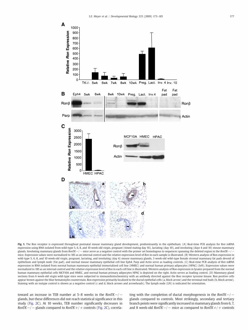
Previous studies on intact mammary glands have shown that RonmRNA expression increases progressively during ductal morphogen-esis, is down regulated at the onset of pregnancy, and remains low
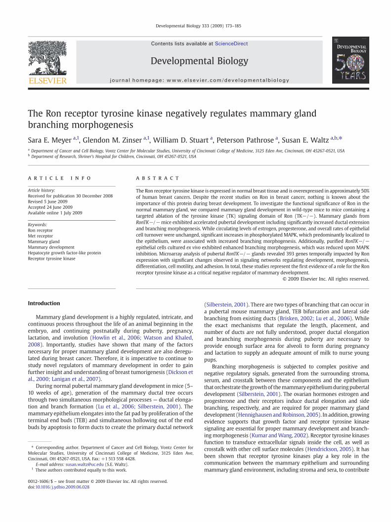
throughout the remainder of postnatal mammary development(Chodosh et al., 2000). Since the ligand for Ron, HGFL, is a motilityfactor that promotes epithelial cell migration (Wang et al., 1996),and since Ron is a member of the c-Met family of receptor tyrosinekinases of which Met and its ligand, hepatocyte growth factor (HGF),have been shown to induce branching morphogenesis (Yang et al.,1995; Yant et al., 1998), we hypothesized that Ron may contribute tothe rapid epithelial migration and branching characteristic ofmammary ductal morphogenesis. Our first objective was to definethe cellular compartment in which Ron is expressed in themammary gland. Prior studies had only demonstrated Ron mRNAexpression from whole gland homogenates. Using primers that spanthe beginning of the tyrosine kinase domain, which is deleted in theRonTK−/− mice (Waltz et al., 2001), we found that Ron mRNA isexpressed in wild-type virgin mouse mammary glands at 5–8 weeksof age, but undetectable at 10 weeks of age (Fig. 1A). Interestingly,Ron expression was also observed in later phases of mammarydevelopment including pregnancy (timed mating day 16), lactation,and involution day 4, but was undetectable by involution day 10(Fig. 1A). We did not detect a PCR product in the RonTK−/−mammary glands using these primers as predicted (Fig. 1A).However, utilizing a set of primers directed to a region upstreamof the TK deletion did amplify a Ron product in the RonTK−/−glands at levels similar to wild-type glands, suggesting that atruncated Ron transcript is expressed at the mRNA level (data notshown). This is consistent with previously published data character-izing the RonTK−/− mice (Waltz et al., 2001). Correspondingly, Ronprotein expression was observed during mammary development inwild-type virgin mouse mammary glands 5–8 weeks of age,pregnancy, and lactation (Fig. 1B). Ron protein expression wasundetectable in mammary glands harvested on involution day 10and in mammary fat pads devoid of epithelium (Fig. 1B). Further,Ron protein expression was also observed in the normal mousemammary epithelial cell line Eph4, and the normal humanmammary epithelial cell lines MCF10A and HMEC (Fig. 1B and C-right). Despite the scant Ron mRNA expression in the humanprimary adipocyte cell line (HPAC) by real-time PCR (Fig. 1C, left),Ron protein was undetectable in these cells by Western analysis (Fig.1C, right). Immunohistochemical detection of Ron in 6 week-oldvirgin wild-type mammary glands showed an intense stainingpattern of Ron in the mammary epithelium of both ducts andterminal end buds (Fig. 1D). Taken together, these data suggest thatRon is expressed during specific time frames throughout mousemammary gland development, primarily in the epithelial compart-ment in both humans and mice with low to undetectable levels ofRon expression in adipose tissue.
Deletion of the Ron tyrosine kinase domain accelerates pubertalmammary gland development
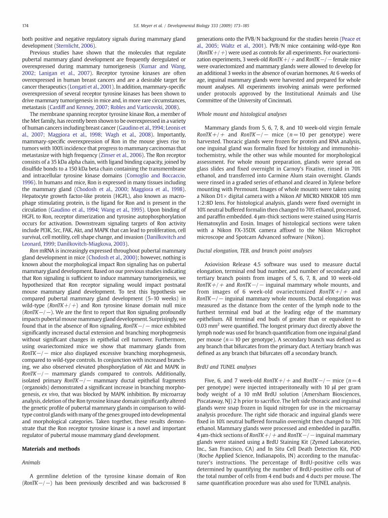
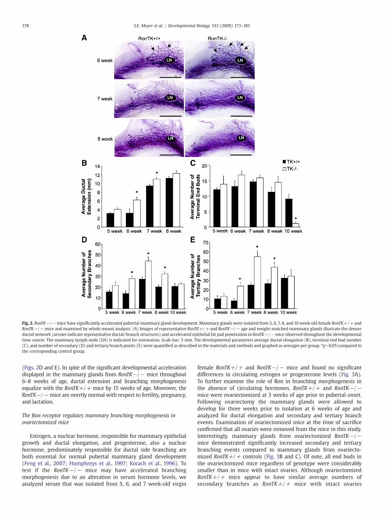
To examine the functional contribution of Ron during mammarydevelopment, a temporal analysis of glandular architecture was under-taken in wild-type mice (RonTK+/+) and mice with homozygousdeletion of the tyrosine kinase domain of Ron (RonTK−/−). Develop-mental analysis of whole mount preparations from 5, 6, 7, 8, and10 week-old virgin female RonTK−/− mouse mammary glands werecompared to age and weight-matched RonTK+/+ control glands.Surprisingly, the RonTK−/− mammary glands displayed acceleratedbranching morphogenesis as evident by the denser mammary ductaltree compared to RonTK+/+ controls (Fig. 2A). To evaluate thedevelopmental progress of the RonTK+/+ and RonTK−/− mammaryglands, terminal end bud (TEB) number, ductal elongation, andsecondaryand tertiarybranchpointswerequantified.Ductal outgrowth,measuring from the center of the lymph node to the furthest TEB, wassignificantly increased inRonTK−/−mammaryglands at 6 and 7weeksof age as compared to RonTK+/+ controls (Fig. 2B).We also sawa trend
Fig. 1. The Ron receptor is expressed throughout postnatal mouse mammary gland development, predominantly in the epithelium. (A) Real-time PCR analysis for Ron mRNAexpression using RNA isolated fromwild-type 5, 6, 8, and 10 week-old virgin, pregnant (timed mating day 16), lactating (day 10), and involuting (days 4 and 10) mouse mammaryglands. Involuting mammary glands from RonTK−/− mice serve as a negative control with the primer set homologous to sequences spanning the deleted region in the RonTK−/−mice. Expression values were normalized to 18S as an internal control and the relative expression level of Ron in each sample is illustrated. (B) Western analysis of Ron expression inwild-type 5, 6, 8, and 10 week-old virgin, pregnant, lactating, and involuting (day 4) mouse mammary glands, 3 week-old wild-type female stromal mammary fat pads devoid ofepithelium and lymph node (Fat pad), and normal mouse mammary epithelial cell line Eph4. Parp and Actin serve as loading controls. (C) Real-time PCR analysis of Ron mRNAexpression in RNA isolated from normal human mammary epithelial immortalized cell line (HMEC) and normal human primary adipocytes (HPAC) (left). Expression values werenormalized to 18S as an internal control and the relative expression level of Ron in each cell line is illustrated. Western analysis of Ron expression in lysates prepared from the normalhuman mammary epithelial cells MCF10A and HMEC, and normal human primary adipocytes HPAC is depicted on the right. Actin serves as loading control. (D) Mammary glandsections from 6 week-old virgin wild-type mice were subjected to immunohistochemistry with an antibody directed against the Ron receptor tyrosine kinase. Ron positive cellsappear brown against the blue hematoxylin counterstain. Ron expression primarily localized to the ductal epithelial cells (a, block arrow) and the terminal end buds (b, block arrow).Staining with an isotype control is shown as a negative control (c and d, block arrows and arrowheads). The lymph node (LN) is indicated for orientation.
177S.E. Meyer et al. / Developmental Biology 333 (2009) 173–185
toward an increase in TEB number at 5–8 weeks in the RonTK−/−glands, but these differences did not reach statistical significance in thisstudy (Fig. 2C). At 10 weeks, TEB number significantly decreases inRonTK−/− glands compared to RonTK+/+ controls (Fig. 2C), correla-
ting with the completion of ductal morphogenesis in the RonTK−/−glands compared to controls. Most strikingly, secondary and tertiarybranchpointswere significantly increased inmammaryglands from6, 7,and 8 week-old RonTK−/− mice as compared to RonTK+/+ controls
Fig. 2. RonTK−/−mice have significantly accelerated pubertal mammary gland development. Mammary glands were isolated from 5, 6, 7, 8, and 10 week-old female RonTK+/+ andRonTK−/− mice and examined by whole mount analysis. (A) Images of representative RonTK+/+ and RonTK−/− age and weight-matched mammary glands illustrate the denserductal network (arrows indicate representative ductal/branch structures) and accelerated epithelial fat pad penetration in RonTK−/−mice observed throughout the developmentaltime course. The mammary lymph node (LN) is indicated for orientation. Scale bar; 5 mm. The developmental parameters average ductal elongation (B), terminal end bud number(C), and number of secondary (D) and tertiary branch points (E) were quantified as described in thematerials andmethods and graphed as averages per group. ⁎pb0.05 compared tothe corresponding control group.
(Figs. 2D and E). In spite of the significant developmental accelerationdisplayed in the mammary glands from RonTK−/− mice throughout6–8 weeks of age, ductal extension and branching morphogenesisequalize with the RonTK+/+ mice by 15 weeks of age. Moreover, theRonTK−/−mice are overtly normal with respect to fertility, pregnancy,and lactation.
The Ron receptor regulates mammary branching morphogenesis inovariectomized mice
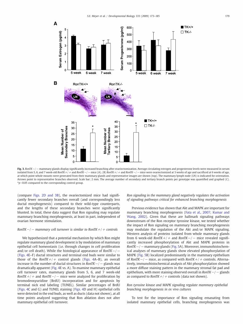
Estrogen, a nuclear hormone, responsible for mammary epithelialgrowth and ductal elongation, and progesterone, also a nuclearhormone, predominately responsible for ductal side branching areboth essential for normal pubertal mammary gland development(Feng et al., 2007; Humphreys et al., 1997; Korach et al., 1996). Totest if the RonTK−/− mice may have accelerated branchingmorphogenesis due to an alteration in serum hormone levels, weanalyzed serum that was isolated from 5, 6, and 7 week-old virgin
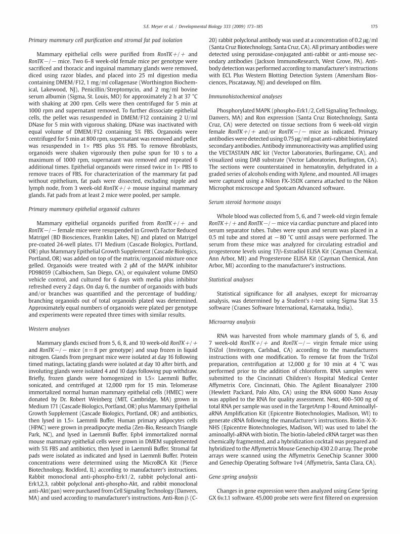
female RonTK+/+ and RonTK−/− mice and found no significantdifferences in circulating estrogen or progesterone levels (Fig. 3A).To further examine the role of Ron in branching morphogenesis inthe absence of circulating hormones, RonTK+/+ and RonTK−/−mice were ovariectomized at 3 weeks of age prior to pubertal onset.Following ovariectomy the mammary glands were allowed todevelop for three weeks prior to isolation at 6 weeks of age andanalyzed for ductal elongation and secondary and tertiary branchevents. Examination of ovariectomized mice at the time of sacrificeconfirmed that all ovaries were removed from the mice in this study.Interestingly, mammary glands from ovariectomized RonTK−/−mice demonstrated significantly increased secondary and tertiarybranching events compared to mammary glands from ovariecto-mized RonTK+/+ controls (Fig. 3B and C). Of note, all end buds inthe ovariectomized mice regardless of genotype were considerablysmaller than in mice with intact ovaries. Although ovariectomizedRonTK+/+ mice appear to have similar average numbers ofsecondary branches as RonTK+/+ mice with intact ovaries
Fig. 3. RonTK−/−mammary glands display significantly increased branching after ovariectomization. Average circulating estrogen and progesterone levels were measured in serumisolated from 5, 6, and 7 week-old RonTK+/+ and RonTK−/−mice (A). (B) RonTK+/+ and RonTK−/−mice were ovariectomized at 3 weeks of age and sacrificed at 6 weeks of age,at which point whole mounts were generated from their mammary glands and representative images are shown (top). The mammary lymph node (LN) is indicated for orientation.Arrows point to representative branches observed. Scale bar; 2 mm. The average number of secondary and tertiary branch points per genotype was quantified and graphed (C).⁎pb0.05 compared to the corresponding control group.
179S.E. Meyer et al. / Developmental Biology 333 (2009) 173–185
(compare Figs. 2D and 3B), the ovariectomized mice had signifi-cantly fewer secondary branches overall (and correspondingly lessductal morphogenesis) compared to their wild-type counterparts,and the lengths of these secondary branches were significantlyblunted. In total, these data suggest that Ron signaling may regulatemammary branching morphogenesis, at least in part, independent ofovarian hormone stimulation.
RonTK−/− mammary cell turnover is similar to RonTK+/+ controls
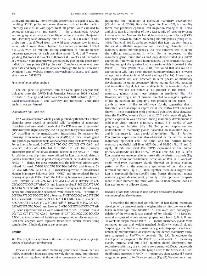
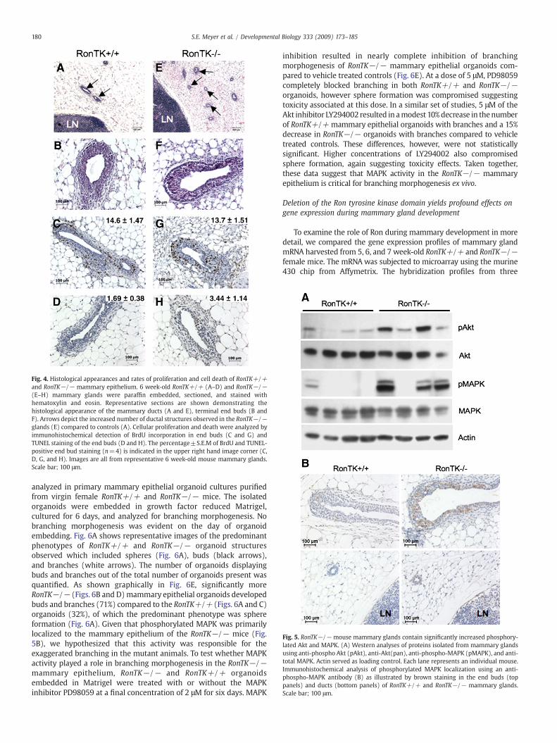
We hypothesized that a potential mechanism by which Ron mightregulate mammary gland development is by modulation of mammaryepithelial cell homeostasis (i.e. through changes in cell proliferationand/or cell death). While the histological appearance of RonTK−/−(Figs. 4E–F) ductal structures and terminal end buds were similar tothose of the RonTK+/+ control glands (Figs. 4A–B), an overallincrease in the number of ductal structures in RonTK−/− glands wasdramatically apparent (Fig. 4E vs. A). To examine mammary epithelialcell turnover rates, mammary glands from 5, 6, and 7 week-oldRonTK+/+ and RonTK−/− mice were analyzed for proliferation bybromodeoxyuridine (BrdU) incorporation and for apoptosis byterminal nick end labeling (TUNEL). Similar percentages of BrdU(Figs. 4C and G) and TUNEL staining (Figs. 4D and H) epithelial cellswere detected in the end buds, as well as ducts (data not shown), at alltime points analyzed suggesting that Ron ablation does not altermammary epithelial cell turnover.
Ron signaling in the mammary gland negatively regulates the activationof signaling pathways critical for enhanced branching morphogenesis
Previous evidence has shown that Akt and MAPK are important formammary branching morphogenesis (Fata et al., 2007; Kumar andWang, 2002). Given that these are hallmark signaling pathwaysdownstream of the Ron receptor tyrosine kinase, we tested whetherthe impact of Ron signaling on mammary branching morphogenesismay modulate the regulation of the Akt and/or MAPK signaling.Western analysis of proteins isolated from whole mammary glandsfrom 6 week-old RonTK+/+ and RonTK−/− mice revealed signifi-cantly increased phosphorylation of Akt and MAPK proteins inRonTK−/−mammary glands (Fig. 5A).Moreover, immunohistochem-ical analyses of mammary glands show elevated phosphorylation ofMAPK (Fig. 5B) localized predominantly in the mammary epitheliumof RonTK−/− mice, as compared with RonTK+/+ controls. Alterna-tively, immunohistochemical analysis of Akt phosphorylation showeda more diffuse staining pattern in the mammary stromal fat pad andepithelium, with more staining observed overall in RonTK−/− glandsas compared to RonTK+/+ controls (data not shown).
Ron tyrosine kinase and MAPK signaling regulate mammary epithelialbranching morphogenesis in ex vivo cultures
To test for the importance of Ron signaling emanating fromisolated mammary epithelial cells, branching morphogenesis was
Fig. 4. Histological appearances and rates of proliferation and cell death of RonTK+/+and RonTK−/− mammary epithelium. 6 week-old RonTK+/+ (A–D) and RonTK−/−(E–H) mammary glands were paraffin embedded, sectioned, and stained withhematoxylin and eosin. Representative sections are shown demonstrating thehistological appearance of the mammary ducts (A and E), terminal end buds (B andF). Arrows depict the increased number of ductal structures observed in the RonTK−/−glands (E) compared to controls (A). Cellular proliferation and death were analyzed byimmunohistochemical detection of BrdU incorporation in end buds (C and G) andTUNEL staining of the end buds (D and H). The percentage±S.E.M of BrdU and TUNEL-positive end bud staining (n=4) is indicated in the upper right hand image corner (C,D, G, and H). Images are all from representative 6 week-old mouse mammary glands.Scale bar; 100 μm.
Fig. 5. RonTK−/− mouse mammary glands contain significantly increased phosphory-lated Akt and MAPK. (A) Western analyses of proteins isolated from mammary glandsusing anti-phospho Akt (pAkt), anti-Akt(pan), anti-phospho-MAPK (pMAPK), and anti-total MAPK. Actin served as loading control. Each lane represents an individual mouse.Immunohistochemical analysis of phosphorylated MAPK localization using an anti-phospho-MAPK antibody (B) as illustrated by brown staining in the end buds (toppanels) and ducts (bottom panels) of RonTK+/+ and RonTK−/− mammary glands.Scale bar; 100 μm.
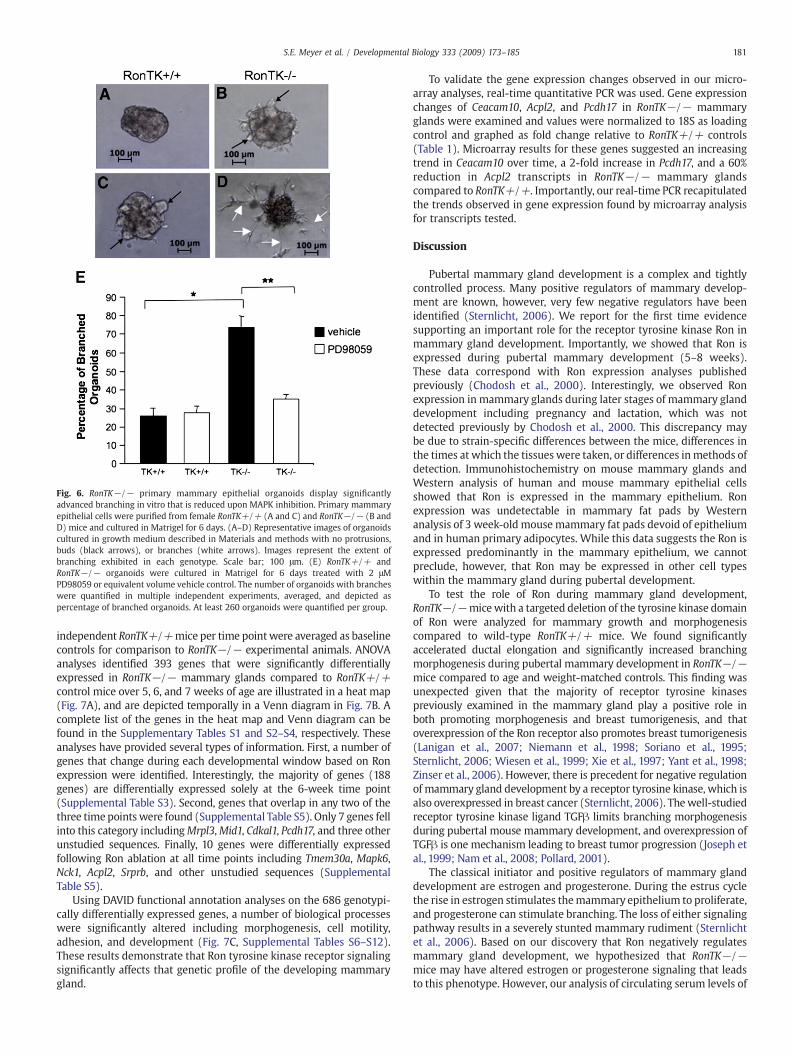
analyzed in primary mammary epithelial organoid cultures purifiedfrom virgin female RonTK+/+ and RonTK−/− mice. The isolatedorganoids were embedded in growth factor reduced Matrigel,cultured for 6 days, and analyzed for branching morphogenesis. Nobranching morphogenesis was evident on the day of organoidembedding. Fig. 6A shows representative images of the predominantphenotypes of RonTK+/+ and RonTK−/− organoid structuresobserved which included spheres (Fig. 6A), buds (black arrows),and branches (white arrows). The number of organoids displayingbuds and branches out of the total number of organoids present wasquantified. As shown graphically in Fig. 6E, significantly moreRonTK−/− (Figs. 6B and D)mammary epithelial organoids developedbuds and branches (71%) compared to the RonTK+/+ (Figs. 6A and C)organoids (32%), of which the predominant phenotype was sphereformation (Fig. 6A). Given that phosphorylated MAPK was primarilylocalized to the mammary epithelium of the RonTK−/− mice (Fig.5B), we hypothesized that this activity was responsible for theexaggerated branching in the mutant animals. To test whether MAPKactivity played a role in branching morphogenesis in the RonTK−/−mammary epithelium, RonTK−/− and RonTK+/+ organoidsembedded in Matrigel were treated with or without the MAPKinhibitor PD98059 at a final concentration of 2 μM for six days. MAPK
inhibition resulted in nearly complete inhibition of branchingmorphogenesis of RonTK−/− mammary epithelial organoids com-pared to vehicle treated controls (Fig. 6E). At a dose of 5 μM, PD98059completely blocked branching in both RonTK+/+ and RonTK−/−organoids, however sphere formation was compromised suggestingtoxicity associated at this dose. In a similar set of studies, 5 μM of theAkt inhibitor LY294002 resulted in amodest 10%decrease in thenumberof RonTK+/+ mammary epithelial organoids with branches and a 15%decrease in RonTK−/− organoids with branches compared to vehicletreated controls. These differences, however, were not statisticallysignificant. Higher concentrations of LY294002 also compromisedsphere formation, again suggesting toxicity effects. Taken together,these data suggest that MAPK activity in the RonTK−/− mammaryepithelium is critical for branching morphogenesis ex vivo.
Deletion of the Ron tyrosine kinase domain yields profound effects ongene expression during mammary gland development
To examine the role of Ron during mammary development in moredetail, we compared the gene expression profiles of mammary glandmRNA harvested from 5, 6, and 7 week-old RonTK+/+ and RonTK−/−female mice. The mRNA was subjected to microarray using the murine430 chip from Affymetrix. The hybridization profiles from three
Fig. 6. RonTK−/− primary mammary epithelial organoids display significantlyadvanced branching in vitro that is reduced upon MAPK inhibition. Primary mammaryepithelial cells were purified from female RonTK+/+ (A and C) and RonTK−/− (B andD) mice and cultured in Matrigel for 6 days. (A–D) Representative images of organoidscultured in growth medium described in Materials and methods with no protrusions,buds (black arrows), or branches (white arrows). Images represent the extent ofbranching exhibited in each genotype. Scale bar; 100 μm. (E) RonTK+/+ andRonTK−/− organoids were cultured in Matrigel for 6 days treated with 2 μMPD98059 or equivalent volume vehicle control. The number of organoids with brancheswere quantified in multiple independent experiments, averaged, and depicted aspercentage of branched organoids. At least 260 organoids were quantified per group.
181S.E. Meyer et al. / Developmental Biology 333 (2009) 173–185
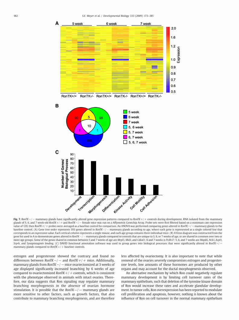
independent RonTK+/+mice per time pointwere averaged as baselinecontrols for comparison to RonTK−/− experimental animals. ANOVAanalyses identified 393 genes that were significantly differentiallyexpressed in RonTK−/− mammary glands compared to RonTK+/+control mice over 5, 6, and 7 weeks of age are illustrated in a heat map(Fig. 7A), and are depicted temporally in a Venn diagram in Fig. 7B. Acomplete list of the genes in the heat map and Venn diagram can befound in the Supplementary Tables S1 and S2–S4, respectively. Theseanalyses have provided several types of information. First, a number ofgenes that change during each developmental window based on Ronexpression were identified. Interestingly, the majority of genes (188genes) are differentially expressed solely at the 6-week time point(Supplemental Table S3). Second, genes that overlap in any two of thethree time points were found (Supplemental Table S5). Only 7 genes fellinto this category includingMrpl3,Mid1, Cdkal1, Pcdh17, and three otherunstudied sequences. Finally, 10 genes were differentially expressedfollowing Ron ablation at all time points including Tmem30a, Mapk6,Nck1, Acpl2, Srprb, and other unstudied sequences (SupplementalTable S5).
Using DAVID functional annotation analyses on the 686 genotypi-cally differentially expressed genes, a number of biological processeswere significantly altered including morphogenesis, cell motility,adhesion, and development (Fig. 7C, Supplemental Tables S6–S12).These results demonstrate that Ron tyrosine kinase receptor signalingsignificantly affects that genetic profile of the developing mammarygland.
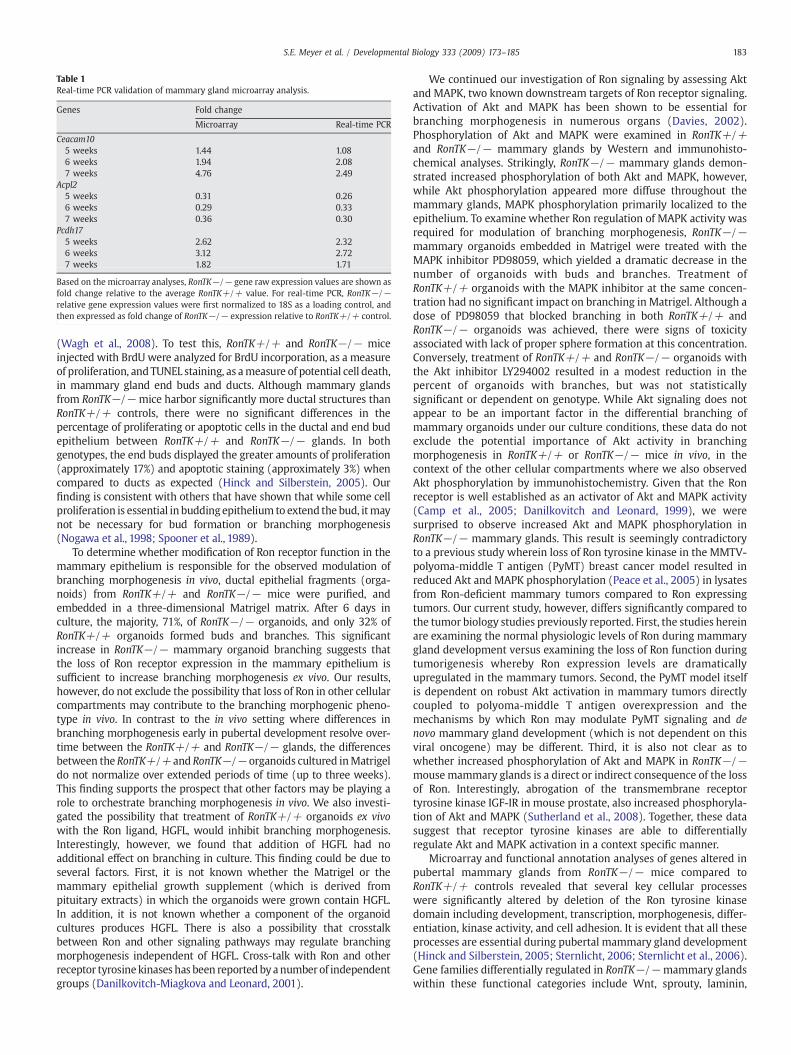
To validate the gene expression changes observed in our micro-array analyses, real-time quantitative PCR was used. Gene expressionchanges of Ceacam10, Acpl2, and Pcdh17 in RonTK−/− mammaryglands were examined and values were normalized to 18S as loadingcontrol and graphed as fold change relative to RonTK+/+ controls(Table 1). Microarray results for these genes suggested an increasingtrend in Ceacam10 over time, a 2-fold increase in Pcdh17, and a 60%reduction in Acpl2 transcripts in RonTK−/− mammary glandscompared to RonTK+/+. Importantly, our real-time PCR recapitulatedthe trends observed in gene expression found by microarray analysisfor transcripts tested.
Discussion
Pubertal mammary gland development is a complex and tightlycontrolled process. Many positive regulators of mammary develop-ment are known, however, very few negative regulators have beenidentified (Sternlicht, 2006). We report for the first time evidencesupporting an important role for the receptor tyrosine kinase Ron inmammary gland development. Importantly, we showed that Ron isexpressed during pubertal mammary development (5–8 weeks).These data correspond with Ron expression analyses publishedpreviously (Chodosh et al., 2000). Interestingly, we observed Ronexpression in mammary glands during later stages of mammary glanddevelopment including pregnancy and lactation, which was notdetected previously by Chodosh et al., 2000. This discrepancy maybe due to strain-specific differences between the mice, differences inthe times at which the tissues were taken, or differences inmethods ofdetection. Immunohistochemistry on mouse mammary glands andWestern analysis of human and mouse mammary epithelial cellsshowed that Ron is expressed in the mammary epithelium. Ronexpression was undetectable in mammary fat pads by Westernanalysis of 3 week-old mousemammary fat pads devoid of epitheliumand in human primary adipocytes. While this data suggests the Ron isexpressed predominantly in the mammary epithelium, we cannotpreclude, however, that Ron may be expressed in other cell typeswithin the mammary gland during pubertal development.
To test the role of Ron during mammary gland development,RonTK−/−micewith a targeted deletion of the tyrosine kinase domainof Ron were analyzed for mammary growth and morphogenesiscompared to wild-type RonTK+/+ mice. We found significantlyaccelerated ductal elongation and significantly increased branchingmorphogenesis during pubertal mammary development in RonTK−/−mice compared to age and weight-matched controls. This finding wasunexpected given that the majority of receptor tyrosine kinasespreviously examined in the mammary gland play a positive role inboth promoting morphogenesis and breast tumorigenesis, and thatoverexpression of the Ron receptor also promotes breast tumorigenesis(Lanigan et al., 2007; Niemann et al., 1998; Soriano et al., 1995;Sternlicht, 2006; Wiesen et al., 1999; Xie et al., 1997; Yant et al., 1998;Zinser et al., 2006). However, there is precedent for negative regulationof mammary gland development by a receptor tyrosine kinase, which isalso overexpressed in breast cancer (Sternlicht, 2006). Thewell-studiedreceptor tyrosine kinase ligand TGFβ limits branching morphogenesisduring pubertal mouse mammary development, and overexpression ofTGFβ is one mechanism leading to breast tumor progression (Joseph etal., 1999; Nam et al., 2008; Pollard, 2001).
The classical initiator and positive regulators of mammary glanddevelopment are estrogen and progesterone. During the estrus cyclethe rise in estrogen stimulates themammary epithelium to proliferate,and progesterone can stimulate branching. The loss of either signalingpathway results in a severely stunted mammary rudiment (Sternlichtet al., 2006). Based on our discovery that Ron negatively regulatesmammary gland development, we hypothesized that RonTK−/−mice may have altered estrogen or progesterone signaling that leadsto this phenotype. However, our analysis of circulating serum levels of
Fig. 7. RonTK−/− mammary glands have significantly altered gene expression patterns compared to RonTK+/+ controls during development. RNA isolated from the mammaryglands of 5, 6, and 7 week-old RonTK+/+ and RonTK−/− female mice was run on a Affymetrix Genechip Array. Probe sets were first filtered based on a minimum raw expressionvalue of 120, then RonTK+/+ probes were averaged as a baseline control for comparison. An ANOVAwas performed comparing genes altered in RonTK−/−mammary glands to thebaseline control. (A) Gene tree order represents 393 genes altered in RonTK−/− mammary glands according to age, where each gene is represented as a single colored line thatcorresponds to an expression value. Each vertical column represents a single mouse, and each age group contains three individual mice. (B) AVenn diagramwas constructed from thegene list used in A to demonstrate genes altered in RonTK−/−mammary glands compared to controls that are unique to 5, 6, or 7 weeks of age, or are shared in common over two ormore age groups. Some of the genes shared in common between 5 and 7 weeks of age areMrpl3,Mid1, and Cdkal1; 6 and 7 weeks is Pcdh17; 5, 6, and 7 weeks areMapk6, Nck1, Acpl1,Srprb, and Synaptotagmin binding. (C) DAVID functional annotation software was used to group genes into biological processes that were significantly altered in RonTK−/−mammary glands compared to RonTK+/+ baseline controls.
estrogen and progesterone showed the contrary and found nodifferences between RonTK−/− and RonTK+/+ mice. Additionally,mammary glands from RonTK−/−mice ovariectomized at 3 weeks ofage displayed significantly increased branching by 6 weeks of agecompared to ovariectomized RonTK+/+ controls, which is consistentwith the phenotype observed in animals with intact ovaries. There-fore, our data suggests that Ron signaling may regulate mammarybranching morphogenesis in the absence of ovarian hormonestimulation. It is possible that the RonTK−/− mammary glands aremore sensitive to other factors, such as growth factors, that alsocontribute to mammary branching morphogenesis, and are therefore
less affected by ovariectomy. It is also important to note that whileremoval of the ovaries severely compromises estrogen and progester-one levels, low amounts of these hormones are produced by otherorgans and may account for the ductal morphogenesis observed.
An alternative mechanism by which Ron could negatively regulatemammary development is by limiting cell turnover rates of themammary epithelium, such that deletion of the tyrosine kinase domainof Ron would increase these rates and accelerate glandular develop-ment. In tumor cells, Ron overexpression has been reported tomodulatecell proliferation and apoptosis, however, nothing is known about theinfluence of Ron on cell turnover in the normal mammary epithelium
Table 1Real-time PCR validation of mammary gland microarray analysis.
Based on the microarray analyses, RonTK−/− gene raw expression values are shown asfold change relative to the average RonTK+/+ value. For real-time PCR, RonTK−/−relative gene expression values were first normalized to 18S as a loading control, andthen expressed as fold change of RonTK−/− expression relative to RonTK+/+ control.
183S.E. Meyer et al. / Developmental Biology 333 (2009) 173–185
(Wagh et al., 2008). To test this, RonTK+/+ and RonTK−/− miceinjected with BrdU were analyzed for BrdU incorporation, as a measureof proliferation, and TUNEL staining, as ameasure of potential cell death,in mammary gland end buds and ducts. Although mammary glandsfrom RonTK−/− mice harbor significantly more ductal structures thanRonTK+/+ controls, there were no significant differences in thepercentage of proliferating or apoptotic cells in the ductal and end budepithelium between RonTK+/+ and RonTK−/− glands. In bothgenotypes, the end buds displayed the greater amounts of proliferation(approximately 17%) and apoptotic staining (approximately 3%) whencompared to ducts as expected (Hinck and Silberstein, 2005). Ourfinding is consistent with others that have shown that while some cellproliferation is essential in buddingepithelium to extend the bud, itmaynot be necessary for bud formation or branching morphogenesis(Nogawa et al., 1998; Spooner et al., 1989).
To determine whether modification of Ron receptor function in themammary epithelium is responsible for the observed modulation ofbranching morphogenesis in vivo, ductal epithelial fragments (orga-noids) from RonTK+/+ and RonTK−/− mice were purified, andembedded in a three-dimensional Matrigel matrix. After 6 days inculture, the majority, 71%, of RonTK−/− organoids, and only 32% ofRonTK+/+ organoids formed buds and branches. This significantincrease in RonTK−/− mammary organoid branching suggests thatthe loss of Ron receptor expression in the mammary epithelium issufficient to increase branching morphogenesis ex vivo. Our results,however, do not exclude the possibility that loss of Ron in other cellularcompartments may contribute to the branching morphogenic pheno-type in vivo. In contrast to the in vivo setting where differences inbranching morphogenesis early in pubertal development resolve over-time between the RonTK+/+ and RonTK−/− glands, the differencesbetween the RonTK+/+ and RonTK−/− organoids cultured inMatrigeldo not normalize over extended periods of time (up to three weeks).This finding supports the prospect that other factors may be playing arole to orchestrate branching morphogenesis in vivo. We also investi-gated the possibility that treatment of RonTK+/+ organoids ex vivowith the Ron ligand, HGFL, would inhibit branching morphogenesis.Interestingly, however, we found that addition of HGFL had noadditional effect on branching in culture. This finding could be due toseveral factors. First, it is not known whether the Matrigel or themammary epithelial growth supplement (which is derived frompituitary extracts) in which the organoids were grown contain HGFL.In addition, it is not known whether a component of the organoidcultures produces HGFL. There is also a possibility that crosstalkbetween Ron and other signaling pathways may regulate branchingmorphogenesis independent of HGFL. Cross-talk with Ron and otherreceptor tyrosinekinaseshas been reportedbyanumberof independentgroups (Danilkovitch-Miagkova and Leonard, 2001).
We continued our investigation of Ron signaling by assessing Aktand MAPK, two known downstream targets of Ron receptor signaling.Activation of Akt and MAPK has been shown to be essential forbranching morphogenesis in numerous organs (Davies, 2002).Phosphorylation of Akt and MAPK were examined in RonTK+/+and RonTK−/− mammary glands by Western and immunohisto-chemical analyses. Strikingly, RonTK−/− mammary glands demon-strated increased phosphorylation of both Akt and MAPK, however,while Akt phosphorylation appeared more diffuse throughout themammary glands, MAPK phosphorylation primarily localized to theepithelium. To examine whether Ron regulation of MAPK activity wasrequired for modulation of branching morphogenesis, RonTK−/−mammary organoids embedded in Matrigel were treated with theMAPK inhibitor PD98059, which yielded a dramatic decrease in thenumber of organoids with buds and branches. Treatment ofRonTK+/+ organoids with the MAPK inhibitor at the same concen-tration had no significant impact on branching in Matrigel. Although adose of PD98059 that blocked branching in both RonTK+/+ andRonTK−/− organoids was achieved, there were signs of toxicityassociated with lack of proper sphere formation at this concentration.Conversely, treatment of RonTK+/+ and RonTK−/− organoids withthe Akt inhibitor LY294002 resulted in a modest reduction in thepercent of organoids with branches, but was not statisticallysignificant or dependent on genotype. While Akt signaling does notappear to be an important factor in the differential branching ofmammary organoids under our culture conditions, these data do notexclude the potential importance of Akt activity in branchingmorphogenesis in RonTK+/+ or RonTK−/− mice in vivo, in thecontext of the other cellular compartments where we also observedAkt phosphorylation by immunohistochemistry. Given that the Ronreceptor is well established as an activator of Akt and MAPK activity(Camp et al., 2005; Danilkovitch and Leonard, 1999), we weresurprised to observe increased Akt and MAPK phosphorylation inRonTK−/− mammary glands. This result is seemingly contradictoryto a previous study wherein loss of Ron tyrosine kinase in the MMTV-polyoma-middle T antigen (PyMT) breast cancer model resulted inreduced Akt and MAPK phosphorylation (Peace et al., 2005) in lysatesfrom Ron-deficient mammary tumors compared to Ron expressingtumors. Our current study, however, differs significantly compared tothe tumor biology studies previously reported. First, the studies hereinare examining the normal physiologic levels of Ron during mammarygland development versus examining the loss of Ron function duringtumorigenesis whereby Ron expression levels are dramaticallyupregulated in the mammary tumors. Second, the PyMT model itselfis dependent on robust Akt activation in mammary tumors directlycoupled to polyoma-middle T antigen overexpression and themechanisms by which Ron may modulate PyMT signaling and denovo mammary gland development (which is not dependent on thisviral oncogene) may be different. Third, it is also not clear as towhether increased phosphorylation of Akt and MAPK in RonTK−/−mouse mammary glands is a direct or indirect consequence of the lossof Ron. Interestingly, abrogation of the transmembrane receptortyrosine kinase IGF-IR in mouse prostate, also increased phosphoryla-tion of Akt and MAPK (Sutherland et al., 2008). Together, these datasuggest that receptor tyrosine kinases are able to differentiallyregulate Akt and MAPK activation in a context specific manner.
Microarray and functional annotation analyses of genes altered inpubertal mammary glands from RonTK−/− mice compared toRonTK+/+ controls revealed that several key cellular processeswere significantly altered by deletion of the Ron tyrosine kinasedomain including development, transcription, morphogenesis, differ-entiation, kinase activity, and cell adhesion. It is evident that all theseprocesses are essential during pubertal mammary gland development(Hinck and Silberstein, 2005; Sternlicht, 2006; Sternlicht et al., 2006).Gene families differentially regulated in RonTK−/−mammary glandswithin these functional categories include Wnt, sprouty, laminin,
protocadherin, and ephrin. We have validated several targetsidentified by the microarray, which have known functions withbroad implications and may potentially play a role in our model, buthave not yet been studied with respect to mammary developmentincluding the glycoprotein Ceacam10 and acid phosphatase Acpl2.Two genes found by DAVID analysis in the cell adhesion and motilitycellular processes important for branching morphogenesis are thetyrosine kinase adapter Nck1 (data not shown) and protocadherinPcdh17. The overall changes observed in our studies are consistentwith other microarray analyses implementing these gene families inmammary gland development (Kouros-Mehr and Werb, 2006).Together, these microarray data support the conclusion that Ronlikely impacts mammary development through a process that ismultifactoral.
In summary, we have shown that ablation of Ron receptor tyrosinekinase accelerates pubertal mammary gland development. Markedly,the loss of Ron impacts mammary gland branching morphogenesisindependently of ovarian estrogen and progesterone stimulation.Moreover, based on three-dimensional ex vivo analyses, the absenceof Ron in the epithelium is sufficient to produce branching. Ronreceptor ablation alters Akt and MAPK phosphorylation, which isknown to be essential for proper mammary epithelial branchingmorphogenesis. Finally, deletion of Ron tyrosine kinase profoundlyalters the genetic program in the pubertal mammary gland. Takentogether, our data demonstrate that Ron is an important regulator ofpubertal mammary gland development.
Acknowledgments
The authors would like to acknowledge the excellent technicalassistance provided by Sarah Kader, Kenya Toney, and Jerilyn Gray aswell as Dr. Nelson Horseman for the helpful discussions and MCF10Aprotein lysates. This work was supported by the Public Health ServicesGrants CA-100002 (S.E.W.), T32-CA59268 (S.E.M.), and the DigestiveDiseases Research Development Center grant DK-064403 (S.E.W.)from the National Institutes of Health, as well as by grant project#8950 (S.E.W.) from Shriner's Hospital for Children.
Appendix A. Supplementary data
Supplementary data associated with this article can be found, inthe online version, at doi:10.1016/j.ydbio.2009.06.028.
References
Brisken, C., 2002. Hormonal control of alveolar development and its implications forbreast carcinogenesis. J. Mammary Gland Biol. Neoplasia 7, 39–48.
Camp, E.R., Liu, W., Fan, F., Yang, A., Somcio, R., Ellis, L.M., 2005. RON, a tyrosine kinasereceptor involved in tumor progression and metastasis. Ann. Surg. Oncol. 12,273–281.
Cardiff, R.D., Kenney, N., 2007. Mouse mammary tumor biology: a short history. Adv.Cancer Res. 98, 53–116.
Comoglio, P.M., Boccaccio, C., 1996. The HGF receptor family: unconventional signaltransducers for invasive cell growth. Genes Cells 1, 347–354.
Danilkovitch, A., Leonard, E.J., 1999. Kinases involved in MSP/RON signaling. J. Leukoc.Biol. 65, 345–348.
Danilkovitch-Miagkova, A., 2003. Oncogenic signaling pathways activated by RONreceptor tyrosine kinase. Curr. Cancer Drug Targets 3, 31–40.
Danilkovitch-Miagkova, A., Leonard, E.J., 2001. Cross-talk between RON receptortyrosine kinase and other transmembrane receptors. Histol. Histopathol. 16,623–631.
Davies, J.A., 2002. Do different branching epithelia use a conserved developmentalmechanism? BioEssays 24, 937–948.
Dickson, C., Creer, A., Fantl, V., 2000. Mammary gland oncogenes as indicators ofpathways important in mammary gland development. Oncogene 19, 1097–1101.
Fata, J.E., Mori, H., Ewald, A.J., Zhang, H., Yao, E., Werb, Z., Bissell, M.J., 2007. The MAPK(ERK-1,2) pathway integrates distinct and antagonistic signals from TGFalpha andFGF7 in morphogenesis of mouse mammary epithelium. Dev. Biol. 306, 193–207.
Feng, Y., Manka, D., Wagner, K.U., Khan, S.A., 2007. Estrogen receptor-alpha expressionin the mammary epithelium is required for ductal and alveolar morphogenesis inmice. Proc. Natl. Acad. Sci. U. S. A. 104, 14718–14723.
Gaudino, G., Follenzi, A., Naldini, L., Collesi, C., Santoro, M., Gallo, K.A., Godowski, P.J.,Comoglio, P.M., 1994. RON is a heterodimeric tyrosine kinase receptor activated bythe HGF homologue MSP. EMBO J. 13, 3524–3532.
Hendrickson, W.A., 2005. Transduction of biochemical signals across cell membranes.Q. Rev. Biophys. 38, 321–330.
Hennighausen, L., Robinson, G.W., 2005. Information networks in the mammary gland.Nat. Rev. Mol. Cell Biol. 6, 715–725.
Hinck, L., Silberstein, G.B., 2005. Key stages in mammary gland development: themammary end bud as a motile organ. Breast Cancer Res. 7, 245–251.
Humphreys, R.C., Lydon, J., O'Malley, B.W., Rosen, J.M., 1997. Mammary glanddevelopment is mediated by both stromal and epithelial progesterone receptors.Mol. Endocrinol. 11, 801–811.
Joseph, H., Gorska, A.E., Sohn, P., Moses, H.L., Serra, R., 1999. Overexpression of a kinase-deficient transforming growth factor-beta type II receptor in mouse mammarystroma results in increased epithelial branching. Mol. Biol. Cell 10, 1221–1234.
Kouros-Mehr, H., Werb, Z., 2006. Candidate regulators of mammary branching morphoge-nesis identified by genome-wide transcript analysis. Dev. Dyn. 235, 3404–3412.
Kumar, R., Wang, R.A., 2002. Protein kinases in mammary gland development andcancer. Microsc. Res. Tech. 59, 49–57.
Lanigan, F., O'Connor, D., Martin, F., Gallagher, W.M., 2007. Molecular links betweenmammary gland development and breast cancer. Cell. Mol. Life Sci. 64, 3159–3184.
Longati, P., Comoglio, P.M., Bardelli, A., 2001. Receptor tyrosine kinases as therapeutictargets: the model of the MET oncogene. Curr. Drug Targets 2, 41–55.
Lu, P., Sternlicht, M.D., Werb, Z., 2006. Comparative mechanisms of branchingmorphogenesis in diverse systems. J. Mammary Gland Biol. Neoplasia 11, 213–228.
Maggiora, P., Marchio, S., Stella, M.C., Giai, M., Belfiore, A., De Bortoli, M., Di Renzo, M.F.,Costantino, A., Sismondi, P., Comoglio, P.M., 1998. Overexpression of the RON genein human breast carcinoma. Oncogene 16, 2927–2933.
Nam, J.S., Terabe, M., Kang, M.J., Chae, H., Voong, N., Yang, Y.A., Laurence, A.,Michalowska, A., Mamura, M., Lonning, S., Berzofsky, J.A., Wakefield, L.M., 2008.Transforming growth factor beta subverts the immune system into directlypromoting tumor growth through interleukin-17. Cancer Res. 68, 3915–3923.
Niemann, C., Brinkmann, V., Spitzer, E., Hartmann, G., Sachs, M., Naundorf, H.,Birchmeier, W., 1998. Reconstitution of mammary gland development in vitro:requirement of c-met and c-erbB2 signaling for branching and alveolar morpho-genesis. J. Cell. Biol. 143, 533–545.
Nogawa, H., Morita, K., Cardoso, W.V., 1998. Bud formation precedes the appearance ofdifferential cell proliferation during branching morphogenesis of mouse lungepithelium in vitro. Dev. Dyn. 213, 228–235.
Peace, B.E., Toney-Earley, K., Collins, M.H., Waltz, S.E., 2005. Ron receptor signalingaugments mammary tumor formation and metastasis in a murine model of breastcancer. Cancer Res. 65, 1285–1293.
Pollard, J.W., 2001. Tumour-stromal interactions. Transforming growth factor-betaisoforms and hepatocyte growth factor/scatter factor in mammary gland ductalmorphogenesis. Breast Cancer Res. 3, 230–237.
Wagh, P.K., Peace, B.E., Waltz, S.E., 2008. Met-related receptor tyrosine kinase Ron intumor growth and metastasis. Adv. Cancer Res. 100, 1–33.
Waltz, S.E., Eaton, L., Toney-Earley, K., Hess, K.A., Peace, B.E., Ihlendorf, J.R., Wang, M.H.,Kaestner, K.H., Degen, S.J., 2001. Ron-mediatedcytoplasmic signaling is dispensable forviability but is required to limit inflammatory responses. J. Clin. Invest. 108, 567–576.
Wang, M.H., Iwama, A., Skeel, A., Suda, T., Leonard, E.J., 1995. The murine stk geneproduct, a transmembrane protein tyrosine kinase, is a receptor for macrophage-stimulating protein. Proc. Natl. Acad. Sci. U. S. A. 92, 3933–3937.
Wang, M.H., Montero-Julian, F.A., Dauny, I., Leonard, E.J., 1996. Requirement ofphosphatidylinositol-3 kinase for epithelial cell migration activated by humanmacrophage stimulating protein. Oncogene 13, 2167–2175.
Watson, C.J., Khaled, W.T., 2008. Mammary development in the embryo and adult: ajourney of morphogenesis and commitment. Development 135, 995–1003.
185S.E. Meyer et al. / Developmental Biology 333 (2009) 173–185
Wiesen, J.F., Young, P., Werb, Z., Cunha, G.R., 1999. Signaling through the stromalepidermal growth factor receptor is necessary for mammary ductal development.Development 126, 335–344.
Xie, W., Paterson, A.J., Chin, E., Nabell, L.M., Kudlow, J.E., 1997. Targeted expression of adominant negative epidermal growth factor receptor in the mammary gland oftransgenic mice inhibits pubertal mammary duct development. Mol. Endocrinol. 11,1766–1781.
Yang, Y., Spitzer, E., Meyer, D., Sachs, M., Niemann, C., Hartmann, G., Weidner, K.M.,Birchmeier, C., Birchmeier, W., 1995. Sequential requirement of hepatocyte growth
factor and neuregulin in the morphogenesis and differentiation of the mammarygland. J. Cell Biol. 131, 215–226.
Yant, J., Buluwela, L., Niranjan, B., Gusterson, B., Kamalati, T., 1998. In vivo effects ofhepatocyte growth factor/scatter factor on mouse mammary gland development.Exp. Cell Res. 241, 476–481.
Zinser, G.M., Leonis, M.A., Toney, K., Pathrose, P., Thobe, M., Kader, S.A., Peace, B.E.,Beauman, S.R., Collins, M.H., Waltz, S.E., 2006. Mammary-specific Ron receptoroverexpression induces highly metastatic mammary tumors associated with beta-catenin activation. Cancer Res. 66, 11967–11974.