Page 1
1
The Saccharomyces cerevisiae 14-3-3 proteins are
required for the G1/S transition, actin cytoskeleton
organization and cell wall integrity
Francisca Lottersberger, Andrea Panza, Giovanna Lucchini, Simonetta Piatti and
Maria Pia Longhese1
Dipartimento di Biotecnologie e Bioscienze, Università di Milano-Bicocca, 20126 Milan,
Italy
1Corresponding author: Dipartimento di Biotecnologie e Bioscienze, Università degli Studi di
Milano-Bicocca, P.zza della Scienza 2, 20126 Milan, Italy.
Phone: 0039-0264483425
Fax: 0039-0264483565
E-mail: [email protected]
Running title: 14-3-3 proteins and the G1/S transition
Keywords: 14-3-3; PKC; G1/S transition; actin cytoskeleton; cell wall integrity
Genetics: Published Articles Ahead of Print, published on April 28, 2006 as 10.1534/genetics.106.058172
Page 2
2
ABSTRACT
14-3-3 proteins are highly conserved polypeptides participating in many biological processes
by binding phosphorylated target proteins. In S. cerevisiae, two functionally redundant 14-3-3
isoforms are encoded by the BMH1 and BMH2 genes, whose concomitant deletion is lethal.
To gain insights into the essential function(s) shared by these proteins, we searched for high
dosage suppressors of the growth defects of temperature-sensitive bmh mutants. Both the
protein kinase C1 (Pkc1) and its upstream regulators Wsc2 and Mid2 were found as high
dosage suppressors of bmh mutants’ temperature-sensitivity, indicating a functional
interaction between 14-3-3 and Pkc1. Consistent with a role of 14-3-3 proteins in Pkc1-
dependent cellular processes, bmh mutants turned out to be severely impaired at restrictive
temperature in initiation of DNA replication, polarization of the actin cytoskeleton and
budding, as well as in cell wall integrity. Since Pkc1 acts in concert with the Swi4-Swi6
(SBF) transcriptional activator to control all these processes, the defective G1/S transition of
bmh might be linked to impaired SBF activity. Consistently, the levels of the G1 cyclin CLN2
transcripts, which are positively regulated by SBF, were dramatically reduced in bmh
mutants. Remarkably, budding and DNA replication defects of bmh mutants were suppressed
by CLN2 expression from an SBF-independent promoter, suggesting that 14-3-3 proteins
could contribute to regulating the late G1 transcriptional program.
Page 3
3
INTRODUCTION
The 14-3-3 proteins are a large family of highly conserved, ubiquitously expressed acidic
polypeptides of 28-33 kDa that are found in all eukaryotes. There are at least seven isoforms
in mammals and up to 15 isoforms in plants, while two isoforms have been identified in yeast,
Drosophila melanogaster and Caernohabditis elegans (reviewed in Hermeking, 2003;
Dougherty and Morrison, 2004). They form homo- and heterodimers able to bind protein
ligands that are usually phosphorylated on serine/threonine residues of consensus binding
motifs (Jones et al., 1995; Muslin et al., 1996; Yaffe et al., 1997; Chaudhri et al., 2003). By
inducing conformational changes or steric hindrance in protein ligands, 14-3-3 proteins can
activate/repress their enzymatic activity, prevent degradation, modulate localization and/or
facilitate/inhibit protein modifications and interactions (reviewed in Hermeking, 2003;
Dougherty and Morrison, 2004). Targets of 14-3-3 family members are found in all
subcellular compartments and include transcription factors, biosynthetic enzymes,
cytoskeletal proteins, signaling molecules, checkpoint and apoptosis factors and tumor
suppressors. This plethora of interacting proteins allows 14-3-3 to play important roles in a
wide range of regulatory processes such as cell cycle control, mitogenic signal transduction
and apoptotic cell death, and to be implicated in cancerogenesis and some human diseases
(reviewed in Dougherty and Morrison, 2004). However, since multiple 14-3-3 isoforms are
present in mammals and 14-3-3 proteins have several binding targets, the mechanisms
underlying 14-3-3 functions are not fully understood.
The two S. cerevisiae members of the 14-3-3 protein family, which share 93% amino acid
identity, are encoded by the BMH1 and BMH2 genes. While single bmh1∆ and bmh2∆
mutants do not show detectable growth defects compared to wild type, the bmh1 bmh2 double
Page 4
4
disruption is lethal in most laboratory strains (van Heusden et al., 1992, 1995; Gelperin et al.,
1995; Roberts et al., 1997).
Although their essential functions are not well understood, budding yeast Bmh proteins
appear to be involved in many cellular processes. For example, they modulate the activity of
some transcription factors. In fact, loss of function mutations in the SIN4 gene, encoding a
global transcriptional regulator, and in the RTG3 gene, encoding a basic helix-loop-helix
transcription factor, suppress the temperature sensitive phenotype of a bmh1 bmh2 mutant
(van Heusden and Steensma, 2001). Moreover, Bmh1 physically interacts with
phosphorylated Rtg3, suggesting that 14-3-3 proteins inhibit Rtg3 transcriptional activation
function by binding its phosphorylated form (van Heusden and Steensma, 2001). Finally,
Bmh1 physically interacts with Msn2 and Msn4, two transcription factors required to activate
a large number of stress-related genes, and retains their phosphorylated forms in the
cytoplasm (Beck and Hall, 1999).
Also vesicular transport and cortical actin network organization likely involve 14-3-3 proteins
(Gelperin et al., 1995; Roth et al., 1999). In fact, S. cerevisiae cells overproducing the
carboxy-terminal region of Bmh2 fail to polarize vesicular transport and show a disrupted
actin cytoskeleton (Roth et al., 1999). Moreover, 14-3-3 proteins interact with many proteins
involved in cytoskeletal regulation in both yeast and mammals (Jin et al., 2004). In particular,
two-hybrid interactions have been reported for Bmh2 with Msb3 (Mayordomo and Sanz,
2002), which is involved in actin cytoskeleton organization (Bach et al., 2000; Bi et al.,
2000), and with Gic2 (Mayordomo and Sanz, 2002), which is required together with Gic1 for
cytoskeletal polarization during bud emergence (Brown et al., 1997; Chen et al., 1997). Both
Bmh1 and Bmh2 interact also with the p21-activated kinase (PAK) Ste20, and this interaction
appears to be specifically required for Ras/MAPK cascade signaling during pseudohyphal
development (Roberts et al., 1997). Finally, mammalian 14-3-3 proteins regulate actin
Page 5
5
dynamics by stabilizing phosphorylated cofilin, a family of proteins essential for high rates of
actin filament turnover through regulation of the actin polymerization/depolymerization
cycles (Gohla and Bokoch, 2002).
By interacting with various regulatory proteins, 14-3-3 proteins participate in diverse signal
transduction pathways. In fact, hyperactivation of the Ras/cAMP-dependent protein kinase A
(PKA) pathway by overproducing Tpk1, the catalytic subunit of PKA, suppresses cell
lethality caused by Bmh depletion (Gelperin et al., 1995). Consistent with a link between 14-
3-3 and PKA, Bmh proteins are dispensable for yeast cell viability in Σ1278b background
(Roberts et al., 1997), where the Ras/cAMP signaling pathway is hyperactivated (Stanhill et
al., 1999). However, bmh1∆ bmh2∆ Σ1278b derivative cells exhibit osmo-remediable
temperature sensitivity and sensitivity to high osmolarity (Roberts et al., 1997), suggesting
that some functions of Bmh proteins are still required at 37°C even in this background.
Moreover, 14-3-3 proteins have been implicated also in Ras/MAPK cascade signaling in
vertebrates (Fantl et al., 1994; Li et al., 1995) and during pseudohyphal development in S.
cerevisiae (Roberts et al., 1997). Finally, vertebrate 14-3-3 proteins were shown to inhibit or
activate Protein Kinase C (PKC), which is involved in many signaling processes (Toker et al.,
1990; Isobe et al., 1992; Tanji et al., 1994), and to stimulate the interaction between PKC and
the mitogen-stimulated Raf1 kinase that controls cell growth (Van Der Hoeven et al., 2000).
In a previous study we isolated 4 bmh1 alleles, whose presence in the cell as the sole 14-3-3
source caused temperature-sensitive growth (Lottersberger et al., 2003). In order to provide
new insights into the essential functions of the S. cerevisiae 14-3-3 proteins, we have carried
out a detailed phenotypic characterization of these mutants and searched for high dosage
suppressors of their temperature sensitivity. We provide evidence that hyperactivation of the
protein kinase C1 (Pkc1)-dependent pathways suppresses the growth defects of these bmh
mutants, suggesting that 14-3-3 proteins functionally interact with Pkc1. Accordingly, bmh
Page 6
6
mutants are impaired in Pkc1-regulated processes at the G1/S transition, such as budding,
initiation of DNA replication, actin cytoskeleton polarization and cell wall integrity. Our data
suggest that both the temperature-sensitivity and the G1/S transition defects of our bmh
mutants might be ascribed to an impaired activity of the SBF transcription factor, which is
known to act in concert with Pkc1 to control all the above processes.
Page 7
7
RESULTS
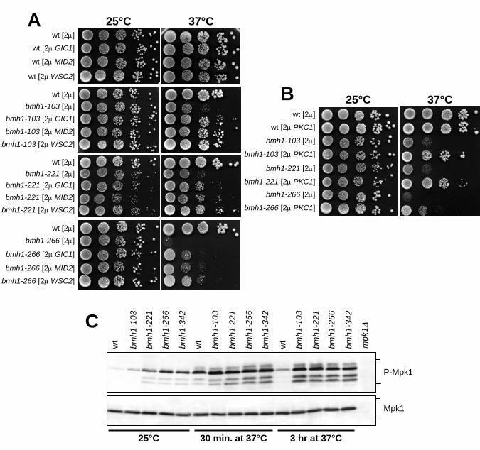
High dosage of MID2, WSC2, PKC1 and GIC1 suppresses bmh mutants’ temperature-
sensitivity: We previously generated bmh1-103, bmh1-221, bmh1-266 and bmh1-342 yeast
temperature-sensitive mutants (Lottersberger et al., 2003 and Fig. 1). In each of our mutant
strains, the bmh1 mutant allele was the sole 14-3-3 source, since all of them carried a BMH2
deletion, which did not cause by itself any of the phenotypes described during this study (data
not shown). Importantly, overproduction of the catalytic subunit of Protein Kinase A (PKA),
Tpk1, was unable to suppress the growth defects of these bmh mutants at 37°C (data not
shown), in contrast to its ability to suppress the cell lethality caused by Bmh depletion at 25°C
(Gelperin et al., 1995), indicating that our bmh mutants are not solely impaired in the
activation of the Ras/cAMP-dependent PKA pathway.
In order to identify cellular partners for 14-3-3 proteins, we searched for high dosage
suppressors of the temperature-sensitivity of the bmh1-266 mutant. To this purpose, bmh1-
266 cells were transformed with a S. cerevisiae genomic library constructed in the YEp24 2µ
vector (Carlson and Botstein, 1982), and 40.000 Ura+ transformants were screened for the
ability to form colonies on YEPD plates at 37°C (see Materials and Methods). Besides 190
BMH2- and 20 BMH1-bearing plasmids, the screen allowed the recovery of ten plasmids
carrying different ORFs. Subcloning of the several ORFs carried by three of these plasmids in
the YEplac195 2µ vector revealed that high copy number of the GIC1, WSC2 or MID2 genes
could partially suppressed the temperature-sensitivity of bmh1-266, bmh1-103 and bmh1-221
cells (Fig. 1A). Unfortunately, we were unable to assess this suppressing ability in bmh1-342
cells, due to their high frequency of 2µ plasmid loss.
While the GIC1 and GIC2 genes encode two homologous proteins required for actin
polarization and bud formation (Brown et al., 1997; Chen et al., 1997), WSC2 and MID2 gene
Page 8
8
products are transmembrane cell surface sensors. They have been proposed to perform
partially overlapping functions in cell wall remodeling during vegetative growth and under
stress conditions (Verna et al., 1997; Marcoux et al., 1998; Rajavel et al. 1999; Ketela et al.,
1999; Philip and Levin, 2001), and to detect and transmit the cell wall status to the Protein
Kinase C1 (Pkc1). The latter is involved in a multiplicity of pathways, including those related
to bud emergence, cell wall integrity, and organization of the actin cytoskeleton in response to
heat shock, pheromone, low osmolarity, nutrient starvation and cell cycle progression
(Heinisch et al., 1999). This prompted us to examine whether an excess of Pkc1 could also
suppress the temperature-sensitivity of our bmh mutants. Indeed, high copy number of PKC1
ameliorated the ability of bmh1-103, bmh1-221 and bmh1-266 cells to form colonies at 37°C
(Fig. 1B), indicating that hyperactivation of a Pkc1-dependent cascade may compensate for
defects in 14-3-3 proteins.
Pkc1 is believed to possess multiple functions (Lee and Levin, 1992; Verna et al., 1997;
Delley and Hall, 1999; Ketela et al., 1999; Andrews and Stark, 2000; Zanelli and Valentini,
2005), only one of which is to regulate the activity of the MAP kinase cascade that ultimately
regulates cell wall integrity, bud emergence, response to hypotonic shock and actin
reorganization (reviewed in Levin and Errede, 1995). Since high levels of Wsc2, Mid2 or
Pkc1, which are predicted to lead to increased signaling through Pkc1, suppressed the
temperature-sensitive growth defects of our bmh mutants, we asked whether the latter were
defective in activating the Pkc1-dependent MAPK cascade. To this end, we monitored Mpk1
phosphorylation, which is an established marker for activation of the Pkc1-MAPK pathway
(Lee et al., 1993; Zarzov et al., 1996; de Nobel et al., 2000). As shown in figure 1C, the
amount of phosphorylated Mpk1 in bmh mutants was higher than in wild type already at the
permissive temperature. Moreover, heat shock, which is known to induce a rapid transient
depolarization of the actin cytoskeleton and cell wall weakening (Delley and Hall, 1999),
Page 9
9
increased the amount of Mpk1 phosphorylated forms in both wild type and bmh mutants after
shift to 37°C for 30 minutes (Fig. 1C). However, phosphorylated Mpk1 level was
significantly decreased in wild type cells after 3 hours at 37°C, due to adaptation to the high
temperature, whereas it remained high in bmh cells under the same conditions (Fig. 1C).
Thus, the growth defects of our bmh mutants are unlikely due to faulty MAPK kinase
signaling, since the latter appears to be instead hyperactivated in these mutants. Rather, these
mutants could be deficient in some Pkc1-regulated pathway that paralleles the one involving
MAP kinases. In this view, the MAPK cascade could be hyperactivated in bmh mutants as a
compensatory mechanism to maintain cell viability in the absence of 14-3-3 function.
Accordingly, we found that MPK1 deletion was lethal for the bmh1-103, bmh1-221, bmh1-
266 and bmh1-342 mutants (data not shown). Therefore, an excess of Wsc2, Mid2 or Pkc1
may suppress temperature-sensitivity of bmh mutants by acting through a Pkc1-dependent
MAPK-independent pathway.
Temperature-sensitive bmh mutants are defective in the G1/S transition and actin
polarization:
Since enhanced Pkc1 signaling contributes to cell viability in the absence of 14-3-3 function,
we asked whether bmh mutants were impaired in Pkc1-regulated cellular processes, such as
bud formation, actin reorganization, cell wall remodeling, and cell cycle progression
(Heinisch et al., 1999). To investigated whether defects in 14-3-3 functions may affect bud
formation at the G1/S transition, exponentially growing cultures of wild type, bmh1-103,
bmh1-221, bmh1-266 and bmh1-342 cells were arrested in G1 with α-factor at 25°C and then
released into fresh medium at 37°C. As shown in figure 2A, most of bmh1-103, bmh1-221,
bmh1-266 and bmh1-342 cells were still largely unbudded after 1 hour at 37°C, when bud
emergence had already occurred in 90% of similarly treated wild type cells. After 3 hours at
Page 10
10
37°C, most of bmh1-221 and bmh1-342 mutant cells were still unbudded, while
approximately 50% of bmh1-103 and bmh1-266 cells managed to bud (Fig. 2A and B).
However, their buds appeared mostly mis-shaped and kept elongating upon further incubation
at 37°C (Fig. 2A and B). Moreover, some elongated budded cells appeared also in bmh1-221
and bmh1-342 mutants at later time points. Thus, bmh mutants might be impaired in the
switch between apical to isotropic growth.
Delayed bud formation in bmh mutants paralleled with defects in DNA synthesis initiation. In
fact, all bmh mutants severely delayed initiation of DNA replication, although to different
extents, after shift to 37°C (Fig. 2C). While wild type cells initiated DNA replication 45-60
minutes after release at 37°C from the G1 block, the onset of DNA replication took place in
bmh1-221 and bmh1-342 cells only around 150 minutes after release under the same
conditions (Fig. 2C). In addition, a major fraction of cells in both mutants was unable to
replicate DNA by 240 minutes (Fig. 2C). The bmh1-103 and bmh1-266 cells started DNA
replication about 120 and 75 minutes, respectively, after release at 37°C and again only a
fraction of these mutant cells managed to complete DNA replication by 240 minutes at 37°C
(Fig. 2C). As shown in figure 2D, initiation of DNA replication upon release at 25°C of the
same G1-arrested cell cultures was delayed by 15-30 minutes in bmh mutant cell cultures
compared to wild type. Altogether, these data indicate that 14-3-3 proteins are required for a
timely G1/S transition.
Both bud emergence and its subsequent surface growth require the polarization of the actin
cytoskeleton, such that cortical patches and actin cables converge at the bud site (reviewed in
Pruyne and Bretscher, 2000). Since 14-3-3 proteins have been previously linked to actin
cytoskeleton organization (Gelperin et al., 1995; Roth et al., 1999), impaired bud formation in
the above bmh mutants might be related to defects in this process. To address this issue, we
analyzed actin polarization upon Alexa-Fluor 546 phalloidin staining of wild type and bmh
Page 11
11
mutant cells that were arrested in G1 by α factor and then released at 37°C for 1 hour. As
shown in figure 3, actin cortical patches, which normally clustered at the bud tips of wild type
cells, were completely missing in bmh1-221 and bmh1-342 cells and appeared only in a small
fraction of bmh1-103 and bmh1-266 cells. Therefore, organization of the actin cytoskeleton at
the future bud emergence sites is perturbed in bmh mutants, thus affecting bud formation at
the G1/S transition.
We then combined the different bmh1 alleles, together with the BMH2 deletion, with the
temperature-sensitive cdc42-1 or cdc24-1 alleles, altering the essential Cdc42 GTPase and its
guanine-nucleotide-exchange factor (GEF) Cdc24 (Adams et al., 1990; Johnson and Pringle,
1990; Van Aelst and D’Souza-Schorey, 1997), which are both required to establish actin
cytoskeleton polarity (reviewed in Pruyne and Bretscher, 2000). As shown in figure 4, the
ability to form colonies at 32°C of all bmh1 bmh2∆ cdc24-1 and bmh1 bmh2∆ cdc42-1 triple
mutants was severely impaired compared to that of the parental mutants. This synthetic effect
between bmh and cdc24 or cdc42 mutant alleles further support a role for budding yeast 14-3-
3 proteins in actin polarization and bud formation.
Bmh defects cause sensitivity to cell wall stress and their effects on the G1/S transition
can be relieved by osmotic support:
Pkc1 controls cell wall metabolism by regulating β-glucan synthesis at the site of wall
remodeling, as well as expression of cell wall biosynthesis genes necessary for maintaining
cellular integrity both during bud formation and in response to heat shock, pheromone and
nutrient starvation (reviewed in Levin, 2005). We therefore asked whether defects in 14-3-3
functions might result in impaired cell wall integrity, by analyzing the ability of bmh mutants
to grow at permissive temperature in the presence of compounds such as the chitin antagonist
calcofluor white and SDS, which have both proved to be powerful tools for revealing yeast
Page 12
12
cell wall defects (Ram et al., 1994). As shown in figure 5A, bmh1-103, bmh1-221, bmh1-266
and bmh1-342 cells were unable to grow on YEPD plates supplemented with 0.01% SDS,
which did not affect wild type cell growth. Moreover, growth of all bmh mutants on YEPD
was compromised, although to different extents, by addition of 0.01 mg/ml calcofluor white
(Fig. 5B). Finally, microscopic examination of the bmh mutant cell cultures revealed
accumulation of cell debris at 37°C (data not shown), suggesting that cell lysis frequently
occurred. Thus, 14-3-3 proteins appear to be required for a stable cell wall structure.
We therefore examined whether osmotic stabilization of the medium might relieve the
temperature sensitivity and the G1/S transition defects of our bmh mutants. As shown in
figure 5C, addition of the osmotic stabilizer sorbitol restored the ability of bmh1-103, bmh1-
221, bmh1-266 and bmh1-342 cells to grow on YEPD plates at 37°C. Moreover, the presence
of sorbitol in the medium largely rescued the defects in bud emergence (Fig. 5D) and
initiation of DNA replication (Fig. 5E) displayed by bmh mutants upon G1 release at 37°C.
Thus, slow-growth and delayed G1/S transition that are caused by defective 14-3-3 proteins
are osmo-remediable, consistent with a primary defect of bmh mutants in cell wall biogenesis.
High dosage of Wsc2, Mid2 or Pkc1 can partially suppress the G1/S transition defects of
bmh mutants:
Since enhanced Pkc1-dependent signaling by high copy number WSC2, MID2 or PKC1
suppressed the temperature sensitivity of bmh mutants, we asked whether it suppressed also
their G1/S transition defects. As shown in figure 6A, bmh1-103, bmh1-221 and bmh1-266
cells carrying WSC2, MID2 or PKC1 on a 2µ plasmid and released from α-factor at 37°C
underwent budding more efficiently than the same mutant cells carrying the empty vector.
Moreover, an excess of Wsc2, Mid2 or Pkc1 attenuated the abnormal bud morphology of
bmh1-103 and bmh1-266 cells after three hours at 37°C (Fig. 6A). Similarly, bud emergence
Page 13
13
took place more efficiently in bmh1-221, bmh1-103 and bmh1-266 cells containing an excess
of Gic1, although this, as expected, caused bud elongation even in wild type cells due to
sustained polarized growth (Fig. 6A) (Brown et al., 1997; Chen et al., 1997).
Suppression of bmh defects in DNA replication initiation were also apparent upon WSC2,
MID2 or PKC1 increased dosage. In fact, bmh1-103, bmh1-221 and bmh1-266 cells carrying
high copy number WSC2-, MID2- or PKC1-bearing plasmids initiated DNA replication at
37°C earlier and more efficiently than the same mutants with the empty vector (Fig. 6B).
Thus, an excess of Wsc2, Mid2 or Pkc1 can partially suppress the G1/S transition defects of
bmh mutants.
Low G1 cyclin-Cdk1 levels may account for the G1/S transition defects of bmh mutants:
During the G1/S transition, Pkc1 acts in concert with the SBF transcription factor to control
the actin cytoskeleton, cell cycle progression and transcription of cell wall biosynthesis genes
(reviewed in Levin, 2005). SBF is composed of the Swi6 and Swi4 subunits, and is
responsible for transcriptional activation of the CLN1 and CLN2 cyclin genes, whose products
associate with the Cyclin-dependent kinase 1 (Cdk1) to promote bud morphogenesis and
DNA replication (reviewed in Levin et al., 1995; Nasmyth, 1996).
Since Pkc1 hyperactivation was shown to partially compensate for the lack of SBF activity
(Gray et al., 1997; Igual et al., 1996), we asked whether the G1/S transition defects of bmh
mutants might be related to impaired formation of G1 Cyclin/Cdk1 complexes. We therefore
measured the levels of CLN2 mRNA in the bmh1-221 and bmh1-342 mutants, which showed
the most severe G1/S transition defects at 37°C compared to the other bmh mutants (Fig. 2).
Exponentially growing cultures of wild type, bmh1-221 and bmh1-342 cells were arrested in
G1 with α-factor and released into the cell cycle at 37°C. At different time points after release
total RNA was analysed by Northern blot with a CLN2 probe. As shown in figure 7A, CLN2
Page 14
14
mRNAs started to appear in wild type cells 30-45 minutes after release, right before bud
emergence (data not shown) and initiation of DNA replication. Conversely, their amount was
dramatically reduced in both bmh1-221 and bmh1-342 mutant cells that remained arrested
with 1C DNA contents for at least 180 minutes after release at 37°C (Fig. 7A). If the G1/S
transition defects of our bmh mutants were due to low amounts of G1 cyclin/Cdk1 complexes
caused by the reduced CLN1 and CLN2 mRNA levels, CLN2 expression from an ectopic
promoter might suppress the G1/S transition defects of our bmh mutants. To test this
hypothesis, cultures of wild type, bmh1-103, bmh1-221, bmh1-266 and bmh1-342 strains,
carrying or lacking a galactose-inducible GAL1-CLN2 construct, were grown in
YEP+raffinose at 25°C, arrested in G1 with α-factor and then released at 25°C or 37°C in
galactose-containing medium to induce CLN2 expression. GAL1-CLN2 induction
significantly rescued the G1/S defects of most bmh mutants. In fact, both bud emergence
(data not shown) and initiation of DNA replication (Fig. 7B) were advanced upon galactose
induction in all bmh GAL1-CLN2 strains compared to the isogenic bmh strains, both at 25°C
and 37°C. In particular, S phase entry took place in GAL-CLN2, bmh1-103 GAL-CLN2,
bmh1-221 GAL-CLN2 and bmh1-342 GAL-CLN2 strains at 30, 45, 30 and 60 minutes,
respectively, after release at 37°C in galactose-containing medium, while similarly treated
bmh1-103, bmh1-221 and bmh1-342 cells neither budded (data not shown) nor initiated DNA
replication up to 4 hours after release (Fig. 7B). Conversely, ectopic CLN2 expression had
only a marginal effect on bmh1-266 cells, allowing only 30% of them to initiate DNA
replication by 4 hours at 37°C (Fig. 7B). This suggests that functions other than activation of
Cln1, 2/Cdk1 might be affected in this mutant. It worth noting that expression of CLN2 from
the GAL1 promoter caused cytokinesis defects at late time points in most strains, leading to
accumulation of cells with elongated buds (data not shown) and more than 2C DNA contents
(Fig. 7B), as previously reported (Lew and Reed, 1993). Altogether these data indicate that
Page 15
15
reduced amounts of G1 cyclin/Cdk1 complexes may partially account for the G1/S transition
defects of our bmh mutants.
Page 16
16
DISCUSSION
In order to understand the essential function(s) of S. cerevisiae 14-3-3 proteins, we searched
for high dosage suppressors of the temperature-sensitivity of bmh mutants, carrying a bmh1
mutant allele as the sole 14-3-3 source (Lottersberger et al., 2003). We found that the growth
defects of bmh1-103, bmh1-221 and bmh1-266 at 37°C can be rescued by overproducing
Pkc1 or its transmembrane cell surface sensors Wsc2 and Mid2, which have been proposed to
perform partially overlapping functions in cell wall remodeling during vegetative growth and
under stress conditions by detecting and transmitting cell wall status to Pkc1 (Verna et al.,
1997; Philip and Levin, 2001; Rajavel et al., 1999; Ketela et al., 1999). Pkc1 is believed to
possess multiple functions (Lee and Levin, 1992; Verna et al., 1997; Delley and Hall, 1999;
Ketela et al., 1999; Andrews and Stark, 2000; Zanelli and Valentini, 2005), one of which is to
regulate the MAP kinase cascade involved in cell wall construction and polarized growth
(reviewed in Levin and Errede, 1995). Based on Mpk1 phosphorylation, the Pkc1-dependent
MAPK cascade appears to be hyperactivated both at 25°C and 37°C in our bmh mutants,
suggesting that defects in 14-3-3 proteins affect a pathway regulated by Pkc1 other than that
Mpk1-dependent. Thus, Wsc2, Mid2 or Pkc1 may act as high dosage suppressors by
stimulating the former pathway, whereas the hyperactivation of the MAPK cascade in our
mutants could be the result of a compensatory mechanism that contributes to their cell
viability at the permissive temperature. Accordingly, deletion of MPK1 was lethal for our
bmh mutants, indicating that 14-3-3 proteins and Mpk1 act in different branches of the Pkc1
pathway to sustain cell viability.
Pkc1, together with its upstream regulators Wsc1-3 and Mid2, controls actin cytoskeleton
reorganization, cell cycle progression and transcription of cell wall biosynthesis genes
involved in synthesis and assembly of cell wall components at the bud (reviewed in Levin,
Page 17
17
2005). Since enhanced Pkc1-dependent signaling can partially suppress the temperature
sensitivity of bmh mutants, some of the above Pkc1-regulated processes might be impaired in
these mutants. Indeed, we found that all our temperature-sensitive bmh1 alleles cause defects
in G1/S transition, actin polarization at the pre-bud site and cell wall integrity at 37°C. In fact,
shift to the restrictive temperature severely impairs bud formation and initiation of DNA
replication in bmh1-221 and bmh1-342 mutants and significantly slows down the same
processes in bmh1-103 and bmh1-266 cells. When entry into S phase and bud emergence
eventually take place in the latter mutants, buds are elongated, suggesting a defective apical-
isotropic switch in bud growth. Consistent with a function for 14-3-3 proteins in bud
formation and actin polarization, bmh mutant alleles also cause synthetic effects at
semipermissive temperature when combined with the cdc42-1 and cdc24-1 temperature-
sensitive alleles, altering the Rho-family GTPase Cdc42 and its guanine-nucleotide-exchange
factor (GEF), respectively, that are essential for polarizing the actin cytoskeleton (reviewed in
Pruyne and Bretscher, 2000). Moreover, high levels of the Cdc42 effector Gic1, which binds
to the activated GTP-bound form of Cdc42 and is required for cytoskeletal polarization during
bud emergence (Chen et al., 1997; Brown et al., 1997), can partially suppress the temperature-
sensitivity of bmh1-103, bmh1-221 and bmh1-266 mutants. Finally, our bmh mutants undergo
cell lysis at restrictive temperature and are hypersensitive to calcofluor and SDS at permissive
temperature, suggesting that they are impaired in cell wall integrity. In agreement with a 14-
3-3 role in cell wall biogenesis, both the growth defects and the G1/S transition delay at 37°C
of our bmh mutants can be rescued by the addition of the osmo-stabilizer sorbitol.
Both initiation of DNA replication and bud morphogenesis require activation of G1
cyclin/Cdk1 complexes (reviewed in Nasmyth, 1996). In particular, when cells reach a critical
size, Cln3-Cdk1 activates the Swi6/Swi4 transcription factor (SBF) that induces transcription
of the CLN1 and CLN2 genes (Nasmyth and Dirick, 1991; Ogas et al., 1991; Dirick et al.,
Page 18
18
1995; Cross et al., 1994). The Pkc1-dependent cascade acts in concert with SBF to control the
actin cytoskeleton and transcription of cell wall biosynthesis genes involved in maintaining
cellular integrity during bud formation (Levin and Bartlett-Heubusch, 1992; Lew and Reed,
1993; Mazzoni et al., 1993; Igual et al., 1996; Marini et al., 1996; Zarzov et al., 1996; Gray et
al., 1997; Madden et al., 1997; Delley and Hall, 1999). Accordingly, swi4∆ and swi6∆
mutants are sensitive to cell wall stresses and the growth defects of swi4∆ cells can be
partially relieved by osmotic stabilization, supporting a role for SBF in cell wall biogenesis
(Igual et al., 1996; Gray et al., 1997). Moreover, swi4 and pkc1 mutations are synthetically
lethal (Madden et al., 1997), whereas the temperature-sensitive growth of swi4∆ cells can be
suppressed by overproduction of Pkc1 or Wsc1, the latter belonging to the Wsc1-3 family of
transmembrane proteins required for heat stress activation of the Pkc1-MAPK cascade (Gray
et al., 1997; Igual et al., 1996). Finally, Pkc1 seems to play redundant functions with G1
cyclins, since deletion of PKC1 causes cell death in a cln1∆ cln2∆ double mutant (Gray et al.,
1997).
The partially redundant function of Pkc1 and SBF-depending pathways and the similarities in
the behaviour of bmh and swi4 or swi6 mutants raise the possibility that the phenotypes of
bmh mutants might be due to defective SBF activity. In agreement with this hypothesis, Cln1,
2/Cdk1 complexes appear to be limiting for execution of the G1/S transition in bmh mutants.
In fact, the amount of CLN2 mRNA is dramatically reduced in both bmh-103 and bmh1-342
mutants at 37°C compared to wild type. Moreover, expression of CLN2 from an SBF-
independent promoter can partially suppress bmh mutant defects in budding and DNA
replication. Consistent with the possibility that defective 14-3-3 proteins may impair SBF-
dependent accumulation of Cln1, 2/Cdk1 complexes, a mutation in the SIN4 gene, whose lack
of function bypasses the requirement for Swi4 and Swi6 to transcribe the HO-LacZ reporter
gene (Nasmyth et al., 1987; Lycan et al., 1994; Li et al., 2005), was shown to suppress the
Page 19
19
temperature sensitivity of a bmh2 bmh1∆ mutant (van Heusden and Steensma, 2001). Since
SBF-dependent induction of Cln1, 2/Cdk1 triggers both entry into S phase by turning on
proteolysis of the cyclin B-Cdk1 inhibitor Sic1 and cytoskeleton polarization for bud
formation (Lew and Reed, 1993; Schwob et al., 1994; Tyers, 1996), impaired SBF-dependent
Cln1, 2/Cdk1 complex formation may account for both budding and DNA replication defects
of our bmh mutants. It should in fact be noted that, unlike in most of the other laboratory
strains, simultaneous deletion of CLN1 and CLN2 is lethal in the W303 genetic background
that we used for all our experiments (Cvrckova et al., 1995). Therefore, low levels of Cln1,
2/Cdk1 complexes in bmh mutants can be the cause of their G1/S transition defects at high
temperatures.
In any case, since SBF and Pkc1 play a partially redundant role in allowing bud formation and
cell integrity (Gray et al., 1997; Igual et al., 1996; Madden et al., 1997), hyperactivation of the
Pkc1-dependent cascades by high levels of Wsc2, Mid2 or Pkc1 might suppress the growth
and G1/S transition defects of bmh mutants by partially compensating defects in SBF activity
and G1 cyclin/Cdk1 complex accumulation.
Taken together, these data indicate that S. cerevisiae 14-3-3 proteins play essential functions
in regulating processes that occur at the G1/S transition. Since 14-3-3 proteins are highly
conserved in evolution, our studies may help to elucidate their essential functions also in
higher eukaryotes.
Page 20
20
MATERIALS AND METHODS
Yeast strains and media: The relevant genotypes of all the yeast strains are listed in Table 1.
All the strains used during this study were derivatives of W303 (MATa or MATα, ade2-1,
can1-100, his3-11,15, leu2-3,112, trp1-1, ura3, ssd1).
Strains YLL1082, YLL1081, YLL1120 and YLL1092 were previously described
(Lottersberger et al., 2003). Wild-type, bmh1-103 bmh2∆, bmh1-221 bmh2∆, bmh1-266
bmh2∆ and bmh1-342 bmh2∆ strains carrying either the 2µ vector, or 2µ WSC2 or 2µ MID2
or 2µ GIC2 plasmids were constructed by transforming strains W303, YLL1082, YLL1081,
YLL1120 and YLL1092 with plasmids YEplac195 (2µ URA3), pML489 (2µ WSC2 URA3),
pML490 (2µ MID2 URA3) and pML493 (2µ GIC2 URA3), respectively.
A MATα strain, carrying the GAL-CLN2 construct integrated at the CLN2 chromosomal locus
and obtained after sporulation of the diploid L96 kindly provided by L. Dirick (Montpellier,
France), was crossed to strains YLL1082, YLL1081, YLL1120 and YLL1092 to obtain
DMP4370/2D, DMP4372/3B, DMP4373/7C and DMP4465/3B strains, respectively. Strain
DMP4357/1B was obtained after sporulation of the diploid L96. Strain YLL1906, carrying
the deletion of the PKC1 gene, was kindly provided by R. Tisi (University of Milano-
Bicocca, Italy).
Strains DMP4436/3C, DMP4439/6B, DMP4440/5C and DMP4441/5A were meiotic
segregants from crosses of strains YLL1082, YLL1081, YLL1120 and YLL1092,
respectively, with a MATα cdc24-1 strain. Strains DMP4430/7A, DMP4433/10B,
DMP4434/7B and DMP4435/4B strains were meiotic segregants from crosses of strains
YLL1082, YLL1081, YLL1120 and YLL1092, respectively, with a MATα cdc42-1 strain.
The accuracy of all gene replacements and integrations was verified by Southern blot analysis
or PCR. Standard yeast genetic techniques and media were according to Rose et al. (1990).
Page 21
21
Cells were grown in YEP medium (1% yeast extract, 2% bactopeptone, 50 mg/l adenine)
supplemented with 2% glucose (YEPD) or 2% raffinose (YEP+raf) or 2% raffinose and 1%
galactose (YEP+raf+gal). Transformants carrying the KANMX4 cassette were selected on
YEPD plates containing 400 µg/ml G418 (US Biological).
Plasmids: To obtain plasmid pML490, containing a MID2 fragment spanning from 663 bp
upstream of the coding region start codon to 425 bp downstream of the stop codon, a 2219 bp
MID2 fragment was amplified by PCR using yeast genomic DNA as template and the
oligonucleotides PRP551 (5’-CGG GAT CCC GAT TGA GAG ATC TCA CGG AAA TG-
3’) and PRP552 (5’-CGG GAT CCC GTC ACA GAA CTC GGT AAG TTT TC-3’) as
primers. The PCR amplification product was then cloned into the BamHI site of plasmid
YEplac195 (Gietz and Sugino, 1988).
To obtain plasmid pML489, containing the WSC2 ORF flanked by 459 bp upstream of the
start codon and 293 bp downstream of the stop codon, a 2264 bp WSC2 fragment was
amplified by PCR using yeast genomic DNA as template and the oligonucleotides PRP543
(5’-CGG GAT CCC GCT ACG GTA AAC ATG CCT GAT GG-3’) and PRP544 (5’-CGG
GAT CCC GTG TGA TCT AGC ACT TCT CCC AG-3’) as primers. The PCR amplification
product was then cloned into the BamHI site of plasmid YEplac195.
To obtain plasmid pML493, containing the GIC1 ORF flanked by 343 bp upstream of the
start codon and 282 bp downstream of the stop codon, a 1570 bp GIC1 fragment was
amplified by PCR using yeast genomic DNA as template and the oligonucleotides PRP561
(5’-GGG GTA CCC CGT TGT CTG AGC AGG AAT AAA GAG-3’) and PRP562 (5’-GGG
GTA CCC CGG GTA GTA GAC ATC GCT ATT ATC-3’) as primers. The PCR
amplification product was then cloned into the KpnI site of plasmid YEplac195.
Plasmids YEplac112 (Gietz and Sugino, 1988), carrying the PKC1 gene, was kindly provided
by R. Tisi (University of Milano-Bicocca, Italy).
Page 22
22
Search for high dosage suppressors: To search for high dosage suppressors of the
temperature-sensitivity caused by the bmh1-266 mutation, strain YLL1120 was transformed
with a S. cerevisiae genomic library based on the multicopy 2� vector YEp24 (Carlson and
Botstein, 1982). Ura+ transformants were tested for their ability to grow at 37°C on YEPD
plates, which inhibited the untransformed strain. Plasmids from transformants showing co-
segregation of the thermo-resistance with the URA3 vector marker were recovered and
introduced again into the YLL1120 strain, to confirm their ability to suppress bmh1-266
temperature-sensitivity. Restriction analysis allowed us to identify several classes of plasmids
containing different yeast genomic fragments. The nucleotide sequences of both ends of the
smallest DNA insert of each plasmid class were determined and compared with the whole
Saccharomyces cerevisiae genomic sequence in the Saccharomyces Genome Database
(SGD). Since most inserts contained several ORFs, the suppressor genes were identified by
cloning subfragments of the inserts into the 2µ plasmid YEplac195 and testing the derivative
plasmids for their ability to suppress the temperature-sensitivity of the bmh1-266 bmh2∆
mutant strain.
Other techniques: Synchronization experiments were performed as described in Lotterberger
et al., 2003. Flow cytometric DNA analysis was determined on a Becton-Dickinson
FACScan. To stain actin cytoskeleton, cells were treated 2’ with Rhodamine-phalloidin
20U/ml (Sigma-Aldrich) in PBS and then washed three in PBS buffer. Digital images were
taken with a CCD camera and software (CoolSNAP; Photometrics). For Western blot
analysis, native protein extracts were prepared in 0.1% SDS, 1% Triton, 50mM Tris pH7.5,
1mM sodium deoxycolate, 120mM β-glicerophosphate, 1,72mM sodium orthovanadate,
10mM DTT, 1mM AEBSF, 15mM para-nitrophenilphosphate and a protease inhibitor
cocktail (Boehringer Mannheim). To detect phosphorylated Mpk1 and Mpk1, polyclonal anti-
phospho p42/p44 (Cell Signaling) and anti-Mpk1 (Santa Cruz Biotechnology) antibodies
Page 23
23
were used, respectively, after 1:1000 dilution in BSA-TBS. Secondary antibodies were
purchased from Amersham and proteins were visualized by an enhanced chemiluminescence
system according to the manufacturer.
Page 24
24
ACKNOWLEDGEMENTS
We thank L. Dirick and R. Tisi for providing yeast strains and plasmids, M. Vai and all the
members of our laboratory for useful discussions and criticisms. This work was supported by
grants from Associazione Italiana per la Ricerca sul Cancro to M.P.L. and S.P.,
Cofinanziamento 2005 MIUR/Università di Milano-Bicocca to M.P.L. and Fondo per gli
investimenti della Ricerca di Base (FIRB) to G.L.. F.L. was supported by a fellowship from
Associazione Italiana per la Ricerca sul Cancro.
Page 25
25
FIGURE LEGENDS
FIGURE 1. High dosage suppressors of the bmh temperature-sensitive growth defects. (A and
B) Exponentially growing cell cultures (selective media at 25°C) of wild type (W303), bmh1-
103 bmh2∆ (YLL1082), bmh1-221 bmh2∆ (YLL1081) and bmh1-266 bmh2∆ (YLL1120)
strains transformed with 2µ plasmids, either empty or carrying the GIC1, MID2, WSC2 (A) or
PKC1 (B), were spotted on YEPD plates and incubated at 25°C or at 37°C for 3 days. (C)
Cell cultures of wild type (K699), bmh1-103 bmh2∆ (YLL1082), bmh1-221 bmh2∆
(YLL1081), bmh1-266 bmh2∆ (YLL1120) and bmh1-342 bmh2∆ (YLL1092), exponentially
growing in YEPD at 25°C, were shifted to 37°C. Aliquots were withdrawn at time zero
(25°C) and 30 minutes or 3 hours after shift at 37°C to prepare protein extracts, which were
subjected to western blot analysis with anti-phospho-p44/p42 antibodies (Cell Signaling) to
detect Mpk1 phosphorylation (top, P-Mpk1). The two faster migrating bands were likely P-
Mpk1 degradation products, and they were not detected by polyclonal antibodies raised
against a C-terminal Mpk1 peptide (Santa Cruz Biotechnology), which were used to measure
total Mpk1 levels in the same samples (bottom, Mpk1). Specificity of the antibodies was
checked by using protein extract prepared from a mpk1∆ (YLL1906) strain incubated 30
minutes at 37°C.
FIGURE 2. Temperature-sensitive bmh mutants are defective in bud emergence and initiation
of DNA replication. Cell cultures of wild type (W303), bmh1-103 bmh2∆ (YLL1082), bmh1-
221 bmh2∆ (YLL1081), bmh1-266 bmh2∆ (YLL1120) and bmh1-342 bmh2∆ (YLL1092)
strains, exponentially growing at 25°C in YEPD, were arrested in G1 with α-factor for 2 hr
and released at time zero in YEPD at 25°C or 37°C. (A) 200 cells for each strain were
analyzed to determine the frequency of cells with no, small, large or elongated buds at 25°C
Page 26
26
(0 hr) and at the indicated time points after shift at 37°C. (B) Photographs were taken 3 hours
after shift at 37°C. (C and D) To determine DNA contents by fluorescence-activated cell
sorting (FACS) analysis, samples were withdrawn at the indicated times after release in
YEPD at 37°C (C), or 25°C (D).
FIGURE 3. Actin organization in the temperature-sensitive bmh mutants. Cell cultures of
wild type (W303), bmh1-103 bmh2∆ (YLL1082), bmh1-221 bmh2∆ (YLL1081), bmh1-266
bmh2∆ (YLL1120) and bmh1-342 bmh2∆ (YLL1092) strains, exponentially growing at 25°C
in YEPD, were synchronized in G1 with α-factor and released at time zero in YEPD at 37°C.
Cells were fixed 1 hour after the release at 37°C, stained with fluorochrome-conjugated
phalloidin, and scored for the presence of cells with polarized actin by fluorescence
microscopy. Differential interference contrast (left) and epi-fluorescence (right) images are
shown as examples.
FIGURE 4. Synthetic effects between bmh and polarization mutant alleles. The following
strains were used: wild type (W303), cdc24-1, cdc42-1, bmh1-103 bmh2∆ (YLL1082), bmh1-
103 cdc24-1 bmh2∆ (DMP4436/3C), bmh1-103 cdc42-1 bmh2∆ (DMP4430/7A), bmh1-221
bmh2∆ (YLL1081), bmh1-221 cdc24-1 bmh2∆ (DMP4439/6B), bmh1-221 cdc42-1 bmh2∆
(DMP4433/10B), bmh1-266 bmh2∆ (YLL1120), bmh1-266 cdc24-1 bmh2∆ (DMP4440/5C),
bmh1-266 cdc42-1 bmh2∆ (DMP4434/7B), bmh1-342 bmh2∆ (YLL1092), bmh1-342 cdc24-1
bmh2∆ (DMP4441/5A) and bmh1-342 cdc42-1 bmh2∆ (DMP4435/4B). Serial dilutions of
cell cultures, exponentially growing in YEPD at 25°C, were spotted on YEPD plates and
incubated at the indicated temperatures for 3 days.
Page 27
27
FIGURE 5. Temperature-sensitive bmh mutants are sensitive to cell wall stress. (A) Serial
dilutions of cell cultures of wild type (W303), bmh1-103 bmh2∆ (YLL1082), bmh1-221
bmh2∆ (YLL1081), bmh1-266 bmh2∆ (YLL1120) and bmh1-342 bmh2∆ (YLL1092),
exponentially growing in YEPD at 25°C, were streaked on SD plates with or without SDS
(0.01%). (B) The same cultures in A were spotted on YEPD plates with or without Calcofluor
(0.01mg/ml). Plates were incubated at 25°C for 4 days. (C) Serial dilution of wild type
(W303), bmh1-103 bmh2∆ (YLL1082), bmh1-221 bmh2∆ (YLL1081), bmh1-266 bmh2∆
(YLL1120) and bmh1-342 bmh2∆ (YLL1092) cell cultures, exponentially growing in YEPD
at 25°C, were spotted on YEPD plates in the absence or presence of 1M sorbitol and
incubated at the indicated temperatures for 3 days. (D and E) Cell cultures of wild type
(W303), bmh1-103 bmh2∆ (YLL1082), bmh1-221 bmh2∆ (YLL1081), bmh1-266 bmh2∆
(YLL1120) and bmh1-342 bmh2∆ (YLL1092) strains, exponentially growing at 25°C in
YEPD, were arrested in G1 with α-factor for 2 hr, and released at time zero in YEPD at 25°C
or 37°C in the absence or presence of 1M sorbitol. Samples were withdrawn at the indicated
times after α-factor release to analyze the kinetics of bud emergence (D) and DNA contents
by FACS analysis (E).
FIGURE 6. WSC2, MID2 and PKC1 overexpression can suppress the G1/S transition delay of
bmh mutants. Exponentially growing (selective media at 25°C) cell cultures of wild type
(W303), bmh1-103 bmh2∆ (YLL1082), bmh1-221 bmh2∆ (YLL1081) and bmh1-266 bmh2∆
(YLL1120) strains transformed with 2µ plasmids, either empty or carrying the WSC2, MID2,
PKC1 or GIC1 genes, were arrested in G1 with α-factor and released at time zero in YEPD at
25°C or 37°C. (A) 200 cells for each strain were analyzed to determine the frequency of cells
with no, small, large or elongated buds after 3 hours at 37°C. (B) Samples were withdrawn at
the indicated times after α-factor release to analyze DNA contents by FACS analysis.
Page 28
28
FIGURE 7. CLN2 mRNA levels and CLN2 ectopic expression in bmh mutants. Cell cultures
of wild type (W303), bmh1-221 bmh2∆ (YLL1081) and bmh1-342 bmh2∆ (YLL1092) strains,
exponentially growing in YEPD at 25°C, were synchronized in G1 with α-factor and released
at time zero into YEPD at 37°C. Samples were taken at the indicated times after the release
into the cell cycle to analyze CLN2 mRNA by Northern analysis (left) and to determine DNA
contents by FACS analysis (right). Loading control of the northern blot is a methylene blue-
stained filter of ribosomal RNAs (rRNAs). (B) Cell cultures of wild type (W303), GAL-CLN2
(DMP4357/1B), bmh1-103 bmh2∆ (YLL1082), bmh1-103 bmh2∆ GAL-CLN2
(DMP4370/2D), bmh1-221 bmh2∆ (YLL1081), bmh1-221 bmh2∆ GAL-CLN2
(DMP4372/3B), bmh1-266 bmh2∆ (YLL1120), bmh1-266 bmh2∆ GAL-CLN2
(DMP4373/7C), bmh1-342 bmh2∆ (YLL1092) and bmh1-342 bmh2∆ GAL-CLN2
(DMP4465/3B) strains, exponentially growing in YEP+raffinose at 25°C, were synchronized
in G1 with α-factor for 2 hr. Galactose was added 30 minutes before release. Synchronized
cells were then released at time zero into YEP+raf+gal at 25°C (top) or at 37°C (bottom).
Samples were taken at the indicated times after release to determine DNA contents by FACS
analysis.
Page 29
29
LITERATURE CITED
ADAMS, A. E., D. I. JOHNSON, R. M. LONGNECKER, B. F. SLOAT, and J. R.
PRINGLE, 1990 CDC42 and CDC43, two additional genes involved in budding and the
establishment of cell polarity in the yeast Saccharomyces cerevisiae. J. Cell. Biol. 111:
131-142.
ANDREWS, P. D., and M. J. STARK, 2000 Type I protein phosphatase is required for
maintenance of cell wall integrity, morphogenesis and cell cycle progression in
Saccharomyces cerevisiae. J. Cell. Sci. 113: 507-520.
BACH, S., O. BOUCHAT, D. PORTETELLE, and M. VANDENBOL, 2000 Co-deletion of
the MSB3 and MSB4 coding regions affects bipolar budding and perturbs the
organization of the actin cytoskeleton. Yeast 16: 1015-1023.
BECK, T., and M. N. HALL, 1999 The TOR signalling pathway controls nuclear localization
of nutrient-regulated transcription factors. Nature 402:689-692.
BI, E., J. B. CHIAVETTA, H. CHEN, G. C. CHEN, C. S. CHAN, and J. R. PRINGLE, 2000
Identification of novel, evolutionarily conserved Cdc42p-interacting proteins and of
redundant pathways linking Cdc24p and Cdc42p to actin polarization in yeast. Mol.
Biol. Cell 11: 773-793.
BROWN, J. L., M. JAQUENOUD, M. P. GULLI, J. CHANT, and M. PETER, 1997 Novel
Cdc42-binding proteins Gic1 and Gic2 control cell polarity in yeast. Genes Dev. 11:
2972-2982.
CARLSON, M., and D. BOTSTEIN, 1982 Two differentially regulated mRNAs with
different 5' ends encode secreted with intracellular forms of yeast invertase. Cell 28:
145-154.
Page 30
30
CHAUDHRI, M., M. SCARABEL, and A. AITKEN, 2003 Mammalian and yeast 14-3-3
isoforms form distinct patterns of dimers in vivo. Biochem. Biophys. Res. Commun.
300: 679-685.
CHEN, G. C., Y. J, KIM, and C. S. CHAN, 1997 The Cdc42 GTPase-associated proteins
Gic1 and Gic2 are required for polarized cell growth in Saccharomyces cerevisiae.
Genes Dev. 11: 2958-2971.
CROSS, F. R., M. HOEK, J. D. MCKINNEY, and A. H. TINKELENBERG, 1994 Role of
Swi4 in cell cycle regulation of CLN2 expression. Mol. Cell. Biol. 14: 4779-4787.
CVRCKOVA, F., C. DE VIRGILIO, E. MANSER, J. R. PRINGLE, and K. NASMYTH,
1995 Ste20-like protein kinases are required for normal localization of cell growth and
for cytokinesis in budding yeast. Genes Dev. 9: 1817-1830.
DE NOBEL, H., C. RUIZ, H. MARTIN, W. MORRIS, S. BRUL, M. MOLINA, and F. M.
KLIS, 2000 Cell wall perturbation in yeast results in dual phosphorylation of the
Slt2/Mpk1 MAP kinase and in an Slt2-mediated increase in FKS2-lacZ expression,
glucanase resistance and thermotolerance. Microbiology 146: 2121-2132.
DELLEY, P. A., and M. N. HALL, 1999 Cell wall stress depolarizes cell growth via
hyperactivation of RHO1. J. Cell. Biol. 147: 163-174.
DIRICK, L., T. BOHM, and K. NASMYTH, 1995 Roles and regulation of Cln-Cdc28 kinases
at the start of the cell cycle of Saccharomyces cerevisiae. EMBO J. 14: 4803-4813.
DOUGHERTY, M. K., and D. K. MORRISON, 2004 Unlocking the code of 14-3-3. J. Cell.
Sci. 117: 1875-1884.
FANTL, W. J., A. J. MUSLIN, A. KIKUCHI, J. A MARTIN, A. M. MACNICOL, R. W.
GROSS and L. T. WILLIAMS, 1994 Activation of Raf-1 by 14-3-3 proteins. Nature
371: 612-614.
Page 31
31
GELPERIN, D., J. WEIGLE, K. NELSON, P. ROSEBOOM, K. IRIE, K. MATSUMOTO,
and S. LEMMON, 1995 14-3-3 proteins: potential roles in vesicular transport and Ras
signaling in Saccharomyces cerevisiae. Proc. Natl. Acad. Sci. U S A 92: 11539-11543.
GIETZ, R. D., and A. SUGINO, 1988 New yeast-Escherichia coli shuttle vectors constructed
with in vitro mutagenized yeast genes lacking six-base pair restriction sites. Gene 74:
527-534.
GOHLA, A., and G. M. BOKOCH, 2002 14-3-3 regulates actin dynamics by stabilizing
phosphorylated cofilin. Curr. Biol. 12: 1704-1710.
GRAY, J. V., J. P. OGAS, Y. KAMADA, M. STONE, D. E. LEVIN, and I. HERSKOWITZ,
1997 A role for the Pkc1 MAP kinase pathway of Saccharomyces cerevisiae in bud
emergence and identification of a putative upstream regulator. EMBO J. 16: 4924-4937.
HEINISCH, J. J., A. LORBERG, H. P. SCHMITZ, and J. J. JACOBY, 1999 The protein
kinase C-mediated MAP kinase pathway involved in the maintenance of cellular
integrity in Saccharomyces cerevisiae. Mol. Microbiol. 32: 671-680.
HERMEKING, H., 2003 The 14-3-3 cancer connection. Nat. Rev. Cancer 3: 931-943.
IGUAL, J. C., A. L. JOHNSON, and L. H. JOHNSTON, 1996 Coordinated regulation of gene
expression by the cell cycle transcription factor Swi4 and the protein kinase C MAP
kinase pathway for yeast cell integrity. EMBO J. 15: 5001-5013.
ISOBE, T., Y. HIYANE, T. ICHIMURA, T. OKUYAMA, N. TAKAHASHI, S. NAKAJO,
and K. NAKAYA, 1992 Activation of protein kinase C by the 14-3-3 proteins
homologous with Exo1 protein that stimulates calcium-dependent exocytosis. FEBS
Lett. 308: 121-124.
JIN, J., F. D. SMITH, C. STARK, C. D. WELLS, J. P. FAWCETT, S. KULKARNI, P.
METALNIKOV, P. O'DONNELL, P. TAYLOR, L. TAYLOR, A. ZOUGMAN, J. R.
WOODGETT, L. K. LANGEBERG, J. D. SCOTT, and T. PAWSON, 2004 Proteomic,
Page 32
32
functional, and domain-based analysis of in vivo 14-3-3 binding proteins involved in
cytoskeletal regulation and cellular organization. Curr. Biol. 14: 1436-1450.
JOHNSON, D. I., and J. R, PRINGLE, 1990 Molecular characterization of CDC42, a
Saccharomyces cerevisiae gene involved in the development of cell polarity. J. Cell
Biol. 111: 143-152.
JONES, D. H., S. LEY, and A. AITKEN, 1995 Isoforms of 14-3-3 protein can form homo-
and heterodimers in vivo and in vitro: implications for function as adapter proteins.
FEBS Lett. 368: 55-58.
KETELA, T., R. GREEN, and H. BUSSEY, 1999 Saccharomyces cerevisiae Mid2p is a
potential cell wall stress sensor and upstream activator of the PKC1-MPK1 cell integrity
pathway. J. Bacteriol. 181: 3330-3340.
LEE, K. S. and D. E. LEVIN, 1992 Dominant mutations in a gene encoding a putative protein
kinase (BCK1) bypass the requirement for a Saccharomyces cerevisiae protein kinase C
homolog. Mol. Cell. Biol. 12: 172-182.
LEE, K. S., K. IRIE, Y. GOTOH, Y. WATANABE, H. ARAKI, E. NISHIDA, K.
MATSUMOTO, and D. E. LEVIN, 1993 A yeast mitogen-activated protein kinase
homolog (Mpk1p) mediates signalling by protein kinase C. Mol. Cell. Biol. 13: 3067-
3075.
LEVIN, D. E., 2005 Cell wall integrity signalling in Saccharomyces cerevisiae. Micr. Mol.
Biol. Reviews 69: 262-291.
LEVIN, D. E., and B. ERREDE, 1995 The proliferation of MAP kinase signaling pathways in
yeast. Curr. Opin. Cell Biol. 7: 197-202.
LEVIN, D. E., and E. BARTLETT-HEUBUSCH, 1992 Mutants in the S. cerevisiae PKC1
gene display a cell cycle-specific osmotic stability defect. J. Cell. Biol. 116: 1221-1229.
Page 33
33
LEVIN, K., A. H. TINKELENBERG, and F. CROSS, 1995 The CLN gene family: central
regulators of cell cycle Start in budding yeast. Prog. Cell Cycle Res. 1: 101-114.
LEW, D. J., and S. I. REED, 1993 Morphogenesis in the yeast cell cycle: regulation by Cdc28
and cyclins. J. Cell. Biol. 120: 1305-1320.
LI, L., T. QUINTON, S. MILES, and L. L. BREEDEN, 2005 Genetic interactions between
mediator and the late G1-specific transcription factor Swi6 in Saccharomyces
cerevisiae. Genetics 171: 477-88.
LI, S., P. JANOSCH, M. TANJI, G. C. ROSENFELD, J. C. WAYMIRE, H. MISCHAK, W.
KOLCH and J. M.SEDIVY, 1995 Regulation of Raf-1 kinase activity by the 14-3-3
family of proteins. EMBO J. 14: 685-696.
LOTTERSBERGER, F., F. RUBERT, V. BALDO, G. LUCCHINI, and M. P. LONGHESE,
2003 Functions of Saccharomyces cerevisiae 14-3-3 proteins in response to DNA
damage and to DNA replication stress. Genetics 165: 1717-1732.
LYCAN, D., G. MIKESELL, M. BUNGER, and L. BREEDEN, 1994 Differential effects of
Cdc68 on cell cycle-regulated promoters in Saccharomyces cerevisiae. Mol. Cell. Biol.
14: 7455-7465.
MADDEN, K., Y. J. SHEU, K. BAETZ, B. ANDREWS, and M. SNYDER, 1997 SBF cell
cycle regulator as a target of the yeast PKC-MAP kinase pathway. Science 275: 1781-
1784.
MARCOUX, N., Y. BOURBONNAIS, P. M. CHAREST, and D. PALLOTTA, 1998
Overexpression of MID2 suppresses the profilin-deficient phenotype of yeast cells. Mol.
Microbiol. 29: 515-526.
MARINI, N. J., E. MELDRUM, B. BUEHRER, A. V. HUBBERSTEY, D. E. STONE, A.
TRAYNOR-KAPLAN, and S. I. REED, 1996 A pathway in the yeast cell division cycle
Page 34
34
linking protein kinase C (Pkc1) to activation of Cdc28 at START. EMBO J. 15: 3040-
3052.
MAYORDOMO, I., and P. SANZ, 2002 The Saccharomyces cerevisiae 14-3-3 protein Bmh2
is required for regulation of the phosphorylation status of Fin1, a novel intermediate
filament protein. Biochem. J. 365: 51-56.
MAZZONI, C., P. ZAROV, A. RAMBOURG, and C. MANN, 1993 The SLT2 (MPK1)
MAP kinase homolog is involved in polarized cell growth in Saccharomyces cerevisiae.
J. Cell Biol. 123: 1821-1833.
MUSLIN, A. J., J. W. TANNER, P. M. ALLEN, and A. S. SHAW, 1996 Interaction of 14-3-
3 with signaling proteins is mediated by the recognition of phosphoserine. Cell 84: 889-
897.
NASMYTH, K., 1996 Viewpoint: putting the cell cycle in order. Science 274: 1643-1645.
NASMYTH, K., and L. DIRICK, 1991 The role of SWI4 and SWI6 in the activity of G1
cyclins in yeast. Cell 66: 995-1013.
NASMYTH, K., D. STILLMAN, and D. KIPLING, 1987 Both positive and negative
regulators of HO transcription are required for mother-cell-specific mating-type
switching in yeast. Cell 48: 579-587.
OGAS, J., B. J. ANDREWS, and I. HERSKOWITZ, 1991 Transcriptional activation of
CLN1, CLN2, and a putative new G1 cyclin (HCS26) by SWI4, a positive regulator of
G1-specific transcription. Cell 66: 1015-1026.
PHILIP, B, and D. E. LEVIN, 2001 Wsc1 and Mid2 are cell surface sensors for cell wall
integrity signaling that act through Rom2, a guanine nucleotide exchange factor for
Rho1. Mol. Cell. Biol. 21: 271-280.
PRUYNE, D., and A. BRETSCHER, 2000 Polarization of cell growth in yeast. J. Cell. Sci.
113: 571-585.
Page 35
35
RAJAVEL, M., B. PHILIP, B. M. BUEHRER, B. ERREDE, and D. E. LEVIN, 1999 Mid2 is
a putative sensor for cell integrity signaling in Saccharomyces cerevisiae. Mol. Cell.
Biol. 19: 3969-3976.
RAM, A. F., A. WOLTERS, R. TEN HOOPEN, and F. M. KLIS, 1994 A new approach for
isolating cell wall mutants in Saccharomyces cerevisiae by screening for
hypersensitivity to calcofluor white. Yeast 10: 1019-1030.
ROBERTS, R. L., H. U. MOSCH, and G. R. FINK, 1997 14-3-3 proteins are essential for
RAS/MAPK cascade signaling during pseudohyphal development in S. cerevisiae. Cell
89: 1055-1065.
ROSE, M. D., F. WINSTON, and P. HIETER, 1990 Methods in yeast genetics. Cold Spring
Harbor Laboratory Press, Cold Spring Harbor, NY.
ROTH, D., J. BIRKENFELD, and H. BETZ, 1999 Dominant-negative alleles of 14-3-3
proteins cause defects in actin organization and vesicle targeting in the yeast
Saccharomyces cerevisiae. FEBS Lett. 460: 411-416.
SCHWOB, E., T. BOHM, M. D. MENDENHALL, and K. NASMYTH, 1994 The B-type
cyclin kinase inhibitor p40SIC1 controls the G1 to S transition in S. cerevisiae. Cell 79:
233-244.
STANHILL, A., N. SCHICK, and D. ENGELBERG, 1999 The yeast ras/cyclic AMP
pathway induces invasive growth by suppressing the cellular stress response. Mol. Cell.
Biol. 19: 7529-7538.
TANJI, M., R. HORWITZ, G. ROSENFELD, and J. C. WAYMIRE, 1994 Activation of
protein kinase C by purified bovine brain 14-3-3: comparison with tyrosine hydroxylase
activation. J. Neurochem. 63: 1908-1916.
Page 36
36
TOKER, A., C. A. ELLIS, L. A. SELLERS, and A. AITKEN, 1990 Protein kinase C inhibitor
proteins. Purification from sheep brain and sequence similarity to lipocortins and 14-3-3
protein. Eur. J. Biochem. 191: 421-429.
TYERS, M., 1996 The cyclin-dependent kinase inhibitor p40SIC1 imposes the requirement
for Cln G1 cyclin function at Start. Proc. Natl. Acad. Sci. U S A 93: 7772-7776.
VAN AELST, L., and C. D'SOUZA-SCHOREY, 1997 Rho GTPases and signaling networks.
Genes Dev. 11: 2295-2322.
VAN DER HOEVEN, P.C., J. C. VAN DER WAL, P. RUURS, and W. J. VAN
BLITTERSWIJK, 2000 Protein kinase C activation by acidic proteins including 14-3-3.
Biochem. J. 347: 781-785.
VAN HEUSDEN, G. P., and H. Y. STEENSMA, 2001 14-3-3 Proteins are essential for
regulation of RTG3-dependent transcription in Saccharomyces cerevisiae. Yeast 18:
1479-1491.
VAN HEUSDEN, G. P., D. J. GRIFFITHS, J. C. FORD, T. F. CHIN-A-WOENG, P. A.
SCHRADER, A. M. CARR, and H. Y. STEENSMA, 1995 The 14-3-3 proteins encoded
by the BMH1 and BMH2 genes are essential in the yeast Saccharomyces cerevisiae and
can be replaced by a plant homologue. Eur. J. Biochem. 229: 45-53.
VAN HEUSDEN, G. P., T. J. WENZEL, E. L. LAGENDIJK, H. Y. DE STEENSMA, and J.
A. VAN DEN BERG, 1992 Characterization of the yeast BMH1 gene encoding a
putative protein homologous to mammalian protein kinase II activators and protein
kinase C inhibitors. FEBS Lett. 302: 145-150.
VERNA, J., A. LODDER, K. LEE, A. VAGTS, and R. BALLESTER, 1997 A family of
genes required for maintenance of cell wall integrity and for the stress response in
Saccharomyces cerevisiae. Proc. Natl. Acad. Sci. U S A 94: 13804-13809.
Page 37
37
YAFFE, M. B., K. RITTINGER, S. VOLINIA, P. R. CARON, A. AITKEN, H. LEFFERS, S.
J. GAMBLIN, S. J. SMERDON, and L. C. CANTLEY, 1997 The structural basis for
14-3-3:phosphopeptide binding specificity. Cell 91: 961-971.
ZANELLI, C. F., and S. R. VALENTINI, 2005 Pkc1 acts through Zds1 and Gic1 to suppress
growth and cell polarity defects of a yeast eIF5A mutant. Genetics 171: 1571-1581.
ZARZOV, P., C. MAZZONI, and C. MANN, 1996 The SLT2(MPK1) MAP kinase is
activated during periods of polarized cell growth in yeast. EMBO J. 15: 83-91.
Page 38
25°C 37°Cwt [2µ]
wt [2µ GIC1]
wt [2µ MID2]
wt [2µ WSC2]
wt [2µ]
bmh1-221 [2µ GIC1]bmh1-221 [2µ MID2]
bmh1-221 [2µ WSC2]
bmh1-221 [2µ]
wt [2µ]
bmh1-266 [2µ GIC1]
bmh1-266 [2µ MID2]
bmh1-266 [2µ WSC2]
bmh1-266 [2µ]
wt [2µ]
bmh1-103 [2µ GIC1]
bmh1-103 [2µ MID2]
bmh1-103 [2µ WSC2]
bmh1-103 [2µ]wt [2µ]
bmh1-103 [2µ]
bmh1-103 [2µ PKC1]
bmh1-221 [2µ PKC1]
bmh1-266 [2µ]
bmh1-266 [2µ PKC1]
bmh1-221 [2µ]
wt [2µ PKC1]
25°C
A
B 37°C
C
wt
bmh1
-103
bmh1
-221
bmh1
-266
bmh1
-342
wt
bmh1
-103
bmh1
-221
bmh1
-266
bmh1
-342
wt
bmh1
-103
bmh1
-221
bmh1
-266
bmh1
-342
P-Mpk1
25°C 30 min. at 37°C 3 hr at 37°C
Mpk1
mpk
1∆
Page 39
wt
0 hr
bmh1-342bmh1-221
0 hr 1hr 2hr 3hr 4hr
unbudded
small budded
large budded
elongated buds
0 hr
A
1hr 2hr 3hr 4hr1hr 2hr 3hr 4hr
%
bmh1-103
0 hr 1hr 2hr 3hr 4hr
0
20
40
60
80
100
0 hr
bmh1-266
1hr 2hr 3hr 4hr0
20
40
60
80
100
0
20
40
60
80
100
0
30
60
90120
25°C
D
15
45
75
0
20
40
60
80
100
0
20
40
60
80
100
wt bmh1-103 bmh1-266bmh1-221B bmh1-342
(min.)
wt bmh1-103bmh1-221 bmh1-266bmh1-342
0
30
60
90120150180
240
37°C
210
15
45
75
C
Page 40
% cells with polarized actin
after 1hr at 37°C
wt 90%
bmh1-221 1%
bmh1-342 1%
bmh1-103 12%
bmh1-266 15%
Page 41
25°C 30°C 32°Cwt
cdc24-1
cdc42-1
bmh1-103
bmh1-103 cdc24-1
bmh1-103 cdc42-1
bmh1-221
bmh1-221 cdc24-1
bmh1-221 cdc42-1
bmh1-266
bmh1-266 cdc24-1
bmh1-266 cdc42-1
bmh1-342
bmh1-342 cdc24-1
bmh1-342 cdc42-1
Page 42
Budd
ed c
ells
(%)
Time after α-factor release (min.) Time after α-factor release (min.)
wtbmh1-103bmh1-221bmh1-266
bmh1-342
25°C 25°C+sorbitol 37°C+sorbitol37°CC
Bwt
bmh1-103
bmh1-221
bmh1-266
bmh1-342
YEPD calcofluor
D
0
30
60
90
120
150
180
240
37°C
wt bmh1-103 bmh1-2210
30
60
90
120
150180
240
37°C
+ s
orbi
tol
E
bmh1-342bmh1-266
0 30 60 90 120 150 180 210 240 270 300 0 30 60 90 120 150 180 210 240 270 3000
20
40
60
80
10037°C wt
0
20
40
60
80
10037°C + sorbitol
bmh1-103bmh1-221
bmh1-342bmh1-266
SD SDS (0.01%)Awt
103
221
342
266
(min.)
Page 43
0
20
40
60
80
100wt
0
20
40
60
80
100
0
20
4060
80
100bmh1-221
0
20
4060
80
100
2µW
SC2
2µM
ID2
2µPK
C1
2µG
IC1
bmh1-103 bmh1-266
2µ
2µW
SC2
2µM
ID2
2µPK
C1
2µG
IC12µ
2µW
SC2
2µM
ID2
2µPK
C1
2µG
IC12µ
2µW
SC2
2µM
ID2
2µPK
C1
2µG
IC12µ
unbudded small budded large budded elongated budsA%
B
bmh1-221[2µ vector]
bmh1-221[2µ WSC2]
bmh1-221[2µ MID2]
bmh1-221[2µ PKC1]
wt[2µ vector]
wt[2µ WSC2]
0
30
120
60
90
15
45
75
105
240
150
180
135
165
210
37°C
wt[2µ MID2]
wt[2µ PKC1]
(min.)
bmh1-266[2µ vector]
bmh1-266[2µ WSC2]
bmh1-266[2µ MID2]
bmh1-266[2µ PKC1]
bmh1-103[2µ WSC2]
bmh1-103[2µ MID2]
bmh1-103[2µ PKC1]
0
30
120
60
90
15
45
75
105
240
150
180
135
165
210
37°C
bmh1-103[2µ vector]
Page 44
0
30
60
90
120
150
180
240
25°C
wt GAL-CLN2 bmh1-103 bmh1-103GAL-CLN2
bmh1-221 bmh1-221 GAL-CLN2
bmh1-266 bmh1-266GAL-CLN2
bmh1-342 bmh1-342 GAL-CLN2
0
30
60
90
120
150
180
240
37°C
B
A
wt bmh1-221 bmh1-342
03045607590
120180
37°C
exp
wt bmh1-221 bmh1-342
CLN2
0 30 60 90 120
180
exp
45 75 0 30 60 90 120
180
exp
45 75 0 30 60 90 120
180
exp
45 75
rRNAs
(min
. at 3
7°C
)
(min.)
(min.)
Page 45
TABLE 1 S. cerevisiae strains used in this study
Strain Relevant genotype Reference/Source
YLL1081 MATa bmh2∆::KanMX4 bmh1-221::LEU2::bmh1∆::HIS3 Lottersberger et al., 2003
YLL1082 MATa bmh2∆::KanMX4 bmh1-103::LEU2: bmh1∆::HIS3 Lottersberger et al., 2003
YLL1092 MATa bmh2∆::KanMX4 bmh1-342::LEU2: bmh1∆::HIS3 Lottersberger et al., 2003
YLL1120 MATa bmh2∆::KanMX4 bmh1-266::LEU2: bmh1∆::HIS3 Lottersberger et al., 2003
DMP4436/3C MATa bmh2∆::KanMX4 bmh1-103::LEU2::bmh1∆::HIS3 cdc24-1 This study
DMP4430/7A MATa bmh2∆::KanMX4 bmh1-103::LEU2::bmh1∆::HIS3 cdc42-1 This study
DMP4439/6B MATa bmh2∆::KanMX4 bmh1-221::LEU2::bmh1∆::HIS3 cdc24-1 This study
DMP4433/10B MATa bmh2∆::KanMX4 bmh1-221::LEU2::bmh1∆::HIS3 cdc42-1 This study
DMP4440/5C MATa bmh2∆::KanMX4 bmh1-266::LEU2::bmh1∆::HIS3 cdc24-1 This study
DMP4434/7B MATa bmh2∆::KanMX4 bmh1-266::LEU2::bmh1∆::HIS3 cdc42-1 This study
DMP4441/5A MATa bmh2∆::KanMX4 bmh1-342::LEU2::bmh1∆::HIS3 cdc24-1 This study
DMP4435/4B MATa bmh2∆::KanMX4 bmh1-342::LEU2::bmh1∆::HIS3 cdc42-1 This study
L96 MATa/α CLN2/cln2::GAL-CLN2::URA3 L. Dirick
DMP4357/1B MATa cln2::GAL-CLN2::URA3 This study
DMP4370/2D MATa bmh2∆::KanMX4 bmh1-103::LEU2::bmh1∆::HIS3 cln2::GAL-CLN2::URA3 This study
DMP4372/3B MATa bmh2∆::KanMX4 bmh1-221::LEU2::bmh1∆::HIS3 cln2::GAL-CLN2::URA3 This study
DMP4373/7C MATa bmh2∆::KanMX4 bmh1-266::LEU2::bmh1∆::HIS3 cln2::GAL-CLN2::URA3 This study
DMP4465/3B MATa bmh2∆::KanMX4 bmh1-342::LEU2::bmh1∆::HIS3 cln2::GAL-CLN2::URA3 This study