581
| Date post: | 22-Oct-2015 |
| Category: |
Documents |
| Upload: | aurelian-dimitriu |
| View: | 333 times |
| Download: | 3 times |


cover next page >
Cover
title: The Sankara Nethralaya Atlas of Fundus Fluorescein Angiographyauthor: Shetty, Nitin S.
publisher: Informa Healthcareisbn10 | asin: 1841844500print isbn13: 9781841844503
ebook isbn13: 9780203640531language: English
subject Fluorescence angiography, Eye Diseases--radiography--Atlases, FluoresceinAngiography--Atlases, Retina--pathology--Atlases, Retina--radiography--Atlases, Uvea--pathology--Atlases, Uvea--radiography--Atlases.
publication date: 2004lcc: RE79.A5S26 2004eb
ddc: 617.71557subject: Fluorescence angiography, Eye Diseases--radiography--Atlases, Fluorescein
Angiography--Atlases, Retina--pathology--Atlases, Retina--radiography--Atlases, Uvea--pathology--Atlases, Uvea--radiography--Atlases.
cover next page >

< previous page page_i next page >
Page iThe Sankara Nethralaya Atlas of Fundus Fluorescein Angiography
< previous page page_i next page >

< previous page page_ii next page >
Page iiThis page intentionally left blank.
< previous page page_ii next page >

< previous page page_iii next page >
Page iii

The Sankara Nethralaya Atlas of Fundus Fluorescein AngiographyNitin S ShettyTarun SharmaMahesh P ShanmugamMuna P BhendeLekha GopalPreetam SamantLingam GopalAll authors are Consultants of Sri Bhagwan Mahavir Vitreoretinal Service, Sankara Nethralaya, Chennai, IndiaWith a foreword byLawrence A Yannuzzi MD Vice-Chairman Department of Ophthalmology and Director of Retinal Services Manhattan Eye, Ear and Throat Hospital Professor of Clinical Ophthalmology College of Physicians and Surgeons Columbia University, New York NY, USA
LONDON AND NEW YORKA MARTIN DUNITZ BOOK
< previous page page_iii next page >

< previous page page_iv next page >
Page iv© 2004 Nitin S Shetty, Tarun Sharma, Mahesh P Shanmugam, Muna P Behende, Lekha Gopal, Preetam Samant, Lingam Gopal
First published in India in 2004 by Jaypee Brothers Medical Publishers (P) Ltd, New Delhi, India. EMCA House, 23/23B Ansari Road, Daryaganj, New Delhi 110 002, India Phones: 23272143, 23272703, 23282021, 23245672 m\, Fax: +91–011–23276490 e-mail: [email protected], Visit our website: www.jaypeebrothers.comThis edition published in the Taylor & Francis e-Library, 2005.
To purchase your own copy of this or any of Taylor & Francis or Routledge’s collection of thousands of eBooks please go towww.eBookstore.tandf.co.uk.First published in the United Kingdom by Taylor & Francis, a member of the Taylor & Francis Group in 2004. Exclusively distributed worldwide (excluding the Indian Subcontinent) by Martin Dunitz, a member of the Taylor & Francis Group.Tel.: +44 (0) 20 7583 9855Fax.: +44 (0) 20 7842 2298E-mail: [email protected]: http://www.dunitz.co.ukAll rights reserved. No part of this publication may be reproduced, stored in a retrieval system, or transmitted, in any form or by any means, electronic, mechanical, photocopying, recording, or otherwise, without the prior permission of the publisher or in accordance with the provisions of the Copyright, Designs and Patents Act 1988 or under the terms of any licence permitting limited copying issued by the Copyright Licensing Agency, 90 Tottenham Court Road, London W1P 0LP.Although every effort has been made to ensure that all owners of copyright material have been acknowledged in this publication, we would be glad to acknowledge in subsequent reprints or editions any omissions brought to our attention.Although every effort has been made to ensure that drug doses and other information are presented accurately in this publication, the ultimate responsibility rests with the prescribing physician. Neither the publishers nor the authors can be held responsible for errors or for any consequences arising from the use of information contained herein. For detailed prescribing information or instructions on the use of any product or procedure discussed herein, please consult the prescribing information or instructional material issued by the manufacturer.A CIP record for this book is available from the British Library.ISBN 0-203-64053-5 Master e-book ISBNISBN 0-203-69010-9 (OEB Format)ISBN 1 84184 450 0 (Print Edition)Distributed in North and South America by Taylor & Francis 2000 NW Corporate Blvd Boca Raton, FL 33431, USAWithin Continental USA Tel.: 800 272 7737; Fax.: 800 374 3401 Outside Continental USA Tel.: 561 994 0555; Fax.: 561 361 6018 E-mail: [email protected] in the rest of the world (excluding the Indian Subcontinent) by Thomson Publishing Services Cheriton House North Way Andover, Hampshire SP10 5BE, UK Tel.: +44 (0)1264 332424 E-mail: [email protected]

< previous page page_iv next page >

< previous page page_v next page >
Page v

ForewordFor more than three decades, retinal specialists and ophthalmologists have enhanced their diagnostic and therapeutic skills through the diagnosticadjunct, fluorescein angiography. Numerous texts and atlases have been written on the interpretation of this procedure in English, French, Italian,German, and Japanese. The authors of this text have taken full advantage of their regional experience and encyclopedic knowledge of chorioretinaldiseases by focusing on seven distinct divisions of retinal and macular disease in categorizing a myriad of abnormalities.Thanks to them, there is for the first time an atlas on the subject of fluorescein angiography with a preferential emphasis on disorders prevalent inIndia. Arrays of excellent photographs have been used to fulfill this purpose, preceded by a section on technique and methodology to providephysicians-in-training and ophthalmic technicians, with the fundamentals needed to incorporate this discipline in their overall ophthalmicevaluations. The text is not meant for to describe new disease or new clinical manifestations and pathogenetic mechanisms in old disease. Nor does itprovide an extensive bibliography to expand the potential for research of a given subject. The principal purposes are to introduce images for patternrecognition by clinicians to use in their practice, to enable a more accurate diagnosis, and in turn, to offer patients a more appropriate and effectivetreatment. Yet, the authors have in their preparation introduced new insight into pathophysiological mechanism of disease in the ocular fundus andoffered unusual and challenging images which enhances our appreciation of these peculiar diseases.In short, this volume represents a high level of industry, initiative, intuition, and intelligence by a group of skilled ophthalmologists and clinicalscientists. Their efforts will be reciprocally rewarded by the gratitude of the ophthalmologists and retinal specialists who read this text and by theincalculable pleasure their photographs will bring, even to the casual and discerning reader.Lawrence A Yannuzzi MDVice-ChairmanDepartment of Ophthalmologyand Director of Retinal ServicesManhattan Eye, Ear and Throat HospitalProfessor of Clinical OphthalmologyCollege of Physicians and SurgeonsColumbia University, USA
< previous page page_v next page >

< previous page page_vi next page >
Page viThis page intentionally left blank.
< previous page page_vi next page >

< previous page page_vii next page >
Page vii

PrefaceFundus fluorescein angiography has played a crucial role in our understanding of different disease processes affecting the eye. A good knowledge ofthe changes occurring in the fluorescein angiogram is important for correct diagnosis and management of eye disorders. With this in mind, we decidedto compile the cases seen at Sankara Nethralaya and publish it in the form of an Atlas. The aim of bringing out this Atlas is to have a comprehensivebook on the fluorescein angiographic findings in different disorders, so as to help the reader in understanding the disorder and in managing thesetypes of cases.The Vitreoretinal services at Sankara Nethralaya examines about 300 patients and undertakes 25 to 30 fluorescein angiography procedures everyday.These images are stored in the digital archival system, an excellent source for selecting cases for this Atlas. The meticulous manner in which recordsare maintained has greatly helped in making of this Atlas. All the cases, without exception, have been seen at Sankara Nethralaya, Chennai.The book is not intended to be a textbook on fluorescein angiography; the script has been kept to the minimum. On the other hand, a color fundusphotograph, a red free photograph and pictures of different phases of the angiogram are included for almost all cases. A detailed legend is given for allphotographs.The book has been divided into seven major sections with separate color coding for each. This was done to make the book as user-friendly aspossible. Every attempt has been made to include as many representative cases as possible in each section.Authors
< previous page page_vii next page >

< previous page page_viii next page >
Page viiiThis page intentionally left blank.
< previous page page_viii next page >

< previous page page_ix next page >
Page ix

AcknowledgementsDr SS Badrinath has been the driving force in bringing out this Atlas. His constant emphasis on maintaining good clinical records proved extremelyuseful in making of this Atlas. Without his encouragement, this book would not have come into existence. We are deeply indebted to his activesupport of our efforts.This Atlas is a testimony of the excellent teamwork involving all the Consultants and the Vitreoretinal fellows at Sankara Nethralaya. Casespublished in this Atlas were contributed by the Consultants. The vitreoretinal fellows helped in collecting and compiling the cases. Our sincerethanks and gratitude to all the following:Consultants Vitreoretinal fellows Dr Pramod Bhende Dr Kasinathan Dr V Vasudeva ChakravarthyDr Dhanashree Ratra Dr Pratima Kathil Dr Parag C ApteDr Candaee D’ Souza Dr Krishna R Murthy Dr Chetan RaoDr Pratik Ranjan Sen Dr Pooja Sinha Dr Anuj GogiDr Parveen Sen Dr Vikas Khetan Dr Nishank MittalDr Sourav Sinha Dr Debajit Deka Dr Ramesh N PatelDr Ajeet Wagle Dr N SriBhargava Dr Maneesh BapayeeDr Ashok Dr Rajeev Kumar Reddy Dr Rupak Kanti BiswasDr Rajeev Raman Dr Manisha Agarwal Dr Aarti Rupauliha PankajDr Madhav Rao Dr Sumitha Sharma Dr Unnikrishnan NairDr Niren Dongre Dr Girish A Gadre Dr Pukhraj Rishi
Dr Sachin B Kabra Dr Debraj ShomeDr Amit Nagpal Dr PJ Ramana KumarDr MR Sriram Gopal Dr G SunilDr Pradeep S
Special thanks go to our photography department headed by Chief photographer, Mr SP Govindarajan and ably supported by Mr Jayaraman(photographer), Mr Anand (photographer) and Mrs Lakshmi (Nursing staff). The entire credit for the quality of photographs goes to thephotography team.Mr Murali, Mr Sridhar, Mr Mohan, Mr Elango and Mr Chidambaram of the multimedia department helped in scanning and organization of theimages.Our sincere thanks to the publishers Jaypee Brothers Medical Publishers, for their technical assistance and for printing and publishing the Atlas ontime.Last but by no means the least, we offer our humble gratitude to all our patients for trusting us and letting us participate in the management of theireye problems.
< previous page page_ix next page >

< previous page page_x next page >
Page xThis page intentionally left blank.
< previous page page_x next page >

< previous page page_xi next page >
Page xi

Contents SECTION 1: GETTING STARTED
1. Introduction 32. Normal Fluorescein Angiogram 43. Abnormal Fluorescence 10
SECTION 2: MACULAR DISORDERS
4. Drusen 175. Epiretinal Membranes 286. Macular Hole 327. Myopia 378. Polypoidal Choroidal Vasculopathy (PCV) 419. Pigment Epithelial Detachment 48
10. RPE Rip 5611. Choroidal Rupture 6012. Acute Macular Neuroretinopathy 6413. Age-related Macular Degeneration—Dry Type 6914. Age-related Macular Degeneration—Wet Type 7915. Angioid Streaks 9116. Choroidal Folds 9917. Choroidal Neovascular Membranes (Other than ARMD) 10518. Central Serous Chorioretinopathy (CSC) 11619. Cystoid Macular Edema 130
SECTION 3: VASCULAR DISORDERS
20. Arterial Occlusion 13921. Coats’ Disease 14822. Diabetic Retinopathy 15223. Eales’ Disease 16224. Hypertensive Retinopathy 17025. Macroaneurysm 17326. Parafoveal Telangiectasia 17727. Purtscher’s Retinopathy 18428. Radiation Retinopathy 18829. Venous Occlusion 193
SECTION 4: INFLAMMATORY DISORDERS
30. Acute Multifocal Posterior Placoid Pigment Epitheliopathy (AMPPPE) 21731. Geographic Helicoid Peripapillary Choroidopathy (GHPC) 22132. Vogt-Koyanagi-Harada Syndrome 22733. Intermediate Uveitis 23334. Birdshot Retinochoroidopathy 23735. Other Inflammatory Disorders 239
—Parasitic Tracts 239 —Disseminated Choroiditis 240 —Toxoplasmic Retinochoroiditis 242 SECTION 5: HEREDOMACULAR DYSTROPHY
36. Best Disease 24737. Stargardt’s Disease (Fundus Flavimaculatus) 25338. Progressive Cone Dystrophy 261

39. Other Heredomacular Dystrophies 267 —Pattern Dystrophy 267 —Foveal Schisis 269 —Foveal Hypoplasia 271
40. Ocular Albinism 274
< previous page page_xi next page >

< previous page page_xii next page >
Page xii41. Retinitis Pigmentosa and Allied Disorders 278
—Choroideremia 288 —Gyrate Atrophy 290 —Pigmented Paravenous Chorioretinal Atrophy 292 —Fundus Albipunctatus 294 —Benign Fleck Retina 296 SECTION 6: OPTIC NERVE DISORDERS
42. Optic Nerve Disorders 301 —Optic pit 302 —Optic Nerve Head Drusen 304 —Papilledema 305 —Neuroretinitis 307 —Optic Neuritis 308 —Lebers Hereditary Optic Neuropathy (LHON) 309 —Anterior Ischemic Optic Neuropathy (AION) 311 SECTION 7: TUMORS
43. Choroidal Nevus 31544. Melanocytoma 31745. Malignant Melanoma 31946. Choroidal Hemangioma 32147. Retinoblastoma 32348. Retinal Angiomatosis 32549. Other Tumors 327
—Optic Nerve Hemangioma 327 —Retinal Cavernous Hemangioma 328 —Combined Hamartoma 329
Glossary 331
< previous page page_xii next page >

< previous page page_1 next page >
Page 1

Section 1 Getting Started
< previous page page_1 next page >

< previous page page_2 next page >
Page 2This page intentionally left blank.
< previous page page_2 next page >

< previous page page_3 next page >
Page 31 IntroductionLuminescence: Emission of light from any source other than high temperature.Fluorescence: Luminescence that is maintained only by continuous excitation.Phosphorescence: Luminescence where the emission continues long after the excitation has stopped.BASIC PRINCIPLES OF FLUORESCEIN ANGIOGRAPHY■ Sodium fluorescein is the fluorescent dye used for performing fundus fluorescein angiography. It absorbs light energy between 465 nm to 490 nm(blue) wavelength and emits light of 520 nm to 530 nm (green) wavelength. Thus blue light is used to excite the sodium fluorescein. It absorbs thelight and emits green light.■ A blue filter is placed in front of the flash of the fundus camera. This allows only the blue light to enter the eye.■ Sodium fluorescein dye is injected intravenously. Eighty percent of the dye is bound to plasma protein and is not available for fluorescence; theremaining 20% of unbound fluorescein is responsible for the fluorescence seen during the angiography procedure.■ The blue light from the fundus camera strikes the fluorescein molecules circulating in the retinal and choroidal blood vessels. It is absorbed andreemitted as green light.■ In addition to the green light, part of the blue light which is reflected from the fundus structures leaves the patient’s eyes and travels back to thefundus camera.■ The reflected blue light is prevented from striking the film by placing a barrier filter in front of the film. This barrier filter allows only green light topass through. Thus light that strikes the film is only true emitted fluorescent light.
< previous page page_3 next page >

< previous page page_4 next page >
Page 42 Normal Fluorescein AngiogramCHOROIDAL PHASE■ Begins 10 to 12 seconds after dye injection in young patients and 12 to 15 seconds after injection in older patients.■ Early choroidal fluorescence is faint, patchy and irregular: called the choroidal flush.■ Areas of choroidal filling and non filling becomes more distinct: called patchy choroidal filling.■ Cilioretinal artery fills during this phase.ARTERIAL PHASE■ Starts 1 to 3 seconds after choroidal fluorescence with filling of the central retinal artery.■ After the central retinal artery begins to fill, the dye flows into the retinal arterioles, precapillary arterioles, the capillaries, the postcapillaryvenules, and finally the retinal veins.
Cilioretinal artery (when present) fills in the choroidal phase before the filling of the retinal arteries. The dye is more brighter in the cilioretinalartery than retinal arterioles that just begins to fill
Arterial phase of the angiogram showing the filling of the arterioles; note the patchy background choroidal fluorescence
< previous page page_4 next page >

< previous page page_5 next page >
Page 5EARLY ARTERIOVENOUS PHASE■ The fluorescein dye from the smaller venules enters the vein along their walls resulting in a laminar flow of the dye in the vein.■ As the vascular flow is faster in the center of the vessel than on its side, the fluorescein dye sticks to the walls of the vein: another contributingfactor for laminar flow.■ With time the laminae along the walls of the veins become thicker.■ At the junction of two veins, the inner lamina of each vein merge creating a trilaminar flow.
Early arteriovenous phase of angiogram showing laminar flow in the veins
Magnified view showing the lamina along the walls of the tributaries joining to form a third central lamina-trilaminar flow-in the proximalvein
< previous page page_5 next page >

< previous page page_6 next page >
Page 6ARTERIOVENOUS PHASE■ The dye completely fills the lumen of the vein.■ Perifoveal capillary network is best visualized at 20 to 25 seconds after the injection of the dye when the concentration of the dye is maximum.■ The fovea appears hypofluorescent because of the absence of blood vessels in the foveal avascular zone and due to blockage of the backgroundchoroidal fluorescence by the increased pigment in the tall RPE cells at the fovea.
Arteriovenous phase of the angiogram showing complete filling of the veins
Magnified view of the parafoveal capillary network and foveal avascular zone
< previous page page_6 next page >

< previous page page_7 next page >
Page 7RECIRCULATION PHASE■ Begins about 30 seconds after the dye injection.■ Fluorescence within the vessels reduces as lower concentration of fluorescein recirculates.
Recirculation phase of the angiogram showing reduced intensity of the dye in the veinsLATE PHASE■ Retinal vessels are empty of the fluorescein dye by 10 minutes after injection.■ Staining of the Bruch’s membrane, choroid and the sclera result in a diffuse background fluorescence.■ Large choroidal vessels may be seen in silhouette against this background.■ Disc remains hyperfluorescent in late films due to staining.
Late phase of the angiogram shows marked reduction in the intensity of the fluorescence throughout the fundus
< previous page page_7 next page >

< previous page page_8 next page >
Page 8TIME NORMAL ABNORMALArm to Choroid 10–15 seconds >30 secondsComplete Choroid Filling(From initial appearance of the dye to complete choroidal filling.)
3–5 seconds >5 seconds
AV transit(From initial appearance of the dye in the retinal arteriole to complete venous filling.)
8–12 seconds >15 seconds
< previous page page_8 next page >

< previous page page_9 next page >
Page 9NORMAL DISC FLUORESCENCE
Arterial phase of the angiogram shows a hyperfluorescent disc due to the early filling of the deep and the surface capillary plexus from theposterior ciliary arterial circulation and from branches of the central retinal artery
Early arteriovenous phase of the angiogram showing the nasal half of the disc and the adjacent choroid in this case, to be less fluorescent thanthe temporal half. This is due to the normal sectoral filling of the posteior ciliary arterial circulation
Increased filling of the disc capillaries is seen as the angiogram proceeds. A hypofluorescent zone is seen surrounding the disc, separating it fromthe choroidal fluorescence. Temporally, additional blocked hypofluorescence is seen due to pigment at the disc margin
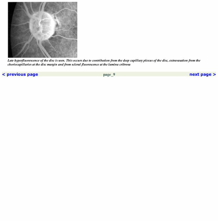
Late hyperfluorescence of the disc is seen. This occurs due to contribution from the deep capillary plexus of the disc, extravasation from thechoriocapillaries at the disc margin and from scleral fluorescence at the lamina cribrosa
< previous page page_9 next page >

< previous page page_10 next page >
Page 103 Abnormal FluorescenceHYPOFLUORESCENCEBlocked HypofluorescenceBlocked Retinal and Choroidal Fluorescence
The large subhyaloid hemorrhage in the inferior macula blocks the underlying retinal and choroidal fluorescence through all phases of theangiogram. However, the subretinal hemorrhage along the superotemporal arcade blocks only the underlying choroidal fluorescenceBlocked Choroidal Fluorescence
The lipofuscin like material in the retinal pigment epithelium in patients with Stargardt’s dystrophy, blocks all the underlying choroidalfluorescence. Choroidal fluorescence is seen only in areas of deficient retinal pigment epithelium at the fovea—an example of transmission orwindow defectBlocked Disc Fluorescence
Pigment in the melanocytoma at the disc blocks all the fluorescence emanating from the underlying disc vessels
< previous page page_10 next page >

< previous page page_11 next page >
Page 11HYPOFLUORESCENCEVascular Filling DefectRetinal Vascular Filling Defect
Areas of non-filling of the retinal vessels due to branch retinal vein occlusion, appear hypofluorescent on the angiogramChoroidal Vascular Filling Defect
Occlusion of the posterior ciliary artery gives rise to hypofluorescence due to non-filling of the affected segment of the choroid. Hypofluorescencedue to choroidal non-filling can also occur in conditions like choroideremia where the choriocapillaries are absentDisc Vascular Filling Defect
Absence of vessels at the optic nerve head pit makes it appear hypofluorescent
Absence of filling of superior half of the disc giving rise to hypofluorescence, is seen in patients with AIONPreinjection Hyperfluorescence
The dehemoglobinised blood reflects sufficient blue light to pass through the barrier filter causing it to appear hyperfluorescent in thepreinjection photograph. This can occur despite the filters being well matchedPreinjection hyperfluorescence can be either due to autofluorescence or pseudofluorescenceAutofluorescence is an innate property of certain structures to emit fluorescent light even in the absence of sodium fluorescein. Optic nerve headdrusen and astrocytic hamartoma are examples of autofluorescencePseudofluorescence occurs due to mismatched barrier and exciter filters
< previous page page_11 next page >
< previous page page_12 next page >
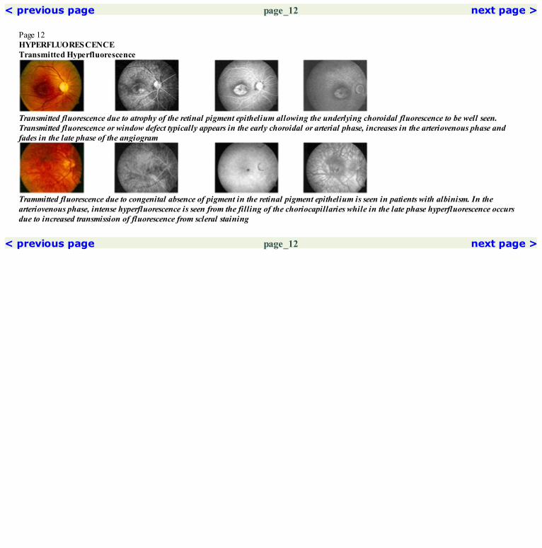
Page 12HYPERFLUORESCENCETransmitted Hyperfluorescence
Transmitted fluorescence due to atrophy of the retinal pigment epithelium allowing the underlying choroidal fluorescence to be well seen.Transmitted fluorescence or window defect typically appears in the early choroidal or arterial phase, increases in the arteriovenous phase andfades in the late phase of the angiogram
Trammitted fluorescence due to congenital absence of pigment in the retinal pigment epithelium is seen in patients with albinism. In thearteriovenous phase, intense hyperfluorescence is seen from the filling of the choriocapillaries while in the late phase hyperfluorescence occursdue to increased transmission of fluorescence from scleral staining
< previous page page_12 next page >

< previous page page_13 next page >
Page 13Hyperfluorescence Due to Leakage
Hyperfluorescence is seen at the disc due to leakage from neovascularization at the disc; the hyperfluorescence (with fuzzy margins) increases inthe late phase of the angiogram
Hyperfluorescence is seen due to presence of neovascularization in the subretinal space. This neovascular membrane increases inhyperfluorescence as the angiogram progresses and is intensely hyperfluorescent in the late filmsHyperfluorescence Due to Pooling
Pooling of the dye in the subretinal space is seen in eyes with central serous retinopathy. Pooling is accumulation of the dye in an anatomicalspace
Pooling of the dye in the sub-RPE space is seen in eyes with pigment epithelial detachmentHyperfluorescence Due to Staining
Staining is attachment of the dye molecule to the tissue. It can occur normally in structures like the sclera or in pathological states like stainingof the scar as seen in the above case. Unlike leakage, the margins of hyperfluorescence due to staining do not go beyond the scar
< previous page page_13 next page >

< previous page page_14 next page >
Page 14This page intentionally left blank.
< previous page page_14 next page >

< previous page page_15 next page >
Page 15

Section 2 Macular Disorders
< previous page page_15 next page >

< previous page page_16 next page >
Page 16This page intentionally left blank.
< previous page page_16 next page >
< previous page page_17 next page >
Page 174 Drusen
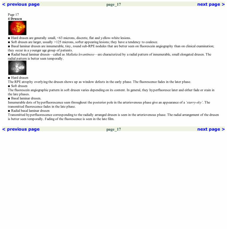
■ Hard drasen are generally small, <63 microns, discrete, flat and yellow-white lesions.■ Soft drusen are larger, usually >125 microns, softer appearing lesions; they have a tendency to coalesce.■ Basal laminar drusen are innumerable, tiny, round sub-RPE nodules that are better seen on fluorescein angiography than on clinical examination;they occur in a younger age group of patients.■ Radial basal laminar drusen—called as Mallatia levantinese—are characterized by a radial pattern of innumerable, small elongated drusen. Theradial pattern is better seen temporally.
■ Hard drasenThe RPE atrophy overlying the drusen shows up as window defects in the early phase. The fluorescence fades in the later phase.■ Soft drusenThe fluorescein angiographic pattern in soft drusen varies depending on its content. In general, they hyperfluoresce later and either fade or stain inthe late phases.■ Basal laminar drusen.Innumerable dots of hyperfluorescence seen throughout the posterior pole in the arteriovenous phase give an appearance of a ‘starry-sky’. Thetransmitted fluorescence fades in the late phase.■ Radial basal laminar drusenTransmitted hyperfluorescence corresponding to the radially arranged drusen is seen in the arteriovenous phase. The radial arrangement of the drusenis better seen temporally. Fading of the fluorescence is seen in the late film.
< previous page page_17 next page >

< previous page page_18 next page >
Page 18Case 1. A 48-year-old male presented with gradual decrease in vision in both the eyes over the past one year. Visual acuity was 6/12; N12 in botheyes.
Fundus photograph of the right eye showing multiple discrete drusen at the fovea
Arteriovenous phase showing discrete early hyperfluorescence corresponding to the drusen location. Note increased number of drusen evident onthe angiogram as compared to clinical appearance
Fading of the hyperfluorescence is seen in the late phase

Red free photograph of the right eye showing the drusen
The hyperfluorescence at the drusen has increased in the mid arteriovenous phaseCOMMENTThe RPE atrophy overlying the drusen allows the background choroidal fluorescence to be seen as transmitted hyperfluorescence in theangiogram.
< previous page page_18 next page >

< previous page page_19 next page >
Page 19Case 2. A 49-year-old male presented with history of reduced vision in the right eye of one month duration. Visual acuity was 1/60 in the right eye and6/6; N6 in the left eye. The right eye had a subfoveal CNVM, and the left eye, drusen at the macula.
Fundus photograph of the left eye showing innumerable discrete hard drusen scattered throughout the macula
Early hyperfluorescene corresponding to the drusen is seen in the arteriovenous phase
Fading of the hyperfluorescence is seen in the late phase

Red free photograph of the left eye showing the drusen
Increased hyperfluorescence is seen in the mid arteriovenous phaseCOMMENTThe hyperfluorescence seen in the angiogram corresponds to the drusen seen cilinically.
< previous page page_19 next page >

< previous page page_20 next page >
Page 20Case 3. A 58-year-old female presented with gradual reduction in vision in both the eyes over the last two years; visual acuity was 6/36; N10 in theright eye and 6/6; N6 in the left eye. Central lens changes were noted in the right eye.
Fundus photograph of the right eye showing multiple discrete hard drusen
Early hyperfluorescence is seen corresponding to the drusen in the early arteriovenous phase of the angiogram
Fading of the hyperfluorescence is seen in the late phase of the angiogram

Red free photograph of the right eye
Increased hyperfluorescence is seen in the mid arterioveneous phaseCOMMENTHard drusen show early hyperfluorescence which increases in the arteriovenous phase and fades in the late phase of the angiogram.
< previous page page_20 next page >

< previous page page_21 next page >
Page 21Left eye of the patient
Fundus photograph of the left eye showing multiple discrete hard drusen at the macula
Hyperfluorescene due to transmission defect is noted corresponding to the drusen in the arteriovenous phase of the angiogram
Red free photograph of the left eye

Late phase shows reduction in the intensity of the hyperfluorescence
< previous page page_21 next page >

< previous page page_22 next page >
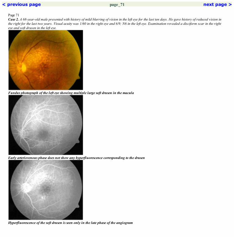
Page 22Case 4. A 68-year-old male presented with history of mild blurring of vision in the left eye for the last ten days. He gave history of reduced vision inthe right eye for the last two years. Visual acuity was 1/60 in the right eye and 6/9; N6 in the left eye. Examination revealed a disciform scar in theright eye and soft drusen in the left eye.
Fundus photograph of the left eye showing multiple large soft drusen in the macula
Early arteriovenous phase does not show any hyperfluorescence corresponding to the drusen
Hyperfluorescence at the drusen is seen only in the late phase of the angiogram

Red free photograph of the left eye
Faint hyperfluorescence is seen around the fovea in the arteriovenous phase. However, the drusen are still not made outCOMMENTDue to the hydrophobic nature of the soft drusen material, the entry of the dye into the drusen is delayed. Hence the soft drusen do not showhyperfluorescence until the late stages.
< previous page page_22 next page >

< previous page page_23 next page >
Page 23Case 5. A 56-year-old male presented with metamorphopsia in the left eye for the past four months; visual acuity was 6/9; N6. Fundus examinationshowed a drusenoid pigment epithelial detachment.
Fundus photograph of the left eye showing two drusenoid RPE detachments superior and inferior to the foveal center. Surrounding drusen areseen. A pigment clump is seen inferonasal to the foveal center
Arteriovenous phase showing filling up of the drusenoid detachments. Blocked hypofluorescence is seen within these due to pigment.Hyperfluorescene is seen in the rest of the macula corresponding to the drusen
Red free photograph of the left eye

Late phase of the angiogram showing persistent hyperfluorescence at the drusenoid detachment. Hyperfluorescence has faded elsewhereCOMMENTConfluence of adjacent soft drusen results in a large drusenoid type of detachment.
< previous page page_23 next page >
< previous page page_24 next page >
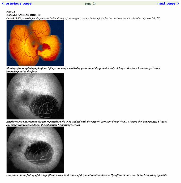
Page 24BASAL LAMINAR DRUSENCase 6. A 37-year-old female presented with history of noticing a scotoma in the left eye for the past one month; visual acuity was 6/9; N6.
Montage fundus photograph of the left eye showing a mottled appearance at the posterior pole. A large subretinal hemorrhage is seeninferotemporal to the fovea
Arteriovenous phase shows the entire posterior pole to be studded with tiny hyperfluorescent dots giving it a ‘starry-sky’ appearance. Blockedchoroidal fluorescence due to the subretinal hemorrhage is seen
Late phase shows fading of the hyperfluorescence in the area of the basal laminar drusen. Hypofluorescence due to the hemorrhage persists
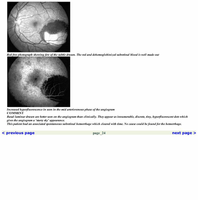
Red free photograph showing few of the subtle drusen. The red and dehemoglobinized subretinal blood is well made out
Increased hyperfluorescence in seen in the mid arteriovenous phase of the angiogramCOMMENTBasal laminar drusen are better seen on the angiogram than clinically. They appear as innumerable, discrete, tiny, hyperfluorescent dots whichgives the angiogram a ‘starry sky’ appearance.This patient had an associated spontaneous subretinal hemorrhage which cleared with time. No cause could be found for the hemorrhage.
< previous page page_24 next page >

< previous page page_25 next page >
Page 25RADIAL BASAL LAMINAR DRUSEN (MALLATIA LEVANTINESE)Case 7. A 34-year-old male presented with reduced vision in both the eyes for the past several years; visual acuity was 6/60; N36 in both eyes.
Fundus photograph of the right eye showing a large number of drusen at the posterior pole. Scar with pigmentation is noted at the fovea. Thedrusen temporally are arranged in a radiating pattern
Arteriovenous phase showing the faint radiating lines at the periphery of the lesion. Central blocked fluorescence is seen due to the scar andpigment
Red free photography of the right eye. The radial arrangement of the drusen temporally is better seen
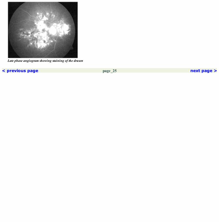
Late phase angiogram showing staining of the drusen
< previous page page_25 next page >

< previous page page_26 next page >
Page 26Left eye of the patient.
Fundus photograph of the left eye showing similar features as the right eye
Early arteriovenous phase showing the peripheral radial spokes. Central hypofluorescence due to the scar and pigment is seen
Late phase showing staining of the central scar. The hyperfluorescence at the radial spokes hasfaded

Red free photograph of the left eye
The hyperfluorescence at radial spokes has increased in the mid arteriovenous phaseCOMMENTThe typical radiating spoke like pattern of arrangement of drusen is seen more clearly in the temporal macula.
< previous page page_26 next page >

< previous page page_27 next page >
Page 27CLINICAL NUGGETS1. Patients with soft drusen are more likely to develop choroidal neovascular membrane.2. Amsler grid chart is useful in following patients with drusen.SELECTED READINGS1. Bressler NM, Bressler SB, Fine SL. Age related macular degeneration. Surv Ophthalmol 32:375–413, 1988.2. Gass JDM. Stereoscopic atlas of macular diseases. Diagnosis and treatment, Vol 2, ed 4 1997, CV Mosby, Chapter 3, pp 78–79.3. Folk JC: Aging macular degeneration: clinical features of treatable diseases. Ophthalmology 92:594–602, 1985.4. Sarks SH: Drusen and their relationship to senile macular degeneration Austral J Ophthalmol 8:117–130, 1980.5. Pauleikhoff D, Barondes MJ, Minassian D et al: Drusen as risk factors in age related macular disease. Am J Ophthalmol 109:38–43, 1990.6. Tanenbaum HL, Eshagian M, Senile disciform degeneration of macula: the other eye—a fluorescein angiographic study: Can J. Ophthalmol 7:280–284, 1972.7. Arnold JJ, Quaranta M, Soubrane G, Sarks SH, Coscos G. ICG angiography of drusen, Am J. Ophthalmol 124:344–356, 1997.8. Barendes M, Pauleikhoff D, Chisholm I, C Minassian D, Bird AC: Bilaterality of drusen, BJO 74:180–182, 1990.
< previous page page_27 next page >

< previous page page_28 next page >
Page 285 Epiretinal Membranes
■ Epiretinal membranes can be either idiopathic or secondary.■ Secondary causes include retinal vascular occlusions, diabetic retinopathy, following surgical procedures like retinal detachment surgery or cataractsurgery, ocular inflammatory conditions and following laser or cryotherapy.■ It varies in severity from very subtle membranes to thick membranes covering the macular region.
■ The vascular tortuosity caused by the epiretinal membranes is very well seen on the fluorescein angiogram.■ The fovea may be displaced and the perifoveal capillary network distorted.■ Varying degree of leakage from the retinal capillaries can be seen in the area of traction.■ Late cystoid changes may be evident.■ In cases of epiretinal membranes secondary to vascular occlusion, the diagnosis is quite evident on the angiogram due to the presence of areas ofcapillary non-perfusion, microaneurysms or collateral vessels.
< previous page page_28 next page >

< previous page page_29 next page >
Page 29Case 1. A 42-year-old male presented with distortion of vision in the right eye for the past three months; visual acuity was 6/12:N8.
Fundus photograph of the right eye showing an idiopathic epiretinal membrane at the macula
The macula retinal vessels appear distorted in the arteriovenous phase of the angiogram
Red free photograph showing the distortion of the macular blood vessels caused by the epiretinal membrane

Late phase of the angiogram showing accumulation of the dye at the foveaCOMMENTDistortion of the macular vessels and late CME are common angiographic findings in cases of epiretinal membrane.
< previous page page_29 next page >

< previous page page_30 next page >
Page 30Case 2. A 74-year-old female (known case of BRVO) presented with history of decreased vision in the left eye for the past one year; visual acuity was1/60.
Fundus photograph of the left eye showing a thick epiretinal membrane at the macula
Marked distortion of the retinal vessels is seen. Few hyperfluorescent dots caused by microaneurysms are seen. Areas of capillary non-perfusionare noted in the superior part of the macula.
Increased leakage of the dye is seen in the late phase
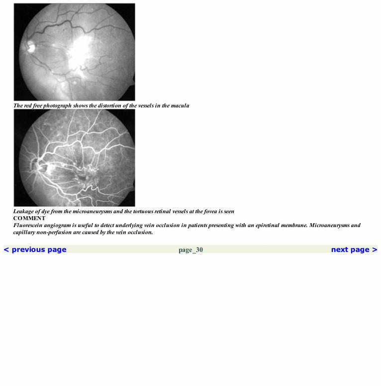
The red free photograph shows the distortion of the vessels in the macula
Leakage of dye from the microaneurysms and the tortuous retinal vessels at the fovea is seenCOMMENTFluorescein angiogram is useful to detect underlying vein occlusion in patients presenting with an epiretinal membrane. Microaneurysms andcapillary non-perfusion are caused by the vein occlusion.
< previous page page_30 next page >

< previous page page_31 next page >
Page 31CLINICAL NUGGETS1. Use of red free illumination during slit lamp biomicroscopy facilitates easy recognition of the epiretinal membranes.2. Superficial retinal hemorrhages and cystoid macular edema can occur in patients with epiretinal membranes.3. Vitrectomy with surgical removal of the membrane is the treatment of choice for those epiretinal membranes causing visual morbidity.4. The reduction in visual acuity caused by epiretinal membrane is related to distortion produced in the outer retinal layers and does not depend onthe size or degree of transluscency of the membrane.5. OCT is very useful to image the epiretinal membrane.SELECTED READING1. Wise GN: Pre retinal macular fibrosis: An analysis of 90 cases. Trans Ophthalmol Soc UK; 92:131–140, 1972.
< previous page page_31 next page >

< previous page page_32 next page >
Page 326 Macular Hole
■ Various causes of macular hole are idiopathic, trauma, accidental foveal burns with laser, long-standing cystoid macular edema and myopia.■ Idiopathic macular holes are classified as follows:Stage 1A and 1B—Impending macular hole.Stage 2—Small central or eccentric full thickness macular hole.Stage 3—Large full thickness macular hole with no posterior vitreous detachment.Stage 4—Large full thickness macular hole with posterior vitreous detachment.
■ Depends on the status of the RPE at the base of the macular hole.■ If the RPE is healthy and intact, no change in the normal fluorescence is seen on the angiogram.■ If RPE atrophy has occurred at the base, corresponding hyperfluorescence due to transmission defect is seen in the angiogram; thishyperfluorescence fades in the late film.
< previous page page_32 next page >

< previous page page_33 next page >
Page 33Case 1. A 54-year-old female presented with reduced vision in the right eye for the past four month; visual acuity was 6/60; N36.
Fundus photograph of the right eye showing a full thickness macular hole at the fovea surrounded by a broad cuff of subretianal fluid
Early arteriovenous phase showing hyperfluorescence due to transmission defect at the base of the macular hole
Magnified view of the macular hole showing the hyperfluorescence due to transmission defect at the base of the macular hole
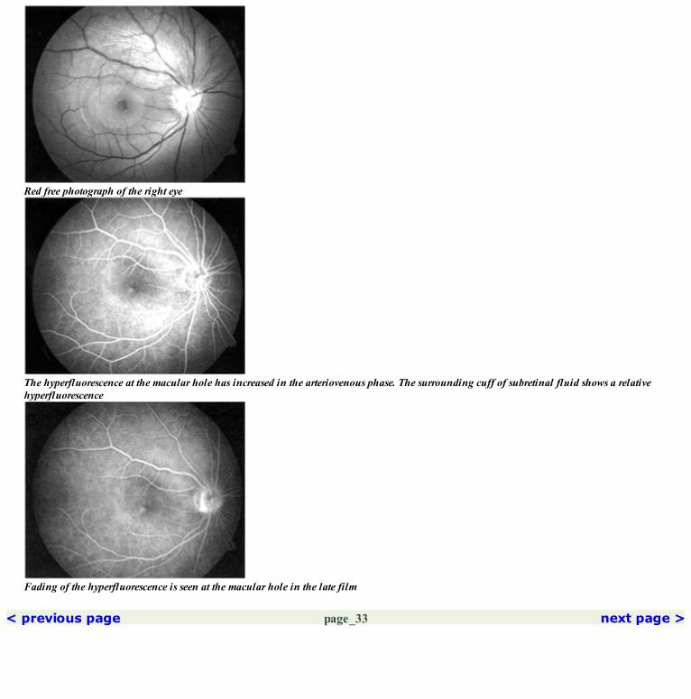
Red free photograph of the right eye
The hyperfluorescence at the macular hole has increased in the arteriovenous phase. The surrounding cuff of subretinal fluid shows a relativehyperfluorescence
Fading of the hyperfluorescence is seen at the macular hole in the late film
< previous page page_33 next page >

< previous page page_34 next page >
Page 34Case 2. A 36-year-old male presented with history of noticing drop in vision in the right eye following accidental exposure to the laser beam of a rangefinding device; visual acuity was 6/9; N6. A full thickness macular hole was noted with minimal RPE alteration at the base.
Fundus photograph of the right eye showing the full thickness macular hole
A faint rim of hyperfluorescence is seen at the edge of the macular hole in the early arteriovenous phase
Some fading of the hyperfluorescence is seen in the later part of the angiogram

Red free photograph showing some RPE alterations at the base of the macular hole
Arteriovenous phase showing increase in the hyperfluorescence at the rim. A faint hyperfluorescence is seen at the base of the macular hole
< previous page page_34 next page >

< previous page page_35 next page >
Page 35Two-months later, the vision had dropped to 6/24; N36. Increased RPE alterations were seen at the base of the macular hole.
Fundus photograph of the right eye showing the full thickness macular hole with increased RPE alterations at the base
Hyperfluorescence due to transmission defect is seen at the base of the macular hole
Red free photograph of the right eye

Fading of the transmitted hyperfluorescence is seen in the late phase of the angiogramCOMMENTThe RPE alteration at the base of the macular hole results in transmission defect.
< previous page page_35 next page >

< previous page page_36 next page >
Page 36CLINICAL NUGGETS1. A positive Watzke Allen sign is seen in 95% of eyes with full thickness macular hole.2. All patients will report a positive laser beam (50—microns) sign.3. Stage 1 macular holes can regress spontaneously.4. Vitrectomy with or without peeling of the internal limiting membrane with gas tamponade is useful in eyes with Stage 2–4 macular holes.SELECTED READINGS1. Gass JDM: Idiopathic senile macular hole, it’s early stages and pathogenesis: Arch Ophthalmol; 106:629–639, 1988.2. Gass JDM: Reappraisal of biomicroscopic classification of stages of development of macular hole: Am J Ophthalmol; 119:752–759, 1995.3. Stereoscopic Atlas of Macular Diseases; Diagnosis and Treatment: Gass JDM: Page 922:4th edition; 1997, CV Mosby, St. Louis4. Gass JDM, Joondeph BC: Observations concerning patients with suspected impending macular holes: Am J Ophthalmol; 109: 638–646:1990
< previous page page_36 next page >

< previous page page_37 next page >
Page 377 Myopia
■ Degenerative myopia is associated with increased axial length and progressive choroidal degeneration at the posterior pole.■ Tesselated fundus, tilted and oblique optic disc, temporal crescent or myopic conus, breaks in Bruch’s membrane (lacquer cracks), atrophy of theRPE and choroid at the posterior pole, and posterior staphyloma are common changes in degenerative myopia.■ Choroidal neovascularization can occur.■ Pathogenesis is not clearly understood and could be due to heredo-degenerative factors.
■ Lacquer cracks show irregular hyperfluorescence in the early phase of the angiogram. This increases in the arteriovenous phase and fades to someextent in the late phase. They are linear or stellate and may be multiple.■ Large areas of atrophy at the posterior pole may be hypofluorescent in the early stages due to the atrophy of the choriocapillaries. The largerchoroidal vessels may be seen through. Scleral staining may be seen in the late phase.
< previous page page_37 next page >
< previous page page_38 next page >
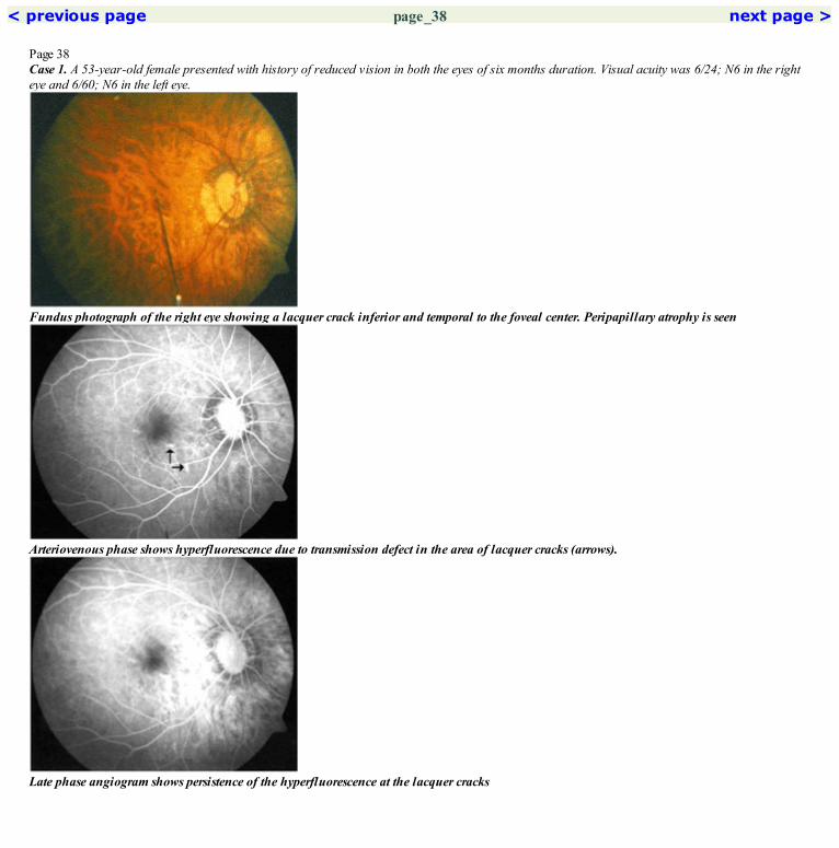
Page 38Case 1. A 53-year-old female presented with history of reduced vision in both the eyes of six months duration. Visual acuity was 6/24; N6 in the righteye and 6/60; N6 in the left eye.
Fundus photograph of the right eye showing a lacquer crack inferior and temporal to the foveal center. Peripapillary atrophy is seen
Arteriovenous phase shows hyperfluorescence due to transmission defect in the area of lacquer cracks (arrows).
Late phase angiogram shows persistence of the hyperfluorescence at the lacquer cracks

Red free photograph of the right eye
Increased hyperfluorescence is seen at these lesions in the late arteriovenous phase
< previous page page_38 next page >
< previous page page_39 next page >
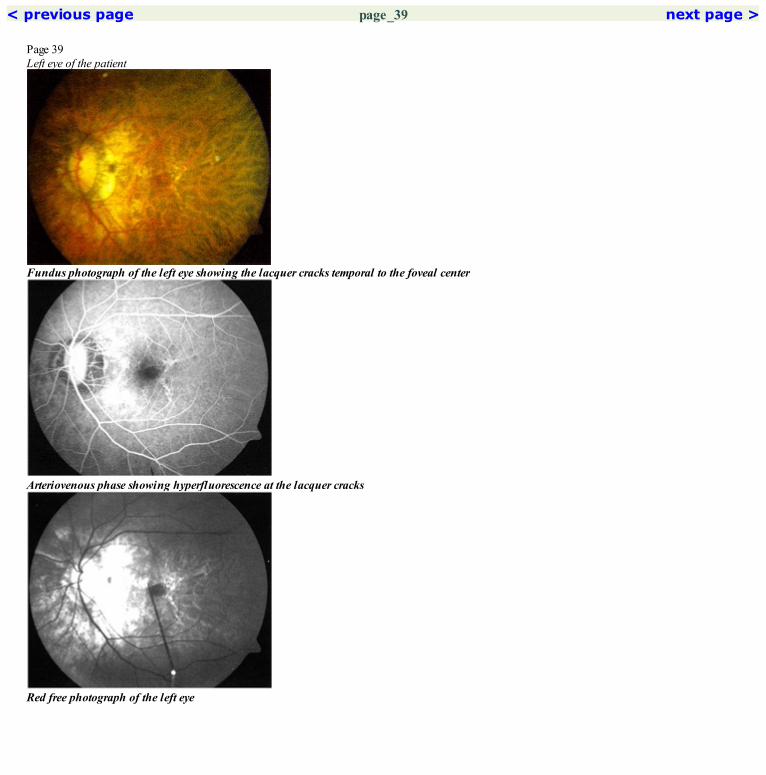
Page 39Left eye of the patient
Fundus photograph of the left eye showing the lacquer cracks temporal to the foveal center
Arteriovenous phase showing hyperfluorescence at the lacquer cracks
Red free photograph of the left eye
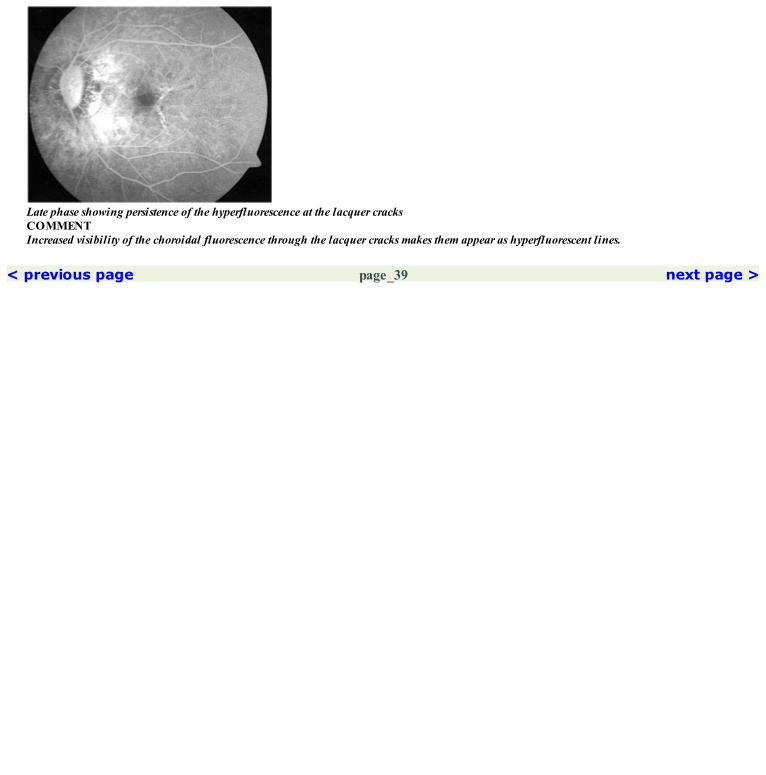
Late phase showing persistence of the hyperfluorescence at the lacquer cracksCOMMENTIncreased visibility of the choroidal fluorescence through the lacquer cracks makes them appear as hyperfluorescent lines.
< previous page page_39 next page >

< previous page page_40 next page >
Page 40CLINICAL NUGGETS1. Progressive myopia is characterized by increased axial length with progressive choroidal degeneration in the posterior pole.2. Fuch’s spot, which are round or oval areas of subretinal hyperpigmentation, may be sequelae of previous subretinal hemorrhage or choroidalnonvascular membranes.3. Spontaneous subretinal hemorrhage can occur at the fovea without any evidence of a neovascular membrane; these usually resolve spontaneously.SELECTED READINGS1. Levy JH, Pollock HM, Curtin BJ. The Fuchs’ Spot. An ophthalmoscopic and Fluorescein Angiographic study. Ann Ophthalmol; 9: 1433–1443,1977.2. Spitznas M, Böker T: Idiopathic posterior subretinal neovascularisation (IPSN) is related to myopia. Graefes Arch Clin Exp Ophthalmol.229:536–538, 1991.3. Quaranta M, Arnold J, Coscas G, Francis C, et al. Indocyanine green angiographic features of pathologic myopia. Am J Ophthalmology. 122:663–671, 1996.4. Ohno-Matsui K, Tokoro T. The progression of lacquer cracks in pathologic myopia. Retina; 16(1): 29–37, 1996.
< previous page page_40 next page >

< previous page page_41 next page >
Page 418 Polypoidal Choroidal Vasculopathy (PCV)
■ Lesion in PCV arises from the inner choroidal plexus and consists of a network of choroidal vessels which terminate in spheroidal polyp likelesions.■ Clinically the polyps are seen as orange-red subretinal nodules.■ Patients usually present with hemorrhagic pigment epithelial detachments associated with hard exudates.
■ Fluorescein angiogram shows areas of stippled hyperfluorescence in the region of the polyps which increases in intensity and may become fuzzyin the late films.■ Occasionally, polyps may be seen in the fluorescein angiogram.
■ ICG angiogram is essential for detecting the choroidal network of vessels and the polyps.■ Polyps may not be seen in the early phase of the ICG.■ In the mid-phase they are seen to appear usually in clusters at the termination of the choroidal network of vessels. Occasionally the polyp may besolitary and large.■ Late phase angiogram shows either a washout of the dye from the polyp with staining of its walls or may show retention of the dye within thepolyp with leakage into the surrounding tissue.
< previous page page_41 next page >

< previous page page_42 next page >
Page 42Case 1. A 64-year-old male presented with hazy vision in the left eye for the past six months; visual acuity was 6/12; N8.
Fundus photograph of the left eye showing orange-red subretinal lesion inferior and inferotemporal to the foveal center. Associated minimalhard exudates are also seen
Arteriovenous phase shows a stippled hyperfluorescence at the fovea. Small polyp like lesions can be made out temporally
Mid-phase ICG angiography shows a small central network of choroidal vessels terminating as polyps at the periphery. The temporal polyps aremore prominent

Red free photograph of the left eye
Late phase shows increased fuzzy hyperfluorescence especially temporally indicative of leakage of dye from the polyps
Late phase ICG angiogram showing some leakage of the dye from the temporal polyps
< previous page page_42 next page >

< previous page page_43 next page >
Page 43Case 2. A 62-year-old male presented with reduced vision in the left eye for the past two months; visual acuity was 6/9; N8.
Fundus photograph of the left eye showing orange-red subretinal lesion just nasal to the foveal center. A pocket of subretinal fluid is noted in theinferior macula. Hard exudates are seen superiorly
Arteriovenous phase showing hyperfluorescent spots at the fovea
Mid-phase ICG shows a cluster of polyps. The network of choroldal vessels is not made out. The polyps roughly correspond to the hyperfluorescentspots seen on the fluorescein angiogram

Red free photograph of the left eye
Late phase shows increased intensity of the hyperfluorescent spots with leakage of the dye into the surrounding tissue
Late phase ICG picture shows retention of the dye within the polyps with minimal leakage through its walls
< previous page page_43 next page >
< previous page page_44 next page >
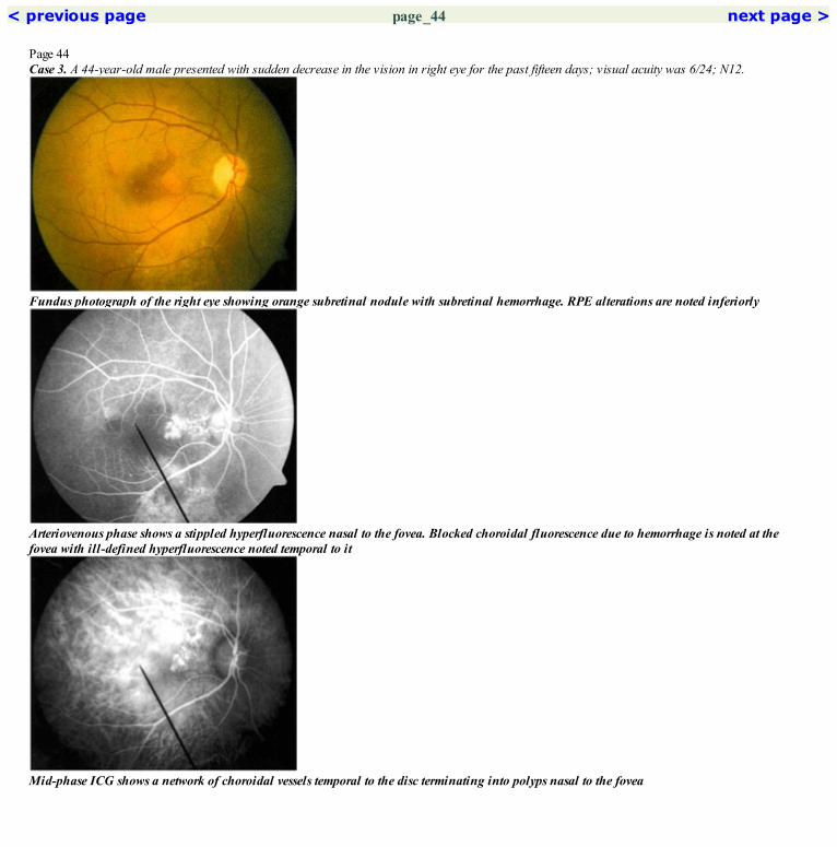
Page 44Case 3. A 44-year-old male presented with sudden decrease in the vision in right eye for the past fifteen days; visual acuity was 6/24; N12.
Fundus photograph of the right eye showing orange subretinal nodule with subretinal hemorrhage. RPE alterations are noted inferiorly
Arteriovenous phase shows a stippled hyperfluorescence nasal to the fovea. Blocked choroidal fluorescence due to hemorrhage is noted at thefovea with ill-defined hyperfluorescence noted temporal to it
Mid-phase ICG shows a network of choroidal vessels temporal to the disc terminating into polyps nasal to the fovea

Red free photograph of the right eye. The inferior RPE alterations are well seen
Intense fuzzy hyperfluorescence is seen nasal to the fovea. Faint hyperfluorescence is seen in the temporal macula. Transmitted hyperfluorescenceis noted inferiorly
The polyps nasal to the fovea have emptied with staining of their walls seen in the late phase of ICG angiogram. Some dye retention is seen in thetemporal macula probably in small polyps which are not well seen in the earlier phases of the angiogram
< previous page page_44 next page >

< previous page page_45 next page >
Page 45Case 4. A 60-year-old female came for a routine eye examination; visual acuity was 6/6; N6 in both eyes.
Fundus photograph of the left eye showing a partially hemorrhagic PED nasal to the disc. A serous PED is seen along with inferotemporalarcade
Arteriovenous phase shows filling of the PED inferotemporal to the disc. Filling of nasal PED is also seen with some blockedfluorescence due tohemorrhage seen adjacent to the disc
Mid-phase ICG angiogram showing a large network of choroidal vessels ending in clusters of polyps
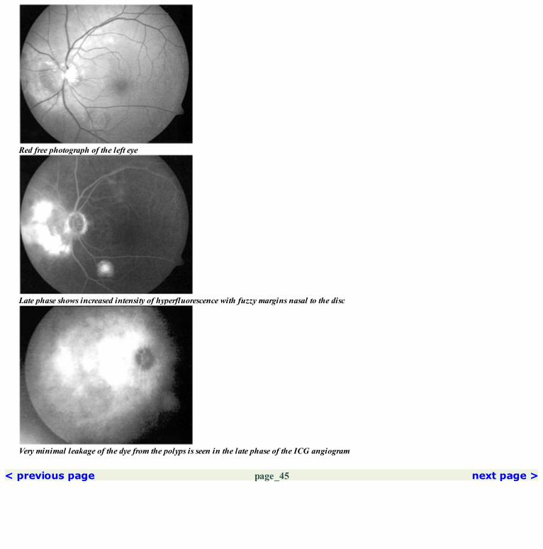
Red free photograph of the left eye
Late phase shows increased intensity of hyperfluorescence with fuzzy margins nasal to the disc
Very minimal leakage of the dye from the polyps is seen in the late phase of the ICG angiogram
< previous page page_45 next page >
< previous page page_46 next page >
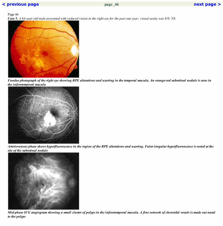
Page 46Case 5. A 64-year-old male presented with reduced vision in the right eye for the past one year; visual acuity was 6/9; N8.
Fundus photograph of the right eye showing RPE alterations and scarring in the temporal macula. An orange-red subretinal nodule is seen inthe inferotemporal macula
Arteriovenous phase shows hyperfluorescence in the region of the RPE alterations and scarring. Faint irregular hyperfluorescence is noted at thesite of the subretinal nodule
Mid-phase ICG angiogram showing a small cluster of polyps in the inferotemporal macula. A fine network of choroidal vessels is made out nasalto the polyps

Red free photograph of the right eye showing the dark subretinal nodule in the inferotemporal macula
Late phase shows staining of the scar. Very minimal increase of fluorescence is noted at the site of the nodule
Late phase of the ICG shows staining of the walls of the polyps
< previous page page_46 next page >

< previous page page_47 next page >
Page 47CLINICAL NUGGETS1. Indocyanine green angiography is essential for diagnosing polypoidal choroidal vasculopathy.2. Subretinal orange nodules and hemorrhagic PEDs are common findings in eyes with polypoidal choroidal vasculopathy.3. Photodynamic therapy is being tried for the treatment of polypoidal choroidal vasculopathy.SELECTED READINGS1. Uyama M, Wada M, Nagai Y, Matsubara T, Matsunaga H, Fukushima I, Takahashi K, Matsumura M. Polypoidal choroidal vasculopathy:natural history. Am J Ophthalmol 133(5): 639–48, 2002.2. Ciardella AP, Donsoff IM, Yannuzzi LA. Polypoidal choroidal vasculopathy. Ophthalmol Clin North Am 15(4): 537–54, 2002.3. Moorthy RS, Lyon AT, Rabb MF, Spaide RF, Yannuzzi LA, Jampol LM. Idiopathic polypoidal choroidal vasculopathy of the macula.Ophthalmology 1946.4. Yannuzzi LA, Sorenson J, Spaide RF, Lipson B. Idiopathic polypoidal choroidal vasculopathy (IPCV). Retina 10(1): 1–8. 98, 105(8): 1380–5,1990.
< previous page page_47 next page >

< previous page page_48 next page >
Page 489 Pigment Epithelial Detachment
■ Retinal pigment epithelial detachment (PED) can occur by itself or in association with central serous chorioretinopathy, age-related maculardegeneration or with polypoidal choroidal vasculopathy.■ It can be serous or hemorrhagic depending on the fluid that accumulates under the detached RPE.■ PED appears as a well-delineated, blister like round or oval area of RPE detachment, yellow or grey in color. A pink halo is usually seen aroundthe PED.■ Serous PEDs are usually around one fourth disc diameter in size.■ Hemorrhagic PEDs are darker appearing and usually larger in size.■ Long standing PEDs may show pigment migration along their walls.
■ Serous PED● Shows hyperfluorescence in the early phase of the angiogram. The entire extent of the PED is hyperfluorescent.● Increase in intensity of the fluorescence is seen as the angiogram progresses due to increased accumulation of the dye. The extent of the lesionremains the same.● Late stage shows pooling of the dye. The margins are still well-defined.● Irregular blocked fluorescence may be seen within the PED in longstanding cases. This is caused by pigment migration.■ PED associated with age-related macular degeneration● PED is slow filling.● Presence of an underlying CNVM is indicated by irregular filling of the PED, delayed filling of the PED or notching of the PED.
< previous page page_48 next page >

< previous page page_49 next page >
Page 49Case 1. A 39-year-old male presented with history of decreased vision in the right eye of one year duration; visual acuity was 6/9; N12.
Fundus photograph of the right eye shows a PED at the fovea
Early arteriovenous phase showing hyperfluorescence at the PED. The margins of the PED is well-defined
Marked increase in the intensity of the hyperfluorescence is seen within the PED. The margins of the PED remain well-defined
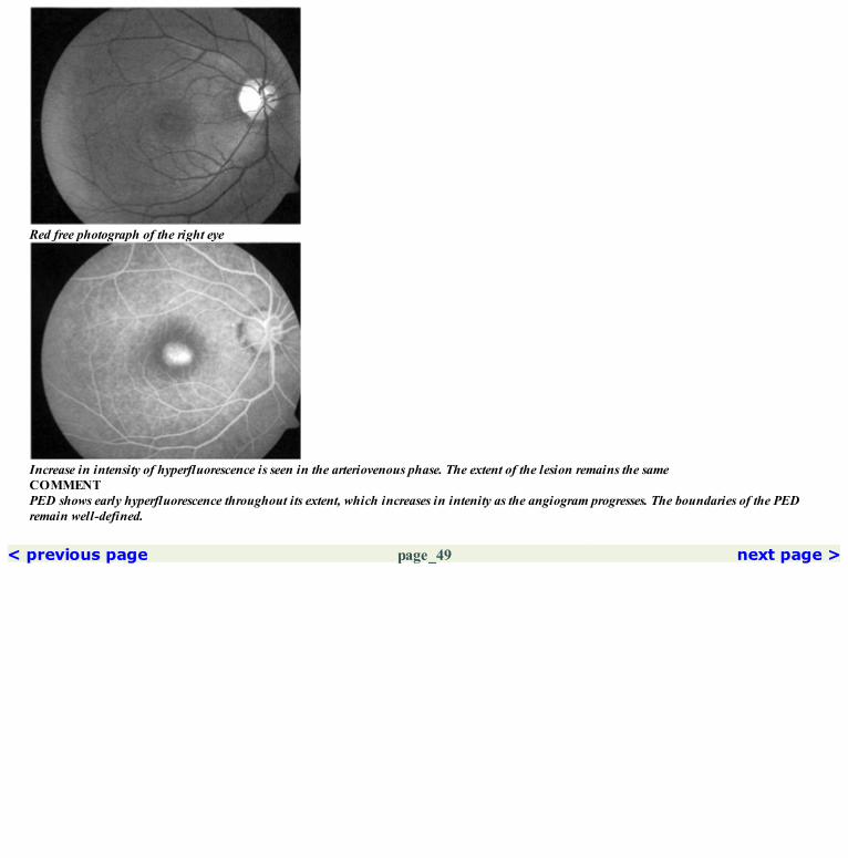
Red free photograph of the right eye
Increase in intensity of hyperfluorescence is seen in the arteriovenous phase. The extent of the lesion remains the sameCOMMENTPED shows early hyperfluorescence throughout its extent, which increases in intenity as the angiogram progresses. The boundaries of the PEDremain well-defined.
< previous page page_49 next page >

< previous page page_50 next page >
Page 50Case 2. A 39-year-old male presented with distortion of vision in the right eye for the past one month; visual acuity was 6/6; N6.
Fundus photograph of the right eye shows a PED at the fovea
Arteriovenous phase showing hyperfluorescence within the PED. Few patches of blocked fluorescence within this are due to pigment migrationalong the walls of the PED. Few hyperfluorescent transmission defects are seen inferonasal to the PED
Red free photograph clearly shows the extent of the PED

The hyperfluorescence within the PED has increased—consequently the areas of blocked fluorescence are better appreciatedPatient was seen again nine months later. Visual acuity was maintained at 6/6; N6
Arteriovenous phase of the same lesion shows a change in the pattern of blocked fluorescence within the PED
Late phase showing increased intensity of the hyperfluorescence in the PED. The hyperfluorescence inferonasal to the PED has faded indicativeof a transmission defect
< previous page page_50 next page >

< previous page page_51 next page >
Page 51Case 3. A 46-year-old male presented with difficulty in near vision in the right eye for the past six months; visual acuity was 6/6; N6.
Fundus photograph of the right eye showing a pocket of subretinal fluid superior to the fovea. Afew small PEDs are seen inferiorly. A very largePED is seen superotemporally, the lower edge of which is seen in the photograph
Arteriovenous phase shows filling of the small PEDs in the inferior and temporal part of the macula. Filling of dye is also seen in the lower partof the superotemporal PED
The intensity of hyperfluorescence in the large PED has increased in the late phase. An ink-blot type of central serous chorioretinopathy leak isseen in the superior part of the macula

Red free photograph shows the lower edge of the PED in the superotemporal quadrant
Filling of the large PED is seen. A point leake of dye is seen in the superior part of the maculaCOMMENTLarge, uniformaly filling PEDs are usually found in central serous chorioretinopathy.
< previous page page_51 next page >

< previous page page_52 next page >
Page 52Case 4. A 45-year-old male underwent an ocular examination for complaints of glare and floaters in both eyes for the past 6 months. Patient was noton any medication and did not have any systemic problem. Visual acuity was 6/5; N6 in both eyes.
Fundus photograph of the right eye shows innumerable PEDs scattered throughout the posterior pole
Early arteriovenous phase shows filling of the PEDs
Further increase in the intensity of the dye is seen in the late phase. PEDs are seen even beyond the superior arcade

PEDs are well seen in this red free photograph
Arteriovenous phase shows increased intensity of the flourescence within the PEDs
< previous page page_52 next page >

< previous page page_53 next page >
Page 53Left eye of the patient.
Fundus photograph of the left eye showing the scattered PEDs
Arterioveneous phase showing dye filling thePEDs some of which are seen nasal to the disc
Red free photograph of the left eye
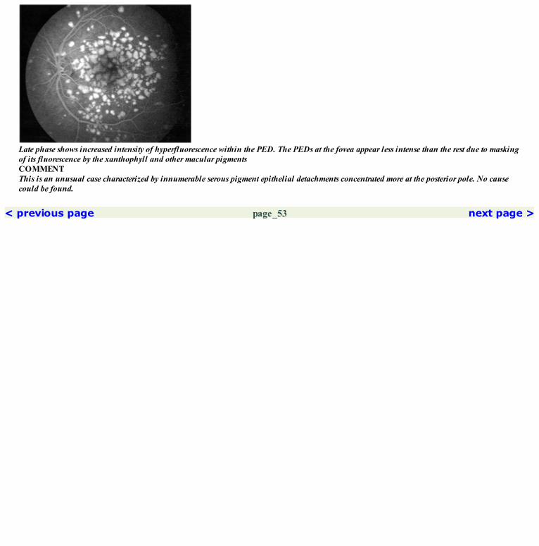
Late phase shows increased intensity of hyperfluorescence within the PED. The PEDs at the fovea appear less intense than the rest due to maskingof its fluorescence by the xanthophyll and other macular pigmentsCOMMENTThis is an unusual case characterized by innumerable serous pigment epithelial detachments concentrated more at the posterior pole. No causecould be found.
< previous page page_53 next page >

< previous page page_54 next page >
Page 54Case 5 A 56-year-old male presented with history of reduced vision in the left eye of three months duration; visual acuity was 6/18; N12.
Fundus photograph of the left eye showing a large PED just superior to the fovea. Turbid fluid was noted within the PED
Arteriovenous phase showing filling of the PED. Stippled hyperfluorescence is seen adjacent to the inferior border of the PED. Ahyperfluorescent point is seen along its superotemporal border
Arteriovenous phase shows a change in the shape and size of the PED two months later. The irregular hyperfluorescence at the fovea hasincreased causing a notch along the inferior border of the PED. Irregular hyperfluorescence is seen along the superior border
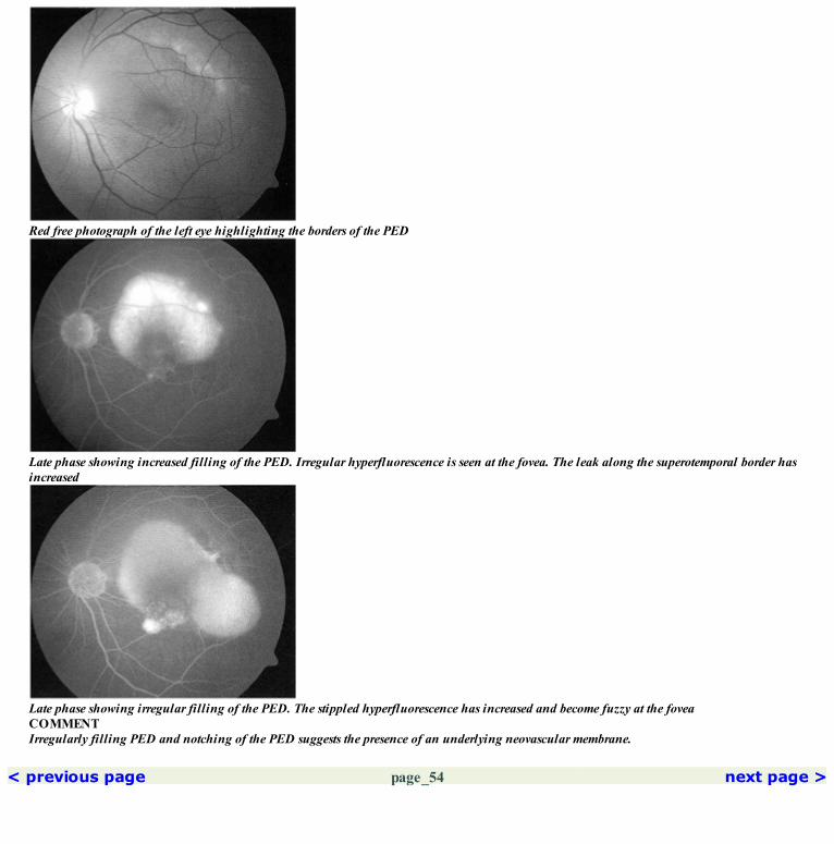
Red free photograph of the left eye highlighting the borders of the PED
Late phase showing increased filling of the PED. Irregular hyperfluorescence is seen at the fovea. The leak along the superotemporal border hasincreased
Late phase showing irregular filling of the PED. The stippled hyperfluorescence has increased and become fuzzy at the foveaCOMMENTIrregularly filling PED and notching of the PED suggests the presence of an underlying neovascular membrane.
< previous page page_54 next page >

< previous page page_55 next page >
Page 55CLINICAL NUGGETS1. Slow filling PEDs, notched PED and PED with a turbid fluid are highly suspicious of an underlying neovascular membrane.2. Laser photocoagulation at the edge of a PED carries a high risk of developing a RPE tear.SELECTED READINGS1. Gass JDM. Stereoscopic atlas of macular diseases. Diagnosis and treatment., Vol 2, ed 4, CV Mosby, 24–26, 1997.2. Gass JDM. Serous Retinal pigment epithelium detachment with a notch; a sign of occult choroidal neovascularisation. Retina 4:205–220, 1984.3. Coscas G, Koenig F, Soubrane G. The pre tear characteristics of pigment epithelial detachments; a study of 40 eyes. Arch Ophthalmol; 108:1687–1693, 1990.4. Nasrallah F, Jalkh AE, Trempe CL et al. Subretinal haemorrhage in atrophic ARMD. Am J Ophthalmol 107:38–41, 1989.5. Bennett SR, Folk JC, Blodi CF, Klugman M. Factors prognostic of visual outcome in patients with Subretinal haemorrhage. Am J Ophthalmol109:33–37, 1990.
< previous page page_55 next page >

< previous page page_56 next page >
Page 5610 RPE Rip
■ Retinal pigment epithelial rip or tears can occur in patients with a pigment epithelial detachment associated with central serous chorioretinopathyor with a choroidal neovascular membrane. It may follow laser photocoagulation as well.■ RPE layer is torn from one edge of the pigment epithelial detachment and scrolls under the opposite edge.
■ The area devoid of RPE appears hyperfluorescent due to transmission of the underlying choroidal fluorescence that is easily seen through.■ Staining of the choroidal tissue and the underlying sclera cause the rip to appear hyperfluorescent in the late phase.■ The area of scrolled RPE appears hypofluorescent throughout the angiogram. However if this scrolled edge harbours a choroidal neovascularmembrane, some irregular hyperfluorescence may be seen.
< previous page page_56 next page >
< previous page page_57 next page >
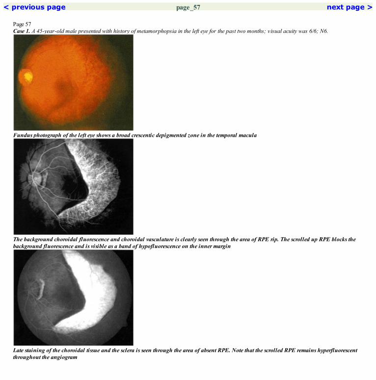
Page 57Case 1. A 45-year-old male presented with history of metamorphopsia in the left eye for the past two months; visual acuity was 6/6; N6.
Fundus photograph of the left eye shows a broad crescentic depigmented zone in the temporal macula
The background choroidal fluorescence and choroidal vasculature is clearly seen through the area of RPE rip. The scrolled up RPE blocks thebackground fluorescence and is visible as a band of hypofluorescence on the inner margin
Late staining of the choroidal tissue and the sclera is seen through the area of absent RPE. Note that the scrolled RPE remains hyperfluorescentthroughout the angiogram

Red free photograph shows the large RPE tear. The scrolled up RPE layer is seen along the inner margin of the tear
Increased hyperfluorescence is seen in the area of RPE rip. The hypofluorescence along the inner margins is better appreciatedCOMMENTAbscence of the overlying RPE in the region of the tear allows increased visibility of the choroidal fluorescence.The scrolled edge of the RPE blocks the background choroidal fluorescence throughout the angiogram.
< previous page page_57 next page >
< previous page page_58 next page >
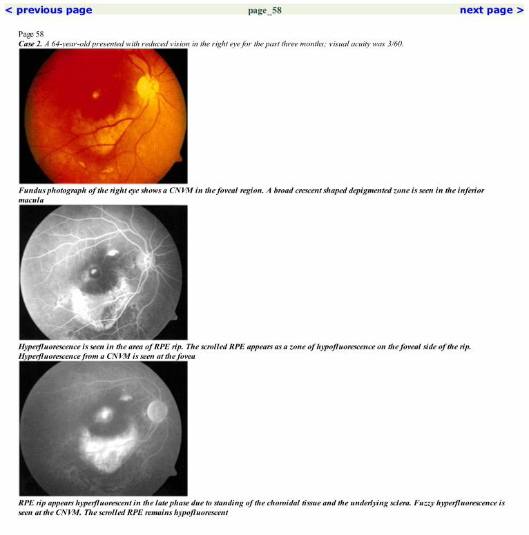
Page 58Case 2. A 64-year-old presented with reduced vision in the right eye for the past three months; visual acuity was 3/60.
Fundus photograph of the right eye shows a CNVM in the foveal region. A broad crescent shaped depigmented zone is seen in the inferiormacula
Hyperfluorescence is seen in the area of RPE rip. The scrolled RPE appears as a zone of hypofluorescence on the foveal side of the rip.Hyperfluorescence from a CNVM is seen at the fovea
RPE rip appears hyperfluorescent in the late phase due to standing of the choroidal tissue and the underlying sclera. Fuzzy hyperfluorescence isseen at the CNVM. The scrolled RPE remains hypofluorescent
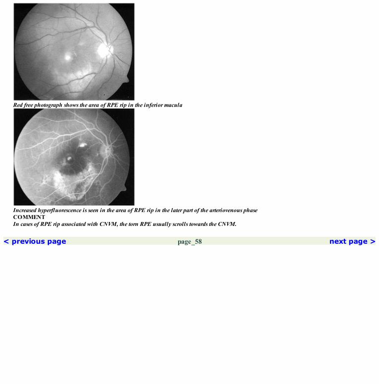
Red free photograph shows the area of RPE rip in the inferior macula
Increased hyperfluorescence is seen in the area of RPE rip in the later part of the arteriovenous phaseCOMMENTIn cases of RPE rip associated with CNVM, the torn RPE usually scrolls towards the CNVM.
< previous page page_58 next page >

< previous page page_59 next page >
Page 59CLINICAL NUGGETS1. RPE rip involving the fovea can cause a sudden drop in vision.2. There is no effective treatment for RPE rip.SELECTED READINGS1. Gass JDM. Stereoscopic atlas of macular diseases. Diagnosis and treatment. Vol 2, ed 4, CV Mosby, Chapter 3, 88–91, 1997.2. Gass JDM. Retinal pigment epithelium rip during krypton red photocoagulation. Am J Ophthalmol 98:700–706, 1984.3. Machemer R, Heriot W. RPE Tears through fovea with preservation of good visual acuity. Arch Ophthalmol 109:1492–1493, 1991.4. Bressler NM, Finkelstein D, Sunness JS et al. RPE Tears through the fovea with preservation of good visual acuity. Arch Ophthalmol 108:1694–1697, 1990.
< previous page page_59 next page >

< previous page page_60 next page >
Page 6011 Choroidal Rupture
■ It follows blunt injury to the eye.■ Due to distortion of the globe, the relatively inelastic Bruch’s membrane ruptures along with the closely apposed RPE and choriocapillaries.■ They are usually crescent shaped and occur concentric to the optic disc.■ Associated with intrachoroidal, subretinal or intraretinal hemorrhage.■ Can be associated with fibrous proliferation, RPE hyperplasia and development of choroidal neovascular membrane.
■ Early phase may show relative hypofluorescence due to absence of the choriocapillaries. The underlying larger choroidal vessels may be seenthrough.■ Late hyperfluorescence is seen due to staining of the tissue at the choroidal rupture and the sclera.■ Areas of RPE hyperplasia are associated with blocked fluorescence.■ Fibrous tissue within the rupture stains in the late film.■ CNVM appear as hyperfluorescent area which increases and has fuzzy margins in the late phase.
< previous page page_60 next page >
< previous page page_61 next page >
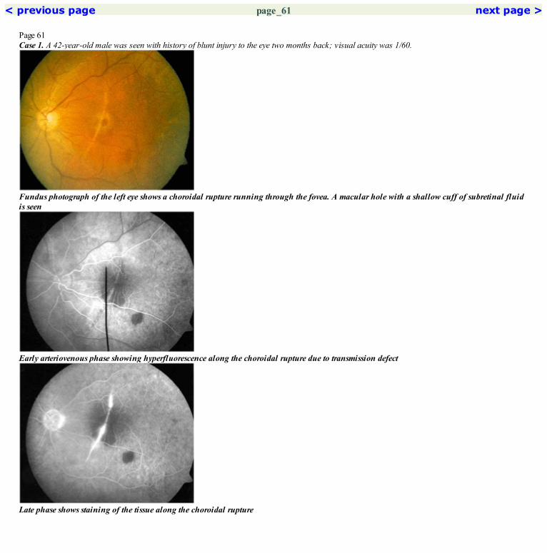
Page 61Case 1. A 42-year-old male was seen with history of blunt injury to the eye two months back; visual acuity was 1/60.
Fundus photograph of the left eye shows a choroidal rupture running through the fovea. A macular hole with a shallow cuff of subretinal fluidis seen
Early arteriovenous phase showing hyperfluorescence along the choroidal rupture due to transmission defect
Late phase shows staining of the tissue along the choroidal rupture

Red free photograph of the left eye. A small patch of subretinal hemorrhage is seen inferotemporal to the fovea
The hyperfluorescence along the choroidal ruptures has increased in the mid arteriovenous phase. Blocked choroidal fluorescence is notedinferotemporal to the fovea caused by the subretinal hemorrhageCOMMENTThis case shows the choroidal rupture traversing close to the foveal center. It shows minimal hyperfluorescence in the early phase which increasesin the later phase of the angiogram.
< previous page page_61 next page >

< previous page page_62 next page >
Page 62Case 2. A 25-year-old female presented with history of injury to the right eye with a tennis ball, one year back; visual acuity was 6/18; N18.
Fundus photograph of the right eye showing multiple choroidal ruptures at the macula. Areas of scarring and pigmentation is seen along theruptures
Early arteriovenous phase showing variable fluorescence along the choroidal rupture. Vascular network can be made out in the scar at the fovea.Blocked fluorescence is seen in areas of pigmentation
Intense staining is seen in the scar at the fovea. Hyperfluorescence elsewhere along the choroidal rupture has faded

Red free photograph of the right eye
The hyperfluorescence at the scar has increased in the mid arteriovenous phase. Areas of blocked fluorescence and transmission hyperfluorescenceis seen in the other areas of choroidal ruptureCOMMENTGrowth of CNVM through the choroidal tear is common. The CNVM shows intense hyperfluorescence with leakage and fuzzy margins in the latephase.
< previous page page_62 next page >

< previous page page_63 next page >
Page 63CLINICAL NUGGET1. Eyes with choroidal ruptures should be followed-up for the development of choroidal neovascular membrane.SELECTED READINGS1. Duke-Elder WS. Textbook of Ophthalmology, Vol 6. St Louis, CV Mosby Co, 5829–5837, 1954.2. Aguilar JP, Green WR. Choroidal rupture: a histopathological study of 47 cases. Retina 4:269–275, 1984.3. Gass JD. Stereoscopic Atlas of Macular Disease. Diagnosis and Treatment, 3rd edition. St Louis, CV Mosby Co, 170, 1987.4. Wyszynski RE, Grossniklaus HE, Frank KE. Indirect choroidal rupture secondary to blunt ocular trauma: a review of eight eyes. Retina 8:237–243, 1988.
< previous page page_63 next page >
< previous page page_64 next page >
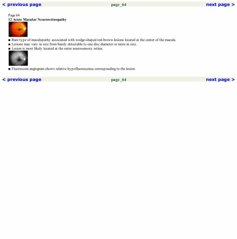
Page 6412 Acute Macular Neuroretinopathy
■ Rare type of maculopathy associated with wedge-shaped red-brown lesions located at the center of the macula.■ Lesions may vary in size from barely detectable to one disc diameter or more in size.■ Lesion is most likely located at the outer neurosensory retina.
■ Fluorescein angiogram shows relative hypofluorescence corresponding to the lesion.
< previous page page_64 next page >

< previous page page_65 next page >
Page 65Case 1. A 16-year-old male patient presented with complaints of reduced vision in the left eye of two months duration. He gave history of a head injurysustained in a road accident, two months back for which he was hospitalized. There was no direct injury to the eyes reported at that time. Visualacuity was 6/6; N6 in the right eye and 6/36; N36 in the left eye. The right eye fundus was normal.
Fundus photograph of the left eye showing a red-brown irregular lesion located at the fovea
Early arteriovenous phase shows a relative hypofluorescence in the region of the foveal lesion
Late phase shows persistence of the hypofluorescence corresponding to the lesion seen clinically

The lesion at the fovea is well seen in the red free photograph
Some hypofluorescence persists at the fovea in the arteriovenous phaseCOMMENTMinimal hypofluorescence is seen at the site of the lesion in the maculaThis lesion usually persists for a long time.
< previous page page_65 next page >

< previous page page_66 next page >
Page 66Case 2. A 39-year-old male patient presented with a history of being involved in a road accident one month back. He sustained injuries to themandible without any injury to the eye. Four days after the injury he noticed decreased vision in the left eye. Visual acuity was 6/6; N6 in the right eyeand 1/60 in the left eye.
Fundus photograph of the right eye showing a red-brown wedge shaped defect at the fovea
Early arteriovenous phase of the angiogram showing hypofluorescence at the site of the lesion
Late phase showing hypofluorescence at the lesion site

Red free photograph demarcates the lesion better
Hypofluorescence persists in the arteriovenous phase
< previous page page_66 next page >

< previous page page_67 next page >
Page 67Left eye of the patient.
Fundus photograph of the left eye showing a red-brown irregular lesion located at the fovea
Arteriovenous phase showing hypofluorescence at the site of the lesion
Red free photograph highlights the lesion
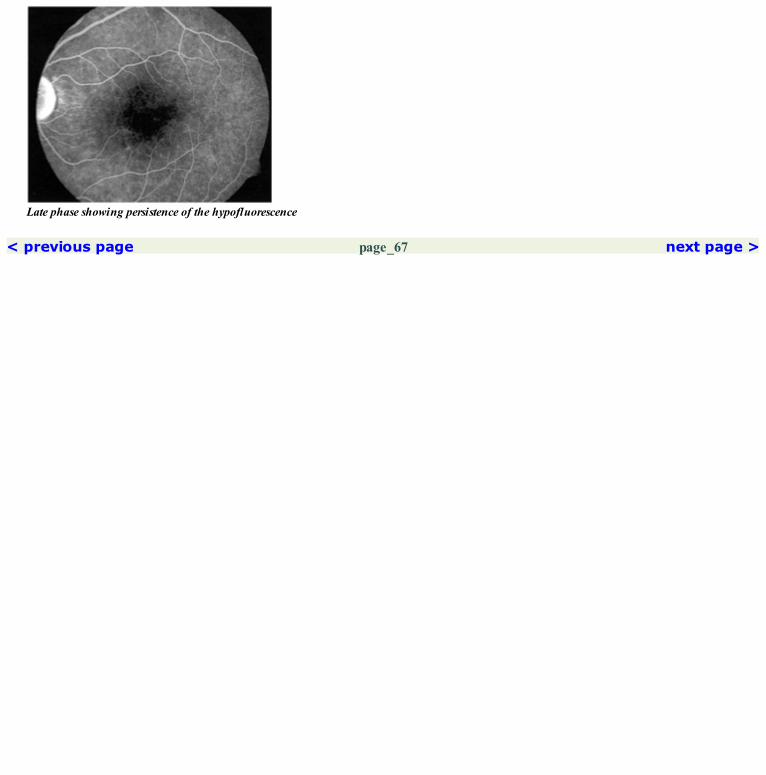
Late phase showing persistence of the hypofluorescence
< previous page page_67 next page >

< previous page page_68 next page >
Page 68CLINICAL NUGGETS1. In patients with acute macular neuroretinopathy, the scotoma seen on the Amsler grid corresponds exactly to the lesion seen at the macula.2. There is no effective treatment for acute macular neuroretinopathy.3. The macular lesion in acute macular neuroretinopathy may persist for a very long time.SELECTED READINGS1. Turbeville SD, Cowan LD, Gass JDM. Acute macular neuroretinopathy. A review of the literature. Surv Ophthalmol 48; 1:1–11, 2003.2. Bos PJ, Deutman AF. Acute macular neuroretinopathy. Am J Ophthalmol 80:573–584, 1975.3. Miller MH, Spalton DJ, Fitzke FW, Bird AC. Acute macular neuroretinopathy. Ophthalmology 96:265–269, 1989.4. O’Brfen DM, Farmer SG, Kalina RE, Leon JA. Acute macular neuroretinopathy following intravenous sympathomimetics. Retina 9:281–286,1989.
< previous page page_68 next page >

< previous page page_69 next page >
Page 6913 Age-related Macular Degeneration—Dry Type
■ Dry, atrophic or non-neovascular ARMD is characterized by the presence of drusen, focal hyperpigmentation, RPE degeneration and geographicatrophy of retinal pigment epithelium.
■ The angiographic features depend on the type of lesion that is present.■ DrusenHard drusen: These generally show up as window defects in the region of the drusen in the early phases due to atrophy of the overlying retinalpigment epithelium. The fluorescence fades in the late phase of the angiogram.Soft drusen: The fluorescein angiographic pattern in soft drusen varies depending on their content. In general they hyperfluorescence later and eitherfade or stain in the late phase of angiogram.■ Focal HyperpigmentationThese areas of focal pigment block the background choroidal fluorescence.■ Nongeographic atrophy/RPE degeneration/Incipient atrophyDiffuse stippled hyperfluorescence is noted in the region of pigment mottling at the posterior pole. A pattern of reticular or punctate blockage isseen corresponding to areas of pigment clumps.■ Geographic atrophy of the RPEEarly hyperfluorescence is noted within the areas of geographic atrophy due to the window defect in the RPE. The choriocapillaries fill slowlywithin the lesion. However the choriocapillaries may be entirely absent and filling of the larger choroidal vessels may be easily visualized. The extentof the lesion does not change in the late phases. In the late phase angiogram, hyperfluorescence due to staining of the choroidal tissue and the sclera isseen in the region of the geographic atrophy.
< previous page page_69 next page >
< previous page page_70 next page >
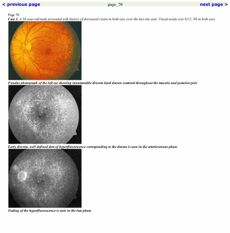
Page 70Case 1. A 58-year-old male presented with history of decreased vision in both eyes over the last one year. Visual acuity was 6/12; N6 in both eyes.
Fundus photograph of the left eye showing innumerable discrete hard drusen scattered throughout the macula and posterior pole
Early discrete, well defined dots of hyperfluorescence corresponding to the drusen is seen in the arteriovenous phase
Fading of the hyperfluorescence is seen in the late phase

Red free photograph of the left eye showing the drusen
Increased hyperfluorescence is seen over drusen in the mid arteriovenous phaseCOMMENTHard drusen show early hyperfluorescence which increases in the mid arteriovenous phase and fades in the late phase of the angiogram.
< previous page page_70 next page >
< previous page page_71 next page >
Page 71Case 2. A 68-year-old male presented with history of mild blurring of vision in the left eye for the last ten days. He gave history of reduced vision inthe right for the last two years. Visual acuity was 1/60 in the right eye and 6/9; N6 in the left eye. Examination revealed a disciform scar in the righteye and soft drusen in the left eye.
Fundus photograph of the left eye showing multiple large soft drusen in the macula
Early arteriovenous phase does not show any hyperfluorescence corresponding to the drusen
Hyperfluorescence of the soft drusen is seen only in the late phase of the angiogram

Red free photograph of the left eye delineating the soft drusen
Faint hyperfluorescence in seen around the fovea in the arteriovenous phase. However, the drusen are still not made outCOMMENTSoft drusen show hyperfluorescence only in the later part of the angiogram. They generally stain in the late phase of the angiogram.
< previous page page_71 next page >

< previous page page_72 next page >
Page 72Case 3. A 65-year-old male was found to have age-related macular degeneration in both eyes during an eye examination. Visual acuity was 6/6; N6 inboth eyes.
Fundus photograph of the right eye showing large soft drusen. Few calcified drusen are also seen (arrow)
Faint hyperfluorescence is seen at the drusen in early arteriovenous phase
Maximum hyperfluorescence is seen in the late phase due to staining of the soft drusen

Red free photograph of the right eye
Further increase in the hyperfluorescence is noted as the angiogram progressesCOMMENTThe late fluorescence of soft drusen is due to the hydrophobic nature of its content which does not allow easy entry of the dye.
< previous page page_72 next page >

< previous page page_73 next page >
Page 73Case 4. A 66-year-old female presented with history of reduced vision in both eyes for the last three months. Visual acuity was 6/9; N6 in both eyes.
Fundus photograph of the left eye showing a large drusenoid detachment at the fovea. Pigment aggregation can be seen within this detachment.Adjacent large soft drusen are seen
Hyperfluorescence is seen at the drusenoid detachment in the mid arteriovenous phase. Blocked hypofluorescence is seen due to pigment. Thesurrounding soft drusen are not very well seen
Hyperfluorescence persists at the drusenoid detachment in the late phase of the angiogram. The stained soft drusen are better seen now

Red free photograph of the left eye. The pigment within the drusenoid detachment is well made out
Hyperfluorescence at the drusenoid detachment has increased in the late arteriovenous phase. Faint hyperfluorescence of the surrounding softdrusen can be made outCOMMENTConfluence of the soft drusen gives rise to the drusenoid detachment.
< previous page page_73 next page >

< previous page page_74 next page >
Page 74Case 5. An 83-year-old male patient was seen with complaints of decreased vision in the right eye for the past five years. Visual acuity was 6/18; N6 inthe right eye and 6/9; N6 in the left eye.
Fundus photograph of the right eye showing large atrophic patches arranged in an annular fashion around the fovea. RPE alteration is noted inthe foveal region
Arteriovenous phase shows relative hyporfluorescence in the region of atrophy. The large choroidal vessels are visible. Irregular hyperfluorescenceis seen at the rest of the macula
Red free photograph shows well demarcated atrophic areas with large choroidal vessels

Normal scleral staining is seen through the atrophic areas making them appear hyperfluorescent in the late phaseCOMMENTEarly hypofluorescence within an atrophic lesion indicates non-filling of the underlying atrophic choriocapillaris along with the atrophy of theRPE.
< previous page page_74 next page >
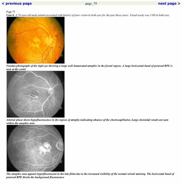
< previous page page_75 next page >
Page 75Case 6. A 74-year-old male patient presented with history of poor vision in both eyes for the past three years. Visual acuity was 1/60 in both eyes.
Fundus photograph of the right eye showing a large well demarcated atrophic in the foveal region. A large horizontal band of preseved RPE isseen at the center
Arterial phase shows hypofluorescence in the region of atrophy indicating absence of the choriocapillaries. Large choroidal vessels are seenwithin the atrophic area
The atrophic area appears hyperfluorescent in the late films due to the increased visibility of the normal scleral staining. The horizontal band ofpreseved RPE blocks the background fluorescence

Red free photograph highlights these findings. Peripapillary atrophy is seen
The hypofluorescence is well made out in the area of atrophy as the choriocapillaries fills up elsewhere. Irregular hyperfluorescence is notedtemporally
< previous page page_75 next page >

< previous page page_76 next page >
Page 76Left eye of the patient.
Fundus photograph of the left eye showing multiple discrete atrophic patches around the foveal center
The atrophic patches appear hyperfluorescent in the arteriovenous phase due to transmission window defect indicating preservation of theunderlying choriocapillaries.
Red free photograph shows well democrated atrophic patches

Some fading of the hyperfluorescence in the atrophic area is seen in the late phaseCOMMENTEarly hyperfluorescence in the region of RPE atrophy indicates the filling of the underlying choriocapillaris which has not atrophied alongwith the RPE.
< previous page page_76 next page >

< previous page page_77 next page >
Page 77Case 7. A 62-year-old male presented with history of reduced vision in the left eye of one year duration. He was asymptomatic in the right eye. Visualacuity was 6/6; N6 in the right eye and 6/60; N36 in the left eye. A cicatrized CNVM was seen in the left eye.
Fundus photograph of the right eye showing atrophic patches arranged in an annular fashion around the fovea. Regressing drusen are seenscattered throughout the macula
Hyperfluorescence is seen in the region of the atrophy. Irregular stippled hyperfluorescence is seen at the rest of the macula
Fading of the hyperfluorescence is seen in the late phase

These features are well made out in the red free photograph
The hyperfluorescence has increased in the region of RPE atrophy. Discrete hyperfluorescence is noted at the sites of the drusenCOMMENTThe foveal center is usually involved late in the course of developement of geographic atrophy.
< previous page page_77 next page >

< previous page page_78 next page >
Page 78CLINICAL NUGGETS1. The AREDS study has shown the beneficial effect of micronutrients in non-neovascular ARMD.2. Eyes with focal hyperpigmentation are more prone for development of choroidal neovascular membranes.3. Choroidal neovascularization rarely develops in areas of geographic atrophy.SELECTED READINGS1. Tanenbaum HL, Eshagian M. Senile disciform degeneration of macula: the other eye—a fluorescein angiographic study: Can J. Ophthalmol 7:280–284, 1972.2. Barendes M, Pauleikhoff D, Chisholm I, C Minassian D, Bird AC. Bilaterality of drusen, BJO 74:180–182, 1990.3. Koenig F, Saubrane G, Coscas G. Angiographic aspect of senile macular degeneration. Jap Journal of ophthalmol 33; 57–65, 1989.4. Bressler NM, Bressler SB, Fine SL. Age related macular degeneration. Surv Ophthalmol 32:375–413, 1988.5. Bird AC, Bressler NM, Bressler SB et al. An international classification and grading system for age related maculopathy and age related maculardegeneration. Surv Ophthalmol 39:367–376, 1995.
< previous page page_78 next page >

< previous page page_79 next page >
Page 7914 Age-related Macular Degeneration—Wet Type
■ Characterized by the development of choroidal neovascular membrane (CNVM).■ Clinically there is presence of subretinal fluid, subretinal or sub-RPE hemorrhage, lipid exudation; more often a grey subretinal neovascularmembrane is seen at the macula.
■ Two basic angiographic patterns of wet ARMD have been described: the classic and the occult choroidal neovascular membrane.■ Classic CNV shows up in the early phase as a bright, well demarcated area of hyperfluorescence which increases in intensity and extent in themid and late films due to progressive leakage of the dye which obscures the boundaries of the CNV. Occasionally, the actual vascular network maybe seen in the early phase.■ Occult CNV includes two fluorescein angiographic patterns.a. Fibrovascular PED. This is seen as a speckled hyperfluorescence within 1 to 2 minutes of dye injection with persistence of staining or leakage ofthe fluorescein dye in the late phase. This could be well demarcated or poorly demarcated.b. Late leakage of undetermined source. This consists of areas of leakage of dye at the level of the RPE in the late phases of the angiogram. It doesnot correspond to an area of CNV or a fibrovascular PED to account for the leakage.
< previous page page_79 next page >
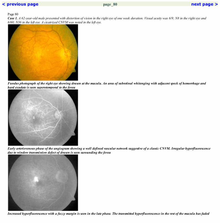
< previous page page_80 next page >
Page 80Case 1. A 62-year-old male presented with distortion of vision in the right eye of one week duration. Visual acuity was 6/9; N8 in the right eye and6/60; N36 in the left eye. A cicatrized CNVM was noted in the left eye.
Fundus photograph of the right eye showing drusen at the macula. An area of subretinal whitenging with adjacent speck of hemorrhage andhard exudate is seen superotemporal to the fovea
Early arteriovenous phase of the angiogram showing a well defined vascular network suggestive of a classic CNVM. Irregular hyperfluorescencedue to window transmission defect of drusen is seen surounding the fovea
Increased hyperfluorescence with a fuzzy margin is seen in the late phase. The transmitted hyperfluorescence in the rest of the macula has faded

Red free photograph showing the changes superotemporal to the foveal center
Arteriovenous phase showing increased hyperfluorescence of the CNVM. Hyperfluorescence due to drusen in the surrounding area has alsoincreasedCOMMENTA small juxtafoveal, classic CNVM is seen in this case. The vessels constituting the CNVM are made out in the early phase. Dye leakage fromthese vessels blurs out its details in the late phase of the angiogram.
< previous page page_80 next page >

< previous page page_81 next page >
Page 81Case 2. A 57-year-old male presented with reduced vision in the right eye for the past ten days. Visual acuity was 6/18; N12 in the right eye.
Fudus photograph of the right eye showing a juxtafoveal membrane with surrounding rim of subretinal hemorrhage. Subretinal fluid and hardexudates are also seen
Arteriovenous phase shows filling up of the juxtafoveal classics CNVM. Surrounding blocked choroidal fluorescence is seen due to subretinalblood
Intense hyperfluorescence of the CNVM is seen in the late phase
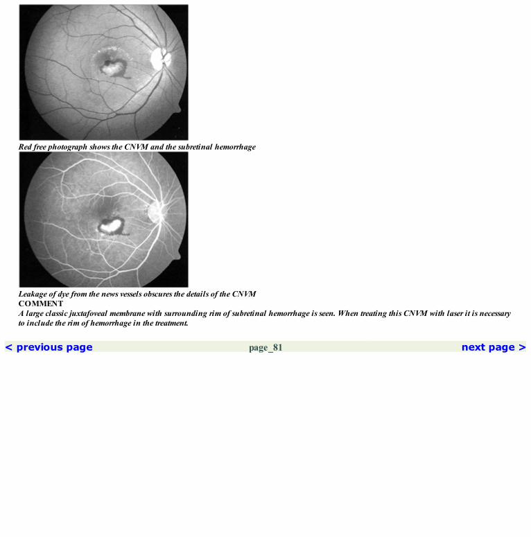
Red free photograph shows the CNVM and the subretinal hemorrhage
Leakage of dye from the news vessels obscures the details of the CNVMCOMMENTA large classic juxtafoveal membrane with surrounding rim of subretinal hemorrhage is seen. When treating this CNVM with laser it is necessaryto include the rim of hemorrhage in the treatment.
< previous page page_81 next page >

< previous page page_82 next page >
Page 82Argon laser photocoagulation was done to the juxtafoveal CNVM. One month later the visual acuity in the right eye was 6/9; N8.
Laser photocoagulation scar is seen inferior to the foveal center. No subretinal fluid, hard exudate or hemorrhage is seen
Hypofluorescence with a rim of hyperfluorescence is seen at the laser scar in the arteriovenous phase
Red free photograph of the right eye

Late staining of the scar is seen. Note that the margins are well defined and the hyperfluorescence has not extended beyond the lesion boundaryCOMMENTFollowing laser photocoagulation of the CNVM, hyperfluorescence within the center of the laser scar which does not extend beyond the marginsof the scar in the late phase, is generally not considered as recurrence of the membrane.
< previous page page_82 next page >

< previous page page_83 next page >
Page 83Case 3. A 63-year-old male presented with distortion of vision in the right eye of two weeks duration. Visual acuity was 6/6; N6.
Fundus photograph of the right eye showing a CNVM inferior to the foveal center. Soft drusen are seen in the superior macula
Early arteriovenous phase of the angiogram showing the filling up of the extrafoveal, classic CNVM
Intense hyperfluorescence is seen at the CNVM in the late phase. The soft drusen show only minimal staining even in the late films

Red free photograph showing the CNVM and the drusen
Increased hyperfluorescence is seen in the mid arteriovenuos phase of the angiogram. Note the rim of hypofluorescence surrounding the CNVM
< previous page page_83 next page >

< previous page page_84 next page >
Page 84Argon laser photocoagulation was done to the extrafoveal CNVM. Patient was lost to follow-up and presented only six months later with reducedvision in the right eye. Visual acuity was 6/24; N18.
Fundus photograph of the right eye showing the laser scar inferior to the foveal center with a subretinal hemorrhage along its lower edge. Thefoveal edge of the scar appears heaped up
Hyperfluorescence is seen at the superior edge of the scar. A vascular network suggestive of a recurrent CNVM can be made out in thishyperfluorescent area
Intense hyperfluorescence with fuzzy margins is seen at the recurrent CNVM in the late phase. Staining of the inferior laser scar is seen

Red free photograph of the right eye
Leakage of dye causes increased hyperfluorescence of the recurrent membrane in the arteriovenous phaseCOMMENTFollowing laser photocoagulation of the CNVM, hyperfluorescence with leakage in the later phase of the angiogram, occuring at the peripheryof the laser scar is generally considered as recurrence of the CNVM.
< previous page page_84 next page >
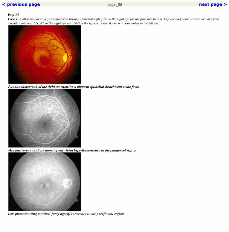
< previous page page_85 next page >
Page 85Case 4. A 60-year-old male presented with history of metamorphopsia in the right eye for the past one month. Left eye had poor vision since one year.Visual acuity was 6/9; N6 in the right eye and 1/60 in the left eye. A disciform scar was noted in the left eye.
Fundus photograph of the right eye showing a pigment epithelial detachment at the fovea
Mid arteriovenous phase showing only faint hyperfluorescence in the parafoveal region
Late phase showing minimal fuzzy hyperfluorescence in the parafloveal region

Red free photograph of the right eye shows the PED
The hyperfluorescence in the center has marginally increased in the late arteriovenous phase. No subretinal vascular network is seen
< previous page page_85 next page >

< previous page page_86 next page >
Page 863-months later, visual acuity in the right eye had dropped to 6/24; N36.
Fundus photograph of the righ eye showing a PED at the fovea. Hemorrhage is seen at the foveal center. Note the increase in size of the PED
Mid arteriovenous phase shows filling of the upper part of the PED. Faint irregular hyperfluorescence is seen in the rest of the area of PED
Late phase shows increased stippled hyperfluorescence around the fovea suggestive of an occult CNVM. Pooling of the dye is noted superiorly

Red free photograph of the right eye
The stippled hyperfluorescence in the lower part of the PED has increased. Increased filling of the superior part of the PED is seenCOMMENTA slow filling PED is highly suspicious of an occult CNVM.
< previous page page_86 next page >
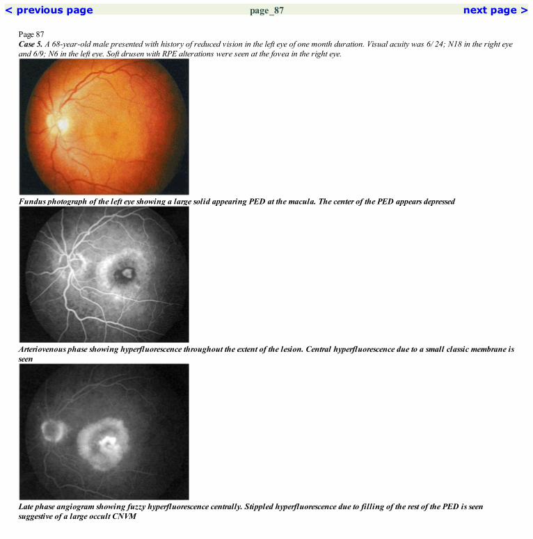
< previous page page_87 next page >
Page 87Case 5. A 68-year-old male presented with history of reduced vision in the left eye of one month duration. Visual acuity was 6/ 24; N18 in the right eyeand 6/9; N6 in the left eye. Soft drusen with RPE alterations were seen at the fovea in the right eye.
Fundus photograph of the left eye showing a large solid appearing PED at the macula. The center of the PED appears depressed
Arteriovenous phase showing hyperfluorescence throughout the extent of the lesion. Central hyperfluorescence due to a small classic membrane isseen
Late phase angiogram showing fuzzy hyperfluorescence centrally. Stippled hyperfluorescence due to filling of the rest of the PED is seensuggestive of a large occult CNVM

Red free photograph of the right eye. A speck of hemorrhage is noted at the center
Overall hyperfluorescence throughout the lesion has increased. The intensity of fluorescence at the central classic membrane has also increasedCOMMENTPatient is having a large occult membrane with a small classic component which is less that fifty percent of the entire lesion. This is a minimallyclassic CNVM
< previous page page_87 next page >
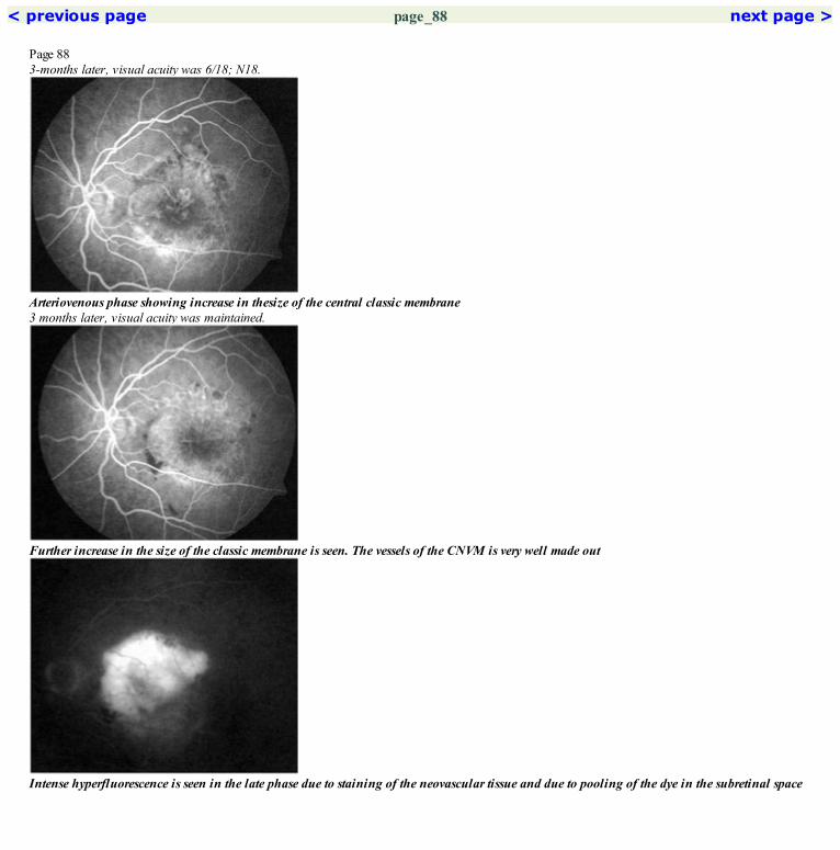
< previous page page_88 next page >
Page 883-months later, visual acuity was 6/18; N18.
Arteriovenous phase showing increase in thesize of the central classic membrane3 months later, visual acuity was maintained.
Further increase in the size of the classic membrane is seen. The vessels of the CNVM is very well made out
Intense hyperfluorescence is seen in the late phase due to staining of the neovascular tissue and due to pooling of the dye in the subretinal space

Late phase showing increased intensity of hyperfluorescence
Leakage of the dye from the neovascular vessels is seenCOMMENTThe classic part of the CNVM has increased and now constitutes more than fifty percent of the entire lesion. This is a predominantly classicmembrane.
< previous page page_88 next page >

< previous page page_89 next page >
Page 89Case 6. A 73-years-old male patient presented with a history of reduced vision in both eyes for the past two years. Visual acuity was 1/60 in the righteye and 6/24; N36 in the left eye. Both eyes showed changes of wet type of age related macular degeneration.
Fundus photograph of the right eye showing a large scarring CNVM at the macula
Early arteriovenous phase shows the large network of vessels of the choroidal neovascular membrane
Intense hyperfluorescence is seen in the late phase due to staining of the tissue and due to leakage and pooling of the dye in the subretinal space

Red free photograph of the right eye
Late arteriovenous phase of the angiogram showing leakage of the dye from the edge of the CVNMCOMMENTPatient has a scarring CNVM with some activity still noted on the angiogram. The vascular network of the CNVM is well made out even in themid arteriovenous phase indicating less leakage from the new vessels.
< previous page page_89 next page >

< previous page page_90 next page >
Page 90CLINICAL NUGGETS1. Photodyanamic therapy can be considered for classic, predominantly classic and fully occult choroidal neovascular membranes.2. Transpupillary thermotherapy can be a treatment option for occult choroidal neovascular membranes.3. Newer treatment modalities being tried for ARMD include use of antiangiogenic factors and steroids.SELECTED READINGS1. MPS Group: Argon laser photocoagulation for senile macular degeneration: results of a randomized clinical trial. Arch Ophthalmol 100:912–918,1982.2. MPS Group: Visual outcome after laser photocoagulation for subfoveal choroidal neovascularisation secondary to age-related maculardegeneration: the influence of initial lesion size and initial visual acuity. Arch Ophthalmol 112:480–488, 1994.3. MPS Group: Subfoveal neovascular lesions in age related macular degenerations. Guidelines for evaluation and treatment in the MacularPhotocoagulation study. Arch Ophthalmol 109; 1242, 1991.4. MPS Group: Persistent and recurrent neovascularisation after krypton laser photocoagulation for neovascular lesions of ocular histoplasmosis.Arch Ophthalmol 107:344, 1989.5. Bressler SB, Bressler NM, Fine SL et al. Natural course of choroidal neovascular membranes within the foveal avascular zone in senile maculardegeneration. Am J Ophthalmol 93:157–163, 1982.6. Bressler NM, Bressler SB, Fine SL. Age related macular degeneration. Surv Ophthalmol 32:375–413, 1988.7. Bird AC, Bressler NM, Bressler SB et al. An International classification and grading system for age related maculopathy and age related maculardegeneration. Surv Ophthalmol 39:367–376, 1995.8. Sylus SO, Bressler NM, Maguire MG, Sacheat AP, Bressler SB. Detecting recurrent choroidal neovascularisation: comparison of clinical examwith and without fundus fluorescein angiography. Arch Ophthalmol 111:1561–1566, 1994.
< previous page page_90 next page >

< previous page page_91 next page >
Page 9115 Angioid Streaks
■ Angioid streaks are usually bilateral and represent visible breaks in the Bruch’s membrane.■ They appear as irregular radiating, reddish brown, ribbon like bands extending from the peripapillary area towards the periphery. Near the opticdisc they are interconnected by circular ring. The retina, especially temporal to the macula has a mottled ‘orange peel’ or ‘peau d’orange’ appearance.■ Excessive deposition of calcium in the Bruch’s membrane has been found in patients with angioid streaks. This deposition makes the layer brittleand susceptible to breaks.■ The systemic association of angioid streaks includes pseudoxanthoma elasticum, Ehler-Danlos syndrome, Paget’s disease, Sickle cell disease etc.
■ Angiographic pattern of angioid streaks depends on the status of the overlying RPE, the degree of pigmentation in the region of the streaks and thestatus of the underlying choriocapillaris.■ Typically, angioid streaks become hyperfluorescent in the arterial phase due to transmitted choroidal fluorescence, which increases slightly in thelater phases. No leakage of dye is seen.■ Choroidal neovascular membranes are common in patients with angioid streaks.
< previous page page_91 next page >

< previous page page_92 next page >
Page 92Case 1. A 48-year-old female presented with history of floaters in both eyes. Visual acuity was 6/9; N6 in the right eye and 6/ 12; N6 in the left eye. Shewas found to have angioid streaks with CNVM in both eyes.
Fundus photograph of the right eye showing the prominent angioid streaks radiating from an annular ring around the disc. Note the pigmentclump seen along the angioid streak superotemporal to the disc. A CNVM is noted just superior to this pigment clump
The angioid streaks are well seen in the arteriovenous phase as hyperfluorescent radiating lines. Faint mottled hyperfluorescence is seentemporally. Note the speckled hyperfluorescence seen just superior to the blocked hypofluorescence caused by the pigment clump
Late phase shows fading of the hyperfluorescence along the angioid streaks. Leakage of dye is noted in the area of the suspected CNVM

Red free photograph highlights the changes seen in the fundus photograph
Increased hyperfluorescence is seen along the angioid streaks in the late arteriovenous phase. There is marked increase in the hyperfluorescencein the lesion superotemporal to the disc suggesting presence of a CNVMCOMMENTIn this patient, the angioid streaks are much better seen clinically than on the angiogram. This is probably due to pigment and fibrous tissuewithin the angioid streak blocking the transmitted fluorescence to some extentCNVM occurs very commonly in eyes with angioid streaks and should be particularly looked for in the angiogram.
< previous page page_92 next page >

< previous page page_93 next page >
Page 93Left eye of the patient.
Fundus photograph of the left eye showing the prominent angioid streaks radiating from an annular ring around the disc. A CNVM is notedtemporal to the fovea
The late arteriovenous phase shows hyperfluorescence along the angioid streaks. Marked hyperfluorescence is noted in the region of the CNVM
Red free photograph highlights the changes seen in the fundus photograph

Late phase angiogram showing fading of the hyperfluorescence along the angioid steaks. Leakage of dye from the CNVM is seenCOMMENTGrowth of the CNVM through the breaks in the Bruchs’ membrane is common in patients with angioid streaks
< previous page page_93 next page >
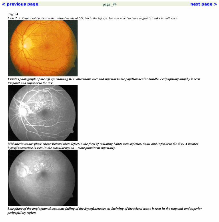
< previous page page_94 next page >
Page 94Case 2. A 55-year-old patient with a visual acuity of 6/9; N6 in the left eye. He was noted to have angioid streaks in both eyes.
Fundus photograph of the left eye showing RPE alterations over and superior to the papillomacular bundle. Peripapillary atrophy is seentemporal and superior to the disc
Mid arteriovenous phase shows transmission defect in the form of radiating bands seen superior, nasal and inferior to the disc. A mottledhyperfluorescence is seen in the macular region—more prominent superiorly.
Late phase of the angiogram shows some fading of the hyperfluorescence. Staining of the scleral tissue is seen in the temporal and superiorperipapillary region

Red free photograph shows the angioid streaks as dark bands radiating away from the disc—both superior and inferior to the disc
Recirculation phase shows increased mottled hyperfluorescence in the macular regionCOMMENTAngioid streaks are seen as hyperfluorescent bands, more prominent in the mid arteriovenous phase. The mottled or stippled pattern ofhyperfluorescence is a common finding in patients with angioid streaks and is usually seen at the posterior pole—especially the temporal macularregion. It is caused by small clumps of pigment epithelium which block the choroidal fluorescence.
< previous page page_94 next page >

< previous page page_95 next page >
Page 95Case 3. A 55-year-old male patient presented with decreased vision in the right eye. Visual acuity was 6/36; N36. He was noted to have angioidstreaks in both eyes with a choroidol neovascular membrane in the right eye
Fundus photograph of the right eye shows a CNVM surrounded by subretinal hemorrhage, just superior to the foveal center. Concentric whitelines (choroidal rupture) are seen along the superior arcade with a large subretinal scar seen superotemporally
Mid arteriovenous phase shows the filling of the CNVM. Hyperfluorescent lines (due to transmission defect) concentric to the disc margin arenoted superotemporal to the disc
Late phase of the angiogram shows leakage of the dye from the CNVM. Some fading of the hyperfluorescence is seen in the region of thechoroidal rupture. Staining of the scar in the superotemporal region is also seen

Red free photograph highlights these changes. The peripapillary atrophy is well made out temporal and superior to the optic nerve head
Arteriovenous phase shows increased filling of the CNVM. Faint linear hyperfluorescent bands are noted radiating away from the disc—nasallyand inferiorlyCOMMENTEyes with angioid streaks are very susceptible to trauma. The concentric lines seen along the superotemporal arcade do not follow the generalpattern of the angioid streaks and probably represents choroidal rupture due to some trivial eye injury in the past. The fellow eye of this patienthad typical pattern of angioid streaks.
< previous page page_95 next page >

< previous page page_96 next page >
Page 96Case 4. A 36-year-old male patient diagnosed to have pseudoxanthoma elasticum presented with decreased vision in the right eye. Visual acuity was6/24; N36.
Fundus photograph of the right eye of a patient with angioid streak with a partly cicatrized CNVM. The angioid streaks are seen as dark brownbands radiating away from the disc
In this case the angioid streaks are not evident in the arterial phase of the angiogram. The normal patchy choroidal filling is well seen in thephase
Angioid streaks became evident in the late arteriovenous phase of the angiogram as hyperfluorescent bands. The mottled hyperfluorescence in thetemporal macular region is better seen

The angioid streaks are well made out in the red free photograph
The early arteriovenous phase shows filling of the CNVM. The normal choroidal patchy filling is persisting. The angioid streaks are not yetclearly seen. There is mottled hyperfluorescence in the temporal macular region
Late phase angiogram shows increased staining of the scarred CNVM. The hyperfluorescence along the angioid streaks has faded to some extent.The temporal mottled hyperfluorescence is persisting
< previous page page_96 next page >

< previous page page_97 next page >
Page 97Case 5. A 47-year-old male patient was examined with a history of decreased vision in the right eye for the past two years. Visual acuity was 3/60 inthe right eye.
Fundus photograph of the right eye showing a large disciform scar in the macular region. Prominent pigment hypertrophy is seen within the scar.Angioid streaks were noted (not evident in this photo)
Early arteriovenous phase shows some hypofluorescence in the region of the scar. Faint hyperfluorescence is seen along the angioid streaks
Late phase shows fading of the hyperfluorescence along the angioid streaks. Staining of the disciform scar is seen

Red free photograph highlights the changes seen in the fundus photograph
The mid arteriovenous phase showing the hyperfluorescence along the angioid streaks. Some hyperfluorescence is noted in the disciform scar dueto the filling of its vasculature. Mottled hyperfluorescence is seen in the adjacent retinaCOMMENTUntreated or recurrent CNVM in patients with angioid streaks, usually results in the formation of a disciform scar. This is an important cause ofsevere visual loss in these patients.
< previous page page_97 next page >

< previous page page_98 next page >
Page 98CLINICAL NUGGETS1. Eyes with angioid streaks are susceptible to trivial trauma and the patient should be advised accordingly.2. Causes for poor vision in eyes with angioid streaks is either due to involvement of the fovea by the breaks in the Bruch’s membrane ordevelopment of choroidal neovascular membrane.SELECTED READINGS1. Gass JD, Clarkson JG: Angioid streaks and disciform macular detachment in Paget’s disease. Am J Ophthalmol 30:955–968, 1974.2. Federman JL, Shields JA, Tomer TL: Angioid streaks II. Fluorescein angiographic features. Arch Ophthalmol 93:921–926. 1976.3. Clarkson JG, Altman RD. Angioid streaks. Surv Ophthalmol 26:235–246, 1983.4. Gass JD, Stereoscopic Atlas of Macular Diseases, 3rd edition, St Louis, CV Mosby Co, 102–109, 1987.5. Hagedoorn A: Angioid streaks and traumatic rupture of Bruch’s membrane. Br J Ophthalmol 59:267, 1975.
< previous page page_98 next page >

< previous page page_99 next page >
Page 9916 Choroidal Folds
■ Choroidal folds or chorioretinal folds can be either idiopathic or secondary to hypotony, posterior scleritis, retrobulbar mass lesion, choroidalneovascular membrane, focal chorioretinal scarring etc.■ They involve the inner layers of the choroid, Bruch’s membrane and the retinal pigment epithelium.■ The folds may be horizontal, vertical or oblique and are parallel to each other.■ They are usually present at the posterior pole and appear as alternate yellow and dark streaks-yellow corresponding to the crest of the folds.
■ Alternate dark and light bands are seen corresponding to the choroidal folds.■ The crest of the fold appears hyperfluorescent This could be due to thinning of the retinal pigment epithelium at the crest of the folds.■ The trough appears as hypofluorescent streaks probably due to crowding of the retinal pigment epithelium causing blockage of the choroidalfluorescence.
< previous page page_99 next page >

< previous page page_100 next page >
Page 100Case 1. A 43-year-old female was noted to have choroidal folds in both eyes on a routine eye examination. Visual acuity was 6/6; N6 in both eyes.
Fundus photograph of the right eye showing parallel choroidal folds
Arteriovenous phase showing obliquely running choroidal folds. A hyperfluorescent spot is noted superonasal to the fovea
Some fading of the hyperfluorescent lines is noted in the late film. The hyperfluorescent spot persists-however it remains discrete with minimalfading-probably representing a RPE defect

Red free photograph highlighting the choroidal folds
The alternating hypo- and hyperfluorescent streaks due to choroidal folds are well seen in the later part of the arteriovenous phaseCOMMENTThe crest of the choroidal folds appears as hyperfluorescent lines which fade to some extent in the late films.
< previous page page_100 next page >
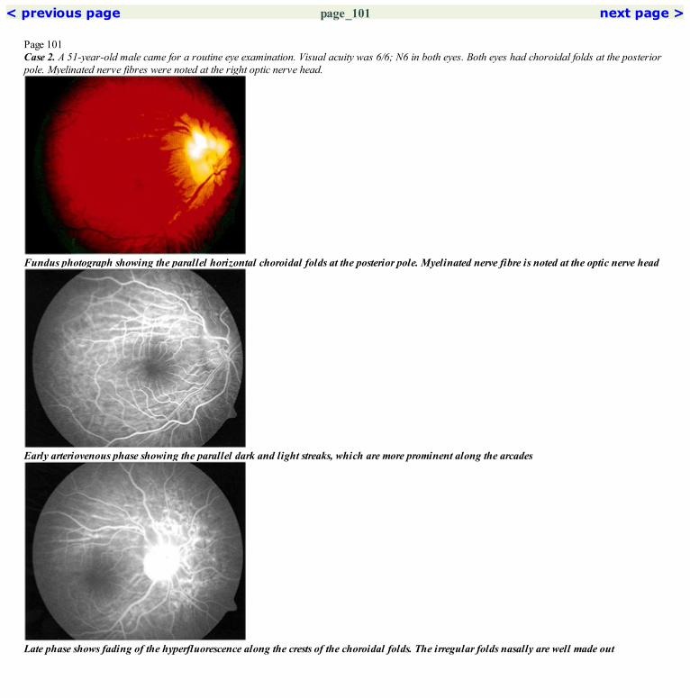
< previous page page_101 next page >
Page 101Case 2. A 51-year-old male came for a routine eye examination. Visual acuity was 6/6; N6 in both eyes. Both eyes had choroidal folds at the posteriorpole. Myelinated nerve fibres were noted at the right optic nerve head.
Fundus photograph showing the parallel horizontal choroidal folds at the posterior pole. Myelinated nerve fibre is noted at the optic nerve head
Early arteriovenous phase showing the parallel dark and light streaks, which are more prominent along the arcades
Late phase shows fading of the hyperfluorescence along the crests of the choroidal folds. The irregular folds nasally are well made out

The red free photograph shows the choroidal folds. A blooming artifact is noted at the optic nerve head
The choroidal folds appear more prominent in the late arteriovenous phase. Note the irregular appearing folds nasal to the disc. Minimal bluringof details is noted in the superior part of the disc due to myelinated nerve fibre
< previous page page_101 next page >

< previous page page_102 next page >
Page 102Case 3. A 45-year-old male presented with blurring of vision in the both eyes. The visual acuity was 6/9; N10 in the right eye and 6/9; N6 in the lefteye. Both eyes had similar fundus findings.
Fundus photograph showing very subtle choroidal folds in the upper part of the macula. There is an elevation of the sensory retina inferiorly
Arteriovenous phase shows a hyperfluorescent dot in the inferior macula. Note subtle choroidal folds in the superior part of the macula
Late phase shows pooling of dye in the upper part of the sensory RD. The hyperfluorescence along the choroidal folds has diminished

Red free photograph highlights the sensory retinal elevation in the inferior part of the macula
The dye is seen to leak from the hyperfluorescent point and ascend upwardsCOMMENTCentral serous chorioretinopathy can occur with idiopathic choroidal folds.
< previous page page_102 next page >

< previous page page_103 next page >
Page 103Case 4. A 43-year-old female was referred for pain and decreased vision in the right eye of 6 months duration. Visual acuity was 6/18; N36 atpresentation. Left eye was normal. Based on clinical and ultrasound findings she was diagnosed to have posterior scleritis in the right eye. Visionimproved to 6/6; N6 following treatment with systemic steroids.
Fundus photograph of a patient who presented with very prominent chorioretinal folds at the posterior pole
The hyperfluorescent crest is more broader than the hyperfluorescent trough which appears as narrow streaks. These represent broad choroidalfolds
There is fading of the hyperfluorescence seen along the choroidal folds. Increased staining of the disc is seen

Red free photograph showing the parallel choroidal folds at the posterior pole
The broad choroidal folds are well made out in the late arteriovenous phaseCOMMENTIn scleritis, increased thickening of the sclera and the choroid results in the inner choroidal layers, Brachs’ membrane and the RPE to be throwninto folds.
< previous page page_103 next page >

< previous page page_104 next page >
Page 104CLINICAL NUGGET1. Idiopathic choroidal folds, per se do not affect the visual acuity and do not require any treatment.SELECTED READINGS1. Gass JDM. Stereoscopic atlas of macular diseases: Diagnosis and treatment. 3rd edition St. Louis Mosby Year Book 1987.2. Francois De Laey JJ. Fluorescein angiographic aspects of acquired chorioretinal folds-Mod. Probl Ophthalmol 9:29–135, 1970.3. Norton EWD. A characteristic fluorescein angiographic pattern in choroidal folds. Proc R Soc Med 62:119–128, 1969.
< previous page page_104 next page >
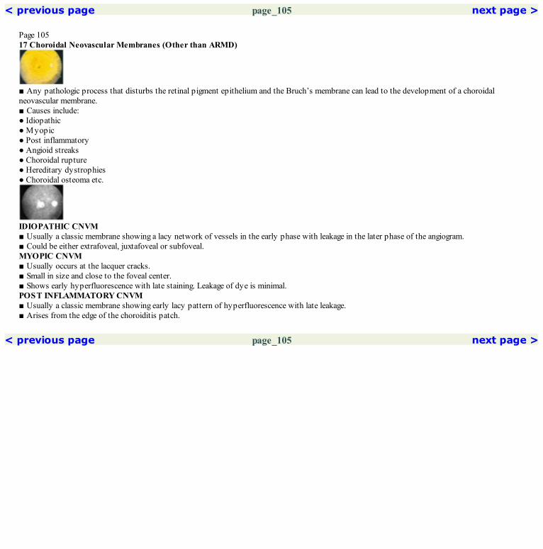
< previous page page_105 next page >
Page 10517 Choroidal Neovascular Membranes (Other than ARMD)
■ Any pathologic process that disturbs the retinal pigment epithelium and the Bruch’s membrane can lead to the development of a choroidalneovascular membrane.■ Causes include:● Idiopathic● Myopic● Post inflammatory● Angioid streaks● Choroidal rupture● Hereditary dystrophies● Choroidal osteoma etc.
IDIOPATHIC CNVM■ Usually a classic membrane showing a lacy network of vessels in the early phase with leakage in the later phase of the angiogram.■ Could be either extrafoveal, juxtafoveal or subfoveal.MYOPIC CNVM■ Usually occurs at the lacquer cracks.■ Small in size and close to the foveal center.■ Shows early hyperfluorescence with late staining. Leakage of dye is minimal.POST INFLAMMATORY CNVM■ Usually a classic membrane showing early lacy pattern of hyperfluorescence with late leakage.■ Arises from the edge of the choroiditis patch.
< previous page page_105 next page >
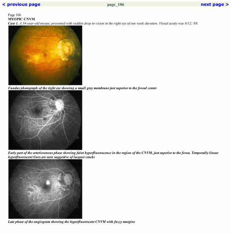
< previous page page_106 next page >
Page 106MYOPIC CNVMCase 1. A 34-year-old myope, presented with sudden drop in vision in the right eye of one week duration. Visual acuity was 6/12; N8.
Fundus photograph of the right eye showing a small grey membrane just superior to the foveal center
Early part of the arteriovenous phase showing faint hyperfluorescence in the region of the CNVM, just superior to the fovea. Temporally linearhyperfluorescent lines are seen suggestive of lacquer cracks
Late phase of the angiogram showing the hyperfluorescent CNVM with fuzzy margins
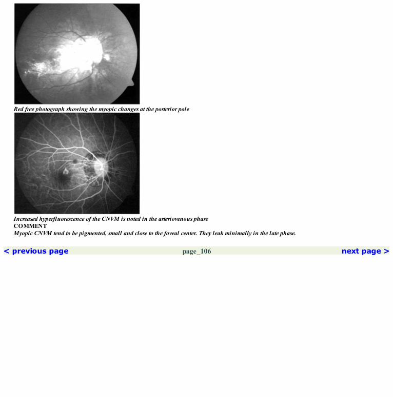
Red free photograph showing the myopic changes at the posterior pole
Increased hyperfluorescence of the CNVM is noted in the arteriovenous phaseCOMMENTMyopic CNVM tend to be pigmented, small and close to the foveal center. They leak minimally in the late phase.
< previous page page_106 next page >
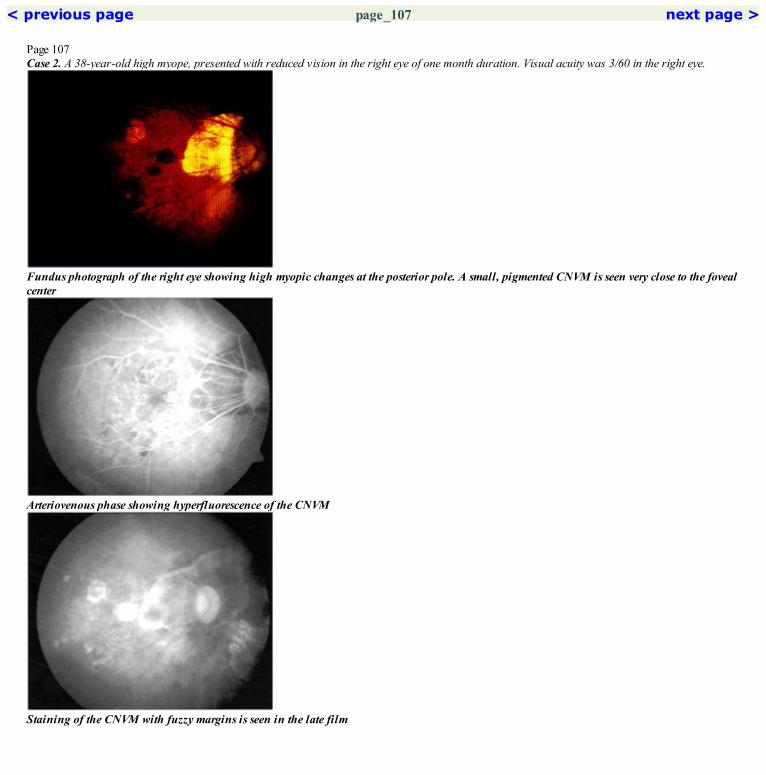
< previous page page_107 next page >
Page 107Case 2. A 38-year-old high myope, presented with reduced vision in the right eye of one month duration. Visual acuity was 3/60 in the right eye.
Fundus photograph of the right eye showing high myopic changes at the posterior pole. A small, pigmented CNVM is seen very close to the fovealcenter
Arteriovenous phase showing hyperfluorescence of the CNVM
Staining of the CNVM with fuzzy margins is seen in the late film

Red free photograph of the right eye
Increased hyperfluorescence is seen in the later part of the angiogramCOMMENTA small subfoveal myopic CNVM is seen in this case.
< previous page page_107 next page >

< previous page page_108 next page >
Page 108IDIOPATHIC CNVMCase 3. A 32-year-old female presented with blurring of vision in the left eye for the past one week. Visual acuity was 6/9; N8.
Fundus photograph of the left eye showing a small subretinal membrane temporal to the fovea
The CNVM is seen to fill in the arterial phase of the angiogram. A rim of hypofluorescence is seen surrounding the CNVM. This is furthersurrounded by a zone of hyperfluorescence
Late phase showing increased hyperfluorescence of the CNVM. The margins are fuzzy
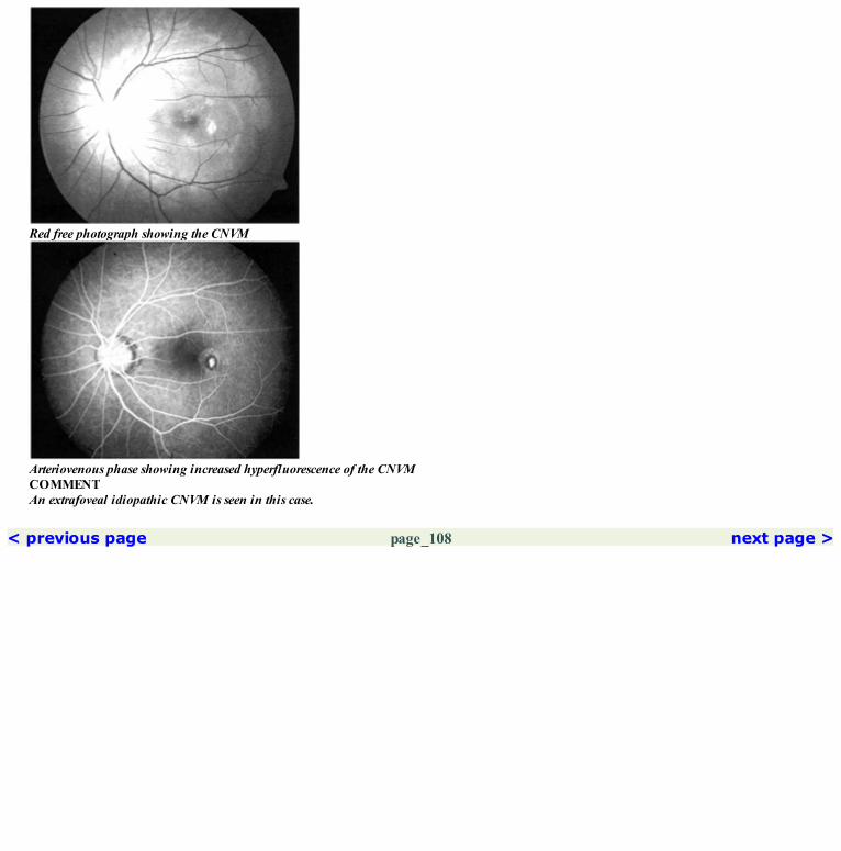
Red free photograph showing the CNVM
Arteriovenous phase showing increased hyperfluorescence of the CNVMCOMMENTAn extrafoveal idiopathic CNVM is seen in this case.
< previous page page_108 next page >
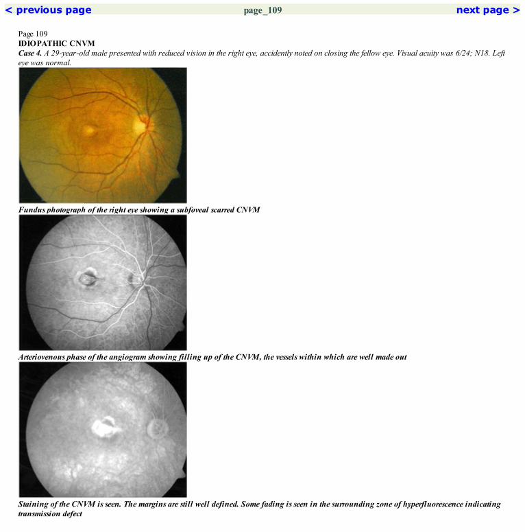
< previous page page_109 next page >
Page 109IDIOPATHIC CNVMCase 4. A 29-year-old male presented with reduced vision in the right eye, accidently noted on closing the fellow eye. Visual acuity was 6/24; N18. Lefteye was normal.
Fundus photograph of the right eye showing a subfoveal scarred CNVM
Arteriovenous phase of the angiogram showing filling up of the CNVM, the vessels within which are well made out
Staining of the CNVM is seen. The margins are still well defined. Some fading is seen in the surrounding zone of hyperfluorescence indicatingtransmission defect

Red free photograph showing the subfoveal scar
Increased hyperfluorescence of the CNVM is seen. A zone of hypofluorescence surrounded by another zone of hyperfluorescence is seen aroundthe CNVMCOMMENTThis case shows a cicatrized idiopathic CNVM. The scar generally tends to remain small in these cases.
< previous page page_109 next page >

< previous page page_110 next page >
Page 110JUXTAPAPILLARY CNVMCase 5. A 25-year-old male presented with metamorphopsia in the left eye of two weeks duration. Visual acuity was 6/12; N8. He had previously beendiagnosed to have a juxtapapillary CNVM in the right eye which was treated with laser photocoagulation.
Fundus photograph of the left eye showing a juxtapapillary CNVM. Associated subretinal fluid and hemorrhage are present
Arteriovenous phase showing filling up of the vessels of the juxtapapillary membrane
Staining of the CNVM and pooling of the dye in the subretinal space is seen in the late phase
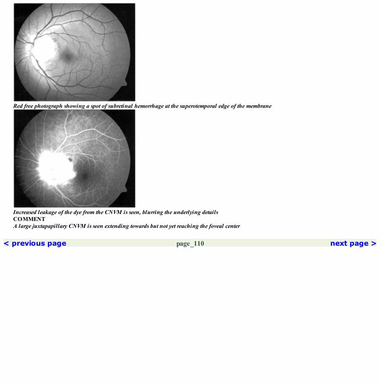
Red free photograph showing a spot of subretinal hemorrhage at the superotemporal edge of the membrane
Increased leakage of the dye from the CNVM is seen, blurring the underlying detailsCOMMENTA large juxtapapillary CNVM is seen extending towards but not yet reaching the foveal center
< previous page page_110 next page >

< previous page page_111 next page >
Page 111Argon laser photocoagulation was done to the juxtapapillary CNVM. One month later the visual acuity was 6/6; N6.
Fundus photograph of the left eye showing a flat chorioretinal atrophy in the juxtapapillary region
Early part of the arteriovenous phase of the angiogram showing hypofluorescence in the juxtapapillary region. A rim of hyperfluorescence is seenat the edge
Late hyperfluorescence due to staining of the sclera and the tissue in the juxtapapillary region is seen. Note that the margins are still well defined

Red free photograph of the left eye
Arteriovenous phase showing persistence of the hyperfluorescenceCOMMENTA well ablated CNVM is seen. There is no evidence of persistence of the CNVM.
< previous page page_111 next page >

< previous page page_112 next page >
Page 112POST INFLAMMATORY CNVM-GHPCCase 6. A 36-year-old male with GHPC in both eyes, presented with distortion of vision in the right eye of four days duration. Visual acuity was 6/9;N6 in the right eye.
Fundus photograph of the right eye showing healed GHPC. A CNVM and associated subretinal fluid was noted inferior to the foveal center
The fine lacy network of vessels are seen in the CNVM located inferotemporal to the foveal center
Intense hyperfluorescence is seen at the CNVM in the late phase
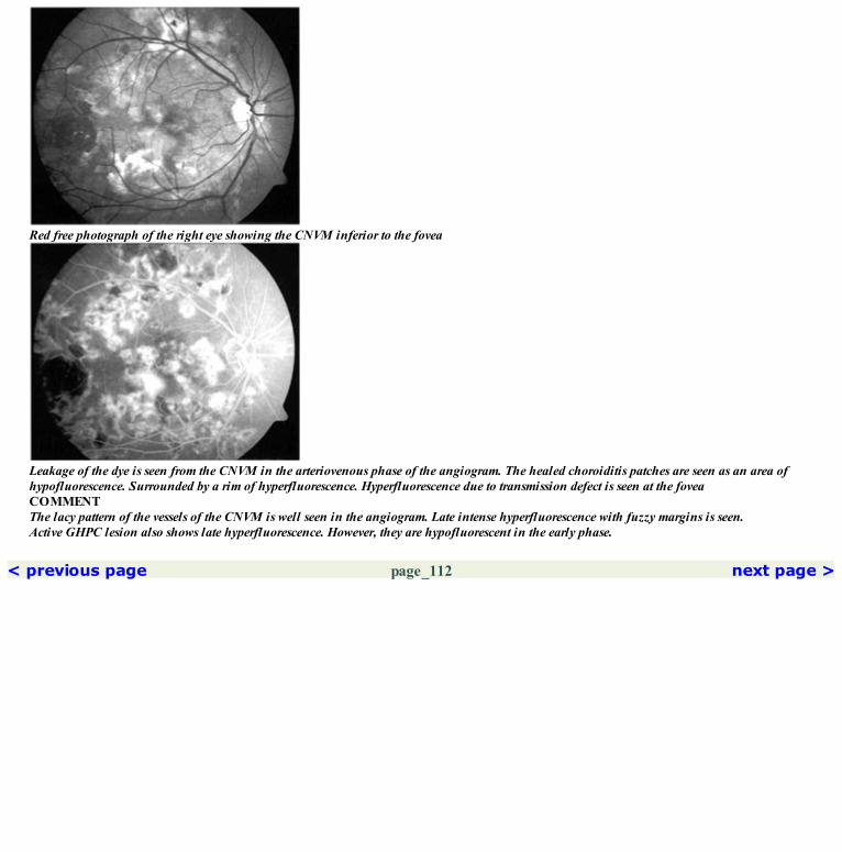
Red free photograph of the right eye showing the CNVM inferior to the fovea
Leakage of the dye is seen from the CNVM in the arteriovenous phase of the angiogram. The healed choroiditis patches are seen as an area ofhypofluorescence. Surrounded by a rim of hyperfluorescence. Hyperfluorescence due to transmission defect is seen at the foveaCOMMENTThe lacy pattern of the vessels of the CNVM is well seen in the angiogram. Late intense hyperfluorescence with fuzzy margins is seen.Active GHPC lesion also shows late hyperfluorescence. However, they are hypofluorescent in the early phase.
< previous page page_112 next page >
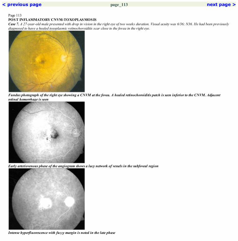
< previous page page_113 next page >
Page 113POST INFLAMMATORY CNVM-TOXOPLASMOSISCase 7. A 27-year-old male presented with drop in vision in the right eye of two weeks duration. Visual acuity was 6/36; N36. He had been previouslydiagnosed to have a healed toxoplasmic retinochoroiditis scar close to the fovea in the right eye.
Fundus photograph of the right eye showing a CNVM at the fovea. A healed retinochoroiditis patch is seen inferior to the CNVM. Adjacentretinal hemorrhage is seen
Early arteriovenous phase of the angiogram shows a lacy network of vessels in the subfoveal region
Intense hyperfluorescence with fuzzy margin is noted in the late phase
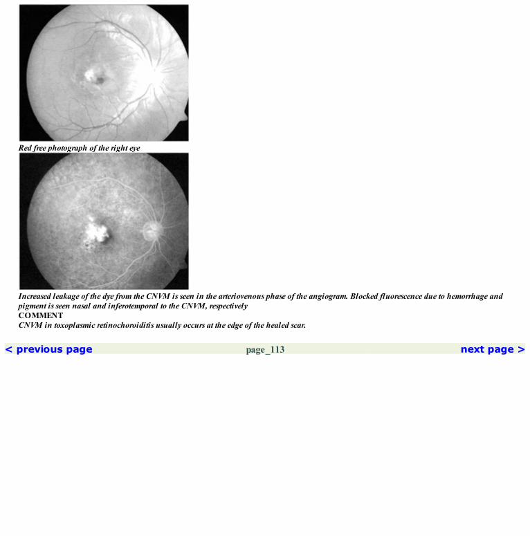
Red free photograph of the right eye
Increased leakage of the dye from the CNVM is seen in the arteriovenous phase of the angiogram. Blocked fluorescence due to hemorrhage andpigment is seen nasal and inferotemporal to the CNVM, respectivelyCOMMENTCNVM in toxoplasmic retinochoroiditis usually occurs at the edge of the healed scar.
< previous page page_113 next page >

< previous page page_114 next page >
Page 114CNVM AT DEMARCATION LINECase 8 A 46-year-old female who had undergone a scleral buckling surgery in the left eye, presented with sudden drop in vision of one monthduration. Visual acuity was 6/60; N36. The retina was attached.
Red free photograph of the left eye showing a demarcation line running across the fundus inferior to the disc. A subfoveal CNVM with adjacentsubretinal hemorrhage is noted. Lower half of the fundus shows RPE alteration
Increased leakage of the dye is noted at the CNVM
Late phase shows staining of the CNVM with fuzzy margins

Early arteriovenous phase showing the lacy network of the subfoveal CNVM
Increased hyperfluorescence due to leakage of the dye is noted at the CNVM in the later part of the arteriovenous phaseCOMMENTThis case shows rare occurrence of a CNVM at the demarcation line that develops at the junction of attached and detached retina in cases of longstanding rhegmatogenous retinal detachment.
< previous page page_114 next page >

< previous page page_115 next page >
Page 115CLINICAL NUGGETS1. Visual prognosis in idiopathic CNVM is significantly better than that in Age related macular degeneration.2. CNVM associated with myopia appears as a grayish circumscribed lesion occurring close to fovea.3. Natural history of CNVM in myopia is variable.4. Photodynamic therapy can be considered as a treatment modality for the treatment of subfoveal myopic CNVM.SELECTED READINGS1. Gass JDM. Stereoscopic atlas of macular diseases. Diagnosis and treatment, ed 4, CV Mosby, Chapter 3, 2:1045, 1997.2. Ho AC, Yannuzzi LA, Pisicano K, DeRosa J. The natural history of idiopathic subfoveal choroidal neovascularisation. Ophthalmology 102:782–789, 1995.3. Hotchkiss ML, Fine SL. Pathologic myopia and choroidal neovascularisation. Am J Ophthalmol 91:177–183, 1981.4. Wojno T, Meredith TA. Unusual findings in serpiginous choroiditis. Am J Ophthalmol 94:650–655, 1982.5. Jampol LM, Orth L, Daily MT, Rabb MF: Subretinal neovascularisation with geographic (serpiginous) choroiditis. Am J Ophthalmol. 88:683–689, 1979.
< previous page page_115 next page >
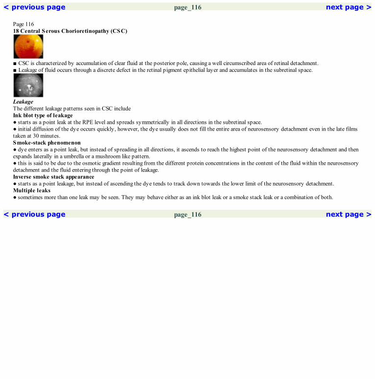
< previous page page_116 next page >
Page 11618 Central Serous Chorioretinopathy (CSC)
■ CSC is characterized by accumulation of clear fluid at the posterior pole, causing a well circumscribed area of retinal detachment.■ Leakage of fluid occurs through a discrete defect in the retinal pigment epithelial layer and accumulates in the subretinal space.
LeakageThe different leakage patterns seen in CSC includeInk blot type of leakage● starts as a point leak at the RPE level and spreads symmetrically in all directions in the subretinal space.● initial diffusion of the dye occurs quickly, however, the dye usually does not fill the entire area of neurosensory detachment even in the late filmstaken at 30 minutes.Smoke-stack phenomenon● dye enters as a point leak, but instead of spreading in all directions, it ascends to reach the highest point of the neurosensory detachment and thenexpands laterally in a umbrella or a mushroom like pattern.● this is said to be due to the osmotic gradient resulting from the different protein concentrations in the content of the fluid within the neurosensorydetachment and the fluid entering through the point of leakage.Inverse smoke stack appearance● starts as a point leakage, but instead of ascending the dye tends to track down towards the lower limit of the neurosensory detachment.Multiple leaks● sometimes more than one leak may be seen. They may behave either as an ink blot leak or a smoke stack leak or a combination of both.
< previous page page_116 next page >

< previous page page_117 next page >
Page 117Diffuse leakage● no definite leakage pattern is seen, instead a diffuse area of leakage may be seen.No leakage seen even though neurosensory detachment is present● this probably indicates a resolving stage of CSC where the leakage has closed; however, the fluid has not yet absorbed; in such cases optic pitassociated sensory retinal elevation should also be looked for.● important to look at the retina superior to the neurosensory elevation, or outside the arcade to look for causative leaks.Pigment epithelial detachment● may be located within the neurosensory detachment or outside it.● entire area of the PED shows hyperfluorescence in the early film. This increases in intensity towards the later films; however the extent remainsthe same.● area of hypofluorescence may be noted within the PED; this occurs due to pigment clumping and can produce a cruciate or a triradiate pattern ofhypofluorescence within the PED.RPE transmission defects● seen in resolved CSC cases.● usually quite extensive, extent depending on the area of previous neurosensory detachment.RPE tracts● transmission defects seen in the form of a tract extending downwards from the posterior pole and expanding inferiorly to have a ‘flask’, ‘teardrop’,‘dumb-bell’, or ‘hourglass’ shaped appearance.
< previous page page_117 next page >

< previous page page_118 next page >
Page 118Case 1. A 32-year-old male presented with history of noticing a scotoma in front of the left eye for the past one month. Visual acuity was 6/9; N6 in theright eye and 6/6; N6 in the left eye. The right eye shows a shallow sensory elevation of the retina at the macula. A small PED was seen in thetemporal part of the macula in the left eye.
Fundus photograph of the right eye showing a sensory elevation of the retina at the macula
An intense dot hyperfluorescence is noted superotemporal to the foveal center. Few faint hyperfluorescent areas are noted within the FAZ and alsosuperotemporal to the disc
The dot hyperfluorescence has increased in intensity and extent in the late films, showing the typical ink blot appearance of CSC. The other areasof hyperfluorescense have faded suggestive of a window transmission defect
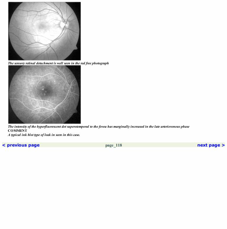
The sensory retinal detachment is well seen in the red free photograph
The intensity of the hyperfluorescent dot superotemporal to the fovea has marginally increased in the late arteriovenous phaseCOMMENTA typical ink blot type of leak in seen in this case.
< previous page page_118 next page >

< previous page page_119 next page >
Page 119Case 2. A 30-year-old male presented with history of blurring of vision in the right eye of one month duration; visual acuity was 6/12; N6.
Fundus photograph of the right eye showing a serous elevation of the retina at the macula
The arteriovenous phase shows a point hyerpfluorescence superior to the fovea
Typical ink blot appearance of the leak is seen in the late film. The extent of the detachment is well made out

Sub-retinal precipitates superior to the fovea and RPE alterations inferior to the fovea are well made out in the red free photograph
The hyperfluorescence has increasd in intensity and extent due to leakage of the dyeCOMMENTLeakage of dye never fills up the entire extent of the sensory detachment even in very late films.
< previous page page_119 next page >

< previous page page_120 next page >
Page 120Case 3. A 30-year-old male presented with history of sudden onset of blurring of vision in the left eye of 15 days duration. Visual acuity was 6/12;N18. He gave history of being treated with periocular steroid injection. The right eye was normal.
Red free photograph of the left eye showing a serous RD at the macula
Dye is seen to leak out from the central hyperfluorescence and ascend vertically
A typical smoke stack leak is seen. The dye has reached the upper border of the detachment and is seen to spread sideways

Faint hyperfluorescence is seen at the fovea in the mid arteriovenous phase
Further leakage of the dye is seenCOMMENTA typical smoke stack type of leak is seen in this case.
< previous page page_120 next page >

< previous page page_121 next page >
Page 121Case 4. A 46-year-old male presented with history of decrease in vision in the left eye of 2 weeks duration. He was being treated for allergic rhinitis—details of the medication were not known. The visual acuity was 6/9; N6 in the left eye. Right eye was normal.
A serous elevation of the retina is seen involving the fovea
A point hyperfluorescence is seen within the foveal avascular zone
A smoke stack appearance is seen in the late phase. Even though the dye has not filled the entire detachment, its extent is well highlighted

Red free photograph clearly showing the extent of the detachment
Dye is seen to leak out and ascend vertically upwardsCOMMENTSCSC leaks can occur within the FAZ. The resultant RPE alterations that follow resolution of CSC, may compromise the final visual outcome.
< previous page page_121 next page >
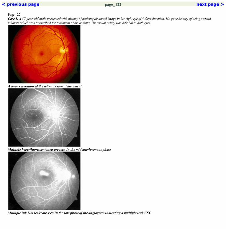
< previous page page_122 next page >
Page 122Case 5. A 37-year-old male presented with history of noticing distorted image in his right eye of 4 days duration. He gave history of using steroidinhalers which was prescribed for treatment of his asthma. His visual acuity was 6/6; N6 in both eyes.
A serous elevation of the retina is seen at the macula
Multiple hyperfluorescent spots are seen in the mid arteriovenous phase
Multiple ink blot leaks are seen in the late phase of the angiogram indicating a multiple leak CSC

The extent of the detachment is well made out in the red free photograph
The hyperfluorescent spots have increased in intensity and size indicating leakage of the dyeCOMMENTMultiple leak CSC is more commonly associated with the use of steroids.
< previous page page_122 next page >

< previous page page_123 next page >
Page 123Case 6. A 41-year-old male presented with history of blurred vision in the right eye of three months duration. He gave history of being treated withsystemic steroids. His visual acuity was 6/9; N6.
Fundus photograph of the right eye showing the large sensory retinal elevation at the macula. An additional pocket of fluid is notedsuperotemporal to the disc
In the early arteriovenous phase, faint hyperfluorescence is seen inferior to the fovea and in the superior macula
Multiple smoke stack and ink blot leaks are seen in the late arteriovenous phase

Red free photograph shows the two areas of sensory retinal detachment
The arteriovenous phase shows multiple points of leakage of dye. At two points inferior to the fovea, the dye is seen to ascend vertically
The dye has leaked from multiple points and pooled in the upper part of the detachment in the late phase of the angiogram
< previous page page_123 next page >

< previous page page_124 next page >
Page 124Case 7. A 40-year-old male patient presented with history of blurred vision in the left eye for the past three months. Visual acuity was 6/12; N8. Righteye was normal.
A serous elevation of the retina is seen at the macula. A pigment epithelial detachment is in the center
Mid arteriovenous phase of the angiogram showing hyperfluorescence due to leakage of the dye into the pigment epithelium detachment
Dye leakage from the pigment epithelial detachment into the subretinal space is seen. The dye is seen to ascend and reach the upper border of thedetachment
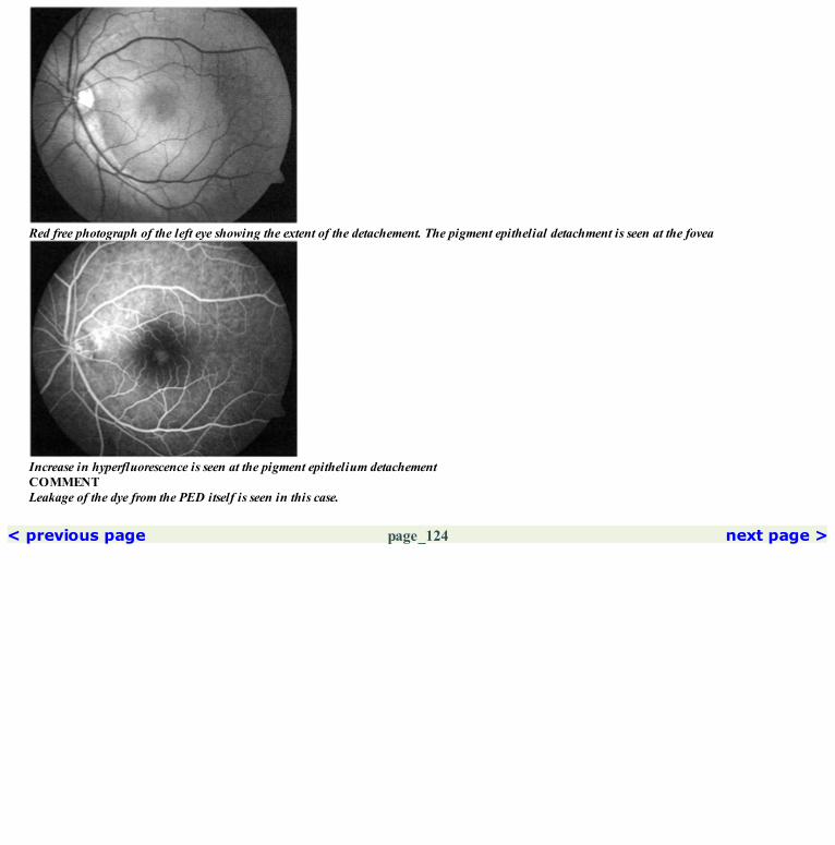
Red free photograph of the left eye showing the extent of the detachement. The pigment epithelial detachment is seen at the fovea
Increase in hyperfluorescence is seen at the pigment epithelium detachementCOMMENTLeakage of the dye from the PED itself is seen in this case.
< previous page page_124 next page >

< previous page page_125 next page >
Page 125Case 8. A 32-year-old female patient gave history of reduced vision in the left eye of three months duration. She had been diagnosed elsewhere ashaving choroiditis and was started on systemic steroids. At presentation her vision in the left eye was 6/18; N36. She was found to have central serouschorioretinopathy in the left eye.
Fundus photograph of the left eye showing multiple PEDs at the macula. Extensive subretinal whitening (fibrin) is seen temporally andsuperiorly
Hyperfluorescence due to early filling of the PED is seen. Point hyperfluorescence are also seen: one point leak temporal to the fovea and twopoint leaks inferotemporally
The bullous inferior retinal detachment is seen. Blurring of vascular details due to retinal elevation; choroidal pattern is not seen
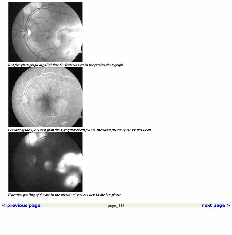
Red free photograph highlighting the features seen in the fundus photograph
Leakage of the dye is seen from the hyperfluorescent points. Increased filling of the PEDs is seen
Extensive pooling of the dye in the subretinal space is seen in the late phase
< previous page page_125 next page >
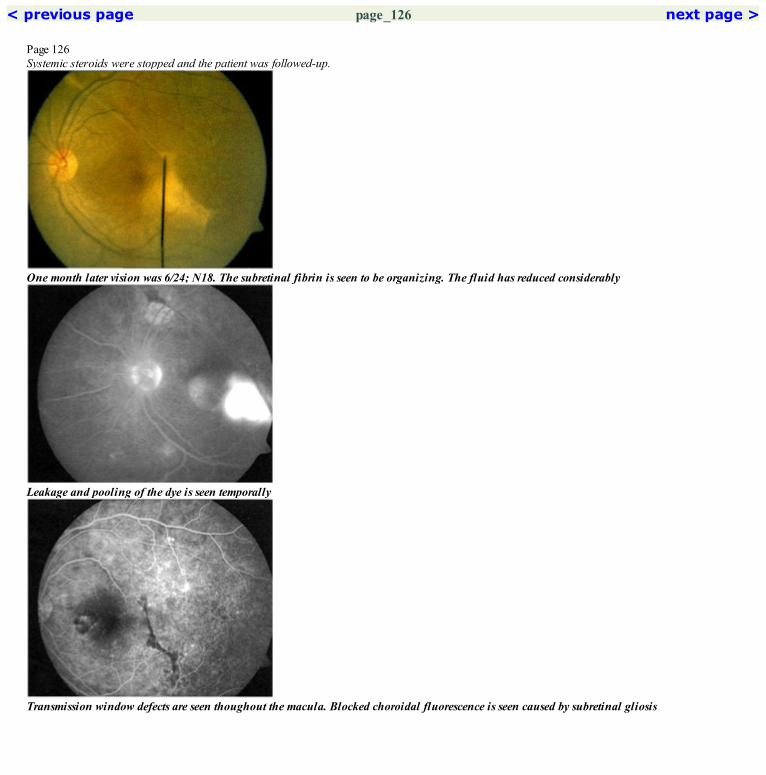
< previous page page_126 next page >
Page 126Systemic steroids were stopped and the patient was followed-up.
One month later vision was 6/24; N18. The subretinal fibrin is seen to be organizing. The fluid has reduced considerably
Leakage and pooling of the dye is seen temporally
Transmission window defects are seen thoughout the macula. Blocked choroidal fluorescence is seen caused by subretinal gliosis

Arteriovenous phase showing a point leak temporally. Blocked choroidal fluorescence (temporal to fovea) is seen caused by the subretinal fibrin
Three months later, visual acuity was 6/24; N6. RPE alteration is seen at the macula. The subretinal fibrin has resorbed. Subretinal gliotic bandis seen inferotemporally
Late phase picture shows minimal fading of the hyperfluorescence temporally suggestive of window defects
< previous page page_126 next page >

< previous page page_127 next page >
Page 127Case 9. A 39-year-old male presented with history of decreased vision in the left eye one month back. Visual acuity was 6/9; N6 in the left eye. Theright eye was normal.
Fundus photograph of the left eye showing the sensory retinal detachment at the center of macula
Arteriovenous phase showing faint point hyperfluorescence spots superonasal to the foveal
No leakage of dye is seen even in the late films
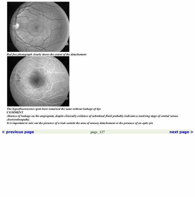
Red free photograph clearly shows the extent of the detachement
The hyperfluorescence spots have remained the same without leakage of dyeCOMMENTAbsence of leakage on the angiogram, despite clinically evidence of subretinal fluid probably indicates a resolving stage of central serouschorioretinopathy.It is important to rule out the presence of a leak outside the area of sensory detachement or the presence of an optic pit.
< previous page page_127 next page >

< previous page page_128 next page >
Page 128Case 10. A 48-year-old male presented with decreased vision in the right eye for the past five months. He gave history of an episode of decreasedvision in the left eye eleven years back. Visual acuity was 6/36; N18 in the right eye and 6/6; N6 in the left eye. Both eyes had features of resolvedcentral serous chorioretinopathy.
Fundus photograph of the right eye showing RPE alterations in the inferior macular region
Stippled hyperfluorescence is noted corresponding to the region of RPE alterations. Hyperfluorescence is also noted superotemporal to the discand to the fovea
An RPE track is seen extending from the macula inferiorly

Red free photograph shows RPE alterations in the inferior part of the macula
Increase in hyperfluorescence in these areas is seen. Hyperfluorescence is also noted at the foveal centerCOMMENTPresence of widespread RPE alterations at the posterior pole and the characteristic RPE track are hallmarks of old resolved central serouschorioretinopathy.
< previous page page_128 next page >

< previous page page_129 next page >
Page 129CLINICAL NUGGETS1. Use of corticosteroids in any form should be avoided in patients with central serous chorioretinopathy.2. Argon laser photocoagulation to the leaks can be considered in cases of non-resolving central serous chorioretinopathy that hampers with dailyactivities; majority of leaks gets obliterated spontaneously.SELECTED READINGS1. Fujisawa Y. Clinical studies on central serous chorioretinitis by fluorescein fundus photography. Jpn J Ophthalmol 10:19–26, 1966.2. Friberg TR, Campagna J. Central serous chorioretinopathy: An analysis of the clinical morphology using image processing techniques. GraefesArch Clin Exp Ophthalmol 227:201–205, 1989.3. Spitznas M, Huke J. Number, shape and topography of leakage points in acute type I central serous retinopathy. Graefes Arch Clin ExpOphthalmol 225:437–440, 19874. Yamada K, Hayasaka S, Setogawa T. Fluorescein angiographic patterns in patients with central serous chorioretinopathy at initial visit.Ophthalmologica 205:69–76, 1992.5. Castro-Correia J, Coutinho MF, Rosas V, Maia J. Long-term follow-up of central serous retinopathy in 150 patients. DOC ophthalmol81(4):379–86, 1992.6. Bennett G. Central serous chorioretinopathy. Br J Ophthamol 39:605–618, 1955.7. Gass JDM. Idiopathic central serous chorioretinopathy. Stereoscopic atlas of macular diseases. Diagnosis and Treatment. 4th edn, St Louis,Mosby, 52–70, 1997.
< previous page page_129 next page >

< previous page page_130 next page >
Page 13019 Cystoid Macular Edema
■ Fluid accumulation in cystic spaces localized to the outer plexiform and inner nuclear layers of the retina is called cystoid macular edema.■ The common causes of cystoid macular edema includePostsurgical Cystoid Macular Edema■ Angiographic macular edema—The macular edema is not obvious on clinical examination, however, the fluorescein angiography shows leakageof the dye. The visual acuity is usually unaffected.■ Clinically significant macular edema—Here the macular edema is associated with a drop in visual acuity.■ The area of dye leakage on fluorescein angiography correlates poorly with the visual acuity. A better predictor in such cases would be theestimation of the retinal thickness.Diabetic Macular Edema■ In addition to cystoid macular edema, the other changes occurring in diabetic maculopathy are well seen on the angiogram. This includes capillaryleakage, microaneurysms, capillary dropout areas and intraretinal microvascular abnormalities. In patients with diabetic retinopathy undergoingcataract surgery it may be difficult to determine whether the postoperative macular edema is due to the surgical intervention or occurring as part ofthe diabetic process. In diabetic macular edema the leakage may be more diffuse and not localized to the perifoveal region. In addition the late discstaining, which is commonly seen in postsurgical cystoid macular edema, is absent in diabetic macular edema.Intermediate UveitisVascular OcclusionsEpiretinal membraneRetinitis PigmentosaDrug Toxicity e.g., Epinephrine, Nicotinic AcidDominantly Inherited Cystoid Macular Edema
< previous page page_130 next page >

< previous page page_131 next page >
Page 131
■ In the early phase of the angiogram, capillary dilation is seen in the perifoveal region.■ Late pooling of the dye is seen, occurring in a petaloid pattern in the perifoveal region. The fusiform cystoid spaces are arranged in a radial pattern.■ Often patchy hyperfluorescence is seen extending beyond the region of the cystic spaces indicating more widespread capillary leakage.■ Leakage of the dye from the disc capillaries gives rise to late staining of the optic nerve head.■ When cystoid spaces occur outside the perifoveal area, fluorescein angiography shows a honeycomb appearance rather than a petaloid pattern.
< previous page page_131 next page >

< previous page page_132 next page >
Page 132Case 1. A 62-year-old female gave history of reduced vision in the right eye. She had undergone a cataract surgery with intraocular lens implantationin the past, with no visual improvement seen in the postoperative period. Vision was 2/60 in the right eye.
Fundus photograph of the right eye. A large lamellar macular hole with surrounding cystoid changes are seen
Dilation of the perifoveal capillaries is seen. Early leakage of the dye is seen from the capillaries in the inferior macular region
Significant accumulation of the dye is noted throughout the macula. Honeycomb pattern of cystoid macular edema is noted in the areas outsidethe fovea. Note the hypofluorescent appearing lamellar macular hole

Red free photograph of the right eye. Large cystoid changes are seen in the foveal region with a large central lamellar macular hole
Increased leakage from the capillaries in the macula is evidentCOMMENTNo dye accumulation occurs in the late lamellar macular hole as the dye which leaks into its cavity easily escapes into the vitreous. Hence, itappears hyperfluorescent.Cystoid changes occurring outside the fovea assumes a honeycomb pattern.
< previous page page_132 next page >

< previous page page_133 next page >
Page 133Case 2. A 36-year-old female presented with history of blurring of vision in both eyes for the past one month. Visual acuity was 6/9; N6 in the right eyeand 6/24; N8 in the left eye. Examination revealed exudates over the pars plana in both eyes suggestive of intermediate uveitis.
Cystoid changes are seen in the foveal region of the right eye
The arteriovenous phase shows dilation and early leakage of the dye from the perifoveal capillaries
Late leakage and accumulation of the dye in cystoid spaces in seen. Increased staining of the disc is seen

Red free photograph clearly demarcates the cystoid changes
Increased leakage of the dye is noted in the perifoveal region
< previous page page_133 next page >

< previous page page_134 next page >
Page 134Left eye of the patient
Fundus photograph of the left eye of the same patient showing cystoid changes at the fovea
Dilation of the perifoveal capillaries is well seen. Minimal leakage of the dye from these dilated capillaries is seen
Red free photograph of the left eye

Petaloid pattern of cystoid macular edema is well seen. Non-cystoid accumulation of the dye is noted in the superior macular regionCOMMENTIntraocular inflammation caused by intermediate uveitis results in the dilation of the perifoveal capillaries and leakage of fluid; this leads tocystoid macular edema.
< previous page page_134 next page >

< previous page page_135 next page >
Page 135Case 3. A 56-year-old presented with diminution of vision in the left eye, one month following an uneventful phacoemulsification with intraocular lensimplantation surgery. The visual acuity was 6/60:N18.
Fundus photograph of the left eye showing the cystoid changes at the fovea
Arteriovenous phase showing the dilated perifoveal capillaries
Late leakage of the dye and its collections in cystoid spaces is seen. The cystoid spaces are arranged in a petaloid pattern. Noncystoidaccumulation of the dye is noted in the superior macular region. Increased staining of the disc is seen.

Red free photograph shows the large cystoid spaces at the fovea
Early leakage from the dilated perifoveal capillaries is seenCOMMENTLeakage of the dye from the perifoveal capillaries causes the cystoid pattern.
< previous page page_135 next page >

< previous page page_136 next page >
Page 136CLINICAL NUGGETS1. The petaloid pattern of cystoid macular edema is best seen in the late phase of the angiogram.2. Fluorescein angiography and OCT are useful investigative tools to document and follow-up of eyes with cystoid macular edema.SELECTED READINGS1. Jaffe NS, Luscombe SM, Clayman HM, Gass JDM: A fluorescein angiographic study of cystoid macular edema. Am J Ophthalmol 92:775–780,1981.2. Wright PL, Wilkinson CP, Balyeat HB et al. Fluorescein angiographic study of cystoid macular edema after PCIOL. Arch. Ophthalmol: 106:740–744, 1988.3. Fine BS, Bruker AJ. macular edema and cystoid macular edema. Am J Ophthalmol 92:466–481,19814. Allen AW, Jaffe NS. Cystoid macular edema: a preliminary study, Trans Ame Acad Ophthalmol and Otolaryngology 81:133–134, 1976.5. Gass JDM, Anderson DR, Davis EB. A clinical fluorescein angiographic and electron microscopic correlation of cystoid macular edema. AmericanJournal of Ophthalmol 100:82–86, 1985.6. Gass JDM, Mortan EWD. Cystoid macular edema and papilledema following cataract extraction, a fluorescein fundoscopic and angiographicstudy. Arch Ophthalmol 76; 646–66, 1966.
< previous page page_136 next page >

< previous page page_137 next page >
Page 137

Section 3 Vascular Disorders
< previous page page_137 next page >

< previous page page_138 next page >
Page 138This page intentionally left blank.
< previous page page_138 next page >

< previous page page_139 next page >
Page 13920 Arterial Occlusion
■ Acute central retinal artery occlusion causes a sudden painless visual loss occurring over several seconds.■ The fundus is characterized by opacification of the retina especially at the posterior pole; the opacification may take hours to become apparent.■ A cherry red spot is present in most of the cases.■ The retinal arterioles are thinned and may show ‘boxcarring’ of the blood column.■ Approximately 25% of eyes with central retinal artery occlusion have a patent cilioretinal artery in which case the retinal area supplied by this isspared.■ Sometimes, the occlusion may involve only the branch arteriole in which case the changes are restricted to the affected segment.■ Isolated occlusion of the cilioretinal artery can also occur by itself or in association with a central retinal vein occlusion.
■ In the acute stage there may be considerable amount of masking of choroidal fluorescence due to the cloudy swelling of the overlying retina.■ The arterial phase is extremely delayed; it is often possible to see the slow progress of the dye front in the arteriole. There is marked delay in thearteriovenous transit time. The column of blood is seen to be broken into segments and hardly any dye reaches the capillaries. Venous return is alsodelayed and incomplete.■ By about the 2nd week following the occlusion, dye begins to flow into the vessels but shows a high degree of irregularity of caliber. Venousreturn improves, however the capillary details do not recover. Optic disc shows staining during the late phase of the angiogram.
< previous page page_139 next page >

< previous page page_140 next page >
Page 140Case 1. A 42-year-old male presented with sudden loss of vision in the right eye of one week duration. Visual acuity was perception of light withinaccurate projection of rays in the right eye. Left eye was normal.
Fundus photograph of the right eye showing a pale optic disc with markedly narrowed arterioles. Few white deposites are seen in the inferiormacular region
Delayed filling of the arteries is evident from the fact that the arteries show only minimal hyperfluorescence whereas the choroidal filling is moreor less complete
Markedly delayed filling of the veins is seen. All the vessels appear narrowed. The optic pit is still hyperfluorescent

Red free photograph of the right eye shows an optic pit in the inferotemporal part of the disc
The concentration of the dye in the arteries has increased. Hyperfluorescence due to optic pit is seen in the inferotemporal part of the optic nervehead
Late phase shows the washout of the dye. Hyperfluorescence of the optic pit is seen
< previous page page_140 next page >

< previous page page_141 next page >
Page 141Case 2. A 29-year-old female with systemic lupus erythematosus presented with sudden loss of vision in the right eye of one day duration. Visualacuity was perception of light.
Fundus photograph showing multiple scattered retinal hemorrhages and retinal whitening at the posterior pole. Note the dilated retinal veins
Filling of the arteries is markedly delayed with dye appearing in the arteries only after 40 seconds. The leading edge of the dye column is seenwithin the arteries
Further filling of the arteries is seen at 5 minutes. However the dye has not yet entered the veins
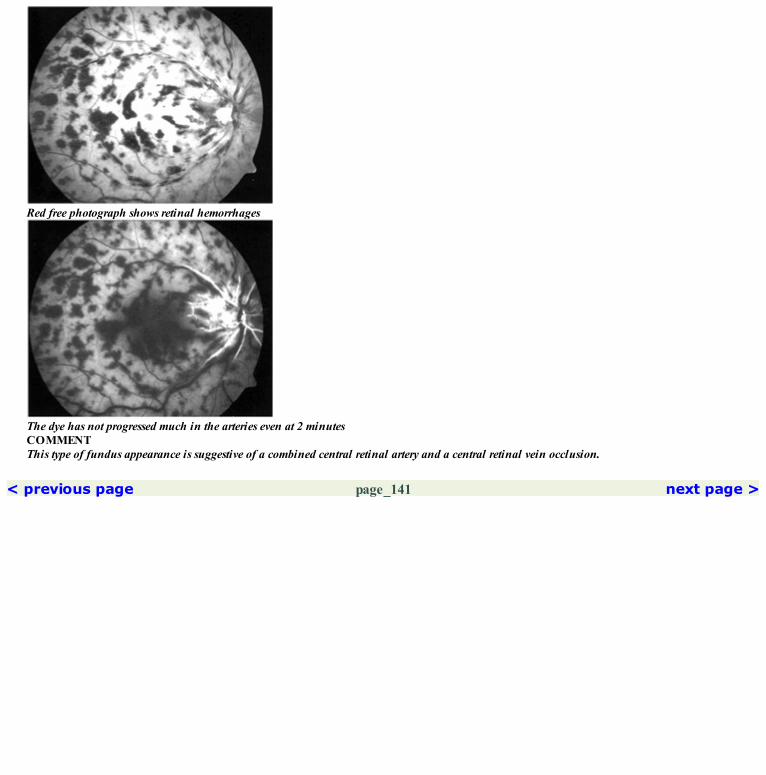
Red free photograph shows retinal hemorrhages
The dye has not progressed much in the arteries even at 2 minutesCOMMENTThis type of fundus appearance is suggestive of a combined central retinal artery and a central retinal vein occlusion.
< previous page page_141 next page >

< previous page page_142 next page >
Page 142Case 3. A 51-year-old patient with history of hypertension for the past fifteen years presented with loss of vision in the right eye of one month duration;visual acuity was perception of light.
Fundus photograph of the right eye showing areas of retinal whitening at the posterior pole; cherry red spot is seen. An area of subretinalwhitening with associated subretinal fluid is seen superotemporally
The early disc hyperfluorescence is seen. However the dye has not entered the arteries even at 28 seconds
The dye has progressed in the arteries and some venous return is noted in the veins close to the disc. The point leak superotemporally has increasedin intensity and extent

Red free photograph highhights the features seen in the color photograph
Delayed entry of the dye is noted in the arteries. The disc capillaries have filled. A point leak is noted superotemporally
Increased staining of the disc and the arteries is seen in the late film. A smoke stack like leak is seen superotemporally
< previous page page_142 next page >

< previous page page_143 next page >
Page 143Case 4. A 52-year-old male presented with sudden loss of vision in the left eye of 2 days duration. He was a diabetic for the past fifteen years and hadundergone pars plana vitrectomy in the left eye eight months back with a postoperative visual acuity of 6/24 N36. His visual acuity was recorded asperception of light in the left eye.
Fundus photograph of the left eye showing retinal whitening and cherry red spot in the macula
At 25 seconds, filling of few disc capillaries is seen. However, the dye has not yet entered the arteries
At 5 minutes the dye has progressed to some extent in the arterioles, however it has not yet completely filled. Some filling of the veins close to thedisc is seen
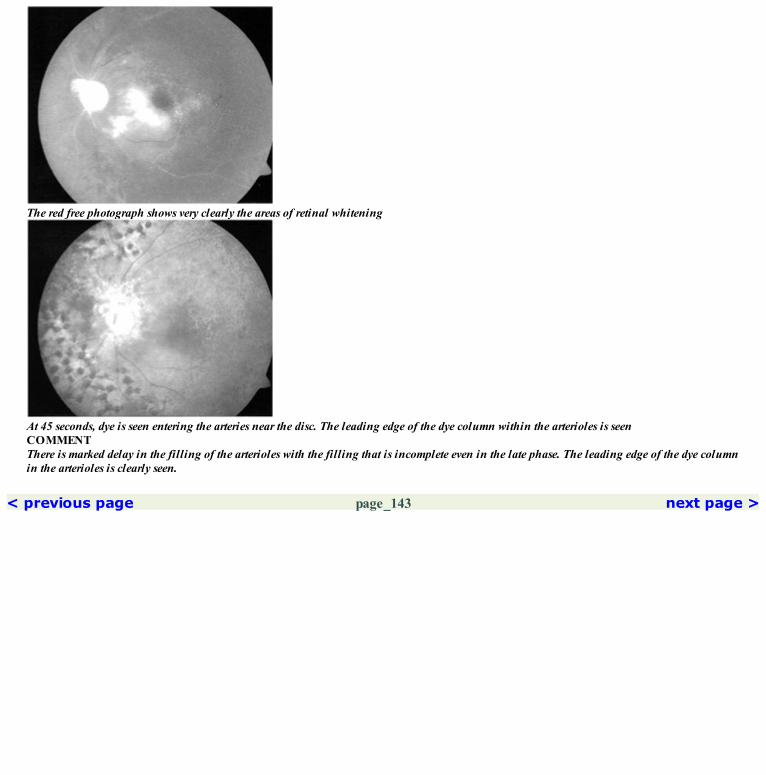
The red free photograph shows very clearly the areas of retinal whitening
At 45 seconds, dye is seen entering the arteries near the disc. The leading edge of the dye column within the arterioles is seenCOMMENTThere is marked delay in the filling of the arterioles with the filling that is incomplete even in the late phase. The leading edge of the dye columnin the arterioles is clearly seen.
< previous page page_143 next page >

< previous page page_144 next page >
Page 144Case 5. 25-year-old male presented with sudden decrease in vision of the left eye of fifteen days duration. Visual acuity was 6/36; N18 in the left eye.
Fundus photograph of the left eye showing areas of retinal whitening. A small island of retina adjacent to the disc is spared due to the presence ofa cilioretinal artery
Arterial filling is delayed and low concentration of the dye is seen initially in the arteries due to its sluggish filling
Arterioveneous phase showing capillary filling only in the region of the cilioretinal artery
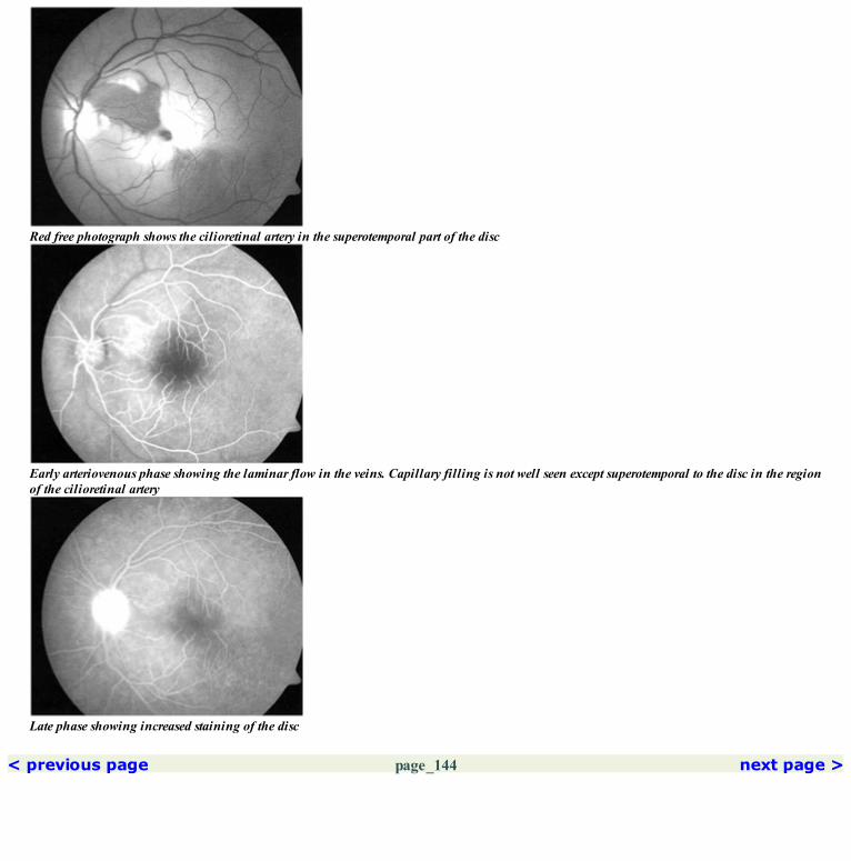
Red free photograph shows the cilioretinal artery in the superotemporal part of the disc
Early arteriovenous phase showing the laminar flow in the veins. Capillary filling is not well seen except superotemporal to the disc in the regionof the cilioretinal artery
Late phase showing increased staining of the disc
< previous page page_144 next page >
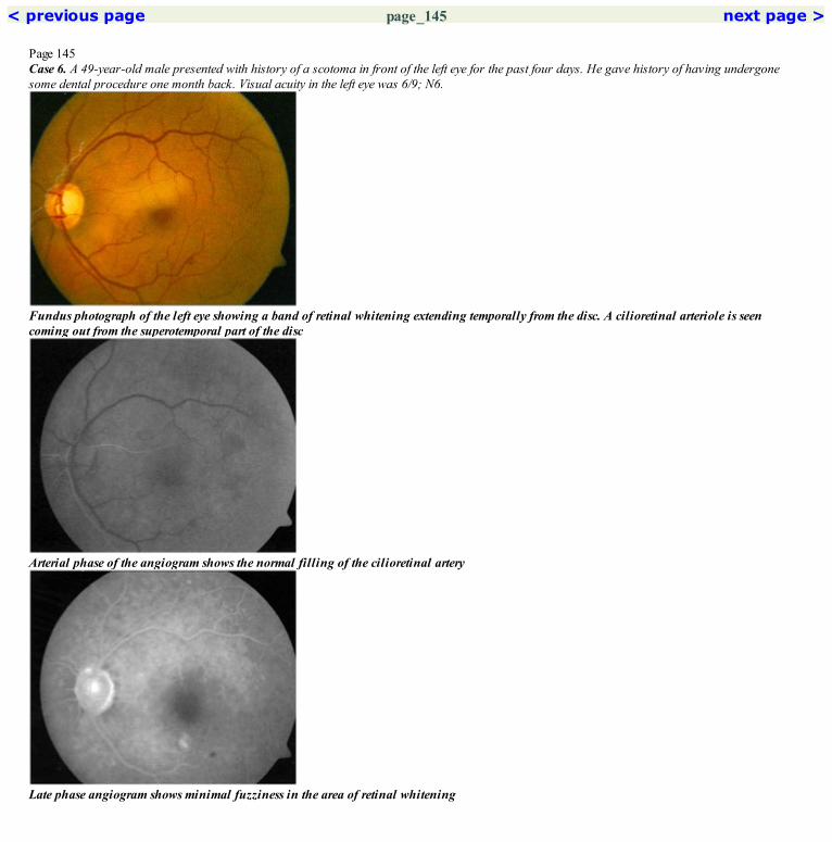
< previous page page_145 next page >
Page 145Case 6. A 49-year-old male presented with history of a scotoma in front of the left eye for the past four days. He gave history of having undergonesome dental procedure one month back. Visual acuity in the left eye was 6/9; N6.
Fundus photograph of the left eye showing a band of retinal whitening extending temporally from the disc. A cilioretinal arteriole is seencoming out from the superotemporal part of the disc
Arterial phase of the angiogram shows the normal filling of the cilioretinal artery
Late phase angiogram shows minimal fuzziness in the area of retinal whitening

Red free photograph shows the area of retinal whitening and the cilioretianal vessels. Few specks of retinal hemorrhage are seen in the inferiormacular region
Late arterioveneous phase showing some blurring of the capillary details in the region of the retinal whiteningCOMMENTThis patient has a cilioretinal artery occlusion with reopening of the cilioretinal artery. However despite reopening, permanent closure of thecapillary bed in the region of the affected cilioretinal artery is seen.
< previous page page_145 next page >

< previous page page_146 next page >
Page 146Case 7. A 38-year-old female presented with sudden onset reduced vision in the right eye of three weeks duration. Visual acuity was 6/9; N6.
Fundus photograph of the right eye showing retinal whitening in the inferior part of the fovea
Arterail phase showing delayed filling of the arteriole exiting from the inferotemporal part of the the disc (arrows). The arterioles superiror to thefovea have filled up
Mid arteriovenous phase showing absence of capillary details in the lower part of the macula. There is some progression of the dye column in theinferotemporal macular arteriole (arrows)
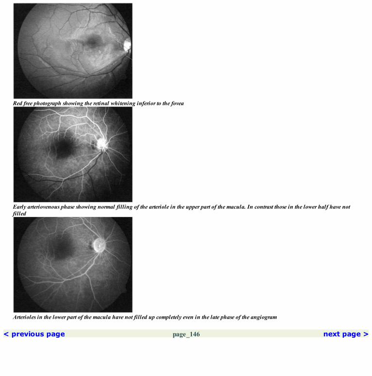
Red free photograph showing the retinal whitening inferior to the fovea
Early arteriovenous phase showing normal filling of the arteriole in the upper part of the macula. In contrast those in the lower half have notfilled
Arterioles in the lower part of the macula have not filled up completely even in the late phase of the angiogram
< previous page page_146 next page >

< previous page page_147 next page >
Page 147CLINICAL NUGGETS1. A cilioretinal artery is normally present in 20% of the eyes.2. Neovascularization of iris can develop in 80% of eyes with combined central retinal artery and vein occlusion.3. Posterior ophthalmic artery occlusion is usually not associated with a cherry red spot.4. Electroretinogram shows a reduction in the b-wave (negative waveform) in eyes with central retinal artery occlusion.SELECTED READINGS1. Brown GC, Magargal LE. Central retinal artery obstruction and visual acuity. Ophthalmology 89:14–19, 1982.2. David NJ, Gass JDM, Beauchamp J. Fluorescein angiography in central retinal artery occlusion. Arch Ophthalmol 77:619–629, 1967.3. Eagling EM, Sanders MD, Miller SJH. Ischemic papillopathy: clinical and fluorescein angiographic review of forty cases. Br J Ophthalmol58:990–1008, 1974.
< previous page page_147 next page >

< previous page page_148 next page >
Page 14821 Coats’ Disease
■ Coats’ disease is an idiopathic condition occurring predominantly in males with no racial predilection.■ It is unilateral in 80% of the cases.■ The common changes seen in the fundus include vascular abnormalities such as telangiectasia and aneurysmal dilations, and zones of capillary non-perfusion.■ The extent of vascular abnormalities can range from minimal to very extensive.■ It is associated with intraretinal and subretinal lipid deposits.■ Leber’s miliary aneurysm is considered to be a localized form of Coats’ disease.
■ Fluorescein angiogram shows numerous localized anomalies of the retinal vasculature.■ Telangiectasia, aneurysms, beading of vessel walls, and vascular communicating channels are seen.■ These are very well made out in the early and mid arteriovenous phases of the angiogram.■ The retinal capillary bed shows marked dilation and tortuosity with multiple saccular and aneurysmal outpouchings.■ Leakage from the vascular abnormalities is seen in the late films.■ Areas of diffuse capillary non-perfusion may be seen in the midperiphery.
< previous page page_148 next page >

< previous page page_149 next page >
Page 149Case 1. A 27-year-old male presented with history of decreased vision in the left eye often days duration. Visual acuity was 6/9; N6. He was found tohave extensive exudation at the posterior pole with vascular abnormalities seen in the periphery. The right eye was normal.
Fundus photograph showing hard exudates at the macula and inferiorly
Dilated retinal capillaries and telangiectasias are clearly made out in the arteriovenous phase. Vascular abnormalities are also noted close to thefovea. Aneurysmal dilations are seen inferiorly
Late phase shows leakage of the dye from the vascular abnormalities

Red free photograph highlights these features
Leakage of dye is noted from the dilated capillaries and the aneurysmal dilatations. Areas of capillary non-perfusion are also seenCOMMENTCapillary telangiectasias, capillary dilatations, aneurysmal dilatations and areas of capillary non-perfusion are hallmarks of Coat’s disease.
< previous page page_149 next page >
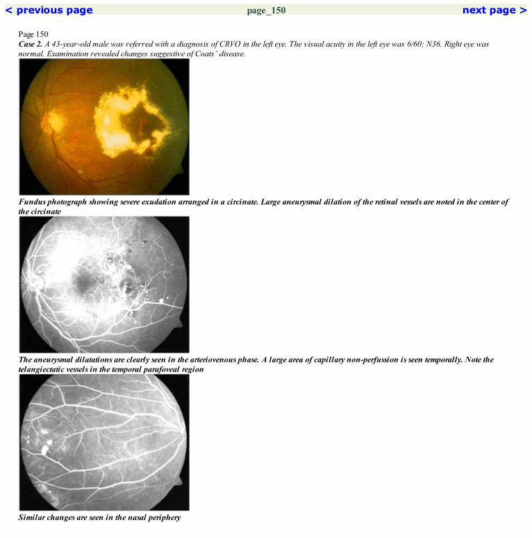
< previous page page_150 next page >
Page 150Case 2. A 43-year-old male was referred with a diagnosis of CRVO in the left eye. The visual acuity in the left eye was 6/60; N36. Right eye wasnormal. Examination revealed changes suggestive of Coats’ disease.
Fundus photograph showing severe exudation arranged in a circinate. Large aneurysmal dilation of the retinal vessels are noted in the center ofthe circinate
The aneurysmal dilatations are clearly seen in the arteriovenous phase. A large area of capillary non-perfussion is seen temporally. Note thetelangiectatic vessels in the temporal parafoveal region
Similar changes are seen in the nasal periphery

Red free photograph showing the circinate with vascular abnormalities in the center
Extensive telangiectasias with areas of capillary non-perfusion and aneurysmal dilatations is seen in the supero-temoral periphery
Late phase picture showing leakage of the dye from the vascular abnormalities
< previous page page_150 next page >

< previous page page_151 next page >
Page 151CLINICAL NUGGETS1. Early treatment of the vascular abnormalities in Coats’ disease provides the best control of leakage and prevents severe visual loss.2. Laser or cryotherapy can be used to treat the vascular abnormalities.3. Multiple treatment sessions may be required.4. Coats’ disease with severe exudative retinal detachment may present with leukocoria.SELECTED READINGS1. Gass JDM. A fluorescein angiographic study of macular dysfunction secondary to retinal vascular disease. V. Retinal telangiectasias. ArchOphthalmol 80:592–605, 1968.2. Gass JDM, Oyakawa RT. Idiopathic juxtafoveolar retinal telangiectasias. Arch Ophthalmol 100:769–780, 1982.3. Chopdar A. Retinal telangiectasis in adults: fluorescein angiographic findings and treatment by argon laser. Br J Ophthalmol 62: 243–250, 1978.4. Egerer I, Tasman W, Tomer TL. Coats’ disease. Arch Ophthalmol 92:109–112, 1974.5. Panleikhoff D, Kruger K, Heinreich T, Wessing A. Epidemiological features and therapeutic result in Coats’ disease. Invest Ophthalmol 29:335,1988.
< previous page page_151 next page >

< previous page page_152 next page >
Page 15222 Diabetic Retinopathy
■ Diabetic retinopathy is classified as● Non-Proliferative Diabetic Retinopathy (NPDR)—Mild/Moderate/Severe● Proliferative Diabetic Retinopathy (PDR)■ Diabetic maculopathy can be either● Focal—microaneurysms leaking fluid into the retina.● Diffuse—dilated retinal capillaries leaking fluid into the retina.● Ischemic—capillary non-perfused area in the macula.● Mixed—combination of any of the above.■ Clinically significant macular edema● Thickening of the retina located <500 microns from the center of the macula.● Hard exudates with thickening of the adjacent retina located <500 microns from the center of the macula.● A zone of retinal thickening, 1 DA or larger in size located <1 DA from the center of the macula.■ Basic pathology involves● Increased permeability of the retinal capillaries.● Closure of the retinal capillaries.
■ Microaneurysms—appear as hyperfluorescent dots which leak dye in the later phases.■ Dilation of the retinal capillaries may occur.■ Capillary closure appears as areas of hypofluorescence due to non-filling; they are usually bordered by dilated capillaries and microaneurysms.■ Intraretinal microvascular abnormalities (IRMA)—irregular segmental dilatation of the retinal capillary bed. They usually occur in areas ofcapillary non-perfusion and usually appear to originate and drain into the venous side of the circulation. They remain within the confines of theretina and do not leak fluorescein except minimally at their growing tips. They probably represent intraretinal neovascularization or dilatation ofexisting retinal channels.■ Superficial and deep retinal hemorrhages cause blocked choroidal fluorescence.■ Venous abnormalities in the form of venous dilation, beading, loops and reduplication may be seen.■ Neovascularization on the disc or the retinal surface appears as irregularly branching vessels which freely leaks the dye.
< previous page page_152 next page >

< previous page page_153 next page >
Page 153Case 1. A 56-year-old male presented with history of decreased vision in both eyes for the past 6 months. He was a known diabetic for the past 6years and a hypertensive for the last 1 year. Visual acuity was 6/9; N6 in both eyes.
Fundus photograph of the left eye showing hard exudates arranged in a lose circinate pattern in the superior part of the macula
Arteriovenous phase showing the multiple hyperfluorescent points caused by microaneurysms. These are seen boarding the areas of capillary non-perfusion
Red free photograph highlights the scattered hard exudates and the retinal hemorrhages

Increased fuzziness is seen around the microaneurysms due to leakage of the dye in the late phase. The area of capillary non-perfusion remainhyperfluorescentCOMMENTFocal maculopathy is characterized by presence of leaking microaneurysms. Fluorescein angiography can be used to guide laserphotocoagulation of these microaneurysms.
< previous page page_153 next page >

< previous page page_154 next page >
Page 154Case 2. A 54-year-old male with diabetes for the past ten years presented with history of reduced vision in both eyes for the last one month. Visualacuity was 6/12; N8 in the right eye and 6/9; N10 in the left eye.
Fundus photograph of the left eye. Retinal thickening was noted in the macular region. Note the absence of hard exudates or retinalhemorrhages
Mid arteriovenous phase shows leakage of dye from the dilated capillaries throughout the macula. A fuzzy hyperfluorescence is seen along theinferotemporal arcade indicative of a small neovascular frond. Few microaneurysms are noted superiorly
Late film shows accumulation of the dye in cystoid spaces around the fovea. Non-cystoid collection of the dye is also noted in the other parts ofthe macula. The leakage from the neovascular frond has also increased

Red free photograph highlighting the cystoid changes in the foveal region
The leakage from the capillaries has increased and the dye is seen to be collecting in cystoid spaces around the fovea
< previous page page_154 next page >
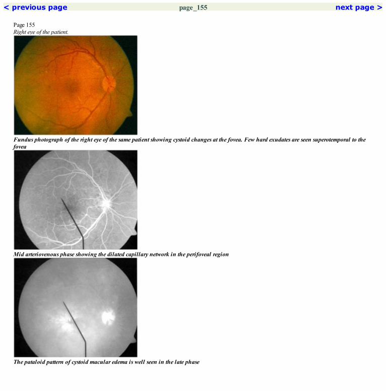
< previous page page_155 next page >
Page 155Right eye of the patient.
Fundus photograph of the right eye of the same patient showing cystoid changes at the fovea. Few hard exudates are seen superotemporal to thefovea
Mid arteriovenous phase showing the dilated capillary network in the perifoveal region
The pataloid pattern of cystoid macular edema is well seen in the late phase

Red free photograph highlights the cystoid changes at the fovea
Significant leakage of the dye is seen from the dilated capillariesCOMMENTDiffuse maculopathy is characterised by dilated capillaries which leak fluorescein dye.Hard exudates are not usually associated with diffuse type of diabetic maculopathy.
< previous page page_155 next page >

< previous page page_156 next page >
Page 156Case 3. A 44-year-old male diabetic patient presented with marked reduction in vision in both eyes for the past one year. Visual acuity was 2/60 in theright eye and 1/60 in the left eye.
A large neovascular frond is seen at the disc. Hard exudates are seen in the upper and lower part of the macula while the foveal area has a dullwashed out appearance
Marked leakage of the dye is seen from the NVD. A large area of capillary non-perfusion is seen in the upper part of the macula involving thefovea; dilated capillaries are seen around this
The red free photograph highlights the features seen in the fundus photograph

Marked leakage of the dye is seen from the NVD. Increased staining of the retinal tissue in the macula is seen except in the region of thecapililary droupout which remains hyperfluorescentCOMMENTIschemic maculopathy is characterized by areas of capillary dropout at the macula.They have a very poor visual prognosis.
< previous page page_156 next page >
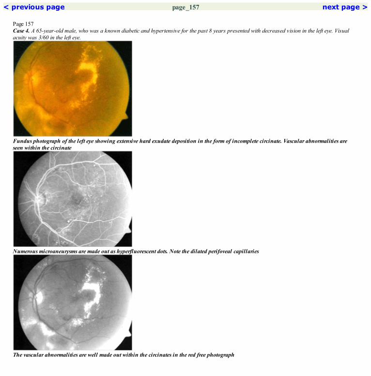
< previous page page_157 next page >
Page 157Case 4. A 65-year-old male, who was a known diabetic and hypertensive for the past 8 years presented with decreased vision in the left eye. Visualacuity was 3/60 in the left eye.
Fundus photograph of the left eye showing extensive hard exudate deposition in the form of incomplete circinate. Vascular abnormalities areseen within the circinate
Numerous microaneurysms are made out as hyperfluorescent dots. Note the dilated perifoveal capillaries
The vascular abnormalities are well made out within the circinates in the red free photograph

Marked leakage of the dye is seen from the microaneurysms and the dilated capillaries. Cystoid pattern of dye accumulation is clearly evidentCOMMENTPatient is having both focal and diffuse type of maculopathy.Mixed type of maculopathy is quite common.
< previous page page_157 next page >

< previous page page_158 next page >
Page 158Case 5. A 48-year-old male patient with a history of diabetes for the past 20 years presented with decreased vision in the left eye of one week duration.Visual acuity was 6/18; N36.
Fundus photograph of the left eye showing scattered hard exudates and hemorrhages in the macula
Innumerable microaneurysms are seen as hyperfluorescent dots. The perifoveal capillary network is broken superotemporally
Red free photograph highlights these retinal changes

Marked leakage of the dye into the retinal tissue is seen from the microaneurysmsCOMMENTFluorescein angiography is very useful to assess the para- and perifoveal capillary network.
< previous page page_158 next page >

< previous page page_159 next page >
Page 159Case 6. A 50-year-old male presented with decreased vision in the right eye of two months duration. He was a known diabetic for the last three years.Visual acuity was 6/12; N36 in the right eye.
Fundus photograph of the right eye showing asteroid hyalosis which is obscuring the view of the fundus. Gliosis is seen in the temporal macularregion
Mid arteriovenous phase showing the large neovascular network. Area of capillary non-perfusion is seen involving the parafoveal capillarynetwork. Microaneurysms are noted bordering this
Marked leakage of the dye is evident in the late phase

The view of the retina is obscured by the asteroid hyalosis even in the red free photograph
Increased leakage of the dye is seen from the neovascular network. Areas of capillary non-perfusion are seen temporallyCOMMENTFluorescein angiography is extremely useful to evaluate the retina in patients with asteriod hyalosis.The presence of a barrier filter prevents the blue light reflected from the asteroids to enter the camera. Only the green light from the retina isallowed to enter. Hence the retinal details are very well seen in the angiogram despite the presence of asteroid hyalosis.
< previous page page_159 next page >

< previous page page_160 next page >
Page 160Case 7. A 51-year-old male diabetic presented with history of panretinal photocoagulation done for both eyes in the past. Visual acuity was 6/6; N6 inboth eyes.
Red free photograph of the right eye showing old laser photocoagulation marks temporally. A neovascular frond is seen along the inferotemporalarcade
Multiple areas of intense hyperfluorescence due to leakage of the dye from the neovascular fronds are seen. Large areas of capillary dropout arealso seen
Increased leakage from the neovascular fronds is seen in the late phase. The rim of hyperfluorescence around the laser marks is more prominent

Hyperfluorescence due to leakage of dye from the neovascular frond is seen in the mid arteriovenous phase. The laser marks appear as centralhypofluorescence surrounded by a rim of hyperfluorescence
The leakage of dye from the inferotemporal frond has increased in the recirculation phase. Extensive areas of capillary non-perfusion is seeninferiorlyCOMMENTFlat, subtle NVEs which may be difficult to appreciate clinically in a heavily ablated eye can be picked up quite well in the fluoresceinangiogram.
< previous page page_160 next page >

< previous page page_161 next page >
Page 161CLINICAL NUGGETS1. Close follow-up of patients with severe NPDR is needed, as the risk of progression to PDR is high.2. In eyes requiring laser for PDR and CSME, it is better to do the treatment for the CSME first followed by a panretinal photocoagulation 2–3weeks later. This is to avoid the aggravation of the macular edema that may follow panretinal photocoagulation.3. It is not unusual to have a transient mild increase in macular edema immediately following focal laser photocoagulation.4. Systemic hypertension can accelerate the progression of diabetic retinopathy.SELECTED READINGS1. Early Treatment Diabetic Retinopathy Study Research Group—Report no 11, classification of diabetic retinopathy from fluorescein angiograms.Ophthalmology 98:807–822, 1991.2. Takeshi N, Mukara K, Shimuzi K. Distribution of capillary non-perfusion in early stage of diabetic retinopathy. Ophthalmology 91: 1431–39,1984.3. Early Treatment Diabetic Retinopathy Study Research Group: Focal photocoagulation treatment of diabetic macular edema: relationship oftreatment effect to fluorescein angiographic and other retinal characteristics at baseline. ETDRS report no. 19. Arch Ophthalmol 113:1144–1155,19954. Early Treatment Diabetic Retinopathy Study Research Group: Fundus photographic risk factors for the progression of diabetic retinopathy.ETDRS report no 12. Ophthalmology 98:823–833, 1991.5. Norton EW, Gutman F. Diabetic retinopathy studied by fluorescein angiography. Ophthalmologica 150:5–17, 1965.6. Kohner EM, Dollery CT, Peterson JW, Oakley NW. Arterial fluorescein studies in diabetic retinopathy. Diabetes, 16:1–10, 1967.7. Diabetic Retinopathy Study Research Group: Photocoagulation treatment of proliferative diabetic retinopathy. Ophthalmology 88: 583–600,1981.
< previous page page_161 next page >

< previous page page_162 next page >
Page 16223 Eales’ Disease
■ Common clinical condition in the Indian subcontinent typically affecting young healthy males.■ Idiopathic retinal periphlebitis in the active stage is more commonly seen in the periphery. This leads to areas of capillary non-perfusion whichmay lead to the development of neovascularization.■ Recurrent vitreous hemorrhage is common.■ Rarely the larger posterior veins are involved and the patient may present with a branch retinal vein or a central retinal vein occlusion.
■ Narrowing of the lumen may be seen in the areas of active periphlebitis.■ The wall of the vein in the affected part stains and leaks dye in the late phase of the angiogram.■ Areas of capillary non-perfusion are seen in the periphery. Associated vascular abnormalities like collaterals are also common.■ Neovascularization is commonly seen in the periphery.
< previous page page_162 next page >

< previous page page_163 next page >
Page 163Case 1. A 33-year-old male presented with history of reduced vision in the left eye for the past one year. Visual acuity was 3/60. The right eye had 6/5;N6 visual acuity and showed few areas of peripheral vascular sheathing.
Fundus photograph of the left eye showing a sheathed superotemporal vessel. Hard exudates are seen in the macula
Arteriovenous phase showing leakage of the dye from the vessel walls. Areas of capillary non-perfusion are seen
Lute phase shows extensive staining of the vessel walls and the adjacent retina

Fundus photograph of the temporal periphery showing areas of resolving periphlebitis
Extensive leakage of the dye from the walls of the veins and its draining channels is seen. The arterioles are unaffectedCOMMENTSActive stage of Eales’ disease is characterized by periphlebitis which causes leakage of dye and staining of the walls of the vein.Eales’ disease predominantly affects the peripheral veins but can also involve the larger posterior veins.
< previous page page_163 next page >

< previous page page_164 next page >
Page 164Case 2. A 30-year-old male presented with minimal blurring of vision in the left eye. Visual acuity was 6/5; N6 in both eye. Vitreous cells were seen inboth eye.
Fundus photograph of the right eye showing multiple foci of active periphlebitis in the inferior periphery
Minimal leakage of the dye is noted through the walls of the vein in the areas of perivasculitis. Areas of capillary non-perfusion are seen
Staining of the vessel wall and the adjacent retina is seen in the later phase of the angiogram

Red free photograph shows the active cuffs of periphlebitis
The leakage of dye in the areas of perivasculitis has increasedCOMMENTMultiple peripheral cuffs of periphlebitis is a common finding in Eales’ disease
< previous page page_164 next page >
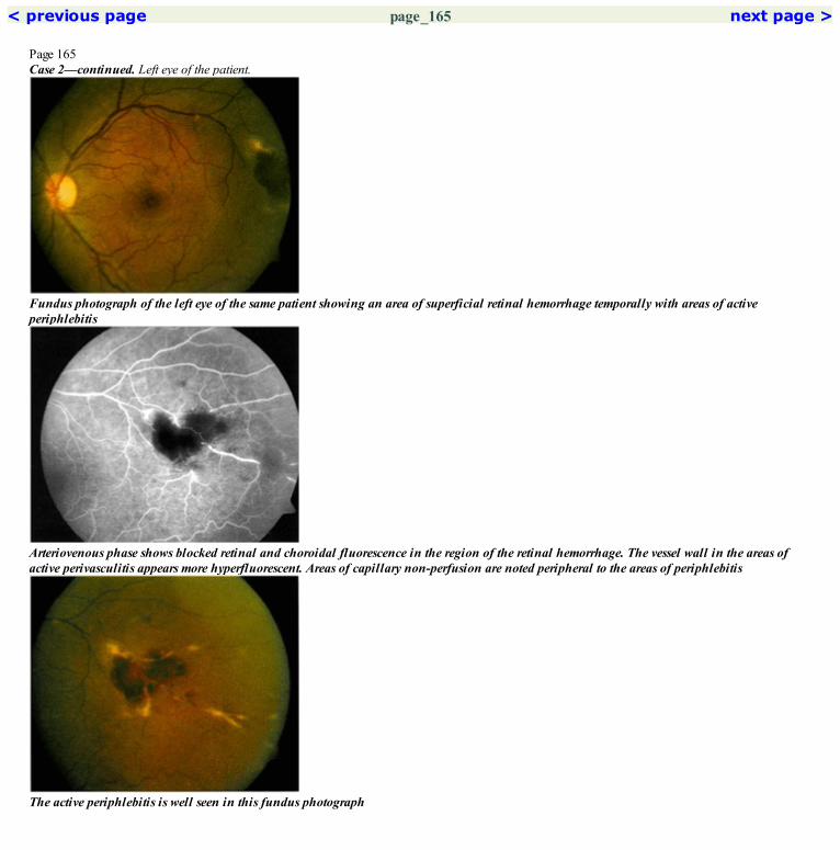
< previous page page_165 next page >
Page 165Case 2—continued. Left eye of the patient.
Fundus photograph of the left eye of the same patient showing an area of superficial retinal hemorrhage temporally with areas of activeperiphlebitis
Arteriovenous phase shows blocked retinal and choroidal fluorescence in the region of the retinal hemorrhage. The vessel wall in the areas ofactive perivasculitis appears more hyperfluorescent. Areas of capillary non-perfusion are noted peripheral to the areas of periphlebitis
The active periphlebitis is well seen in this fundus photograph

Increased staining of the vessel wall is seen in the late phaseCOMMENTPeriphlebitis can lead to retinal hemorrhages and can even cause a vitreous hemorrhage.
< previous page page_165 next page >

< previous page page_166 next page >
Page 166Case 3. A 41-year-old male was examined for floaters noticed in front of both eyes for the past two years. Visual acuity was 6/6; N6 in both eyes.
Fundus photograph of the left eye showing small patches of RPE atrophy around the foveal center. Sheathing of the inferotemporal vein is seenin the periphery
Stippled hyperfluorescence is seen along the veins in the superior periphery.
Areas of hypofluorescence surrounded by a ring of hyperfluorescence are seen superiorly corresponding to the ‘sun-burst’ like lesion seenclinically

Arteriovenous phase showing hyperfluorescence in the areas of atrophy due to transmission window defect. Stippled hyperfluorescence is seenaround the vein temporally
Similar changes are seen temporally. Aneurysmal dilatation is seen peripherally. An area of blocked fluorescence is seen due to RPE hypertrophy
Late phase photograph showing fading of the hyperfluorescence in the atrophic patches at the fovea
< previous page page_166 next page >

< previous page page_167 next page >
Page 167Case 4. A 36-year-old patient presented with an episode of decreased vision one year back following which the vision had cleared. Visual acuity was6/6; N6 in both eyes.
Fundus photograph of the right eye showing the normal posterior pole
Arteriovenous phase showing collateral channels in the superotemporal periphery. Areas of capillary non-perfusion are seen peripherally
Temporal periphery shows extensive areas of capillary non-perfusion. Few foci of intense fuzzy hyperfluorescence are seen indicative ofneovascularization
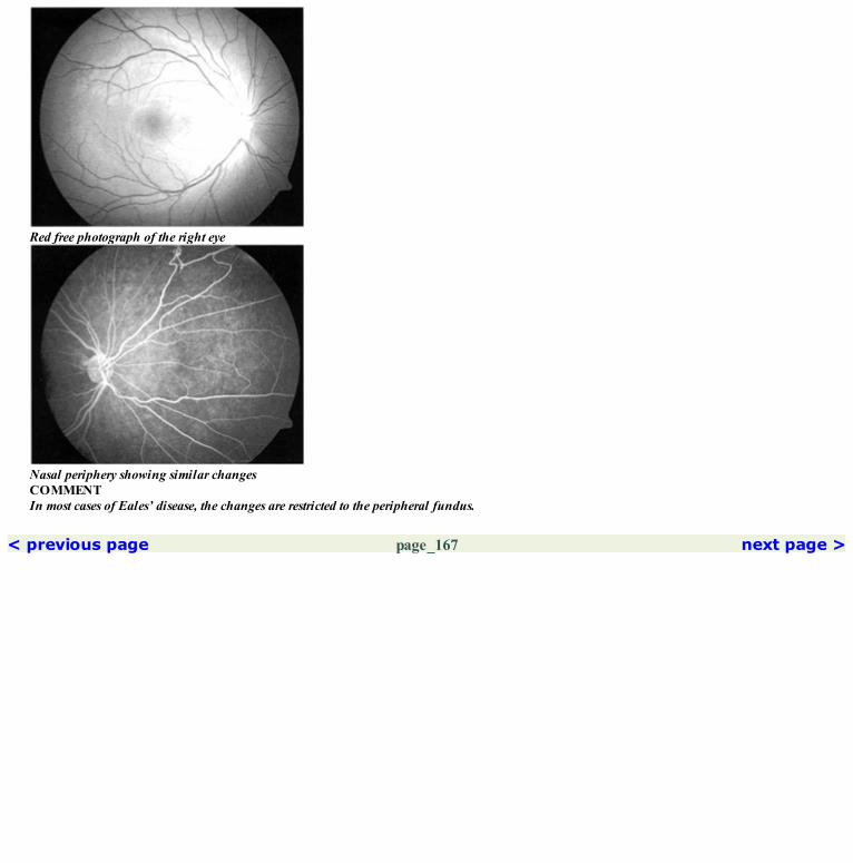
Red free photograph of the right eye
Nasal periphery showing similar changesCOMMENTIn most cases of Eales’ disease, the changes are restricted to the peripheral fundus.
< previous page page_167 next page >
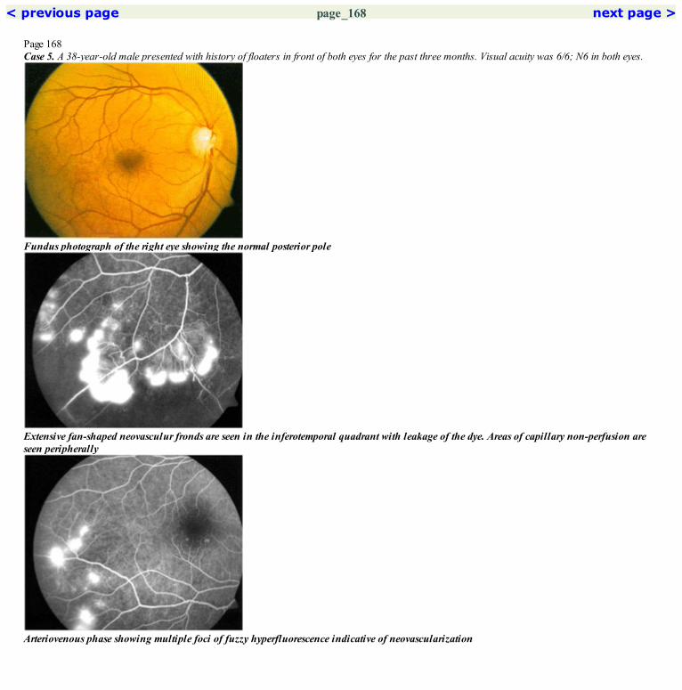
< previous page page_168 next page >
Page 168Case 5. A 38-year-old male presented with history of floaters in front of both eyes for the past three months. Visual acuity was 6/6; N6 in both eyes.
Fundus photograph of the right eye showing the normal posterior pole
Extensive fan-shaped neovasculur fronds are seen in the inferotemporal quadrant with leakage of the dye. Areas of capillary non-perfusion areseen peripherally
Arteriovenous phase showing multiple foci of fuzzy hyperfluorescence indicative of neovascularization
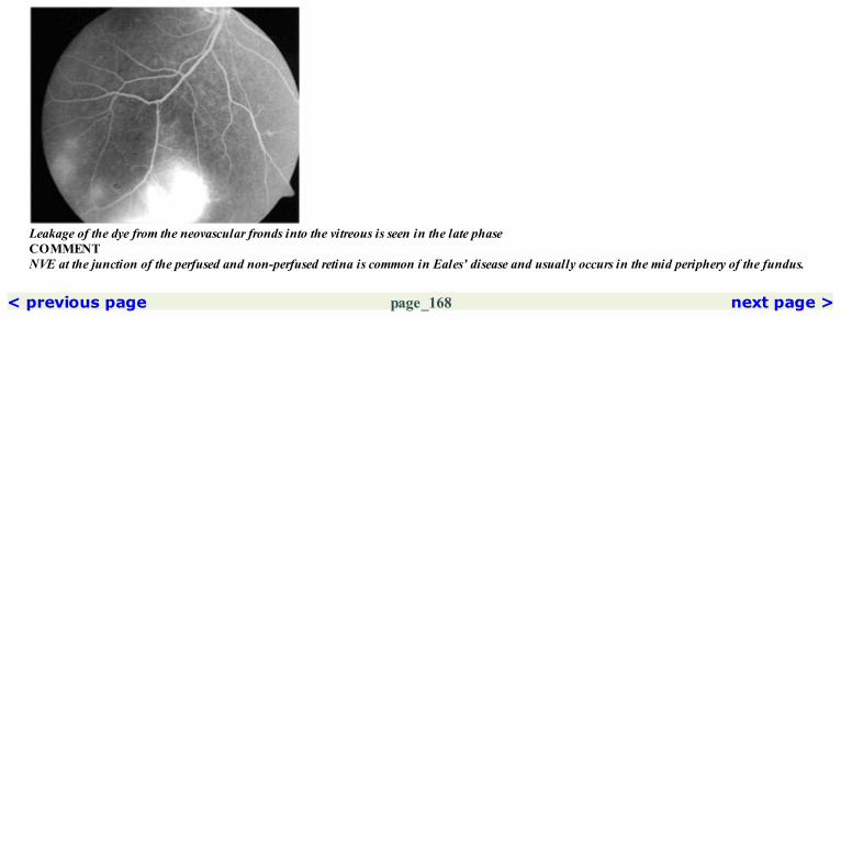
Leakage of the dye from the neovascular fronds into the vitreous is seen in the late phaseCOMMENTNVE at the junction of the perfused and non-perfused retina is common in Eales’ disease and usually occurs in the mid periphery of the fundus.
< previous page page_168 next page >

< previous page page_169 next page >
Page 169CLINICAL NUGGETS1. Eales’ disease is common in young, healthy, males, usually occurring in the third decade of life.2. Macula is usually spared in Eales’ disease.3. Patients with active periphlebitis are treated with systemic steroids.4. Laser photocoagulation is required in eyes that develop neovascularization.SELECTED READINGS1. Theodossiadis G. Fluorescein angiography in Eales’ disease. Am J Ophthalmol 69:271–277, 1970.2. Malik SR, Patnaik B. Fluorescein angiography in Eales’ disease. Indian J Ophthalmol 21:5–15, 1973.3. Karel I, Votoková J, Pele[ka M. Fluorescein angiography in unusual forms of idiopathic retinal vasculitis. Ophthalmologica 168:446–461, 1974.
< previous page page_169 next page >

< previous page page_170 next page >
Page 17024 Hypertensive Retinopathy
■ Features seen clinically are those associated with hypertensive retinopathy, hypertensive choroidopathy or hypertensive optic neuropathy.■ Hypertensive retinopathy is characterized by retinal arteriolar attenuation, changes in the arteriolar reflex, arteriovenous crossing changes, cottonwool spots, flame shaped hemorrhages and vascular occlusion.■ Hypertensive choroidopathy is characterized by Elschnig’s spot at the posterior pole and and Siegrist’s streak peripherally. Both these occur as aresult of choroidal infarction.■ Hypertensive optic neuropathy is characterized by disc swelling, associated with retinal edema and exudation.
■ Arteriovenous crossing changes with evidence of vein compression may be seen.■ Areas of cotton wool spots show up as areas of capillary nonperfusion.■ Dilation and at times occlusion of the terminal arterioles may be seen.■ In acute stage, Elschnig’s spot shows up as hyperfluorescence in the early phase of the angiogram with leakage of the dye seen in the later phases.Healed Elschnig’s spots block fluorescence in the region of the central hyperpigmentation with a halo of surrounding hyperfluorescence due totransmission defect.■ Siegrist’s streaks shows areas of blocked fluorescence in the region of hyperpigmentation and non-filling in the region of atrophy. Late staining ofthe underlying sclera is seen.
< previous page page_170 next page >
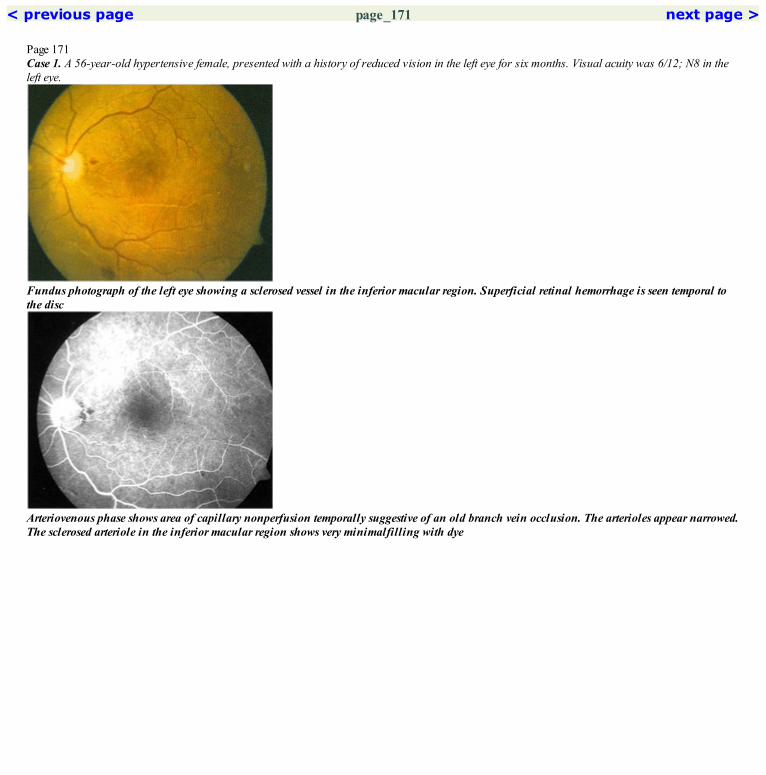
< previous page page_171 next page >
Page 171Case 1. A 56-year-old hypertensive female, presented with a history of reduced vision in the left eye for six months. Visual acuity was 6/12; N8 in theleft eye.
Fundus photograph of the left eye showing a sclerosed vessel in the inferior macular region. Superficial retinal hemorrhage is seen temporal tothe disc
Arteriovenous phase shows area of capillary nonperfusion temporally suggestive of an old branch vein occlusion. The arterioles appear narrowed.The sclerosed arteriole in the inferior macular region shows very minimalfilling with dye

Montage picture shows Siegrist’s streaks in the temporal, inferior and inferotemporal periphery
Red free photograph of the left eye
Tongue shaped areas of hypofluorescence with a rim of hyperfluorescein is seen in the temporal peripheryCOMMENTSiegrist’s streaks are tongue shaped atrophic patches seen in the periphery of the fundus. They result from small choroidal infarcts which leads toatrophy of the overlying RPE.
< previous page page_171 next page >

< previous page page_172 next page >
Page 172CLINICAL NUGGETS1. Elevated high blood pressure causes both focal and generalized constriction of the retinal arterioles due to autoregulation.2. Elschnig’s spots occur at the posterior pole and are seen as white spots in the acute stage.3. Siegrist’s streaks occur as tongue shaped lesion in the retinal periphery.4. Malignant hypertension is associated with disc swelling and retinal hemorrhages around the disc.SELECTED READINGS1. Dollery CT, Hodge JV. Hypertensive retinopathy studied with fluorescein. Trans Ophthalmol Soc UK 83:115–132, 19632. Gass JDM: Fluorescein angiographic study of macular dysfunction secondary to retinal vascular disease III. Hypertensive retinopathy. ArchOphthalmol 80:569–582, 1968.3. Scheie HG. Evaluation of ophthalmoscopic changes of hypertension and arteriolar sclerosis. Arch Ophthalmol 49:117, 1953.
< previous page page_172 next page >

< previous page page_173 next page >
Page 17325 Macroaneurysm
■ A retinal arterial macroaneurysm is a round or fusiform dilation occurring in an arterioles within the first three orders of the arteriolar bifurcation.■ It is usually unilateral, more common in females and occurs in the fifth to sixth decades of life.■ It usually occurs unrelated to other ocular disorders, but has been described in Eales’ disease, angiomatosis retinae, hypertensive retinopathy andretinal vascular occlusions.■ It is often located at the site of an arteriolar bifurcation or an AV crossing.■ Bleeding from a macroaneurysm can occur in the subretinal space, into the retina, beneath the internal limiting membrane or into the vitreous.
■ Typically, a macroaneurysm fills in the early arterial phase of the angiogram.■ Occasionally, it may be hypofluorescent in the early films, probably due to the slow filling of the macroaneurysm.■ The appearance in the late phase varies, ranging from minimal staining of the vessel wall to marked leakage.■ Dilation of surrounding capillaries, adjacent areas of capillary nonperfusion and microaneurysms may be seen in some cases.■ If retinal hemorrhage overlies the macroaneurysm, it may not be seen on a fluorescein angiogram.
< previous page page_173 next page >

< previous page page_174 next page >
Page 174Case 1. A 63-year-old hypertensive male, presented with sudden onset of decreased vision in the right eye of six weeks duration. The visual acuity was1/60 in the right eye.
Fundus photograph of the right eye showing subretinal dark and altered hemorrhage. The macroaneurysm is seen as a white round lesion (arrow)superior to the fovea, along the retinal arteriole
Early arteriovenous phase showing blocked choroidal fluorescence caused by the hemorrhage. The macroaneurysm is seen as a hyperfluorescentspot along the retinal arteriole
Late phase angiogram shows some fuzziness aound the borders of the macroaneurysm due to leakage of the dye through its wall

Red free photograph of the right eye
Arteriovenous phase shows increased hyperfluorescence of the macroaneurysmCOMMENTMacroaneurysm appears as a hyperfluorescent spot along the arteriole with increasing intensity of fluorescence in later phases.
< previous page page_174 next page >
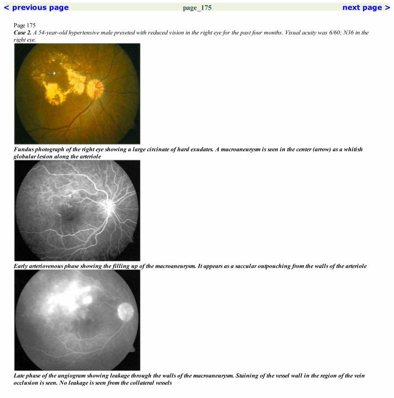
< previous page page_175 next page >
Page 175Case 2. A 54-year-old hypertensive male preseted with reduced vision in the right eye for the past four months. Visual acuity was 6/60; N36 in theright eye.
Fundus photograph of the right eye showing a large circinate of hard exudates. A macroaneurysm is seen in the center (arrow) as a whitishglobular lesion along the arteriole
Early arteriovenous phase showing the filling up of the macroaneurysm. It appears as a saccular outpouching from the walls of the arteriole
Late phase of the angiogram showing leakage through the walls of the macroaneurysm. Staining of the vessel wall in the region of the veinocclusion is seen. No leakage is seen from the collateral vessels
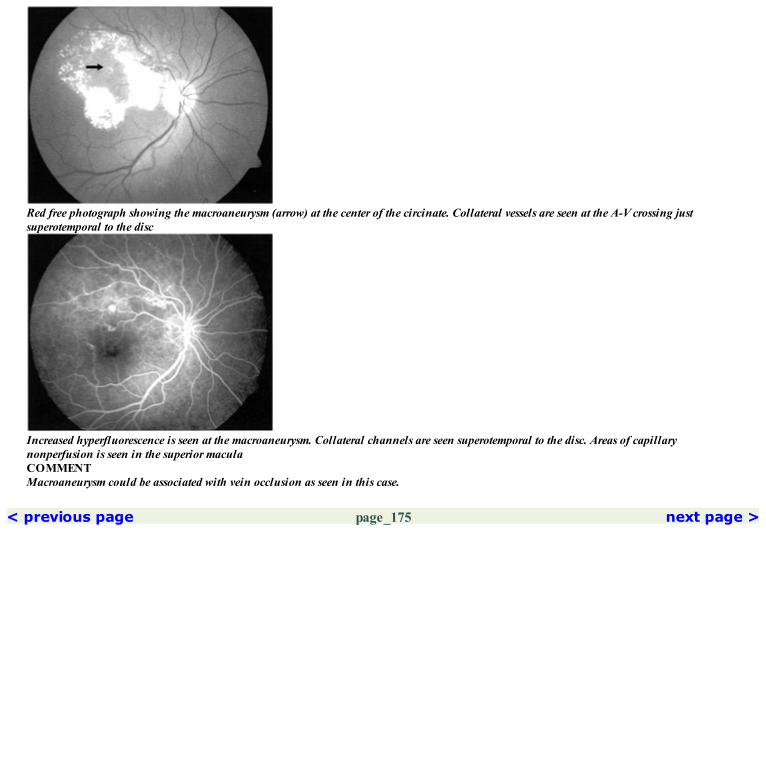
Red free photograph showing the macroaneurysm (arrow) at the center of the circinate. Collateral vessels are seen at the A-V crossing justsuperotemporal to the disc
Increased hyperfluorescence is seen at the macroaneurysm. Collateral channels are seen superotemporal to the disc. Areas of capillarynonperfusion is seen in the superior maculaCOMMENTMacroaneurysm could be associated with vein occlusion as seen in this case.
< previous page page_175 next page >

< previous page page_176 next page >
Page 176CLINICAL NUGGETS1. The superotemporal artery is more commonly involved.2. Macroaneurysms are more common in females and generally occur in the 6th and 7th decade of life.3. Patients with macroaneurysm may be asymptomatic if the fovea is not involved.4. Macroaneurysms with exudation can be treated with direct and mild photocoagulation; a heavy burn might cause arterial occlusion.SELECTED READINGS1. Abdel- Khalek MN, Richardson J. Retinal macroaneurysm: natural history and guidelines for management. Br J Ophthalmol; 70:2–11:1986.2. Panton RW, Goldberg MF, Farber MD: Retinal arterial macroaneurysm: Risk factors and natural history. Br J Ophthalmol 74:595–600, 1990.3. Cleary PE, KohnerEM, Hamilton AM. et al. Retinal macroaneurysms. Br J Ophthalmol; 59:355–361, 1975.
< previous page page_176 next page >

< previous page page_177 next page >
Page 17726 Parafoveal Telangiectasia■ Parafoveal telangiectasia is characterized by the presence of focal microaneury smal or saccular dilatation of some portion of the parafovealcapillary network.■ Three types have been described:Group 11A—Unilateral Congenital Parafoveal TelangiectasiaIt is more common in men, with a mean age of onset at 40 years of age. It is unilateral with the temporal 1 to 2 disc diameter of the parafovealcapillary network involved. Macular edema and hard exudation is common. This could be a variant of Coats’ disease.1B—Unilateral Idiopathic Parafoveal TelangiectasiaFound in middle aged men with a very small area of capillary telangiectasia confined to 1 clock hour. Exudation is not very common.Angiographic patternFluorescein angiography shows microaneurysmal and saccular dilatation of the temporal parafoveal capillaries with staining of the retina seen in thelate phase.Group 2. Bilateral Acquired Parafoveal TelangiectasiaBilateral, symmetric and involving the area temporal to the fovea. May involve the entire parafoveal capillary network. Right-angled retinal venulesare commonly seen. Lipid exudation is uncommon. RPE hyperplasia may develop along the right angled venule.Angiographic patternFluorescein angiography shows dilated parafoveal capillaries with staining of the adjacent retina in the mid and late phases. This does not extend tothe center of the fovea and no cystoid changes are seen. The area of RPE hyperplasia blocks fluorescence. The right angled venules are bettervisualized on the angiogram. They appear as abruptly ending vessels in the parafoveal venules.Presence of a neovascular membrane is associated with increased leakage of the fluorescein dye and its accumulation in cystoid spaces in the latefilms.Group 3. Bilateral Idiopathic Perifoveal Telangiectasia and Capillary ObliterationTelangiectasia with progressive obliteration of the perifoveal network is seen.These changes are very well seen on the fluorescein angiogram.
< previous page page_177 next page >

< previous page page_178 next page >
Page 178Case 1. A 48-year-old male patient was referred with a diagnosis of a branch retinal vein occlusion in the right eye. He complained of reduced visionin that eye for the past eight months. Visual acuity was 6/24; N36. Left eye was normal.
Fundus photograph of the right eye showing hard exudates arranged in a circinate pattern temporal to the fovea. Vascular abnormalities are seenwithin
Arteriovenous phase showing vascular abnormalities including capillary dilation and microaneurysmal dilation of the parafoveal capillarynetwork, more marked temporally
Late staining of the parafoveal retina is seen. Note that the foveal center remains hypofluorescent

Red free photograph shows vascular abnormalities within the circinate
Increased leakage of the dye is seen from the vascular abnormalitiesCOMMENTThis case represents Group 1A type of parafoveal telangiectasia. Hard exudates are common in this type.Involvement of the entire parafoveal network differentiates this from a small tributary vein occlusion in which the changes would be restricted tothe affected segment.
< previous page page_178 next page >

< previous page page_179 next page >
Page 179Case 2. A 47-year-old male presented with history of noticing reduced vision in both eyes for the past two months. Visual acuity was 6/9; N8 in theright eye and 6/6; N6 in the left eye.
Fundus photograph of the right eye showing a zone of retinal whitening in theparafoveal region
Arteriovenous phase shows capillary dilation and telangiectasia especially in the temporal parafoveal region
Late staining of parafoveal retina is seen, much more prominent on the temporal aspect

Red free photograph of the right eye
Increased intensity of hyperfluorescence is seen in the temporal parafoveal region
< previous page page_179 next page >

< previous page page_180 next page >
Page 180Left eye of the patient.
Fundus photograph of the left eye showing the retinal whitening in the parafoveal region
Very subtle vascular abnormality is seen at the superotemporal edge of the foveal avascular zone
Red free photograph of the left eye

Late minimal staining of the superior parafoveal region is seenCOMMENTThe temporal parafoveal capillary network is more commonly involved in the initial stages.Note that no cystoid changes are seen and the center of the FAZ remains hypofluorescent even in the late phase.
< previous page page_180 next page >
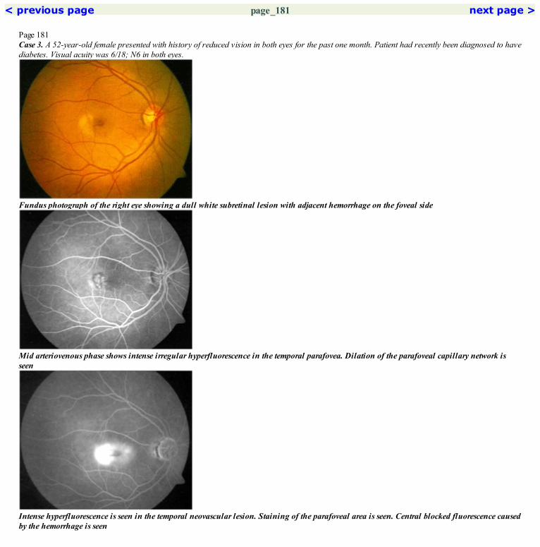
< previous page page_181 next page >
Page 181Case 3. A 52-year-old female presented with history of reduced vision in both eyes for the past one month. Patient had recently been diagnosed to havediabetes. Visual acuity was 6/18; N6 in both eyes.
Fundus photograph of the right eye showing a dull white subretinal lesion with adjacent hemorrhage on the foveal side
Mid arteriovenous phase shows intense irregular hyperfluorescence in the temporal parafovea. Dilation of the parafoveal capillary network isseen
Intense hyperfluorescence is seen in the temporal neovascular lesion. Staining of the parafoveal area is seen. Central blocked fluorescence causedby the hemorrhage is seen

Red free photograph of the right eye
Hyperfluorescence in the temporal parafoveal lesion has increased. The vascular abnormalities are better made outCOMMENTOccurrence of subretinal hemorrhage or fluid, cystoid macular edema and presence of focal, intense hyperfluorescent lesion in the angiogram aresuggestive of development of neovascular membrane in eyes with parafoveal telangiectasia.
< previous page page_181 next page >

< previous page page_182 next page >
Page 182Left eye of the patient.
Annular zone of whitening is seen in the parafoveal region in the left eye
Leakage of dye from the vascular abnormalities in the parafoveal region is seen
Red free photograph of the left eye
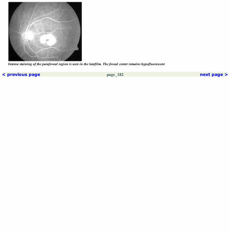
Intense staining of the parafoveal region is seen in the latefilm. The foveal center remains hypofluorescent
< previous page page_182 next page >

< previous page page_183 next page >
Page 183CLINICAL NUGGETS1. Group 2 is the most common form of parafoveal telangiectasia.2. Abnormal glucose tolerance test is seen in 35% of patients with unilateral disease and 60% of patients with bilateral disease.3. Patients with parafoveal telangiectasia should be followed up to look for the development of choroidal neovascular membrane.4. Treatment of parafoveal telangiectasia with laser photocoagulation is controversial.SELECTED READINGS1. Gass JDM, Oyakawa RT. Idiopathic juxtafoveolar retinal telangiectasia.; Arch. Ophthalmology; 100:769–780, 1982.2. Gass JDM. Stereoscopic atlas of macular diseases: diagnosis and treatment. 4th edition Vol. 1, Mosby, St Louis, Chapter 6—Maculardysfunction caused by retinal vascular diseases 506–511, 1997.3. Gass JDM. A fluorescein angiographic study of macular dysfunction secondary to retinal vascular diseases Vs retinal telangiectasia. Arch.Ophthalmol; 80:592–605, 1968.4. Chopdar A: Retinal telangiectasias in adults: Fluorescein angiographic findings and treatment by argon laser. Br J Ophthalmol; 62:243–250, 1978.
< previous page page_183 next page >

< previous page page_184 next page >
Page 18427 Purtscher’s Retinopathy
■ It is characterized by the presence of multiple patches of retinal whitening and retinal hemorrhages at the posterior pole.■ It usually follows compressive injuries of the head and trunk.■ It has also been reported in cases of pancreatitis, chronic renal failure, following child birth, in connective tissue disorders etc.■ It is possibly due to complement induced leukocyte aggregation and leukoembolism.
■ Areas of capillary non-perfusion is seen in the affected areas.■ Minimal leakage of the dye from the retinal vasculature and perivascular staining may be seen.
< previous page page_184 next page >

< previous page page_185 next page >
Page 185Case 1 A 35-year-old male presented with loss of vision noted in both eyes following a road traffic accident. Visual acuity was hand movements closeto face in both eyes.
Fundus photograph of the right eye showing patches of retinal whitening and retinal hemorrhages at the posterior pole
Arteriovenous phase of the right eye showing a large hypofluorescent patch in the macula due to a combination of blocked fluorescence and non-filling of the vasculature
Late phase angiogram showing persistent hypofluorescence at the macula. Perivascular and disc staining is also seen

Red free photograph of the right eye
The hypofluorescence is persisting in the later arteriovenous phase. Note there is no filling of the vessels in the macula. Minimal leakage of thedye is seen through the walls of the larger vesselsCOMMENTOcclusion of the arterioles possibly due to leukoembolization, results in closure of the capillaries in the macula resulting in visual impairment.
< previous page page_185 next page >

< previous page page_186 next page >
Page 186Left eye of the patient.
Fundus photograph of the left eye showing patches of retinal whitening and retinal hemorrhages at the posterior pole
Arteriovenous phase shows areas of blocked hypofluorescence due to hemorrhage and areas of hypofluorescence due to non-filing of thevasculature
Red free photograph of the left eye

Late perivascular and disc staining is noted. The macula remains hypofluorescentCOMMENTPurtscher’s retinopathy is characterized by the presence of areas of non-perfusion at the macula.
< previous page page_186 next page >

< previous page page_187 next page >
Page 187CLINICAL NUGGETS1. Leukocyte aggregation resulting from activation of the complement system due to varied etiology, leads to leukoembolization resulting inPurtscher’s retinopathy.2. Visual prognosis is poor in patients with Purtscher’s retinopathy.3. There is no specific treatment for Purtscher’s retinopathy.SELECTED READINGS1. Kelley JS. Purtscher’s retinopathy related to chest compression by safety belts. Fluorescein Angiographic findings, Am J Ophthalmol, 74:278–283, 1972.2. .Burton TC. Unilateral Purtscher’s retinopathy. Ophthalmology; 87:1096–1105, 1980.
< previous page page_187 next page >

< previous page page_188 next page >
Page 18828 Radiation Retinopathy
■ Radiation retinopathy is a slowly progressive condition with characteristic fundus changes.■ It is predominantly a retinal vascular disorder in which the permeability of the retinal and optic nerve vasculature is affected.■ The changes seen in radiation retinopathy can mimic those seen in diabetic retinopathy.■ Three stages have been described:Background radiation retinopathy—this is characterized by microaneurysms, retinal telangiectasia, dot and blot hemorrhages, cotton wool spots,hard exudates and macular edema.Preproliferative radiation retinopathy—this is characterized by intraretinal microvascular abnormalities, venous beading and increased number ofcotton wool spots.Proliferative radiation retinopathy—neovascularization may occur at the disc or elsewhere in the retina. Vitreous hemorrhage and neovascularglaucoma can result.■ Optic nerve involvement would result in optic disc edema or optic atrophy.
■ These changes correlate with the clinical findings and can include areas of capillary non-perfusion, increased capillary permeability and leakage,microaneurysms, IRMA’s and neovascularization.■ If optic nerve is affected, then non-perfusion of the optic nerve capillaries is also seen.
< previous page page_188 next page >

< previous page page_189 next page >
Page 189Case 1. A 29-year-old male presented with history of reduced vision in the left eye of one month duration. He had previously been diagnosed to havean orbital lymphoma and had undergone radiotherapy nine months back. Visual acuity was 6/6; N6 in the right eye and 6/18; N8 in the left eye. Theright eye fundus was normal.
Fundus photograph of the left eye showing cotton wool spots along the superotemporal arcade and temporal to the disc. The macula has a dullappearance
Mid arteriovenousphase showing areas of capil lary non-perfusion temporal and superotemporal to the disc. Leakage of the dye from dilatedcapillaries around the non-perfused areas is seen
Staining of the retinal tissue from incompetent dilated capillaries is seen in the late phase
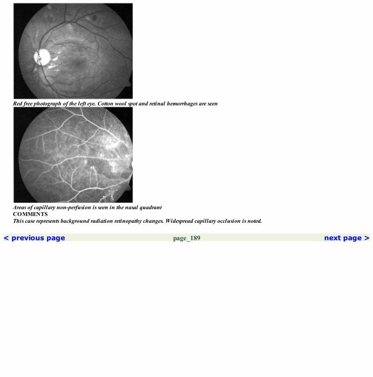
Red free photograph of the left eye. Cotton wool spot and retinal hemorrhages are seen
Areas of capillary non-perfusion is seen in the nasal quadrantCOMMENTSThis case represents background radiation retinopathy changes. Widespread capillary occlusion is noted.
< previous page page_189 next page >
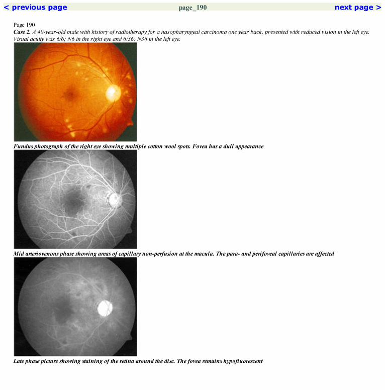
< previous page page_190 next page >
Page 190Case 2. A 40-year-old male with history of radiotherapy for a nasopharyngeal carcinoma one year back, presented with reduced vision in the left eye.Visual acuity was 6/6; N6 in the right eye and 6/36; N36 in the left eye.
Fundus photograph of the right eye showing multiple cotton wool spots. Fovea has a dull appearance
Mid arteriovenous phase showing areas of capillary non-perfusion at the macula. The para- and perifoveal capillaries are affected
Late phase picture showing staining of the retina around the disc. The fovea remains hypofluorescent

Red free photograph of the right eye
Mid arteriovenous phase shows extensive capillary non-perfusion seen in the superior and nasal quadrants
< previous page page_190 next page >

< previous page page_191 next page >
Page 191Left eye of the patient.
Fundus photograph of the left eye showing cotton wool spots and retinal hemorrhages
Extensive areas of capillary non-perfusion are seen in all quadrants and at the fovea
Red free photograph of the left eye

Late phase shows minimal vessel wall staining of the capillaries bordering the areas of non-perfusion. Fovea remains hypofluorescentCOMMENTRetinal changes seen in radiation retinopathy resembles those seen in diabetic retinopathy.
< previous page page_191 next page >

< previous page page_192 next page >
Page 192CLINICAL NUGGETS1. Radiation retinopathy can develop 6 months to 8 years following exposure to radiation. Average time is 2–3 years.2. Radiation retinopathy is rare when the total dose of radiation is less than 3000 rads but has been reported with as little as 1500 rads.SELECTED READINGS1. Hayreh SS: Post radiation retinopathy: A fluorescein fundus angiographic study. Br J Ophthalmol 54:705–714, 1970.2. Amoaku WMK, Archer DB. Fluorescein angiographic features, natural course and treatment of radiation retinopathy. Eye 4:657–667, 1990.3. Gass JDM. A fluorescein angiographic study of macular dysfunction secondary to retinal vascular disease. VI. X-ray irradiation, carotid arteryand vitritis. Arch Ophthalmol 80:606–617, 1968.
< previous page page_192 next page >

< previous page page_193 next page >
Page 19329 Venous Occlusion
CENTRAL RETINAL VEIN OCCLUSION■ Central retinal vein occlusion is characterized by diffuse retinal hemorrhages in all quadrants and dilated and tortuous retinal veins.■ The clinical appearance may vary from a few scattered retinal hemorrhages with a few cotton-wool spots to very extensive retinal hemorrhagesoccurring throughout the fundus.■ Macular edema is present in most cases.■ Based on the visual acuity, presence or absence of an afferent pupillary defect, ophthalmoscopic findings, fluorescein angiographic findings, visualfields and electroretinographic findings, central retinal vein occlusion is divided into two groups:1. Nonischemic/perfused CRVO (NICRVO)2. Ischemic/nonperfused CRVO (ICRVO)
NICRVO■ The superficial retinal hemorrhages cause blocked retinal and choroidal fluorescence through all phases of the angiogram.■ There is a delay in the filling of the veins.■ Veins are markedly engorged, tortuous and appear disrupted because of hemorrhage and retinal edema masking their view.■ The disc capillaries are also dilated and engorged.■ Venous return is severely delayed.■ Capillaries are dilated, tortuous and may show microaneurysmal changes. Perifoveal arcade may show marked congestion and dilatation.■ Few areas of capillary nonperfusion may be seen in the midperipheral retina. However this is not as extensive as in ischemic CRVO.■ Late phase shows considerable staining of the vein walls and leakage of dye to the surrounding retina. In the macula this leads to cystoid macularedema.ICRVO■ In addition to the above changes, extensive areas of capillary closure are seen throughout the fundus.■ There is marked delay in the filling of the veins.■ Ischemic changes usually extend to the macular region.
< previous page page_193 next page >
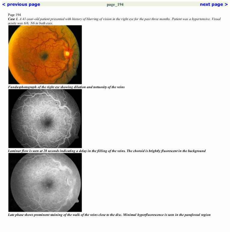
< previous page page_194 next page >
Page 194Case 1. A 41-year-old patient presented with history of blurring of vision in the right eye for the past three months. Patient was a hypertensive. Visualacuity was 6/6; N6 in both eyes.
Fundusphotograph of the right eye showing dilation and tortuosity of the veins
Laminar flow is seen at 28 seconds indicating a delay in the filling of the veins. The choroid is brightly fluorescent in the background
Late phase shows prominent staining of the walk of the veins close to the disc. Minimal hyperfluorescence is seen in the parafoveal region

Red freephotograph of the right eye showing the tortuosity of the veins. A superficial retinal hemorrhage is seen along the superotemporal vein
Complete filling of the veins is seen at 36 secondsCOMMENTPatient is having a NICRVO with very minimal macular edema suggesting a lesser degree of occlusion of the central retinal vein.
< previous page page_194 next page >

< previous page page_195 next page >
Page 195Case 2. A 47-year-old patient presented with history of decreased vision in the right eye of one day duration. Visual acuity was 6/6; N10 in the righteye. Left eye was normal.
Fundus photograph of the right eye showing mild dilation and tortuosity of the veins and scattered superficial and deep retinal hemorrhages
Marked delay in the filling of the veins is seen; no filling is seen even at 30 seconds
Late stage shows extensive staining of the vessel wall throughout the macula
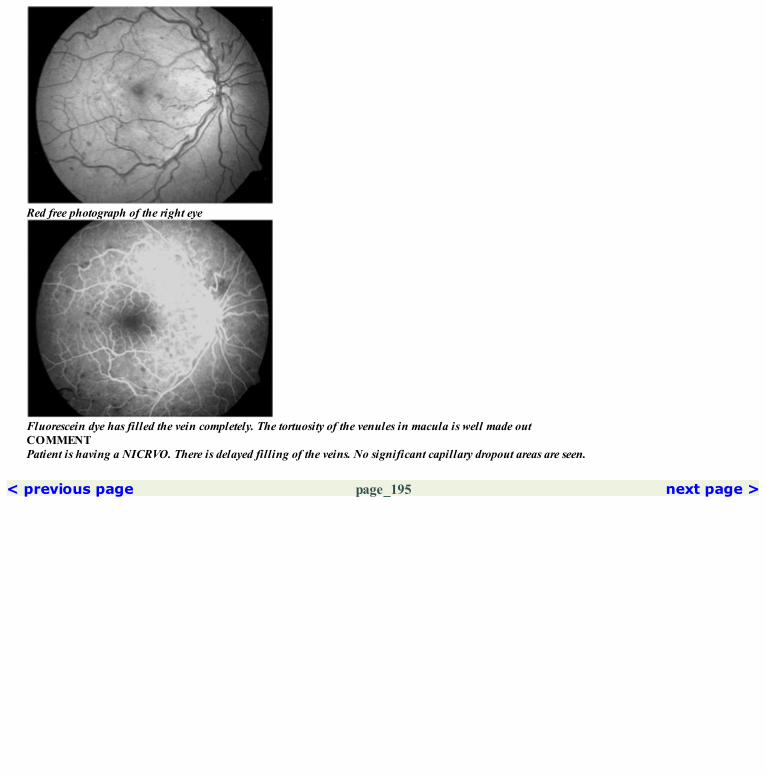
Red free photograph of the right eye
Fluorescein dye has filled the vein completely. The tortuosity of the venules in macula is well made outCOMMENTPatient is having a NICRVO. There is delayed filling of the veins. No significant capillary dropout areas are seen.
< previous page page_195 next page >
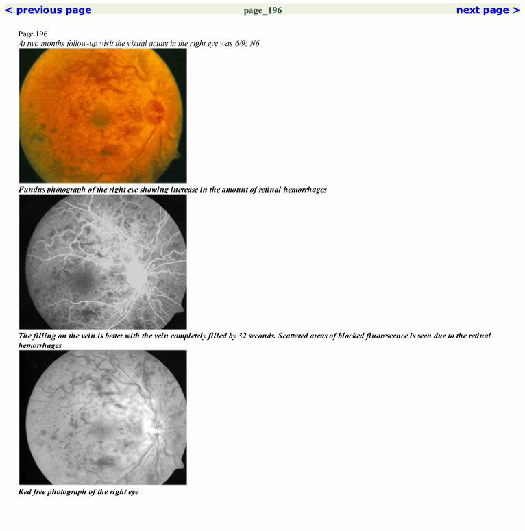
< previous page page_196 next page >
Page 196At two months follow-up visit the visual acuity in the right eye was 6/9; N6.
Fundus photograph of the right eye showing increase in the amount of retinal hemorrhages
The filling on the vein is better with the vein completely filled by 32 seconds. Scattered areas of blocked fluorescence is seen due to the retinalhemorrhages
Red free photograph of the right eye

Late phase angiogram shows increased staining of the retina at the macula. No cystoid changes are seenCOMMENTIncreased tortuosity of the veins and more delayed emptying of the veins is seen in the follow-up angiogram. This suggests progression of theCRVO.
< previous page page_196 next page >

< previous page page_197 next page >
Page 197Case 3. A 39-year-old male presented with history of sudden reduction of vision in the left eye two months back. Visual acuity was 6/9; N6. He wasfound to have a NICRVO.
Fundus photograph of the left eye showing scattered retinal hemorrhages. Few cotton-wool spots are seen
Dilation of the disc capillaries is seen. Scattered blocked fluorescence due to the retinal hemorrhage is evident
Few areas of capillary non-perfusion are seen in the superior mid periphery

Red free photograph of the left eye
No significant areas of capillary non-perfusion is seen in the inferotemporal periphery
Late phase angiogram showing accumulation of the dye at the macula
< previous page page_197 next page >
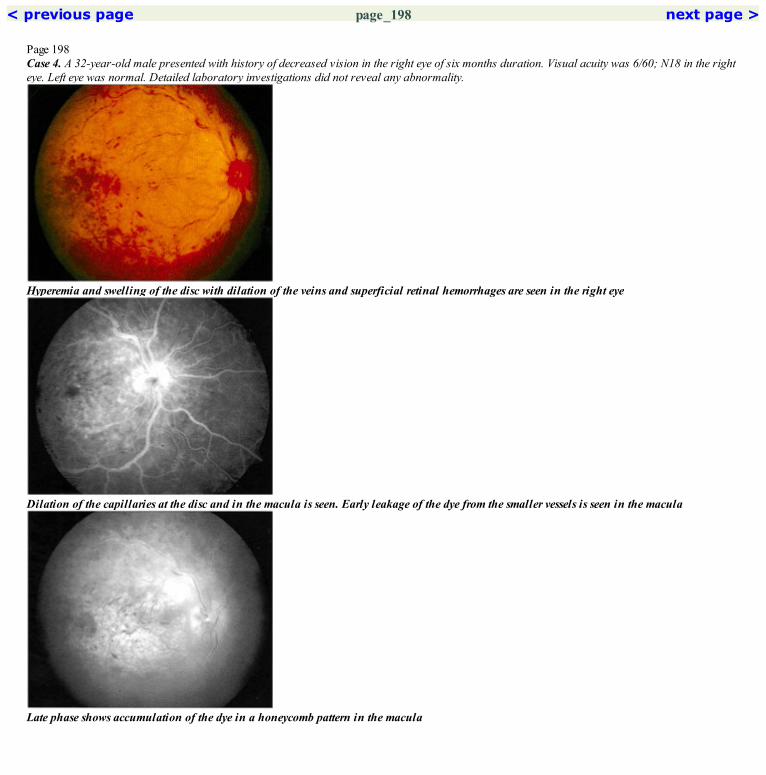
< previous page page_198 next page >
Page 198Case 4. A 32-year-old male presented with history of decreased vision in the right eye of six months duration. Visual acuity was 6/60; N18 in the righteye. Left eye was normal. Detailed laboratory investigations did not reveal any abnormality.
Hyperemia and swelling of the disc with dilation of the veins and superficial retinal hemorrhages are seen in the right eye
Dilation of the capillaries at the disc and in the macula is seen. Early leakage of the dye from the smaller vessels is seen in the macula
Late phase shows accumulation of the dye in a honeycomb pattern in the macula

Red free photograph of the right eye. Cystoid changes are seen at the fovea
Further leakage of the dye from the vessels is seen at the maculaCOMMENTPatient has a NICRVO with gross macular edema as indicated by the cystoid accumulation of the dye in the late phase.
< previous page page_198 next page >

< previous page page_199 next page >
Page 199Case 5. A 55-year-old male presented with history of decreased vision in both eyes for the past fifteen days. He was a known hypertensive for the pastten years on treatment. Visual acuity was 6/60; N36 in the right eye and 3/60 in the left eye. Investigations showed the patient to havehypergammaglobulinemia.
Fundus photograph of the right eye showing swelling of the disc with marked dilation and tortuosity of the veins
Marked delay in the filling of the veins was noted. Dilation of the capillaries is seen in the macula
Extensive areas of capillary non-perfusion are seen. The parafoveal capillary network is affected
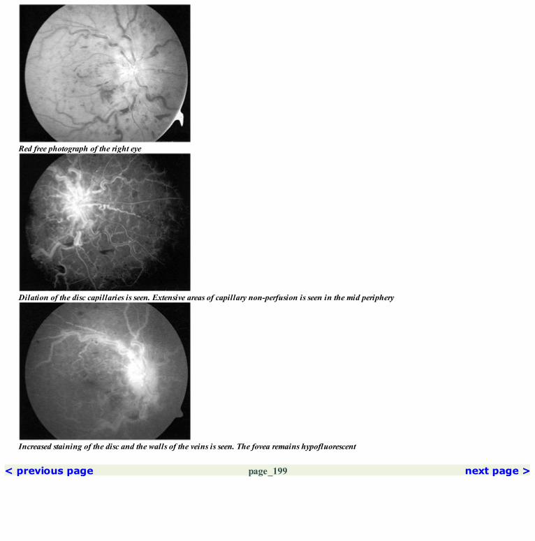
Red free photograph of the right eye
Dilation of the disc capillaries is seen. Extensive areas of capillary non-perfusion is seen in the mid periphery
Increased staining of the disc and the walls of the veins is seen. The fovea remains hypofluorescent
< previous page page_199 next page >

< previous page page_200 next page >
Page 200Left eye of the patient.
Fundus photograph of the left eye showing a similar picture as that in the right eye
Marked dilation of the disc capillaries is seen. Extensive areas of capillary non-perfusion including the fovea are seen
Red free photograph of the left eye

Staining of the disc and the walls of the veins is seen. The fovea remains hypofluorescentCOMMENTPatient has an ICRVO in both eyes with extensive areas of capillary non-perfusion seen in the mid periphery.Patients presenting with simultaneous bilateral CRVO should always be investigated for hematological disorders.
< previous page page_200 next page >
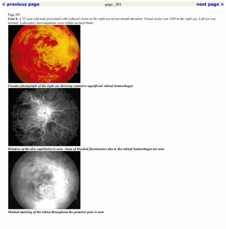
< previous page page_201 next page >
Page 201Case 6. A 32-year-old male presented with reduced vision in the right eye of one month duration. Visual acuity was 3/60 in the right eye. Left eye wasnormal. Laboratory investigations were within normal limits.
Fundus photograph of the right eye showing extensive superficial retinal hemorrhages
Dilation of the disc capillaries is seen. Areas of blocked fluorescence due to the retinal hemorrhages are seen
Marked staining of the retina throughout the posterior pole is seen

Red free photograph of the right eye
Extensive areas of blocked fluorescence due to retinal hemorrhage are seenCOMMENTFluorescein angiography is not very informative in presence of extensive retinal hemorrhages as it blocks the underlying fluorescence.
< previous page page_201 next page >

< previous page page_202 next page >
Page 202CLINICAL NUGGETS1. Bilateral simultaneous central retinal vein occlusion is more common in patients with underlying systemic disorders.2. Laser photocoagulation has not been found to be useful for the treatment of macular edema secondary to a central retinal vein occlusion.3. Patients with CRVO should be followed up on a monthly basis to look for the development of ocular neovascularization; if so, panretinalphotocoagulation is performed.4. Recently, intravitreal injection of Triamcinolone acetonide has been used to treat macular edema.SELECTED READINGS1. Gass JDM. A Fluorescein angiographic study of macular dysfunction secondary to retinal vascular disease. II. Retinal vein obstruction: ArchOphthalmol 80:550–568, 1968.2. Central vein occlusion study group: Baseline and early natural history report: The central venous occlusion study group: Arch Ophthalmol111:1087–1095, 1993.3. A randomized clinical trial of panretinal photocoagulation for central vein occlusion: The central venous occlusion study group N report:Ophthalmology 102:1434–1444, 1995.4. Gutman FA. Evaluation of a patient with central retinal vein occlusion. Ophthalmology 90:481–483, 1983.5. Hayreh SS. Classification of central retinal vein occlusion. Ophthalmology 90:458–474, 1988.
< previous page page_202 next page >

< previous page page_203 next page >
Page 203
BRANCH RETINAL VEIN OCCLUSION■ Branch retinal vein occlusion is characterized by dilation and tortuosity of the affected vein associated with retinal hemorrhages, cotton-woolspots and edema in the area drained by the vein.
■ Blocked fluorescence due to the retinal hemorrhages is seen through all phases of the angiogram.■ There is delayed filling and emptying of the affected vein.■ Dilated capillaries, microaneurysms and telangiectatic changes are well made out on the angiogram.■ Areas of capillary closure may be present in the involved segment.■ Perfused BRVO—area of capillary closure is less than five disc diameters.■ Non-perfused BRVO—area of capillary closure is more than five disc diameters.■ Macular edema may be perfused, non-perfused or mixed.Perfused macular edema—an intact parafoveal capillary network is seen during the arteriovenous phase, followed by late accumulation of the dyeinvolving the foveal center.Non-perfused macular edema—areas of parafoveal and perifoveal capillary non-perfusion are seen with no accumulation of the dye in the late phase.Mixed macular edema—a combination of capillary dilation and leakage and areas of capillary non-perfusion in the parafoveal region. Late phaseshows some degree of accumulation of the dye.■ Late staining and leakage from the affected veins is seen.
HEMICENTRAL RETINAL VEIN OCCLUSION (HCRVO)■ The two venous channels that develop during the third month of intrauterine life may fail to join in front of the lamina cribrosa—the two trunkstravel inside the optic nerve independently for a considerable distance before joining. They are usually seen entering the optic nerve separately.Occlusion of one of the channels leads to a hemicentral retinal vein occlusion.
Angiographic appearance■ Pattern is similar to CRVO except that only half the fundus area is involved.
< previous page page_203 next page >

< previous page page_204 next page >
Page 204Case 7. A 56-year-old male hypertensive presented with mild blurring of vision in the right eye. Visual acuity was 6/6; N6 in both eyes.
Fundus photograph of the right eye
Early arteriovenous phase of the angiogram. Laminar flow is seen in the superotemporal vein upto the A-V crossing only
Late phase angiogram picture of the right eye

Red free photograph of the right eye. Note the small deflection of the vein seen at the first A-V crossing (arrow) superotemporal to the disc
Magnified view of the A-V crossing. Note the absence of laminar flow proximal to the A-V crossing indicating turbulence of flow due tocompression of the vein at the crossingCOMMENTDisturbance of the laminar flow in the vein at the A-V crossing is probably an early sign of an impending vein occlusion.
< previous page page_204 next page >

< previous page page_205 next page >
Page 205Case 8. A 48-year-old male presented with history of disturbance of vision in the left eye of 3 days duration. He was on medication for systemichypertension for the past two years and was noted to have raised serum cholesterol. Visual acuity was 6/6; N6 in the left eye. The right eye wasnormal.
Fundus photograph of the left eye showing a small tributary vein occlusion superotemporal to the disc
Early arteriovenous phase showing delayed filling of the affected vein (white arrows). The site of occlusion at the AV crossing is well seen (blackarrow)
Late phase angiogram showing staining of the retina in the affected segment

Red free photograph showing superficial retinal hemorrhages and cotton-wool spots along the affected segment
The affected vein has filled up. Blocked fluorescence is seen due to the retinal hemorrhagesCOMMENTA macular tributary vein occlusion involves a small segment of the retina and does not give rise to significant ischemic changes. Vision howeveris compromised if the FAZ is involved. Fluorescein angiogram is useful to assess the status of the FAZ.
< previous page page_205 next page >

< previous page page_206 next page >
Page 206Seven months later, visual acuity in the left eye was maintained at 6/6; N6.
Fundus photograph of the left eye showing clearing of the superficial retinal hemorrhages
Arteriovenous phase showing normal filling of the affected vein. A collateral channel has developed at the site of previous occlusion at the AVcrossing (arrow). The capillary details in the affected segment is not well made out
Red free photograph of the left eye

Late phase angiogram does not show a significant staining of the retinaCOMMENTTributary vein occlusions generally have a good prognosis provided the parafoveal capillaries are not disturbed.Almost all BRVO occur at the site of A-V crossing. Exception to this would be BRVO occurring secondary to perivasculitis.
< previous page page_206 next page >

< previous page page_207 next page >
Page 207Case 9. A 22-year-old male presented with decreased vision in the right eye of 3 months duration. No relevant systemic history was present andlaboratory investigations were within normal limits. Visual acuity was 6/12; N6. In view of similar changes in the left eye he was suspected of havingEales’ disease.
Fundus photograph of the right eye showing a fresh superotemporal BRVO
Arteriovenous phase showing the delayed filling of the affected vein (arrows)
Late phase angiogram showing persistence of the hypofluorescence. Some leakage of the dye from the capillaries in the affected segment is seen.Staining of the vein due to periphlebitis is seen superior and inferonasal to the disc
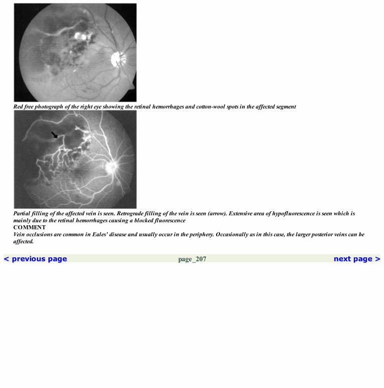
Red free photograph of the right eye showing the retinal hemorrhages and cotton-wool spots in the affected segment
Partial filling of the affected vein is seen. Retrograde filling of the vein is seen (arrow). Extensive area of hypofluorescence is seen which ismainly due to the retinal hemorrhages causing a blocked fluorescenceCOMMENTVein occlusions are common in Eales’ disease and usually occur in the periphery. Occasionally as in this case, the larger posterior veins can beaffected.
< previous page page_207 next page >

< previous page page_208 next page >
Page 208Case 10. A 48-year-old female presented with history of reduced vision in the left eye for the past six months. Visual acuity was 6/24; N36.
Fundus photograph of the left eye showing a BRVO in the inferotemporal quadrant. Note retinal hemorrhages inferotemporally
Arteriovenous phase showing dilation of the inferotemporal vein. Delayed filling in it is evident from the persistence of laminar pattern of fillingclose to the disc (arrow). Dilated capillaries are made out in the affected segment. The parafoveal capillaries network is intact
Red free photograph of the left eye showing a few scattered hemorrhages in the inferotemporal quadrant
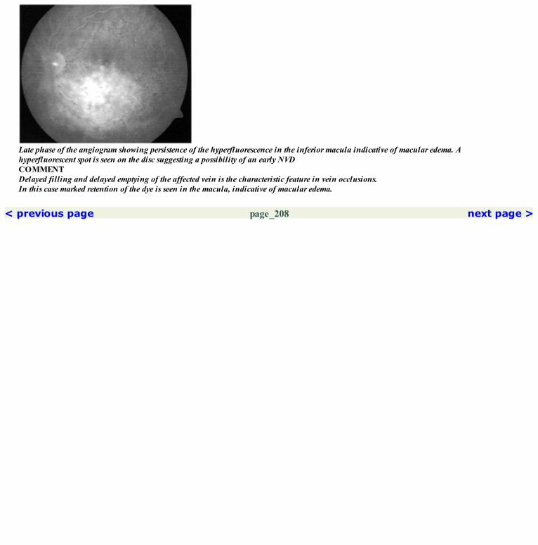
Late phase of the angiogram showing persistence of the hyperfluorescence in the inferior macula indicative of macular edema. Ahyperfluorescent spot is seen on the disc suggesting a possibility of an early NVDCOMMENTDelayed filling and delayed emptying of the affected vein is the characteristic feature in vein occlusions.In this case marked retention of the dye is seen in the macula, indicative of macular edema.
< previous page page_208 next page >

< previous page page_209 next page >
Page 209Case 11. A 65-year-old male presented with history of decreased vision in the left eye for the past four months. He was diagnosed to havehypertension after the onset of ocular symptoms. Visual acuity was 3/60 in the left eye. Right eye was normal.
Fundus photograph of the left eye showing a hemicentral vein occlusion in the inferior half
Extensive areas of capillary non-perfusion are seen in the inferior half. Dilated capillaries are seen surrounding these areas of non-perfusion.The lower half of the parafoveal capillary network is affected
Late phase showing staining of the vessel walls in the affected segment. The capillary non-perfused areas remain hypofluorescent
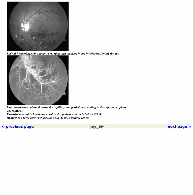
Retinal hemorrhages and cotton-wool spots seen scattered in the inferior half of the fundus
Late arteriovenous phase showing the capillary non perfusion extending to the inferior peripheryCOMMENTExtensive areas of ischemia are noted in this patient with an inferior HCRVO.HCRVO to a large extent behave like a CRVO in its natural course.
< previous page page_209 next page >

< previous page page_210 next page >
Page 210Case 12. A 57-year-old male presented with history of decreased vision in the left eye for the past six months. Patient was a known hypertensive andgave history of a cerebrovascular accident a year back. Visual acuity was 6/12; N6 in the left eye. Right eye was normal.
Fundus photograph of the left eye showing a sheathed superotemporal vein. Few absorbing cotton-wool spots are seen
Arteriovenous phase showing extensive non-perfused area in the superotemporal quadrant. The superotemporal vein has not filled (white arrows).The site of occlusion is at the AVcrossing superotemporal to the disc (black arrow)
Late phase angiogram showing the non-perfused area in the temporal periphery

Red free photograph of the left eye
Late phase showing hypofluorescence in the area of non-perfusion. The superotemporal vein has not yet filledCOMMENTPresence of extensive areas of non-perfusion indicates a high probability of the patient developing NVD or NVE.
< previous page page_210 next page >

< previous page page_211 next page >
Page 211Case 13. A 56-year-old female came for a routine ocular evaluation. Visual acuity was 6/9; N6 in both eyes. She was found to have an oldsuperotemporal branch retinal vein occlusion in the left eye.
Fundus photograph of the left eye showing collateral vessels just superotemporal to the disc
Arteriovenous phase showing the delayed filling of the superotemporal vein
Late phase showing absence of significant staining of the retina in the macula

Red free photograph of the left eye
The superotemporal vein has filled. Dilated capillaries and collateral vessles are seen. Some areas of capillary non-perfusion are made out.Collateral channels crossing the horizontal raphe are seen temporallyCOMMENTCollaterals act as alternate channels for venous drainage and help to bypass the site of occlusion.
< previous page page_211 next page >

< previous page page_212 next page >
Page 212Case 14. A 64-year-old hypertensive male presented with reduced vision in the right eye for the past six months. Visual acuity was 6/36; N36.
Fundus photograph of the right eye showing a fibrovascular proliferation superotemporal to the disc
Mid arteriovenous phase showing the filling up of the neovascular network. Complete wash-out of the capillaries is seen peripherally
Late phase showing leakage of the dye into the vitreous
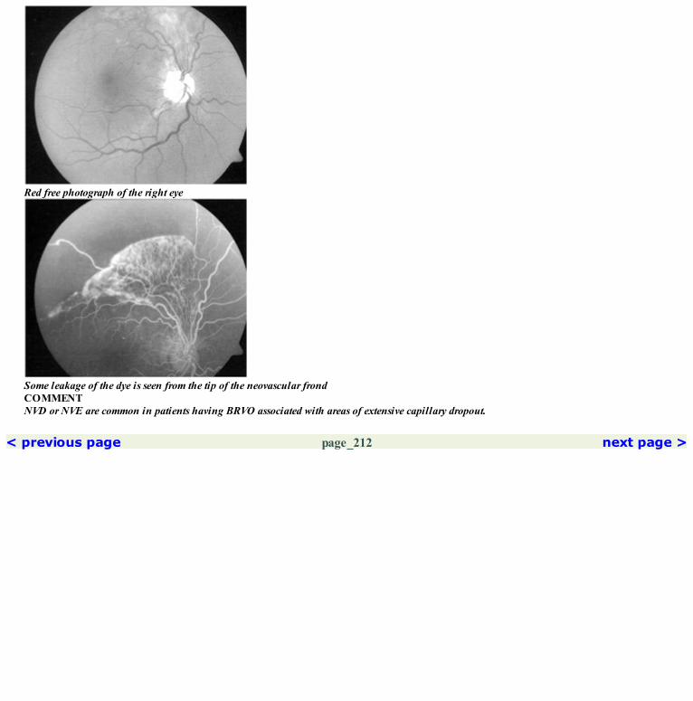
Red free photograph of the right eye
Some leakage of the dye is seen from the tip of the neovascular frondCOMMENTNVD or NVE are common in patients having BRVO associated with areas of extensive capillary dropout.
< previous page page_212 next page >

< previous page page_213 next page >
Page 213CLINICAL NUGGETS1. Superotemporal vein is more commonly involved in BRVO.2. Except in inflammatory conditions, BRVO almost always develops at the A-V crossing.3. Sector laser photocoagulation in the affected segment is done for patients with BRVO who develop neovascularization.4. Laser photocoagulation for macular edema secondary to BRVO can be considered if it is of more than three months duration and associated with avisual acuity less than 6/12.SELECTED READINGS1. Argon laser scatter photocoagulation for prevention of neovascularization and vitreous haemorrhage in branch vein occlusion: A randomizedclinical trial: Branch vein occlusion study group: Arch Ophthalmol 104:34–41, 1986.2. Argon laser photocoagulation for macular edema in branch vein occlusion: Branch vein occlusion study group: Am J Ophthalmol 98:271–282,1984.3. Finkelstein D. Ischemic macular edema, recognition and favorable natural history in branch vein occlusion: Arch Ophthalmol 119: 1427–1434,1992.
< previous page page_213 next page >

< previous page page_214 next page >
Page 214This page intentionally left blank.
< previous page page_214 next page >

< previous page page_215 next page >
Page 215

Section 4 Inflammatory Disorders
< previous page page_215 next page >

< previous page page_216 next page >
Page 216This page intentionally left blank.
< previous page page_216 next page >

< previous page page_217 next page >
Page 21730 Acute Multifocal Posterior Placoid Pigment Epitheliopathy (AMPPPE)
■ AMPPPE is characterized by multiple yellow-white placoid lesions in the posterior pole occurring at the RPE/choroid level.■ The lesions are usually discrete and may show considerable variation in size and shape.■ Yellow lesions either represent infiltration of the choroid by inflammatory cells or choroidal hypoperfusion due to obstructive vasculitis or both,leading to opacification of the overlying retinal pigment epithelium.■ Lesions may become confluent.■ The placoid lesions fade and show retinal pigment epithelial changes on resolution. A crop of new active lesions may develop during the healing ofthe older lesions.
■ In the acute stage the lesions are hypofluorescent due to either blockage of fluorescence by inflammatory cells or due to non-perfusion ofchoriocapillaris.■ Late hyperfluorescence of the lesions is seen.■ Healed lesions show irregular hyperfluorescent (transmission defects) in the early phase which fades in the later part of the angiogram.
< previous page page_217 next page >

< previous page page_218 next page >
Page 218Case 1. A 40-year-old male presented with history of noticing a central scotoma in the left eye for the past one week. Visual acuity was 6/9; N8 in theleft eye.
Fundus photograph of the left eye showing multiple dull yellow placoid lesions at the macula
Early arteriovenous phase showing hypofluorescence in the area of the placoid lesions
Faint hyperfluorescence is seen within the lesions in the late arteriovenous phase

Red free photograph of the left eye
The hypofluorescence is more prominent in the mid arteriovenous phase due to contrast from the normal surrounding choroidal fluorescence
Placoid lesions are hyperfluorescent with fuuy margins in the late phase
< previous page page_218 next page >

< previous page page_219 next page >
Page 219Case 2. A 49-year-old male presented with history of decreased vision in the right eye for the past ten days. He did not give history of any precedingillness. Visual acuity was 6/60; N36 in the right eye. Left eye was normal.
Fundus photograph of the right eye showing multiple placoid lesions at the posterior pole
Placoid lesions appear hypofluorescent in the arteriovenous phase. Peripapillary hypofluorescence is seen corresponding to the lesions seen in thefundus photograph
Red free photograph of the right eye

Placoid lesions appear hyperfluorescent in the late phase. Hyperfluorescence is also noted in the peripapillary regionCOMMENTActive placoid lesions of AMPPPE appear hypofluorescent in the early phase and hyperfluorescent with fuzzy margins in the late phase.
< previous page page_219 next page >

< previous page page_220 next page >
Page 220The patient was observed without treatment. Six weeks later vision had improved to 6/18; N12. Fundus examination showed a combination of healedand active lesions.
Lesions just nasal and temporal to the fovea appear healed with pigmentary changes seen in the center. Faint dull yellow placoid lesions are stillmade out in the temporal and superior part of the macula
Arteriovenous phase shows the healed lesions to have a central hypofluoresence with a rim of hyperfluorescence due to transmission defect. Theactive lesions are still clearly visualized
All the lesions appear hypofluorescent. Faint hyperfluorescence due to transmission defect is seen around the healed lesion just temporal to thefovea. The hypofluorescence around the disc seen in the active stage has reduced
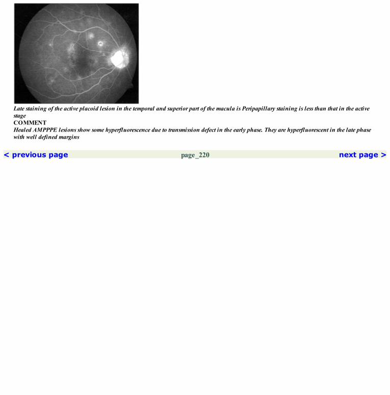
Late staining of the active placoid lesion in the temporal and superior part of the macula is Peripapillary staining is less than that in the activestageCOMMENTHealed AMPPPE lesions show some hyperfluorescence due to transmission defect in the early phase. They are hyperfluorescent in the late phasewith well defined margins
< previous page page_220 next page >

< previous page page_221 next page >
Page 22131Geographic Helicoid Peripapillary Choroidopathy (GHPC)
■ Serpiginous choroiditis or geographic helicoid peripapillary choroidopathy (GHPC) is a bilateral, chronic progressive inflammatory conditionaffecting the retinal pigment epithelium and the inner choroid at the posterior pole.■ It is typically seen in young and middle aged patients with no sex predilection.■ In its active stage, the cream or yellow colored lesions are seen at the level of the retinal pigment epithelium extending in a helicoid manner from theoptic disc. Typical pseudopod like extensions are seen around the edge of the lesions.■ Healing starts centrally and results in pigmentary alteration within the lesion.■ Skip or satellite lesions can occur.
■ Active lesions are hypofluorescent in the early phases and show staining with fuzzy borders in the later part of the angiogram.■ Angiographic appearance of healed lesions depends on the extent of atrophy of the retinal pigment epithelium and choriocapillaris. When retinalpigment epithelium is predominantly affected with relative preservation of the choriocapillaris, the angiogram will show mottled hyperfluorescentdefects within the lesion in the early phase. If the choriocapillaries are also significantly atrophied, the angiogram will remain hypofluorescent in theearly phases with the scleral staining seen through in the late phase. Pigment clumps associated with the lesion will block the choroidal fluorescence.
< previous page page_221 next page >
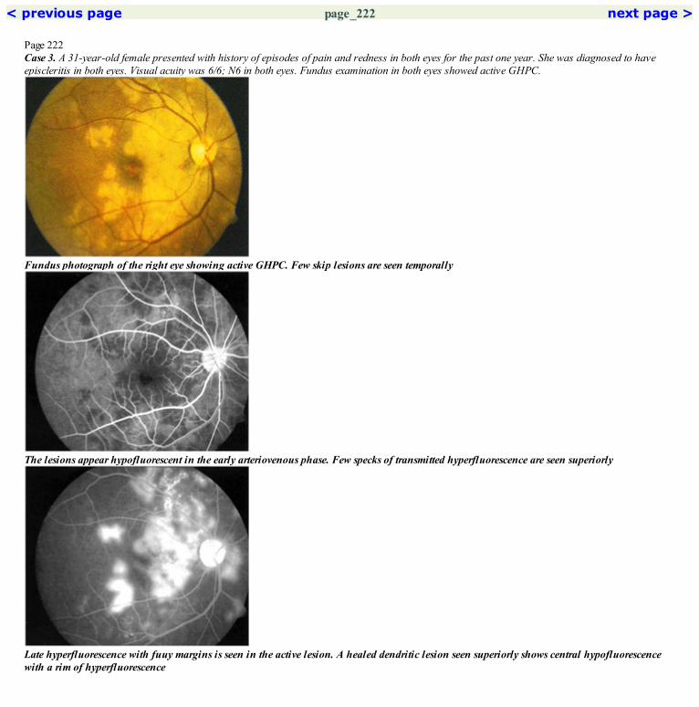
< previous page page_222 next page >
Page 222Case 3. A 31-year-old female presented with history of episodes of pain and redness in both eyes for the past one year. She was diagnosed to haveepiscleritis in both eyes. Visual acuity was 6/6; N6 in both eyes. Fundus examination in both eyes showed active GHPC.
Fundus photograph of the right eye showing active GHPC. Few skip lesions are seen temporally
The lesions appear hypofluorescent in the early arteriovenous phase. Few specks of transmitted hyperfluorescence are seen superiorly
Late hyperfluorescence with fuuy margins is seen in the active lesion. A healed dendritic lesion seen superiorly shows central hypofluorescencewith a rim of hyperfluorescence

Red free photograph of the right eye
The lesions remain hypofluorescent in the mid arteriovenous phaseCOMMENTActive GHPC lesions are hypofluorescent in the early phase and hyperfluorescent with fuzzy margins in the late film.
< previous page page_222 next page >
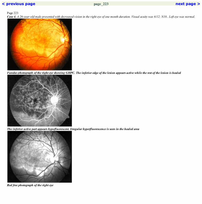
< previous page page_223 next page >
Page 223Case 4. A 26-year-old male presented with decreased vision in the right eye of one month duration. Visual acuity was 6/12; N10.. Left eye was normal.
Fundus photograph of the right eye showing GHPC. The inferior edge of the lesion appears active while the rest of the lesion is healed
The inferior active part appears hypofluorescent. Irregular hyperfluorescence is seen in the healed area
Red free photograph of the right eye

Marked fuzzy hyperfluorescence is seen in the area of the active disease. Hyperfluorescence due to staining is also noted in the healed part,however its edges remain well defined in contrast to the fuzzy margins of the inferior active partCOMMENTThis case illustrates the difference between active and healed GHPC. Healed lesions have well defined borders even in the late phase while theactive lesion shows increased hyperfluorescence with fuzzy margins in the late phase.
< previous page page_223 next page >
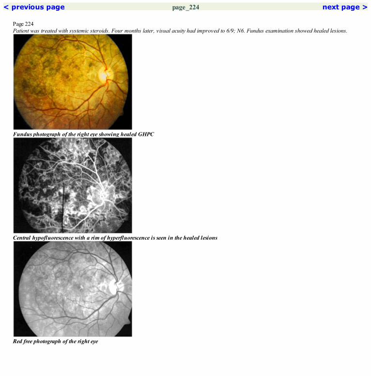
< previous page page_224 next page >
Page 224Patient was treated with systemic steroids. Four months later, visual acuity had improved to 6/9; N6. Fundus examination showed healed lesions.
Fundus photograph of the right eye showing healed GHPC
Central hypofluorescence with a rim of hyperfluorescence is seen in the healed lesions
Red free photograph of the right eye

Late hyperfluorescence of the healed lesion occurs due to increased visibility of the scleral staining and due to staining of the scar tissue. Notethat the edges of the lesions remain discreteCOMMENTGHPC lesions progressively increase in size. Fluorescein angiography is extremely useful to monitor this progression.
< previous page page_224 next page >
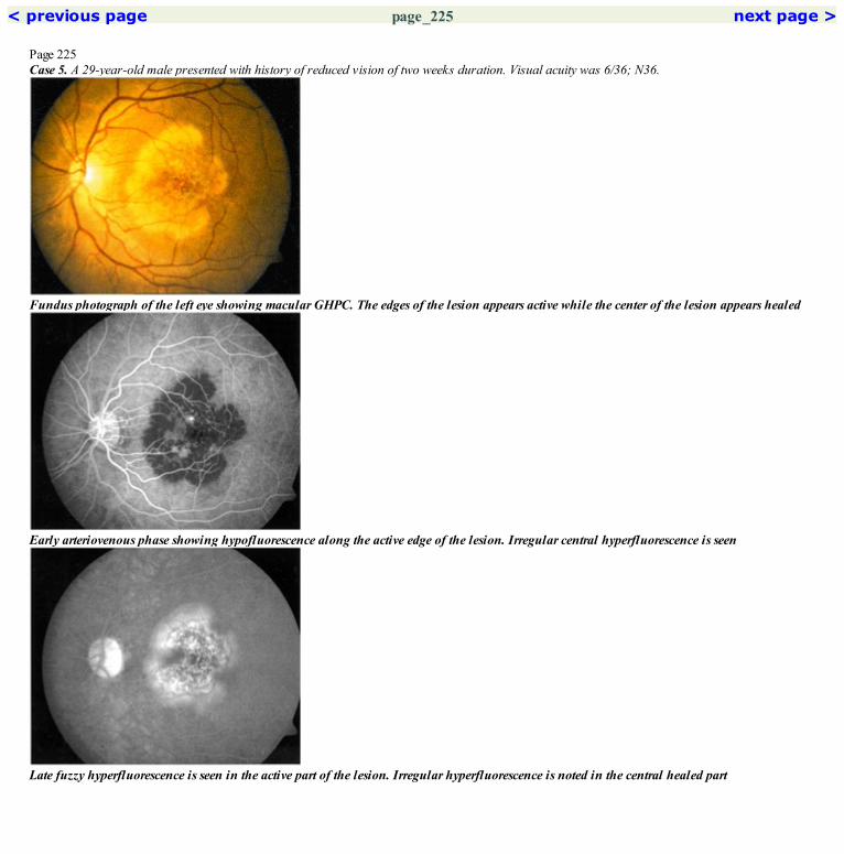
< previous page page_225 next page >
Page 225Case 5. A 29-year-old male presented with history of reduced vision of two weeks duration. Visual acuity was 6/36; N36.
Fundus photograph of the left eye showing macular GHPC. The edges of the lesion appears active while the center of the lesion appears healed
Early arteriovenous phase showing hypofluorescence along the active edge of the lesion. Irregular central hyperfluorescence is seen
Late fuzzy hyperfluorescence is seen in the active part of the lesion. Irregular hyperfluorescence is noted in the central healed part

Red free photograph of the left eye
Midarteriovenous phase show hypofluorescence of the active edgeCOMMENTGHPC need not always be peripapillary, as seen in this case of macular GHPC.
< previous page page_225 next page >

< previous page page_226 next page >
Page 226CLINICAL NUGGETS1. GHPC is a chronic, typically bilateral, episodic disease with a poor visual prognosis.2. GHPC typically affects the young to middle-aged patient with no sex predilection.3. GHPC lesions are typically peripapillary, but macular lesions can also occur.4. Skip or non-contiguous lesions can occur and probably represent new foci of involvement.5. Systemic steroids, antiviral and immunosuppressive drugs have been tried in the treatment of GHPC.
< previous page page_226 next page >

< previous page page_227 next page >
Page 22732Vogt-Koyanagi-Harada Syndrome
■ Vogt-Koyanagi-Harada (VKH) syndrome is a bilateral disorder, commonly seen in the second to fourth decade of life.■ It is a panuveitis associated with exudative retinal detachment and is more common in darkly pigmented individuals and in patients with Orientalancestry.■ Acute stage is characterized by massive infiltration of the choroid with inflammatory cells.■ Localized or extensive exudative retinal detachment is the most common finding and is associated with cells in the vitreous and anterior chamber.The detachment usually begins in the posterior pole and may be multifocal. Focal yellow-white lesions may be seen at the retinal pigment epitheliallayer. The optic nerve head is hyperemic.■ The resolved stage is characterized by a ‘sunset glow’ fundus.
■ Typically, innumerable tiny leaks at the level of the retinal pigment epithelium are seen in the early phase of the angiogram.■ In the late phase characteristic pooling of the dye occurs in the areas of exudative retinal detachments.■ Late staining of the optic nerve head is seen.■ In the resolved stage, transmission window defects are noted in the areas of retinal pigment epithelium atrophy. Areas of hyperpigmentation causeblocked fluorescence.
< previous page page_227 next page >

< previous page page_228 next page >
Page 228Case 1. A 55-year-old female presented with sudden loss of vision in the right eye for the past 4 days. Visual acuity was counting fingers close to facein the right eye and 6/6; N6 in the left eye. Vitreous cells were seen in the right eye.
Fundus photograph of the right eye showing pockets of subretinal fluid at the posterior pole. The disc is hyperemic
Mid arteriovenous phase showing blurring of the choroidal fluorescence in the region of the subretinal fluid. A cluster of pinpointhyperfluorescence is seen temporal to the fovea
Characteristic extensive pooling of the dye in the subretinal space is seen in the late phase

Red free photograph of the right eye
The areas of pinpoint hyperfluorescence are better seen in the late arteriovenous phaseCOMMENTMultiple pinpoint hyperfluorescent spots in the arteriovenous phase with extensive late pooling of the dye in the subretinal space is typical ofVKH.
< previous page page_228 next page >
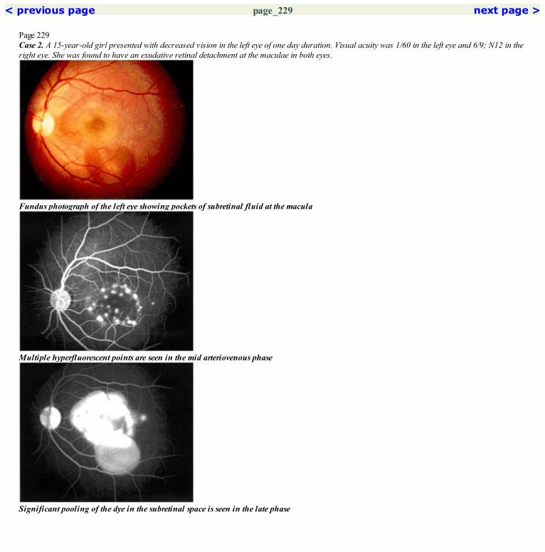
< previous page page_229 next page >
Page 229Case 2. A 15-year-old girl presented with decreased vision in the left eye of one day duration. Visual acuity was 1/60 in the left eye and 6/9; N12 in theright eye. She was found to have an exudative retinal detachment at the maculae in both eyes.
Fundus photograph of the left eye showing pockets of subretinal fluid at the macula
Multiple hyperfluorescent points are seen in the mid arteriovenous phase
Significant pooling of the dye in the subretinal space is seen in the late phase

Red free photograph of the left eye
The leakage is seen to increase as the angiogram progresses
< previous page page_229 next page >

< previous page page_230 next page >
Page 230Right eye of the patient.
Fundus photograph of the right eye showing the exudative detachment at the macula
Mid arteriovenous phase showing multiple points of dye leakage at the RPE level
Red free photograph of the right eye

Late phase showing extensive pooling of the dye in the subretinal spaceCOMMENTUnlike in CSC, the leaks in VKH are innumerable.Late pooling of the dye that occurs in CSC is never so extensive as in VKH. In CSC, the entire extent of neurosensory elevation is never filled upwith dye even in the very late phase.Late staining of the disc is a common finding in VKH.
< previous page page_230 next page >

< previous page page_231 next page >
Page 231Case 3. A 17-year-old girl presented with history of decreased vision in the right eye of one month duration. Visual acuity was 1/60 in the right eyeand 6/6; N6 in the left eye.
Fundus photograph of the right eye showing an exudative detachment at the macula. The disc is hyperemic
Mid arteriovenous phase showing pinpoint hyperfluorescence temporally. The disc capillaries are dilated and the margins are blurred
Pooling of the dye in the subretinal space is seen in the late film. Increased disc staining is evident

Red free photograph of the right eye
The pinpoint hyperfluorescence is well seen in the late arteriovenous phase
< previous page page_231 next page >

< previous page page_232 next page >
Page 232Right eye of the patient.
Fundus photograph of the left eye showing a pocket of turbid subretinal fluid inferotemporal to the foveal center
Arteriovenous phase showing hyperfluorescent points inferotemporal to the fovea
Red free photograph of the left eye

Late pooling of the dye is seen. The dye fills the entire area of neuro-sensory detachment. Increased staining of the disc is seenCOMMENTIt is important to differentiate VKH from central serous chorioretinopathy as the treatment of the two conditions is different.Age, visual acuity, presence of vitreous cells, disc involvement, ultrasound and the angiographic findings help to differentiate the two.
< previous page page_232 next page >

< previous page page_233 next page >
Page 23333 Intermediate Uveitis
■ Intermediate uveitis or pars planitis is a common chronic inflammatory disorder causing a marked inflammatory exudative response in theperipheral retina.■ Vitreous cells are invariably present and may range from mild to severe.■ Yellow-white exudates may collect over the inferior vitreous base overlying the pars plana and the peripheral retina—when this is significant it iscalled ‘snowbanking’.■ Cystoid macular edema is very common and important cause of visual loss in this condition.■ Disc edema can also be present.
■ Fluorescein angiography is useful in evaluating cystoid macular edema in intermediate uveitis. This is especially true in cases in which the vitritisdoes not allow a very clear visualization of the posterior pole.■ Leakage from the perifoveal capillaries is seen in the arteriovenous phase of the angiogram. This is generally more extensive than that seen inaphakic cystoid macular edema. Cystoid changes are very well noted in the late phase of the angiogram.■ Leakage of the dye and staining of the vessel wall may occur in larger vessels. Increased disc staining is common and well seen in the late phase.
< previous page page_233 next page >

< previous page page_234 next page >
Page 234Case 1. A 42-year-old female presented with blurring of vision in the right eye for the past two months. Visual acuity was 6/24; N18. Exudates werenoted over the pars plana.
Fundus photograph of the right eye. Vitreous haze precludes evaluation of the finer retinal details
Increased hyperfluorescence of the disc is seen. Faint hyperfluorescence is seen in the perifoveal region
Red free photograph of the right eye
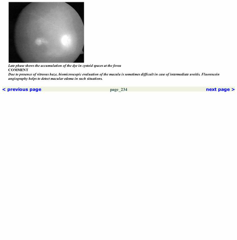
Late phase shows the accumulation of the dye in cystoid spaces at the foveaCOMMENTDue to presence of vitreous haze, biomicroscopic evaluation of the macula is sometimes difficult in case of intermediate uveitis. Fluoresceinangiography helps to detect macular edema in such situations.
< previous page page_234 next page >
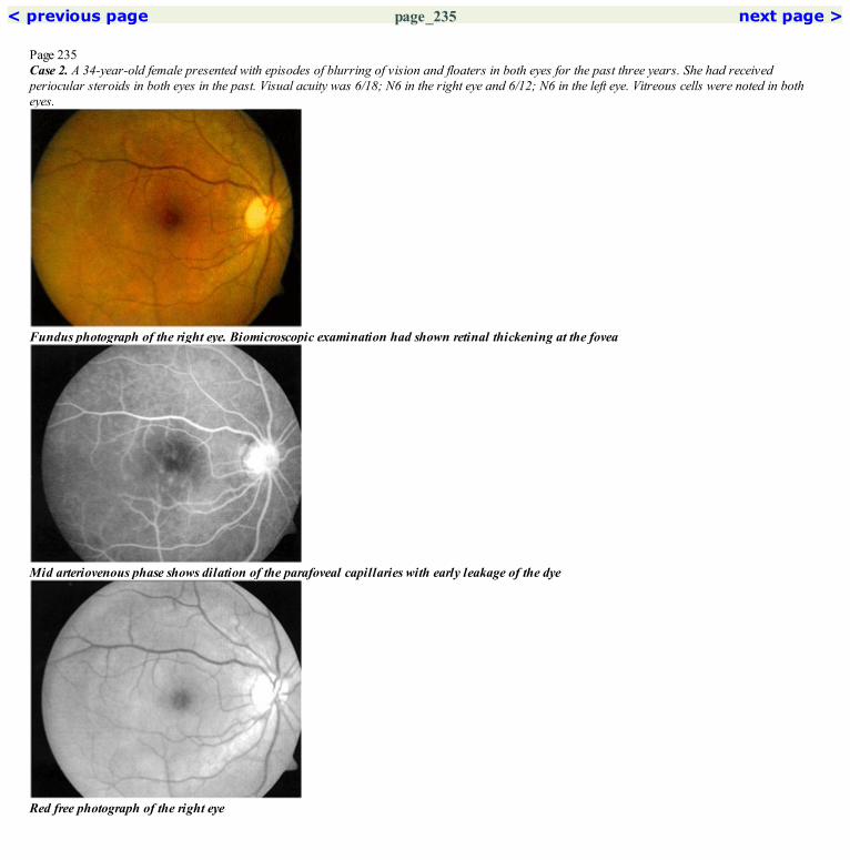
< previous page page_235 next page >
Page 235Case 2. A 34-year-old female presented with episodes of blurring of vision and floaters in both eyes for the past three years. She had receivedperiocular steroids in both eyes in the past. Visual acuity was 6/18; N6 in the right eye and 6/12; N6 in the left eye. Vitreous cells were noted in botheyes.
Fundus photograph of the right eye. Biomicroscopic examination had shown retinal thickening at the fovea
Mid arteriovenous phase shows dilation of the parafoveal capillaries with early leakage of the dye
Red free photograph of the right eye
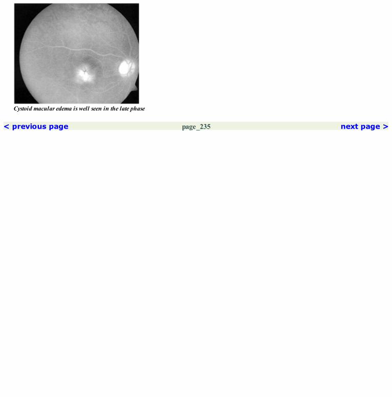
Cystoid macular edema is well seen in the late phase
< previous page page_235 next page >
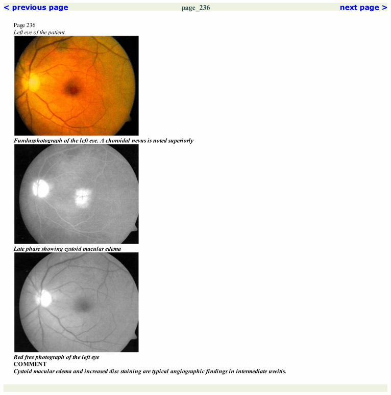
< previous page page_236 next page >
Page 236Left eye of the patient.
Fundusphotograph of the left eye. A choroidal nevus is noted superiorly
Late phase showing cystoid macular edema
Red free photograph of the left eyeCOMMENTCystoid macular edema and increased disc staining are typical angiographic findings in intermediate uveitis.

< previous page page_236 next page >

< previous page page_237 next page >
Page 23734 Birdshot Retinochoroidopathy
■ Birdshot retinochoroidopathy is a rare, bilateral disorder occurring more commonly in females in the fifth to seventh decade of life.■ The lesions are typically flat, creamy yellow in color and distributed throughout the fundus.■ They appear to radiate linearly from the optic disc.■ These lesions are choroidal infiltrates.■ Some degree of vitritis is common in this disorder.■ Cystoid macular edema and disc edema are frequently present.
■ The lesions of birdshot retinochoroidopathy usually do not show hypo- or hyperfluorescence in the early phase. Mild hyperfluorescence is seenin the late phase of the angiogram.■ Arterial filling may be delayed.■ Leakage from the perifoveal capillaries and from larger vessels may be seen in the late phase.■ Cystoid macular edema is commonly seen.
< previous page page_237 next page >

< previous page page_238 next page >
Page 238Case 1. A 60-year-old male patient presented with history of reduced vision in the left eye for the past several years. He had been diagnosed to havepanuveitis in both eyes and had lost vision in the right due to secondary glaucoma. Visual acuity in the left eye was 6/36; N36.
Fundus photograph of the left eye showing mulitple flat creamy yellow lesions
Red free photograph of the left eye
Late phase showing hyperfluorescence of the lesions
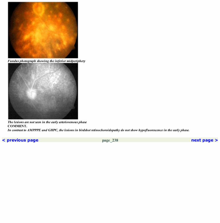
Fundus photograph showing the inferior midperiphery
The lesions are not seen in the early arteriovenous phaseCOMMENT.In contrast to AMPPPE and GHPC, the lesions in birdshot retinochoroidopathy do not show hypofluorescence in the early phase.
< previous page page_238 next page >

< previous page page_239 next page >
Page 23935 Other Inflammatory DisordersPARASITICTRACTSCase 1. A 49-year-old female presented with decreased vision in the right eye for the past one month. Visual acuity was 6/12; N8 in the right eye.
Fundus photograph showing the dead parasite in the inferonasal periphery. The parasitic tracts are seen
The parasitic tracts appear hyperfluorescent due to transmission window defects
Red free photograph showing the posterior pole. The linear tracts are well seen temporally

Tracts seen nasally
< previous page page_239 next page >

< previous page page_240 next page >
Page 240
The criss-crossing tracts seen in the temporal periphery
Late phase angiogram showing some fading of the hyperfluorescence along the tractsDISSEMINATED CHOROIDITISCase 2. A 22-year-old male was examined for decreased vision in the right eye for the past six months. He had been treated with systemic steroids forhis ocular problem. Visual acuity was 6/24; N6 in the right eye and 6/6; N6 in the left eye.
Montage picture of the right eye fundus showing disseminated healed choroiditis

Red free photograph of the right eye
< previous page page_240 next page >
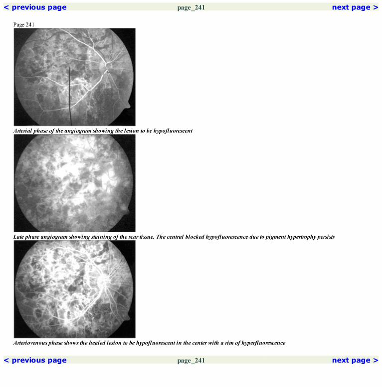
< previous page page_241 next page >
Page 241
Arterial phase of the angiogram showing the lesion to be hypofluorescent
Late phase angiogram showing staining of the scar tissue. The central blocked hypofluorescence due to pigment hypertrophy persists
Arteriovenous phase shows the healed lesion to be hypofluorescent in the center with a rim of hyperfluorescence
< previous page page_241 next page >

< previous page page_242 next page >
Page 242TOXOPLASMIC RETINOCHOROIDITISCase 3. A 24-year-old male presented with reduced vision in the right eye of one week duration. Visual acuity was 6/9; N6. He had been previouslydiagnosed to have healed toxoplasmic retinochoroiditis.
Fundus photograph of the right eye showing an active retinochoroiditis patch adjacent to a healed lesion. Overlying vitreous haze is seen
Early arteriovenous phase showing hypofluorescence in the region of the active and healed lesion
Late phase shows hyperfluorescence of the active lesion. Staining of the vessels wall and the disc are also seen

Red free photograph of the right eye
Hypofluorescence is persisting in the late part of the arteriovenous phase. Minimal leakage of the dye from the walls of the adjacent retinal vesselsis seenCOMMENTEarly hypofluorescence with late intense hyperfluorescence with fuzzy margins are typical findings in active toxoplasmic retinochoroiditis.Recurrent lesions usually occur at the edge of the old healed lesion.
< previous page page_242 next page >

< previous page page_243 next page >
Page 243CLINICAL NUGGETS1. AMPPPE is usually a self-limiting disease; rarely it recurs.2. Vogt-Koyanagi-Harada syndrome is a systemic disorder involving the eye, ear, skin and the meninges.3. Dallen-Fuchs nodules can occur in VKH syndrome.4. Ultrasonography shows a characteristic low reflective thickening of the choroid in VKH5. Birdshot chorioretinopathy is closely linked to HLA-A29.SELECTED READINGS1. Gass JD. Acute posterior multifocal placoid pigment epitheliopathy. Arch Ophthalmol 80:177–185, 1968.2. Hyvarinen L, Maumenee AE, George T, Weinstein GW. Fluorescein angiography of the choriocapillaris. Am J Ophthalmol 67:653–666,1969.3. Schatz H, Maumenee AE, Patz A. Geographic helicoids peripapillary choroidopathy. Trans Am Acad Ophthalmol Otolaryngol 78:OP747–761,1974.4. Gass JD. Vitiliginous chorioretinitis. Arch Ophthalmol 99:1778–1787, 1981.5. De Laey JJ. Fluorescein angiography in posterior uveitis. Int Ophthalmol Clin 35:33–58, 1995.
< previous page page_243 next page >

< previous page page_244 next page >
Page 244This page intentionally left blank.
< previous page page_244 next page >

< previous page page_245 next page >
Page 245

Section 5 Heredomacular Dystrophy
< previous page page_245 next page >

< previous page page_246 next page >
Page 246This page intentionally left blank.
< previous page page_246 next page >

< previous page page_247 next page >
Page 24736 Best Disease
■ Best disease or vitelliform macular dystrophy is an autosomal dominant disorder with variable penetrance and expressivity.■ It primarily affects the RPE layer.■ The Arden’s index in the electrooculogram is markedly reduced in this condition.■ The various stages described include:● Previtelliform stage● Vitelliform stage● Pseudohypopyon stage● Vitelliruptive stage● Atrophic stage.
■ Depends on the stage of the disease.■ In the vitelliform stage the early phases of angiogram shows some blocked choroidal fluorescence in the region of the lesion. In the later phase ofthe angiogram, the vitelliform lesion may appear nonfluorescent or may appear to fluoresce slightly.■ In the subsequent stages when the yellow material disrupts, the angiogram shows areas of hyperfluorescence within the lesion and late staining.■ In the atrophic stage, the large choroidal vessels may be visualized within the atrophic lesion.■ Angiography in Best disease permits the detection of choroidal neovascularization.
< previous page page_247 next page >

< previous page page_248 next page >
Page 248Case 1. A 36-year-old female presented with history of decreased vision in both eyes for the last ten years. Visual acuity was 6/60; N36 in both eyes.Electroretinogram was normal in both eyes while the electrooculogram showed markedly subnormal Arden’s index. Both eyes had similar fundusfindings.
Fundus photograph of the right eye showing a well-demarcated round lesion in the macula with dull yellow organised subretinal deposits in thecenter. RPE alteration is seen within the lesion
Early arteriovenous phase shows hyperfluorescence within the lesion. A rim of hypofluorescence is seen surrounding the lesion
Late phase angiogram showing fading of the hyperfluorescence. Staining of the subretinal organised deposits is seen

Red free photograph of the right eye
The hyperfluorescence has increased in the arteriovenous phase with one area of more intense hyperfluorescenceCOMMENTIrregular hyperfluorescence is commonly seen within the lesion and occurs due to RPE alterations. Some fuzziness within the lesion is usuallyseen in the late phase.
< previous page page_248 next page >

< previous page page_249 next page >
Page 249Case 2. A 41-year-old male presented with history of difficulty in reading small print. Visual acuity was 6/18; N10 in the right eye and 6/9; N8 in theleft eye. Electrooculogram showed markedly subnormal Arden’s index.
Fundus photograph of the right eye showing scattered dull yellow subretinal deposits in the macula. RPE alterations are seen
Arterial phase shows irregular hyperfluorescence in the area of the lesion
Late phase angiogram show fading of the fluorescence indicating transmission defect

Red free photograph highlights these changes
The hyperfluorescence has increased in the arteriovenous phase with some fuzziness of the fluorescence
< previous page page_249 next page >

< previous page page_250 next page >
Page 250Left eye of the patient.
Fundus photograph of the left eye showing RPE alterations with scattered subretinal yellow deposits in the macula
Early hyperfluorescence is seen within the lesion due to transmission defect
The hyperfluorescence fades in the late films

Red free photograph of the left eye
Increase in the hyperfluorescence is seen in the mid arteriovenous phaseCOMMENTIn cases with bilateral, symmetrical, round lesions at the fovea with some evidence of subretinal deposits, Best dystrophy should be considered.Fluorescein angiogram will show transmission defects at the site of the lesion. Electrooculogram is diagnostic.
< previous page page_250 next page >

< previous page page_251 next page >
Page 251Case 3. A 29-year-old male presented with history of decreased vision in the right eye noticed for the last one year. Visual acuity was 6/9; N6 in theright eye and 6/6; N6 in the left eye. The Arden’s index was reduced.
Fundus photograph of the right eye showing the vitelliruptive stage of Best disease. Subretinal deposits are seen as a rim inferiorly. A scar is notedat the fovea
Hyperfluorescence is seen in the area of the lesion in the arteriovenous phase. Increased hyperfluorescence is seen in the center and a rim ofhypofluorescence is seen surrounding the lesion
Red free photograph of the right eye

Fading of the hyperfluorescence is seen. Staining of the central scar is seen in the late film
< previous page page_251 next page >

< previous page page_252 next page >
Page 252Left eye of the patient.
Fundus photograph of the left eye showing a pseudohypopyon. A subretinal scar is seen centrally
Arteriovenous phase showing transmitted hyperfluorescence within the lesion. The scar shows intense hyperfluorescence
Red free photograph of the left eye

Late phase shows staining of the central scarCOMMENTDevelopment of CNVM is known to occur in patients with Best dystrophy. The CNVM tends to remain small and results in a scar formation.
< previous page page_252 next page >

< previous page page_253 next page >
Page 25337 Stargardt’s Disease (Fundus Flavimaculatus)
■ Stargardt’s disease and fundus flavimaculatus can be considered to represent different stages of the same disease.■ It is predominantly an autosomal recessive disorder.■ Visual loss starts early, usually in childhood or early adulthood.■ Fundus may be normal in the early stages. However with time, atrophic macular changes and associated retinal flecks develop.
■ The characteristic feature of Stargardt’s disease on a fluorescein angiogram is the absence of choroidal fluorescence or the ‘silent choroid’. This isconsidered almost pathognomonic of Stargardt’s disease.■ Lipofuscin like material deposition in the retinal pigment epithelium is probably responsible for blockage of the underlying choroidal fluorescence.■ The retinal vessels especially the retinal capillary network are seen more prominently against the dark background.■ The atrophic macular lesion shows up as a hyperfluorescent area due to window defect.■ Multiple irregular hyperfluorescent spots that do not precisely correspond to the flecks are seen in the midperipheral retina.
< previous page page_253 next page >

< previous page page_254 next page >
Page 254Case 1. A 14-year-old male presented with inability to see the blackboard at school for the last six months. Visual acuity was 6/24; N12 in both eyes.
Fundus photograph of the right eye showing RPE alterations at the fovea
Arteriovenous phase of the angiogram showing stippled hyperfluorescence in foveal region due to transmission defects. Choroidal fluorescence isnot seen in the rest of the areas
Fading of the hyperfluorescence is seen in the foveal region in the latephase, indicating a transmission window defect. Silent choroid can still beappreciated
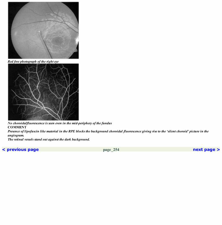
Red free photograph of the right eye
No choroidalfluorescence is seen even in the mid-periphery of the fundusCOMMENTPresence of lipofuscin like material in the RPE blocks the background choroidal fluorescence giving rise to the ‘silent choroid’ picture in theangiogram.The retinal vessels stand out against the dark background.
< previous page page_254 next page >

< previous page page_255 next page >
Page 255Case 2. A 16-year-old male presented with a history of decreased vision in both eyes for the past four years. Visual acuity was 6/36; N12 in both eyes.
Fundus photograph of the right eye showing an atrophic lesion at the fovea with multiple irregular flecks at the posterior pole
Arterial phase showing irregular transmitted hyperfluorescence. The choroidalfluorescence is absent in the peripapillary region
Fading of the transmitted hyperfluorescence is seen in the late phase
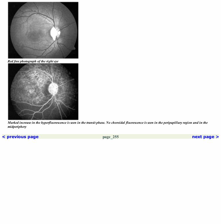
Red free photograph of the right eye
Marked increase in the hyperfluorescence is seen in the transit phase. No choroidal fluorescence is seen in the peripapillary region and in themidperiphery
< previous page page_255 next page >

< previous page page_256 next page >
Page 256Left eye of the patient
Fundus photograph of the left eye showing RPE alterations at the fovea. Retinal flecks were noted throughout the posterior pole
Transmitted hyperfluorescence is seen in the region of the flecks and the atrophic macular lesion. No choroidal fluorescence is seen in theperipapillary region and in the midperiphery
Absence of the choroidal fluorescence is seen in the inferior midperiphery

Red free photograph of the left eye
Some fading of the hyperfluorescence is seen in the later phase of the angiogramCOMMENTEven in patients with extensive flecks, the silent choroid is still evident in theperipapillary region, in between flecks and in the midperiphery.
< previous page page_256 next page >

< previous page page_257 next page >
Page 257Case 3. A 13-year-old child was brought for examination with history of defective vision for the past six months. Visual acuity was 6/18; N6 in theright eye and 6/36; N10 in the left eye.
Fundus photograph of right eye with retinal flecks. No atrophic lesion is seen at the fovea
Hyperfluorescence is seen in the region of the retinal flecks. No choroidal fluorescence is seen elesewhere
Fading of the hyperfluorescence is seen suggesting a transmission window defect

Red free photograph showing the retinal flecks
Increased hyperfluorescence at the flecks with a silent choroid is seen in the nasal retina
< previous page page_257 next page >
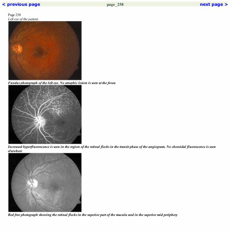
< previous page page_258 next page >
Page 258Left eye of the patient.
Fundus photograph of the left eye. No atrophic lesion is seen at the fovea
Increased hyperfluorescence is seen in the region of the retinal flecks in the transit phase of the angiogram. No choroidal fluorescence is seenelsewhere
Red free photograph showing the retinal flecks in the superior part of the macula and in the superior mid periphery
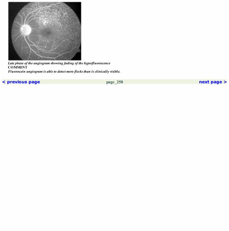
Late phase of the angiogram showing fading of the hyperfluorescenceCOMMENTFluorescein angiogram is able to detect more flecks than is clinically visible.
< previous page page_258 next page >

< previous page page_259 next page >
Page 259Case 4. A 33-year-old male presented with history of decreased vision in both eyes for the past five years. Visual acuity was 6/60; N12 in both eyes.
Fundus photograph of the right eye showing a large atrophic patch at the fovea. Numerous atrophic flecks are seen throughout the posterior pole
Mid arteriovenous phase shows hypofluorescence in the region of the foveal atrophic lesion. Large choroidal vessels are seen within. Irregularhyperfluorescence is seen elsewhere in the macula. No choroidal fluorescence is seen in the peripapillary region and in the midperiphery
Fading of the hyperfluorescence is seen in the area of the flecks. The central atrophic area is hyperfluorescent due to the scleral staining beingseen through

Red free photograph of the right eye
Irregular hyperfluorescence has increased in the arteriovenous phase of the angiogram
< previous page page_259 next page >

< previous page page_260 next page >
Page 260Left eye of the patient.
Fundus phototgraphy of the left eye showing similar changes as the right eye
Increased hyperfluorescence is seen in the area of the flecks. Choroidal silence is seen in the peripapillary region and in the mid periphery
Red free photograph of the left eye

Late phase showing fading of the irregular hyperfluorescence. The atrophic central lesion shows hyperfluorescence due to scleral stainingCOMMENTAngiogram showing the absence of the choroidal fluorescence in the peripapillary region and in the midperiphery.The atrophic changes occurring at the fovea is well seen in the angiogram.
< previous page page_260 next page >

< previous page page_261 next page >
Page 26138 Progressive Cone Dystrophy
■ The onset of symptoms is usually in early childhood. However a subgroup of patients may have onset of symptoms in the third or fourth decadeof life.■ Common symptoms include reduced vision, photoaversion and color vision defect.■ The fundus appearance in this condition can vary. It could be normal in early stages. A classical bull’s eye lesion may develop in the macularregion. Alternately pigment stippling or a large atrophic area may develop in the macular region.
■ Depends on the type of lesion seen in the fundus.■ In cases with a bull’s eye maculopathy, a horizontally ovoid zone of hyperfluorescence is seen surrounding a hypofluorescent center.■ In patients with pigment stippling, the fluorescein angiogram show diffuse stippled hyperfluorescence.■ In patient with a large atrophic lesion, a hyperfluorescent window defect is seen in the area of the lesion.
< previous page page_261 next page >

< previous page page_262 next page >
Page 262Case 1. A 39-year-old male presented with reduced vision in both eyes for the past five years. Visual acuity was 6/12; N6 in both eyes. A bull’s eye likelesion was seen in both eyes. Color vision was severely affected. The cone responses in the electroretinogram was markedly reduced.
Fundus photograph of the right eye showing a bull’s eye like lesion at the fovea
Arterial phase of the angiogram showing the hyperfluorescence due to transmission defect at the fovea
Late phase shows fading of the hyperfluorescence indicating it to be a transmission window defect

Red free photograph showing the foveal lesion
Arteriovenous phase showing increased transmission defect seen through the annular atrophy at the foveaCOMMENTThe bull’s eye lesion is very well seen in the angiogram. The central normal hypofluorescence is surrounded by a ring of hyperfluorescence due totransmission defects.
< previous page page_262 next page >

< previous page page_263 next page >
Page 263Case 2. A 34-year-old male presented with history of photoaversion and reduced vision in both eyes. Visual acuity was 6/24; N12 in the right eye and6/60; N12 in the left eye. The cone response was significanlty reduced in the electroretinogram.
Fundus photograph of the right eye showing an atrophic lesion at the fovea
Arteriovenous phase of the angiogram showing transmitted irregular hyperfluorescence within the atrophic lesion
Red free photograph of the right eye
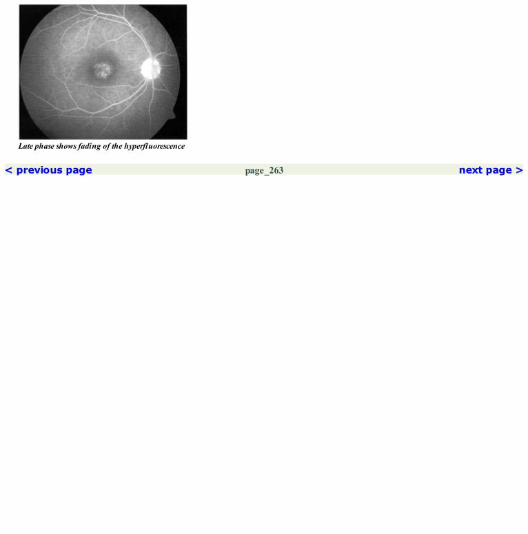
Late phase shows fading of the hyperfluorescence
< previous page page_263 next page >
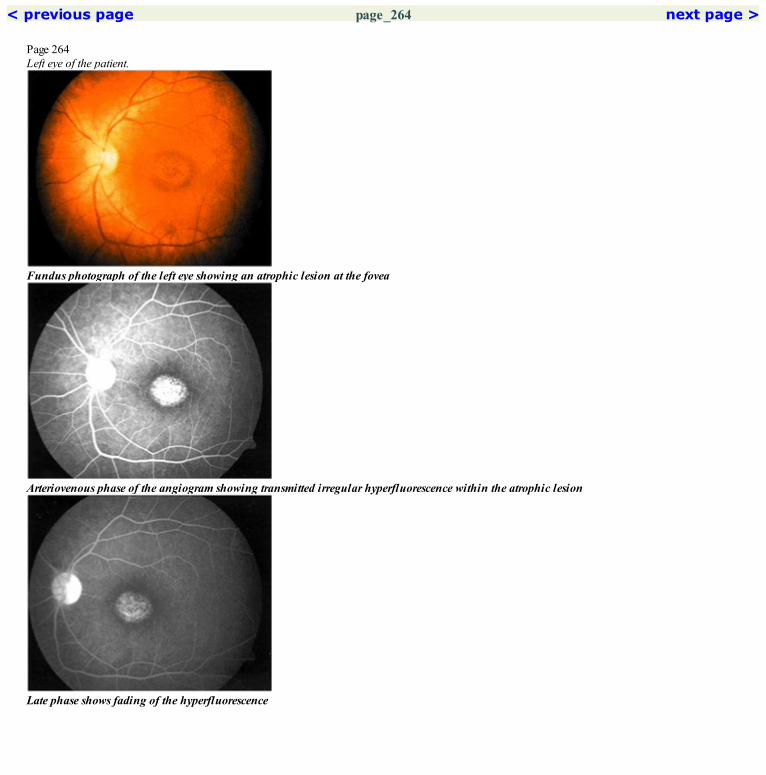
< previous page page_264 next page >
Page 264Left eye of the patient.
Fundus photograph of the left eye showing an atrophic lesion at the fovea
Arteriovenous phase of the angiogram showing transmitted irregular hyperfluorescence within the atrophic lesion
Late phase shows fading of the hyperfluorescence

Red free photograph of the left eye
Increase in the hyperfluorescence seen as the angiogram proceedsCOMMENTAngiogram is showing transmission defects due to RPE alterations at the fovea.Fluorescein angiogram picks up even very subtle RPE alterations.
< previous page page_264 next page >

< previous page page_265 next page >
Page 265Case 3. A 21-year-old male presented with history of reduced vision in both eyes of four years duration. Visual acuity was 6/60; N6 in both eyes. Theelectroretinogram showed reduced cone responses.
Fundus photograph showing a bull’s eye like lesion at the fovea. A reflective Mizuo like sheen is seen temporal to this lesion
Early arteriovenous phase showing stippled hyperfluorescence within the atrophic foveal lesion
Fading of the hyperfluorescence is seen in the late phase
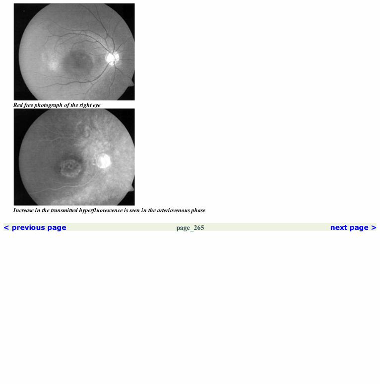
Red free photograph of the right eye
Increase in the transmitted hyperfluorescence is seen in the arteriovenous phase
< previous page page_265 next page >

< previous page page_266 next page >
Page 266Left eye of the patient.
Fundus photograph of the left eye showing a bull’s eye like lesion at the fovea with a Mizuo like sheen superotemporal to it
Arteriovenous phase showing an annular hyperfluorescent ring with central hypofluorescence at the fovea
Red freephotograph of the left eye
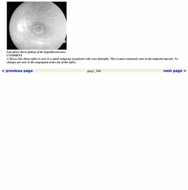
Late phase shows fading of the hyperfluorescenceCOMMENTA Mizuo like shiny reflex is seen in a small subgroup of patients with cone dystrophy. This is more commonly seen in the temporal macula. Nochanges are seen in the angiogram at the site of the reflex.
< previous page page_266 next page >

< previous page page_267 next page >
Page 26739 Other Heredomacular DystrophiesPATTERN DYSTROPHYCase 1. A 26-year-old male presented with history of distortion of vision for the past one year. Visual acuity was 6/6; N6 in both eyes.
Fundus photograph of the right eye showing irregular subretinal deposits at the fovea
Arterial phase showing a central area of hypofluorescence with radiating spokes
Red free photograph of the right eye
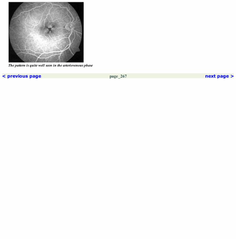
The pattern is quite well seen in the arteriovenous phase
< previous page page_267 next page >

< previous page page_268 next page >
Page 268Left eye of the patient.
Late phase of the angiogram
Fundus photograph of the left eye showing subretinal deposits at the foveaLeft eye of the patient.
Red free photograph of the left eye

Late phase angiogram picture
A pattern similar to that seen in the right eye is noted at the foveaCOMMENTThere is deposition of a yellow-orange material in the subretinal space in pattern dystrophy. This blocks the background choroidal fluorescencegiving rise to the pattern seen angiographically.
< previous page page_268 next page >
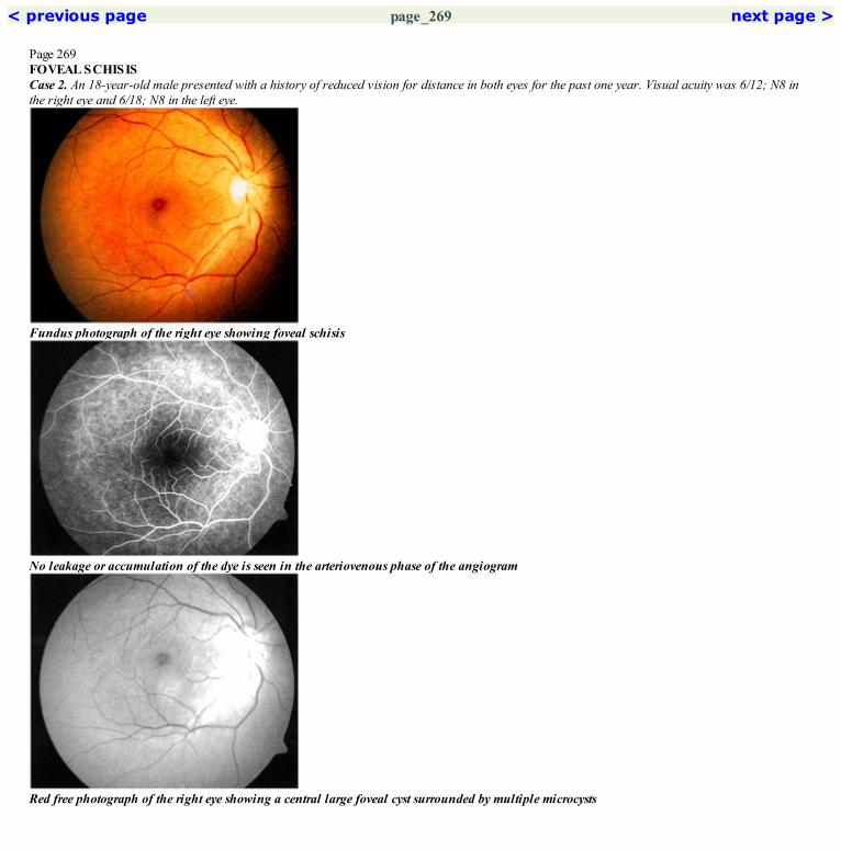
< previous page page_269 next page >
Page 269FOVEAL SCHISISCase 2. An 18-year-old male presented with a history of reduced vision for distance in both eyes for the past one year. Visual acuity was 6/12; N8 inthe right eye and 6/18; N8 in the left eye.
Fundus photograph of the right eye showing foveal schisis
No leakage or accumulation of the dye is seen in the arteriovenous phase of the angiogram
Red free photograph of the right eye showing a central large foveal cyst surrounded by multiple microcysts
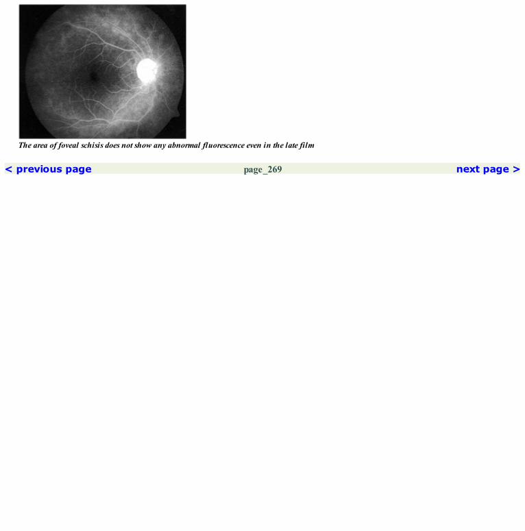
The area of foveal schisis does not show any abnormal fluorescence even in the late film
< previous page page_269 next page >

< previous page page_270 next page >
Page 270Left eye of the patient.
Fundus photograph of the left eye showing foveal schisis
Arteriovenous phase not showing any abnormal fluorescence at the fovea
Red freephotograph showing the foveal microcysts

Late phase angiogram not showing any dye accumulation in the cystsCOMMENTUnlike in cystoid macular edema, no pooling of the dye is seen in eyes with foveal schisis.Angiogram is usually normal or may show a few transmission defects at the fovea.
< previous page page_270 next page >

< previous page page_271 next page >
Page 271FOVEAL HYPOPLASIACase 3. A 17-year-old male presented with reduced vision in both eyes since early childhood. Visual acuity was 3/60 in both eyes.
Fundus photograph of the right eye showing absence of a well-formed fovea
Arteriovenous phase does not show a foveal avascular zone. The normal hypofluorescence at the fovea is absent
Red free photograph shows lack of a foveal avascular zone

Late phase angiogram
< previous page page_271 next page >

< previous page page_272 next page >
Page 272Left eye of the patient.
Fundus photograph of the left eye does not show a foveal reflex
Arteriovenous phase shows lack of a foveal avascular zone or a parafoveal capillary plexus. The normal foveal hypofluorescence is absent
Red free photograph shows absence of a capillary free zone at the center of the macula

Late phase of the angiogramCOMMENTThe FAZ in foveal hypoplasia is either absent or ill-formed. Blood vessels may be seen traversing the normal site of the fovea
< previous page page_272 next page >

< previous page page_273 next page >
Page 273CLINICAL NUGGETS1. Heredomacular dystrophies are familial, bilaterally symmetrical disorders occurring at the posterior pole.2. History, clinical features, color vision, visual fields, electrodiagnostic tests, and fluorescein angiography are useful investigative tools in patientswith heredomacular dystrophies.SELECTED READINGS1. Curry H F Jr, Moorman LT. Fluorescein photography of vitelliform macular degeneration. Arch Ophthalmol 79:705–709, 1968.2. Miller SA. Fluorescence in Bests vitelliform dystrophy, lipofuschin and fundus flavimaculatus. Br J Ophthalmol 62:256–260, 1978.3. Morse PH, MacLean AL. Fluorescein fundus studies in hereditary vitelloeruptive macular degeneration. Am J Ophthalmol 66:485–494,1968.4. Klien BA, Krill AE. Fundus flavimaculatus: clinical functions and histopathologic observations. Am J Ophthalmol 64:3, 1967.5. Franceschetti A, François J. Fundus flavimaculatus. Arch Ophthalmol 25:505, 1965.6. Anmarkrud N. Fundus Fluorescein angiography in Fundus flavimaculatus and Stargardts disease. Acta Ophthalmol 57:172–182, 1979.7. Uliss AE, Moore AT, Bird AC. The dark choroid in posterior retinal dystrophies. Ophthalmology 94(11):1423–1427, 1987.8. von Ruckmann A, Fitzke FW, Bird AC. In vivo fundus autofluorescence in macular dystrophies: Arch Ophthalmol 115(5):609–15, 1997.9. Krause U, Vainio-Mattila B, Eriksson A, Forsius. Fluorescein angiographic studies on X-chromosomal retinoschisis. Acta Ophthalmol 48:794,1970.10. Deutman A F, van Blommestein JDA, Henkes H E et al. Butterfly-shaped pigment dystrophy of the fovea. Arch Ophthalmol 83:558–569,1970.11. Ayazi S, Fagan R. Pattern dystrophy of the pigment epithelium. Retina 1:287–289; 1981.
< previous page page_273 next page >

< previous page page_274 next page >
Page 27440 Ocular Albinism
■ Albinism is a group of disorders associated with an inborn error of amino acid metabolism affecting the production of melanin.■ Oculocutaneous albinism comprises of ocular involvement along with the skin and hair involvement. This can be either tyrosinase negative ortyrosinase positive.■ Ocular albinism is characterized by changes restricted to the ocular structures.■ Retinal changes associated with albinism include a depigmented fundus, foveal hypoplasia and optic disc hypoplasia.
■ Increased visibility of the choroidal vessels is noted due to lack of masking by the overlying pigment.■ Foveal hypoplasia may be evident by absence of a well formed foveal avascular zone.■ Late phase shows the choroidal and retinal vessels silhouetted against the background fluorescence from scleral staining.
< previous page page_274 next page >

< previous page page_275 next page >
Page 275Case 1. A 19-year-old female with ocular albinism presented with history of strabismus since early childhood. Her brother was noted to have similarcomplaints. Visual acuity was 6/24; N6 in the right eye and 6/18; N6 in the left eye.
Fundus photograph of the right eye showing a markedly depigmented fundus. Few patches of pigmented RPE are seen in the macula
Arterial phase of the angiogram shows increased visibility of the larger choroidal vessels due to lack of blockage by the normally pigmented RPE.The choroidal details are blurred in the few areas of preserved RPE in the macula
Late phase shows the relatively hypofluorescent larger choroidal vessels and the retinal vessels silhouetted against the backgroundfluorescencedue to scleral staining

Red freephotograph shows increased visibility of the larger choroidal vessels due to luck ofpigment in the more anterior layers
A diffuse choroidal fluorescence is seen due to leakage of dye from the choriocapillaris. Note the retinal vessels are not clearly visualized due tolack of contrastCOMMENTThere is increased visibility of the large choroidal vessels as the pigment in the RPE, which normally blocks the choroidal fluorescence, isdeficient.
< previous page page_275 next page >

< previous page page_276 next page >
Page 276Left eye of the patient.
Fundus photograph of the left eye showing the depigmented fundus. Areas of normalpigmented RPE is noted in the macula
Arteriovenous phase of the angiogram showing increased visibility of the larger choroidal vessels due to lack of blockage by thepigment. It isdifficult to appreciate the retinal vessels due to lack of contrast. The normal foveal hypofluorescence is absent
Red free photograph of the left eye
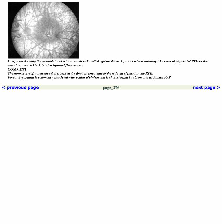
Late phase showing the choroidal and retinal vessels silhouetted against the background scleral staining. The areas of pigmented RPE in themacula is seen to block this background fluorescenceCOMMENTThe normal hypofluorescence that is seen at the fovea is absent due to the reduced pigment in the RPE.Foveal hypoplasia is commonly associated with ocular albinism and is characterized by absent or a ill formed FAZ.
< previous page page_276 next page >

< previous page page_277 next page >
Page 277CLINICAL NUGGETS1. Albinism is a group of inherited disorders characterized by absent or deficient pigmentation.2. Skin, hair and eyes are involved in oculocutaneous albinism; abnormal pigmentation primarily involves the eyes in ocular albinism.SELECTED READINGS1. Gregor: The perifoveal vasculature in albinism. Br J Ophthalmology 62:554–557, 1978.2. Spedick MJ, Beauchamp GR. Retinal vascular and optic nerve abnormality in albinism. J Pediatric Ophthalmology Strabismus 23: 58–63, 1986.
< previous page page_277 next page >

< previous page page_278 next page >
Page 27841 Retinitis Pigmentosa and Allied Disorders
■ Retinitis pigmentosa (RP) is characterized by arteriolar attenuation, disc pallor and bony spicule pigmentation in the mid-peripheral fundus.■ Mottled retinal pigment epithelial changes are seen throughout the fundus.■ In early stages of retinitis pigmentosa, fundus findings are better delineated on fluorescein angiogram.
■ The angiographic appearance in retinitis pigmentosa depends on the severity on the retinal changes.■ In early RP, the choroidal flush is normal, with a mottled hyperfluorescence seen more prominently in the mid-peripheral retina.■ As the disease progresses, patches of choriocapillary non-filling are noted.■ In advanced disease, due to loss of the choriocapillaris the choroidal flush may be absent.■ Leakage of dye from the retinal capillary causes increased staining of the retinal tissue in the late phase. This may be confined to the macular areaor in the region of the vascular arcades, or it may be widespread. It is difficult to determine whether most of the fluorescein staining is derived fromleaking retinal capillaries or choriocapillaris through the abnormal RPE.■ Cystoid macular edema is seen in a small percentage of patients of RP.■ An increased retinal circulation time is seen.
< previous page page_278 next page >

< previous page page_279 next page >
Page 279Case 1. A 40-year-old female presented with history of decreased vision in the left eye for the past one year. Visual acuity was 6/36; N36 in the left eye.Right eye had similar fundus findings. Electroretinogram showed markedly reduced responses in both eyes.
Fundus photograph of the right eye showing RPE alteration and pigment clumping along the arcades. The peripheral retina was relativelyunaffected
Transmitted hyperfluorescence is seen in the affected areas
Arterial phase of the angiogram showing large choroidal vessels in areas of RPE and choriocapillaries atrophy

Late phase angiogram showing the background scleral staining as hyperfluorescence through the overlying atrophoid layersCOMMENTFluorescein angiography is useful to assess the status of the choriocapillaris in RP.Early hyperfluorescence through the window defects in the RPE suggests an intact choriocapillaris.Delayed hyperfluorescence indicates atrophy of the choriocapillaris.Fluorescein angiography can thus be of prognostic value in patients with RP.
< previous page page_279 next page >

< previous page page_280 next page >
Page 280Case 2. A 45-year-old female presented with a history of night blindness for the past fifteen years. She noticed floaters in front of both eyes for the lastthree months. Examination did not reveal any inflammatory process in the eye. Visual acuity was 6/9; N18 in both eyes.
Fundus photograph of the right eye showing attenuated arterioles. Retinal thickening was noted at the fovea on biomicroscopic examination
Arteriovenous phase showing dilation of the parafoveal capillaries with minimal leakage of the dye. Irregular hyperfluorescence due totransmission defect is seen elsewhere in the fundus
Red free photograph of the right eye

Late phase shows accumulation of the dye in cystoid spaces at the fovea. Minimal staining of the retina is also evident along the inferotemporalarcade
< previous page page_280 next page >

< previous page page_281 next page >
Page 281Left eye of the patient.
Fundus photograph of the left eye showing arteriolar attenuation and RPE alterations beyond the arcade. Cystoid changes were seen clinically atthe fovea
Arteriovenous phase showing leakage of the dye from the capillaries around the fovea. Widespread irregular hyperfluorescence is seen due towindow defects
Red free photograph of the left eye

Late phase showing cystoid macular edemaCOMMENTA small percentage of patients with RP develop cystoid macular changes. The leakage is most likely from the parafoveal and perifovealcapillaries.
< previous page page_281 next page >

< previous page page_282 next page >
Page 282Case 3. A 19-year-old male presented with progressive decrease of vision in dim illumination for the past four years. Visual acuity was 6/36; N18 inboth eyes. Electroretinogram showed grossly reduced responses.
Fundus photograph of the right eye showing an annular atrophy around the fovea. Arteriolar attenuation and widespread RPE alteration arealso seen
Hyperfluorescence due to transmission defect is seen at the atrophic ring around the fovea. Elsewhere in the fundus, irregular stippledhypefluorescence due to RPE atrophy is seen. The normal hypofluorescence at the fovea is accentuated by the surrounding hyperfluorescence
Red free photograph of the right eye

Late phase angiogram shows fading of the hyperfluorescence
< previous page page_282 next page >

< previous page page_283 next page >
Page 283Left eye of the patient.
Fundus photograph of the left eye showing similar changes as that in the right eye
Arteriovenous phase showing hyperfluorescence due to transmission defects. The annular atrophy around the fovea is well made out
Red free photograph of the left eye
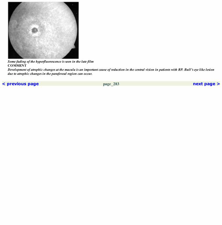
Some fading of the hyperfluorescence is seen in the late filmCOMMENTDevelopment of atrophic changes at the macula is an important cause of reduction in the central vision in patients with RP. Bull’s eye like lesiondue to atrophic changes in the parafoveal region can occur.
< previous page page_283 next page >
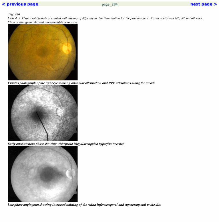
< previous page page_284 next page >
Page 284Case 4. A 37-year-old female presented with history of difficulty in dim illumination for the past one year. Visual acuity was 6/6; N6 in both eyes.Electroretinogram showed unrecordable responses.
Fundus photograph of the right eye showing arteriolar attenuation and RPE alterations along the arcade
Early arteriovenous phase showing widespread irregular stippled hyperfluorescence
Late phase angiogram showing increased staining of the retina inferotemporal and superotemporal to the disc

Red free photograph of the right eye
Increase in the transmitted hyperfluorescence is seen in the arteriovenous phase
< previous page page_284 next page >

< previous page page_285 next page >
Page 285Left eye of the patient.
Fundus photograph of the left eye showing similar changes as in the right eye
Arteriovenous phase showing transmitted hyperfluorescence throughout the fundus. Normal hypofluorescence at the fovea is seen
Red free photograph of the left eye

Late phase angiogram showing some staining of the retina along the arcadesCOMMENTLeakage of the dye into the retinal tissue can occur either from the retinal blood vessels or from choriocapillaris through the defects in the RPElayer. Late phase angiogram shows diffuse staining of the retina in these cases.
< previous page page_285 next page >
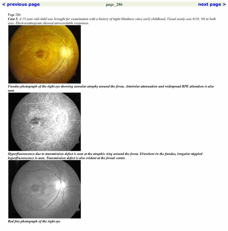
< previous page page_286 next page >
Page 286Case 5. A 15-year-old child was brought for examination with a history of night blindness since early childhood. Visual acuity was 6/18; N6 in botheyes. Electroretinogram showed unrecordable responses.
Fundus photograph of the right eye showing annular atrophy around the fovea. Arteriolar attenuation and widespread RPE alteration is alsoseen
Hyperfluorescence due to transmission defect is seen at the atrophic ring around the fovea. Elsewhere in the fundus, irregular stippledhyperfluorescence is seen. Transmission defect is also evident at the foveal center.
Red free photograph of the right eye

Late phase angiogram shows some fading of the hyperfluorescence
< previous page page_286 next page >

< previous page page_287 next page >
Page 287Left eye of the patient.
Fundus photograph of the left eye showing similar changes as that in the right eye
Arteriovenous phase showing hyperfluorescence due to transmission defects. Transmission defects are also seen at the foveal center
Red free photograph of the left eye
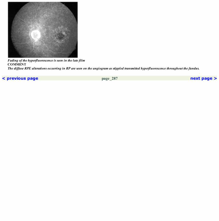
Fading of the hyperfluorescence is seen in the late filmCOMMENTThe diffuse RPE alterations occurring in RP are seen on the angiogram as stippled transmitted hyperfluorescence throughout the fundus.
< previous page page_287 next page >

< previous page page_288 next page >
Page 288CHOROIDEREMIACase 6. A 31-year-old male presented with a history of night blindness since early childhood. He complained of decreased vision in the right eye forthe past 15 days. Visual acuity was 6/12; N10 in the right eye and 6/9; N6 in the left eye.
Fundus photograph of the right eye shows extensive atrophy of the RPE and choriocapillaris. A strip of RPE is preserved at the fovea; pigmentclumps are seen
Arteriovenous phase showing the absence of the choriocapillaris except for a vertical strip at the fovea. The large choroidal vessels are seen
Late phase angiogram showing very minimal staining of the sclera in the areas of atrophy
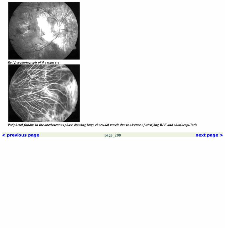
Red free photograph of the right eye
Peripheral fundus in the arteriovenous phase showing large choroidal vessels due to absence of overlying RPE and choriocapillaris
< previous page page_288 next page >

< previous page page_289 next page >
Page 289Left eye of the patient.
Fundus photograph of the left eye showing absense of RPE and choriocapillaris except for a small patch at the fovea
Late phase showing the central patch of preserved RPE
Red free photograph of the left eyeCOMMENTDiffuse loss of the choriocapillaris is the hallmark of choroideremia.The normal prominent scleral staining that occurs in the late phase is not seen in this condition as the leakage of dye is minimal from theatrophic choriocapillaris.

< previous page page_289 next page >
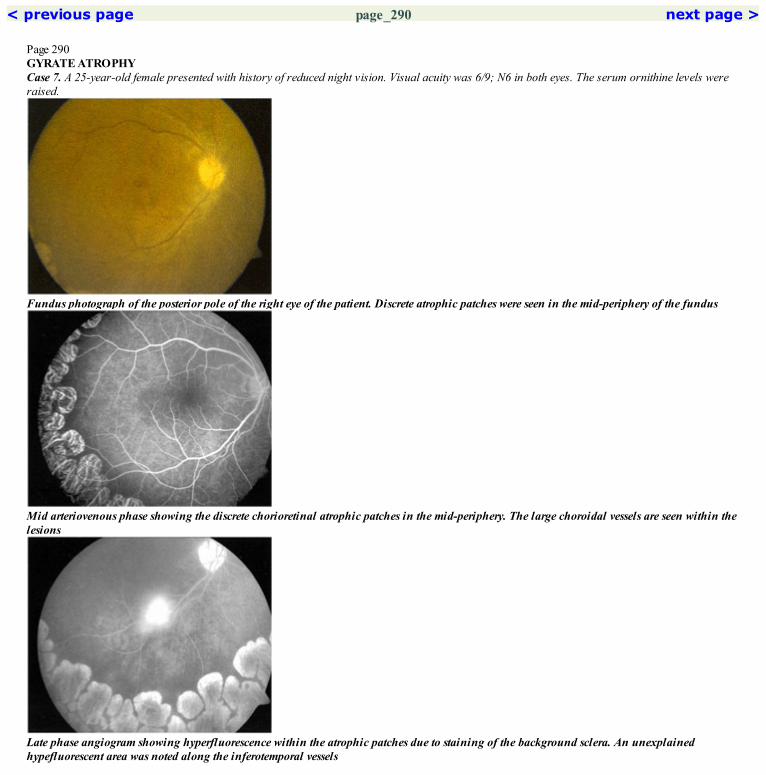
< previous page page_290 next page >
Page 290GYRATE ATROPHYCase 7. A 25-year-old female presented with history of reduced night vision. Visual acuity was 6/9; N6 in both eyes. The serum ornithine levels wereraised.
Fundus photograph of the posterior pole of the right eye of the patient. Discrete atrophic patches were seen in the mid-periphery of the fundus
Mid arteriovenous phase showing the discrete chorioretinal atrophic patches in the mid-periphery. The large choroidal vessels are seen within thelesions
Late phase angiogram showing hyperfluorescence within the atrophic patches due to staining of the background sclera. An unexplainedhypefluorescent area was noted along the inferotemporal vessels

Red free photograph of the right eye
Atrophic patches seen in the superotemporal mid-peripheryCOMMENTGyrate atrophy is characterized by the loss of discrete patches of RPE and choriocapillaris.
< previous page page_290 next page >

< previous page page_291 next page >
Page 291Left eye of the patient.
Fundus photograph of the left eye
Arteriovenous phase showing the atrophic patches temporally
Superotemporal quadrant showing the discrete atrophic patches

Red free photograph
The discrete atrophic patches seen in the inferotemporal quadrant
Late phase angiogram showing the normal posterior pole
< previous page page_291 next page >
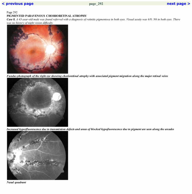
< previous page page_292 next page >
Page 292PIGMENTED PARAVENOUS CHORIORETINAL ATROPHYCase 8. A 43-year-old male was found referred with a diagnosis of retinitis pigmentosa in both eyes. Visual acuity was 6/9; N6 in both eyes. Therewas no history of night vision difficulty.
Fundus photograph of the right eye showing chorioretinal atrophy with associated pigment migration along the major retinal veins
Increased hyperfluorescence due to transmission defects and areas of blocked hypojluorescence due to pigment are seen along the arcades
Nasal quadrant
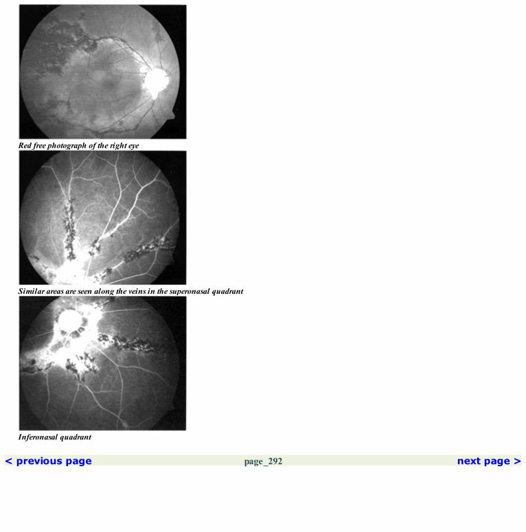
Red free photograph of the right eye
Similar areas are seen along the veins in the superonasal quadrant
Inferonasal quadrant
< previous page page_292 next page >

< previous page page_293 next page >
Page 293Left eye of the patient.
Fundus photograph of the left eye showing chorioretinal atrophy and pigmentary changes along the inferotemporal vessels
Areas of transmitted hyperfluorescence with areas of blocked hypofluorescence are seen around the disc and along the inferotemporal vein
Red free photograph of the left eye
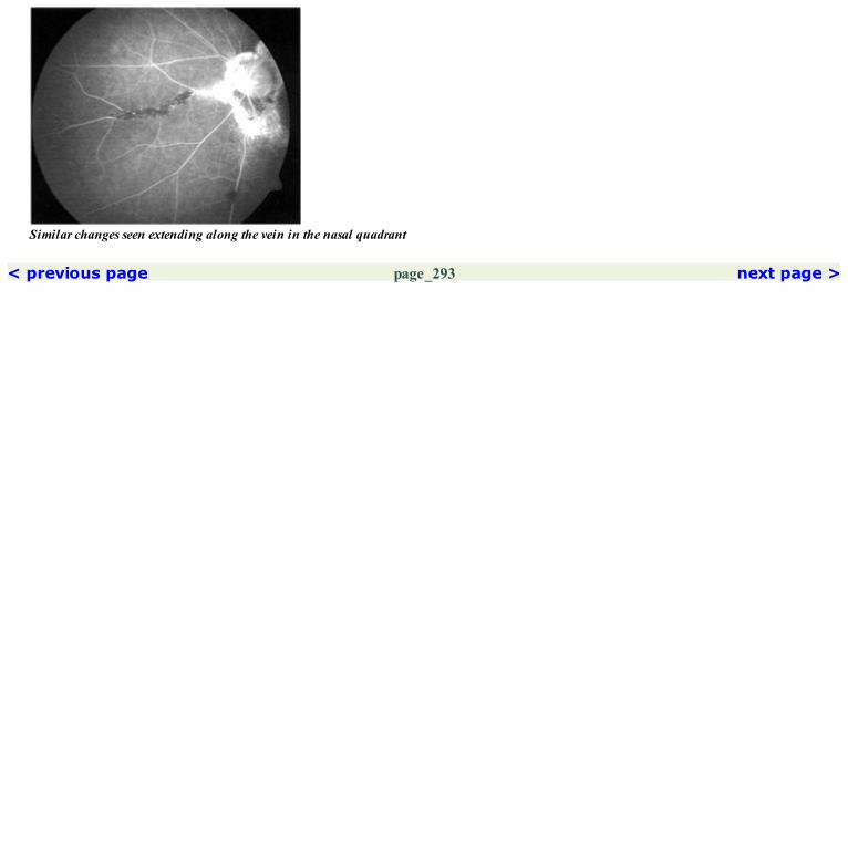
Similar changes seen extending along the vein in the nasal quadrant
< previous page page_293 next page >

< previous page page_294 next page >
Page 294FUNDUS ALBIPUNCTATUSCase 9. A 27-year-old male presented with history of night vision difficulty since early childhood. There was no worsening of the symptoms overs theyears. Visual acuity was 6/6; N6 in both eyes. Electroretinogram showed reduced responses which improved after prolonged dark adaptation.
Fundus photograph of the right eye. Innumer able tiny white dots were seen scattered throughout the fundus
A fine stippled hyperfluorescence due to transmission defect is seen throughout the fundus in the mid arteriovenous phase
Similar stippled hyperfluorescence is seen in the inferonasal periphery

Red free photograph showing the fine dots in the temporal region
Stippled hyperfluorescence is seen in the temporal periphely in the late arteriovenous phase
Fading of the hyperfluorescence is seen in the late phase
< previous page page_294 next page >

< previous page page_295 next page >
Page 295Left eye of the patient.
Fundus photograph of the left eye. Changes similar to those in the right eye were seen
Stippled hyperfluorescence due to transmission defect is seen throughout the posterior pole
Fading of the hyperfluorescence is seen in the late film

Red free photograph of the left eye
Stippled hyperfluorescence is seen in the inferior peripheryCOMMENTStippled hyperfluorescence seen in fundus albipunctatus does not exactly correspond to the innumerable tiny flecks seen clinically.
< previous page page_295 next page >

< previous page page_296 next page >
Page 296BENIGN FLECK RETINACase 10. A 32-year-old female came for a routine eye examination. She was asymptomatic and had a visual acuity of 6/6; N6 in both eyes. Fundusexamination showed innumerable flecks scattered throughout the fundus.
Fundus photograph of the right eye shows innumerable yellow-white flecks seen throughout the posteriorpole. The fovea is spared
Early arteriovenous phase shows irregular hyperfluorescence, more marked temporally
Late phase showing fading of the hypefluorescence
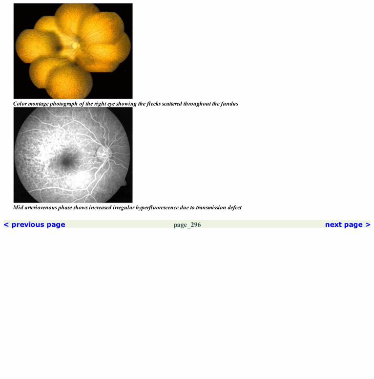
Color montage photograph of the right eye showing the flecks scattered throughout the fundus
Mid arteriovenous phase shows increased irregular hyperfluorescence due to transmission defect
< previous page page_296 next page >

< previous page page_297 next page >
Page 297Left eye of the patient.
Fundus photograph of the left eye showing the flecks at the posterior pole
Irregular transmitted hyperfluorescence seen in the arteriovenous phase of the angiogram
Color montage photograph of the left eye showing the flecks scattered throughout the fundus
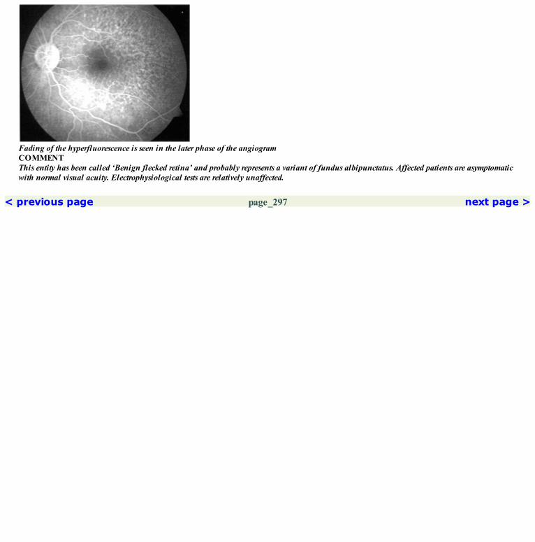
Fading of the hyperfluorescence is seen in the later phase of the angiogramCOMMENTThis entity has been called ‘Benign flecked retina’ and probably represents a variant of fundus albipunctatus. Affected patients are asymptomaticwith normal visual acuity. Electrophysiological tests are relatively unaffected.
< previous page page_297 next page >

< previous page page_298 next page >
Page 298CLINICAL NUGGETS1. Posterior subcapsular cataract, macular degeneration and cystoid macular edema are important causes for loss of central vision in patients with RP.2. In RP, approximately 16–18% loss of ERG amplitudes per year is seen.3. In RP, approximately 4.6% of the remaining visual field loss occurs per year.SELECTED READINGS1. Ronald E Carr. The generalized heredoretinal disorders in vitreoretinal diseases, the essentials. (Ist edn); (Eds: CD Regillo; GC Brown and HWFlynn) page 309.2. Newsome DA. Retinal fluorescein leakage in retinitis pigmentosa. Am J. Ophthalmol; 101:354–360, 1986.3. Piermarocchi S, Segato T, Midena E. Retinal fluorescein leakage in RP. Am J Ophthalmol 102:674, 1986.4. Khan JA, I de CH, Strickland MP. Coat’s—type RP, Surv Ophthalmol 32:317–332, 1988.5. Spallone A: Retinal fluorescein leakage in RP. Am J Ophthalmol 102:408, 1986.
< previous page page_298 next page >

< previous page page_299 next page >
Page 299

Section 6 Optic Nerve Disorders
< previous page page_299 next page >

< previous page page_300 next page >
Page 300This page intentionally left blank.
< previous page page_300 next page >

< previous page page_301 next page >
Page 30142 Optic Nerve Disorders
■ The conditions included in this section are:● Optic pit● Optic nerve head drusen● Papilledema● Neuroretinitis● Optic neuritis● Lebers hereditary optic neuropathy (LHON)● Anterior ischemic optic neuropathy (AION)
■ Optic Pit● Early hypofluorescence is seen at the pit due to absence of vessels in the optic pit. Late staining of the tissue in the optic pit is seen.■ Optic Nerve Head Drusen● Autofluorescence is seen in the pre-injection photograph. Late staining of the optic nerve head drusen is seen.■ Papilledema● Dilatation of the disc capillaries with leakage of the dye is seen in the early phase of the angiogram. Late staining of the disc is seen.■ Neuroretinitis● Dilated disc capillaries with leakage of the dye is seen in the early part of the angiogram. Late staining of the disc occurs.■ Optic Neuritis● Dilated disc capillaries which leak dye resulting in staining of the disc is seen in the late phase.■ Lebers Hereditary Optic Neuropathy (LHON)● Dilated and tortuous capillaries are seen in the peripapillary region. No abnormal staining of the disc is seen in the late phase.■ Anterior Ischemic Optic Neuropathy (AION)● Differential filling of the disc capillaries in the upper and lower half of the optic disc is seen.
< previous page page_301 next page >
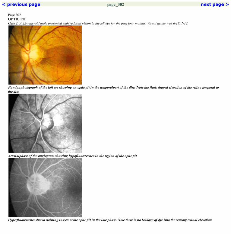
< previous page page_302 next page >
Page 302OPTIC PITCase 1. A 22-year-old male presented with reduced vision in the left eye for the past four months. Visual acuity was 6/18; N12.
Fundus photograph of the left eye showing an optic pit in the temporalpart of the disc. Note the flask shaped elevation of the retina temporal tothe disc
Arterialphase of the angiogram showing hypofluorescence in the region of the optic pit
Hyperfluorescence due to staining is seen at the optic pit in the late phase. Note there is no leakage of dye into the sensory retinal elevation
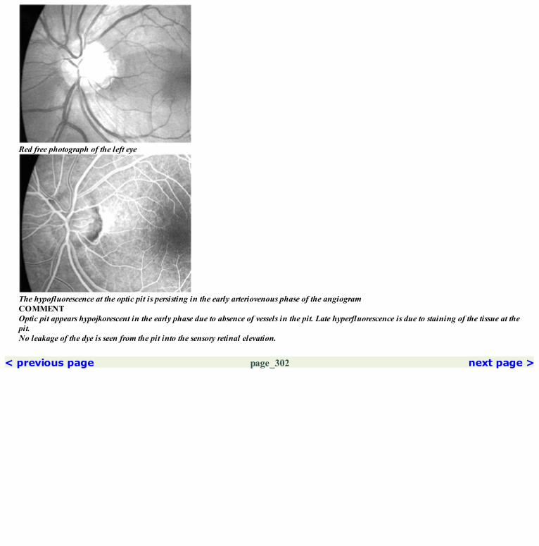
Red free photograph of the left eye
The hypofluorescence at the optic pit is persisting in the early arteriovenous phase of the angiogramCOMMENTOptic pit appears hypojkorescent in the early phase due to absence of vessels in the pit. Late hyperfluorescence is due to staining of the tissue at thepit.No leakage of the dye is seen from the pit into the sensory retinal elevation.
< previous page page_302 next page >
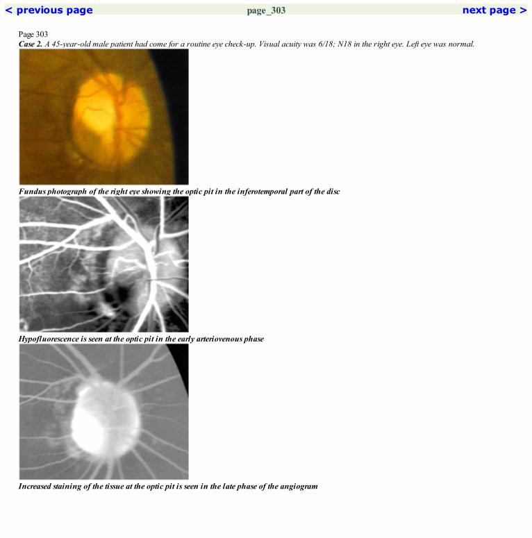
< previous page page_303 next page >
Page 303Case 2. A 45-year-old male patient had come for a routine eye check-up. Visual acuity was 6/18; N18 in the right eye. Left eye was normal.
Fundus photograph of the right eye showing the optic pit in the inferotemporal part of the disc
Hypofluorescence is seen at the optic pit in the early arteriovenous phase
Increased staining of the tissue at the optic pit is seen in the late phase of the angiogram

Red free photograph of the right eye
Minimal hyperfluorescence is seen at the optic pit in the mid arteriovenous phase of the angiogramCOMMENTCase shows the typical angiographic picture of the optic pit.
< previous page page_303 next page >

< previous page page_304 next page >
Page 304OPTIC NERVE HEAD DRUSENCase 3. A 24-year-old female was referred with a diagnosis of papillitis in the left eye. Visual acuity was 6/6; N6 in both eyes. Nodular elevation of thedisc was seen. Ultrasound examination showed a high reflective echo at the optic nerve head, suggestive of an optic nerve head drusen.
Fundus photograph of the left eye showing the nodular elevation along the disc margin
Irregularity along the disc margin is seen in the mid arteriovenous phase of the angiogram
Late phase showing the increased staining of the disc drusen

Red free photograph of the left eye. Note the crowding of the vessels at the disc
Late arteriovenous phase of the angiogram showing the irregularity along the disc margin. Note that there is no dilatation of the disc capillariesCOMMENTOptic nerve head drusen shows autofluorescence in pre-injection photograph.
< previous page page_304 next page >

< previous page page_305 next page >
Page 305PAPILLEDEMACase 4. A 26-year-old female, diagnosed to have pseudotumor cerebri was found to have papilledema in both eyes and referred for an ophthalmiccheckup. Visual acuity was 6/6; N6 in both eyes.
Fundus photograph of the right eye showing the swollen hyperemic disc
Mid arteriovenous phase of the angiogram showing dilation of the disc capillaries. The margins of the disc are blurred
Red free photograph of the right eye

Late phase of the angiogram showing increased staining of the disc
< previous page page_305 next page >
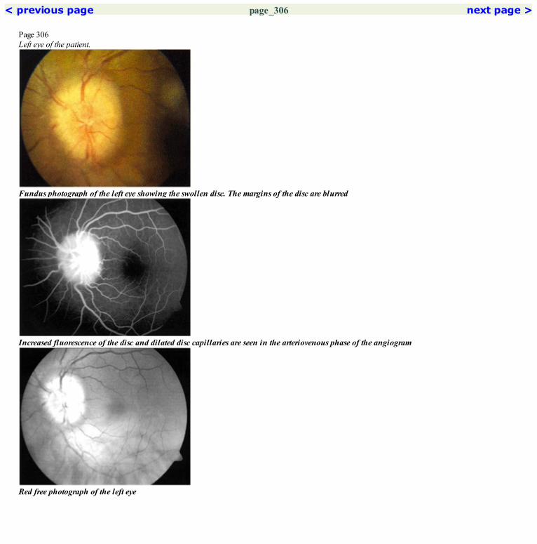
< previous page page_306 next page >
Page 306Left eye of the patient.
Fundus photograph of the left eye showing the swollen disc. The margins of the disc are blurred
Increased fluorescence of the disc and dilated disc capillaries are seen in the arteriovenous phase of the angiogram
Red free photograph of the left eye

Lute phase of the angiogram showing increased staining of the discCOMMENTLeakage of the dye from the dilated disc capillaries is seen in papilledema; late staining of the disc is also seen.
< previous page page_306 next page >
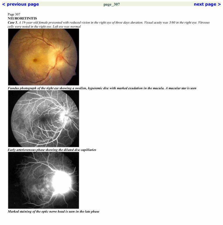
< previous page page_307 next page >
Page 307NEURORETINITISCase 5. A 19-year-old female presented with reduced vision in the right eye of three days duration. Visual acuity was 3/60 in the right eye. Vitreouscells were noted in the right eye. Left eye was normal
Fundus photograph of the right eye showing a swollen, hyperemic disc with marked exudation in the macula. A macular star is seen
Early arteriovenous phase showing the dilated disc capillaries
Marked staining of the optic nerve head is seen in the late phase

Red free photograph of the right eye showing the macular star
Late arteriovenous phase showing leakage of the dye from the disc capillaries. Note that no leakage is seen from the macular retinal vesselsCOMMENTDilatation of the disc capillaries with leakage of the dye and late staining of the disc is seen in patients with neuroretinitis.The lipid exudation is most likely from the disc capillaries as no leakage is seen from the macular retinal vessels.
< previous page page_307 next page >

< previous page page_308 next page >
Page 308OPTIC NEURITISCase 6. A 32-year-old female with multiple sclerosis, presented with sudden onset drop in vision in the right eye of one day duration. Visual acuity was2/60 in the right eye.
Fundus photograph of the right eye showing a swollen, hyperemic disc. The disc margins are blurred
Marked dilatation of the disc capillaries is seen in the arteriovenous phase of the angiogram
Red free photograph of the right eye

Increased staining of the disc is seen in the late phaseCOMMENTLipid exudation is not common in patients with optic neuritis occurring in demyelinating disorders.
< previous page page_308 next page >

< previous page page_309 next page >
Page 309LEBERS HEREDITARY OPTIC NEUROPATHY (LHON)Case 7. A 14-year-old male presented with reduced vision in both eyes of one month duration. Visual acuity was 3/60 in both eyes. CT scan wasnormal. Serum lactate, pyruvate and ammonia were found to be raised.
Fundus photograph of the right eye. Dilated capillaries are seen in the peripapillary region
Dilated and tortuous capillaries are seen around the disc in the early arteriovenous phase
Late phase showing normal disc fluorescence. No abnormal staining of the disc is seen

Red free photograph of the right eye
No leakage of the dye is seen from the dilated capillaries in the late arteriovenous phase
< previous page page_309 next page >

< previous page page_310 next page >
Page 310Left eye of the patient.
Fundus photograph of the left eye showing the dilated peripapillary capillaries
Arteriovenous phase showing the dilated and tortuous capillaries around the disc
Red free photograph of the left eye

No abnormal staining of the disc is seen in the late phaseCOMMENTLebers hereditary optic neuropathy is characterized by the presence of telangiectatic vessels in the peripapillary region. Typically, no abnormalstaining of the disc is seen in the late phase.
< previous page page_310 next page >

< previous page page_311 next page >
Page 311ANTERIOR ISCHEMIC OPTIC NEUROPATHY (AION)Case 8. A 50-year-old diabetic male presented with an inferior altitudinal field defect in the left eye. Visual acuity was 6/18; N10.
Fundus photograph of the left eye showing pallor in the upper half of the disc. Microaneurysms and hard exudates due to diabetic retinopathyare seen in the macula
Filling of the disc capillaries is seen in the lower half. The upper half of the disc shows relative hypofuorescence. Microvascular abnormalitiesare seen in the macula
Red free photograph of the left eye

Increased staining of the disc is seen. Retention of dye is seen in the macula, indicative of macular edemaCOMMENTDifferential filling of the disc capillaries is seen in AION.
< previous page page_311 next page >

< previous page page_312 next page >
Page 312CLINICAL NUGGET1. Visual acuity, pupillary reaction, color vision, visual fields and visual evoked potential are useful for assessement of the optic nerve function.SELECTED READINGS1. Papilledema. In Walsh and Hoyt’s Clinical Neuro-ophthalmology, 5th edn. The Essentials. Editors: Miller NR, Newman NJ, Baltimore, Williamsand Wilkins, chapter 6, 166–195, 1999.2. Oosterhuis JA, Boen-Tan TN. Fluorescein angiography in papilledema and pseudopapilledema. Ophthalmologica 159:96–110, 1969.3. Arnold AC, Helper RS. Fluorescein angiography in acute nonarteritic anterior ischemic optic neuropathy. Am J Ophthalmol 117:222–230, 1994.4. Boghen DR, Glaser JS. Ischemic optic neuropathy: The clinical profile and natural history. Brain 98:689–708, 1975.5. Bonnet M. Serous macular detachment associated with optic nerve pits. Graefe’s Arch Clin Exp Ophthalmol 229:526–532, 1991.6. Brown GC, Shields JA, Goldberg RE. Congenital pits of the optic nerve head. II. Clinical Studies in humans. Ophthalmology 87:51–65, 1980.7. Kelley JS. Autofluorescence of the optic nerve head. Arch Ophthalmol 92:263–264, 1974.8. Sanders MD. A classification of papilledema based on a fluorescein angiographic study. Trans Ophthalmol Soc UK 89:177–192, 1969.9. Mustonen E. Pseudopapilledema with and without verified optic disc drusen clinical analysis I. Acta Ophthalmol 61:1037–1056, 1983.10. Mustonen E. Pseudopapilledema with and without verified optic disc drusen: A clinical analysis II. Acta Ophthalmol 61:1057–1066, 1983.11. Yen MY, Wei YH, Liu JH. Peripapillary fluorescein leakage in 11778 Leber’s optic neuropathy. J Neuro-ophthalmol 16(3):178–81, 1996.
< previous page page_312 next page >

< previous page page_313 next page >
Page 313

Section 7 Tumors
< previous page page_313 next page >

< previous page page_314 next page >
Page 314This page intentionally left blank.
< previous page page_314 next page >

< previous page page_315 next page >
Page 31543 Choroidal Nevus
■ Choroidal nevus appears as a flat, brown to slate gray lesion occurring more commonly in the postequatorial region.■ The degree of pigmentation can vary with some being amelanotic.■ Histopathologically, these are variably pigmented placoid lesions located in the outer choroid.■ The choriocapillaris is generally spared.
■ Angiographic appearance depends on the degree of pigmentation of the nevus, its exact location in the choroid and changes in the overlying retinalpigment epithelium.■ If the nevus is confined to the outer choroid, sparing the choriocapillaris, the fluorescein angiogram may be relatively normal.■ If the choriocapillaris is partially involved, it may show up as a relatively hypofluorescent lesion.■ If changes in the overlying retinal pigment epithelium are present, it may show up as hyperfluorescent transmission defects within thehypofluorescent lesion.
< previous page page_315 next page >
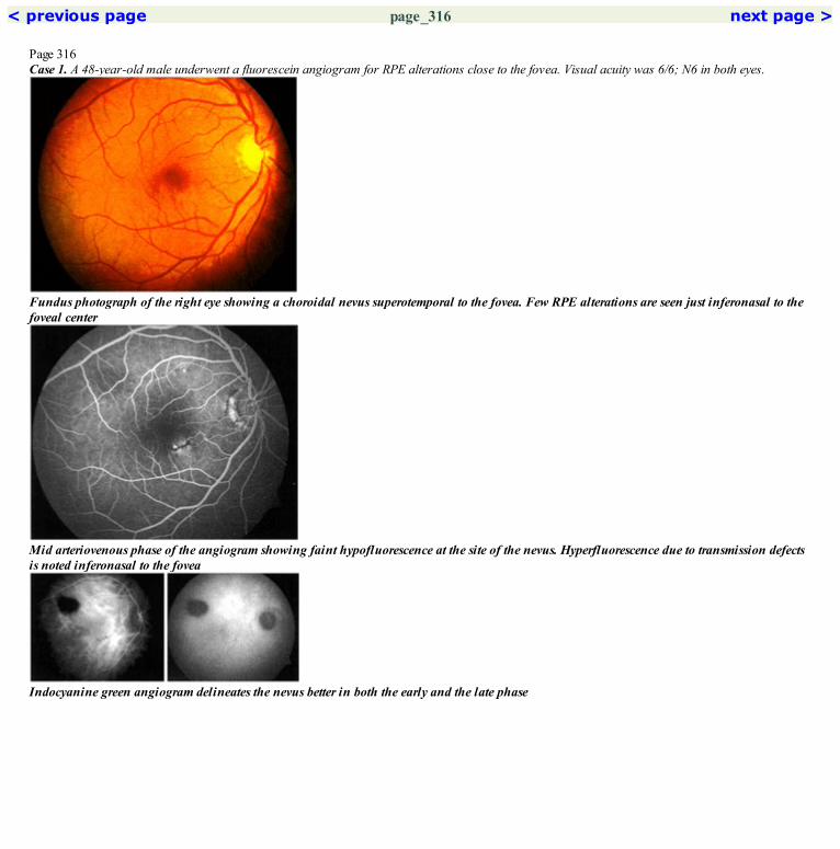
< previous page page_316 next page >
Page 316Case 1. A 48-year-old male underwent a fluorescein angiogram for RPE alterations close to the fovea. Visual acuity was 6/6; N6 in both eyes.
Fundus photograph of the right eye showing a choroidal nevus superotemporal to the fovea. Few RPE alterations are seen just inferonasal to thefoveal center
Mid arteriovenous phase of the angiogram showing faint hypofluorescence at the site of the nevus. Hyperfluorescence due to transmission defectsis noted inferonasal to the fovea
Indocyanine green angiogram delineates the nevus better in both the early and the late phase
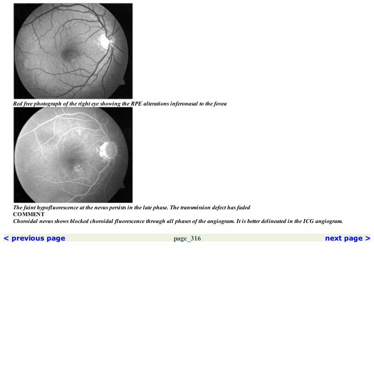
Red free photograph of the right eye showing the RPE alterations inferonasal to the fovea
The faint hypofluorescence at the nevus persists in the late phase. The transmission defect has fadedCOMMENTChoroidal nevus shows blocked choroidal fluorescence through all phases of the angiogram. It is better delineated in the ICG angiogram.
< previous page page_316 next page >

< previous page page_317 next page >
Page 31744Melanocytoma
■ It is a benign tumor arising from the optic nerve head.■ It is said to be derived from the uveal dendritic melanocytes.■ It appears as dark pigmented mass lesion on the optic nerve head.■ Malignant transformation of the melanocytoma is rare.
■ Blocked disc hypofluorescence is seen through all phases of the angiogram.
< previous page page_317 next page >

< previous page page_318 next page >
Page 318Case 1. A 33-year-old female presented with history of reduced vision in the right eye. Visual acuity was 6/12; N6.
Fundus photograph of the right eye showing a typical melanocytoma at the optic nerve head
A blocked hypofluorescence due to the melanocytoma is seen in the early arteriovenous phase. An arteriole is seen coursing over the tumor
Hypofluorescence is seen to persist in the late phase of the angiogram

Red free photograph of the right eye
Arteriovenous phase showing the blocked hypofluorescence at the discCOMMENTThe pigment in the tumor blocks the underlying disc fluorescence through all phases of the angiogram.
< previous page page_318 next page >

< previous page page_319 next page >
Page 31945 Malignant Melanoma
■ Malignant melanoma is the most common primary intraocular tumor.■ It presents as a dark pigmented elevated ovoid mass lesion.■ The degree of pigmentation can vary.■ Some of the tumors may be amelanotic.■ It may assume a mushroom shape if it breaks through the Bruch’s membrane.■ Overlying orange pigmentation may be seen.
■ The fluorescein angiographic picture of a malignant melanoma can vary depending on the size of the tumor and the degree of pigmentation.■ A small melanoma shows mottled hyperfluorescence in the arteriovenous phase. Increased staining of the mass lesion is seen in the late phase ofthe angiogram.■ Larger melanomas may show the filling up of the large tumor vessels (double circulation—simultaneous filling up of the normal retinal vessels andthe tumor vessels).■ Late diffuse staining of the mass lesion is seen.
< previous page page_319 next page >

< previous page page_320 next page >
Page 320Case 1. A 32-year-old male was detected to have a mass lesion in the right eye and referred for further management. Visual acuity was 6/18; N8.
Fundus photograph of the right eye showing a dome shaped, pigmented, subretinal mass lesion in the peripapillary region
Arteriovenous phase of the angiogram showing patchy speckled hyperfuorescence over the mass lesion. The tumor vessels are made out within themass lesionCOMMENTMottled hyperfluorescence in the mass lesion is seen in the early phase of the angiogram. Leakage of the dye from the tumor vessels occurs as theangiogram progresses leading to diffuse staining in the late phase.In less pigmented tumors, both the retinal and tumor vessels are readily evident—this is called double circulation.
< previous page page_320 next page >

< previous page page_321 next page >
Page 32146 Choroidal Hemangioma
■ Choroidal hemangioma is a vascular hamartoma and can either be circumscribed or diffuse.■ Circumscribed choroidal hemangioma is seen as a well defined red-orange, slightly elevated dome shaped lesion usually located at the posteriorpole.■ It may be associated with a serous retinal detachment.■ Histopathologically it may show a cavernous, capillary or a mixed type of a hemangioma. Large, thin walled, blood filled vascular channels areseen. Cystic changes are commonly seen in the outer retinal layers overlying the tumor.
■ Arterial and early arteriovenous phase may show the reticular vascular pattern due to the large choroidal vessels within the tumor.■ The arteriovenous phase shows stippled hyperfluorescence.■ Increased staining and leakage is seen in the late phase of the angiogram.
< previous page page_321 next page >

< previous page page_322 next page >
Page 322Case 1. A 34-year-old male was referred for a mass lesion detected in the left eye. Visual acuity was 6/6; N6.
Fundus photograph of the left eye showing a red orange, minimally elevated mass lesion temporal to the macula. Note extensive pigmentoverlying the mass lesion and a subretinal gliotic band
Arterial phase showing intense hyperfluorescence of the tumor indicating its choroidal origin and vascularity
Late phase angiogram showing leakage of the dye from the tumor vessels

Red free photograph of the left eye
Mid arteriovenous phase showing increase in hyperfluorescence indicating leakage of the dye. Note patches of blocked fluorescence due topigmentCOMMENTChoroidal hemangioma shows early hyperfluorescence with late leakage of the dye and staining of the tumor.
< previous page page_322 next page >

< previous page page_323 next page >
Page 32347 Retinoblastoma
■ It is the most common intraocular malignancy of childhood.■ It can be unilateral or bilateral.■ It starts as a small transparent lesion in the retina. Later on it becomes more opaque with development of tumor vessels.■ The growth of the tumor can be towards the subretinal space (exophytic) or towards the vitreous (endophytic). Rarely it may be diffuse.
■ Early phase shows filling of the tumor vessels with leakage of the dye seen in the later phases.■ Late hyperfluorescence of the tumor is seen.
< previous page page_323 next page >

< previous page page_324 next page >
Page 324Case 1. An 8-year-old child with retinoblastoma was referred for reactivation of the tumor in the right eye. Left eye had been enucleated six year backand he had undergone cryotherapy, radiation and chemotherapy for the tumor in the right eye. Visual acuity was 6/12; N6.
Fundus photograph of the right eye showing chorioretinal atrophy along the superotemporal arcade
Early arteriovenous phase showing the intrinsic vasculature of the tumor vessels and its connection to the retinal circulation
Late phase shows leakage of the dye from the tumor
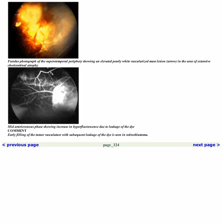
Fundus photograph of the superotemporal periphery showing an elevated pearly white vascularized mass lesion (arrow) in the area of extensivechorioretinal atrophy
Mid arteriovenous phase showing increase in hyperfluorescence due to leakage of the dyeCOMMENTEarly filling of the tumor vasculature with subsequent leakage of the dye is seen in retinoblastoma.
< previous page page_324 next page >

< previous page page_325 next page >
Page 32548 Retinal Angiomatosis
■ It is usually diagnosed in children or young adults.■ It can occur as a solitary tumor without any systemic associations or as a part of the von Hippel-Lindau (VHL) syndrome.■ It appears as a reddish-pink tumor in the retina or on the optic disc.■ Associated feeder vessels are seen.■ It may be associated with extensive fluid and lipid exudation.
■ Capillary hemangioma shows rapid filling of the tumor mass in the early phase of the angiogram.■ Leakage of the dye with intense hyperfluorescence is seen in the late phase.
< previous page page_325 next page >

< previous page page_326 next page >
Page 326Case 1. A 15-year-old male patient presented with history of loss of vision in the left eye for the past three years. He had been diagnosed to have aretinal angioma with exudative retinal detachment in the left eye and had undergone cryotherapy in the past. Visual acuity was 6/5; N6 in the right eyeand no perception of light in the left eye. He was found to have retinal angiomas in the right eye.
Fundus photograph of the right eye showing dilated twin feeder vessels with exudates at the macula
Arterial phase helps to differentiate the feeder arteriole from the venule

The multiple capillary hemangiomas are evident in this montage
Red free photograph of the right eye
Rapid flow through the angioma is reflected in early filling of the draining venule in the arteriovenous phase of the angiogram
Profuse leakage of the dye from the tumor vessels is seen in the late phase of the angiogram
< previous page page_326 next page >

< previous page page_327 next page >
Page 32749 Other TumorsOPTIC NERVE HEMANGIOMACase 1. A 49-year-old male presented with reduced vision in the right eye for the past five years. Visual acuity was 6/60; N36 in the right eye.
Fundus photograph of the right eye showing a orange-red well defined lesion at the disc margin. Hard exudates are seen along theinferotemporal arcade indicating exudation from the tumor
Lesion shows intense hyperfluorescence in the early arteriovenous phase indicating the vascular nature of the lesion
Red free photograph of the right eye

Late arteriovenous phase showing increased area of hyperfluorescence due to leakage of the dye. Note intraretinal dye leakage, presumed to be dueto vascular ‘steal’ by the tumor
< previous page page_327 next page >

< previous page page_328 next page >
Page 328
Late phase angiogram shows dye leakage from the tumor and increased intraretinal dye leakageCOMMENTThe vascular nature of the lesion gives rise to the intense hyperfluorescence seen on the angiogram.RETINAL CAVERNOUS HEMANGIOMACase 2. A 31-year-old female presented with complaints of occasional headache for the past three months. Visual acuity was 6/6; N6 in both eyes.
Fundus photograph of the left eye showing a cluster of grapes like appearing cavernous hemangioma of the optic nerve head
Late phase showing the classical picture of cavernous hemangioma. The dye has filled the upper half of the saccular aneurysms while the lowerhalf remains hypofluorescent

COMMENTThe dark red color of the hemorrhage within the tumor indicates a sluggish circulation. Sedimentation of cellular elements and supernatantserum is seen as a fluid level in each globular lesion. The fluorescein dye enters only the upper supernatant plasma; the sedimented cellularelements in the lower half do not allow fluorescein to percolate, causing it to remain hypofluorescent.
< previous page page_328 next page >
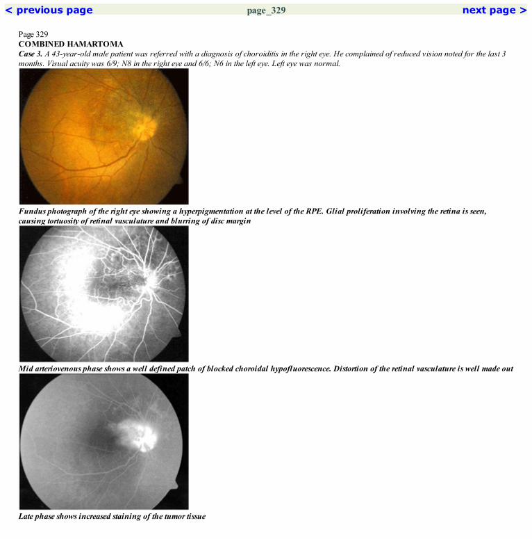
< previous page page_329 next page >
Page 329COMBINED HAMARTOMACase 3. A 43-year-old male patient was referred with a diagnosis of choroiditis in the right eye. He complained of reduced vision noted for the last 3months. Visual acuity was 6/9; N8 in the right eye and 6/6; N6 in the left eye. Left eye was normal.
Fundus photograph of the right eye showing a hyperpigmentation at the level of the RPE. Glial proliferation involving the retina is seen,causing tortuosity of retinal vasculature and blurring of disc margin
Mid arteriovenous phase shows a well defined patch of blocked choroidal hypofluorescence. Distortion of the retinal vasculature is well made out
Late phase shows increased staining of the tumor tissue
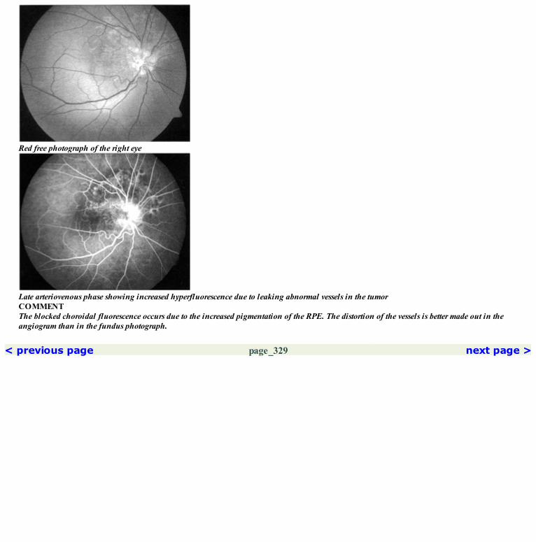
Red free photograph of the right eye
Late arteriovenous phase showing increased hyperfluorescence due to leaking abnormal vessels in the tumorCOMMENTThe blocked choroidal fluorescence occurs due to the increased pigmentation of the RPE. The distortion of the vessels is better made out in theangiogram than in the fundus photograph.
< previous page page_329 next page >

< previous page page_330 next page >
Page 330CLINICAL NUGGET1. Ultrasonography, CT scan and MRI are useful investigative tools in the management of ocular tumors.SELECTED READINGS1. Joffe L, Shields JA, Osher R et al. Clinical and follow-up studies of melanocytomas of the optic disc. Ophthalmology 86:1067–1078, 1979.2. Edwards WC, Layden WE, MC Donald R. Fundus fluorescein angiography of malignant melanoma of the choroid. Am Journal of Ophthalmol.68:797–808, 1969.3. Flindall RJ, Gass JDM. A histopathologic and fluorescein angiographic correlative study of malignant melanomas of the choroid. Can JOphthalmol. 6:258–267, 1971.4. Hayreh SS. Choroidal melanomata. Fundus fluorescein angiography and histopathologic study. Br J Ophthalmol 54:145–160, 1970.5. Sheilds JA, Current approaches to the diagnosis and management of choroidal melanoma. Surv. Ophthalmol 21:443–463, 1977.6. Norton EWD, Gutman F. Fundus fluorescein angiography of Choroidal hemangioma.. Arch Ophthalmol 78:121–125, 1967.7. Smith JL, David NJ, Hart LM et al. Choroidal hemangioma Fundus fluorescein photography and photocoagulation. Arch Ophthalmol 9:51–54,1963.8. Nortan EWD, Smith JL, Curtin VJ. Fundus fluorescein angiography; An aid in differential diagnosis of posterior ocular lesion. Trans AmericanAcad Ophthalmol and Otolaryngol 68; 755–765, 1964.9. Norton EWD, Gutman F. Fundus fluorescein angiography of Choroidal hemangioma.. Arch Ophthalmol 78:121–125, 1967.10. Gass JDM: Fundus fluorescein angiography: An aid in the differential diagnosis of intraocular tumors. Inter Ophthalmol Clin 12; 85–120, 1972.
< previous page page_330 next page >

< previous page page_331
Page 331

GlossaryArterial phase: Appearance of fluorescein dye in the retinal arterioles to its first appearance in the retinal veins.Arteriovenous phase: The stage of fluorescein angiogram when fluorescein dye is present in the retinal arteries and veins; this stage begins withretinal venous laminar filling.Autofluorescence: Preinjection fluorescence produced by lesions that naturally fluoresce when exposed to excitatory blue light.Background fluorescence: A complete sheet of fluorescence beneath the retinal vessels that has a ground glass pattern; it varies according to thefilling pattern of the choroid and density of the retinal pigment epithelium.Barrier filter: A filter used to transmit fluorescein light and prevent transmission of reflected light from the fundus.Blocked fluorescence: A form of hypofluorescence that occurs when tissue or fluid obscures retinal or choroidal vascular fluorescence.Capillary dropout: Non-perfusion of retinal capillaries.Choroidal phase: The stage of fluorescein angiogram when dye first enters the choroidal vasculature. It precedes retinal arterial phase.Collateral vessels: An anastomotic vascular communication that develops in response to vascular obstruction. It joins obstructed vessels to openvessels or bypasses a blocked area in a single vessel.Cystoid edema: Accumulation of dye in discrete spaces in the outer plexiform layer.Exciter filter: An absorption filter that selectively transmits light of the wavelengths needed to cause the fluorescence. It is placed between the lightsource and the fundusFeeder vessel: Inflow channels to an abnormal frond of vessel in the retina or to a tumor.Hot spot: A focal area of intense hyperfluorescence beneath a pigment epithelium detachment indicative of choroidal neovascularisation.Hyperfluorescence: An abnormal increase in fluorescence produced by vascular leakage, vascular changes or window defects.Laminar flow: The fluorescent flow seen along the walls of veins, caused by the slower, nonturbulent movement of blood along the vessel wall.Light bulb fluorescence: A discrete area of hyperfluorescence seen in the late stages of a fluorescein angiogram at the site of a retinalmacroaneurysm.Pooling: Late hyperfluorescence produced by leakage into an anatomical space.Pseudofluorescence: Preinjection fluorescence that emanates from highly reflective surfaces in the fundus, and that is produced by poorly matchedfilters.Red free photo: A photograph using a green filter that eliminates the normal red reflex of the fundus and increases fundus contrast.Staining: Late hyperfluorescence produced by leakage of fluorescein into tissues.Telangiectasia: A retinal vascular disorder consisting of variable degrees of capillary dilatation and aneurysm formation.Transmitted fluorescence: Early hyperfluorescence produced by accentuation of normal choroidal fluorescence through defects in the retinalpigment epithelium (Window defects).Vascular filling defects: Hypofluorescence produced by delay or absence in perfusion of the retinal or choroidal vasculature.
< previous page page_331


