The Second Heart Sound in Atrial Septal Defect SUDARSHAN KUMAR, MB, MRCP, FRCP(C) ALDO A. LUISADA, MD, FACC Chicago, Illinois From the Division of Cardiology (Medi- cine), and the Division of Cardiovascular Research (Physiology) of the Chicago Medi- cal School, University of Health Sciences and the Mount Sinai Hospital Medical Cen- ter. This study was aided by Research Grant HE-09350 and was made during tenure of Undergraduate Training Grant HE-5002 of the National Heart and Lung Institute. U. S. Public Health Service. Bethesda, Md. Address for reprints: Aldo A. Luisada. MD, the Division of Cardiovascular Re- search, the Chicago Medical School, 2020 W. Ogden Ave., Chicago, Ill. 60612. Ventricular and arterial pressure tracings, electrocardiograms and phonocardiograms were studied in 4 normal subjects and 52 patients with atrial septal defect. After excluding cases in which complicating factors were involved, 10 cases of secundum type and 3 cases of primum type were studied in detail, Contrary to previously held beliefs, right ventricular systole is not longer than left ventricular systole in these patients. Therefore, pul- monary valve closure still occurs very close to aortic valve closure, as in normal subjects. However, the incisuras of the large arteries are more widely separated than in normal subjects. The explanation for the wide splitting of the second heart sound is based on recent work that attributes the sound components to vibra- tions of the blood, valves and walls of the large arteries at the time of the aortic and pulmonary arterial rebounds. Patients with atrial septal defect have a dilated pulmonary artery. This causes a slower reaction of the wall and a later occurrence of both the pulmonary incisura and the pulmonary component of the second heart sound. Since the study of Barber et al.,’ wide and “fixed” splitting of the second heart sound has been considered a typical sign of atria1 septal defect. Barber et al. attributed this type of splitting to “right bundle branch block or delay in the emptying time of an overfilled right ventricle.” Subsequent investigators explained the typical electrocardiographic pattern as the result of right ven- tricular hypertrophy. Thus,, the delay of the pulmonary compo- nent was generally attributed to delayed closure of the pulmonary valve, in turn caused by delayed ending of right ventricular sys- tole as a result of prolonged ejection time. Observation of a typical case of atria1 septal defect in which the 2 ventricles terminated systole at the same time induced US to study this problem in all our cases of atria1 septal defect. Material and Method Fifty-six patients were studied. They included 48 patients with the “septum secundum” type of atria1 septal defect, 4 with the “septum primum” type and 4 normal subjects used as controls. All patients underwent a detailed physical examination ; an electrocardiogram, chest roentgenogram, right and left cardiac catheterization and angiocardio- graphic studies were performed. The diagnosis of atria1 septal defect was accepted on cardiac cathe- terization by passage of the catheter across the defect plus (1) signifi- cant step-up in blood oxygen saturation at the atria1 level; and (2) demonstration of a shunt by either an angiocardiogram or indicator- dilution curve, or both. Isa no Amdcan Journal of CARPIOLOOY
Transcript
The Second Heart Sound in Atrial Septal Defect
SUDARSHAN KUMAR, MB, MRCP, FRCP(C)
ALDO A. LUISADA, MD, FACC
Chicago, Illinois
From the Division of Cardiology (Medi- cine), and the Division of Cardiovascular Research (Physiology) of the Chicago Medi- cal School, University of Health Sciences and the Mount Sinai Hospital Medical Cen- ter. This study was aided by Research Grant HE-09350 and was made during tenure of Undergraduate Training Grant HE-5002 of the National Heart and Lung Institute. U. S. Public Health Service. Bethesda, Md.
Address for reprints: Aldo A. Luisada. MD, the Division of Cardiovascular Re- search, the Chicago Medical School, 2020 W. Ogden Ave., Chicago, Ill. 60612.
Ventricular and arterial pressure tracings, electrocardiograms and phonocardiograms were studied in 4 normal subjects and 52 patients with atrial septal defect. After excluding cases in which complicating factors were involved, 10 cases of secundum type and 3 cases of primum type were studied in detail,
Contrary to previously held beliefs, right ventricular systole is not longer than left ventricular systole in these patients. Therefore, pul- monary valve closure still occurs very close to aortic valve closure, as in normal subjects. However, the incisuras of the large arteries are more widely separated than in normal subjects.
The explanation for the wide splitting of the second heart sound is based on recent work that attributes the sound components to vibra- tions of the blood, valves and walls of the large arteries at the time of the aortic and pulmonary arterial rebounds.
Patients with atrial septal defect have a dilated pulmonary artery. This causes a slower reaction of the wall and a later occurrence of both the pulmonary incisura and the pulmonary component of the second heart sound.
Since the study of Barber et al.,’ wide and “fixed” splitting of the second heart sound has been considered a typical sign of atria1 septal defect. Barber et al. attributed this type of splitting to “right bundle branch block or delay in the emptying time of an overfilled right ventricle.” Subsequent investigators explained the typical electrocardiographic pattern as the result of right ven- tricular hypertrophy. Thus,, the delay of the pulmonary compo- nent was generally attributed to delayed closure of the pulmonary valve, in turn caused by delayed ending of right ventricular sys- tole as a result of prolonged ejection time.
Observation of a typical case of atria1 septal defect in which the 2 ventricles terminated systole at the same time induced US to
study this problem in all our cases of atria1 septal defect.
Material and Method
Fifty-six patients were studied. They included 48 patients with the “septum secundum” type of atria1 septal defect, 4 with the “septum primum” type and 4 normal subjects used as controls. All patients underwent a detailed physical examination ; an electrocardiogram, chest roentgenogram, right and left cardiac catheterization and angiocardio- graphic studies were performed.
The diagnosis of atria1 septal defect was accepted on cardiac cathe- terization by passage of the catheter across the defect plus (1) signifi- cant step-up in blood oxygen saturation at the atria1 level; and (2) demonstration of a shunt by either an angiocardiogram or indicator- dilution curve, or both.
Isa no Amdcan Journal of CARPIOLOOY
In all patients, phonocardiograms, apex cardiogram and external carotid arteriograms were recorded on a Sanborn multichannel recorder. The paper speed was 100 mm/set with time lines separated by an int’erval of 40 msec.
Selection of Cases
To avoid complicating causes for either the splitting of the second sound or the duration of ventricular sys- toles, we excluded patients who had any of the follow- ing conditions : (1) complete right bundle branch block; (2) pulmonary hypertension ; (3) obstruction to the right ventricular outflow tract (pulmonary valvular or infundibular stenosis) ; (4) clinical or hemodynamic evidence of right or left ventricular failure; or (5) any significant degree of arterial desaturation. Also excluded were patients with a small shunt and a few cases in which the pressure curves of the right ven- tricle, left ventricle, pulmonary artery or aorta were marred by artifacts. All subjects had normal sinus rhythm.
Atria1 septal defect (secundum type): Of 48 pa- tients with an ostium secundum defect, 10 were even- tually selected on the basis of the criteria cited. Their ages ranged from 14 to 59 years ; 8 were female and 2 were male. All had a moderate to large left to right atria1 shunt. One patient (Case 6) with a pulmonary to systemic flow ratio of 1.5: 1 was included in this group because she had a very wide splitting of the second heart sound.
Septum primum defect : The 3 patients included in this group ranged in age from 6 to 24 years; 2 were male, and 1 female. In addition to the presence of all the cited criteria, significant mitral insufficiency was revealed by both an apical panysystolic murmur and the results of left ventricular angiocardiography.
Normal subjects: Four patients ranging in age from 13 to 26 years were studied ; 2 were male, and 2 female. None had either congenital or acquired heart
SECOND HEAR1 SOUND IN ATRIAL SEPTAL DEFECT
i
ECG / /
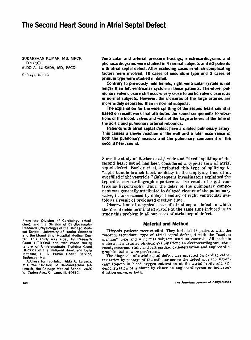
Figure 1. Pressure tracings of the 2 ventricles in a normal subject (same amplification). The right ventricle terminates its systole 35 msec after the left in the first cycle and 20 msec after the left in the second cycle. IDLV = first deriva- tive of the left ventricular pressure; LV = left ventricular pressure; PCG z phonocardiogram; RV = right ventricular pressure.
disease; their study was prompted by the observation of a systolic murmur over the pulmonary area.
Measurements
An average of the measurements of 5 complexes was made by studying ventricular and arterial pressure tracings. Whenever available, simultaneous tracings (ventricular or arterial) were studied. Otherwise, care was taken that the measurements compared had been obtained at periods with identical heart rate.
Q-end RV to Q-end LV: The end of each ventricu- lar systole is marked by a sharp downward turn of the ventricular pressure curve (Fig. 1 and 2). By compar-
ID --i RV
Figure 2. Case 9. Phonocardiogram and pressure tracings in atrial septal defect (septum secundum type). Left, upper tracing shows phonocardiogram at apex; middle tracing, phonocardiogram at base. Right, symbols as in Figure 1. Compari- son of phonocardiogram with first derivative of right ventricular pressure, electro- cardiogram, and pressures of the 2 ventricles. Right ventricular pressure is more amplified than left ventricular pressure.
insp. = during inspiration; LV = left ventricular; RV = right ventricular.
ing the values for the end of right and left ventricular systole (Q-RV, Q-LV), one can ascertain if one ven- tricle ends its contraction later than the other, thus causing a delay in the closure of the respective semi- lunar valve.
IIA-IIP interval: The interval between the onset of the aortic component (IIA) and the onset of the pul- monary component (IIP) of the second heart sound is used to measure the degree of splitting of this sound. In normal subjects, this interval was determined dur- ing inspiration, thus giving the greatest possible de- gree of splitting. In atria1 septal defect, no attention was paid to the respiratory phase, which was considered of minor importance because of the “fixed” splitting.
Aortic-pulmonary pulse incisuras: The interval between these incisuras is used to measure the interval between the onsets of the rebounds in the 2 main ar- teries.
Q-HA, actual and predicted : Q-HA is used to mea- sure the actual interval between the Q wave and the onset of the aortic component of the second heart sound. This interval was compared with the predicted interval for normal subjects, based on Shah and Slodki’s formula,* which includes heart rate and a constant.
Q-IIP, actual and predicted: The same calcula- tions were made for the pulmonary component.
Results Normal subjects: The 4 young people included
in our study (Table I) were between the ages of 13 and 26 years (average 17.2 years). The interval between the end of left ventricular systole (Q-LV) and right ventricular systole (Q-RV) averaged 23 msec (range 20 to 36 msec). An example of the longest interval is shown in Figure 1.
The interval between the aortic (A) and pul- monary (P) components of the second heart sound at the end of inspiration averaged 45 msec (30 to 50 msec) and was much narrower during expira- tion.
Comparing the Q-HA and Q-IIP intervals with the predicted figures according to the formulas of Shah and Slodki,* we found a close correlation; the minor differences were probably related to the younger age of our subjeds.
3801375 430/403 Moderate Ml 390/369 450/402 Mild MI 3601354 400/389 Severe Ml
376/366 + 10 4261398 + 28
The average interval between the incisuras of the aortic and pulmonary arterial pulsations was 47.5 msec.
Patients with atria1 septal defect (secundum type) : The average age of these patients was 31.3 years (14 to 55) (Table II). They had an average ratio between pulmonary and systemic flow of 2.3: 1 (minimal 1.5: 1; maximal 4:l). In 4 patients right ventricular systole ended slightly after the left (+20 to +30 msec) but was within physiologic limits; in 2 patients left ventricular systole ended after the right; in 4 patients right and left ventricular systole ended simultaneously (Fig. 2). The average values for Q-end RV and Q-end LV were identical, indicating no prolonga- tion of right ventricular systole.
The interval between the aortic (A) and pul- monary (P) components of the second heart sound ranged from 40 to 75 msec ; the average value was 67.5 msec, which is definitely longer than the nor- mal average. The average interval between aortic and pulmonary incisuras was 66.6 (60 to 80 msec), which is very close to the value for the A-P inter- val and is longer than in normal subjects.
Comparison of the actual with the predicted values for Q-IIA and Q-IIP shows that the Q-IIA interval was slightly prolonged (+26) and the Q-IIP interval was markedly prolonged (+4i) over the predicted values.
Patients with atria1 septal defect (primum type) : In our 3 cases, the average age was 19.3 years and the average ratio of pulmonary to aortic flow was 2.1: 1 (Table III).
There was a minimal average interval between the end of left ventricular and the end of right ventricular systole (13 msec). This difference was within normal limits ; however, it should be noted that left ventricular systole ended after the right in the only patient with severe mitral insufficiency. The A-P interval was somewhat shorter than in the secundum type of defect (50 msec), possibly because of the younger age of the patients. The Q-IIA interval was close to the expected interval,
but the Q-IIP interval was prolonged, although less than in the secundum type.
Discussion
After the description of Barber et al,1 who at- tributed the wide splitting of the second heart sound to either right bundle branch block or pro- longed ejection of the right ventricle, many inves- tigators studied this problem and excluded the first possibility. Thus, some author@ attributed the wide splitting of the second heart sound to greater stroke volume of the right ventricle causing a longer duration of right ventricular systole and delayed closure of the pulmonary valve. On the other hand, Castle9 questioned the prolongation of right ventricular systole although he did not ex- clude it.
In the present study, the normal subjects were observed in order to make a direct comparison with our clinical cases. In these normal subjects, right and left ventricular systole ended very close to each ‘other, although with a minor delay of the right in relation to the left ventricle.
Patients with secundum type of atria1 septal defect were the main object of our study. Table II shows that right ventricular systole not only did not end after the left, but also, was on average, even closer to that of the left ventricle than in normal subjects. This did not surprise us because, according to an experimental study of Wallace and co-workers,“’ an increase in stroke volume slightly prolongs the ejection period, abbreviates the tension period and does not prolong the total duration of systole.
Closure of the semilunar valves ‘occurs very shortly after the end of ventricular systole. There- fore, we have to assume that no delay in pulmonary valve closure occurs in uncomplicated atria1 septal defect. This statement may seem paradoxical in the light of an older theory that considers the sound component to be caused by and coincident with valve closure. However, we have developed a
VOLUME 28. AUOUST 1971 171
KUMAR AND LUISADA
newer theory in our laboratory. The sound compo- longer than normal in all subjects in whom it could nent (aortic or pulmonary, respectively) occurs at be studied. It was equal to the A-P interval in 3 the time of the incisura of the large artery, is in- cases and longer in the other 3. This can be ex- timately connected with the flow and pressure re- plained by the fact that, in some cases, the pul- bound over the closed valve, and is caused by vi- monary arterial catheter was pushed close to the bration of the semilunar valve, the infundibulum bifurcation so that an additional delay was caused and the root of the artery (MacCanon et al.,** Mori by transmission of vibrations from the arterial et a1.12 and Luisadals). root to the catheter tip.
The distance between end of ventricular systole (,and valve closure) and incisura (and sound com- ponent) is longer for the pulmonary artery than the aorta in normal subjects and is affected by vascular factors. A dilated pulmonary artery with normal pressure has a lower compliance than the aorta and causes a late rebound over the closed pulmonary valve and thus a late pulmonary com- ponent compared to the aortic component of the second sound in atria1 septal defect. It also ac- counts for the increased delay that occurs with age” because dilatation of the pulmonary artery probably progresses with age. It may also explain the wide splitting observed in idiopathic dilatation of the pulmonary artery, as well as the frequent persistence of the wide splitting in cases of atria1 septal defect for several months (or even years) after surgical repair.
The subjects with primum type of atria1 septal defect were studied separately because of the pos- sible complicating influence of the coexisting mitral insufficiency. Tn these cases, left ventricular systole may have been slightly shortened, as in other cases of mitral insufficiency. The average in- terval between left and right ventricular systole, however, was very short (13 msec) and still within normal limits. The only patient with severe mitral insufficiency (Case 3) actually had longer left ven- tricular systole in comparison with the right while having the shortest interval between the aortic and pulmonary components.
The interval between the 2 incisuras was much
The present study did not try to explain the rea- son for the absent or minor respiratory changes of the interval between the aor.tic and pulmonary components because current explanations, based on the communication between the 2 atria, seem adequate.
Brit Heart J 12:277-292, 1950 2. Shah PM, Skxlki SJ: The Q-II interval. Circulation 29:
551-561, 1964 3. Leatham D, Gray I: Auscultatoty and phonocardio-
graphic signs of atrial septal defect. Brit Heart J 18:193- 208. 1956
4. Perloff JK, Harvey WP: Mechanisms of fixed splitting of the second heart sound. Circulation 18:998-1009, 1958
5. Shafter HA: Splitting of the second heart sound. Amer J Cardiol 6:1013-1022, 1960
6. Aygen MM, Braunwald E: The splitting of the second heart sound in normal subjects and in patients with con- genital heart disease. Circulation 25:328-345. 1962
7. Braunwald E: Physiological principles regulating the timing of closure of the heart valves. In, The Theory and Practice of Auscultation (Segal B, ed). Philadelphia, FA Davis, 1964, p 113-116
8. Braunwald E, Aygen MM, Ross J.: Auscultatory and
phonocardiographic findings in atrial septal defect. In Ref 7, p 183-199
9. Castle RF: Variables affecting the splitting of the sec- ond heart sound in atrial septal defect. Amer Heart J 73468-473, 1967
10. Wallace AG, Mitchell JH, Skinner NS, et al: Duration of the phases of left ventricular systole. Circ Res 12:611- 619, 1963
11. Ma&anon DM, Arevalo F, Meyer EC: Direct detection and timing of aortic valve closure. Circ Res 14:387-391, 1964
12. Mori M, Shah PM, MacCanon DM, et al: Hemodynamic correlates of the various components of the second heart sound. Cardiologia (Basel) 44:65-77, 1964
13. Luisada AA: Phonocardiography: a dynamic interpreta- tion of the normal and abnormal cardiac vibrations. In, Clinical Cardiopulmonary Physiology, third edition (Gor- don BL, Carleton RA, Faber PL, ed). New York, Grune & Stratton, 1970, p 122-139