19
The Senses “Sights and Sounds”
| Date post: | 27-Dec-2015 |
| Category: |
Documents |
| Upload: | bryan-bennett |
| View: | 218 times |
| Download: | 0 times |

The Senses
“Sights and Sounds”

Anatomy of External Eye• Eyes protected by eyelids,
which meet at canthus• Eyelashes at borders• Tarsal glands – secrete oil that
lubricates eyes• Conjuctiva lines the eyelids and
secretes mucus to lubricate eyeball
• Lacrimal glands secrete tears (salt solution)

Anatomy of Inner Eye• Three Layers:
1. Fibrous Layer:• Sclera – white part• Cornea – transparent
2. Vascular Layer:• Choroid – nutritive tunic• Ciliary Body – lens attached
by suspensory ligament• Iris – pigmented region,
regulates amount of light• Pupil – light passes thru
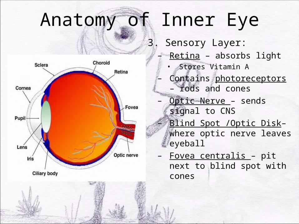
Anatomy of Inner Eye3. Sensory Layer:– Retina – absorbs light • Stores Vitamin A
– Contains photoreceptors – rods and cones
– Optic Nerve – sends signal to CNS
– Blind Spot /Optic Disk– where optic nerve leaves eyeball
– Fovea centralis – pit next to blind spot with cones
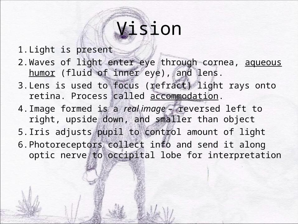
Vision1. Light is present2. Waves of light enter eye through cornea, aqueous
humor (fluid of inner eye), and lens.3. Lens is used to focus (refract) light rays onto retina.
Process called accommodation. 4. Image formed is a real image – reversed left to right,
upside down, and smaller than object5. Iris adjusts pupil to control amount of light6. Photoreceptors collect info and send it along optic
nerve to occipital lobe for interpretation
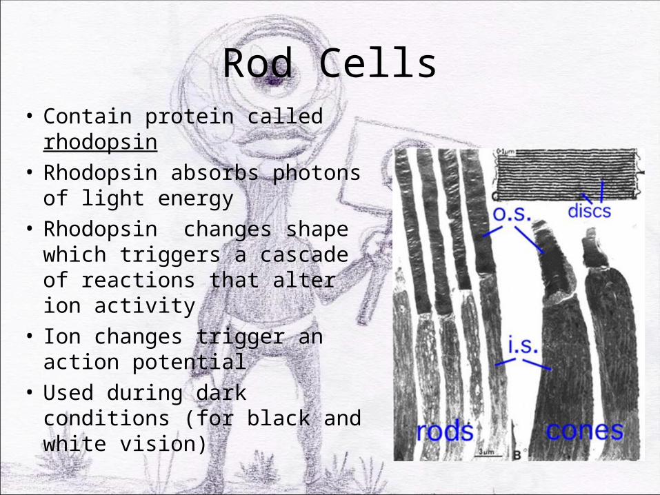
Rod Cells• Contain protein called
rhodopsin• Rhodopsin absorbs photons of
light energy• Rhodopsin changes shape
which triggers a cascade of reactions that alter ion activity
• Ion changes trigger an action potential
• Used during dark conditions (for black and white vision)

Cone Cells• Responsible for seeing in color.
(Interpretation of color occurs in brain not in retina)
• Three varieties of cones, each one sensitive to a particular wavelength of visible light: blue, green, and red.
• When all three are stimulated, we see white.
• Color blindness – occurs when person lacks cones of certain type, or all three.

Eye Health• Emmetropia – eye that focuses images correctly on
retina• Myopia – nearsightedness. Light fails to reach retina• Hyperopia – farsightedness. Light focused behind
retina.• Astigmatism – unequal curvatures in lens/cornea• Glaucoma – pressure within eye when aqueous
humor is blocked, leads to compression of nerve• Cataracts – hardened lens, leads to hazy vision

Left Eye
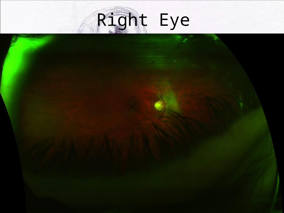
Right Eye

DSAEK - Descemet's Stripping Automated Endothelial Keratoplasty
http://www.youtube.com/watch?v=yOh_4hG8gJw

Anatomy of the Ear
• Outer Ear:• Auricle / pinna – ear• External acoustic meatus /
auditory canal – chamber into skull
• Ceruminous glands – secrete cerumen (earwax)
• Tympanic membrane - eardrum
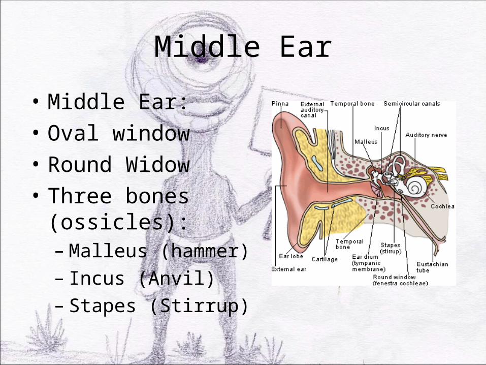
Middle Ear
• Middle Ear:• Oval window• Round Widow• Three bones (ossicles):– Malleus (hammer)– Incus (Anvil)– Stapes (Stirrup)

Inner Ear
• Osseus Labyrinth –– Cochlea – spiraling
structure– Vestible– Semicircular canals
• Filled with perilymph
Hearing
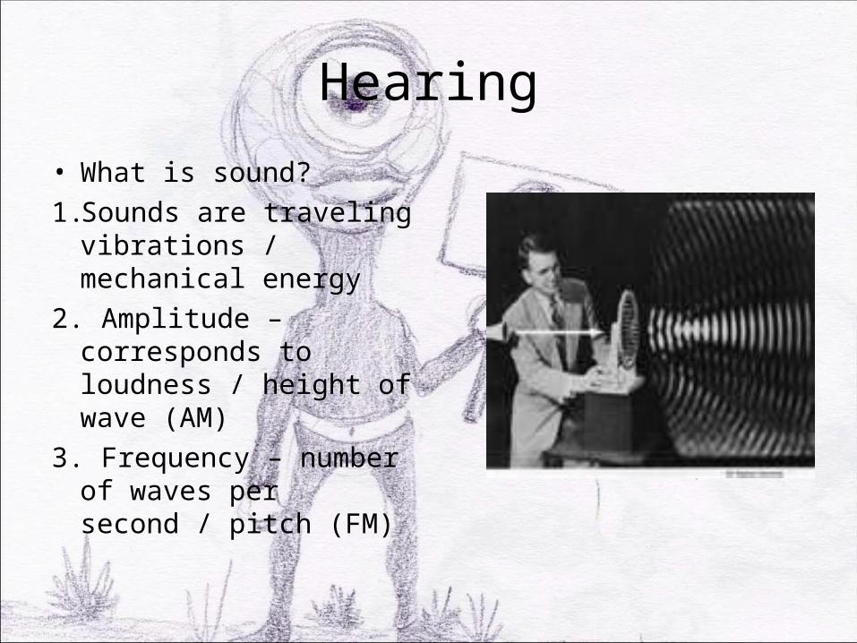
• What is sound?1. Sounds are traveling
vibrations / mechanical energy
2. Amplitude – corresponds to loudness / height of wave (AM)
3. Frequency – number of waves per second / pitch (FM)
Sensory Reception1. External flaps of outer ear collect sound waves and
direct them into auditory canal to ear drum2. Sounds waves make drum vibrate. Bones of middle
ear pick up vibrations and amplify stimulus. Moves waves of pressure to oval window (entrance to inner ear)
3. Oval window vibrations move fluids within inner ear. Causes membrane in cochlea to vibrate and sort out frequencies.
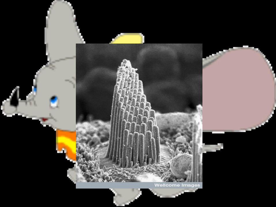
4. Vibrations then move hair cells that when bent, generate action potential. Send stimulus along auditory nerve (VIII) to brain.
Hearing
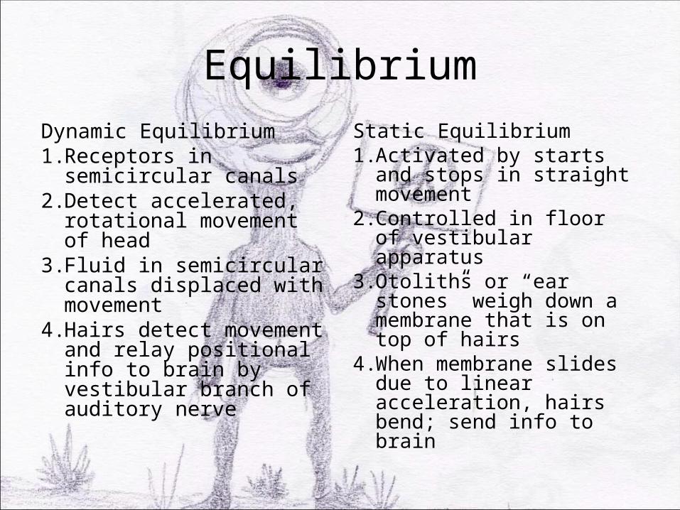
EquilibriumDynamic Equilibrium1. Receptors in semicircular
canals2. Detect accelerated,
rotational movement of head
3. Fluid in semicircular canals displaced with movement
4. Hairs detect movement and relay positional info to brain by vestibular branch of auditory nerve
Static Equilibrium1. Activated by starts and
stops in straight movement2. Controlled in floor of
vestibular apparatus3. Otoliths or “ear stones”
weigh down a membrane that is on top of hairs
4. When membrane slides due to linear acceleration, hairs bend; send info to brain

Physiology of Balance


