61
THE SEPSIS MANUAL Responsible management of sepsis, severe infection and antimicrobial stewardship. 4th edition 2017 - 2018
THE
SEP
SIS
MA
NU
AL
Responsible management of sepsis, severe infection and antimicrobial stewardship.
4th edition 2017 - 2018
2 3
THE SEPSISMANUAL4th edition 2017 - 2018
Edited by: Dr Ron Daniels and Professor Tim Nutbeam
Contributing authors:
• Dr Ron Daniels BEM - Chief Executive of the UK Sepsis
Trust and Global Sepsis Alliance
• George McNamara
• Dr Tim Nutbeam
• Samantha Fox
• Verity Sangan
• Sian Annakin
• Dr Emma Joynes
• Dr Natasha Ratnaraja
• Larry Matthews
• Dr Viral Thakerar
Publisher: United Kindgom Sepsis Trust
Published by:
United Kingdom Sepsis Trust
Level 2, 36 Bennetts Hill
Birmingham
B2 5SN
Email: [email protected]
Website: sepsistrust.org
United Kingdom Sepsis Trust is a Registered Charity: 1146234
The United Kingdom Sepsis Trust is for people who want to help fix the way sepsis is dealt with
by the NHS. We combine clinical expertise and comprehensive practical toolkits with the right
people to help save lives. Unlike other charities that focus on commissioning expensive
research and magic bullets, we act directly to help the public and the NHS see sepsis as a
medical emergency; planning and designing systems to deliver better care.
2017 United Kingdom Sepsis Trust
All rights reserved. No part of this book may be stored in a retrieval system or reproduced in
any form whatsoever without prior permission in writing from the publisher. This book is sold
subject to the condition that it shall not, by trade or otherwise, be lent, resold, hired out or
otherwise circulated without the publishers prior permission in any form of binding or cover
other than that in which published, and without a similar condition including this condition
being imposed on the subsequent purchaser.
ISBN: 978-0-9928155-0-9

4 5
In 2017, the World Health Assembly, the decision-making body of the World Health Organisation (WHO), adopted a resolution to improve the prevention, diagnosis and management of sepsis.
This resolution marks a new era in our fight against sepsis. All 194 member states of the United Nations will now need to develop national action plans against the condition, which is one of our most prolific killers.
Often the final common pathway to death from infection, sepsis claims an almost unbelievable six million lives each year worldwide, and this estimate is likely to grow as we improve our understanding and measurement. Many of these fatalities are in children, particularly in low and middle income countries.
In the United Kingdom, there are more than 250,000 episodes of sepsis annually, with at least 44,000 people dying as a result. Sepsis claims more lives than breast, bowel and prostate cancer put together, but until recently, few had heard of it.
We need to work hard to reduce the many thousands of available deaths from sepsis. In the context of the rising threat of antimicrobial resistance, however, we must do so responsibly. Antimicrobials must be preserved for the sickest patients, and used correctly- otherwise we risk the very real threat of being unable to treat our patients in the future.
We have come a long way since I, and others around the world, started this fight a number of years ago. We understand sepsis better, we have designed effective clinical systems around it, we have secured commissioning for better care, and in some countries (including the UK) these steps have resulted in gradual reductions in mortality rates.
But we have a long way to go. To achieve our dream of preventing any avoidable death from sepsis, we’ll need continued effort from governments, policy makers, professional bodies, the public, the media – and from you. I hope that this manual will mark the start, or begin a new and reinvigorated phase, of your fight against sepsis, because this involves every one of us.
With very best wishes
Dr Ron Daniels B.E.M, FFICM, FRCA, FRCP(Ed)CEO - Global Sepsis AllianceCEO - UK Sepsis Trust
FOREWORDI commend member states for the content of the resolution on sepsis which points to the key actions needed to start reversing these shocking statistics. Some successful examples of turning the recommendations into reality at country level already exist, and should be highlighted and used as models. WHO is committed to strongly supporting countries’ and stakeholders’ improvement efforts in the area of sepsis.
– Dr Margaret Chan, Director General of the World Health Organisation (WHO)“
CON
TEN
TS
THE BURDEN OF SEPSIS AND SEVERE INFECTION
DEFINING SEPSIS
SOURCES OF INFECTION
THE PATHOPHYSIOLOGY OF SEPSIS
THE SEPSIS 6
ONGOING CARE
SPECIAL PATIENT GROUPS
MICROBIOLOGY
HUMAN FACTORS
AFTER SEPSIS - SURVIVOR ISSUES
UK & GLOBAL POLITICS
06
1230
40
54808693100104108

7
THE
BU
RD
EN O
F SE
PSI
SA
ND
SEV
ERE
INFE
CTIO
N
Accurate record keeping is a vital part of good clinical practice. What we write in the notes affects not only the care of the individual patient, but also coding. In turn, coding affects, for example, how much a hospital gets paid; and more importantly our broader societal understanding of clinical and public health issues.
If we write in the notes ‘possible sepsis’, or ‘? sepsis’ and no one subsequently confirms the clinical diagnosis, the patient will not be coded as having sepsis even if they end up on Intensive Care with multi-organ failure as a result.
So, if you think sepsis, remember to say ‘sepsis’, write ‘Diagnosis: sepsis’ or ‘Δ sepsis’, and assess and record the level of severity, or acuity. More about this below, but remember, coding matters!
ESTIMATING THE BURDEN OF SEPSIS & SEVERE INFECTION01
Sepsis and severe infection are one of the most common reasons for admission to hospital, and perhaps the most common cause of inpatient deterioration.
Whilst this statement might well be true, and other than knowing that it is a significant issue, the reality is that we don’t truly understand the burden of sepsis. This introductory chapter will start by describing how we use the best available data and how these data are sense checked against data from other sources to estimate: i) How many cases of sepsis we see each year across the United Kingdom ii) How many people die as a result of sepsisiii) The economic burden to our healthcare system and to the wider economy
Across each country, hospital coded data are collected at national level in order to examine disease trends and inform policy and commissioning of healthcare. Whilst efficient, there are a number of issues with this approach with respect to sepsis:
1. Such administrative data collect ‘episodes’ of care, which is not necessarily the same as the number of people affected (one person might have two or three episodes of sepsis in a given year). This issue will tend to overestimate the number of people affected.
2. The ‘codes’ with which the data are derived tend to lag behind clinical terminology and practice. We use International Classification of Diseases (ICD) as our main coding source, currently in its 10th iter-ation. Since ICD was last updated, international definitions and descriptors of sepsis (see below) have changed. Whilst coders attempt to embrace such changes by writing and updating coding ‘rules’, such a system is never perfect. This issue will tend to underestimate the number of people affected.
3. Codes are only ever as good as the words we write in the notes! Coders stick to strict rules, and cannot make a diagnosis someone has missed or written incorrectly. A 2015 report, ‘Just Say Sepsis’, by the National Confidential Enquiry into Patient Outcome and Death (NCEPOD) found that, where patients with sepsis had died, it was only recorded on the death certificate in 40% of cases. This will tend to underestimate the number of people affected.
CLINICAL PRACTICE TIP
8 9
NUMBER OF DEATHS03
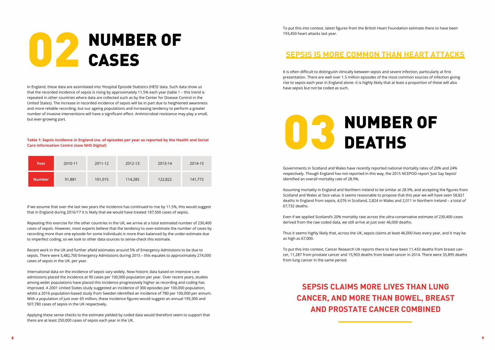
To put this into context, latest figures from the British Heart Foundation estimate there to have been 193,450 heart attacks last year.
It is often difficult to distinguish clinically between sepsis and severe infection, particularly at first presentation. There are well over 1.5 million episodes of the most common sources of infection giving rise to sepsis each year in England alone- it is highly likely that at least a proportion of these will also have sepsis but not be coded as such.
SEPSIS IS MORE COMMON THAN HEART ATTACKS
SEPSIS CLAIMS MORE LIVES THAN LUNG CANCER, AND MORE THAN BOWEL, BREAST
AND PROSTATE CANCER COMBINED
Governments in Scotland and Wales have recently reported national mortality rates of 20% and 24% respectively. Though England has not reported in this way, the 2015 NCEPOD report ‘Just Say Sepsis’identified an overall mortality rate of 28.9%.
Assuming mortality in England and Northern Ireland to be similar at 28.9%, and accepting the figures from Scotland and Wales at face value, it seems reasonable to propose that this year we will have seen 58,821 deaths in England from sepsis, 4,076 in Scotland, 2,824 in Wales and 2,011 in Northern Ireland – a total of 67,732 deaths.
Even if we applied Scotland’s 20% mortality rate across the ultra-conservative estimate of 230,400 cases derived from the raw coded data, we still arrive at just over 46,000 deaths.
Thus it seems highly likely that, across the UK, sepsis claims at least 46,000 lives every year, and it may be as high as 67,000.
To put this into context, Cancer Research UK reports there to have been 11,433 deaths from breast can-cer, 11,287 from prostate cancer and 15,903 deaths from bowel cancer in 2014. There were 35,895 deaths from lung cancer in the same period.
NUMBER OF CASES02
In England, these data are assimilated into ‘Hospital Episode Statistics (HES)’ data. Such data show us that the recorded incidence of sepsis is rising by approximately 11.5% each year (table 1 – this trend is repeated in other countries where data are collected such as by the Center for Disease Control in the United States). The increase in recorded incidence of sepsis will be in part due to heightened awareness and more reliable recording, but our ageing populations and increasing tendency to perform a greater number of invasive interventions will have a significant effect. Antimicrobial resistance may play a small, but ever-growing part.
Year 2010-11 2011-12 2012-13 2013-14 2014-15
Number 91,881 101,015 114,285 122,822 141,772
Table 1: Sepsis incidence in England (no. of episodes per year as reported by the Health and Social Care Information Centre (now NHS Digital)
If we assume that over the last two years the incidence has continued to rise by 11.5%, this would suggest that in England during 2016/17 it is likely that we would have treated 187,500 cases of sepsis.
Repeating this exercise for the other countries in the UK, we arrive at a total estimated number of 230,400 cases of sepsis. However, most experts believe that the tendency to over-estimate the number of cases by recording more than one episode for some individuals is more than balanced by the under-estimate due to imperfect coding, so we look to other data sources to sense-check this estimate.
Recent work in the UK and further afield estimates around 5% of Emergency Admissions to be due to sepsis. There were 5,482,700 Emergency Admissions during 2015 – this equates to approximately 274,000 cases of sepsis in the UK. per year.
International data on the incidence of sepsis vary widely. Now historic data based on intensive care admissions placed the incidence at 90 cases per 100,000 population per year. Over recent years, studies among wider populations have placed this incidence progressively higher as recording and coding has improved. A 2001 United States study suggested an incidence of 300 episodes per 100,000 population, whilst a 2016 population-based study from Sweden identified an incidence of 780 per 100,000 per annum. With a population of just over 65 million, these incidence figures would suggest an annual 195,300 and 507,780 cases of sepsis in the UK respectively.
Applying these sense checks to the estimate yielded by coded data would therefore seem to support that there are at least 250,000 cases of sepsis each year in the UK.

10 11
CONCLUSIONWhilst we have improved our recording of the number of cases of sepsis and understand better its impact on the NHS and society, we still have to estimate figures based on the best available data.
Conservative estimates would suggest that we see at least 250,000 cases of sepsis in the UK each year, with at least 46,000 deaths and a direct cost to the NHS of at least £1.5 billion. Sepsis costs our society as much as £15.6 billion every year. It is highly likely that these numbers are significant under-estimates, since a proportion of the more than 1.5 million patients suffering severe infection in England every year are likely to have uncoded sepsis.
Whichever way we cut it, sepsis is huge.
THE COST OF SEPSIS04
In 2017, the UK Sepsis Trust commissioned an independent piece of work from the York Health Economics Consortium (YHEC) to estimate the cost burden of sepsis to the NHS, and to our wider economy.
YHEC estimated direct costs to the NHS based upon the use of consumables, drugs, clinical time and bed days in hospital, together with the need for rehabilitation, ongoing organ support and other access to healthcare. The group also estimated indirect costs, primarily due to lost productivity but also in litigation.
Clearly, if a patient has died because of sepsis, they are unable to return to productive life, and they will not be able to pay taxes. However, the same might be true for survivors. We know, for example, that 22% of survivors of sepsis who have needed Intensive Care have post-traumatic stress disorder; and that 17% of survivors have moderate-to-severe cognitive decline. Even if we do save a life, and particularly if we delay diagnosis and treatment, the burden of survival might mean that sufferers are unable to return to work at their previous level of function, if at all.
YHEC estimated, given that there are at least 250,000 cases of sepsis every year, that sepsis costs the NHS between £1.5 and £2 billion each year, and our wider economy at least £11 billion and possibly as high as £15.6 billion.
To put this into context, the Asthma UK Centre in Applied Research estimates the cost to the NHS of treating Asthma to be £1.1 billion.
SEPSIS COSTS THE NHS MORE THAN ASTHMA

12 13
INTRODUCTIONThe definition of sepsis has changed over time, and will continue to do so. These changes have, at times, created confusion, but it is hoped that from the time of writing there will be a period of stability for some years while we continue to advance improvements in clinical systems.
There are various purposes to a definition for any condition, including:
• The use of a common language to improve communication between health professionals, and between healthcare and its patients
• The use of language suitable to educate the public about the condition• The establishment of criteria and thresholds beyond which intervention is recommended• Provision of criteria to determine eligibility for inclusion in a clinical trial.
Often, a single description is unable to fulfil all of these purposes. For example, in a complex condition like sepsis (which can affect multiple organ systems, can strike at any age and can occur as a result of almost any infection caused by a vast range of pathogens) it is likely that any ‘official’ and necessarily precise definition using a wide array of criteria would be operationally challenging to deliver at the bedside. Thus, for sepsis, we have multiple components to our definition. This chapter will describe the definitions of sepsis in non-pregnant adults, and will draw on the recommendations of the Task Force for the Third In-ternational Consensus Definitions for sepsis and septic shock (known as ‘Sepsis-3’ and published in 2016), together with operational ‘bedside’ solutions proposed jointly by the UK Sepsis Trust and by the National Institute for Health and Care Excellence (NICE) in National Guideline NG51, also published in 2016.
No definition is currently perfect, and we do not yet enjoy the routine adoption of any one set of crtieria to prompt either a screen for sepsis or treatment for sepsis. Organisations may elect to choose between various strategies- we have attempted to make clear where alternatives are available within this chap-ter. Precision is not always possible. From a patient’s perspective, and often that of an organisation, the difference between sepsis and a severe infection requiring hospital admission for intravenous antibiotics is somewhat semantic!
It should be noted that NICE applies only to Wales, England and Northern Ireland, but in the absence of an equivalent guideline from the Scottish Intercollegiate Guidelines Network (SIGN) it is customary for Scotland to follow NICE’s recommendations.
Where it is felt it will add clarity, make reference to now historic aspects of sepsis definitions.
There follows a fairly detailed description of how we’ve arrived at where we are now: detail is included as many will have existing knowledge, and some might feel confused as to the various terms and definitions around sepsis. If you’d simply like to know how to operationally deliver the NICE guideline on sepsis, please feel free to focus only on Sections 1, 2b, 3, Section 4 part iii and Section 5.“D
EFIN
ING
SEP
SIS

14 15
SCREENING PROMPTS02
Now that we know that sepsis is caused by an infection, but describes only patients who have evidence of organ dysfunction, we need to know in which patients we should start looking for sepsis.
Risk factors for sepsis (outlined below in section 2.iii) should always prompt a high index of suspicion for sepsis – health professionals should always ‘think sepsis’. But in a resource-constrained, busy healthcare system, this is not always 100% reliable. It is important to have a set of criteria which indicate potential acute illness, and which in the context of infection should prompt a health professional to actively look for organ dysfunction.
SIRS was originally described back in 1991 by the first international consensus conference led by Roger Bone. Intended to describe infection, it was felt to be a suitable ‘starting point’ in the definition of sepsis.
Originally, four criteria were proposed for SIRS, with the presence of any two meaning that the patient should be assumed to have a systemic inflammatory response to infection. The 2001 Task Force, developing the second consensus definitions, expanded the list of criteria significantly.
This wider set of criteria, numbering 12, was too unwieldy to use at the bedside, so when the Surviving Sepsis campaign issued its first release of International Guidelines for the Management of Severe Sepsis and Septic Shock in 2004, it narrowed the list down to the six which will be familiar to many readers:
Why are we telling you this, if we no longer use SIRS criteria?
Two reasons:
1. SIRS are still relevant in the identification of infection2. The presence of two SIRS criteria in the presence of infection used to define ‘uncomplicated’ sepsis
– i.e. that without evidence of organ dysfunction. People with evidence of a systemic inflammatory response to infection, but without organ dysfunction, remain an at-risk group but are no longer described as having ‘sepsis’.
i. HISTORIC – the Systemic Inflammatory Response Syndrome (SIRS)
Box 1: Modified SIRS criteria, adapted from the Surviving Sepsis campaign
Temperature >38.3 or <36.00C New confusion/drowsiness
Pulse >90/min WBC >12 or <4.0 x 10⁹/L
RR >20/min Blood glucose >7.7 mmol/L (not if diabetic)
NARRATIVE DEFINITIONS01
The 2015 NCEPOD study ‘Just Say Sepsis’ found around 80% of episodes of sepsis in the UK to be occurring in response to community-acquired infections. That same study also found that patients delayed accessing healthcare, often by two days or longer. For this reason, it is essential that we have a narrative definition, using accessible language, which can be used to describe sepsis to the public and in collaboration with the media.
In 2010 in New Jersey, the Global Sepsis Alliance penned what is now accepted by all parties as the best way to encapsulate what we know about sepsis in such communication. This definition, termed the ‘Merinoff definition’ after the family who sponsored the meeting, was considered by the Sepsis-3 Task Force to be the most suitable for current use:
However, the Task Force considered it appropriate to modify this slightly for use by health professionals to reinforce the fact that sepsis is used to describe only those patients who have organ dysfunction:
Importantly, both describe sepsis not as ‘a bad infection’, but as the body’s response to infection. This is helpful in order for us and our patients to understand that antibiotics alone will not fix the problem.
Septic shock is a subset of sepsis. In Sepsis-3, septic shock was redefined:
Lay definition of sepsis: the Merinoff definition
‘Sepsis is a life-threatening condition that arises when the body’s response to an infection injures its own tissues and organs.’
Professional narrative definition of Sepsis: Singer M et al (‘Sepsis-3’)
‘Sepsis is characterised by a life-threatening organ dysfunction due to a
dysregulated host response to infection.’
Definition of septic shock: Singer M et al (‘Sepsis-3’)
‘Septic shock is a subset of sepsis where particularly profound circulatory, cellular and metabolic abnormalities substantially increase mortality.’

16 17
Of course, though patients with risk factors are more prone to developing sepsis, it is important not to rely upon risk factors alone. NICE, in NG51, also recommend the application of clinical acumen – to think sepsis’ if a patient looks unwell, if they are deteriorating unexpectedly or failing to improve as expected. It is particularly important to listen to the concerns of colleagues, the patient, and their advocates, carers or family. Subtle cues such as ‘she’s not normally like this’ and ‘I’ve never seen him so unwell’ should be ignored at your peril!
(adapted from NICE guideline [NG51], Sepsis: recognition, diagnosis and early management, 2016)
The very young (under one year) and older people (over 75 years) or people who are very frail
People who have impaired immune systems because of illness or drugs, including:• people being treated for cancer with chemotherapy • people who have impaired immune function (for example, people with diabetes, people who
have had a splenectomy, or people with sickle cell disease)• people taking long-term steroids• people taking immunosuppressant drugs to treat non-malignant disorders such as
rheumatoid arthritis• people who have had surgery, or other invasive procedures, in the past 6 weeks• people with any breach of skin integrity (for example, cuts, burns, blisters or skin infections)• people who misuse drugs intravenously• people with indwelling lines or catheters
Women who are pregnant, have given birth or had a termination of pregnancy or miscarriage in the past 6 weeks are in a high-srisk group for sepsis. In particular, women who:• have impaired immune systems because of illness or drugs • have gestational diabetes or diabetes or other comorbidities• needed invasive procedures (for example, caesarean section, forceps delivery, removal of
retained products of conception)• had prolonged rupture of membranes• have or have been in close contact with people with group A streptococcal infection, for example,
scarlet fever• have continued vaginal bleeding or an offensive vaginal discharge
For neonates, risk factors include:• invasive group B streptococcal infection in a previous baby• maternal group B streptococcal colonisation, bacteriuria or infection in the current pregnancy• prelabour rupture of membranes• preterm birth following spontaneous labour (before 37 weeks’ gestation)• suspected or confirmed rupture of membranes for more than 18 hours in a preterm birth• intrapartum fever higher than 38°C, or confirmed or suspected chorioamnionitis• parenteral antibiotic treatment given to the woman for confirmed or suspected invasive bacterial
infection at any time during labour, or in the 24-hour periods before and after the birth (this does not refer to intrapartum antibiotic prophylaxis)
• suspected or confirmed infection in another baby in the case of a multiple pregnancy
RISK FACTORS FOR SEPSIS
It is critically important to note that, with Sepsis-3, the term ‘sepsis’ is now used only to define those patients who have evidence of organ dysfunction – who would have been described as having ‘severe sepsis’ (or septic shock) prior to 2016.
The presence of risk factors for sepsis should attune health professionals to its possibility – they should ‘think sepsis’ particularly when faced with a patient with one or more risk factors.
qSOFA, or ‘quick-SOFA’, is a tool proposed by the Sepsis-3 Task Force to aid in the identification of patients with infection who have a high risk of death. ‘SOFA’ is derived from the Sequential (or Sepsis-related) Organ Failure Assessment (SOFA) score, which is described below.
It is important to note that the Task Force did not represent qSOFA as part of the diagnostic criteria for sepsis – it was proposed as a screening prompt.
The Task Force undertook retrospective analyses of large patient databases from North America and Germany to identify an evidence-based predictor of death or Critical Care admission for three days or longer. qSOFA is considered positive if the patient has at least 2 of the following clinical criteria:
qSOFA remains highly relevant in systems where there is no existing use of a track-and-trigger scoring system to identify risk of deterioration, and as a useful redundancy in electronic systems recording physiology. Its use requires further prospective validation, particularly in comparison with existing, embedded track-and-trigger scoring systems such as the National Early Warning Score (NEWS).
Concerns were raised about the applicability of qSOFA in several health systems including in the UK. The thresholds (such as a respiratory rate of 22 or higher) were not aligned with the thresholds of NEWS. The use of admission to Critical Care as an endpoint in its derivation seemed less relevant in countries with one-tenth the number of Critical Care beds per capita than North America, from where the vast majority of the derivation data originated. The mortality rates associated with a qSOFA of two or more were also felt to be too high to use it as a stand-alone screening prompt. While the tool is highly predictive of poor outcome, it may do so too late for some patients.
In 2017, a study by Churpek et al indicated that NEWS and other track-and-trigger scores might outperform qSOFA in the identification of acutely ill patients with infection.
ii. qSOFA
iii. Risk factors and clinical concern
Respiratory rate of 22/min or greater
Altered mentation (glasgow coma scale of less than 15)
Systolic blood pressure of 100 mm hg or less

19
There are many early warning scores in use across the UK and beyond. Northern Ireland, Scotland and Wales have delivered national adoption of NEWS, leaving England somewhat lagging. Because it is expected that NEWS will increasingly become the norm, we shall discuss only NEWS here.
In late 2017, the Royal College of Physicians launched the second incarnation of NEWS for national role out. The first version was a highly validated tool in the identification of deterioration from any cause. In the study mentioned above by Churpek et al, NEWS outperformed both a modified early warning score (MEWS) and qSOFA, which in turn performed better than SIRS, in predicting adverse outcome in patients with infection. It’s worth noting here, however, that Churpek’s paper used a higher threshold of NEWS than we would typically use to trigger a response.
The UK Sepsis Trust agrees broadly with NICE, and with the NHS England-led Cross System Programme Board on Sepsis. We would recommend that a screen for sepsis be triggered when a patient has an aggregate (combined) NEWS score of five or more, when one of the risk factors described above is present, or when a health professional or carer/advocate is unduly worried. This is summarised in Box 1 of the Screening Tool for sepsis:
Some organisations may elect to use a diferent threshold of NEWS (or local equivalent) to trigger a screen for sepsis, which may be driven either by local evidence/ practice preference or by capacity to respond. Whilst NICE found no evidence to support the use of NEWS as a screening prompt for sepsis, consensus would strongly support its use as a starting point. Organisations will need to determine which track-and-trigger score they will use in general, whether they will use it as a screening prompt for sepsis, and at what threshold it will trigger a screen.
iv. Track-and-trigger warning scores, such as NEWS
QUALIFYING NEED FOR SCREENING – CONFIRMING INFECTION SUSPECTED03
We’ve now identified a patient who has a risk factor for sepsis, a NEWS score of five or above (or locally determined equivalent), or looks unwell to a health professional or concerned relative/carer/advocate.
However, it’s important to be mindful that other things can cause deterioration. Before we move on to look for signs of organ dysfunction (and therefore ‘diagnose sepsis’), we need to confirm we’re on the right track – we need to look for infection.
Although any infection can give rise to sepsis, the most common sources are shown in Box 3.
i. Could this be sepsis?
Patient looks sick
Patient, carer or relative very worried
NEWS (or similar) triggering
Risk factors present
e.g. age over 75, recent surgery, trauma or invasive procedure, immunosuppressed, indwelling device or skin integrity breached
Tick
BOX 3: the National Early Warning Score (NEWS2), from the Royal College of Physicians

20 21
Box 5: The SOFA score
Remember that the narrative definition of sepsis requires the patient to have one or more ‘dysfunctional’, or failing organs.
Here is where we really see the need for formal, ‘research-centred’ definitions, and distinct pragmatic solutions to aid those working in front line healthcare.
Sepsis-3 recommends the use of an increase in a patient’s Sequential (or Sepsis-related) Organ Failure Assessment Score (SOFA) of two points (or a score of two where a patient presents for the first time and the baseline isn’t known) as the ‘official’ definition of sepsis, and it is likely that this score is the most appropriate measure available at present to formally identify organ dysfunction.
LOOKING FOR ORGAN DYSFUNCTION: ‘DIAGNOSING’ SEPSIS, AND DETERMINING ITS SEVERITY04
i. The SOFA score
Measurement Score
Respiratory
PaO2/FiO2 (mmHg) <400 1
<300 2
<200 + ventilated 3
<100 + ventilated 4
Nervous system
GCS 13-14 1
10-12 2
6-9 3
<6 4
Measurement Score
Liver
Bilirubin(μmol/l) 20-32 1
33-101 2
102-204 3
>204 4
Coagulation
Platelets x103/μl <150 1
<100 2
<50 3
<20 4
Cardiovascular system
Mean arterial pressure <70 mmHg 1
Receiving dopamine ≤5 µg/kg/min or dobutamine (any dose) 2
Dopamine >5 µg/kg/min OR epinephrine OR norepinephrine ≤0.1 µg/kg/min 3
Dopamine >15 µg/kg/min OR epinephrine OR norepinephrine >0.1 µg/kg/min 4
Renal system
Creatinine (μmol/l) (or urine output) 110-170 1
171-229 2
(or <500 ml UO per day) 300-440 3
(or <200 ml UO per day) >440 4
Box 4: Common infections precipitating sepsis
All that is needed is a reasonable clinical suspicion of infection, so a chesty cough with green sputum, or pain on passing offensive-smelling urine in someone who’s been feeling unwell are as good as a chest X-ray, and arguably better than a urine dipstick!
Sometimes, of course, you might think a patient has an infection but have no idea (at first) where. Such a patient might clearly describe a history of fever, they might be running a high (or low) temperature, or show other signs of infection such as sweating or looking flushed. That’s fine – clinical suspicion of an infection is all that’s needed.
If you’re really unsure whether this is an infective or non-infective cause of illness, it’s always best to check. Ask a senior, make sure someone orders tests such as a chest X-ray, and revisit the diagnosis once you have more information. It’s not good practice to proceed to looking for organ dysfunction and treating with broad-spectrum antibiotics ‘just in case’, and it might lead the entire team down the path of wrongly assuming the patient has sepsis and failing to treat another condition.
Source
PneumoniaUrinary tractAbdomenSkin, soft tissue, bone and jointEndocarditisDevice-related infectionMeningitisOthers
% of cases (approx.)
50%20%15%10%
1%1%1%2%
ii. Sources of infection giving rise to sepsis
Yes, source unclear
Urinary Tract Infection
Joint or skin infection
Meningitis
Other (specify)
Lungs
Abdomen
Device-related infection
Endocarditis
TickTick

22 23
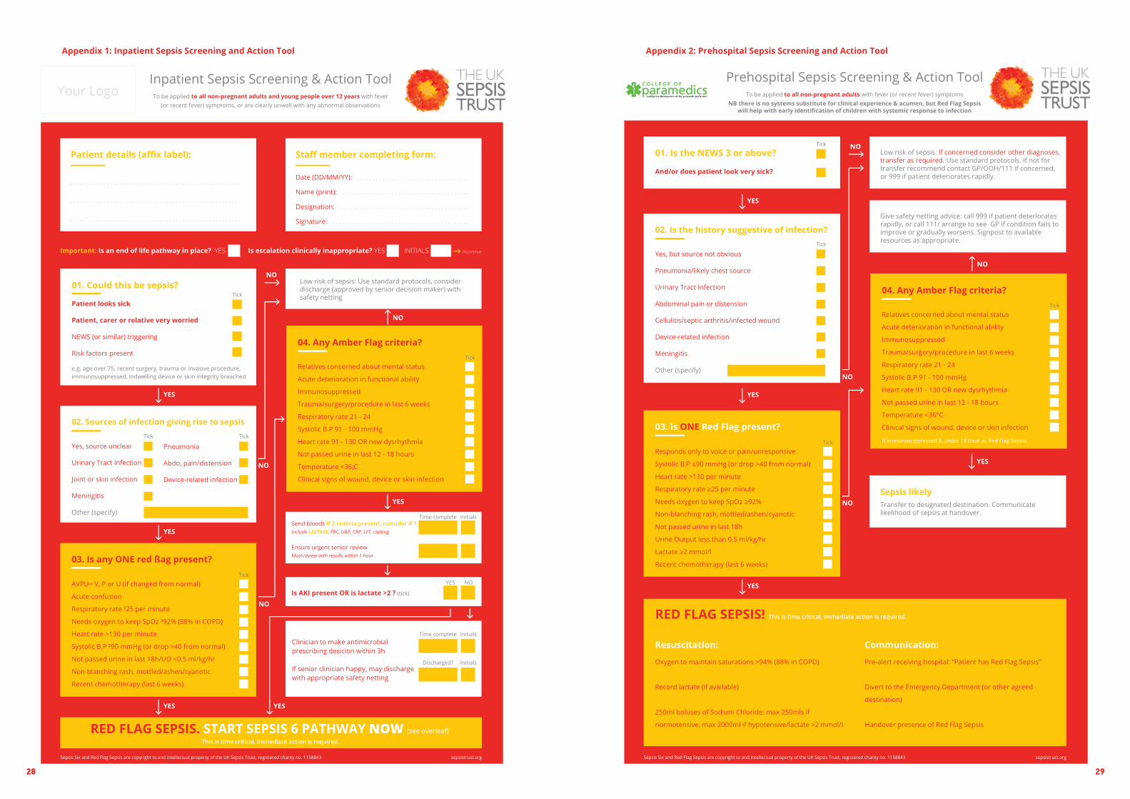
In July 2016, NICE issued NG51, which dealt with the identification and management of sepsis in the community and in hospitals, but did not include Critical Care management of sepsis.
NG51 provides a series of algorithms for the identification and severity assessment of sepsis at the bedside for adults, children and pregnant women. We include here an extract of those algorithms, including their ‘high risk’ (Red Flag) criteria and their ‘intermediate risk’ (‘Amber Flag’) criteria.
NG51’s high risk criteria were derived from the earlier Red Flag Sepsis criteria, with only minor changes. We present only the amended NICE criteria here for clarity.
Whilst there are some language inconsistencies between NG51 and the table below (for example, replac-ing ‘objective evidence of altered mental state’ (NG51) with a change in AVPU score), the Red Flag criteria are intended as an operational solution and are generally accepted as clinically relevant. Some organisa-tions may prefer to revert to NG51 language in these instances. Others, where this cn be resourced, may prefer to replace rigid protocols with decisions based upon clinical assessment by a competent deci-sion-maker: in this context, Red Flags may be deemed unnecessary. The risk here is the ready availability of clinical staff with sufficient eperience and gestalt.
iii. NICE Guideline NG51
‘RED FLAG’ criteria indicating a high risk of deterioration
iii. Is any ONE RED FLAG present?
AVPU= V, P or U (if changed from normal)
Acute confusion
Respiratory rate ≥25 per minute
Needs oxygen to keep SpO2 ≥92% (88% in COPD)
Heart rate >130 per minute
Systolic B.P ≤90 mmHg (or drop >40 from normal)
Not passed urine in last 18h/UO <0.5 ml/kg/hr
Non-blanching rash, mottled/ashen/cyanotic
Recent chemotherapy (last 6 weeks)
Tick
Clearly not all Red Flag criteria can be measured in all clinical settings. Whilst most General Practitioners now have access to pulse oximetry (for adults), it would be difficult to accurately measure hourly urine outputs in the back of an ambulance. The UK Sepsis Trust website has examples of clinical tools tailored to each clinical area.
Lactate measurement was included in NG51 later in the pathway – to be measured if a patient had one or more Amber Flag criteria (see below). Organisations who had adopted Red Flag Sepsis were used to measuring lactate at the bedside at the time of presentation – many such organisations retain lactate measurement as a Red Flag. Operationally, there is little difference.
So, a change in SOFA score is the ‘official’ diagnostic criterion for the diagnosis of organ dysfunction and therefore sepsis. However, it is difficult to envisage an NHS organisation reliably calculating formal SOFA scores on a busy ward or in a overstretched Emergency Department. It also shares some of the issues of previous organ dysfunction criteria lists, including a reliance on blood tests which are typically not ‘back’ at the time of first assessment of the patient.
Some readers will, rightly, be asking what on earth a PaO2/FiO2 ratio is, how you measure mean arterial pressure or indeed what a dose of norepinephrine of >0.1 µg/kg/min means! You’re not alone. This was a definition created for the most part for use by Critical Care specialists.
A change in SOFA score remains the most robust definitiion of organ dysfunction in this patient population, and where resources allow we would not discourage its use. Recognising, however, that
few clinical areas have the resource to formally measure SOFA scores, we need something more pragmatic.
CLINICAL PRACTICE TIP
Red Flag Sepsis was developed in September 2015 by the UK Sepsis Trust in collaboration with NHS England and the Royal Colleges as a pragmatic, operational solution for use at the bedside. Red Flag Sepsis is not, and never will be, a formal ‘diagnosis’ of sepsis: it is a set of criteria we can measure rapidly which suggest it is highly likely the patient has a degree of organ dysfunction.
The criteria were derived from what was already being used at the bedside – the National Early Warning Score. Those thresholds for respiratory rate, blood pressure and so on which would score ‘3’ on the NEWS score were included, as all expert contributors felt that these indicated a sicker cohort of patients. We also included two criteria from the then current second international consensus definitions which could be measured at the bedside: lactate, and the presence of a purpuric rash similar to that seen (and feared) in meningococcal sepsis.
ii. Red Flag Sepsis
Red Flag Sepsis (RFS) is not the same as an ‘official’ diagnosis of sepsis, which would be made by identifying a deterioration in SOFA score of two points.
However, many, in fact most, NHS organisations lack resource to measure SOFA scores routinely, and the UK Sepsis Trust, NICE, the Royal Colleges and NHS Digital accept RFS as a suitable surrogate.
If you identify one or more Red Flags, assume the patient has sepsis. It’s a tool designed to help you to get on and ensure the patient gets the treat-ment they need.
Red Flag Sepsis will be coded as ‘sepsis’ providing it is written as a state-ment or diagnosis rather than a query.

24 25
Pathway in the presence of one or more Amber Flags: action if Acute Kidney Injury is present or lactate is >2 mmol/l is to commence Sepsis 6 immediately
Send bloods if 2 criteria present, consider if 1Include LACTATE, FBC, U&E, CRP, LFT, clotting
Clinician to make antimicrobial prescribing desicion within 3h
If senior clinician happy, may discharge with appropriate safety netting
Is AKI present OR is lactate >2 ? (tick)
Ensure urgent senior reviewMust review with results within 1 hour
Time complete
Time complete
Discharged?
Initials
Initials
Initials
NOYES
clotting) and arrange for review of the patient by a senior doctor or equivalent nurse, midwife or allied health professional.
The presence of any two (or more) Amber Flags in the community might reinforce the need for hospital assessment (unless this is not appropriate given any advance directive or advanced stages of terminal illness). For health professionals in hospital, NICE recommends that for any patient with two or more Amber Flags, bloods should be sent in addition to ensuring review by a senior clinician.
Decision-making, once Amber Flag(s) have been identified, is based upon clinical judgement and should take into account both patient and environmental factors.
If blood tests reveal the presence of an acute kidney injury, or if the lactate is found to be higher than 2 mmol/l, the patient moves to the Red Flag Sepsis pathway and the Sepsis 6 should be immediately commenced.
If there is no acute kidney injury, and the lactate is less than 2 mmol/l, the responsible clinician should make a decision: if in the community, whether hospital assessment is urgently required; and if in hospital whether antibiotics are needed urgently.
Should a decision not to transfer to hospital be made by a clinician in the community, this should be documented, appropriate verbal and preferably written safety netting should be given, and consideration given to a scheduled review.
Should a clinician in hospital decide urgent antimicrobials are unnecessary, they should consider alternative diagnoses and assess severity of illness in that context. Discharge from hospital, possibly with oral antibiotics, may be appropriate with safety netting and consideration given to a scheduled review.
If neither a Red Flag nor an Amber Flag are present, this indicates a low risk of adverse outcome from infection. This does not mean the patient is necessarily ‘fine’! Other conditions should be considered, and standard protocols followed. If care in the community is considered suitable, then verbal and written safety netting instructions should be provided where appropriate.
NICE recognised that the absence of a Red Flag did not necessarily indicate a patient was well – it would be illogical to think a patient with, say, a heart rate of 131 needed ‘blues and twos’ to hospital whereas another with a heart rate of 129 could be left at home.
NG51 therefore introduced a second, ‘safety net’, risk assessment – the presence (or absence) of ‘moderate risk’, or Amber Flag criteria.
CLINICAL PRACTICE TIPRed Flag Sepsis (RFS) identifies patients likely to be at high risk of deterioration. You should view the presence of one or more Red Flags as empowering you to act immediately.
Actions will vary according to your clinical setting – for a General Practitioner or Community Nurse, this might be to call 999 and communicate using the term ‘Red Flag Sepsis’. For a Paramedic, this might be immediate transfer to hospital. For a health professional in hospital, this will be to ensure delivery of the Sepsis 6.
‘Amber Flag’ criteria indicating a moderate to high risk of deterioration:
iv. Any Amber Flag criteria?
Relatives concerned about mental status
Acute deterioration in functional ability
Immunosuppressed
Trauma/surgery/procedure in last 6 weeks
Respiratory rate 21 - 24
Systolic B.P 91 - 100 mmHg
Heart rate 91 - 130 OR new dysrhythmia
Not passed urine in last 12 - 18 hours
Temperature <36 °C
Clinical signs of wound, device or skin infection
Tick
According to NICE, the presence of a single Amber Flag should prompt the health professional to consider action. Health professionals outside hospital should consider whether the patient can safely be cared for in the community or requires hospital assessment (with appropriate documentation of the decision and safety netting if the patient is to be managed in the community). For health professionals in hospital, NICE recommends that a single Amber Flag should prompt consideration of the need for blood tests (to include lactate, Full Blood Count, urea and electrolytes, C-reactive Protein, liver function tests and enzymes and

26 27
Term ‘Official’ meaning Notes
Red Flag Sepsis
A term used to describe the presence of any one Red Flag criterion from the UK Sepsis Trust pathways
Some organisations may prefer to use the term ‘sepsis with one or more high risk criteria’ as per NG51
Amber Flag Sepsis
A term used to describe the presence of any one Amber Flag criterion from the UK Sepsis Trust pathways
Some organisations may prefer to use the term ‘sepsis with one or more moderate risk criteria’ as per NG51
Septic Shock
Sepsis and (despite adequate volume resuscitation) persis-tent hypotension requiring vasopressors to maintain Mean Arterial Pressure (MAP) greater than or equal to 65 mm Hg, and lactate greater than or equal to 2 mmol/l
Pragmatically and usually as a trigger to call Critical Care, a patient who is hypotensive (Red Flag criterion, systolic blood pressure (SBP) <90 mmHg) AND who has a lactate >2 mmol/l following fluid resuscitation
Within this chapter, we have explored the balance between the need for an ‘official’ definition of sepsis – primarily for use in Critical Care, and important for ensuring we are entering the right patients into clinical trials – and a more pragmatic, ‘bedside’ definition. Regarding the latter, we have discussed the NICE sepsis guideline NG51, and how the UK Sepsis Trust has worked with NICE and other stakeholders to translate this into tools for use across the healthcare system.
The UK Sepsis Trust website (sepsistrust.org) has tools for use by any health professional in the community, in General Practice, in prehospital services and in hospitals. Whilst it would be clumsy to include all within this manual, over the next two pages you will find examples for use in hospitals and in Ambulance Services for reference. Others are similar.
CLINICAL PRACTICE TIP
Pragmatically, septic shock should be a term used in written and verbal communication to describe ‘presumed’ septic shock: a patient who is hypotensive (Red Flag criterion, systolic blood pressure (SBP) <90 mmHg) AND who has a lactate >2 mmol/l following fluid resuscitation.
SUMMARY: WE’VE STRATIFIED SEVERITY. WHAT TERMS DO WE USE, AND HOW DO WE IDENTIFY SEPTIC SHOCK?05
Sepsis and (despite adequate volume resuscitation) both of:
• Persistent hypotension requiring vasopressors to maintain Mean Arterial Pressure (MAP) greater than or equal to 65 mm Hg, and
• Lactate greater than or equal to 2 mmol/l.
Having followed the UK Sepsis Trust tools, which are based upon NG51, we have determined whether the patient with infection has any high-risk criteria (Red Flag Sepsis), intermediate risk criteria (Amber Flag Sepsis), or in the absence of any of these has a low risk of deterioration from infection.
We described Septic Shock in narrative terms in Section 1, as ‘a subset of sepsis where particularly profound circulatory, cellular and metabolic abnormalities substantially increase mortality’. The Sepsis-3 authors were looking to identify patients with a particularly high risk of death in this group, and from a recognition perspective described septic shock as:
It seems that a formal diagnosis of septic shock is outside the remit of most people working outside Critical Care. This issue was a criticism levelled at the Sepsis-3 definitions, in particular by those working in low and middle income countries where it would seem septic shock might not exist according to a definition requiring the use of vasopressors.
Box 5 describes appropriate terms to use in written and verbal communication when discussing sepsis. Clinical coders are familiar with these terms, and cases will be coded appropriately when we write ‘sepsis’.
Term ‘Official’ meaning Notes
Infection
The invasion of a normally sterile cavity by organisms, or inflammation caused by organisms in parts of the body which are not normally sterile
May also be used to describe patients who are presumed to have an infection, but who have no Red or Amber Flag criteria
SepsisA deterioration in the Sequential Organ Failure As-sessment score of 2 points
Pragmatically, sepsis is a con-venient term to describe the presence of either Red Flag Sepsis (including Septic Shock) or Amber Flag Sepsis
Box 5
28 29
Appendix 2: Prehospital Sepsis Screening and Action ToolAppendix 1: Inpatient Sepsis Screening and Action Tool
Inpatient Sepsis Screening & Action Tool
RED FLAG SEPSIS. START SEPSIS 6 PATHWAY NOW (see overleaf)
Your Logo To be applied to all non-pregnant adults and young people over 12 years with fever (or recent fever) symptoms, or are clearly unwell with any abnormal observations
This is time critical, immediate action is required.
Sepsis Six and Red Flag Sepsis are copyright to and intellectual property of the UK Sepsis Trust, registered charity no. 1158843 sepsistrust.org
Date (DD/MM/YY):
Name (print):
Designation:
Signature:
Important: Is an end of life pathway in place? YES Is escalation clinically inappropriate? YES INITIALS Discontinue
01. Could this be sepsis?
Patient looks sick
Patient, carer or relative very worried
NEWS (or similar) triggering
Risk factors present
e.g. age over 75, recent surgery, trauma or invasive procedure, immunosuppressed, indwelling device or skin integrity breached
Low risk of sepsis: Use standard protocols, consider discharge (approved by senior decision maker) with safety nettingTick
02. Sources of infection giving rise to sepsis
03. Is any ONE red ßag present?
Yes, source unclear
Urinary Tract Infection
Joint or skin infection
Meningitis
Other (specify)
Pneumonia
Abdo, pain/distension
Device-related infection
Tick Tick
Tick
AVPU= V, P or U (if changed from normal)
Acute confusion
Respiratory rate ³25 per minute
Needs oxygen to keep SpO ³92% (88% in COPD)
Heart rate >130 per minute
Systolic B.P ²90 mmHg (or drop >40 from normal)
Not passed urine in last 18h/UO <0.5 ml/kg/hr
Non-blanching rash, mottled/ashen/cyanotic
Recent chemotherapy (last 6 weeks)
04. Any Amber Flag criteria?Tick
Relatives concerned about mental status
Acute deterioration in functional ability
Immunosuppressed
Trauma/surgery/procedure in last 6 weeks
Respiratory rate 21 - 24
Systolic B.P 91 - 100 mmHg
Heart rate 91 - 130 OR new dysrhythmia
Not passed urine in last 12 - 18 hours
Temperature <36¡C
Clinical signs of wound, device or skin infection
Send bloods if 2 criteria present, consider if 1Include LACTATE, FBC, U&E, CRP, LFT, clotting
Ensure urgent senior reviewMust review with results within 1 hour
Time complete Initials
NOYES
Is AKI present OR is lactate >2 ? (tick)
Clinician to make antimicrobial prescribing desicion within 3h
If senior clinician happy, may discharge with appropriate safety netting
Time complete Initials
Discharged? Initials
YES
NO
NO
NO
NO
YES
YES
YES YES

30 31
The clinical signs and symptoms of early sepsis can be vague, subtle or non-specific; for instance, a mild tachycardia or fever. This can make early diagnosis challenging, as early signs can often be missed by healthcare providers. Few doctors can describe the definition of sepsis accurately, so it is no surprise that sepsis can be difficult to identify and therefore that delays in initiating treatment are common. Regular screening of patients at risk of sepsis and early, and judicious treatment of those presenting with likely sepsis, are key to improving patient outcomes.
An understanding of the potential and common sources of infection and their modes of presentation will help you to identify those at risk of sepsis and choose an appropriate treatment regime.
A search for the source of infection is critically important toward ensuring that we use antimicrobial agents responsibly by allowing us to target treatment with evidence-based, often narrower spectrum choices of agents.
PNEUMONIAWhat is it?
How will the patient present?
Diagnosis
Additional
Pneumonia is an infection of the lung tissue, and as a source of infection is responsible for approximately 50% of all episodes of sepsis. When a person has pneumonia, the lungs become filled with microorgan-isms, fluid, and inflammatory cells which make the work of breathing difficult and prevent the lungs from working properly.
Diagnosis of pneumonia is based on the signs and symptoms of an acute lower respiratory tract infection. These might include a productive cough, tachypnoea, noisy breathing (sometimes audible from the end of the bed), or respiratory distress. In the later stages of this condition impending respiratory failure might be recognised through the development of cyanosis, severe fatigue or even a reduced conscious level due to exhaustion or hypercapnia.
Pneumonia can be confirmed by a chest X ray showing new shadowing that is not due to any other cause (such as pulmonary oedema or infarction).
Do not wait for a chest X-ray to confirm pneumonia before starting treatment if sepsis is suspected!
Pneumonia can be classified as community acquired pneumonia (CAP) or hospital acquired pneumonia (HAP). HAP is defined as pneumonia that occurs 48 hours or more after hospital admission and which was not incubating (present within the patient) at hospital admission. You must have a strong suspicion for HAP in patients who have recently been discharged from hospital and those from high risk environments (e.g. nursing homes). SO
UR
CES
OF
INFE
CTIO
N

32 33
INTRA-ABDOMINAL SEPSISWhat is it?
How will the patient present?
Additional
The incidence of urinary tract infection is highest in young women. Most infections in adult men are complicated and related to abnormalities of the urinary tract, although some can occur spontaneously in otherwise healthy young men. HES data suggest that we see at least between 300,000 and 700,000 UTIs in England each year (code N39.0).Catheter associated UTIs (CAUTIs) are a common cause of urinary infection and sepsis. The risks associated with catheter use must be judiciously balanced against the benefits on an individual patient basis: • catheters should be inserted for the minimal time in the minimum number of patients (not for
‘routine use’ and never for monitoring urine output in ambulatory patients)• alternatives to an indwelling catheter should always be considered • ensure proper aseptic technique for insertion and after care by properly trained individuals • ensure adequate maintenance and regular checks of catheter function.
Intra-abdominal infections are the third commonest cause of sepsis in the general population, accounting for between 15 and 20% of cases. Intra-abdominal infections commonly arise from the biliary tract (e.g. cholangitis, cholecystitis) or as a complication of a perforation of the bowel (such as following an episode of diverticulitis or due to a bowel obstruction). When the bowel is very inflamed (for example, if it is ischaemic), bacteria can ‘translocate’ across the lining of the bowel into the bloodstream, precipitating sepsis in the absence of a perforation. There are between 30,000 and 50,000 cases of such infections each year in England.
In complicated intra-abdominal infections, the infection progresses from a single organ and affects the peritoneum, which can lead to the formation of intra-abdominal abscesses or diffuse peritonitis. Peritoneal contamination may result from mishandling of bowel contents during surgery, or from trauma or a spontaneous perforation (for example, appendicitis, perforated ulcer or diverticulitis).
Non-specific symptoms can be a sign that the patient is acutely unwell, such as fever, warm skin (from vasodilation) or altered mental state. More specific symptoms include abdominal pain, an inability to eat or drink, nausea, vomiting, diarrhoea or constipation. Symptoms tend to be localised initially (such as in the right iliac fossa in appendicitis), but as peritonitis develops they tend to become generalised. An ‘acute abdomen’ is characterised by a rigid, often distended abdominal wall which is exquisitely tender to palpa-tion. Patients may exhibit ‘guarding’, where they tense their muscles to prevent the palpating hand from pressing down; and ‘rebound tenderness’, where they might wince as the palpating hand is removed.
HAP is associated with a higher mortality than CAP, and is more likely to be resistant to standard antibiotic regimes.
In addition, some pneumonias can be considered “atypical” (caused by uncommon microorgansims). An atypical pneumonia might be suspected in patients with a prolonged prodromal illness, a dry cough, or failure to respond to first line therapy. If you suspect your patient may have an atypical pneumonia it is always best to liaise with infection specialist services such as Microbiology.
Hospital Episode Statistics (HES) data suggest that we see a minimum of between 450,000 and 700,000 episodes of pneumonia annually in England. Between 1.2% and 10% of adults admitted to hospital with community-acquired pneumonia are managed in an intensive care unit, and for these patients the risk of dying is over 30% (NICE, 2016). More than half of pneumonia-related deaths occur in people older than 84 years.
Hospital-acquired pneumonia is estimated to increase a hospital stay by about eight days and has a re-ported mortality rate ranging from 30–70% (NICE, 2017). There are variations in clinical management and outcomes across the UK.
URINARY TRACT INFECTIONWhat is it?
How will the patient present?
Diagnosis
Urinary tract infections (UTIs) are caused by the presence and multiplication of microorganisms in the urinary tract. A urinary tract infection can result in several clinical syndromes, including acute and chronic pyelonephritis (infection of the kidney and renal pelvis), cystitis (infection of the bladder), urethritis (infection of the urethra), epididymitis (infection of the epididymis) and prostatitis (infection of the prostate gland). Infection may spread to surrounding tissues (for example, perinephric abscess) or to the bloodstream.
Symptoms reported can include dysuria, frequency, offensive-smelling or discoloured urine, loin pain and haematuria. As a source of infection UTIs are responsible for approximately 20-25% of episodes of sepsis.
Whilst sending urine and blood cultures will aid in the confirmation of a UTI, clinical suspicion based upon signs and symptoms is sufficient to initiate therapy. A positive urine dipstick in the absence of symptoms is NEVER a reason to start an antibiotic. Common organisms causing urinary tract sepsis are gram-negative bacteria such as E. coli and Klebsiella. It is important to follow local antimicrobial guidelines (or if in any doubt to seek antimicrobial advice) as these organisms can be antibiotic resistant. Most microbiologists would no longer recommend the routine use of trimethoprim due to increasing resistance.

34 35
MENINGITISWhat is it?
How will the patient present?
Diagnosis
A post-operative wound infection is recognised by pain, erythema, a purulent discharge or heat around the incision. ‘Surgical Site Infection’ is defined as clinical evidence of an infection arising at a surgical incision site within 28 days of surgery. Poor healing may be the first marker of a lower grade infection. Post-operative wounds should be inspected daily and if there is evidence of discharge the clips or sutures should be removed and the potential space opened up using a sterile-gloved finger. Antibiotics are not needed unless a patient is immunosuppressed or there is evidence of surrounding cellulitis. Considera-tion should be given to the presence of a deeper infection – for example, an infected joint prosthesis or leaking abdominal anastomosis.
It is important to differentiate between meningitis (inflammation of the meninges, usually due to infec-tion) and ‘meningococcal septicaemia’, which should now be termed meningococcal sepsis. Each can exist without the other. Meningococcal sepsis, if present, carries a far worse prognosis than meningitis alone.
Symptoms of meningitis include headache, photophobia, vomiting, a stiff neck, drowsiness and occasion-ally focal neurological signs. Symptoms of meningococcal sepsis include some of the above plus rigors, cold hands and feet sometimes with severe pain, confusion and myalgia (muscle pain). Worsening neurological signs may indicate the development of cerebral oedema or hydrocephalus (raised pressure in the cranial cavity due to obstruction of cerebrospinal fluid flow).
Particularly with meningococcal disease, a typical purpuric (like small bruises) rash may be noted in late stages, together with signs of circulatory failure – shock, cold and mottled peripheries, low urine output and reduced conscious level.
The presence of a meningococcal rash is suggestive of meningococcal sepsis, but it can occur with other pathogens and in the absence of meningitis. Whatever the cause, the presence of a purpuric rash in the context of suspected infection is a medical emergency and demands the highest level of skill and experience available. It is inappropriate for a junior to manage such cases alone.
A lumbar puncture should be done, after checking clotting, in cases of suspected meningitis to assess white blood cell count and glucose level, as well as to identify causative organisms. If there is doubt about the diagnosis (for instance a subarachnoid haemorrhage may have some similar clinical features) or there is any suspicion of raised intracranial pressure then a CT head may be required to ensure that it is safe to proceed to lumbar puncture.
It is vital not to delay treatment. Intravenous antibiotics with activity against the Meningococcus (Neisseria meningitidis) such as cefotaxime/ceftriaxone should be given immediately. If sampling blood cultures is likely to cause delays and this cannot be avoided, then antibiotics should take priority.
Diagnosis
Identifying intra-abdominal pathology accurately demands advanced assessment skills and often advanced modalities of imaging (CT or Ultrasound) – if intra-abdominal infection is suspected, early involvement of senior clinicians is essential. Early source control (removal of infection) is essential.
CELLULITISWhat is it?
How will the patient present?
Diagnosis
Additional
Cellulitis is the most common of the group of infections known as ‘skin and soft tissue infections’ (SSTIs), which also include the much rarer necrotising fasciitis. SSTIs account for around 10-15% of episodes of sepsis. In 2016/17, there were between 110,000 and 250,000 episodes of sepsis due to cellulitis recorded in HES data.
There is likely to be tenderness, pain and swelling of the affected area, possibly following an injury or something as minor as an insect bite which have resulted in a breach of skin integrity. Cellulitis presents with rapidly spreading erythema, blistering, or even skin necrosis. The skin will feel hot. Although low-tech, carefully marking the margins of the erythema at presentation can help assessment of whether the initial antibiotic therapy is effective or not.
Diabetic patients are particularly prone to cellulitis, so it is important to check for a history of diabetes and perform blood glucose measurement in case of undiagnosed diabetes: you might spot a presentation of diabetic ketoacidosis.
The patient will be diagnosed from their clinical presentation. Swabs taken for culture may confirm the organism involved – treatment will need to be started before results are available.
Beware of rapidly spreading cellulitis, or exquisite pain which is disproportionate to the clinical findings. This may be necrotizing fasciitis, a rare surgical emergency, which spreads along fascial planes with destruction of underlying tissue. It is commonly caused by mixed flora including haemolytic streptococci. This group of organisms release exotoxins which worsen the inflammatory response. Necrotising fasciitis has a high associated mortality and requires rapid and extensive debridement of the affected area in theatre as an emergency. If suspected, the most senior available member of the team should be consulted urgently.

36 37
Additional
What is it?
How will the patient present?
Diagnosis
Diagnosis
Additional
SEPTIC ARTHRITIS
If diagnosis is suspected, the line should be removed, the tip cultured and if symptoms and signs of sepsis are present, treatment started.
Although line sepsis accounts for only around 1% of episodes, it is almost always avoidable so should not be dismissed as unimportant. For every invasive device sited, a plan should be documented for its ongoing care and consideration for removal. At every opportunity, for every device, its removal should be considered.
Central venous catheters (CVCs) are the VADs most commonly associated with bacteraemia. Whilst routine changing of CVCs is no longer recommended, in a patient deteriorating without other obvious source of infection their removal should be considered. Peripheral venous lines are less commonly involved, particularly since the introduction of high impact care bundles for their insertion and management, though due to the sheer number used they remain a significant source of healthcare associated infection.
This is inflammation of a joint (the synovial membranes or fluid within a joint) caused by infection.
Symptoms of an infection include severe pain (particularly on movement), swelling, erythema and heat around the affected joint. The patient will not be keen to move the limb. A history of arthritis can often be elicited. It is important to ask about trauma or recent instrumentation to the joint such as arthroscopic surgery.
Joint aspiration will help to establish the diagnosis and identify the causative organism. Any aspirate should be sent for culture and microscopy together with blood cultures. X-rays or other imaging will be required to establish the extent of any joint destruction.
Any antibiotic therapy must cover Staphylococci and achieve good joint penetration – intravenous benzylpenicillin and flucloxacillin being a good initial choice. It is important to liaise with orthopaedic surgeons and/or rheumatologists. In many cases a joint washout by arthroscopy is warranted (source control), and should be completed within the first six hours (and ideally sooner). In the recovery phase, physiotherapy will be essential to regain joint function.
Additional
The incidence of meningitis has, thankfully, reduced dramatically due to vaccination programmes, and meningitis now accounts for fewer than 1% of episodes of sepsis. However, for the individual patient we must not let our guard down and retain a high index of suspicion.
LINE SEPSIS What is it?
How will the patient present?
Sepsis can be associated with the direct introduction of microbes into the blood stream through insertion, or subsequent colonisation by bacteria, of indwelling devices, and in particular vascular access devices (VADs).
The Visual Infusion Phlebitis (VIP) score can be used to monitor infusion sites. Sites should be inspected daily for pain, erythema and swelling.
IV site appears healthy
One of the following is evident• Slight pain near IV site or• Slight redness near IV site
Two of the following are evident• Pain at IV site• Erythema• Swelling
All of the following signs are evident• Pain along path of cannula• Erythema• Induration
All of the following are evident & extensive• Pain along path of cannula• Erythema• Induration• Palpable venous cord
All of the following are evident & extensive• Pain along path of cannula• Erythema• Induration• Palpable venous cord• Pyrxia
No signs of phlebitis OBSERVE CANNULA
Possible first signs OBSERVE CANNULA
Early stage of phlebitis RESITE CANNULA
Mid-stage of phlebitis RESITE CANNULACONSIDER TREATMENT
Advanced stage of phlebitis or start of thrombophlebitis
RESITE CANNULACONSIDER TREATMENT
Advanced stage of thrombophlebitis INITIATE TREATMENT
0
1
2
3
4
5
38 39
Splinter haemorrhages on the nails may be a feature (but are often innocent due to trauma, particularly if the patient has a manual occupation) but are not necessary for diagnosis. In sub-acute endocarditis, splenomegaly may occur. The patient can appear cachectic, and may be mistakenly thought to have a malignancy. They may have signs of heart failure such as raised jugular venous pressure, peripheral oedema and pulmonary congestion.
Diagnosis
Multiple sets of blood cultures from different sites are mandatory. These may take several days to grow an organism. An echocardiogram should be requested to look for vegetations, but absence of these does not exclude the diagnosis. Trans-oesophageal echocardiography (TOE) may be necessary.
Additional
It is mandatory to involve Cardiology early, as the patient may deteriorate and may require urgent valve replacement surgery. Long durations of antibiotic treatments are typically necessary. Liase with your microbiology team at an early stage.
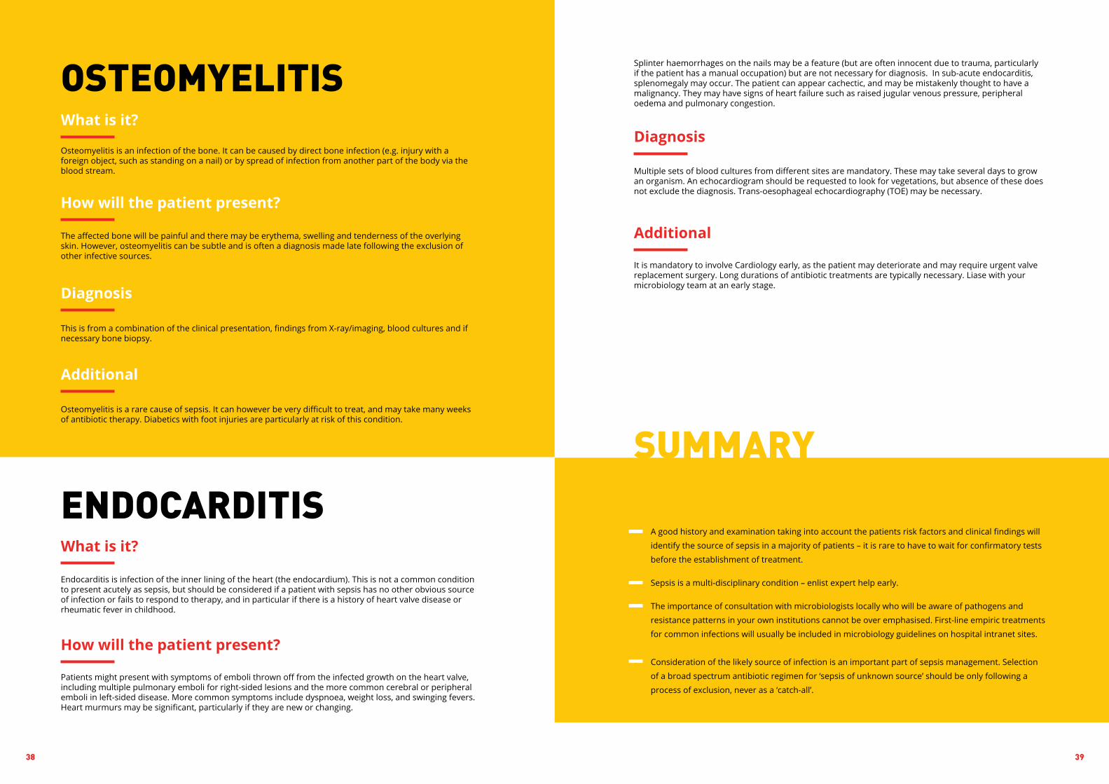
SUMMARY
• A good history and examination taking into account the patients risk factors and clinical findings will
identify the source of sepsis in a majority of patients – it is rare to have to wait for confirmatory tests
before the establishment of treatment.
• Sepsis is a multi-disciplinary condition – enlist expert help early.
• The importance of consultation with microbiologists locally who will be aware of pathogens and
resistance patterns in your own institutions cannot be over emphasised. First-line empiric treatments
for common infections will usually be included in microbiology guidelines on hospital intranet sites.
• Consideration of the likely source of infection is an important part of sepsis management. Selection
of a broad spectrum antibiotic regimen for ‘sepsis of unknown source’ should be only following a
process of exclusion, never as a ‘catch-all’.
OSTEOMYELITIS
ENDOCARDITIS
What is it?
How will the patient present?
Diagnosis
Additional
Osteomyelitis is an infection of the bone. It can be caused by direct bone infection (e.g. injury with a foreign object, such as standing on a nail) or by spread of infection from another part of the body via the blood stream.
The affected bone will be painful and there may be erythema, swelling and tenderness of the overlying skin. However, osteomyelitis can be subtle and is often a diagnosis made late following the exclusion of other infective sources.
This is from a combination of the clinical presentation, findings from X-ray/imaging, blood cultures and if necessary bone biopsy.
Osteomyelitis is a rare cause of sepsis. It can however be very difficult to treat, and may take many weeks of antibiotic therapy. Diabetics with foot injuries are particularly at risk of this condition.
What is it?
Endocarditis is infection of the inner lining of the heart (the endocardium). This is not a common condition to present acutely as sepsis, but should be considered if a patient with sepsis has no other obvious source of infection or fails to respond to therapy, and in particular if there is a history of heart valve disease or rheumatic fever in childhood.
How will the patient present?
Patients might present with symptoms of emboli thrown off from the infected growth on the heart valve, including multiple pulmonary emboli for right-sided lesions and the more common cerebral or peripheral emboli in left-sided disease. More common symptoms include dyspnoea, weight loss, and swinging fevers. Heart murmurs may be significant, particularly if they are new or changing.

40 41
WHAT HAPPENS IN SEPSIS01
Sepsis is a collection of physiological responses to infection, which involves the immune system and the coagulation cascade. It is characterised by a process known as inflammation.
Inflammation in response to infection is largely triggered by receptors in the lining of blood vessels (the endothelium), which detect products on the cell walls of pathogens. The response is from the immune system – this first line of defence then sets off a cascade of reactions. In sepsis, these reactions become dysregulated.
Think about what happens when you cut yourself. The skin around the injury quickly becomes red, it swells slightly; it is also hot to touch and is painful. Doctors, with their obsession with classical language, have historically been taught that these symptoms can be described using the terms ‘rubor’, ‘tumor’, ‘calor’ and ‘dolor’ respectively.
Sepsis is a life-threatening condition arising when an abnormal response to infection causes organ dysfunction. Sepsis can be caused by any bug, including bacteria, fungi or viruses. We refer to these disease-causing microorganisms as pathogens.
It is not clear why some people develop sepsis in response to an infection and others don’t. Several factors are likely to be at play, including:
• The type of pathogen causing the infection – some are more prone to triggering an aggressive response than others (they’re more ‘virulent’)
• The number of pathogens present, and where in the body they are• Individual or ‘host’ factors: these are determined by both genetics and by acquired conditions,
which may predispose to a disordered immune response.
Term Meaning
Rubor Redness
Tumor Swelling
Calor
Dolor
Heat
PainTHE
PATH
OP
HYS
IOLO
GY
OF
SEP
SIS
42 43
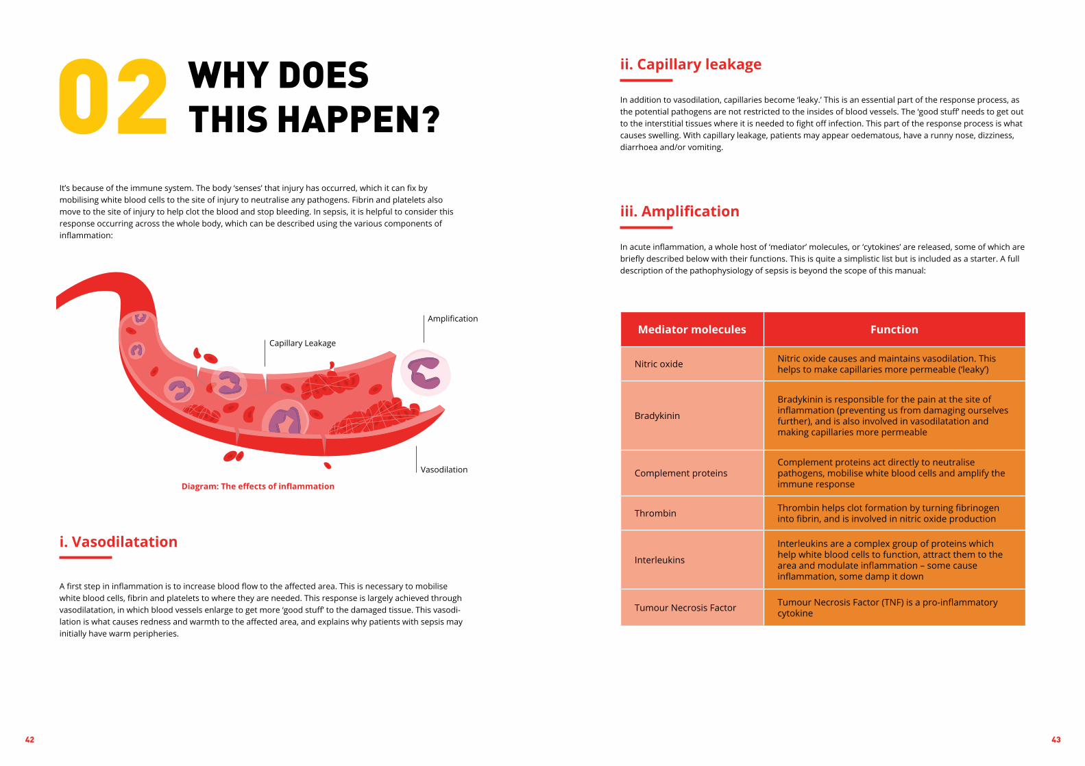
In addition to vasodilation, capillaries become ‘leaky.’ This is an essential part of the response process, as the potential pathogens are not restricted to the insides of blood vessels. The ‘good stuff’ needs to get out to the interstitial tissues where it is needed to fight off infection. This part of the response process is what causes swelling. With capillary leakage, patients may appear oedematous, have a runny nose, dizziness, diarrhoea and/or vomiting.
In acute inflammation, a whole host of ‘mediator’ molecules, or ‘cytokines’ are released, some of which are briefly described below with their functions. This is quite a simplistic list but is included as a starter. A full description of the pathophysiology of sepsis is beyond the scope of this manual:
ii. Capillary leakage
iii. Amplification
Mediator molecules Function
Nitric oxide Nitric oxide causes and maintains vasodilation. This helps to make capillaries more permeable (‘leaky’)
Bradykinin
Bradykinin is responsible for the pain at the site of inflammation (preventing us from damaging ourselves further), and is also involved in vasodilatation and making capillaries more permeable
Complement proteinsComplement proteins act directly to neutralise pathogens, mobilise white blood cells and amplify the immune response
Thrombin Thrombin helps clot formation by turning fibrinogen into fibrin, and is involved in nitric oxide production
Interleukins
Interleukins are a complex group of proteins which help white blood cells to function, attract them to the area and modulate inflammation – some cause inflammation, some damp it down
Tumour Necrosis Factor Tumour Necrosis Factor (TNF) is a pro-inflammatory cytokine
WHY DOES THIS HAPPEN?02
It’s because of the immune system. The body ‘senses’ that injury has occurred, which it can fix by mobilising white blood cells to the site of injury to neutralise any pathogens. Fibrin and platelets also move to the site of injury to help clot the blood and stop bleeding. In sepsis, it is helpful to consider this response occurring across the whole body, which can be described using the various components of inflammation:
Vasodilation
Capillary Leakage
Amplification
A first step in inflammation is to increase blood flow to the affected area. This is necessary to mobilise white blood cells, fibrin and platelets to where they are needed. This response is largely achieved through vasodilatation, in which blood vessels enlarge to get more ‘good stuff’ to the damaged tissue. This vasodi-lation is what causes redness and warmth to the affected area, and explains why patients with sepsis may initially have warm peripheries.
i. Vasodilatation
Diagram: The effects of inflammation

44 45
In essence, these processes mean that the lungs are stiff and cannot transfer oxygen and carbon dioxide in and out of the blood as easily. Patients will struggle to breathe, and will tend to take quick, shallow breaths. This fast respiratory rate is known as tachypnoea, and is often the first noticeable sign that a patient is deteriorating. This mechanism is the body’s way of meeting the oxygen demand of organs, muscles and tissues, as a result of a low circulating volume despite the stiff lungs. It cannot be sustained for long, particularly in the elderly, as it’s hard work. The respiratory rate may also increase in ‘compensation’ for a metabolic acidosis – if the pH of the blood falls because the tissues aren’t getting enough oxygen, the body will try to compensate for this by breathing faster to blow off carbon dioxide (CO2), since this prevents it dissolving to form more acid.
Mechanical ventilation might be necessary in patients with respiratory failure. A pulse oximeter might show low oxygen saturations, and a blood gas might show a low partial pressure of oxygen (PaO2). The PaCO2 might be low because of compensation for a metabolic acidosis, but in later stages may rise as the lungs begin to fail to clear carbon dioxide efficiently.
The below table shows arterial blood gas results from a patient suffering from sepsis. Whilst it is beyond the scope of this manual to fully explain blood gas results, a brief description is given next to each value. Recommendations for further reading to better understand blood gas results are given at the end of this chapter.
Test Value Normal values
pH 7.23 7.35-7.45
PaO2 9.85 11-13 kPa
PaCO2 3.2 4.7-6.0 kPa
BE -16.7 +/– 2
HCO3- 12.6 22-26 mEq/l
Lac 6.2 <2 mmol/l
The airway is not specifically affected by sepsis, unless infection arises due to infection of the throat or soft tissues of the neck. A patient with a low Glasgow Coma Score (GCS) or Alert, Voice, Pain, Unresponsive (AVPU score) score might be at risk of developing an airway problem; particularly if the GCS drops below nine, which approximates to an AVPU of ‘P’.
The lung is involved early in the inflammatory process. Often, a raised respiratory rate is the first sign that a patient is deteriorating.
Fluids and proteins leak into the interstitial tissues causing swelling and decreased oxygen transfer across the alveoli. The protective layer of surfactant which helps lungs to move freely starts to disappear, leading to decreased ‘compliance’ of the lungs (they become stiff) and increased vulnerability to infection. Later, fibrosis may develop, although this won’t affect the early stages. However, particularly with patients presenting late and those in multi-organ failure, the patient may deteriorate further and develop an Acute Lung Injury or the Acute Respiratory Distress Syndrome (ARDS).
The role that systems, pathways and cytokines play in the first phase of inflammation are still not fully understood. However, as the above table demonstrates, inflammation is mediated by a complex set of molecules which are all inter-related. It is therefore inevitable that processes will go wrong and it is understandable why sepsis can have such a rapid progression and poor prognosis.
In sepsis, the delicate balance between pro-inflammatory and anti-inflammatory cytokines, which should regulate the process, becomes disordered. Sepsis is inflammation ‘gone bad’.
So far, we’ve described the body’s ‘first line of defence’ to potential infection, which is mostly mediated by proteins and components of the vascular system. This is largely known as the ‘humoral’ component of the immune response, but to neutralise complex infections requires a cellular component too.
White blood cells are known as leukocytes, and act to neutralise pathogens. Leukocytes are also required to ‘remember’ individual pathogens, in order to act more quickly should another infection with the same pathogen occur. The ‘inbuilt’ immune system is known as the ‘innate’ immune system; the bit with memo-ry as the ‘adaptive’ immune system, although this distinction is not important here. Leukocytes are mobi-lised to the infective site by cytokines which ‘signal’ that they are needed. This pathway also signals to the bone marrow to produce more leukocytes.
There are a number of different types of leukocytes. Some, such as neutrophils, act by releasing ‘granules’ which contain enzymes with which to attack pathogens. Others, such as monocytes, ‘engulf’ pathogens and destroy them internally. B cells make antibodies, which can bind to pathogens and accelerate their destruction. White blood cells are the second line of defence, and are vital to ensuring a sustained attack on invading pathogens.
The effect of sepsis on organ systems can be described using an ABCDE approach:
A. Airway
B. Breathing

46 47
considered in the context of the PaCO2 when analysing blood gas results. If the results are both going in the same direction, then one system is working to compensate the other. If the results are going in opposite directions, then both respiratory and metabolic imbalances are occurring.
LactateLactate can be quite confusing for many people to understand and interpret, and is often debated in the management of sepsis. Lactate is often associated with tissue hypoxia, and whilst lactate does not diagnose sepsis, it can tell us how ‘bad’ the circulation and tissue oxygenation in sepsis is. Lactate is a normal waste product of anaerobic breakdown of tissue glucose – and lactate can actually be helpful. The heart is able to use it as an energy source in times of distress. In sepsis, patients struggle to get rid of this waste product as quickly as it accumulates, hence a rise in lactate. We should be concerned about anyone with sepsis and a lactate greater than 2 mmol/L, as the mortality rate of the septic patient with a high lactate is significantly higher. If the initially high lactate falls with adequate fluid resuscitation to normal levels, this is associated with better outcomes than if it remains elevated.
The effects of inadequate respiration are compounded by a reduced blood flow to the lungs in the later stages of sepsis, when the circulation begins to fail. This causes a ‘dead space’, where bits of lung are ventilated with oxygen-enriched air, but are not perfused with blood. The blood flowing out of these ‘dead space’ areas will not have been oxygenated, and, although it will mix with blood from oxygenated areas, there will be an inevitable further drop in oxygen satura-tions. Receptors called ‘chemoreceptors’ found in the carotid and aortic arteries will sense low oxygen levels. To compensate for these low oxygen levels (hypoxia), further tachypnoea will occur. This compensation is exhausting for patients to maintain, and will cause patients to look and feel very ill and lethargic. They may even express that they ‘I feel like [I] might die.’
As described above, sepsis causes vasodilatation and capillary leakage.
The increased ‘space’ in the circulation caused by vasodilatation means that the same volume of blood is occupying a much larger space. This is called a relative lack of blood volume or ‘relative hypovolaemia’.
Leaky capillaries allow proteins, solutes and water to leave the circulation, making the blood volume smaller. This compounds the ‘relative hypovolaemia’ with an ‘absolute hypovolaemia’. Absolute hypovolaemia is a reduction in circulating volume relating to blood or plasma loss.
Circulating mediators such as interleukins and nitric oxide cause vasodilatation, particularly in arterioles, and precapillary sphincter dysfunction. This leads to loss of systemic vascular resistance and contributes to hypotension. Additionally, regulation of blood flow to organs is impaired leading to hypoperfusion, shock and ultimately organ failure
C. Circulation
pHThe pH determines whether or not the patient is acidic (a low pH) or alkalotic (a high pH). The value given here indicates that the patient is acidotic. Using the other tests, we can determine if this acidosis is a result of respiratory or metabolic imbalances.
PaO2This test tells us how well oxygenated the patient is. The patient will either have hypoxaemia (low PaO2), normal oxygenation or hyperoxaemia (high PaO2). The given value indicates that the patient is hypoxic. Hypoxia is common in sepsis due to the inflammatory process described above compromising lung function.
PaCO2The PaCO2 will help to determine whether or not the acidotic pH is the result of an imbalance in the respiratory or the metabolic system. A high PaCO2 indicates that a patient is hypoventilating, and on its own would cause an acidosis. Rarely, a high PaCO2 can occur to compensate for a met-abolic alkalosis – this would not be a typical feature of sepsis. A low PaCO2, such as the value given here, indicates that the patient is hyperventilating, and on its own would cause an alkalosis. More commonly in patients with sepsis, a low PaCO2 is caused by the body’s attempt to compensate for the metabolic acidosis which arises because of the relative lack of oxygen supply to the tissues.
As the PaCO2 is low and the pH is low (acidosis), this would suggest that the cause of the abnor-malities is a metabolic acidosis. The patient is trying to compensate by breathing faster, but this is only partially effective – they remain acidotic. Looking at the bicarbonate (HCO3-), we can see that the value is low. This tells us that the patient has metabolic acidosis with partial respiratory com-pensation. In the context of the patient with sepsis, this information tells us that organ damage is likely – the sense is that the respiratory and metabolic systems are failing.
CO2 binds with water (H2O) in the body to form H2CO3 (carbonic acid). The formation of H2CO3 acts to decrease pH, and therefore is used as part of ‘homeostasis’ (the body’s way of maintaining equilibrium) to keep the pH within normal ranges. In this case, the patient is ‘blowing off’ their CO2; so the patient has less circulating carbonic acid. This causes the blood to become more alkalotic. In instances where the pH is imbalanced due to a metabolic source, the respiratory system compensates by either retaining or ‘blowing off’ CO2. Physically, we will see an increase or decrease in the patient’s ventilation in order to help the body achieve this compensation to aid the pH to return closer to its normal range. The patient with metabolic acidosis may present with Kussmaul’s respiration (rapid and deep) as they attempt to normalise pH. When considering PaCO2, it is therefore important to consider the pH. If the PaCO2 and the pH are moving in opposite directions, the imbalance will be respiratory in origin. If the PaCO2 and pH are going in the same direction, the imbalance will be metabolic in origin.
BEA Base Excess (BE) which is very low demonstrates that there is low amount of HCO3- in the patient’s blood. This decrease in HCO3- further suggests that the patient has either a metabolic acidosis or is attempting to compensate for a respiratory alkalosis. In sepsis, it’s highly likely to be the former.
HCO3- (bicarbonate)This result is low. As the pH shows an acidosis, the acid-base disturbance is likely to be a result of respiratory acidosis – that is, the disturbance is being caused by the respiratory system. Most likely, the cause of this is insufficient oxygen supply to the tissues and organs, leading to anaerobic respiration by the cells.
HCO3- is a base which ‘mops up’ hydrogen (H+) ions. If the HCO3- is low, then there are more free H+ ions. This increase in free H+ ions causes the patient to be acidotic. The HCO3- should be

48 49
Calculating Capillary Refill Time (CRT)
How to:
CRT is a quick and useful test to determine effective and efficient blood flow around the body.
The circulation, in terms of cardiac output or ‘flow’ of blood can begin to fail for two main reasons:
1. The reduced circulating volume caused by capillary leakage results in less blood returning to the heart. This ‘reduced venous return’ means that the heart is relatively empty before it contracts. If there is little blood to pump, even a strong heart won’t be able to generate a good cardiac output
2. Sepsis can affect the heart’s function, particularly during diastole, meaning that the heart cannot ‘relax’ properly and therefore cannot fill. As a result, cardiac output falls. Not only does blood pressure fall further, but blood flow to organs will diminish.
Adequate filling
Cardiac muscle contracts by the ‘walking’ of actin filaments over myosin filaments in response to calcium release. When the heart muscle is adequately stretched by ‘filling’, there is plenty of room for molecular movement and contraction is strong.
Myosin filament (does not move)
Actin filament (does move)
Apply pressure to centre of patient’s sternum using your thumb, for 5 seconds. Peripheral capillary refill is not very reliable in the very sick patient, and so CRT should be measured centrally.
When practitioner’s thumb is removed, the patient’s skin should return from white to its normal colour in under 3 seconds. A CRT of 3 seconds or more is cause for concern as the patient is not being adequately perfused.
The immediate effect of these changes is a fall in blood pressure. Blood pressure is a product of the amount of blood pumped out by the heart (cardiac output, CO) and the ‘tone’ of the blood vessels, which is termed ‘systemic vascular resistance (SVR)’.
In sepsis we have a drop in SVR which will mean the blood pressure falls even if cardiac output is preserved. The body detects this drop in blood pressure via pressure receptors called ‘baroreceptors’. These receptors trigger the sympathetic nervous system to increase the heart rate and the strength at which the heart pumps, which is further raised by the body releasing catecholamines, such as adrenaline – they attempt to increase the CO to compensate for the fall in SVR.
This rise in heart rate is known as ‘compensatory tachycardia’ and is the body’s attempt to compensate for the low blood pressure. In essence, the body is working incredibly hard to pump what (relatively) little blood there is around the body in order to get oxygen-rich blood to organs and tissues.
Early in the progression of sepsis, the patient may look remarkably well, despite a low blood pressure. They may present with warm peripheries and often normal capillary refill times. Later, as the circulating volume becomes depleted (reducing venous return to the heart) and the compensatory mechanisms become exhausted, the circulation begins to fail and the patient will look much worse; with cool periph-eries, often a prolonged capillary refill time and signs of organ dysfunction. It is critical to ensure that we identify patients before they reach this stage! It is important to remember that some patient groups (such as children) can maintain their blood pressure for a long time before very rapidly compensating.
The equation for blood pressureBP = CO X SVR
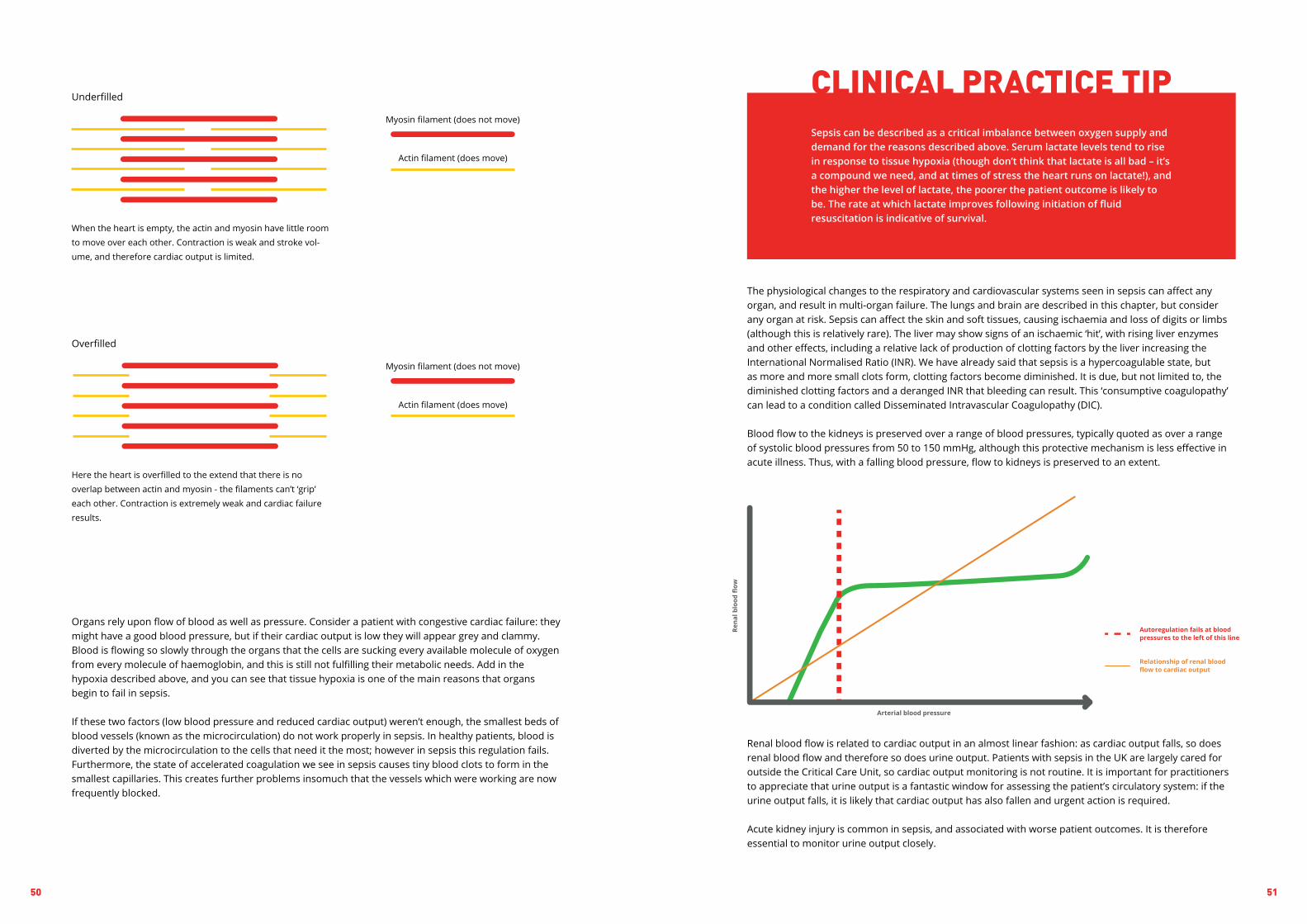
50 51
CLINICAL PRACTICE TIPSepsis can be described as a critical imbalance between oxygen supply and demand for the reasons described above. Serum lactate levels tend to rise in response to tissue hypoxia (though don’t think that lactate is all bad – it’s a compound we need, and at times of stress the heart runs on lactate!), and the higher the level of lactate, the poorer the patient outcome is likely to be. The rate at which lactate improves following initiation of fluid resuscitation is indicative of survival.
The physiological changes to the respiratory and cardiovascular systems seen in sepsis can affect any organ, and result in multi-organ failure. The lungs and brain are described in this chapter, but consider any organ at risk. Sepsis can affect the skin and soft tissues, causing ischaemia and loss of digits or limbs (although this is relatively rare). The liver may show signs of an ischaemic ‘hit’, with rising liver enzymes and other effects, including a relative lack of production of clotting factors by the liver increasing the International Normalised Ratio (INR). We have already said that sepsis is a hypercoagulable state, but as more and more small clots form, clotting factors become diminished. It is due, but not limited to, the diminished clotting factors and a deranged INR that bleeding can result. This ‘consumptive coagulopathy’ can lead to a condition called Disseminated Intravascular Coagulopathy (DIC).
Blood flow to the kidneys is preserved over a range of blood pressures, typically quoted as over a range of systolic blood pressures from 50 to 150 mmHg, although this protective mechanism is less effective in acute illness. Thus, with a falling blood pressure, flow to kidneys is preserved to an extent.
Renal blood flow is related to cardiac output in an almost linear fashion: as cardiac output falls, so does renal blood flow and therefore so does urine output. Patients with sepsis in the UK are largely cared for outside the Critical Care Unit, so cardiac output monitoring is not routine. It is important for practitioners to appreciate that urine output is a fantastic window for assessing the patient’s circulatory system: if the urine output falls, it is likely that cardiac output has also fallen and urgent action is required.
Acute kidney injury is common in sepsis, and associated with worse patient outcomes. It is therefore essential to monitor urine output closely.
Organs rely upon flow of blood as well as pressure. Consider a patient with congestive cardiac failure: they might have a good blood pressure, but if their cardiac output is low they will appear grey and clammy. Blood is flowing so slowly through the organs that the cells are sucking every available molecule of oxygen from every molecule of haemoglobin, and this is still not fulfilling their metabolic needs. Add in the hypoxia described above, and you can see that tissue hypoxia is one of the main reasons that organs begin to fail in sepsis.
If these two factors (low blood pressure and reduced cardiac output) weren’t enough, the smallest beds of blood vessels (known as the microcirculation) do not work properly in sepsis. In healthy patients, blood is diverted by the microcirculation to the cells that need it the most; however in sepsis this regulation fails. Furthermore, the state of accelerated coagulation we see in sepsis causes tiny blood clots to form in the smallest capillaries. This creates further problems insomuch that the vessels which were working are now frequently blocked.
Myosin filament (does not move)
Myosin filament (does not move)
Actin filament (does move)
Actin filament (does move)
Underfilled
Overfilled
When the heart is empty, the actin and myosin have little room to move over each other. Contraction is weak and stroke vol-ume, and therefore cardiac output is limited.
Here the heart is overfilled to the extend that there is no overlap between actin and myosin - the filaments can’t ‘grip’ each other. Contraction is extremely weak and cardiac failure results.

52 53
CONCLUSIONThe pathophysiology of sepsis is a very complex topic. This chapter has touched on the brief principles, but further reading is advised for practitioners to gain more depth of understanding into this multifaceted subject.
The most important messages to take from this chapter are:• Sepsis is complex, and can affect any system of the body. Symptoms may be subtle, or not as we
would expect, but any concern should be given immediate attention.• Ensure that all test results are read and acted upon. Some test results, particularly those obtained
from blood gases, may only show minimal changes, however these subtle changes could be a result of compensation, and need interpretation by those familiar with sepsis and blood gas analysis.
• The higher the patient’s lactate level climbs, the worse their prognosis becomes.• Sepsis is life-threatening and time-critical. By assessing patients in a systematic way (for example
using the ABCDE approach), life-threatening problems should be found quickly and treatment can be delivered promptly. It is easy to panic if you find something wrong or ‘not quite right’ with any patient. The important thing if you do not understand the pathophysiology behind what is happening is to escalate care to the appropriate team members who can provide timely treatment.
Further reading
Adam, S. K. and Osborne, S. (2006) Critical Care Nursing: Science and Practice, 2nd edn. Oxford: Oxford University Press.
Blann, A. (2013) Routine Blood Results Explained, 3rd edn.Cumbria: M&K Publishing.
British Medical Journal (2017) ‘Sepsis in adults,’ Available at: http://bestpractice.bmj.com/best-rnauprac-tice/monograph/245/basics/pathophysiology.html.
Cutler, L. R. and Cutler, J. M. (2010) Critical Care Nursing made incredibly easy, 1st UK edn. London: Lippin-cott Williams and Wilkins.
Daniels, R. and Nutbeam, T. (eds) (2009) ABC of Sepsis, West Sussex, Blackwell Publishing Ltd.
Potter, L. and Zaininger, P. (2017) ‘ABG Interpretation,’ Available at: https://geekymedics.com/abg-interpre-tation/.
Patient.info (2016) ‘Sepsis,’ Available at: https://patient.info/doctor/sepsis-septicaemia-pro#ref-5.
As blood flow to the brain reduces, so conscious levels can be affected. This can present as confusion, drowsiness, slurred speech, agitation, anxiety, or a decreased level of consciousness.
Blood sugar is normally slightly elevated in sepsis, meaning that it is unlikely to be responsible for a reduced conscious level. When the body enters a state of shock, the patient’s fight or flight response is triggered. Simply put, when the brain identifies a stress on the body, adrenaline, noradrenaline and cortisol are released in order to help the body ‘fight’. With the release of these hormones; three things are now happening:
1. Cortisol activates enzymes which are involved in hepatic gluconeogenesis (creation of glucose, or sugar, by the liver), and also inhibit the ability of the peripheral tissues to uptake glucose
2. Adrenaline and noradrenaline activate hepatic gluconeogenesis and glycogenolysis, consequently increasing blood sugar levels
3. As the body is fighting infection, an inflammatory substance called C-reactive protein is released in order to combat the infection. C-reactive protein, however, induces insulin resistance, meaning that the body cannot effectively use its own insulin. The result of this will be a raised blood sugar.
A further consideration is that when the body enters a state of shock, in order to preserve the internal organs, the body pulls its circulating volume into its core. The brain is the only internal organ not to sit in the core of the body. So, when the body pulls its circulating volume into its core, the brain does not receive adequate oxygen to function.
Recent evidence suggests that a high temperature might be a protective response to sepsis, with patients with higher temperatures appearing to fare better. Clinical opinion suggests that hypothermia below 36.0oC is a sinister development associated with worse patient outcome, although this is not conclusively proven.
A high temperature occurs due to a response to infection by the hypothalamus, essentially sending it into disarray. Pathogens, particularly if bacterial, will produce pyrogens which act on the hypothalamus to ‘reset’ the way in which it regulates temperature.
A low or normal temperature may occur because of heat loss, due to vasodilatation, or due to the patient having taken anti-pyretic medication, for example paracetamol.
D. Disability
E. Exposure

54 55
OVERVIEW1. Delivering the Sepsis 6 within one hour is one of the most effective life-saving treatments in medicine2. Each hour’s delay in giving antibiotics increases mortality3. The Sepsis 6 includes strategies to control the source of infection, and to measure and restore
circulation and oxygen delivery4. Always assess the impact of your treatment and adjust accordingly
The Sepsis 6, and the rationale behind each element, is described in the modified version of our Screening and Action Tool below:
Action (complete ALL within 1 hour) Time Initials Why we do this
1. Administer oxygen
Aim to keep saturations >94%(88-92% if at risk of CO2 retention e.g. COPD)
To improve the oxygen content of the blood, and therefore its delivery to the tissues
To help identify pathogens, to determine likely source of infection & guide antimicrobial therapy
To control the underlying infection, removing the trigger for immune overreaction
To improve preload to the heart by correcting hypovolae-mia, improving cardiac output and BP
High lactate indicates hypop-erfusion- response of lactate helps to guide resuscitation
Urine output falls if the patient is hypovolaemic, also provides an indicator of adequate cardiac output
2. Take blood cultures
At least a peripheral set. Consider e.g. CSF, urine, sputum. Think source control! Call surgeon/ radiologist if needed
3. Give IV antibiotics
According to Trust protocolConsider allergies prior to administration
4. Give IV fluids
If hypotensive/ lactate >2 mmol/l, up to 30ml/kgGive 500ml stat if not hypotensive and lactate normal
5. Check serial lactates
Corroborate high VBG lactate with arterial sampleIf lactate >4 mmol/L, recheck after each 10ml/kg challenge and call Critical Care
6. Measure urine output
May require urinary catheterEnsure fluid balance chart commenced & completed hourly TH
E S
EPSI
S 6

56 57
How is oxygen carried in the blood?
Oxygen is transported in two forms:
1. The amount of oxygen bound to haemoglobin – this is really important (98% of total oxygen carried)2. The amount of oxygen dissolved directly in the blood – this is relatively unimportant (2% of total
oxygen carried).
Bound to haemoglobin
Dissolved in plasma
There are a few equations coming up to explain why we do what we do. Rest assured you do not need to memorise them for clinical practice!
Oxygen delivery to the tissues is governed by two things: how much oxygen is in the blood, and how much blood is flowing to the tissue or organ. Writing this as an equation gives:
The O2 content of blood is then given by this equation:
Let’s break down this equation to see how it relates to the Sepsis 6. The first bit of good news: you can essentially ignore the ‘0.003 x PaO2’ bit (which, as we’ve said above, relates to the amount of oxygen which is dissolved in the blood rather than bound to haemoglobin), as the dissolved portion is tiny.
[Hb] is the concentration of haemoglobin in g/dL (note some hospitals use g/L).
The SpO2 (oxygen saturation of haemoglobin) is the amount of oxygen bound to haemoglobin as a percentage of the total amount of oxygen that could potentially be bound to haemoglobin. This is the bit you can fix quickly by giving a little oxygen. We used to advise that high flow oxygen be given routinely to patients with sepsis, but this is now only the case in children or those adults who are already critically ill.
Each gram of haemoglobin can carry up to a maximum of 4 molecules of oxygen which equates to 1.34ml, which is where that number comes from. You can’t change this number! If the SpO2 is lower, it means there is less oxygen than this bound to each haemoglobin molecule.
O2 delivery = O2 content of blood x cardiac output
Oxygen content (measured in ml) = [1.34 x [Hb] x % SpO2] + 0.003 x PaO2
INTRODUCTION
Sepsis is characterised by organ dysfunction mediated, to a large extent, by a relative lack of oxygen to the cells.
In this chapter, we will see how applying the Sepsis 6 works to minimise this by restoring the circulation, assessing risk, monitoring the effect of treatment and switching off the infective trigger. The Sepsis Six is a set of six (!) tasks which can be performed by junior health professionals working together as a team – all it takes is the knowledge, the will and basic prescribing and practical skills. It is simple and effective, and has been shown to greatly increase a patient’s chances of survival if delivered within the first hour.
In 2016, the National Institute for Health and Social Care Excellence (NICE), in their NG51 Guideline on Sepsis, recognised that these elements of care are those with the greatest evidence base in the early resuscitation phase of sepsis.
The Sepsis 6 should be delivered as quickly as possible, but always within the first hour following recognition of sepsis.
GIVE OXYGENTO MAINTAIN TARGETED SATURATIONS 94-98%
STEP
01In sepsis, a critical imbalance exists between oxygen demand by the tissues and its supply. Oxygen delivery is compromised due to a combination of reduced blood pressure and possibly flow, tissue oedema and abnormal flow of blood through capillary beds. Demand of the cells for oxygen is increased as the hypermetabolic state means cells are crying out for oxygen. This means you will need to do what you can to maximise oxygen delivery to your patient’s tissues.
Any patient who is critically ill – for example, who is shocked or unconscious – should immediately receive high flow oxygen at 15 litres per minute via a non-rebreathe facemask with reservoir bag. If the patient is not in immediate danger, current practice for most patients is to use ‘just enough’ oxygen to achieve targeted oxygen saturations of 94-98%.
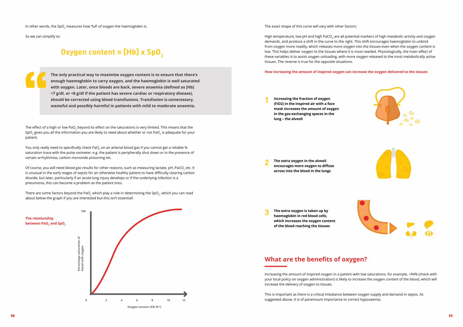
58 59
1
2
3
The exact shape of this curve will vary with other factors: High temperature, low pH and high PaCO2 are all potential markers of high metabolic activity and oxygen demands, and produce a shift in the curve to the right. This shift encourages haemoglobin to unbind from oxygen more readily, which releases more oxygen into the tissues even when the oxygen content is low. This helps deliver oxygen to the tissues where it is most needed. Physiologically, the main effect of these variables is to assist oxygen unloading, with more oxygen released to the most metabolically active tissues. The reverse is true for the opposite situations.
How increasing the amount of inspired oxygen can increase the oxygen delivered to the tissues
Increasing the fraction of oxygen (FiO2) in the inspired air with a face mask increases the amount of oxygen in the gas-exchanging spaces in the lung – the alveoli
The extra oxygen in the alveoli encourages more oxygen to diffuse across into the blood in the lungs
The extra oxygen is taken up by haemoglobin in red blood cells, which increases the oxygen content of the blood reaching the tissues
What are the benefits of oxygen?
Increasing the amount of inspired oxygen in a patient with low saturations, for example, <94% (check with your local policy on oxygen administration) is likely to increase the oxygen content of the blood, which will increase the delivery of oxygen to tissues.
This is important as there is a critical imbalance between oxygen supply and demand in sepsis. As suggested above, it is of paramount importance to correct hypoxaemia.
In other words, the SpO2 measures how ‘full’ of oxygen the haemoglobin is.
So we can simplify to:
The effect of a high or low PaO2 beyond its effect on the saturations is very limited. This means that the SpO2 gives you all the information you are likely to need about whether or not PaO2 is adequate for your patient.
You only really need to specifically check PaO2 on an arterial blood gas if you cannot get a reliable % saturation trace with the pulse oximeter: e.g. the patient is peripherally shut down or in the presence of certain arrhythmias, carbon monoxide poisoning etc.
Of course, you will need blood gas results for other reasons, such as measuring lactate, pH, PaCO2 etc. It is unusual in the early stages of sepsis for an otherwise healthy patient to have difficulty clearing carbon dioxide, but later, particularly if an acute lung injury develops or if the underlying infection is a pneumonia, this can become a problem as the patient tires.
There are some factors beyond the PaO2 which play a role in determining the SpO2, which you can read about below the graph if you are interested but this isn’t essential!
The relationship between PaO2 and SpO2
Oxygen content ≈ [Hb] x SpO2
The only practical way to maximise oxygen content is to ensure that there’s enough haemoglobin to carry oxygen, and the haemoglobin is well saturated with oxygen. Later, once bloods are back, severe anaemia (defined as [Hb] <7 g/dl, or <8 g/dl if the patient has severe cardiac or respiratory disease), should be corrected using blood transfusions. Transfusion is unnecessary, wasteful and possibly harmful in patients with mild to moderate anaemia. “

60 61
1. There are a small group of patients at risk of hypercapnic respiratory failure with high flow inspired oxygen
2. Even in these patients, hypoxia will kill before hypercapnia3. In general, lower oxygen saturation ranges should be targeted, with
blood gases to check for CO2 retention and acidosis4. Senior medical and nursing input should be sought for these patients.
Specific guidance is available in the BTS Guidelines for sepsis:
1. If the patient is shocked or otherwise critically ill, the initial oxygen therapy is a reservoir mask at 15 l/min
2. Once the patient is stable, reduce the oxygen flow and aim for target saturation range of 94–98%
3. If oximetry is unavailable, continue to use a reservoir mask until definitive treatment is available
4. Patients with COPD and other risk factors for hypercapnia who develop critical illness should have the same initial target saturations as other critically ill patients pending the results of blood gas measurements, after which these patients may need controlled oxygen therapy or supported ventilation if there is severe hypoxaemia and/or hypercapnia with respiratory acidosis
Patients should not receive dry, high flow oxygen for more than four to six hours due to the risks of retained secretions, dehydration and loss of heat. If a patient continues to require high inspired oxygen concentrations then this must be humidified.
KEY
PO
INT
PRACTICAL TIP
In contrast, multi-organ failure from hypoxia happens quickly and rapidly becomes a problem that requires HDU/ITU or even becomes irreversible. In other words, hypoxia will kill quicker than hypercapnia.
For patients at risk of hypercapnic failure, close liaison with a senior doctor is essential (Specialist Trainee level and above in the UK) and/or senior nurse with specialist skills (e.g, a Respiratory Nurse Specialist). The aim will generally be to give the highest tolerated amount of inspired oxygen.
One pragmatic approach, supported by NICE though not grounded in a huge evidence base, is to target oxygen saturations of 88-92% in these groups. You can then re-check a blood gas (if the pulse oximeter is working well, a venous gas is acceptable) at 30 minutes. If their CO2 remains normal then continue, whereas if it has risen consider the need for ventilation or reduce the target range of saturatons, but never tolerate life-threatening hypoxia!
So how should we oxygenate the patient?
Oxygen should be titrated to achieve saturations of 94-98% in most patients unless they are immediately recognised to be critically ill. In patients with known COPD, seek senior advice and have a low threshold for repeating arterial blood gas sampling. Once the SpO2 is at 98%, there is little benefit in further increases in the amount of inspired oxygen.
Increasing the inspired oxygen will increase oxygen saturations, which increases the oxygen content of the blood.
Above a saturation of 98% there is little benefit from further increases in oxygenation.
KEY
PO
INT
What are the risks of oxygen therapy?
There is a small risk of hypercapnic respiratory failure.
Normally blood in the lungs flows to the alveoli (air-filled sacs where gas exchange occurs) that are best ventilated. The way the body works this out is by the amount of oxygen in the alveoli. A low amount of oxygen in an alveolus leads to less blood perfusing that alveolus by narrowing the capillaries supplying it with blood. This is known as ‘hypoxic pulmonary vasoconstriction’. This means that the best performing parts of the lung receive most of the blood. Thus, CO2 is easily removed, as most of the blood will go to alveoli that are ventilating well and so will be effective at removing CO2.
When too much oxygen is given, all the alveoli become better oxygenated, and so blood is spread more evenly through the lung rather than focused on the best performing, well ventilated areas. This means that CO2 removal becomes less efficient.
For most people, this is not a problem, as we can increase our tidal volume and respiratory rate to remove this extra CO2. In those with impaired lung function, however, carbon dioxide levels can begin to rise. This theory has largely replaced the theory of ‘hypoxic drive’ in explaining hypercapnia developing in patients with COPD who are given high flow oxygen.
In patients with limited ventilation ability, this effect can result in them retaining CO2. These patients are at risk of hypercapnic respiratory failure, and patient groups at risk will include:
1. Some patients with COPD (particularly those on home oxygen or with previous hypercapnic respiratory failure)
2. Patients with neuromuscular problems affecting their breathing3. Patients with chest wall/spinal deformities4. Very obese patients5. Patients with bronchiectasis, including secondary to cystic fibrosis.
Hypercapnic respiratory failure is dangerous as it can lead to respiratory acidosis. It must be remembered that hypercapnia leading to acidosis generally happens slowly, and that regular blood gas monitoring can identify this. Hypercapnia can be managed with controlled oxygen, NIV (non-invasive ventilation) and/or invasive ventilation.

62 63
GIVE ANTIBIOTICS
STEP
03
tract. If a source of infection amenable to drainage is present, such as a pelvic abscess, intervention is urgent. Such a patient will not get better with antibiotics alone! If in doubt, consult with a radiologist and/or surgeon.
Of course, there is no point in taking samples if they are not followed up on. Most microbiology laboratories will routinely telephone positive samples through, but if you’ve heard nothing it’s always worth checking!
Each hour’s delay in giving antibiotics in septic shock is associated with a significant increase in mortality.
KEY
PO
INT
The 2006 landmark study by Anand Kumar showed an increase in mortality of 7.6% for every hour’s delay in administration of appropriate antibiotic therapy. As overall mortality has reduced with time, the magnitude of this effect might have reduced, but studies still largely concur that each hour’s delay increases the risk of death by 2-5%.
Antibiotic choice should be guided by the suspected focus of infection. This depends on your clinical, microbiological and radiological evidence for infection. The choice of antibiotic should be in line with your local hospital guidelines. If in doubt, discuss with the microbiology or infectious diseases teams.
If you are confident about the source of the infection, then the antibiotic choice should be tailored to cover the likely pathogens according to local antibiotic prescribing guidelines.
If you are less confident about the source of the infection, then a broad spectrum covering gram negatives and gram positives, with consideration to anaerobic and anti-pseudomonal cover can be started. This should certainly not be a default position, however - considering the likely source of infection is a critical step in the responsible use of antimicrobials.
The choice and need for antibiotics should be reviewed daily, and again as soon as culture and sensitivity results are known in order to reduce antibiotic resistance and toxicity.
If appropriate, based upon culture results, antimicrobial therapy should be de-escalated as soon as possible (check for any positive results after 24, 48 and 72 hours) in order to reduce opportunities for the development of antimicrobial resistance and toxicity.
TAKE CULTURES
STEP
02Cultures are essential to identify the organism causing sepsis and its sensitivities, and hence rationalise therapy.
Blood cultures should be taken percutaneously, and from all intravenous access devices that have been in for more than 24 hours (i.e, take more than one set if an intravenous device is in place). Best practice is to always take two sets from separate percutaneous sites. With two or more sets of cultures, the capture rate of bugs is significantly higher.
Cultures should be taken before antibiotics are started, unless this would significantly delay the time to the first antibiotic dose. The only exception to this rule is in patients with purpura fulminans – the characteristic rash seen in meningococcal and streptococcal disease. If you see this, give antibiotics without delay.
There is a direct relationship between the blood volume in the culture bottles and the yield, with an approximately 3% increase in yield for every ml of blood. In the UK, 8-10ml of blood should be in each blood culture bottle. If you are struggling to get sufficient blood from the patient, do not unduly delay antibiotic therapy to obtain cultures.
If you can only get a limited amount of blood, adequately fill the aerobic bottle before filling the anaerobic bottle, as the vast majority of organisms causing sepsis will grow in the aerobic bottle.
PRACTICAL TIP
IT’S NOT JUST ABOUT BLOOD CULTURES…
If you suspect a source of sepsis, send other body fluids too; for example sputum, urine, CSF, or any overt pus. The more samples the lab receives, the greater the chance of identifying the bug. This can help your patient in one of two ways: if the bug is resistant to the antibiotics you have chosen, you can change to the right therapy more quickly, and if it is a sensitive organism you can change to a less toxic, narrower spectrum agent and reduce the risk of causing a secondary infection. The type of bug grown can also point to the source of infection where this is not already known or suspected. Most centres will use rapid molecular techniques for identifying particular bugs, for example, looking for antigens in the urine to Legionella species and Streptococcus pneumoniae.
If the source is unclear, consider imaging such as a chest X-ray, or imaging of the abdomen or urinary

64 65
Cardiac output = stroke volume x heart rate
Cardiac Output
The cardiac output is one of the determinants of oxygen delivery to tissues and organs.
There are two factors governing cardiac output:
Diagram above: Your walking pace is given by the length of your stride (the stroke volume, which is the amount of blood the heart pumps out with each beat) multiplied by the number of strides per minute (heart rate). In a similar way, cardiac output is given by the stroke volume multiplied by the heart rate.
The body will naturally increase the heart rate in an attempt to overcome a low blood pressure or vasodilatation. This effect is frequently seen early in sepsis.
STRIDE LENGTH(STROKE VOLUME)
NUMBER OF STEPS(HEART RATE)
COMBINED = CARDIAC OUTPUT
Your Trust will have a local antibiotic policy, depending on the source of the infection. The commonest two sources are chest and abdominal infections, so a broad spectrum β-lactam, with or without an aminoglycoside and with consideration to anaerobic cover, are good starting points.
PRACTICAL TIPTrust your local experts
Responsible antibiotic stewardship involves reviewing the decision to keep the patient on IV antibiotics at 24, 48 and 72 hours together with a plan to convert to oral therapy once the patient improves, and a fixed course of therapy. Discussion with microbiology or infectious diseases teams can be very helpful here.
It’s important to remember that not all sepsis is caused by bacteria. Certain risk factors should prompt consideration of anti-fungal treatment, including patients with solid organ transplants, those who have received multiple or prolonged courses of antibiotics, or those with complicated bowel perforation. A failure to respond to therapy should alert the clinician to the possibility of an alternative diagnosis, the need to escalate spectrum of antibiotic cover, or to consider fungal or atypical bacterial causes.
CONSIDERFLUIDS
STEP
04Fluids are key to ensuring that the tissues get the oxygen and nutrients they need: helping to restore the imbalance between oxygen supply and demand.
To start with, consider again how oxygen delivery to the tissues is determined:
... or, the amount of oxygen delivered to a tissue or organ depends on how much oxygen is in the blood, and how much blood is flowing to the tissue or organ.
O2 delivery = O2 content of blood x cardiac output

66 67
Absolute hypovolaemia, where there is less circulating volume, compounds relative hypovolaemia. It occurs in sepsis for two reasons:
A. a lack of total body fluid, orB. body fluid in the wrong place
This can be from decreased intake of fluid, or increased fluid losses. Some causes are given in the table:
Lack of total body fluid
Fluid Increased Losses
Lack of appetite Sweating
Lethargy
Confusion
Decreased consciousness
Increased ventilation
Diarrhoea
Vomiting
Bleeding (DIC)
The stroke volume is dependent on three variables:
1. Preload
‘Preload’ describes how ‘full’ the heart is before it contracts to eject blood – it’s determined by the circulating volume. A hypovolaemic patient will have a low preload and therefore a low stroke volume.
Greater circulating volume › Increased venous return › Increased stroke volume
The reason the increased venous return leads to increased force of contraction is because of the Frank-Starling mechanism. This states that the more blood that stretches the heart whilst it is filling, the more forcefully it contracts.
In a healthy heart, preload is often the major determinant of stroke volume.
2. Afterload
This is the pressure that the ventricle must overcome to eject blood, caused by the tone (state of contraction) of the blood vessels, and is otherwise known as the ‘systemic vascular resistance’.
A higher afterload tends to lead to a reduced stroke volume (and therefore cardiac output) because the heart has to work harder to overcome the resistance. In sepsis, the afterload is usually low, and the heart rate and contractility (see below) will need to increase to maintain blood pressure. This is why in early stages of sepsis the circulation is described as hyperdynamic: cardiac output initially rises.
In patients with heart failure, afterload is often a major determinant of stroke volume. In sepsis, the afterload is often low: so for patients with cardiac failure who
develop sepsis, contractility becomes the main determinant of stroke volume, which frequently falls.

68 69
30% of fluid remains in intravascular spaceNot associated with hyperchloraemic metabolic acidosis
30% of fluid remains in intravascular spaceDoes not contain potassium, so may be safer in established renal failure without urine output
Contains potassium, so make sure the patient is not potassium overloadedCaution in liver disease - Hartmann’s contains small amounts of lactate which can accumulate
Risk of hyperchloraemic acidosis if high volumes given
Only 10% of fluid remains in the intravascular space: poor at replenishing circulating volumeCan cause hyponatremia
Starch solutions carry a risk of acute kidney injury compared to crystalloids and are not recommended in patients with sepsis
Very expensive
Risks of blood transfusion Crossmatched blood not immediately availableContains a lot more potassium than Hartmann’s!
None (in the acutely hypovolemic patient)
As for 0.9% sodium chloride
Corrects anaemia and stays in vasculature
Stays predominantly in the vasculature. Consider when large volumes of resusci-tation fluid needed. SAFE study suggestive of benefit in sepsis
FLUID
Hartmann’s
5% dextrose
Albumin
Packed red cells
0.9% Sodium chloride
Colloids (except albumin)
ADVANTAGES DISADVANTAGES
Fluid in the wrong place
Fluid choice
When fluid is in the wrong place, this usually means fluid has moved from the plasma into the tissues, so it’s no longer in the circulation.
For fluid to remain in the blood vessels two things are needed:
1. The forces encouraging fluid to stay in the vessels must be greater than the forces encouraging fluid to leave the vessels
2. The blood vessels must not be leaky.
The first point is based on Starling’s law of the capillaries. This states that the pressure differences between the blood vessels and tissue compartment are the driving force of fluid movement between these compartments. There are two types of pressure – ‘hydrostatic’ and ‘oncotic’, which are briefly outlined below.
Hydrostatic pressure is essentially blood pressure. If the blood pressure is 120/60 in a leaky blood vessel, and the pressure in the tissues around it is 20mmHg, fluid will tend to leak out of the vessel into the tissues.
Oncotic pressure, on the other hand, is a pressure caused by the amount of proteins in the space. Fluid tends to stay in the space containing proteins, particularly albumin. Thus, if the patient has a low level of albumin (as can happen in sepsis), more fluid will tend to leak out of the vessels into the tissues.
The second determinant (leaky blood vessels) is particularly relevant in sepsis. In sepsis, the infectious organism triggers the release of multiple inflammatory messengers or ‘cytokines’. The target for these inflammatory messengers includes the inner lining of the blood vessels (endothelium, particularly in capillaries), where they cause them to leak. As described in a previous chapter, capillary leak is a healthy response when localised to a site of injury, but harmful when it is generalised. Capillary leak will increase whenever there is alteration, damage, or death of the endothelial cell.
The aims of fluid therapy are:
1. To correct absolute and relative hypovolaemia2. To bring the patient’s pulse, blood pressure, mental state, lactate and
urine output within target3. To do this judiciously, and to avoid pushing the patient into overload.K
EY P
OIN
T
Crystalloids are the preferred first line fluid for resuscitation.
Appropriate initial fluid choices in most patients are Hartmann’s solution, or a balanced solution such as the proprietary brand Plasmalyte®.

70 71
Scenario
Again, blood pressure is an important component of perfusion, but flow is also a determinant of the amount of oxygen and nutrients the tissues are receiving. Thus, a normal or even high blood pressure can still be compatible with shock – if a patient has a blood pressure of 130/55, but with a low cardiac output of two litres per minute (normal is around 5 lpm), their tissues will still be starved of oxygen.
The bottom line is that each patient is different, and some (often older) patients will be in shock at blood pressures that others would tolerate with no problem. Whilst a very low blood pressure is likely to cause inadequate perfusion in all patients, we must be careful not to be reassured by a BP of 109/61 when the capillary refill is four seconds, for example.Ideally, we would be able to see the blood pressure, pulse, urine output, lactate production and capillary perfusion change in real time as we give the fluid to help judge the amount and rate required.
In practice, we cannot measure these parameters continuously on the ward. However, we can measure the mental state, pulse, blood pressure and urine output at specific times, and capillary refill is a useful bedside clinical sign.
This is where the idea of a fluid challenge comes from. On the ward, the goal is to improve the haemodynamic markers that we can measure by giving repeated fluid challenges until there is no further improvement or until there are signs of fluid overload.
In sepsis, early aggressive fluid resuscitation to correct hypovolaemia makes sense and should improve the outcome, though evidence is currently scant. NICE recommends that the initial total volume in patients with evidence of poor perfusion should be at least 500ml, delivered as quickly as possible and certainly within 15 minutes. International guidance, supported by NICE, extends this to a total volume of 30 ml per kg body weight. This can be delivered in divided fluid challenges of 500ml of crystalloid, provided that there is a favourable response after each challenge.
Fluid challenges should always be commenced within the first hour (with the first 500ml delivered within 15 minutes) in any patient with a lactate >2 mmol/l and any Red Flag criterion (e.g. systolic blood pressure of <90 mmHg (or a drop of 40 mmHg from their usual levels)) or any two Amber Flag criteria. If the lactate is >4 mmol/l and the patient is hypotensive, Critical Care should also be called. Fluid resuscitation can still be considered in patients with a normal lactate and blood pressure >90 mmHg according to clinical assessment.
A 65-year-old lady (weight 70 kg) has had a fever for the past day and has experienced burning on passing urine for the past two days. She has been screened for sepsis as her NEWS is 6. A urine dipstick is positive for nitrites and leucocytes.
Her observations on admission are:• Pulse 102• BP 99/78• RR 22• Sats 100% on air• Temp 38.5
Clinically, she has a capillary refill of five seconds and her lips seem dry. Her chest is clear, and there is no peripheral oedema.
In the early stages of sepsis (‘warm sepsis’), a previously healthy patient typically ‘looks’ well perfused. Vasodilatation means that their peripheries are pink and warm, and their cardiac output is preserved or higher than normal as they increase their heart rate and contractility. Don’t be fooled, though. Their blood pressure might be already lower than ideal, and these patients will still need guided fluid resuscitation to correct their relative hypovolaemia.
Later in sepsis, the relative hypovolaemia becomes compounded by absolute hypovolaemia. Patients begin to become puffy with oedema as capillaries begin to leak fluid into the tissues, and ultimately their compensatory mechanisms will not be able to keep pace with the losses. Their body attempts to compensate by shutting down peripheral perfusion – the skin at the peripheries becomes cool and clammy, and sometimes takes on a ‘mottled’ appearance. This is sometimes known as ‘cold sepsis’, and means the patient needs urgent and aggressive resuscitation. The situation can be made even worse by circulating factors reducing contractility of the heart in sepsis.
Imagine a tank full of fluid (preload) attached to a pump (contractility), with a hosepipe leading out of the pump. Your thumb is over the end of the hosepipe to restrict flow, to boost pressure (resistance).
If a patient is in shock, this could be because of a lack of fluid (hypovolemic shock). The tank is empty, so even if the pump is working well it can’t work efficiently as it can’t draw enough fluid.
Cardiogenic shock means a lack of pumping power. In distributive shock, which includes septic shock, vasodilation means that although blood flow might be high (the tank is full and the pump is working well), pressure is low and the cells distant from the capillaries won’t receive any oxygen.
The things we measure at the bedside can give us clues as to where the problem lies. The ‘markers of end organ perfusion’ tell us if the patient is in shock.
Their conscious level may become affected as their brain perfusion reduces. A fall in perfusion to the kidneys can cause a low urine output. Poor lung perfusion can result in hypoxaemia which appears unre-lated to lung pathology. Perfusion to the peripheries can reduce later in sepsis (described above) and will result in a delayed capillary refill. Poor global perfusion can be assessed by measuring blood lactate, since anaerobic metabolism causes the production of lactic acid.
Rationalising how the fluids are given
Normal blood volume, normal cardiac function and normal peripheral
pressure means good end organ perfusion
A hypovolemic state leads to poor end organ
perfusion, even if the cardiac function is normal
Poor cardiac function leads to poor end organ
perfusion, despite an adequate blood volume

72 73
2. Normal conscious level
3. Respiratory rate <25 breaths/minute
4. Lactate <2 mmol/l
In this case, there was favourable change in pulse, blood pressure and urine output. The urine output (a further 20ml in just 20 minutes) and blood pressure are now acceptable with just the first fluid challenge. By no means is this always the case – always reassess and be prepared to repeat! Some patients will need the full 30ml/kg.
If these targets are still not reached, or if the initial lactate was >4 mmol/l, call Critical Care urgently.
How do we decide whether to continue or stop fluid challenges?
Our thresholds for continuing (or not) with fluid resuscitation will depend in part on the patient:
A fit, young person has a large therapeutic window for fluids, and so a generous approach to fluid usually works.
A patient with a heart failure has a much narrower therapeutic window for fluids, and so a more cautious approach to fluid therapy is needed.
When you see your patient with sepsis, you can deliver three of the Sepsis 6 as soon as you have IV access:
1. Take blood cultures2. Give a stat IV dose of antibiotics3. Give your first fluids fast.
Your usual challenge will be an initial 500 ml within five minutes, followed by further challenges up to a total of 30ml/kg of Hartmann’s or Plasmalyte (or equivalent) (or ‘normal’ Saline 0.9% if these are unavailable) given in divided
PRACTICAL TIPFirst fluids fast, second set slower
The catheter has drained 30ml of dark urine. You are awaiting the blood results. Your team has already performed five of the Sepsis 6, and has asked you to manage the fluids.
In this scenario, you should prescribe a fluid challenge of 500 ml Hartmann’s or Plasmalyte (or equivalent) stat, and be prepared to repeat should her parameters not return to normal.
Remember, for this 70kg patient we would be aiming to give up to 2100ml in the initial resuscitation period depending on response.
20 minutes later and after your fluid challenge, her observations are:
• Pulse 90• BP 113/82• RR 20 • Sats 100% on air• Temp 38.6
The catheter has drained a further 20 ml in this time. What would you do now? There are three key questions to ask yourself after each fluid challenge:
1. Is the patient showing any signs of fluid overload?If overloaded, stop giving fluids and consider the need for diuresis to offload fluids. Critical Care support is likely to be needed at this point.
2. Have the blood pressure, conscious level, lactate and urine output responded favourably? If these parameters have not responded favourably, look for causes for these markers other than hypo-volemia. It is entirely possible that the patient is severely hypovolemic and needs a further fluid challenge.
If they have responded favorably, proceed to question three:
3. Where are the blood pressure, lactate, conscious level and urine output in relation to my targets?If they have responded and the markers are acceptable in relation to your targets, then stop fluid resuscitation for now, although you must regularly reassess the patient.
NICE NG51 recommends that the following targets be reached:
1. Systolic BP >90 mmHg
Remember this is not absolute – different patients will require different levels of blood pressure. For example, an 80-year-old who is normally hypertensive is likely to be quite unwell if they present with a blood pressure of 110/60 in the context of tachycardia and other signs of reduced perfusion, whereas a healthy 20-year-old may well be have a systolic blood pressure of 89mmHg when they’re normally asleep. These thresholds are guides, and common sense should always prevail in the context of the patient!
Systolic blood pressure
Puls
e pr
essu
re
Diastolic blood pressure
Mean arterial pressure is the diastolic pressure plus one third of the pulse pressure
Diastolic blood pressure

74 75
route’ allows an alternative energy molecule, in the form of lactate, to be produced in times of stress – lactate in normal physiological situations is helpful, not bad!
In sepsis and in other pathological conditions, however, lactate is a marker of anaerobic respiration. It becomes elevated when oxygen delivery is inadequate for oxygen demand, which is known as ischaemia.
Lactate is a marker of anaerobic respiration in disease states or trauma. This could represent local ischaemia, or relative systemic ischaemia.
KEY
PO
INT
A raised arterial lactate is usually because of one of four types of problems:
1. Insufficient oxygen delivery due to circulatory failure (the ‘macrocirculation’), which means a problem with:
2. Insufficient oxygen delivery in the microcirculation (the capillary beds are not working properly)
3. Inability of the tissues to use oxygen (e.g. mitochondrial dysfunction)
4. Excessive oxygen demand (e.g. tonic-clonic seizures, or excessive exercise).
Despite optimising oxygen delivery in the macrocirculation through fluid challenges and optimising oxygen content, the lactate may still remain elevated in sepsis, which is a sinister sign. This failure to improve is partly because in sepsis there may also be microcirculatory derangement – the capillary beds, which normally send blood to where it is needed, have lost their regulatory capacity.
Normally, the microcirculation is regulated by signalling between local cells. These signals help match local tissue oxygen demand with local blood supply. In sepsis, this regulation gets deranged.
The two main issues are:
1. The flow in some capillaries stops altogether, which leaves tissue perfused by those capillaries hypoxic.This is typically caused by physical obstruction of the capillaries, either by red and white blood cells with reduced deformability or by microthrombi (tiny blood clots) formed by the dysfunctional clotting system.
2. Increased blood shunting directly from the arterioles (small arteries) to the venules (small veins) without passing through the capillaries, which may cause tissues dependent on those arterioles to become hypoxic.
A lactate that was high at presentation but which recovers to normal (<2 mmol/L) following the Sepsis 6 and optimisation of oxygen delivery suggests the problem was largely in the macrocirculation, which has been fixed for now. This is important, because early correction of oxygen delivery in the macrocirculation may reduce or even stop the development of microcirculatory problems. This rapid improvement of lactate is associated with a good outcome.
A lactate that remains >4 mmol/l despite optimisation of oxygen delivery is very concerning. This implies that there is also microcirculatory derangement, and mandates urgent Critical Care involvement.
O2 delivery = O2 content of blood x cardiac output
Lactate is useful for three reasons. First, it identifies earlier those patients who have circulatory problems but whose blood pressure is preserved: this is known as ‘cryptic shock’. Second, it predicts outcome: a high lactate means there is greater likelihood of a need for Critical Care admission. Third, it helps guide therapy: if it begins to fall with fluid challenges, then the challenges are helping.
boluses as quickly as possible but always within the first hour, although lower volumes (but not lower rates) should be used in those at risk of overload.
Monitor the response to each fluid challenge, and repeat if the systolic blood pressure remains <90 mmHg, the patient’s mental state has not returned to normal, or their lactate is still >2 mmol/l. Capillary refill time, pulse rate and urine output are good additional signs of adequate restoration of circulating volume.
Stop if there are signs of overload. If you have reached 30ml/kg in total within an hour and the patient remains poorly perfused, or their blood pressure, mental state or lactate have not returned to acceptable levels, then refer immediately to Critical Care and tell your senior.
Once the patient has a systolic blood pressure >90 mmHg, their mental state has returned to normal, and their lactate is <2 mmol/l, ensure the patient has regular observations (at least every 30 minutes initially) and that further fluids will be prescribed if needed. It is a good idea to write up maintenance fluids e.g. eight hourly bags of Hartmann’s if the patient will not have sufficient oral intake.
These are only guides, and some patients will still need senior review even if you have attained these goals. If the patient ‘doesn’t look right’, trust your instinct!
Therefore, in a patient who is known to have congestive cardiac failure you should deliver smaller challenges more slowly, and call for senior help or Critical Care. If your initial fluid challenges, to a volume of 30ml per kg body weight, do not restore blood pressure AND if the lactate remains >4 mmol/l, this is SEPTIC SHOCK. Septic shock is a critical situation and demands immediate referral to Critical Care.
MEASURE SERIAL SERUM LACTATES
STEP
05Why is lactate a marker of severity in sepsis?
Normally, the body metabolises glucose to produce adenosine triphosphate – the ‘energy currency’ of the body, known as ATP. The end product of this process (glycolysis) produces another substance called pyruvate. Glycolysis does not require oxygen. The pyruvate is then metabolised with oxygen in the cells’ mitochondria to produce more ATP.
If there is a lack of oxygen, pyruvate is instead converted to lactate. This conversion of pyruvate to lactate produces other substances that allow further glycolysis to happen. It’s important to note that this ‘escape
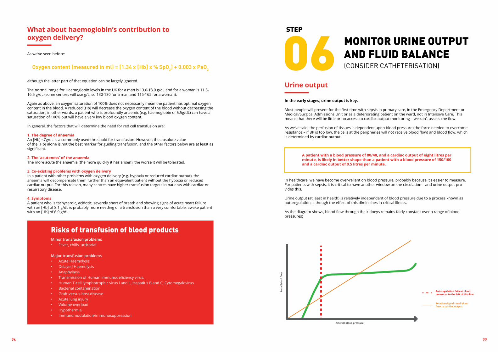
76 77
MONITOR URINE OUTPUT AND FLUID BALANCE(CONSIDER CATHETERISATION)
STEP
06Urine output
In the early stages, urine output is key.
Most people will present for the first time with sepsis in primary care, in the Emergency Department or Medical/Surgical Admissions Unit or as a deteriorating patient on the ward, not in Intensive Care. This means that there will be little or no access to cardiac output monitoring – we can’t assess the flow.
As we’ve said, the perfusion of tissues is dependent upon blood pressure (the force needed to overcome resistance – if BP is too low, the cells at the peripheries will not receive blood flow) and blood flow, which is determined by cardiac output.
A patient with a blood pressure of 80/40, and a cardiac output of eight litres per minute, is likely in better shape than a patient with a blood pressure of 150/100 and a cardiac output of 0.5 litres per minute.
In healthcare, we have become over-reliant on blood pressure, probably because it’s easier to measure. For patients with sepsis, it is critical to have another window on the circulation – and urine output pro-vides this.
Urine output (at least in health) is relatively independent of blood pressure due to a process known as autoregulation, although the effect of this diminishes in critical illness.
As the diagram shows, blood flow through the kidneys remains fairly constant over a range of blood pressures:
As we’ve seen before:
although the latter part of that equation can be largely ignored.
The normal range for Haemoglobin levels in the UK for a man is 13.0-18.0 g/dL and for a woman is 11.5-16.5 g/dL (some centres will use g/L, so 130-180 for a man and 115-165 for a woman).
Again as above, an oxygen saturation of 100% does not necessarily mean the patient has optimal oxygen content in the blood. A reduced [Hb] will decrease the oxygen content of the blood without decreasing the saturation; in other words, a patient who is profoundly anaemic (e.g, haemoglobin of 5.5g/dL) can have a saturation of 100% but will have a very low blood oxygen content.
In general, the factors that will determine the need for red cell transfusion are:
1. The degree of anaemiaAn [Hb] <7g/dL is a commonly used threshold for transfusion. However, the absolute valueof the [Hb] alone is not the best marker for guiding transfusion, and the other factors below are at least as significant.
2. The ‘acuteness’ of the anaemiaThe more acute the anaemia (the more quickly it has arisen), the worse it will be tolerated.
3. Co-existing problems with oxygen delivery In a patient with other problems with oxygen delivery (e.g. hypoxia or reduced cardiac output), the anaemia will decompensate them further than an equivalent patient without the hypoxia or reduced cardiac output. For this reason, many centres have higher transfusion targets in patients with cardiac or respiratory disease.
4. SymptomsA patient who is tachycardic, acidotic, severely short of breath and showing signs of acute heart failure with an [Hb] of 8.1 g/dL is probably more needing of a transfusion than a very comfortable, awake patient with an [Hb] of 6.9 g/dL.
What about haemoglobin’s contribution to oxygen delivery?
Oxygen content (measured in ml) = [1.34 x [Hb] x % SpO2] + 0.003 x PaO2
Minor transfusion problems• Fever, chills, urticarial
Major transfusion problems• Acute Haemolysis• Delayed Haemolysis• Anaphylaxis• Transmission of Human immunodeficiency virus,• Human T-cell lymphotrophic virus I and II, Hepatitis B and C, Cytomegalovirus• Bacterial contamination• Graft-versus-host disease• Acute lung injury• Volume overload• Hypothermia• Immunomodulation/immunosuppression
Risks of transfusion of blood products

78 79
Summary
SEPSIS
01
04
02
05
03
06
GIVE OXYGEN
CONSIDER FLUIDS
TAKE CULTURES
TAKE HB& LACTATE
GIVE ANTIBIOTICS
MONITOR URINE OUTPUT
Spot it. Treat it. Beat it.
Further reading
Trzeciak S, Chansky ME, Dellinger PR et al. Operationalising the use of serum lactate measurement for identifying high risk of death in a clinical practice algorithm for suspected severe sepsis. Acad Emerg Med 2006; 13: 150–1
Daniels R, Nutbeam T, McNamara G, Galvin C. The Sepsis 6 and the severe sepsis resuscitation bundle: a prospective observational cohort study. Emerg Med J 2010; doi:10.1136/emj.2010.095067
British Thoracic Society Emergency Oxygen Group. Guideline for emergency oxygen use in adult patients. Thorax 2008; 63 (Suppl VI): 1-81
NICE NG51
However, the kidneys cannot autoregulate well for changes in blood flow. The relationship here is quite linear – as blood flow to the kidneys falls, so does renal blood flow and therefore urine output.
The urine output is an excellent window on the circulation. As blood flow (cardiac output) falls, so does urine output. This is essential in guiding further fluid challenges, and may identify a problem with the circulation before the blood pressure begins to fall.
Insensible losses
Insensible gain
Can’t we just compare the inputs and the outputs to decide about fluid balance?
The fluid balance chart does not take into account insensible losses and gains.
Skin: about 400-500ml/day. Increased in pyrexia and sweating.
Respiratory: about 400-500ml/day. Increased with hyperventilation, though this effect is decreased if humidified inspired air/oxygen are administered in the context of respiratory distress.
Metabolism: about 400ml/day.
It may seem that the input should be about 400ml greater than the output in a ‘typical’ fluid balance chart. However, the insensible losses are impossible to measure, and what is going on in each patient is so variable that it is meaningless to target a particular number in fluid balance to cover this theoretical difference based on the average relatively well patient.
Instead, we should be guided by:
1. Clinical scenario2. Clinical assessment of fluid status3. Observations4. Markers of end organ perfusion and hydration (mucous membranes, capillary refill, mental status,
urine output, lactate)5. U&Es
The numbers given by daily requirements are just guides; what you actually prescribe is determined by all of these factors.
Don’t forget to start a fluid balance chart once you have put the catheter in!
PRACTICAL TIPSo easily done…

80 81
In this chapter, we will discuss the ongoing care for the patient with sepsis from the ward to Critical Care, including where we are with what was known as ‘Early Goal-Directed Therapy’ (EGDT) in the context of the new recommendations within the updated Surviving Sepsis Campaign in 2016.
The Surviving Sepsis Campaign (SSC) International Guidelines for management of Sepsis and Septic Shock are updated every four years. When first released in 2004, the SSC made more than 50 individual recommendations. Recognising that a long list was unlikely to transform practice, the authors worked with the Institute for Healthcare Improvement (IHI, US) to create two ‘bundles’ of care – one for within the first six hours including basic elements of care and EGDT (see below), and the second to cover the next 24 hours which included largely Critical Care aspects of therapy.
In 2012, the SSC endorsed the original protocol of Early Goal Directed Therapy (EGDT). This strategy was highlighted in 2001 by Rivers et al, as a bundle of treatments to be completed within the first six hours in patients with sepsis, with the aim of normalising the vital signs within a set range of targets utilising a combination of fluid resuscitation, vasopressors, inotropes, blood transfusions and oxygen therapies.
Achieving these goals required transfer to Critical Care, and:• insertion of invasive lines to maintain a central venous pressure (CVP) between 8-10mmHg • using fluids to resuscitate, a mean arterial pressure (MAP) of 65-95mmHg • utilising vasopressors to constrict the dilated blood vessels and maintaining a central venous oxygen
saturation (ScvO2) of >70% • optimising the haemoglobin levels via blood transfusion • if required, giving inotropes to increase the cardiac output.
The 2012 update made amends to the two care bundles as follows: Within three hours, ensure: • lactate is measured• blood cultures are collected• broad spectrum intravenous antibiotics administered and • fluid resuscitation commenced.
Within six hours: • aim for the range of therapeutic goals (physiological targets) using a slightly ‘softer’ adherence to the
EGDT protocol• recheck lactate if the initial level was elevated.
This ‘softer’ application of EGDT was as a result of clinical opinion moving away from EGDT.The 2016 guideline update has softened even further with respect to EGDT after three large multicentre studies across the world – ProCESS, ARISE and ProMISe – failed to show a reduction in mortality when comparing EGDT with standard care. The central venous oxygen saturation (ScvO2) goal, although causing no harm, failed to show a mortality reduction; as in each of the three large trials patients were fluid resuscitated before randomisation, therefore the average baseline ScvO2 was already greater than the target 70% on admission to Critical Care. In essence, patients weren’t as sick at baseline as they were in Rivers’ original study.
Therefore, we have moved further towards using lactate to guide fluid resuscitation instead of ScvO2. This allows more timely intervention as lactate can be measured within a wide range of environments, whereas central pressure measurement was limited to a level three facility (i.e. Critical Care).
INTRODUCTION
ON
GO
ING
CAR
E

82 83
The administration of a combination of broad spectrum antimicrobials within one hour of deterioration and recognition of sepsis or septic shock is vital due to the increased mortality associated with every hour of delay. Ideally, blood cultures should be taken before these are administered, but if these are unobtain-able for any reason it is imperative not to delay the antimicrobials. These antibiotics should be reviewed by a senior clinician between 24 and 72 hours, following any results and sensitivities; the antimicrobials should be narrowed down to treat the specific pathogen. For courses continuing beyond this range, daily assessment should take place to consider the appropriate time to de-escalate from IV to oral. In some patient’s source control may require surgical intervention, removal of invasive lines or even early delivery of baby. Ideally if any of these are required, they are best facilitated as soon as possible, but best practice would be within 12 hours of recognition.
iii. Antimicrobial therapy
iv. Respiratory support
Many patients in septic shock will not only require invasive monitoring and cardiovascular support, but also respiratory support via mechanical ventilation. This could be via non-invasive ventilation (NIV) via a nasal /face mask, or invasive ventilation with an endotracheal tube. Once intubated, a lower tidal volume of 6ml/kg based upon the patient’s predicted body weight is targeted. Other lung protective measures of limiting the plateau airway pressure to 30cm H2O, using lower tidal volumes and using a higher peak end expiratory pressure (PEEP) to recruit alveoli should also be employed. Refractory hypoxemia and ARDS can be managed using ‘recruitment manoeuvres’ or potentially nursing the patient in the prone position rather than the traditional supine. Although lung protective measures are beneficial in sepsis-induced ARDS there is evidence that mortality is higher if novel ventilatory techniques such as high frequency oscillatory ventilation (HFOV) are utilised. Due to the increased vascular permeability in these patients, careful fluid management is paramount and elevating the head of the bed to between 30-45˚ as a preventative measure against the development of Ventilator-Associated Pneumonia (VAP) is beneficial. Ensuring the patient is not too heavily sedated, and using regular sedation ‘holds’ will facilitate weaning protocols from mechanical ventilation by allowing them to breath spontaneously when able, as well as reducing the risk of delirium.
Completion of the Sepsis 6 pathway within 60 minutes is not the end of the treatment for patients with sepsis. In many cases there will still be hypoperfusion within the tissues despite repeated crystalloid boluses. This is evidenced by a persistently elevated lactate level or hypotension <90mmHg despite crystalloid boluses of 30ml/kg – the presence of both together diagnoses septic shock. Fluid management within sepsis can be a fine balance, as too much fluid has also been linked to a higher mortality rate, hence the need for early escalation and referral to Critical Care for the consideration of vasopressors and inotropes.
There is little evidence to support the use of any one type of crystalloid as there are few direct compar-isons between isotonic saline (e.g. 0.9% saline) and balanced salt solutions, however as hyperchloremia should be avoided this often leads to the choice of a balanced salt solution (e.g. Hartmanns or Plasmalyte), since 0.9% carries a high chloride ion content.
An initial fluid challenge of 500mls given rapidly in under 15 minutes, followed by further challenges guided by the repeated sampling of lactate, is recommended for patients with a high lactate or hypotension. If the initial lactate is greater than 4mmol/l, repeated lactate measurements after each 10ml/kg bolus are recommended to guide resuscitation. Several studies suggest that Albumin can also be effectively utilised as part of fluid management in a patient with septic shock, but is not normally as readily available or as cost effective as a crystalloid.
What is clear, however, is the evidence against using starches and gelatins (such as Hydroxyeth-yl starch (HES/HAES) / Voluven or Gelofusine) as these can be detrimental in patients with sepsis, with a higher risk of acute kidney injury. Patients who are bleeding will obviously require blood products, however in those who are not bleeding evidence supports transfusing only when the Hb is <7.0g/dl. There is some evidence that human albumin solution (HAS) may be beneficial in resuscitation due to its physiological nature in reducing water shift out of the circulation, but as yet HAS is not routinely recommended as the benefits have not been found to outweigh either risks or cost.
A continued state of tissue hypoperfusion despite 30ml/kg of fluid may require the introduction of vasoactive medications. Aiming for a MAP of 65mmHg, the first-choice vasopressor recommended by the SSC is norepinephrine (historically called noradrenaline). If blood pressure is not improved with the use of norepinephrine alone, some centres add vasopressin as a second line, though the evidence base for this is weak. Both require the insertion of a central venous catheter and transfer to Critical Care. Some patients will require a higher MAP to maintain their renal function if they are normally hypertensive. Potentially, the Critical Care Outreach Team may facilitate this treatment commencing at the point of deterioration to help stabilise the patient before transfer to a level two or three facility.
The SSC recommends that steroids be considered (i.e. not recommended!) in patients with refractory shock. For many centres, pragmatically this means that they are considered at a similar time to adding vasopressin. Steroids can lead to an increase in hyperglycaemia which is best avoided. By early interven-tion and increasing the systemic vascular resistance with norepinephrine, the aim is to both increase the blood pressure to better perfuse the other major organs but also to increase the oxygen delivery to the tissues and reduce the anaerobic respiration, demonstrated by a decrease in lactate. It is, however, important that the patient is well filled first with crystalloids to 30ml/kg as described above. A bedside ‘echo’ can be beneficial in these patients to gain a better all-round picture, as well as monitoring of the haemodynamic status and cardiac output via equipment such as a PiCCO or LiDCO®.
i. Fluid management
ii. Vasoactive drugs
Many patients in septic shock experience a prolonged period of reduced cardiac output or hypoperfu-sion, which can lead to a prerenal acute kidney injury (AKI). Careful exclusion of any nephrotoxic drugs is important, but, if essential, doses of such drugs should be adjusted to limit further damage. Treatment goals include achieving haemodynamic stability through the restoration of normal circulating volume and the use of vasopressors and inotropes when needed, and treating the precipitating cause. Fluid overload is associated with poorer outcomes and can be the trigger for starting continuous renal replacement therapy (CRRT) to remove excess fluid and aid fluid balance. CRRT is recommended over intermittent RRT in unstable patients and is therefore the method of choice in most Critical Care environments.
v. Renal therapy

84 85
SUMMARYHopefully this brief overview has given an insight into the ongoing care required following the first 60 minutes from recognition of sepsis, including the changes to the international guidelines over the last few years. Early recognition, followed by escalation and treatment in the initial stages should be backed up by a timely referral to Critical Care if the initial resuscitation efforts do not stabilise the patient.
Further reading
Dellinger, R.P. & Schorr, C.A. A Users’ Guide to the 2016 Surviving Sepsis Guidelines. Critical Care Medicine, March 2017:45(3):381-385
Mouncey, P.R. et al Protocolised Management in Sepsis (ProMISe): a multicentre randomised controlled trial of the clinical effectiveness and cost-effectiveness of early, goal-directed, protocolised resuscitation for emerging septic shock. Health Technol Assess, 2015: Nov,19(97)
Rhodes, A et al Surviving Sepsis Campaign: International Guidelines for Management of Sepsis and Septic Shock: 2016. Critical Care Medicine, March 2017:45(3) DOI: 10.1097/CCM.0000000000002255
The ARISE investigators, Goal-Directed Resuscitation for Patients with Early Septic Shock, NEJM Oct 2014; 371: 1496-506
The ProCESS investigators, A Randomised Trial of Protocol-Based Care for Early Septic Shock, NEJM May 2014; 370: 1683-1693
Early establishment of enteral nutrition is vital in any critically ill patient, and may be facilitated by the administration of prokinetics such as metoclopramide or erythromycin. Such medication, though evidence to support their use is slightly questionable, might assist with patients displaying signs of feeding intolerance due to reduced gut perfusion, gastroparesis or just due to the amount of sedation administered and help prevent aspiration of the gastric contents.
Throughout this traumatic time, communication with the patient and their families is of vital importance. During any admission to Critical Care, prognosis and any goals for care will be discussed. Ensuring an open and honest relationship between staff and families will facilitate any end of life planning if required, and will also assist with the expectations for recovery. At this challenging time, it can be helpful to both the family members and the patient to maintain a diary of care, this can be looked at retrospectively and assist to fill in long gaps in time for the patient when they were sedated. This in turn can ease some of the Post Sepsis Syndrome symptoms they might experience in their recovery period. On discharge from the Critical Care environment, follow up should ideally be provided by the Critical Care Outreach team (or equivalent), with regular reviews on discharge to ensure no further problems develop and with a view to preventing re-admission. Literature on sepsis and its after-effects should be provided to the patient and family to provide safety netting advice, but also to inform of the possible side effects that may occur over the next few months.
vi. Nutrition
vii. Communication

86 87
Neutropenic sepsis is time-critical and potentially fatal. It occurs in patients who are immunocompromised due to their anticancer or other immunomodulatory therapies. These therapies suppress the body’s normal response to infection, and the bone marrow cannot maintain production of white cells at the rate required. Neutropenic sepsis can lead to significant mortality in adults – cited as up to 21% but likely much higher. The mortality is significantly higher if treatment is delayed or Critical Care therapy becomes necessary. As the volume of patients receiving systemic cytotoxic therapies increases, the number of patients developing neutropenic sepsis will also rise.
Excellent communication with at risk patients is required to raise their awareness of the risk of sepsis, and their awareness of the symptoms that mean they should seek immediate medical review. Increasingly these therapies are delivered in a day case environment – safety netting advice regarding when to seek medical assistance is of vital importance to ensure early help is sought.
On assessment, a diagnosis of neutropenic sepsis relies upon a neutrophil count of 0.5 x 10⁹/l or less. Whilst the diagnosis of sepsis is the same according to physiological and biochemical parameters as in the general population, a high index of suspicion should be maintained in patients with either a temperature >38˚C or other signs of deterioration such as a NEWS >3. If treatment is reliant on the return of blood results, this can lead to significant delays in treatment, and these patients who are at great risk of sepsis will tend to deteriorate more rapidly than their counterparts without neutropenia. Therefore, rapid assessment and escalation onto the Sepsis 6 pathway as soon as neutropenic sepsis is suspected is recommended.
Febrile neutropenic patients are usually recognised and their treatment started early. Patients with non-febrile neutropenia will often deteriorate further before being recognised, highlighting the importance of a standardised, graduated response system to deteriorating patients even in specialist areas.
Maternal mortality from sepsis varies hugely depending on access to safe and affordable healthcare. Maternal mortality remains extremely high at around 400 per 100,000 live births in low middle income countries (LMICs) as compared with developed countries, where the mortality is lower- for example 8 per 100,000 live births in the UK. This discrepancy is unacceptable, and there is evidence to suggest that maternal sepsis is on the increase, with an estimated 30,000 women dying from sepsis each year globally.
THEIMMUNOCOMPROMISED PATIENT
THE PREGNANTPATIENT
01
02
SPEC
IAL
PATI
ENT
GR
OU
PS

88 89
Sepsis is a major cause of death in the under-five population worldwide, particularly in Sub-Saharan Africa and Asia where many sepsis-related deaths are preventable. This group of patients is vulnerable, and they often present with atypical or vague signs and symptoms, potentially resulting in delayed or inappropriate treatment. You should maintain a high index of suspicion in children, and have a low threshold for admis-sion and observation. It is important to take a detailed history and to listen to the concerns of the parent or carer as they know their child best.
In young children and infants, language and understanding can be a communication barrier, you may need to take a collateral history from a parent or relative and use other means to communicate. If discharging a child or infant from your care, ensure safety-netting advice has been given and that the care givers know the warning signs of sepsis and when they should seek medical help.
Due to the nature of childhood illnesses, a fever can be quite common. Screening should take place for all infants and children who look unwell or are feverish, particularly with a temperature greater than 39˚C, but remembering that in those infants younger than three months a temperature of just 38˚C or more is a Red Flag. A low temperature can be more concerning and is a Red Flag in all children and infants under 12 years.
Children can often compensate well during a disease process like sepsis. This means that subtle changes can be missed until they suddenly decompensate and become extremely unwell. Early escalation to senior support is vital, and use of a Paediatric Early Warning Score (PEWS) with an appropriate escalation plan will ensure this happens.
THE CHILDOR INFANT03
ANY CHILD WHO:1
Is breathing very fast2
Has a ‘fit’ or convulsion3
Looks mottled, bluish, or pale
4 Has a rash that does not fade when you press it
5 Is very lethargic or difficult to wake
6 Feels abnormally cold to touch
MIGHT HAVE SEPSIS
ANY CHILD UNDER 5 WHO:1
Is not feeding 2
Is vomiting repeatedly3
Hasn’t had a wee or wet nappy for 12 hours
MIGHT HAVE SEPSIS
Call 999 and ask: could it be sepsis?
If you’re worried they’re deteriorating call 111 or see your GP
JUST ASK“COULD IT BE SEPSIS?”
IT’S A SIMPLE QUESTION, BUT IT COULD SAVE A LIFE. The UK Sepsis Trust registered charity number (England & Wales) 1158843
Sepsis that occurs during pregnancy is termed ‘maternal sepsis’. If it develops within six weeks of delivery it is termed postpartum, or ‘puerperal’ sepsis. Sepsis is one of the leading causes of direct maternal death in the UK. This is partly because the immunological changes naturally occurring during pregnancy together with the increased exposure to healthcare, and additional risks such as with premature rupture of membranes or gestational diabetes, mean a pregnant woman is more susceptible to infection than her non-pregnant counterpart. The natural adaptations to the body with pregnancy may mask the signs and symptoms of infection or an acute abdomen until the woman deteriorates.
Sepsis can be as a direct result of the pregnancy or an indirect cause unrelated to the pregnancy, for instance pneumonia or a urinary infection. Following a number of maternal deaths from the H1N1 influenza pandemic, the flu vaccine is now routinely offered to pregnant women in industrialised countries. The commonest sources for sepsis are urinary tract prenatally and genital tract postnatally. E. coli accounts for one third of episodes of sepsis, and infection with group A streptococcus can rapidly progress to septic shock.
Due to the physiological changes in pregnancy, the National Early Warning Score (NEWS) is not designed for use in pregnant patients. Use of a modified obstetric early warning score (MOEWS) alongside the Maternal Sepsis screening tool is recommended to facilitate the early recognition and escalation of deteriorating maternal patients.
Risk factors for the development of sepsis in pregnancy
The maternal sepsis screening tools are not only for use in patients who are currently pregnant, but also for those who have recently been pregnant and are within the post-partum period.
A sepsis screening tool may also consider foetal distress. A foetal heart rate >160 bpm is of significant concern and is considered as an equal trigger when screening for sepsis as the woman looking sick. The Red Flags for a pregnant woman are the same as the Red Flags in their non-pregnant counterpart.
Any pregnant woman with suspicion of sepsis requires an urgent senior review and multidisciplinary care. It is highly possible that the timing of delivery may need to be influ-enced by this diagnosis. Consideration should be given toward prophylactic treatment of the new-born if particularly at risk of neonatal sepsis. .
Specific guidance on managing sepsis in the pregnancy and the puerperium are available from the Royal College of Obstetricians and Gynaecologists’ Green Top series.
90 91
SUMMARY• The treatment principles for patients with sepsis are identical regardless of the cause. • Initial assessment and resuscitation should follow the ABCDE format with the application of the Sepsis
Screening and Action Tool. • Patients should be managed using the Sepsis 6 approach. Liaison with Critical Care should be timely,
particularly in the presence of septic shock or multi-organ failure.• Patients with pneumonia represent the largest group of patients with sepsis. • Common causes of sepsis aside from pneumonia include gastrointestinal pathology, urinary tract,
biliary tract and skin infections. • Sources will vary in the pregnant patient.• Remember to keep an open mind when assessing a patient presenting with sepsis.• The importance of consultation with microbiologists locally who will be aware of pathogens and
resistance patterns in your own institutions cannot be over emphasised. • Most organisations now have their recommended first-line empiric treatments for common infections
on their intranet sites.
Further reading
Bonet, M et al “Towards a consensus definition of maternal sepsis: results of a systematic review and expert consultation” Reproductive Health 2017 14:67 DOI 10.1186/s12978-017-0321-6
Naveda Romero, OE, & Naveda Meléndez, AF. “Fluid overload and kidney failure in children with severe sepsis and septic shock: a cohort study” Arch Argent Pediatr 2017; 115(2):118-124
Sarafidis, K et al “Urine metabolomics in neonates with late-onset sepsis in a case-control study” Scientific Reports 2016 7:45506 DOI: 10.1038/srep45506
The Lancet commission on Global Surgery, 2015. Accessed 7/4/2017 at www.lancetglobalsurgery.org
A senior review by a doctor of ST4 or higher is an integral part of the Sepsis 6 in patients under 12. In the case of a critically unwell child with sepsis, non-specialist areas will require sup-port from their local PICU and retrieval teams. Communication using a tool such as SBAR or RSVP will ensure the urgency of the situation is relayed effectively, and no information is missed.
Fluid management in children with sepsis can be difficult. If the infant or child remains decompensated after two initial 10ml/kg fluid boluses, Critical Care advice regarding inotropic sup-port should be sought – usually if needed using dopamine or epinephrine. The fluid of choice is usually sodium chloride 0.9% for the initial 10ml/kg boluses in paediatrics, although if blood is being lost this will also need replacing.
Whilst the management of the neonate with sepsis is beyond the scope of this book, special consideration is needed for this group due to their immature immune system making them more susceptible to infec-tion – particularly respiratory, urinary or line-based infections. Any underlying disease process or low birth weight contributes to a higher mortality; any suspected or proven infection in the mother during the third trimester will often indicate a need for prophylactic treatment in the new-born as well as in twin pregnan-cies where one twin develops an infection or sepsis shortly after birth. If a previous child developed an invasive strep B infection, this also puts a subsequent newborn at greater risk of developing the same.
Management of intravenous access, fluids and antibiotics in these patients is a specialised field. Metabolic changes including lactic acidosis and increased glucose requirements are recognised early responses to sepsis in the neonate, and other differences such as a depletion in vitamin B compounds and glutamine have also been noted. More research in these areas is required.

92 93
The management of infections includes multiple facets, but in essence centres around identification of the pathogen, control of any source of infection including judicious use of antimicrobials where necessary, and management of any sequelae of infection including sepsis.
Source control is, therefore, an essential part of managing sepsis where this is practicable. In this context, source control means physical removal of the source, such as drainage of abdominal collections or surgical removal of infected tissue. Source control will also include re-establishing flow of fluid which has become obstructed – for example relief of biliary obstruction. To achieve effective and rapid source control may therefore demand close liaison with colleagues in surgery and radiology. However, for some conditions (such as pneumonia) where there is neither a collection of infected material amenable to drainage nor a presence of prosthetic material which can be removed, source control is not possible. Here, antimicrobial therapy, usually considered as an adjunct to source control, becomes the only way of controlling the trigger for sepsis.
Initial antibiotic choice (assuming, as in the majority of cases, the likely pathogen is bacterial) is usually based on the suspected focus of infection, determined through clinical suspicion supported by radiolog-ical and microbiological evidence. It is vital that the right antimicrobials are given to control the infection and fight the organisms present, and this will often mean initially using broad spectrum ‘best guess’ agents with a later focus on a narrower spectrum when (if) the organism becomes known. Organisms take a while to grow; therefore taking the right sample and sending it in the right container as soon as possible following the diagnosis of sepsis can help to identify the likely pathogens in a timely fashion.
Taking samples
Wherever possible, a sterile technique should be adopted when taking samples for microbiological inves-tigation. Because a plethora of microorganisms are ubiquitous within our environment, they can easily contaminate samples, resulting in the predominant organism isolated from a culture being an environ-mental contaminant rather than a true pathogen. In the case of blood cultures, commensal skin flora can be picked up instead of true pathogens. If not correctly interpreted, such false positives can result in inappropriate antimicrobial prescribing, which could leave a patient undertreated, and/or put them at risk of acquisition of a multidrug resistant organism or C. difficile infection.
Advice on taking appropriate specimens can usually be obtained from local microbiology or infectious diseases teams. Once a specimen has been taken, it must be placed in a container that maintains viability of any pathogenic microorganisms during transit. For example, formalin kills organisms, so placing any samples in formalin-containing specimen pots is unlikely to yield any pathogens. Likewise, some viruses are easily inactivated by detergents (for example the influenza virus), swab sticks or the transport material (e.g. gel, activated charcoal) contained within wound swab containers. Using the right container for the suspected organism is essential.
Urgent samples
Many laboratories operate an on-call system for urgent microbiological specimens (e.g. tissues taken in theatre, CSF) – if any samples are urgent the laboratory team must be called to come in and process the samples. This is usually not necessary for blood cultures, but it is always better to check with your local laboratory. Many laboratories don’t place blood cultures in the incubator immediately if received overnight – we would encourage this practice to change.M
ICR
OB
IOLO
GY

94 95
Identifying pathogens
These details are important, as different organisms tend to affect and infect different parts of the body, and require different conditions in order to grow. Prior knowledge of the type of infection suspected and the site affected helps the laboratory scientists and clinicians to determine which type of organisms they need to look out for, which can mean using different types of agar plates and techniques such as molecu-lar PCR (polymerase chain reaction) and serological tests.
Recent antimicrobial therapy can affect the ability to detect organisms which are susceptible to the antibiotics given, though modern blood culture media attempt to bind antibiotics present to reduce their masking effect.
Sepsis can occur in patients that have travelled abroad; common examples of travel associated infections that can cause sepsis include malaria, TB and typhoid fever. Some of these infections are extremely infec-tious e.g. typhoid (due to Salmonella typhi and Salmonella paratyphi), TB, viral haemorrhagic fevers (e.g. Ebola) and MERS-CoV infections. These can result in outbreaks, so it is always wise to consult local policies and liaise with your infection specialists – the microbiologist or infectious diseases clinician. Providing a travel history not only allows the infection specialist to advise on the most appropriate tests, it can help to prevent ongoing transmission of infection to laboratory staff, other patients and other staff, including yourself.
Isolating pathogens
Methods of identifying pathogens depend on what is trying to be identified. Most bacterial organisms will grow on standard culture media. Anaerobes, fastidious bacteria, slow growing organisms and most myco-bacteria (including TB) require different media and special growing conditions such as temperature and atmospheric conditions. Other organisms are so difficult to grow they need to be sent away to a specialist laboratory for testing, or require that alternative techniques such as serology and PCR be used. Often this can add to the time taken to identify an organism, hence the importance of clinical suspicion of the source of infection and likely pathogens affecting the patient being conveyed to the infection teams.
If the incubator containing a blood culture sample detects the production of carbon dioxide by bacteria, it will flag the sample as positive. Specimens are then processed and usually spread onto agar plates, or put into special liquid media for harder to grow organisms. When organisms grow, they appear as “colonies” on the plate, which can then be tested for identification and antimicrobial susceptibility testing.
Sometimes there is more than one organism present, in which case the individual organisms need to be “picked off” and cultured again to ensure that there is a pure growth of organism. This helps to ensure that we do not get false results with regards to identification and antimicrobial susceptibility testing, which can lead to inappropriate antibiotic treatment.
Mixed E. coli (pink colonies) and Klebsiella pneumoniae (blue colonies) on a chromogenic agar plate
Pure growth of Pseudomonas putida on a blood agar plate
Labelling of samples
All samples must be labelled correctly to avoid rejection once they reach the laboratory. There are UK standards for specimens, required to ensure that the sample has come from the patient stated on the request form and to ensure traceability back to the requestor and/or person taking the sample in case of queries.
The minimum information required on the sample container is:
Patient’s forename and surname
Important clinical findings e.g. prosthetic heart valve in situ, known infectious condition such as TB or HIV
Location (i.e. ward or department)
Working diagnosis e.g. pneumonia
Date and time of location
Travel history over past 12 months
Type of specimen
Printed (legible) name and registration number of both requestor and person taking sample
Recent and current antimicrobial therapy
The request form must also contain the same information as well as:
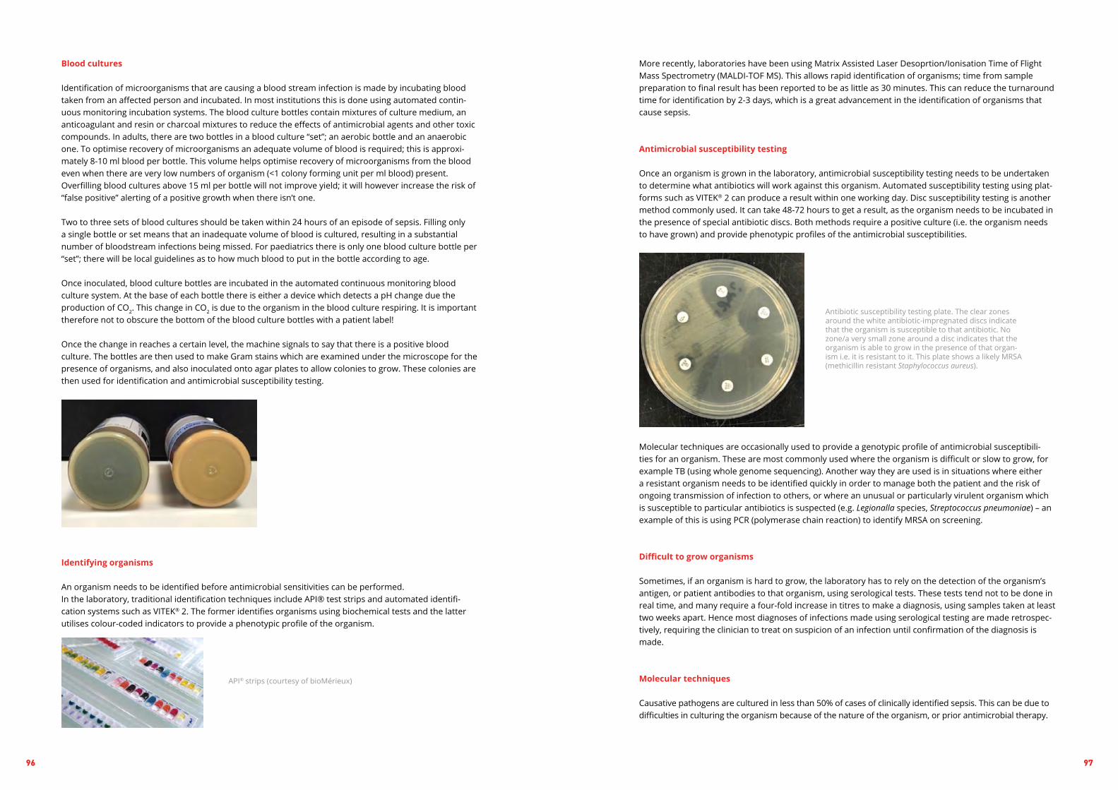
96 97
Antibiotic susceptibility testing plate. The clear zones around the white antibiotic-impregnated discs indicate that the organism is susceptible to that antibiotic. No zone/a very small zone around a disc indicates that the organism is able to grow in the presence of that organ-ism i.e. it is resistant to it. This plate shows a likely MRSA (methicillin resistant Staphylococcus aureus).
More recently, laboratories have been using Matrix Assisted Laser Desoprtion/Ionisation Time of Flight Mass Spectrometry (MALDI-TOF MS). This allows rapid identification of organisms; time from sample preparation to final result has been reported to be as little as 30 minutes. This can reduce the turnaround time for identification by 2-3 days, which is a great advancement in the identification of organisms that cause sepsis.
Antimicrobial susceptibility testing
Once an organism is grown in the laboratory, antimicrobial susceptibility testing needs to be undertaken to determine what antibiotics will work against this organism. Automated susceptibility testing using plat-forms such as VITEK® 2 can produce a result within one working day. Disc susceptibility testing is another method commonly used. It can take 48-72 hours to get a result, as the organism needs to be incubated in the presence of special antibiotic discs. Both methods require a positive culture (i.e. the organism needs to have grown) and provide phenotypic profiles of the antimicrobial susceptibilities.
Molecular techniques are occasionally used to provide a genotypic profile of antimicrobial susceptibili-ties for an organism. These are most commonly used where the organism is difficult or slow to grow, for example TB (using whole genome sequencing). Another way they are used is in situations where either a resistant organism needs to be identified quickly in order to manage both the patient and the risk of ongoing transmission of infection to others, or where an unusual or particularly virulent organism which is susceptible to particular antibiotics is suspected (e.g. Legionalla species, Streptococcus pneumoniae) – an example of this is using PCR (polymerase chain reaction) to identify MRSA on screening.
Difficult to grow organisms
Sometimes, if an organism is hard to grow, the laboratory has to rely on the detection of the organism’s antigen, or patient antibodies to that organism, using serological tests. These tests tend not to be done in real time, and many require a four-fold increase in titres to make a diagnosis, using samples taken at least two weeks apart. Hence most diagnoses of infections made using serological testing are made retrospec-tively, requiring the clinician to treat on suspicion of an infection until confirmation of the diagnosis is made.
Molecular techniques
Causative pathogens are cultured in less than 50% of cases of clinically identified sepsis. This can be due to difficulties in culturing the organism because of the nature of the organism, or prior antimicrobial therapy.
Identifying organisms
An organism needs to be identified before antimicrobial sensitivities can be performed.In the laboratory, traditional identification techniques include API® test strips and automated identifi-cation systems such as VITEK® 2. The former identifies organisms using biochemical tests and the latter utilises colour-coded indicators to provide a phenotypic profile of the organism.
Blood cultures
Identification of microorganisms that are causing a blood stream infection is made by incubating blood taken from an affected person and incubated. In most institutions this is done using automated contin-uous monitoring incubation systems. The blood culture bottles contain mixtures of culture medium, an anticoagulant and resin or charcoal mixtures to reduce the effects of antimicrobial agents and other toxic compounds. In adults, there are two bottles in a blood culture “set”; an aerobic bottle and an anaerobic one. To optimise recovery of microorganisms an adequate volume of blood is required; this is approxi-mately 8-10 ml blood per bottle. This volume helps optimise recovery of microorganisms from the blood even when there are very low numbers of organism (<1 colony forming unit per ml blood) present. Overfilling blood cultures above 15 ml per bottle will not improve yield; it will however increase the risk of “false positive” alerting of a positive growth when there isn’t one.
Two to three sets of blood cultures should be taken within 24 hours of an episode of sepsis. Filling only a single bottle or set means that an inadequate volume of blood is cultured, resulting in a substantial number of bloodstream infections being missed. For paediatrics there is only one blood culture bottle per “set”; there will be local guidelines as to how much blood to put in the bottle according to age.
Once inoculated, blood culture bottles are incubated in the automated continuous monitoring blood culture system. At the base of each bottle there is either a device which detects a pH change due the production of CO2. This change in CO2 is due to the organism in the blood culture respiring. It is important therefore not to obscure the bottom of the blood culture bottles with a patient label!
Once the change in reaches a certain level, the machine signals to say that there is a positive blood culture. The bottles are then used to make Gram stains which are examined under the microscope for the presence of organisms, and also inoculated onto agar plates to allow colonies to grow. These colonies are then used for identification and antimicrobial susceptibility testing.
API® strips (courtesy of bioMérieux)
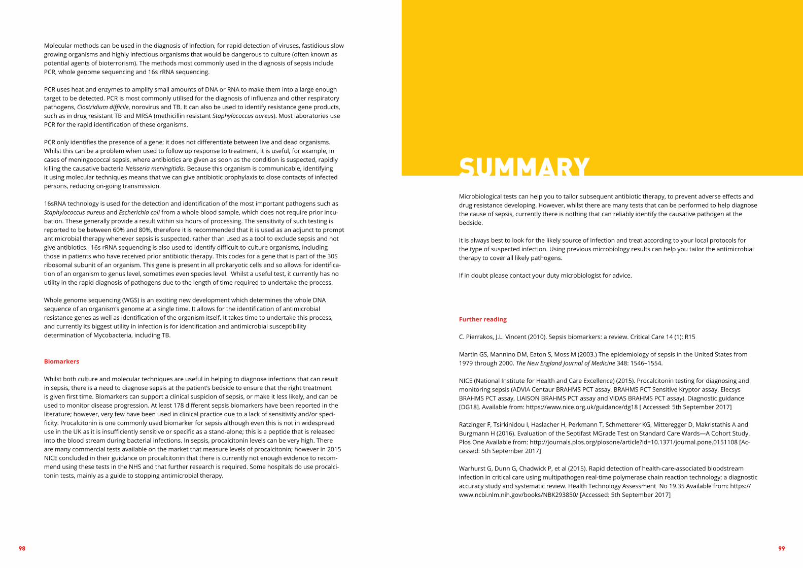
98 99
SUMMARYMicrobiological tests can help you to tailor subsequent antibiotic therapy, to prevent adverse effects and drug resistance developing. However, whilst there are many tests that can be performed to help diagnose the cause of sepsis, currently there is nothing that can reliably identify the causative pathogen at the bedside.
It is always best to look for the likely source of infection and treat according to your local protocols for the type of suspected infection. Using previous microbiology results can help you tailor the antimicrobial therapy to cover all likely pathogens.
If in doubt please contact your duty microbiologist for advice.
Further reading
C. Pierrakos, J.L. Vincent (2010). Sepsis biomarkers: a review. Critical Care 14 (1): R15
Martin GS, Mannino DM, Eaton S, Moss M (2003.) The epidemiology of sepsis in the United States from 1979 through 2000. The New England Journal of Medicine 348: 1546–1554.
NICE (National Institute for Health and Care Excellence) (2015). Procalcitonin testing for diagnosing and monitoring sepsis (ADVIA Centaur BRAHMS PCT assay, BRAHMS PCT Sensitive Kryptor assay, Elecsys BRAHMS PCT assay, LIAISON BRAHMS PCT assay and VIDAS BRAHMS PCT assay). Diagnostic guidance [DG18]. Available from: https://www.nice.org.uk/guidance/dg18 [ Accessed: 5th September 2017]
Ratzinger F, Tsirkinidou I, Haslacher H, Perkmann T, Schmetterer KG, Mitteregger D, Makristathis A and Burgmann H (2016). Evaluation of the Septifast MGrade Test on Standard Care Wards—A Cohort Study. Plos One Available from: http://journals.plos.org/plosone/article?id=10.1371/journal.pone.0151108 [Ac-cessed: 5th September 2017]
Warhurst G, Dunn G, Chadwick P, et al (2015). Rapid detection of health-care-associated bloodstream infection in critical care using multipathogen real-time polymerase chain reaction technology: a diagnostic accuracy study and systematic review. Health Technology Assessment No 19.35 Available from: https://www.ncbi.nlm.nih.gov/books/NBK293850/ [Accessed: 5th September 2017]
Molecular methods can be used in the diagnosis of infection, for rapid detection of viruses, fastidious slow growing organisms and highly infectious organisms that would be dangerous to culture (often known as potential agents of bioterrorism). The methods most commonly used in the diagnosis of sepsis include PCR, whole genome sequencing and 16s rRNA sequencing.
PCR uses heat and enzymes to amplify small amounts of DNA or RNA to make them into a large enough target to be detected. PCR is most commonly utilised for the diagnosis of influenza and other respiratory pathogens, Clostridium difficile, norovirus and TB. It can also be used to identify resistance gene products, such as in drug resistant TB and MRSA (methicillin resistant Staphylococcus aureus). Most laboratories use PCR for the rapid identification of these organisms.
PCR only identifies the presence of a gene; it does not differentiate between live and dead organisms. Whilst this can be a problem when used to follow up response to treatment, it is useful, for example, in cases of meningococcal sepsis, where antibiotics are given as soon as the condition is suspected, rapidly killing the causative bacteria Neisseria meningitidis. Because this organism is communicable, identifying it using molecular techniques means that we can give antibiotic prophylaxis to close contacts of infected persons, reducing on-going transmission.
16sRNA technology is used for the detection and identification of the most important pathogens such as Staphylococcus aureus and Escherichia coli from a whole blood sample, which does not require prior incu-bation. These generally provide a result within six hours of processing. The sensitivity of such testing is reported to be between 60% and 80%, therefore it is recommended that it is used as an adjunct to prompt antimicrobial therapy whenever sepsis is suspected, rather than used as a tool to exclude sepsis and not give antibiotics. 16s rRNA sequencing is also used to identify difficult-to-culture organisms, including those in patients who have received prior antibiotic therapy. This codes for a gene that is part of the 30S ribosomal subunit of an organism. This gene is present in all prokaryotic cells and so allows for identifica-tion of an organism to genus level, sometimes even species level. Whilst a useful test, it currently has no utility in the rapid diagnosis of pathogens due to the length of time required to undertake the process.
Whole genome sequencing (WGS) is an exciting new development which determines the whole DNA sequence of an organism’s genome at a single time. It allows for the identification of antimicrobial resistance genes as well as identification of the organism itself. It takes time to undertake this process, and currently its biggest utility in infection is for identification and antimicrobial susceptibility determination of Mycobacteria, including TB.
Biomarkers
Whilst both culture and molecular techniques are useful in helping to diagnose infections that can result in sepsis, there is a need to diagnose sepsis at the patient’s bedside to ensure that the right treatment is given first time. Biomarkers can support a clinical suspicion of sepsis, or make it less likely, and can be used to monitor disease progression. At least 178 different sepsis biomarkers have been reported in the literature; however, very few have been used in clinical practice due to a lack of sensitivity and/or speci-ficity. Procalcitonin is one commonly used biomarker for sepsis although even this is not in widespread use in the UK as it is insufficiently sensitive or specific as a stand-alone; this is a peptide that is released into the blood stream during bacterial infections. In sepsis, procalcitonin levels can be very high. There are many commercial tests available on the market that measure levels of procalcitonin; however in 2015 NICE concluded in their guidance on procalcitonin that there is currently not enough evidence to recom-mend using these tests in the NHS and that further research is required. Some hospitals do use procalci-tonin tests, mainly as a guide to stopping antimicrobial therapy.

100 101
Human factors (also termed ergonomics) has fast become an important scientific discipline in safety-criti-cal industries such as the military and airline industry. In these complex organisations, non-technical skills including leadership, decision-making and performance all influence how people behave within a system. In recent years, the importance of human factors has been increasingly recognised within the National Health Service (NHS), and human factors teaching underpins patient safety and quality improvement to promote high quality patient care.
There has been a culture shift within the NHS to recognise the importance of human factors at every level within the health service. There is more focus on human interaction with equipment and standardisation of procedures within the NHS to help reduce medical errors and improve patient safety.
The Swiss cheese model by James Reason is used across many industries to describe the causation of accidents. It uses the analogy of Swiss cheese to demonstrate how the holes in the cheese are not usually aligned. These represent potential hazards. It is only when all the holes in each layer align that an accident or adverse event can occur. Another way to visualise errors is to use the tip of the iceberg model. This describes an adverse outcome as the tip of the iceberg, while below the tip are many less visible errors, which occur more frequently. This model was used to promote road safety through means of wearing a seatbelt.
The Swiss Cheese model underpinning errors in healthcare, adapted with permission from James ReasonHU
MA
N F
ACT
OR
SIN
SEP
SIS

102 103
CASE STUDYYou are the SHO on call for intensive care and you are fast bleeped to a cardiac arrest on the ward. When you arrive the scene is chaotic, you do not introduce yourself to everyone, there are many people there already and they do not introduce themselves to you. You are unsure who is in charge, who is a doctor or who is a nurse. You ask a colleague, another junior doctor with a name badge on what you can do to help. He asks you to take blood. This task is difficult and you decide to do a femoral stab. You give the blood to your colleague and he puts it in blood bottles.
After sending the blood your colleague realises he has sent the blood and labelled it with another patient’s details. There were 2 patients with similar names in that bay and the wrong patient stickers were in this patient notes. He calls the lab and tells them immediately about his error. He sent blood for cross match, which could potentially have resulted in disaster.
There were many small errors here and we can see how the holes in the Swiss Cheese are starting to line up. The situation is chaotic and no one took the time to pause, introduce themselves or allocate a team leader and team roles. This is often the case in emergency situations when organisation and structure become even more important. You were then asked to perform a task which you did to help save the patient’s life. However, looking back, you did not check the patients name or see if they had a wrist band on, you then handed the blood to someone else to label. When doing a cross match, no matter how life threatening the situation, the person taking the blood should label the blood themselves against the patient’s wristband. The blood was then labelled incorrectly against the patient label, and this belonged to a different patient.
Luckily the junior doctor labelling the blood realised his mistake. A root cause analysis was done by the blood bank and the team members were educated about the errors that occurred.
Sepsis is a complex condition associated with poor outcomes when the diagnosis is delayed and treat-ment is not started promptly. Time pressures, high stress levels and an unpredictable clinical environ-ment often compound managing such sick patients. Many different teams and healthcare workers will be involved with the care of patients with sepsis, and effective leadership and an organised team approach are vital in the timely delivery of treatment. An understanding of the environment we work in, the role of individuals working with one another and the interactions we have are vital if we are to succeed in opti-mising patient safety and delivering high quality care to patients presenting with sepsis.
We are only human, and therefore we are all bound to make mistakes. Being aware of human errors through human factors training can help us to decrease the risk of both potential hazards and adverse events form occurring. There are a number of important elements involved in human factors, which we can address to improve patient safety. These include: cognition, distraction, physical demands, the envi-ronment, product design, teamwork and process design.
The National Patient Safety Agency (NPSA) has produced a series of booklets called ‘Design for Patient Safety’ which discuss these elements in more detail. These can be found on their website alongside useful alerts, guidance and toolkits for health professionals to help improve patient safety in their organisation. http://www.npsa.nhs.uk/nrls/medication-zone/design-for-patient-safety-medication-topics/.
The NHS demands that high-quality care be delivered to patients through a safe, effective and free health-care system. Human factors affect the entire NHS from individuals to teams within it. People are often working in a dynamic and unpredictable environment and are making difficult decisions under pressure. Effective leadership is vital alongside education and training to raise awareness around the importance of human factors in healthcare and promote a safety culture in the effective management of sepsis.
The lack of human factors training in the National Health Service (NHS) was highlighted after the tragic death of a patient called Elaine Bromiley during routine surgery. Her death was largely attributed to a breakdown in human factors through a lack of leadership, teamwork and communication. She was the wife of Martin Bromiley, a pilot who specialised in human factors training. After his wife’s death in 2005, Martin focused on raising awareness about human factors and founded the Clinical Human Factors Group (CHFG) to improve safety within the NHS (www.chfg.org).
A film of this case study can be viewed at: http://www.institute.nhs.uk/safer_care/ general/human_factors.html
Watch

104 105
For many patients surviving sepsis, leaving hospital is not the end of their problems. Between one-fifth and one-half of survivors of a hospital admission with sepsis experience long-term sequelae.
These are particularly prevalent when a person has spent time in Critical Care, is elderly or has significant health issues before sepsis. For some, reasons for these sequelae are obvious. Microvascular changes and Disseminated Intravascular Coagulopathy can result in loss of digits or limbs, acute lung injury can result in respiratory dysfunction, and acute kidney injuries can lead to a reliance on dialysis.
Increasingly, however, we are beginning to understand what we describe as ‘Post-Sepsis Syndrome’ (PSS). This is a term used to describe a group of problems that commonly occur following sepsis, both physical and psychological (see table 1). Whilst our understanding of the aetiology is incomplete, we suspect that changes in the microcirculation and the action of pro-inflammatory cytokines may play a role.
Post-Sepsis Syndrome can affect people of any age, it commonly takes six to 18 months to recover, with some survivors taking considerably longer and some never resuming their pre-sepsis state of health again. A study from the University of Michigan Health System, (JAMA 2010), found that older sepsis survivors were at higher risk for long-term cognitive impairment and physical problems than others of their age who were treated for different illnesses. Their problems ranged from not being able to walk, even though they could before they became ill, to not being able to undertake everyday activities, such as bathing, toileting, or preparing meals. Changes in mental status can range from no longer being able to perform complicated tasks to not being able to remember everyday things.
Compared to non-sepsis admissions, sepsis survivors have a greater risk of readmission, with 30-day readmission rates averaging between 19% and 32%. The most common reason for admission is treatment either for unresolved/recurrent infection or new infection. The reasons for recurring infections post sepsis are poorly understood – it may be a result of immunosuppression from a persistent compensatory anti-in-flammatory response to the initial pro-inflammatory storm; Immunological investigations will sometimes demonstrate impaired reactivity of immune cells in survivors of sepsis. These recurring infections can be a particularly distressing for survivors and wearing both physically and emotionally; each time impacting on the small improvements that have been made. Many survivors live in constant fear and anxiety about the prospect of acquiring another infection and become preoccupied with the prospect that they may develop sepsis again.
Some sepsis survivors are discharged from hospital without being informed that they have had sepsis, and many are discharged without information on what to expect during recovery. There are survivors that will have uncomplicated recoveries, with some fatigue in the first few weeks but quickly returning to their pre-sepsis condition and resuming life as it was before. It should not be the intention to cause unneces-sary concern to those recovering from sepsis, but many survivors will experience some of the long term physical and mental sequelae, it is important that prior to discharge we inform survivors that they may have some lasting effects as a result of their sepsis and for some recovery can be lengthy process and they may need to make significant adjustments to lifestyle and employment conditions. A
FTER
SEP
SIS
SUR
VIVO
R IS
SUES

106 107
Further reading
Iwashyna TJ, Ely EW, Smith DM, Langa KM. Long-term cognitive impairment and functional disability among survivors of severe sepsis. JAMA 2010; 27: 304.
Winters BD, Eberlein M, Leung J, Needham DM, Pronovost PJ, Sevransky JE. Long-term mortality and quality of life in sepsis: a systematic review. Crit Care Med 2010; 38: 1276-1283
C. Arens1, S. A. Bajwa, C. Koch, B. H. Siegler, E. Schneck, A. Hecker3, S. Weiterer, C. Lichtenstern, M. A. Weigand1 and F. Uhle. Sepsis-induced long-term immune paralysis– results of a descriptive, explorative study. Crit Care. 2016 Feb 29; 20:93
Nicholas Heming, Aurelian Maraud, Franck Verdonk, Fernando A. Bozza, Fabrice Chrétien and Tarek Sharshar. Neuroanatomy of sepsis-associated encephalopathy. Critical Care 2017 21:65
Table: 1
Physical Psychological and emotional
Lethargy / excessive tiredness
Poor mobility / muscle weakness
Breathlessness / chest pains
Vertigo
Swollen limbs (excessive fluid in the tissues)
Joint pains
Hair loss
Dry / flaking skin and nails
Taste changes
Poor appetite
Changes in vision
Changes in sensation in limbs
Repeated infections
Reduced kidney function.
Anxiety / fear of sepsis recurring
Depression
Flashbacks
Nightmares
Insomnia
PTSD (Post Traumatic Stress Disorder)
Poor concentration
Short term memory loss
Mood changes
Loss of confidence and self-esteem.
There is currently little in the way of support for survivors once discharged from hospital. If they have been admitted to Critical Care they may have access to a follow up service providing unit visits and the opportunity to attend a support group. For those whose care was provided on a ward only, as is increas-ingly happening with early diagnosis and treatment, there is no follow up provided and often no discharge information is given relating to recovery. Many of these patients frequently present themselves to General Practitioners, Out of Hours services and Emergency Departments with a variety of unexplained symptoms and problems they were not anticipating.
The UK Sepsis Trust provides a helpline Monday to Friday, manned by trained support nurses who can explain to survivors what sepsis is and can discuss recovery and any problems being experienced. They can offer advice and support on how to manage some of these problems. There is currently no specific follow up and rehabilitation service offered for sepsis survivors and no one particular speciality that ‘owns’ sepsis, thus currently patients are referred to other professionals for help and support such as therapists for treatment of anxiety and PTSD, Occupational Therapists and Physiotherapists for fatigue management, pain clinics for chronic pain management and immunologists for investigation of recurring infections. UKST offers a number of support groups nationwide – informal meetings open to anyone affected by sepsis. These offer an opportunity to meet other survivors and share their experiences and offer peer support. A member of the UKST support team attends the group to answer questions and provide advice and support if needed.
There is a great need for more research into the long-term consequences of sepsis for survivors. As we become more successful at identifying and treating sepsis, this cohort of patients is going to grow with significant economic and resource consequences – we need to identify ways of managing sepsis in order to reduce these effects and develop rehabilitation and follow up services so as to optimise their outcome.
1. Sepsis can result in physical and psychological long-term sequelae. 2. Post Sepsis Syndrome (PSS) is a term used to describe the various
problems that can result following sepsis.3. PSS can occur in any sepsis survivor not just those that have had a
critical care admission.4. Sepsis survivors require follow up and may need referral to specialist
services.5. The long-term effects of sepsis are poorly understood and there is a
need for more research in this area.
KEY
PO
INT

108 109
So said Sir Liam Donaldson, the former Chief Medical Officer for England and the current World Health Organization (WHO) Envoy for Patient Safety, on May 24th, 2017.
Such a statement might make some health professionals a touch uncomfortable. What has brought a respected Professor of Public Health to realise that, in order to effect change, we need to engage with politicians and the media?
It’s almost 20 years since a review by Balas found that it takes, on average, 17 years, to translate research into clinical practice. Since the Surviving Sepsis Campaign first issued guidelines in 2004, and we had our first international sepsis definitions as far back as 1991, surely we must be well on the road to embedding better sepsis care into our clinical systems without the need to engage shady characters from outside our own profession?
Whilst on the face of it, this might seem true, Sir Liam was aware of a number of tangible and significant barriers to health professionals working alone to ‘fix’ sepsis. Despite the oft-quoted figure of 17 years, Balas did not find all robust research findings to have translated into clinical practice in under two decades, but 14% of them. So it is by no means the norm that we professionals have the ability to effect such change.
Some very important clinical issues, some of them affecting life and death, stay largely in a backwater which is inhabited by academics and professionals and enthusiasts, dealt with very well at the clinical and scientific level but not visible to the public, political leaders, leaders of healthcare systems. The public and political space is the space in which sepsis needs to be in order for things to change.“
INTRODUCTIONU
K A
ND
GLO
BA
LP
OLI
TICS

110 111
EFFECTING TRANSFORMATIONAL CHANGE WITHIN THE UNITED KINGDOM02
Further to this, health professionals in general work in silos. We interact primarily with members of our own ‘tribe’, and, with exceptions, are not accustomed to spending time walking in other tribes’ shoes. Our finances are siloed too – there is no readily accessible organisational budget, for example, in an acute hospital, to affect a new cultural and system change across all clinical and support areas.
To overcome these barriers for a condition such as sepsis requires that we work collaboratively not only with other disciplines within healthcare, but also recognise and accept our limitations, and learn to work with experts from outside healthcare. Just as in clinical practice, we need to know when to ask for help.
Fixing sepsis will demand resource, and a lot of it. Investment in improving outcomes through better measurement of burden of disease and efficacy of therapy, heightened public and professional aware-ness, and resilient and responsive systems will reap dividends in the longer term. In 2017, the York Health Economics Consortium estimated sepsis to cost our economy comfortably £10 billion annually (and possibly high as £15.6 billion), with up to £3 billion of this borne in direct costs to the NHS. The same group further estimated that up to £2 billion in lost productivity might be saved through reliable delivery of the basics of care. Investment in better outcomes from sepsis is not an option – it is the only way.
It is beyond dispute that the current professional landscape with respect to sepsis in the United Kingdom is due in part to continued multi-agency working among the Royal Colleges, Societies and health professions, and in part to concerted political influence.
The groundwork toward this commenced back in 2004, when a Steering Group, chaired by Dr Jane Eddle-ston, the then Critical Care Adviser to HM Government, was established to support the implementation of the international Surviving Sepsis Campaign (SSC). It quickly became apparent, however, that the Critical Care focus of the guidelines would make a mass translation into clinical practice problematic.
The future founders of the UK Sepsis Trust set about identifying a solution based upon the content of the SSC guidelines, but deliverable at the bedside, and in 2006 the concept of the Sepsis 6 was born. Initially, this was implemented and tested at a single hospital, the then Good Hope Hospital NHS Foundation Trust in the West Midlands. This was supported by the development of an education programme aimed at a multi-professional audience, which was proven locally to be effective in transforming behaviour. Once proven, the design phase started toward the creation of a slick, marketable product. Survive Sepsis, the forerunner to this educational resource, was launched in late 2006 and actively marketed to other acute Trusts in the region – with uptake being rapid and feedback appreciative, it was launched nationally as a non-profit in 2007.
Over the next three years, Survive Sepsis spread to some 120 hospitals across the British Isles, and by 2010, largely due to its simplicity and empowering nature, the Sepsis 6 had become known at home and in other countries as a pragmatic, effective solution to the bedside delivery of ‘the basics’ of sepsis care.
WHAT ARE THE BARRIERS TO EFFECTING TRANSFORMATIONAL CHANGE FOR SEPSIS?01
Sepsis is an enormously complex clinical issue – perhaps one of the most complex we face. We don’t yet fully understand why or how some patients rapidly become critically unwell in response to an infection, yet some seem to ‘shrug off’ a seemingly similar infectious insult. We have only a basic understanding of which treatments work, and tend to apply a ‘one size fits all’ approach as a result. Despite these limita-tions, and with at least 120 people dying every day from sepsis in the UK alone, few would argue that we should wait for better clinical evidence before trying to effect change.
Sepsis is a condition which every health professional might encounter, and which can touch anyone at any time. In general, patients developing sepsis aren’t ‘labelled’ as being at high risk for that condition (in comparison with, for example, a majority of patients presenting with acute severe asthma or diabetic ketoacidosis). There is no one ‘hallmark’ symptom or sign, unlike the crushing chest pain which the public know might indicate a heart attack. Because of this, patients tend to present to healthcare late, as evi-denced by a 2015 report from the National Confidential Enquiry into Patient Outcome and Death (NCE-POD) which found that, where patients were felt to have presented late to hospital, in nearly 60% of cases it was because they did not ask for help and the delays were typically measured in days rather than hours.
Because fixing sepsis involves collaborative working across all facets of healthcare, and because it requires also that we educate our public, particularly those in higher risk groups, to do so requires transformational change.
Healthcare, particularly within the NHS, is a vast and complex business. It is a business with multiple leaders, and with relatively poor structures and processes as to how these leaders interact and collaborate to change culture. In any successful large business, leaders effecting such massive cultural and systems change recognise that they will need to use the skills of different individuals, teams or departments. They will need the scientific team to develop a new product, the design team to make it functional and aesthetic, the merchandising team to assess the market and appropriate price points, the marketing team to advertise the new product and so forth.
In healthcare, it is all too often true that the professional group conceiving of a concept believes itself to possess of all these skills. In reality, health professionals are typically relatively ill-equipped to take a product, or concept, to mass market.
Transformational change is defined by the Business Dictionary as a shift in the business culture of an organisation resulting from a change in the underlying strategy and processes that the organisation has used in the
past. A transformational change is designed to be organisation-wide and is enacted over a period of time.

112 113
Following the second of these reports, on 1st January 2015 the Secretary of State for Health set out further measures to tackle sepsis, with the aim of making tackling sepsis as important to the NHS as C. difficile and MRSA, where rates had virtually halved since 2010.
Measures included:
• New electronic tools to assist GPs in checking for the signs and symptoms of sepsis in line with NICE clinical guidelines, to start with children under 5 years old, and eventually extend to adults
• New diagnosis and incentivised treatment goals for hospitals to help raise standards• Public Health England to look at the benefits of a new public awareness campaign on the signs and
symptoms of sepsis, aimed at those most at risk.
Each have since been effected.
The 2015 National Confidential Enquiry into Patient Outcome and Death (NCEPOD) report ‘Just Say Sepsis’, proposed by the UK Sepsis Trust and its partners, identified failings in the reliable delivery of sepsis care across the healthcare system and firmly identified sepsis as a community-acquired issue. All have been used to inform policy and continue to focus political attention on sepsis.
Formed at the beginning of 2015, the NHS Cross System Programme Board is contributed to by experts representing all health professions and supporting services, together with healthcare commissioners, reg-ulators and coders, and patient and public involvement. The Board provides clinical expertise and advice on the current barriers and issues to driving quality improvement, and how these can be overcome, advis-es on the overall strategy required to drive improvement in the identification and treatment of sepsis; andIdentifies those areas in which efforts need to be targeted in the short, medium and long-term. A primary function of the Board has been and remains to make decisions and/or recommendations about those tools and levers needed to drive improvement in 2015/16, 2016/17, and beyond.
NHS England, in partnership with the Programme Board member organisations, has published its second cross-system action plan ‘Improving outcomes for patients with sepsis’ outlining a number of actions that will be taken across the health and care landscape.
This report sets out some of the key challenges, and the actions that organisations across the health and care system plan to take. This document is designed to give the public an overview of what actions are being taken to address sepsis. It should also be of use to health and care professionals, those working in national organisations, and commissioners, in highlighting some of the key issues identified and outlining the steps that will be taken.
NHS Cross System Programme Board
During this time, the SSC Steering Group had morphed into the UK Sepsis Group, which importantly carried representation from each of the major Royal Colleges together with Societies such as the Intensive Care Society and Society for Acute Medicine and, vitally, the College of Paramedics.
With a marketable solution, support from the relevant professional bodies, and a growing ground swell of professional (and now public) support, the time was right to become public- and government-facing in order to further change.
The UK Sepsis Trust, established in 2010 and registering with the Charities Commission in March 2012, set out four priorities for action:
1. Providing support to survivors and bereaved families2. Continuing professional education3. Heightening public awareness4. Lobbying government to effect change.
Drivers to achieve these elements are intrinsically interrelated. Resource is required to drive all four, which for a charity will only be forthcoming if the media are supportive and able to act to raise awareness.
Whilst data and persuasive argument are a prerequisite, pressure from families (typically those who have been helped) together with pressure from the media and support from the professions are all useful tools to help persuade Ministers to act. To reach a point of political influence has required a backbone of the right clinical tools, professional support and coalitions coupled to a designed strategic direction, an effec-tive and visible brand, and years of hard slog.
This has been supported in no small part by the creation of an All Party Parliamentary Group (APPG) for Sepsis (supported by an expert political consultancy firm), expert outsourced design, and forming strategic partnerships with agencies such as the Parliamentary and Health Services Ombudsman together with elements of the media including Good Morning Britain and the Daily Mail. These may all seem unpal-atable to some health professionals, but such alliances are essential to achieving transformational change.
As a result, the current political landscape in England is strong – but there is more work to do. Governments in Scotland, Wales and Northern Ireland have all adopted either components of these strategies or bespoke country-specific equivalents, and some elements such as the NICE guideline are applicable across borders. Achievements include:
In 2013, the UK Sepsis Trust supported the Parliamentary and Health Services Ombudsman in the publication of her report ‘Time to Act’. This report made recommendations to statutory agencies including NHS England and NICE, which were examined a little over a year later in a Health Select Committee hearing. As a direct result of these actions, NICE was asked to develop a National Guideline on Sepsis, Health Education England were mandated to develop education resources which have since been completed, and NHS England formed a Cross System Programme Board on Sepsis.
The APPG on Sepsis, to which the UK Sepsis Trust holds secretariat, has to date produced three independent reports, each exploring a different aspect of the reliable delivery of excellent sepsis care. Issues such a lack of investment by Acute Trusts in education of staff and a lack of robust measurement have been clearly identified.
Influential reports

114 115
Following the Secretary of State for Health’s announcement in January 2015, Public Health England were instructed to develop a public awareness campaign to educate parents about the signs of sepsis in children.
Following research to evaluate existing knowledge and need and conceptual development, Public Health England, in part prompted by the Secretary of State, decided to adopt resources proposed by the UK Sepsis Trust, which in turn had been developed following extensive consultation between clinicians including from the Royal Colleges of Paediatrics and Child Health, Emergency Medicine and General Practitioners; and parents of children who had suffered sepsis.
In late 2016, over one million of these resources, including safety netting cards, leaflets and posters, were distributed by Public Health England and complemented by releases by commercial partners. Work to evaluate impact of this print medium-only, single shot campaign is ongoing and will inform future strate-gy. The Public Health England-funded public awareness campaign was designed to complement existing work by the UK Sepsis Trust across mainstream and social media.
Public awareness campaign
It is difficult for any healthcare organisation in a resource-challenged system to divert resources toward the management of any one condition without compromising the quality of care delivered to others. As described above, sepsis as a complex condition requires significant investment.
On April 1st 2015, NHS England initiated a national commissioning incentive known as a Commission-ing for Quality and Improvement lever, or CQuIN. CQuINs exist at local and national level, and rely upon agreements to incentivise improvement being agreed between acute Trusts and Clinical Commissioning Groups. For 2015/16, the CQuIN focused on Emergency Departments and requested that systems be implemented to improve and measure the reliability of screening of appropriate patients (those triggering the NEWS score or similar) and, for patients identified as having Red Flag Sepsis or Septic Shock, the reliability of delivery of antibiotics within one hour following identification.
From April 2016 until April 2019, the CQuIN has been extended throughout hospitals to include all wards. From April 1st 2017, it has been combined with a CQuIN on antimicrobial resistance driving reductions in the use of carbapenems and piperacillin/ tazobactam (‘pip/taz’).
This approach has undoubtedly been successful. Screening rates and rates of antibiotic delivery have improved nationally to delivery in over 80% of opportunities presenting in Emergency Departments, with rates on the wards slightly lower.
Commissioning incentives
In July 2016, NICE introduced the first national guideline on Sepsis, NG51. The development of this guide-line, the clinical implications of which are described elsewhere in this manual, was a key milestone toward and set out the blueprint for the establishment of a national standard of practice.
Whilst a NICE guideline carries no mandate and tends to be viewed as a ‘best practice’ resource, the prag-matic, operationally deliverable nature of the guideline and its applicability across the entire healthcare system prompted the UK Sepsis Trust to agree to formally collaborate in translating the guideline into a series of six clinical toolkits for each main clinical area: Community, General Practice, Prehospital, Emergency Department/AMU and Inpatient. Each toolkit includes a Sepsis Screening and Action Tool for each patient cohort- non-pregnant adults, women who are pregnant or up to six weeks post-partum, children under five, and children aged 5-14.
The Screening and Action Tools, which incorporate sepsis screening prompts, severity assessment using Red and Amber Flags, and where appropriate the Sepsis 6 treatment pathway. They are designed to standardise language across the healthcare system and embed uniform standards of sepsis recognition and management.
This Guideline has now been followed by a NICE Quality Standard, published in late 2017. Quality Stand-ards are developed independently of NICE. They are more concise than Guidelines, but differ in that they set out a ‘standard’ of care. They are used to design and commission high quality healthcare services, and by healthcare providers and regulators alike to monitor service improvements and identify areas of both excellence and poor performance. Thus the Quality Standard will ensure continued attention is paid to service and system improvements for sepsis.
NICE guideline and UK Sepsis Trust Clinical Toolkits As a balancing measure, prescription burdens of total antibiotic use, and carbapenem and pip/taz usage, have been monitored. These measures have shown total antibiotic prescriptions in Emergency Depart-ments to have risen by approximately 20%, but overall antibiotic usage to have remained steady and, importantly, use of carbapenems and pip/taz in hospitals to have decreased by more than 8%. It would appear the CQuIN has resulted in our ‘front loading’ antibiotic administration without increasing prescribing rates.
We have a long way to go.
There is no mandatory training for health professionals for sepsis – uptake is likely to be by those already interested.
We welcome the content of the NICE Quality Standard, but the steps needed to implement it will be challenging.
Whilst the Royal College of General Practitioners has appointed a Sepsis Lead, uptake of the electronic tools has not been universal, and these need further work to improve their utility.
The CQuIN alone is unlikely to embed lasting improvement, and evaluates only the simplest of metrics.
Next steps

116 117
The GSA works closely with the World Health Organization (WHO), national sepsis advocate groups, gov-ernments, and politicians, and on May 26th, 2017, the World Health Assembly (the decision-making body of the WHO) adopted its resolution on sepsis. This resolution is a quantum leap in the global fight against sepsis and will save countless lives all over the world.
The resolution urges the 194 United Nation Member States to implement appropriate measures to reduce the human and health economic burden of sepsis. It also requests the Director-General of the WHO, Dr Tedros, to draw attention to the public health impact of sepsis and to 1) publish a report on sepsis and its global consequences by the end of 2018, 2) support the Member States adequately, 3) collaborate with other UN organizations, and 4) report to the 2020 WHA on the implementation of this resolution. The WHO has allocated $4.6 million USD to help implement their sepsis resolution.
It is recognised that there will be perceived conflict between rapid administration of antibiotics to treat sepsis and efforts to combat antimicrobial resistance. The Global Sepsis Alliance will work hand-in-hand with the WHO to ensure implementation is compatible with the Global Action Plan on Antimicrobial Resist-ance.
This resolution, which recognises sepsis as a major threat to patient safety and global health, has the potential to save millions of lives. To achieve this will demand not only learning from high income countries and spreading excellence, but also engagement with workers on the ground in low and middle income countries to understand the unique challenges faced by health workers in such environments. It is in these countries that the vast majority of the burden of the estimated six million annual global deaths lies, and in these countries that we see a disproportionate representation of children in these figures.
PARTNERSHIP INDRIVING EDUCATION
Health Education England (HEE) Learning MaterialsIn 2016 HEE scoped the provision of sepsis education and training for healthcare staff in England to better understand what resources are already in use and where gaps exist. The report ‘Getting it right - the current state of sepsis education and training for healthcare staff across England’, highlights numerous examples of good practice in relation to sepsis education and training.
Established in 2010, and with early outputs including the Merinoff lay definition of sepsis and the World Sepsis Declaration, the Global Sepsis Alliance is a non-profit charity with the aim to raise awareness of sepsis worldwide and reduce sepsis deaths by 20% over the next decade. It achieves this through advocacy, through the provision of resources and expertise to members, and by learning from and sharing excellence and innovative practice via its Quality Improvement Committee.
The Alliance now unites more than 500,000 caregivers from around the world. It exists to provide a common voice toward reducing preventable infections, improving access to healthcare systems and rapid recognition and management of sepsis, provide access to adequate rehabilitation and heighten public awareness.
With the latter goal, the Global Sepsis Alliance developed World Sepsis Day. Held each year on September 13th and at the time of writing in its 6th year, World Sepsis Day provides opportunity for supporters to engage with health professions, the public and the media, providing a global focus for awareness-raising activities. Each year, World Sepsis Day, which is supported by more than 4,000 professional bodies and individuals from across the globe, sees activities across 70 countries representing all four permanently populated continents.
The Global Sepsis Alliance and the WHO
EFFECTINGINTERNATIONALCHANGE03
Whilst parents might (or might not) be aware of sepsis, the far greater burden of disease is in the general adult population, particularly among the elderly, and these groups require targeting. The public aware-ness campaign to date has received but a fraction of the resource of, for example, the FAST campaign for stroke.
It is our belief that the next steps in tackling sepsis will involve significant investment – in formal education of health professionals rather than by self-directed learning; in robust, sustained and well-resourced pub-lic awareness campaigns; and in better understanding of the burden of disease, efficacy of interventions and impact of better care on quality of life after sepsis via the development of a national Sepsis Registry.

118 119
SUMMARYSepsis is a condition whose time has come. With six million deaths each year globally, with at least 44,000 of these being in the United Kingdom, and with developing a reliable and robust response to sepsis being one of the biggest healthcare challenges we face; we must continue to demand transformational change. If we wish to achieve transformational change, we cannot rely upon health professions alone – saving this many lives demands multi-stakeholder engagement.
It also identifies clear gaps in the provision of sepsis education and training, particularly for healthcare staff working in community and primary care settings, management and executive staff within healthcare providers, and staff in permanent and non-training roles.
Working with partners, HEE developed an awareness-raising teaching aid to help health care professionals spot and respond to the warning signs of sepsis in children. The short film features the story of Jason (who is an actor in real life) and Clara Watkins who tragically lost their daughter Maude aged just three to undiagnosed sepsis in 2011. The film highlights the key signs that healthcare workers should be looking out for and asks them to think: ‘could this be sepsis?’ and encourages all healthcare staff to access the associated educational materials. For more details visit https://www.e-lfh.org.uk/programmes/sepsis/
Identifying and managing sepsis in primary care is an important measure in reducing deaths, with 70% of sepsis cases developing within primary care. HEE created an e-learning module on sepsis in primary care, which is available free to NHS staff.
HEE has also collaborated with the Royal College of General Practitioners to develop a sepsis toolkit made up of a series of educational materials, up-to-date guidance and training resources to support GPs and healthcare professionals to identify and manage the condition in patients.

sepsistrust.org
0800 389 6255
SURVIVESEPSIS


