99
The SKELETAL System The framework of 206 bones, ligaments and cartilage.
| Date post: | 18-Jan-2016 |
| Category: |
Documents |
| Upload: | caren-jones |
| View: | 240 times |
| Download: | 0 times |

The SKELETAL SystemThe framework of 206 bones,
ligaments and cartilage.

Functions of the Skeletal System

Types of Bone Cells
• Osteoblasts form bone by depositing minerals and collagen fibers.
• Osteocytes maintain bone tissue and are known as the bone cells.
• Osteoclasts break down bone tissue by secreting enzymes to destroy bone tissue.
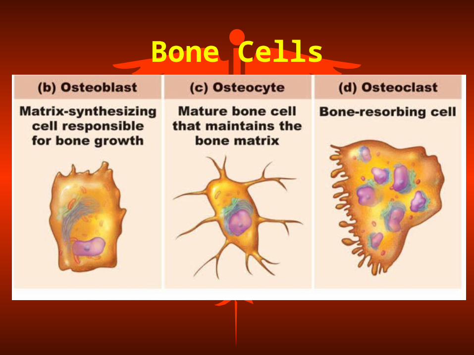
Bone Cells

Ossification• The process by which bones form
in the body (Osteogenesis)• Intramembranous Ossification
– Convert membrane models to bone– (Periosteum – growth in width)
• Endochondral Ossification– Convert cartilage to bone – (Epiphyseal plate - growth in length)

Intramembranous Ossification
Location: Skull

Endochondral OssificationOccurs in Long Bones

EpiphysealPlate
Homeostasis and Bone Remodeling
• Bones constantly undergo ossification and remodeling.
• Replaces older bone matrix with new bone matrix:–bone reabsorption (osteoclasts)
–bone deposition (osteoblasts)• Allows injured or worn bone to
be replaced.

Long Bone Anatomy

Long Bone Features• Periosteum – the outer
connective tissue membrane covering the outer surface of most bones.
• Diaphysis – shaft or midsection of the bone which contains bone marrow and adipose tissue.
• Epiphysis – the rounded ends of a long bone.

Long Bone Features, p. 2
• Medullary Cavity – a central cavity in the diaphysis which contains red bone marrow and yellow bone marrow.
• Red Bone Marrow – composed of soft, gel-like hematopoietic tissue that fills the diaphysis and produces red blood cells (erythrocytes), white blood cells (leukocytes), and platelets (thrombocytes)

Long Bone Features• Yellow Bone Marrow – adipose
tissue that will replace the red bone marrow in the diaphyses (shafts) of long bones during adulthood.

Long Bone Features
• Endosteum – the lining of the medullary cavity.
• Articular Cartilage – hyaline cartilage found on the ends of long bones (epiphyses) to reduce friction during joint movement.
• Compact Bone – densely packed osteocytes to provide strength to the bone.
Long Bone Features
• Spongy Bone – loosely-packed osteocytes which help to reduce the weight of the bone and form the red marrow.
• Compact Bone – densely-packed osteocytes to provide strength to the bone by forming a shell around the spongy bone.

Let’s Quiz

Answers• 1. Articular Cartilage• 2. Spongy Bone• 3. Compact Bone• 4. Medullary Cavity• 5. Yellow Marrow• 6. Periosteum• 7. Proximal Epiphysis• 8. Diaphysis• 9. Distal Epiphysis

Shapes of Bones
• FOUR Categories of Bone Shapes
• Long Bones• Short Bones• Flat Bones• Irregular Bones

Shapes of Bones

Long Bones
• Greater length than width• Have a distinct diaphysis and a
variable number of epiphysis• Slightly curved for strength• Examples: humerus, ulna, radius,
femur, tibia, fibula, metacarpals, metatarsals, phalanges
Short Bones
• Cube-shaped bones• Nearly equal in length and width• Spongy texture on inside of the
bone• Examples: carpal and tarsal bones

Flat Bones
• Generally thin and flat (plate-like)• Compact bone on anterior and posterior
surfaces with spongy bone in the middle• Provides protection to organs• Large surface area for muscle
attachment• Examples: cranial bones, sternum,
scapula, ribs

Irregular Bones
• Complex-shaped bones• Cannot be classified into other
categories• Vary in the amount of spongy and
compact bone• Examples: vertebrae, facial bones,
patella

Classification of Bones
• Compact Bone (Dense Bone)– little space between the solid components
of bone• Spongy Bone (Trabecular Bone)
– made up of an irregular network of thin plates of bone with many intercellular spaces called trabeculae (spicules)
• spaces between trabeculae filled with red bone marrow
• responsible for hematopoiesis

Spongy Bone Structure

Compact Bone

Bone Markings
Refers to any bump, groove,
opening, or depression
associated with a bone. They have a
names and functions

Foramen• An opening or
hole in a bone for the passage of nerves and/or blood vessels.
• Example: Foramen Magnum (spinal cord)

Meatus
• A tube-like passage within a bone.
• Example: • External
Auditory Meatus (green) passageway for sound.

Sinus
• A space within a bone lined with mucus membrane that reduces the weight of a bone.
• Example:• Frontal Sinus
(green)

Fossa
• A depression or groove in the bone.
• Example:• Mandibular Fossa
– the depression where the mandible or jaw contacts the skull.
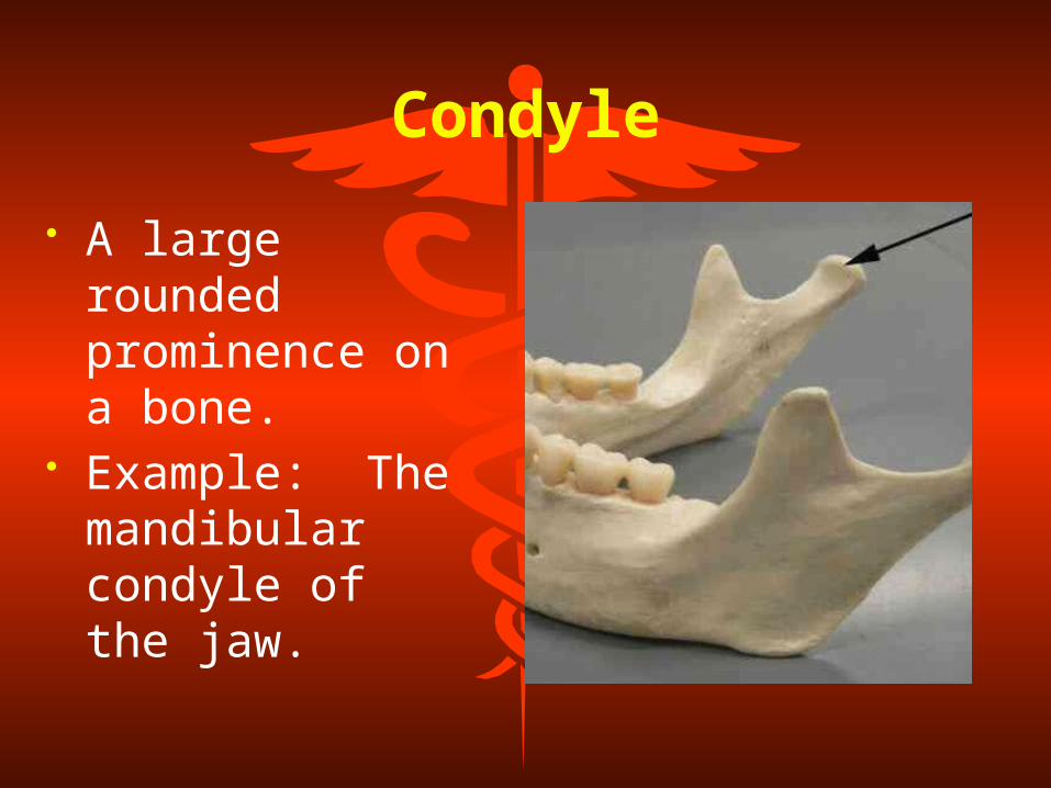
Condyle
• A large rounded prominence on a bone.
• Example: The mandibular condyle of the jaw.

Tuberosity• A large, rounded,
usually roughened area for the attachment of tendons and ligaments.
• Example: Tibial Tuberosity for attachment of patellar tendon
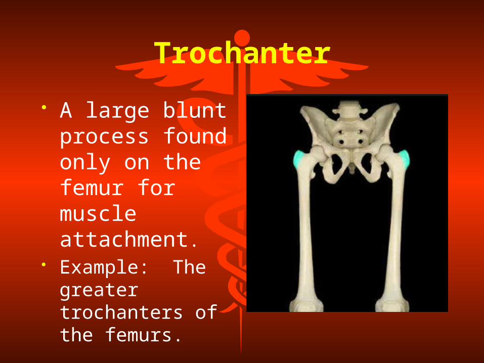
Trochanter
• A large blunt process found only on the femur for muscle attachment.
• Example: The greater trochanters of the femurs.

Tubercle
• A small rounded projection for muscle attachment.
• Example – The greater tubercle of the humerus.

Process
• A growth or extension projecting from a bone used for muscle attachment or to form a joint.
• Example – the mastoid process of the temporal bone.

Fontanels• Found only on the
infant skull, it is a membrane-covered "soft spot” where the bones (sutures) have not fused. The fontanelles allow for growth of the brain and skull during an infant's first year.
• Example – the anterior fontanel.

Fontanels – Birth View

Sutures• Strong,
immovable joints that are formed as the membranes in the fontanels are replaced by tough, fibrous tissue. Sutures hold the cranial bones together.
• Example – coronal suture

Sutures – Fetal View

2 Divisions of the Skeleton
The AXIAL skeleton and the APPENDICULAR skeleton

Axial Skeleton• The axial skeleton
consists of 80 bones and forms the skull, vertebrae, rib cage, and hyoid bone.

Appendicular Skeleton
• The appendicular skeleton is composed of 126 bones.
• It helps in locomotion (pelvic girdle and lower limbs) and manipulation of objects in the environment (pectoral girdle and upper limbs).

The Axial Skeleton
Axial Skeleton80 Bones
• Skull – Cranial and Facial Bones - 22
• Hyoid Bone -1• Vertebral Column - 24• Sternum - 1• Ribs - 24
The Skull Bones
• Mandible• Maxilla• Zygomatic• Frontal • Parietal• Occipital• Temporal
• Sphenoid• Ethmoid

Facial Bones
• Mandible• Maxilla• Zygomati
c

Cranial Bones (8)
• Frontal Bone• Parietal Bones
(2)• Temporal Bones
(2)• Occipital Bone• Sphenoid Bone• Ethmoid Bone

Hyoid Bone
• The hyoid bone is a U-shaped bone found superior to the larynx.
• It holds the tongue in place and provides for muscle attaachment in the neck.

Frontal Bone
• Forms the forehead, the roof of the orbits (eye sockets) and most of the anterior portion of the cranial floor

Temporal Bones (2)
• Form the inferior sides of the cranium and part of the cranial floor
• Temporal bone landmarks:– Zygomatic Process– Mandibular Fossa– External Auditory Meatus– Mastoid Process– Styloid Process

Parietal Bones

Occipital Bone
• The posterior part and prominent portion of the base of the cranium
• Occipital bone landmarks:– Foramen Magnum– Occipital Condyles– External Occipital Protuberance

Sphenoid Bone
• Bone situated in the middle part of the base of the skull
• Shaped like a bat• Only bone that connects to all
other cranial bones• Sphenoid bone landmarks:
– Body - Sella Turcica– Greater Wings -Sphenoid Sinuses

Sphenoid Bone

Ethmoid Bone• Light, spongy bone located in the
anterior floor of the cranium between the orbits
• Makes up much of the structure of the nasal cavity
• Ethmoid bone landmarks:– Lateral Masses (Labyrinths)– Ethmoid Sinuses - Crista Galli– Perpendicular Plate -Cribriform
Plate– Superior Nasal Conchae– Middle Nasal Conchae

Ethmoid Bone

Zygomatic Bones (2)
• cheek bones• form the prominences of the
cheeks and the floor and outer walls of the orbits
• Zygomatic bone landmarks:– temporal processes– zygomatic arches

Maxillary Bones (2)• Pair of bones that unite to form the
upper jaw• Articulate with every bone of the face
except the mandible• Maxillary bone landmarks:
– Alveolar Processes– Alveoli– Palatine Processes - horizontal projection
from the maxillae that forms the anterior three fourths of the hard palate
– Cleft Palate– Cleft Lip

Cleft Palate & Cleft Lip

Facial BonesSagittal Section

Mandible (Lower Jaw) Bone
• Largest and strongest bone in the face
• The only moveable skull bone• Articulates with the temporal bone
to form the Temporal Mandibular Joint (TMJ)
The Appendicular Skeleton
Appendicular Skeleton126 Bones
• clavicle• scapula• humerus• ulna • radius• carpals• metacarpals• phalanges
• pelvis• femur• patella• tibia• fibula• tarsals• metatarsals• phalanges

The Pectoral Girdle
• Clavicles (2)• (collar bones)• Scapulae (2)• (shoulder blades)

The Upper Limb• Humerus (1)• (arm bone)• Radius (1)• (lateral foream)• Ulna (1)• (medial forearm)• Carpals (8)• (wrist)• Metacarpals (5)• (hand bones)• Phalanges (14)• (fingers)

Pelvis (Os Coxae)
• Ilium (2)• Ischium (2)• Pubis (2)

Lower Limb• Femur (1)• (thigh bone)• Patella (1) (knee cap)• Tibia (1)• (shin bone)• Fibula (1)• (lower leg bone)• Tarsals (7)• (ankle bones)• Metatarsals (5)• (foot bones)• Phalanges (14)
(toes)

The Vertebral Column (Spine)
• Composed of 33 (26) bones• Encloses and protects the
spinal cord• Supports the head• Lower vertebrae supports the
weight of the entire upper body

Vertebrae• Bones of the vertebral column• Cervical vertebrae (7) - neck• Thoracic vertebrae (12) - ribs• Lumbar vertebrae (5) - lower back• Sacral vertebrae (5) - pelvic bones• Coccygeal vertebrae (4) - tail bone• Intervertebral Foramina - openings
between the vertebrae for nerve exit

Vertebral Column
Joints (Articulations)
The point of contact between bones, between bones and
cartilage, or between bones and fibrous tissue

Structural Classification of Joints
• Classification of which tissues are holding the bones together:
• Fibrous Joints– held together by fibrous connective
tissue• Cartilaginous Joints
– held together by cartilage• Synovial Joints
– joint enclosed within a synovial or joint capsule

Fibrous Articulations
• Example:• Sutures between • the cranial bones

Cartilaginous Articulations
• Examples:• Intervertebral
discs formed by cartilage between the vertebrae.

Synovial Joints
• Example:• Knee Joint
Functional Classification of Joints
• Based on the degree of movement at a joint.
• Synarthroses – No movement at the joint.
• Amphiarthroses –Small or slight movement at the joint.
• Diarthroses – Freely moveable joints.

Synarthrotic Joints
• Example:• Sutures

Amphiarthrotic Joints
• Example:• Intervertebral
Disks• Symphysis Pubic

Diarthrotic Joints
• Examples:• Elbow• Shoulder
Diseases and Disorders
Herniated disk Osteoarthritis Osteoporosis Scoliosis Kyphosis Lordosis Spina bifida RA (Rheumatoid
arthritis)

Intervertebral Discs
• Discs of fibrocartilage found between the vertebrae from C1 to the sacrum
• Functions to absorb shock• Allows for the multi-directional
motion between each vertebrae– Annulus Fibrosis - outer fibrous ring– Nucleus Pulposus - inner, soft pulpy
portion of the intervertebral discs

Herniated Discs(Slipped Discs)
• Rupture of the fibrocartilage discs• Usually caused by compression forces• Usually occurs between L4 and L5 or L5
and the 1st Sacral Vertebrae• Disc protrudes and exerts pressure on
spinal nerves• To decrease risk of herniated discs:
– 1. maintain optimal body weight– 2. strengthen abdominal muscles– 3. increase lower back flexibility

Herniated Disc

Osteoarthritis• Degenerative joint disease associated
with aging or with trauma• Characteristics:
– degeneration of articular cartilage– development of bone spurs– usually effects large joints (knees, hips, etc)
• Treatment:– rest– removal of bone spurs– joint replacement

Osteoarthritis

Osteoporosis
• Decrease in bone mass and increased susceptibility to fractures.
OsteoporosisContributing Factors
• Decreased estrogen production• Poor nutritional status• Low activity levels• Weight• Smoking • Drugs and alcohol consumption• Gender/race/hereditary factors

Osteoporosis - Treatment
• Calcium supplementation• Estrogen Replacement Therapy• Weight-bearing exercise• Steroid treatment therapy
Spina Bifida• congenital defect where the neural
arch fails to unit• usually involves the lumbar
vertebrae• symptoms may be mild to severe
– usually results in paralysis– partial or complete loss of bladder
control– absence of reflexes
• can be diagnosed during pregnancy by sonography, amniocentesis, blood tests

Spina Bifada

Abnormal Curvatures of the Spine
• Scoliosis - lateral curvature of the spine– usually in thoracic and lumbar region
• Kyphosis - hunchback/humpback– exaggeration of thoracic curvature
• Lordosis - swayback (sprinters butt)– exaggeration of lumbar curvature
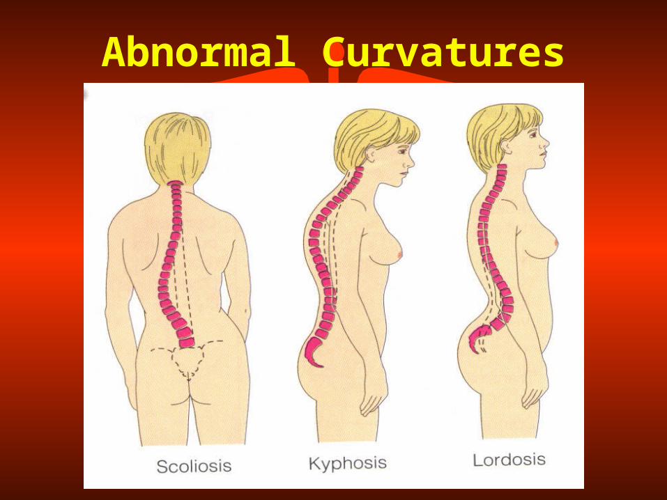
Abnormal Curvatures

Scoliosis

Scoliosis

Kyphosis

Lordosis (swayback)
Rheumatoid ArthritisRheumatoid arthritis is an
autoimmune disease in which the body’s immune system – which normally protects its health by
attacking foreign substances like bacteria and viruses – mistakenly
attacks the joints. The cause of RA is not yet fully understood, although doctors do know that an abnormal
response of the immune system plays a leading role in the inflammation and joint damage that
occurs.

Rheumatoid Arthritis


