The soft body parts offreshwater bryozoans depicted by
scanning electron microscopy
M . G . W A L Z L & E .R .Wöss
Abstract: For the first time, special scanning electron microscopical preparation techniques, i.e. chem-
ical dehydration and air drying with hexamethyldisilacane, were used to study the Phylactolaemata.
This approach depicted the growth form of colonies and the outer structures of zooids of Plumatella cas-
miana and P. fungosa three-dimensionally. The structures of cystids and polypids, for example the cystid
wall, lophophore, gut, funiculus and the muscles, are represented using dissected zooids. Moreover, this
technique revealed the structures of both asexual reproduction (the buds, the generation and germina-
tion of statoblasts) and the organs for sexual reproduction (testis, ovary, embryo sac, larva).
Key words: Phylactolaemata, Plumatella, anatomy, sexual propagation, statoblast formation.
Introduction
The phylum Bryozoa consists of threeclasses, the Phylactolaemata, Gymnolaema-ta and Stenolaemata (RYLAND 1970). Thevast majority of bryozoan taxa are marine,inhabiting depths from the intertidal to theabyssal. Freshwater bryozoans comprise asmaller number of about 60 species (WOOD1989) and can mostly be assigned to theclass of phylactolaemates. This group exclu-sively inhabits freshwater, as do a smallernumber of gymnolaemate species, all be-longing to the order Ctenostomata. Allfreshwater bryozoans lack mineralized skele-tons. This is in striking contrast to most bry-ozoans, which have skeletons made of cal-cite or, less frequently, aragonite. All Steno-laemata and the order Cheilostomata of theGymnolaemata possess calcareous skeletons;they are among the most common groups ofmacrofossils found in the post-Cambrianmarine fossil record (TAYLOR 2005). In to-tal, 14-700 species have been described inthe fossil record (HOROWITZ & PACHUT2000), and about 5.600 extant bryozoans areknown (TODD 2000). The morphologicalfeatures of bryozoan skeletons form the basisfor classifications, and these features differbetween the tubular stenolaemate bryozoans
and the boxlike gymnolaemate cheilo-
stomes. In most stenolaemate bryozoans, the
exterior surface is insufficient to differenti-
ate taxa, and taxonomic separation in that
group relies heavily on characters visible in
petrographic thin sections (SANDBERG 1977,
see also Ernst and Scholz et al. this volume).
The frontal surfaces of cheilostome zooecia,
however, are covered by membranes or by
calcified walls which offer varying amounts
of morphological details (BANTA 1973; see
also Bader & Schäfer, Novosel, and Vävra
this volume). In the latter case, scanning
electron microscopy (SEM) is broadly used
to study bryozoan skeletons and has became
the standard tool in taxonomic work for
both palaeontologists and biologists. Even
in ctenostomate bryozoans, where calcified
skeletons are missing, the process of bioim-
muration (the preservation of an organism
by the skeletal overgrowth of a neighbour-
ing encruster) allows the details of the
zooids to be described using SEM techniques
(VOIGT 1966; TAYLOR 1990; TODD 1994).
In the phylactolaemate group, the soft-bodied zooecia offer less distinctive charac-ters (KRAEPLIN 1887, 1892; BRAEM 1890;
contrast to marine species, where hetero-zooids such as avicularia and vibracularia arepresent (RYLAND & HAYWARD 1977; HAY-
WARD & RYLAND 1979), the uniform shapeof the autozoids of phylactolaemate specieshas raised less interest for SEM studies. Ul-trastructural investigations on spermatozoanstructure and larva (FRANZEN 1982, FRAN-
ZEN & SENSENBAUGH 1983) sporadically in-
volve SEM. Most ultrastructural work, how-ever, has traditionally focused on stato-blasts. These dormant bodies are excellentlysuited for SEM because their shell consistsof a chitinised cuticula. They have been thetarget of numerous comparative morpholog-ical investigations (e.g. WlEBACH 1974;RAO & BUSHNELL 1979; MUKAI 1999;
MUNDY 1980; GOETHALS et al. 1984; ODA &
MUKAI 1985; GEIMER & MASSARD 1986;
POURCHER & D'HONDT 1987; WOOD &WOOD 2000). In some cases, such as withinthe genus Plumatella, SEM has become theonly reliable tool for species distinction(GEIMER &. MASSARD 1987; WOOD 2001;
TATICCHI &. PlERONI 2005 and see also Tat-icchi et al. this volume).
Despite the great benefit of SEM, thisstandard method has rarely been used tostudy the soft-bodied outer and inner partsof the zooids (but see: MUKAI et al. 1997),probably due to the complexity of the prepa-ration techniques required. This SEM studyis an introduction for further ultrastructurework on reproduction in freshwater bry-ozoans. First results are presented in prepa-ration techniques of soft-bodied inner andouter parts of the zooids of two species, PIu-matetta fungosa and P. casmiana. Thesespecies have already been examined with re-spect to the formation of sexual and asexualpropagules and the reproductive cycle infreshwater bryozoans (WÖSS 2002).
Material and methods
Colonies of Plumatella casmiana werecollected on 17.5.1992 from a pond at Lax-enburg (Lower Austria) and colonies of P.fungosa on 7-7. and 3.9.1992 from a backwa-ter of the Danube River at Bad Deutsch Al-tenburg (Lower Austria).
The colonies were transported in pond-water, along with the logs and twigs onwhich they grew, to the laboratory and left
there undisturbed at least until most of thepolypids had protruded. Then, with apipette, a saturated aqueous solution ofchloral hydrate (C13CCH(OH)2) was addeddropwise and carefully to the water surface.The specifically heavier chloral hydrate so-lution sinks down to the colony and narco-tizes the zooids, so that most of the polypi-des remain protuded. After 15 minutes, anequal volume of 1 % aqueous bufferedformaldehyde solution with pH 7.2 (LlLLIE1954) was added. For definitive fixation thecolonies were removed and immersed into a4 % buffered formaldehyde solution. Thecolonies were stored in this medium untilexamination.
For further detailed SEM investigations,selected parts of colonies or single zooidswere separated from the substratum using asharp razor blade and transferred to distilledwater to wash out excessive formaldehyde.The distilled water was changed 3 times af-ter 15 minutes (important to avoid precipi-tation during the following processes). Af-terwards, the samples were dehydratedchemically with acidified 2,2-dimethoxy-propane (DMP) (MULLER &. JACKS 1975).
For acidification and activation, 1 ml 25 %HCL was added to 100 ml DMP shortly be-fore use. For rapid dehydration, 1 part waterin the sample vials was mixed with 3 partsDMP. A rapid cooling of the vials docu-mented the endothermic chemical processthat yields anhydrous methanol and ace-tone. After 20 minutes (although anovernight delay has no negative effect) thesolution was replaced twice with water-freeacetone, for 15 minutes in each case. Theacetone was exchanged with HMDS(1,1,1,3,3,3-hexamethyldisilazane), thesample initially being immersed for 30 minin a 1:1 mixture of acetone and HMDS fol-lowed by 30 minutes in pure HMDS, andthen air dried on filter paper under a fumehood (BRAY et al. 1993; NATION 1983). Af-ter drying, the samples were transferred in-dividually on aluminiumstubs using a finepencil. Single zooids or parts thereof weremounted with TEMPFIX-thermo glue(Neubauer Chemikalien company, Ger-many), and parts of colonies or groups ofzooids were mounted using silver paste. Allsamples were then sputter coated with 40nm of gold in a Agar B 7340 sputter coater.
Fig. 1: Parts of colonies with many protruded polypides of Plumatella casmiana (a) and P. fungosa (b).
Specimens were examined at 10 to 15keV in a Philips XL20 scanning electron mi-croscope and photographed digitally. In or-der to view the internal organs, zooids wereopened with two tungsten needles that weresharpened by repeatedly inserting the nee-dle tips into an aqueous potassium hydrox-ide solution under 6 volts of alternating cur-rent generated by a microscope transformer.The zooids were opened either in the phaseof washing in water or after air drying.
Results and discussion
The colonies of Plumatella casmiana andP. fungosa are characterized by a differentgrowth form. Plumatella casmiana shows anirregularly "knotty" arrangement of zooids,which are attached to the substrate in asheetlike growth form; colonies of P. fun-
gosa, however, are packed more densely,with regular fused zooids resulting in anerect and massive growth form (Fig. 1).
The fully grown monomorphic zooids ofboth species differ in size. Plumatella casmi-
ana zooids are smaller, and the horseshoe-shaped lophophore therefore bears only 25-40 tentacles, whereas 40-60 tentacles arepresent in P. fungosa (Fig. 2). The single
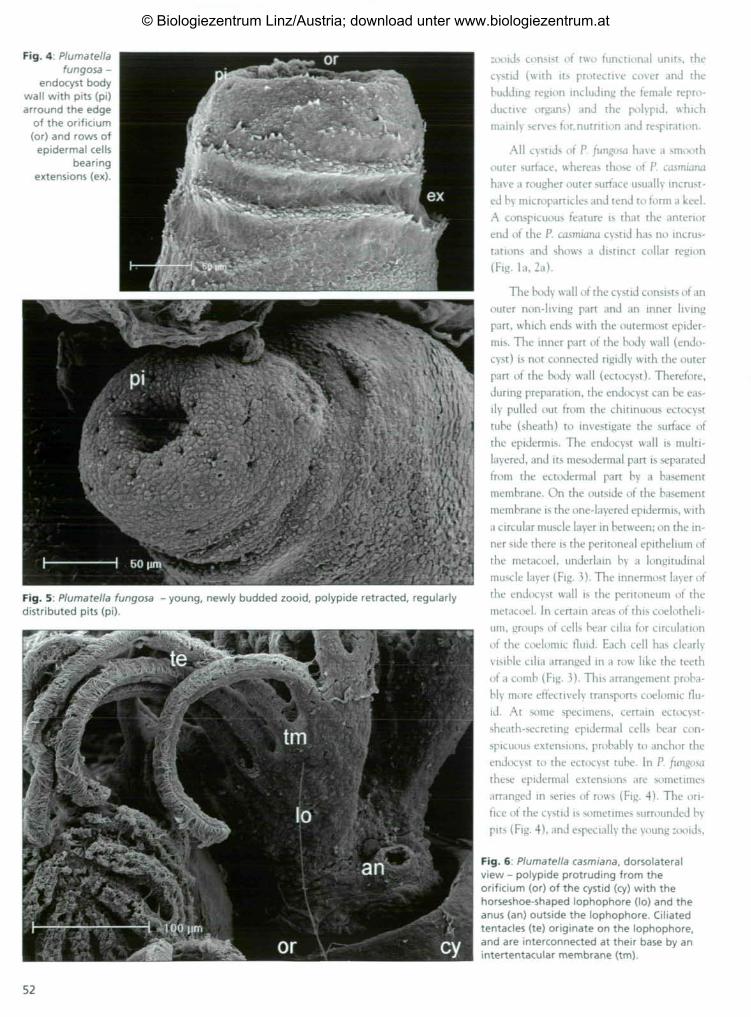
Fig. 2: Zooids with protruded polypides ofPlumatella casmiana (a) with rough,incrusted outer cystid wall and collarregion (co) and P. fungosa (b) with smoothouter surface of cystid wall.
Fig. 3: Plumatella fungosa - endocyst body wall with epidermis (ep), circular muscles (cm),basement membrane (bm), longitudinal muscles (Im) and ciliated peritoneal cells (pe).
looids consist of two functional units, thecystid (with its protective cover and thebudding region including the female repro-ductive organs) and the polypid, whichmainly serves for,nutrition and respiration.
All cystids of P. fungosa have a smoothouter surface, whereas those of P. casmiana
have a rougher outer surface usually incrust-ed by microparticles and tend to form a keel.A conspicuous feature is that the anteriorend of the P. casmiana cystid has no incrus-tations and shows a distinct collar region(Fig. la, 2a).
The body wall of the cystid consists of anouter non-living part and an inner livingpart, which ends with the outermost epider-mis. The inner part of the body wall (endo-cyst) is not connected rigidly with the outerpart of the body wall (ectocyst). Therefore,during preparation, the endocyst can be eas-ily pulled out from the chitinuous ectocysttube (sheath) to investigate the surface ofthe epidermis. The endocyst wall is multi-layered, and its mesodermal part is separatedfrom the ectodermal part by a basementmembrane. On the outside of the basementmembrane is the one-layered epidermis, witha circular muscle layer in between; on the in-ner side there is the peritoneal epithelium ofthe metacoel, underlain by a longitudinalmuscle layer (Fig. 3). The innermost layer ofthe endocyst wall is the peritoneum of themetacoel. In certain areas of this coelotheli-um, groups of cells bear cilia for circulationof the coelomic fluid. Each cell has clearlyvisible cilia arranged in a row like the teethof a comb (Fig. 3). This arrangement proba-bly more effectively transports coelomic flu-id. At some specimens, certain ectocyst-sheath-secreting epidermal cells bear con-spicuous extensions, probably to anchor theendocyst to the ectocyst tube. In P. /ungosathese epidermal extensions are sometimesarranged in series of rows (Fig. 4). The ori-fice of the cystid is sometimes surrounded bypits (Fig. 4), and especially the young :ooids,
Fig. 6: Plumatella casmiana, dorsolateralview - polypide protruding from theorificium (or) of the cystid (cy) with thehorseshoe-shaped lophophore (lo) and theanus (an) outside the lophophore. Ciliatedtentacles (te) originate on the lophophore,and are interconnected at their base by anintertentacular membrane (tm).
Fig. 7: Plumatella fungosa - tentacles withrows of multiciliated cells at their lateral and
inner sides
shortly after evagination, show a regular
arrangement of these pits. These structures
can he interpreted either as sensory pits or a
vestibular pores (Fig. 5).
The polypide consists of the lophophorr
and the V-shaped gut, which is connects
with the ventral cystid wall by a hollow
peritoneal cord, the funiculus.
The lophophore bears tentacles inter-
connected at their bases by the intertentac-
ular membrane, a fold of the lophophore
(Fig. 6). Three rows of epidermal cells of the
tentacles bear cilia directed laterally and to
the inner side of the lophophore. The syn-
chronized beating of the cilia generates ;i
water current that transports particles to-
ward the mouth (Fig. 7, 8). The mouth is
situated centrally on the lophophore and is
encircled by the tentacles. Dorsally, the
mouth is overhung by a flap, the epistome,
which can close the opening (Fig. 8). As op-
posed to the mouth opening, the anus lies
outside the tentacle circle at the dorsally
open side of the lophophore (Fig. 6).
The gut is divided into different parts -the pharynx, oesophagus, cardia, caecum andthe intestine - and hangs into the coelomiccavity of the cystid. A large, paired muscle oneither side of the gut extends across the meta-coel. On one side, the muscle inserts in theventro-lateral cystid wall, and on the otherside at the base of the lophophore, i.e. at thetentacle sheath, with two smaller bundles al-so inserting directly on the gut (Fig. 9). Thegut wall consists of the endothelium and twolayers of muscles, an inner circular musclelayer and an outer longitudinal layer. At thebulged end of the V-shaped gut, the peri-toneum of the gut continues to a hollow peri-toneal cord, the funiculus (Fig. 10).
The funiculus runs across the metacoel
and passes into the peritoneum of the cystid
wall (Fig. 11). Statoblast formation begins
at the insertion area of the border funiculus-
Fig. 9: Plumatella fungosa - two dissectedzooids with opened cystids (cy). Gut (gu) and
Fig. 8: Plumatella fungosa - horseshoe-shaped lophophore with tentacles; the mouth islocated in the bend of the lophophore, overhung dorsally by the epistome (ep).
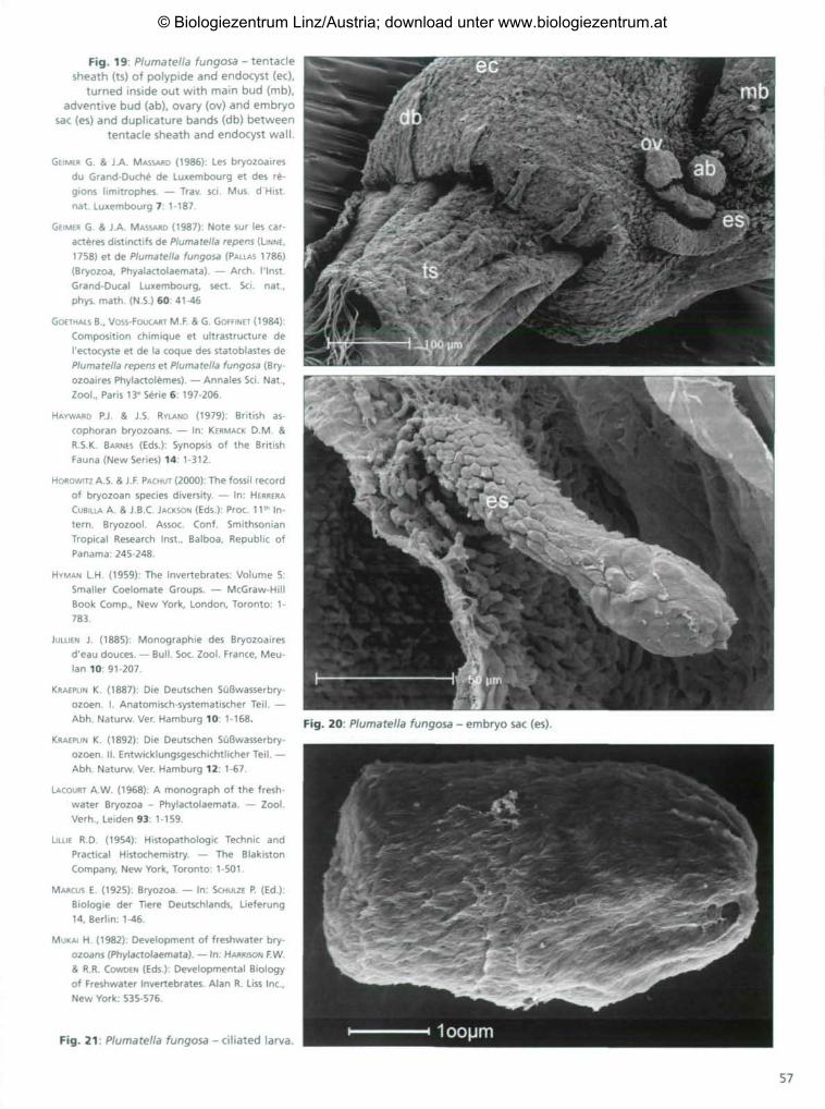
bryo sac. It develops there into the so-calledlarva. After embryogenesis, the ciliated lar-va is released and swims with the aboral poleoriented forward (Fig. 21 ) .
Future prospects
This paper is a preliminary study interms of future work on classical questionsin zooid anatomy and reproduction. Certainissues, e.g. in the field of sexual propagation,were already addressed by the earliest bry-ozoan workers but remain unsolved until to-day. The planned studies will focus on re-production biology as well as on the anato-my of sensory organs.
To investigate details of gametogenesis,sperm release, fertilization and nidation ofthe egg, and embryogenesis, SEM will beused in combination with series of semithinsections and transmission electron mi-croscopy. In asexual reproduction, the docu-
Fig. 13: Statoblasts of Plumatella fungosa (a) and P. casmiana (b) detatched from thefuniculus and rotating within the fluid of the coelomic cavity, covered by the peritoneallayer (pe) of the funiculus and the outer epidermal layer (oe) of the statoblast.
Fig. 14: Plumatella casmiana - mature statoblast, the two valves of the shell demarcatedby the equatorial suture line (si).
Fig. 15: Plumatella casmianasurrounded by their valves.
two germinating statoblasts Fig. 16: Plumatella fungosa - inner side of ventral endocyst wall withmain bud (mb), adventive bud (ab), ovary (ov) and embryo sac (es).
Fig. 19: Plumatella fungosa - tentaclesheath (ts) of polypide and endocyst (ec),
turned inside out with main bud (mb),adventive bud (ab), ovary (ov) and embryo
sac (es) and duplicature bands (db) betweententacle sheath and endocyst wall.
GEIMER G. & J.A. MASSARD (1986): Les bryozoairesdu Grand-Duche de Luxembourg et des re-gions limitrophes. — Trav. sei. Mus. d Histnat. Luxembourg 7: 1-187.
GEIMER G. & J.A. MASSARD (1987): Note sur les car-acteres distinctifs de Plumatella repens (LINNE,1758) et de Plumatella fungosa (PALLAS 1786)(Bryozoa, Phyalactolaemata). — Arch. I'lnst.Grand-Ducal Luxembourg, sect. Sei. nat.,phys. math. (N.S.) 60: 41-46
GOETHALS B., VOSS-FOUCART M.F. & G. GOFFINET (1984):
Composition chimique et ultrastructure deI'ectocyste et de la coque des statoblastes dePlumatella repens et Plumatella fungosa (Bry-ozoaires Phylactolemes). — Annales Sei. Nat.,Zool., Paris 13« Serie 6: 197-206.
HAYWARD P.J. & J.S. RYLAND (1979): British as-cophoran bryozoans. — In: KERMACK D.M. &R.S.K. BARNES (Eds): Synopsis of the BritishFauna (New Series) 14: 1-312.
HOROWITZ A.S. S J.F. PACHUT (2000): The fossil recordof bryozoan species diversity. — In: HERRERACUBILLA A. & J.B.C. JACKSON (Eds.): Proc. 11 th In-tern. Bryozool. Assoc. Conf. SmithsonianTropical Research Inst., Balboa, Republic ofPanama: 245-248.
HYMAN L.H. (1959): The Invertebrates: Volume 5:Smaller Coelomate Groups. — McGraw-HillBook Comp., New York, London, Toronto: 1-783.
JULLIEN J. (1885): Monographie des Bryozoairesd'eau douces. — Bull. Soc. Zool. France, Meu-lan 10: 91-207.
KRAEPLIN K. (1887): Die Deutschen Süßwasserbry-ozoen. I. Anatomisch-systematischer Teil. —Abh. Naturw. Ver. Hamburg 10: 1-168.
KRAEPLIN K. (1892): Die Deutschen Süßwasserbry-ozoen. II. Entwicklungsgeschichtlicher Teil. —Abh. Naturw. Ver. Hamburg 12: 1-67.
LACOURT A.W. (1968): A monograph of the fresh-water Bryozoa - Phylactolaemata. — ZoolVerh., Leiden 93: 1-159.
LILLIE R.D. (1954): Histopathologic Technic andPractical Histochemistry. — The BlakistonCompany, New York, Toronto: 1-501.
MARCUS E. (1925): Bryozoa. — In: SCHULZE P. (Ed.):Biologie der Tiere Deutschlands, Lieferung14, Berlin: 1-46.
MUKAI H. (1982): Development of freshwater bry-ozoans (Phylactolaemata). — In: HARRISON F.W.& R.R. COWDEN (Eds): Developmental Biologyof Freshwater Invertebrates. Alan R. Liss Inc.,New York: 535-576.
Microscopic Anatomy of Invertebrates 13.Wiley-Liss, New York: 45-206.
MUKAI H. (1999): Comparative morphological stud-ies on the statoblasts of lower phylactolae-mate bryozoans, with discussion on the sys-tematics of Phylactolaemata. — Science Rep.Fac. Educ, Hunma Univ. 46: 51-91.
MULLER L.L. S T.J. JACKS (1975): Rapid chemical de-hydration of samples for electron microscopicexaminations. — J. Histochem. Cytochem.23(2): 107-110.
MUNDY S.P. (1980): Stereoscan stdies of Phylacto-laemata bryozoan statoblasts including a keyto the statoblasts of the British and EuropeanPhylactolaemata. — J.Zool. 912: 511-530.
NATION J.L (1983): A new method using hexa-methyldisilazane for preparation of soft in-sect tissues for scanning electron microscopy.
— Stain Tech. 58: 347-351.
ODA S. & H. MUKAI (1985): Fine surface structure ofthe statoblasts of higher phylactolaematebryozoans. — In: NIELSEN C. & G.P. LARWOOD
(Eds.): Bryozoa: Ordovician to Recent. Olsen &Olsen, Fredensborg, Denmark: 234-244.
POURCHER A.-M. & J.-L D'HONDT (1987): Etude ultra-structurale du sessoblaste et du flottoblastechez Plumatella fungosa (PALLAS, 1768) (Bry-ozoaires, Phylactolaemates). — Annal. Sei.Nat., Zool., Paris 13' Serie 8: 209-216.
RAO K.S. & J.H. BUSHNELL (1979) : New Structures inBinding Designs of Freshwater EctoproctaDormant Bodies (Statoblasts). — Acta Zool.(Stockh.)60: 123-127.
RYLAND J.S. (1970): Bryozoans. — Hutchinson Univ.
Library, London: 1-175.
RYLAND J.S. & P.J. HAYWARD (1977): British anascanbryozoans. — In: KERMACK D.M. (Ed.): Synopsisof the British Fauna (New Series) 10: 1-187.
SANDBERG P.A. (1977): Ultrastructure, mineraloga,and development of bryozoans skeletons. —In: WOLLACOTT R.M. & R.L. ZIMMER (Eds.): Biolo-gy of Bryozoans. Acad. Press, New York, SanFrancisco, London 143-181.
TATICCHI M.I. S G. PIERONI (2005): Freshwater bry-ozoa of Italy. A survey of some species fromthe Italian bryozoan collection of A. Viganöwith new records. — In MOYANO G.H.I., CANCI-NO J.M. & P.N. WYSE JACKSON (Eds.): BryozoanStudies 2004. Proc. 13* Intern. Bryozool. As-soc. A.A. Balkema Publ., Leiden, London, NewYork, Philadelphia, Singapore: 317-327.
TAYLOR P.D. (1990): Preservation of soft-bodied andother organisms by bioimmuration - a re-view. — Palaeontology 33: 19-35.
TAYLOR P.D. (2005): Bryozoans. — In: SEUEY R.C.,COOKS L.R.M. S I.R. PUMER (Eds.): Encyclopae-dia of Geology 2, Elsevier, Amsterdam: 310-320.
TODD J.A. (2000): The central role of ctenostomesin bryozoan phylogeny. — In: HERRERA CUBILLA
A. & J.B.C. JACKSON (Eds.): Proc. 11 th Intern.Bryozool. Assoc. Conf. Smithsonian TropicalResearch Institute, Panama: 104-135.
TODD J.A. (1994): The role of bioimmuration in theexceptional preservation of fossil ctenostom-ates, including a new Jurassic species ofBuskia. — In: HAYWARD P.J., RYIAND J.F. & P.D.
TAYLOR (Eds.): Biology and Paleobiology ofBryozoans. Olsen & Olsen, Fredensborg, Den-mark: 187-192.
VOIGT E. (1966): Die Erhaltung vergänglicher Or-ganismen durch Abformung infolge Inkrusta-tion durch sessile Tiere. — Neues Jahrb. Geol.Palaeontol., Abh. 125: 401-422.
WIEBACH F. (1974): Specific structures of sessoblasts
(Bryozoa: Phylactolaemata). — Docum. Lab.
Geol. Fac. Sei. Lyon H.S. 3 (fasc. 1): 149-154.
WOOLLACOTT R.M. & R.L. ZIMMER (1977): Biology of
Bryozoans. — Acad. Press, New York, San
Francisco London: 1-566.
WOOD T.S. (1989): Ectoproct bryozoans of Ohio. —
Bull. Ohio Biol. Survey New Series (2): 1-70.
WOOD T.S. (2001): Bryozoans. — In: THORP J. & A.CORVICH (Eds.): Ecology and Classification ofNorth American Freshwater Invertebrates, 2nd
ed. Acad. Press, New York: 505-525.
WOOD T.S. & L.J. WOOD (2000): Statoblast morphol-ogy in historical specimens of phylactolae-mate bryozoans. — In: HERRERA CUBILLA A. SJ.B.C. JACKSON (Eds.): Proc. 11* Intern. Bryozo-ol. Assoc. Conf. Smithsonian Tropical ResearchInstitute, Panama: 421-430.
Wöss E.R. (2002): The reproductive cycle of Plu-matella casmiana (Phylactolaemata: Plu-matellidae). — In: BUTLER C, SPENCER JONES M. &
P. WYSE JACKSON (Eds.): Bryozoan Studies 2001.Proc. 12th Intern. Bryozool. Assoc. A.A. Balke-ma Publ., Rotterdam: 347-352.