The structure and protein binding of amyloid-specific dye reagents *. Barbara Stopa 1½ , Barbara Piekarska 1 , Leszek Konieczny 1 , Janina Rybarska 1 , Paweł Spólnik 1 , Grzegorz Zemanek 1 , Irena Roterman 2 and Marcin Król 2 1 Institute of Medical Biochemistry, and 2 Department of Bioinformatics and Telemedicine, Collegium Medicum, Jagiellonian University, Kraków, Poland Received: 12 May, 2003; revised: 28 August, 2003; accepted: 15 September, 2003 Key words: Congo red, amyloid, supramolecularity, dye–protein complexation, immunoglobulin light chain The self-assembling tendency and protein complexation capability of dyes related to Congo red and also some dyes of different structure were compared to explain the mechanism of Congo red binding and the reason for its specific affinity for b-struc- ture. Complexation with proteins was measured directly and expressed as the num- ber of dye molecules bound to heat-aggregated IgG and to two light chains with dif- ferent structural stability. Binding of dyes to rabbit antibodies was measured indi- rectly as the enhancement effect of the dye on immune complex formation. Self-as- sembling was tested using dynamic light scattering to measure the size of the supra- molecular assemblies. In general the results show that the supramolecular form of a dye is the main factor determining its complexation capability. Dyes that in their compact supramolecular organization are ribbon-shaped may adhere to polypeptides of b-conformation due to the architectural compatibility in this unique structural form. The optimal fit in complexation seems to depend on two contradictory factors involving, on the one hand, the compactness of the non-covalently stabilized supra- molecular ligand, and the dynamic character producing its plasticity on the other. As a result, the highest protein binding capability is shown by dyes with a moderate self-assembling tendency, while those arranging into either very rigid or very unsta- ble supramolecular entities are less able to bind. Vol. 50 No. 4/2003 1213–1227 QUARTERLY * Report on the same subject was presented at the Fifth European Symposium of The Protein Society, 2003, Florence, Italy. . This work was supported by the State Committee for Scientific Research (KBN, Poland) grant No. 6 P05F 012 20. ½ Barbara Stopa, Institute of Medical Biochemistry, Collegium Medicum, Jagiellonian University, M. Kopernika 7, 31-034 Kraków, Poland; tel/fax: (48 12) 422 3272; e-mail: [email protected]Abbreviations: R, hydrodynamic radius of dye supramolecular assembly; SRBC, sheep red blood cells.
Transcript
The structure and protein binding of amyloid-specificdye reagents��
Barbara Stopa1�, Barbara Piekarska1, Leszek Konieczny1, Janina Rybarska1,Paweł Spólnik1, Grzegorz Zemanek1, Irena Roterman2 and Marcin Król2
1Institute of Medical Biochemistry, and 2Department of Bioinformatics and Telemedicine,Collegium Medicum, Jagiellonian University, Kraków, Poland
The self-assembling tendency and protein complexation capability of dyes relatedto Congo red and also some dyes of different structure were compared to explain themechanism of Congo red binding and the reason for its specific affinity for �-struc-ture. Complexation with proteins was measured directly and expressed as the num-ber of dye molecules bound to heat-aggregated IgG and to two light chains with dif-ferent structural stability. Binding of dyes to rabbit antibodies was measured indi-rectly as the enhancement effect of the dye on immune complex formation. Self-as-sembling was tested using dynamic light scattering to measure the size of the supra-molecular assemblies. In general the results show that the supramolecular form of adye is the main factor determining its complexation capability. Dyes that in theircompact supramolecular organization are ribbon-shaped may adhere to polypeptidesof �-conformation due to the architectural compatibility in this unique structuralform. The optimal fit in complexation seems to depend on two contradictory factorsinvolving, on the one hand, the compactness of the non-covalently stabilized supra-molecular ligand, and the dynamic character producing its plasticity on the other. Asa result, the highest protein binding capability is shown by dyes with a moderateself-assembling tendency, while those arranging into either very rigid or very unsta-ble supramolecular entities are less able to bind.
Vol. 50 No. 4/2003
1213–1227
QUARTERLY
�Report on the same subject was presented at the Fifth European Symposium of The Protein Society,2003, Florence, Italy.�This work was supported by the State Committee for Scientific Research (KBN, Poland) grant No. 6P05F 012 20.
�Barbara Stopa, Institute of Medical Biochemistry, Collegium Medicum, Jagiellonian University, M.Kopernika 7, 31-034 Kraków, Poland; tel/fax: (48 12) 422 3272; e-mail: [email protected]
Abbreviations: R, hydrodynamic radius of dye supramolecular assembly; SRBC, sheep red blood cells.
The dye Congo red, which for years was re-garded as an amyloid-specific stain, recentlywas found to form complexes with some otherproteins (Rybarska et al.,1988; Piekarska etal., 1996; Roterman et al., 1998; Khurana etal., 2001; Zemanek et al., 2002). Global or lo-cal structural instability seems to character-ize all the proteins that bind the dye (Wetzel,1997; Wall et al., 1999; Raffen et al., 1999;Ramirez-Alvarado et al., 2000; Kim et al.,2000; Randolph et al., 2002). Another com-mon feature correlated with dye binding is ac-cessibility of �-conformation polypeptidechain backbones (Glenner et al., 1972; 1974;Serpel et al., 1997; Sunde & Blake, 1998;Kallberg et al., 2001). Many approaches havebeen employed to identify the mechanism ofcomplexation and the structure of the proteinsites responsible for specific dye attachment(Westermark et al., 1999; Demaimay et al.,1998; Li et al., 1999; Gazit, 2002). Surpris-ingly, many dyes not necessarily similar toCongo red appear to bind to amyloid proteins(Klunk et al., 1994; Pollack et al., 1995;Caughey et al., 1998; Carter & Chou 1998;Howlett et al., 1999a; 1999b; Priola et al.,1999; Kuner et al., 2000; Reixach et al., 2000;Rudyk et al., 2000; Awan et al., 2001; Lin etal., 2001), and many quite different proteinsmay form amyloid deposits that create bind-ing sites specific for Congo red (Sipe, 1992;Sunde & Blacke, 1997; Chiti et al., 1999;Lansbury, 1999; Janek et al., 1999; Rostagnoet al., 1999; West et al., 1999; Sinha et al.,2001). This suggests that the standard en-zyme–substrate model of ligand complexa-tion to a specific binding site may not apply inthis case.We put forward an alternative proposal to
explain these discrepancies, arguing thatCongo red binds to proteins not as a singlemolecule but as a ligand represented by agroup of self-assembled dye molecules(Roterman et al., 1993; Skowronek et al.,1998; Piekarska et al., 1999; Roterman et al.,2001). The polyaromatic ring character ofthese dyes, correlated with the elongated
shape, planarity and significant hydro-phobicity of the molecules, favors self-assem-bling by face-to-face ring stacking (Wasilew-skaya et al., 1989; Attwood et al., 1990; Skow-ronek et al., 2000; Piekarska et al., 2001).This results in the creation of unique rib-bon-like micellar species capable of interact-ing with a protein — not by entering its natu-ral binding site but by adhesion to the back-bone of �-conformation polypeptide chains,due to the structural similarity of the interact-ing elements (Roterman et al., 1993;Piekarska et al., 1999; Roterman et al., 2001).The large interaction surface of the rib-bon-shaped dye micelle, its plasticity, and inparticular the exposure of the hydrophobicportions of the dye molecules to solvent —which in micellar organizations is exceptional— favor adhesion to periodic polymers includ-ing polypeptide chains, and thus the forma-tion of highly stable complexes. In such acomplexation mechanism several assembleddye molecules interact with the protein as asingle ligand (Roterman et al., 1998;Piekarska et al., 1999; Roterman et al., 2001).The model only works if the dye–dye interac-tion is strong enough to make the assemblycompact and if this large supramolecularligand can penetrate the �-pleated sheet eas-ily. Protein instability seems necessary forthe penetration to occur. However, antibodiesand serpins may bind the dye when alteredupon complexation with their natural ligands(Piekarska et al., 1994; Rybarska et al., 1995).The diversity of the supramolecular forms of
dyes results from differences in the struc-tures of the monomers, and consequently dif-ferent arrangements of their self-assembledmolecules, making their protein complexa-tion capability vary. This work compares thestructures, self-assembling and protein bind-ing activity of many dyes related and not re-lated to Congo red, to verify the hypothesisthat their binding specificity derives fromtheir supramolecular character. We mea-sured the protein-binding capability of thedyes using unstable proteins of immunoglob-
1214 B. Stopa and others 2003
ulin origin often found as sources of amyloiddeposits and hence expected to representcomplexation characteristics similar to thatof amyloid.
MATERIALS AND METHODS
Dyes. Some dyes of the Congo red family[2,7-bis(1-amino-4-sulfonaphthyl-2-azo)fluore-ne, 1,4-bis(1-amino-4-sulfonaphthyl-2-azo)phe-nylene, 1-amino-2-[4�-(4-acetylamino-biphe-nyl)azo]naphthalene-4-sulfonic acid, 1-amino-2-phenylazo-naphthalene-4-sulfonic acid]were synthesized, according to describedmethods (Neri, 1929; Rollet & Bacher, 1940;Novelli & Ruiz, 1928; Stopa et al., 1998). Thedyes: 2-formylamino-4,4�-bis(1-amino-4-sulfo-naphthyl-2-azo)biphenyl, 2-acetylamino-4,4�-bis(1-amino-4-sulfonaphthyl-2-azo)biphenyland 2-benzoylamino-4,4�-bis(1-amino-4-sulfo-naphthyl-2-azo)biphenyl were synthesizedfrom 2-amino-4,4�-dinitrobiphenyl (Zhen etal., 1999). Suitable acid chlorides were thenadded to make the final products. Chrysa-mine G and 4,4�-bis(1-amino-5-sulfonaph-thyl-2-azo)biphenyl were synthesized by diazo-tization of benzidine and coupling with sali-cylic acid and 1-amino-naphthalene-5-sulfonicacid, respectively. Other dyes were purchasedfrom Sigma-Aldrich.Dye purity. Quantitative evaluation of
self-assembling dyes poses particular difficul-ties (Lyon, 2002). Readily self-assemblingdyes may incorporate derivatives producedduring synthesis, very difficult to remove be-cause of their structural similarity to themain fraction. In addition, these dyes are ba-sically unattainable in crystal forms; they areoften associated with impurities, moreover,self-assembling usually produces some heter-ogeneity.We did preliminary testing of the pro-
tein-binding properties of the dyes. Dyes thatshowed very low protein-binding capabilityand migrated (in thin-layer silica gel chroma-tography) as a single band or were associated
with small amounts of color impurities wereused directly without further purification. Inthese cases the absorption coefficients werecalculated from weights, taking into accountthe producer’s declaration on dye purity.Dyes with significant impurity content werepurified for analysis by chromatography us-ing preparative thin-layer silica gel plates(Merck silica gel 60). Dyes of higher reactiv-ity, mostly of the Congo red and Evans bluefamilies were purified by preparativethin-layer chromatography if necessary. Theabsorption coefficients used for Evans blue,Trypan blue and Chicago Sky blue 6B weretaken from the producer’s website (Sigma)and independently verified experimentally.The absorption coefficients of the Congo redfamily (all dyes containing naphthionic acid)were established based on the fluorescence ofnaphthionic acid derivatives (1,2-diamino-na-phthalene-4-sulfonic acid) released from thedye by reduction of its azo bonds with sodiumdithionite. The corresponding Congo red fluo-rescent derivatives were used to standardizethe method.Dye structure. Formulas of the dyes were
drawn using the ISIS Draw 2.4 program.Three-dimensional forms of dye structurewere obtained using HyperChem ver. 5 withthe energy optimization procedures availablein this program.Evaluation of self-assembling by mea-
surement of the hydrodynamic radius.The hydrodynamic radii (R) of dye supramo-lecular assemblies were measured using Dy-namic Light Scattering (DLS) with a DynaProMS 800 instrument (Protein Solutions Inc.,U.S.A.). Measurements were performed at25�C in 0.06 M barbitate buffer, pH 8.6, 0.1 MNaCl, at 2 mM dye concentration. When thepresence of polymeric impurities was sus-pected, R was also measured at 60�C to iden-tify the non-covalently stabilized dye speciesby the significant alteration of their size (R).Prior to measurements the dye solutions
were heated in a boiling bath and then slowlycooled to room temperature. Each dye mea-
Vol. 50 Binding of amyloid-specific dye reagents 1215
surement was repeated several times and themean values were calculated. The main com-ponent appearing in DLS analysis was consid-ered to represent the micellar species typicalfor the given dye.Proteins. Immunoglobulin G was pur-
chased from Sandoz Pharma Ltd. (Switzer-land). Immunoglobulin light chain dimersLAn and LKok were isolated from the urine ofpatients with multiple myeloma as describedearlier (Piekarska et al., 1996).Dye–protein complexation The protein
complexation capability of a dye was evalu-ated by measuring the number of dye mole-cules bound to a protein molecule. Dye andprotein solutions were mixed in a ratio of 0.6�mol dye per 1 mg protein (in 0.06 Mbarbitate buffer, pH 8.6, 0.1 M NaCl). A largedye excess was used for complexation to mini-mize the effect of possible impurities. Sam-ples were incubated for 20 min at 20�C forLKok, at 45�C for LAn and 63�C for IgG. Afterincubation the dye–protein complexes wereseparated from the dye excess by gel filtrationon Biogel P6 (Biorad) columns (4.5 � 0.5 cm)in the same buffer. The dye/protein ratio inthe eluates was calculated based on theknown amount of protein applied to the col-umn and on spectral measurement of the dyeconcentration.Differences in the adsorption of the dyes to
the Biogel influenced the results and necessi-tated correction. Biogel filtration removes thedye excess and some of the weakly boundsupramolecular dye attached to the proteinbut not anchored deeply. The specific adsorp-tion of the dyes (assessed in an independentexperiment) was equalized to minimize the ar-tifact in comparing the dye/protein binding:the number of dye molecules bound was cor-rected to the deviation of its specific adsorp-tion from the mean adsorption calculated forall studied dyes. This was done using the spe-cific coefficient of adsorption to Biogel esti-mated for each individual dye. In conse-quence, the number of dye molecules addedfor correction of the experimentally deter-
mined value was found by multiplying thenumber of dye molecules bound to protein bythe coefficient, calculated as the ratio of devi-ation of adsorption of a given dye to Biogelfrom the mean value (mean specific adsorp-tion of all studied dyes) to the mean adsorp-tion value.Agglutination enhancement. The in-
creased stability of the immune complexcaused by the dye increases the antibody dilu-tion at which red cells (SRBC, sheep red bloodcells) still agglutinate with IgG anti-SRBC.The effect of the dyes on agglutination wasstudied at increasing dye concentrations. En-hancement is measured as the maximum dilu-tion of antibodies at which agglutination isobserved in the presence of a given dye, at thedye concentration at which the plateau of en-hancement is reached (Stopa et al., 1998).The effect observed in the presence of Congored was used as an internal reference for allother dyes.
RESULTS
The yield of protein–dye complexation wascompared in four dye-binding proteins of dif-ferent packing stability and hence differentaccessibility for dyes. All were of immuno-globulin origin, having similar folds with�-conformation predominant.Heat-aggregated human IgG was used as the
model protein form with the most unstablestructure and the lowest structural specificityrequirement for the ligand. Two human im-munoglobulin L chains � (Kok and An) ob-tained from the urine of myeloma patientsrepresented protein molecules having stabil-ity lower than that of a well-packed proteinbut higher than its molten globule version.The first (L chain Kok) was an amyloidogenicprotein binding Congo red at room tempera-ture but still unable to bind ANS(1-anilino-8-naphtalenesulfonate) — in con-trast to heat-aggregated IgG (Piekarska et al.,1996). The second one (L chain An) was
1216 B. Stopa and others 2003
non-amyloidogenic � chain able to bind Congored upon moderate heating to 40–45�C(Piekarska et al., 2001). The binding capabil-ity of these three proteins was evaluated di-rectly by measuring the amount of dye at-tached. The number of dye molecules boundto a protein was measured after the com-plexes were separated by filtration on aBiogel P6 column or by agarose gel electro-phoresis, in the same conditions for all stud-ied dyes. The fourth protein system used wasrepresented by rabbit antibodies specific tosheep red cells. In this case, dye binding wasmeasured indirectly by the enhancement ofagglutination in the SRBC–antiSRBC sys-tem, caused by ligation of the dye to the anti-bodies interacting with the antigen (Rybarskaet al., 1991; Stopa et al., 1998).Congo red and some related dyes have been
found to strengthen immune complexesthrough the involvement of low-affinity anti-bodies that become transiently susceptible todye complexation during their brief contactwith the antigen (Rybarska et al., 1991). At in-creasing dye concentrations the enhance-ment effect increases until it reaches a pla-teau indicating that the dye cannot furtherengage the weak antibodies to the immunecomplex. Thus, the enhancement is measuredas the maximum possible dilution ofanti-SRBC antibodies at which agglutinationoccurs. This value, labeled D in this work,differs for different dyes.We assumed that in the case of ribbon- or
rod-like micellar products of self-associationthe self-assembling tendency of the dyes maybe expressed by the diffusion coefficient or bythe size of the supramolecular species. Themeasurement used dynamic light scatteringto determine the hydrodynamic radius of themicellar species, called R here.Some of the dyes selected for this study
were basically related to the molecular archi-tecture of Congo red. They were representedby elongated, symmetric polyaromatic ringmolecules of planar structure, with a hydro-phobic central fragment and charged ends.
Each dye differed from Congo red in itscharge distribution, steric effects, planarityand/or symmetry. We used some dyes unlikeCongo red in molecular structure to verify thehypothesis of supramolecular dye ligand in-teraction with protein. The dyes with similarstructural features are presented in groups tohighlight the effects of structural differences(Figs. 1 a–i). The adjoined comments directattention to the dyes’ particular structuralfeatures and properties that may affect theirself-assembling and/or complexation capabil-ity.The results are summarized in plots of bind-
ing properties versus R (Figs. 2, 3). They showthat supramolecular organization seems nec-essary for protein binding and that the opti-mum R value is in the range of 1.3–2.2 nm.Protein complexation of dyes that show avery high self-assembling tendency is not fa-vored. However, deviations from predictedbehavior may be expected if the size of themonomers is larger than the size of dyes ofthe Congo red family and/or if the dye createsa different micellar organization, thus affect-ing the reading of R, as is likely in the case ofthioflavin S. Also, dyes not assembled orpoorly assembled fail to form complexes withthe proteins used. The spectrum of pro-tein-binding activity versus self-assemblingtendency differs, however, when complexa-tion to proteins and enhancement of aggluti-nation are compared (Fig. 2). More flexiblesupramolecular dye ligands seem preferablefor the enhancement of agglutination. Thisexplains why fluorene analog of Congo red(dye No. 3), which shows the highest proteincomplexation activity in respect to L chainsand heat-aggregated IgG amongst the studieddyes, loses in the agglutination enhancementtest to Congo red and Evans blue, dyes ofhigher plasticity which offer rapid fitting tothe sites of their binding in the protein.Binding or non-binding of a dye was inde-
pendently verified by analyzing migration ofthe protein–dye complex in agarose electro-phoresis (Fig. 4). The number of dye mole-
Vol. 50 Binding of amyloid-specific dye reagents 1217
1218 B. Stopa and others 2003
Figure. 1 a. Effect of intra- and inter-molecular rotational freedom of the dye molecule on self-assem-bling and protein complexation.
Figure 1 b. Congo red properties altered by substituting residues of different sizes and shapes to thebiphenyl fragment.
Figure 1 c. Effect of low dye polarity and solubility.
Expected clustering of individual micellar entities. Low solubility in the conditions used makes comparison withother dyes difficult. *Not determined due to very low solubility.
Vol. 50 Binding of amyloid-specific dye reagents 1219
Figure 1 d. Role of the hydrophobic biphenyl fragment and/or charged naphthyl rings (imposing theparallel orientation of self-assembling molecules) in the formation of suitable for protein complexationsupramolecular dye ligands.
Figure 1 e. Evans blue family. Self-assembling balanced by increased (versus Congo red) hydrophobicity(methylated biphenyl) and charge repulsion (four sulfonic groups).
Figure 1 f. Molecules having general architecture corresponding to symmetric halves of Congo red. Lossof Congo red-type self-assembling attributes.
ANS, 1-anilino-8-naphtalenesulfonate.
1220 B. Stopa and others 2003
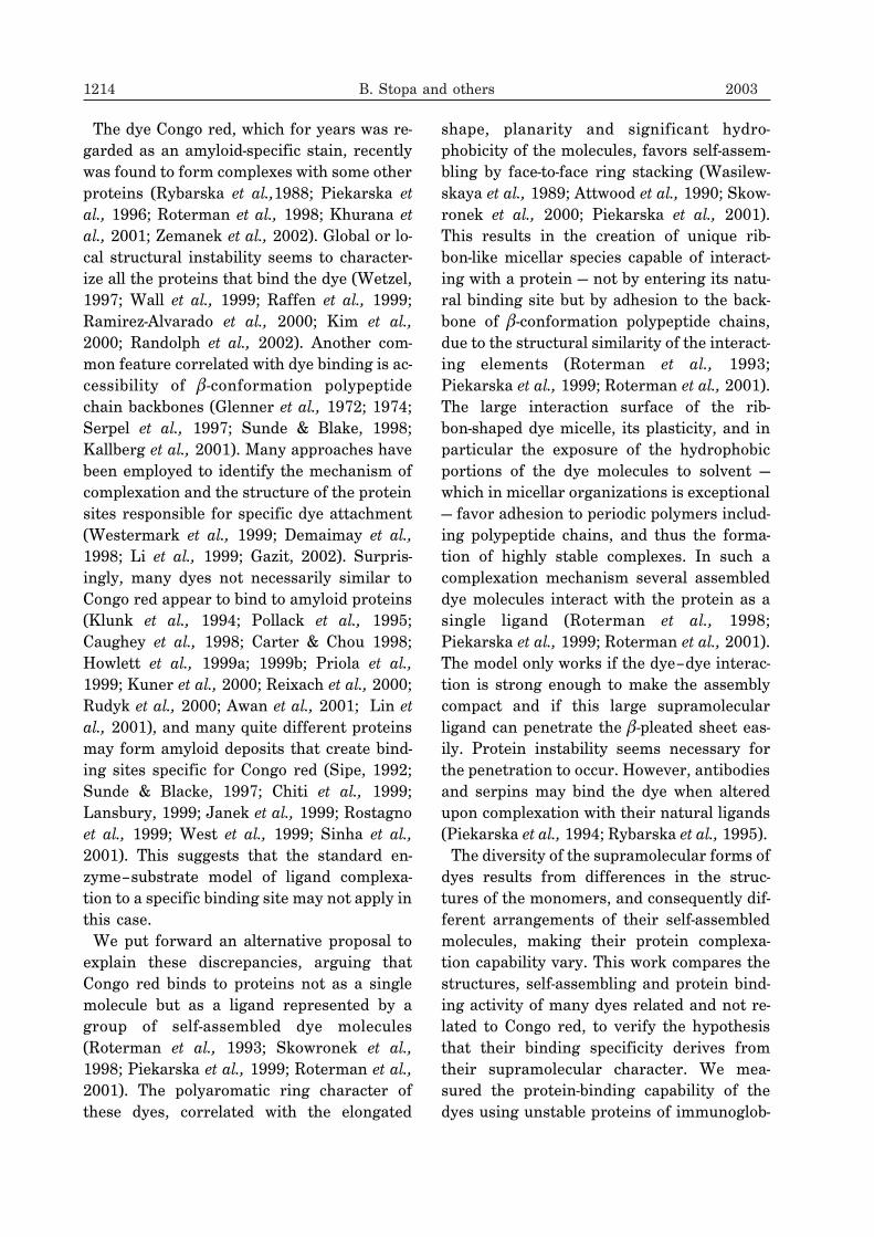
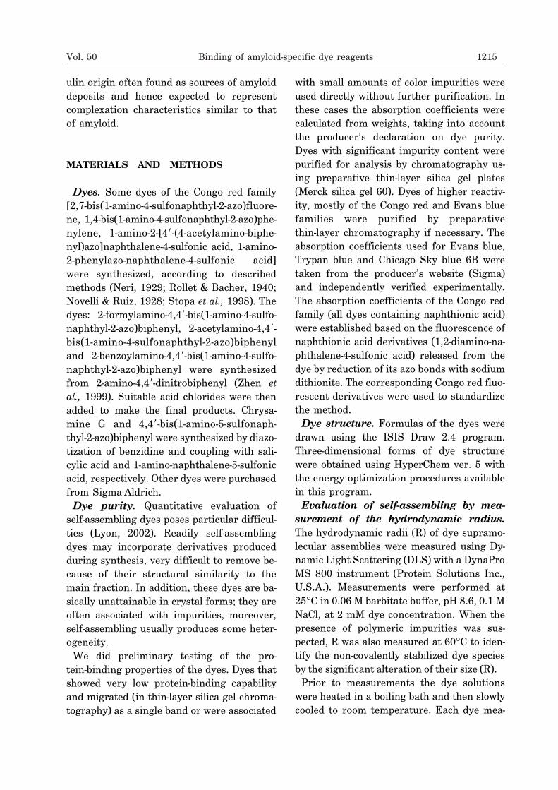
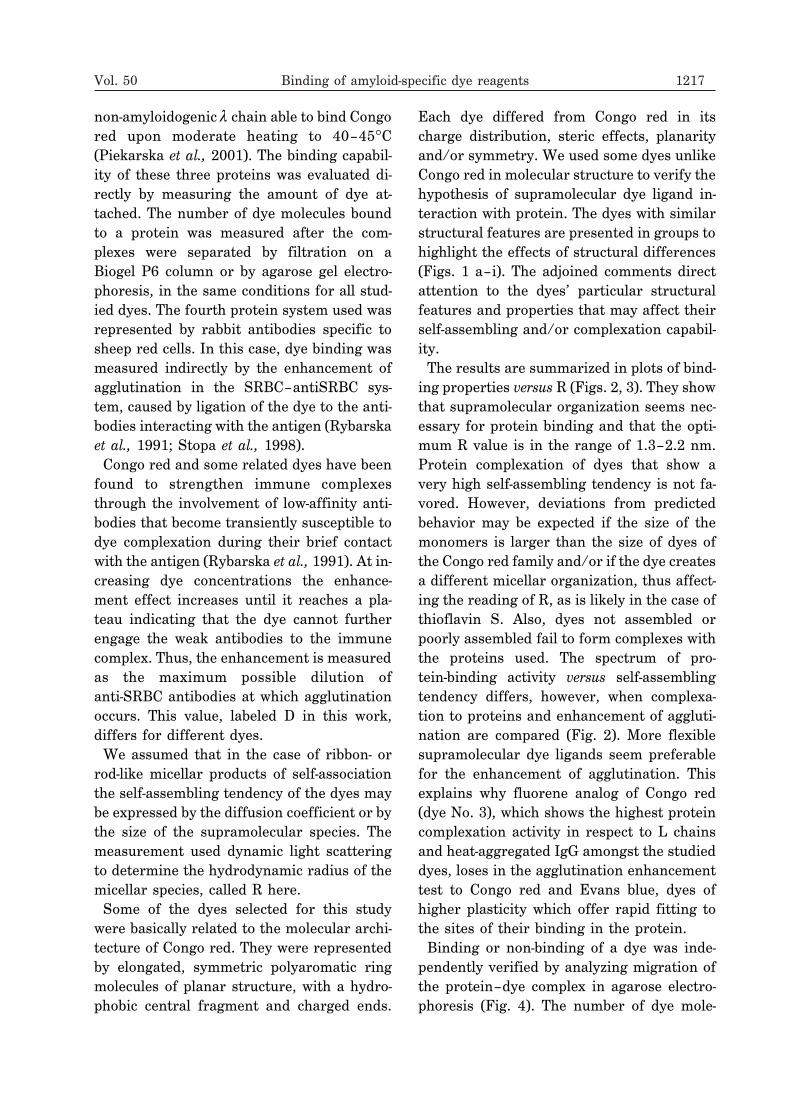
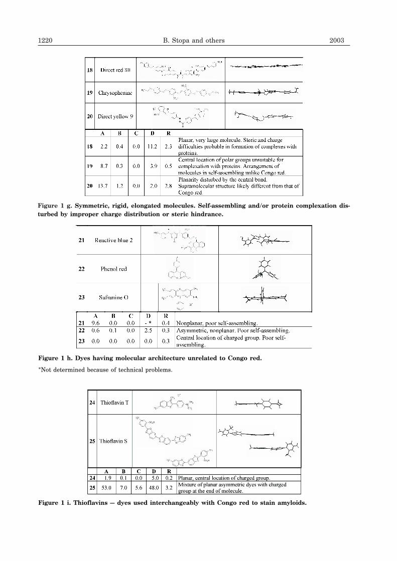
Figure 1 g. Symmetric, rigid, elongated molecules. Self-assembling and/or protein complexation dis-turbed by improper charge distribution or steric hindrance.
Figure 1 h. Dyes having molecular architecture unrelated to Congo red.
*Not determined because of technical problems.
Figure 1 i. Thioflavins — dyes used interchangeably with Congo red to stain amyloids.
cules bound to protein observed in this test-ing system is basically higher than in thechromatographic technique because the bedabsorbs only weakly (not shown). Readingprecision is poor in this test, and it is difficultto use for quantitative analysis, but themethod was performed to support the obser-vations of the dye-binding capability revealedby the gel filtration tests.
DISCUSSION
The evidence supplied by our studies sup-ports the hypothesis of Congo red’s specificbinding capability arising from the particularsupramolecular form of this dye (Skowroneket al., 1998; Piekarska et al., 1999; Rotermanet al., 2001, Rybarska et al., 2001). It explainsthe binding specificity by the adhesion of rib-bon-like supramolecular dye species to �-con-formation polypeptides.Two somewhat contradictory factors seem
to determine the particular dye binding mech-anism that finally ensures the best possible fitand adhesion of the supramolecular dye lig-ands within the protein: (1) the strength of
Vol. 50 Binding of amyloid-specific dye reagents 1221
Figure 1 (a–i). Relation of molecular architecture to self-assembling and protein-complexation capabil-ity in groups of dyes.
Symbols used: A, number of dye molecules bound to heat-aggregated IgG (160 kDa); B, number of dye moleculesbound to monomer of L chain � Kok (23.5 kDa, 20�C); C, number of dye molecules bound to L chain � An (23.5 kDa,45�C); D, enhancement of agglutination caused by dyes (SRBC–anti-SRBC system); R, hydrodynamic radius ofpredominant micellar species (nm).
Figure 2. Comparison of protein complexation ca-pabilities of dyes, in order of increasing self-as-sembling tendency.
R, hydrodynamic radius of the main supramolecularform. Numbers at the top of each graph correspond todye numbers in Fig. 1. Panels A, B and C, the numberof dye molecules bound to each protein after gel filtra-tion was recalculated for 100 kDa. Panel D, the en-hancement effect of dyes on immune complex stability,evaluated in SRBC–antiSRBC system.
Figure 3. Correlation between hydrodynamic ra-dius (R) of dye self-assemblies and their ability toform complexes with heat-aggregated IgG.
self-association increasing the stability of thedye assemblies, sufficient to make them inter-act with polypeptides as compact ligands; and(2) their plasticity, which arises from the dy-namic micellar form of the dye and facilitatesoptimal fitting within the complex. The move-ments of individual dye molecules allowed inthe supramolecular ligand, which are neces-sary for micellar plasticity, involve rotationsaround the long axis of the ribbon-like micelleas well as around the long axis of the particu-lar molecule. The latter consists in rotationsaround the central bond of a symmetric dyemolecule, producing syn and anti conformers(Skowronek et al., 2000). The compactness ofthe micellar structures increases with the pla-narity of the dye molecule, its hydrophobicity,and enlargement of the stacking area. In-creasing charge repulsion and/or steric hin-drance reduce micellar rigidity, associated inconsequence with higher freedom of molecu-lar movements. These contradictory effectsdetermining protein complexation make thebinding capability optimal not at a very highbut rather at an intermediate level of thedye’s self-assembling. The central hydropho-bic parts of the dye molecules seem to be pref-erentially engaged in interaction with the
polypeptide backbone (Demaimay et al.,1998). The role of the charged parts of themolecule, and in particular of the charge dis-tribution, does not seem negligible, however.Only symmetric, rigid, planar, polyaromaticdye molecules with charged groups located atthe ends, with the necessary elongated, hy-drophobic central fragment (biphenyl), mayimpose the orientation of stacked moleculesthat ensures the formation of ribbon-shapedsupramolecular structures (Wasilewskaya etal., 1989; Attwood et al., 1990; Piekarska etal., 1999).Among the many different supramolecular
assemblies, ribbon-shaped micellar speciesseem to expose the hydrophobic surface mostefficiently, favoring adhesion. The poly-peptide backbone likely represents the corre-sponding structure for adhesion. Althoughboth adhering elements — the dye andpolypeptide — are similar in their chain-likecharacter, fitting optimization seems neces-sary for complex formation since their chemi-cal nature is significantly different. Thistakes time, particularly when the interactingstructures are rigid. This explains why dyesof high micellar rigidity that complex effi-ciently when incubated with proteins of un-
1222 B. Stopa and others 2003
Figure 4. Protein-binding ability of selected dyes studied by agarose gel electrophoresis (pH 8.6). Num-bers below each lane correspond to those used in Fig. 1.
0, control sample (no dye). A, gel before staining for protein; B, after staining with bromophenol blue and removalof dyes by reduction with sodium dithionite.
stable structure appear less suitable for liga-tion to low-affinity antibodies (largely repre-sented in the polyvalent IgG fraction); thoseantibodies offer only brief contact with theantigen and consequently brief or too briefprotein accessibility to allow for optimizationof the dye–protein adhesion structure andcomplexation (Rybarska et al., 1991; Kaszubaet al., 1993; Stopa et al., 1998).Although growing evidence links Congo
red’s specific binding property with itssupramolecularity and implicates �-conforma-tion polypeptides as the receptor structure inthe protein, other ligation mechanisms mayalso be engaged in particular situations.Thus, a single Congo red molecule bound to aspecific site in the protein was found in pig in-sulin crystals (Turnell & Finch, 1992). Otherstudies have supplied evidence of Congored-bridging protein molecules (Khurana etal., 2001). However, the hypothesis that thebinding of Congo red-related dyes is associ-ated with their supramolecular characterseems to offer a general explanation of theirspecific affinity to �-structure (Piekarska etal., 1996; 2001; Roterman et al., 2001). Al-though supramolecularity seems the most es-sential factor in the binding capability of thegroup of studied dyes, a direct relation to theR value cannot be expected due to the shape-and charge-derived polymorphism of supra-molecular ligands.While the specific affinity of Congo red to
amyloid deposits can be explained by the par-ticular complexation character of the supra-molecular form of this dye, there is some dis-crepancy in respect to thioflavins T and S,which, although differing essentially in struc-ture and properties, are used interchangeablyto stain amyloids (Le Vine 1999; Carrotta etal., 2001; Nielsen et al., 2001; Zhuang et al.,2001; Pavlov et al., 2002; Devlin et al., 2002).The planar, elongated components (Fig. 1 i) ofthioflavin S, negatively charged at the ends,may overlap, producing symmetric dimersand then longer micellar structures, finally fa-voring complexation in the manner of Congo
red. This explanation is supported by the ob-served high R value for this dye. In contrast,the polarity distribution of the thioflavin Tmolecule seems unsuited to self-assemblingalthough its molecule is planar. Consequen-tly, the yield of its complexation with proteinsappeared also low. This seems consistent withthe known direct dependence of this dye’s flu-orescence on growing amyloid fibrils, while itis poor or absent in the presence of individualmolecules of amyloidogenic proteins. Thestandard quenching of fluorescence in solu-tions probably can be prevented here by im-mobilizing the dye molecules adsorbed to thehighly ordered amyloid protein structure,even if the binding specificity is not necessar-ily of the Congo red type (Nakashima et al.,1995).For optimal adhesion, complexation of
supramolecular Congo red-related dye lig-ands to proteins requires a special ribbon-likeorganization and an extra adaptation to theprotein receptor surface. Although the com-pact self-assembled form of the dye is re-quired for adhesion to polypeptides, very highrigidity of the supramolecular ligand seemsunsuitable, since the increasing micellar ri-gidity makes the adhering structures moredifficult to adjust to each other.
The dyes were synthesized by Dr. Jan Boksafrom the Institute of Pharmacology of the Pol-ish Academy of Sciences (Kraków).
R E F E R E N C E S
Attwood TK, Lydon JE, Hall C, Tiddy GJT.(1990) The distinction between chromonicand amphiphilic lyotropic mesophases. Liq-uid Crystals.; 7: 657–68.
Awan T, Forloni G, Ragg E, Iussich S, Rossi G,Colombo L, Girola L, Massignan T, BugianiO, Salmona M, Tagliavini F. (2001) Thera-peutic approaches to prion diseases: In vitrostudies with tetracycline compounds. In: Alz-heimer’s disease: advances in etiology,pathogenesis and therapeutics. Iqbal K,
Vol. 50 Binding of amyloid-specific dye reagents 1223
Sisodia SS, Winblad B. eds, pp 809–20. JohnWiley & Sons Ltd.
Carrotta R, Bauer R, Waninge R, Rischel C.(2001) Conformational characterization ofoligomeric intermediates and aggregates in�-lactoglobulin heat aggregation. Protein Sci.;10: 1312–8.
Carter DB, Chou K-C. (1998) A model for struc-ture-dependent binding of Congo red to Alz-heimer �-amyloid fibrils. Neurobiol Aging.;19: 37–40.
Caughey WS, Raymond LD, Horiuchi M,Caughey B. (1998) Inhibition of protease-re-sistant prion protein formation by porphy-rins and phthalocyanines. Proc Natl AcadSci U S A.; 95: 12117–22.
Chiti F, Webster P, Taddei N, Chiti F, WebsterP, Taddei N, Clark A, Stefani M, Ramponi G,Dobson CM. (1999) Designing conditions forin vitro formation of amyloid protofilamentsand fibrils. Proc Natl Acad Sci U S A.; 96:3590–4.
Demaimay R, Harper J, Gordon H, Weaver D,Chesebro B, Caughey B. (1998) Structuralaspects of Congo red as an inhibitor of pro-tease-resistant prion protein formation. JNeurochem.; 71: 2534–41.
Devlin GL, Chow MKM, Howlett GJ, BottomleySP. (2002) Acid denaturation of�1-antitrypsin: characterization of a novelmechanism of serpin polymerization. J MolBiol.; 324: 859–70.
Gazit E. (2002) A possible role for �-stacking inthe self-assembly of amyloid fibrils. FASEBJ.; 16: 77–83.
Glenner GG, Eanes ED, Page DL. (1972) Therelation of the properties of Congored-stained amyloid fibrils to the �-conforma-tion. J Histochem Cytochem.; 20: 821–6.
Glenner GG, Eanes ED, Bladen HA, Linke RP,Termine JD. (1974) �-pleated sheet fibrils acomparison of native amyloid with syntheticprotein fibrils. J Histochem Cytochem.; 22:1141–58.
Howlett DR, Perry AE, Godfrey F, Swatton JE,Jennings KH, Spitzfaden C, Wadsworth H,Wood SJ, Markwell RE. (1999) Inhibition of
fibril formation in �-amyloid peptide by anovel series of benzofurans. Biochem J.;340: 283–9.
Howlett DR, George AR, Owen DE, Ward RV,Markwell RE. (1999) Common structural fea-tures determine the effectiveness ofcarvedilol, daunomycin and rolitetracyclineas inhibitors of Alzheimer �-amyloid fibrilformation. Biochem J.; 343: 419–23.
Janek K, Behlke J, Zipper J, Fabian H,Georgalis Y, Beyermann M, Bienert M,Krause E. (1999) Water-soluble �-sheet mod-els which self-assemble into fibrillar struc-tures. Biochemistry.; 38: 8246–52.
Kallberg Y, Gustafsson M, Persson B, ThybergJ, Johansson J. (2001) Prediction of amyloidfibril-forming proteins. J Biol Chem.; 276:12945–50.
Kaszuba J, Konieczny L, Piekarska B,Roterman I, Rybarska J. (1993) Bis-azo dyesinterference with effector activation of anti-bodies. J Physiol Pharmacol.; 44: 233–42.
Khurana R, Uversky VN, Nielsen L, Fink AL.(2001) Is Congo red an amyloid-specific dye?J Biol Chem.; 276: 22715–21.
Klunk WE, Debnath ML, Pettegrew JW. (1994)Development of small molecule probes forthe �-amyloid protein of Alzheimer’s disease.Neurobiol Aging.; 15: 691–8.
Kuner P, Bohrmann B, Tjernberg LO, NäslundJ, Huber G, Celenk S, Grüninger-Leitch F,Richards JG, Jakob-R�tne R, Kemp JA,Nordstedt C. (2000) Controlling polymeriza-tion of �-amyloid and prion-derived peptideswith synthetic small molecule ligands. J BiolChem.; 275: 1673–8.
Lansbury PT Jr. (1999) Evolution of amyloid:What normal protein folding may tell usabout fibrillogenesis and disease. Proc NatlAcad Sci U S A.; 96: 3342–4.
1224 B. Stopa and others 2003
Le Vine III H. (1999) Quantification of �-sheetamyloid fibril structures with thioflavin T.Methods Enzymol.; 309: 274–84.
Li L, Darden TA, Bartolotti L, Kominos D,Pedersen LG. (1999) An atomic model forthe pleated �-sheet structure of A� amyloidprotofilaments. Biophys J.; 76: 2871–8.
Lin Y-M, Raffen R, Zhou Y, Cassidy CS, FlavinMT, Steven FJ. (2001) Amyloid fibril forma-tion in microwell plates for screening of in-hibitors. Amyloid.; 8: 182–93.
Lyon HO. (2002) Dye purity and dye standard-ization for biological staining. BiotechHistochem.; 77: 57–80.
Maeda N, Chen N, Tirrell M, Israelachvili JN.(2002) Adhesion and friction mechanisms ofpolymer-on-polymer surfaces. Science.; 297:379–82.
Nakashima K, Fujimoto Y, Kido N. (1995) Fluo-rescence studies on the adsorption ofoctadecylrhodamine B onto a latex surface.Photochem Photobiol.; 62: 674–9.
Neri A. (1929) Monosulfonic derivatives of2-N-phenyl-1,2-naphtho-1,2,3-triazole. GazzChim Ital.; 59: 384–91.
Nielsen L, Khurana R, Coats A. (2001) Effect ofenvironmental factors on the kinetics of in-sulin fibril formation: elucidation of the mo-lecular mechanism. Biochemistry.; 40:6036–46.
Novelli A, Ruiz C. (1928) New substantive dyesderived from 2,7-diaminofluorene. AnnAsocn Quim Argent.; 16: 56–64.
Pavlov NA, Cherny DI, Heim G, Jovin TM,Subramaniam V. (2002) Amyloid fibrils fromthe mammalian protein prothymosin �.FEBS Lett.; 517: 37–40.
Piekarska B, Roterman I, Rybarska J,Konieczny L, Kaszuba J. (1994) The meltingof native domain structure in effector activa-tion of IgG studied by using Congo red as aspecific probe. J Physiol Pharmacol.; 45:147–62.
Piekarska B, Skowronek M, Rybarska J, StopaB, Roterman I, Konieczny L. (1996) Congored-stabilized intermediates in the � light
chain transition from native to molten state.Biochimie.; 78: 183–9.
Piekarska B, Rybarska J, Stopa B, Zemanek G,Król M, Konieczny L. (1999) Supramole-cularity creates nonstandard protein ligands.Acta Biochim Polon.; 46: 841–51.
Piekarska B, Konieczny L, Rybarska J, Stopa B,Zemanek G, Szneler E, Król M, Nowak M,Roterman I. (2001) Heat-induced formationof a specific binding site for self-assembledCongo red in the V domain of immunoglobu-lin L chain �. Biopolymers.; 59: 446–56.
Pollack SJ, Sadler IIJ, Hawtin SR, Tailor VJ,Shearman MS. (1995) Sulfonated dyes atten-uate the toxic effects of �-amyloid in a struc-ture-specific fashion. Neurosci Lett.; 197:211–4.
Priola SA, Caughey B, Caughey WS. (1999)Novel therapeutic uses for porphyrins andphthalocyanines in the transmissiblespongiform encephalopathies. Curr OpinMicrobiol.; 2: 563–6.
Raffen R, Dieckman LJ, Szpunar M, WunschlC, Pokkuluri PR, Dave P, Stevens PW, CaiX, Schiffer M, Stevens FJ. (1999) Physico-chemical consequences of amino acid varia-tions that contribute to fibril formation byimmunoglobulin light chains. Protein Sci.; 8:509–17.
Ramirez-Alvarado M, Merkel JS, Regan L.(2000) A systematic exploration of the influ-ence of the protein stability on amyloid fibrilformation in vitro. Proc Natl Acad SciU S A.; 97: 8979–84.
Randolph TW, Seefeldt M, Carpenter JF. (2002)High hydrostatic pressure as a tool to studyprotein aggregation and amyloidosis. BiochimBiophys Acta.; 1595: 224–34.
Reixach N, Crooks E, Ostresh JM, HoughtenRA, Blondelle SE. (2000) Inhibition of �-amy-loid-induced neurotoxicity by imidazopyri-doindoles derived from a synthetic combina-tion library. J Struct Biol.; 130: 247–58.
Rollet A, Bacher W. (1940) Azo dyes IV.Monatsh.; 73: 20–4.
Rostagno A, Vidal R, Kaplan B, Chuba J,Kumar A, Elliott JI, Frangione B, Gallo G,
Vol. 50 Binding of amyloid-specific dye reagents 1225
Ghiso J. (1999) pH-dependent fibrillogenesisof a V [�] III Bence Jones protein. Br JHaematol.; 107: 835–43.
Roterman I, No K-T, Piekarska B, Kaszuba J,Pawlicki R, Rybarska J, Konieczny L. (1993)Bis azo dyes — studies on the mechanism ofcomplex formation with IgG modulated byheating or antigen binding. J PhysiolPharmacol.; 44: 213–32.
Roterman I, Rybarska J, Konieczny L,Skowronek M, Stopa B, Piekarska B. (1998)Congo red bound to �-1-proteinase inhibitoras a model of supramolecular ligand andprotein complex. Comput Chem.; 22: 61–70.
Roterman I, Król M, Nowak M, Konieczny L,Rybarska J, Stopa B, Piekarska B, ZemanekG. (2001) Why Congo red binding is specificfor amyloid proteins — model studies and acomputer analysis approach. Med Sci Monit.;7: 771–84.
Rudyk H, Vasiljevic S, Hennion RM, Birkett CR,Hope J, Gilbert IH. (2000) Screening Congored and its analogues for their ability to pre-vent the formation of PrP-res in scrapie-in-fected cells. J Gen Virol.; 81: 1155–64.
Rybarska J, Piekarska B, Konieczny L,Roterman I. (1988) The formation of solubleheat IgG aggregates for immunological stud-ies. Arch Immunol Ther Exp (Warsz).; 36:609–22.
Rybarska J, Konieczny L, Roterman I,Piekarska B. (1991) The effect of azo dyes onthe formation of immune complexes. ArchImmunol Ther Exp (Warsz).; 39: 317–27.
Rybarska J, Konieczny L, Piekarska B, Stopa B,Roterman I. (1995) The detection of acutephase serum protein complexes and immunecomplexes by Congo red binding. J PhysiolPharmacol.; 46: 221–31.
Rybarska J, Piekarska B, Stopa B, Zemanek G,Konieczny L, Nowak M, Król M, Roterman I,Szymczakiewicz-Multanowska A. (2001) Evi-dence that supramolecular Congo red is thesole ligation form of this dye for L chain �
Serpel LC, Sunde M, Blake CCF. (1997) Themolecular basis of amyloidosis. Cell Mol LifeSci.; 53: 871–87.
Sinha N, Tsai C-J, Nussinov R. (2001) A pro-posed structural model for amyloid fibrilelongation: domain swapping forms aninterdigitating �-structure polymer. ProteinEng.; 14: 93–103.
Sipe JD. (1992) Amyloidosis. Annu RevBiochem.; 61: 947–75.
Skowronek M, Stopa B, Konieczny L, RybarskaJ, Piekarska B, Szneler E, Bakalarski G,Roterman I. (1998) Self-assembly of Congored — a theoretical and experimental ap-proach to identify its supramolecular organi-zation in water and salt solutions.Biopolymers.; 46: 267–81.
Skowronek M, Roterman I, Konieczny L, StopaB, Rybarska J, Piekarska B. (2000) Why doCongo red, Evans blue and Trypan blue dif-fer in their complexation properties? JComputat Chem.; 21: 656–67.
Stopa B, Górny M, Konieczny L, Piekarska B,Rybarska J, Skowronek M, Roterman I.(1998) Supramolecular ligands: Monomerstructure and protein ligation capability.Biochimie.; 80: 963–8.
Sunde M, Blake C. (1997) The structure of amy-loid fibrils by electron microscopy and X-raydiffraction. Adv Protein Chem.; 50: 123–59.
Sunde M, Blake CCF. (1998) From the globularto the fibrous state: protein structure andstructural conversion in amyloid formation.Quat Rev Biophys.; 31: 1–39.
Turnell WG, Finch JT. (1992) Binding of thedye Congo red to the amyloid protein pig in-sulin reveals a novel homology amongst amy-loid-forming peptide sequences. J Mol Biol.;227: 1205–23.
Wall J, Schell M, Murphy C, Hrncic C, StevensFJ, Solomon A. (1999) Thermodynamic in-stability of human �6 light chains: Correla-tion with fibrillogenicity. Biochemistry.; 38:14101–8.
West MW, Wang W, Patterson J, Mancias JD,Beasley JR, Hecht MH. (1999) De novo amy-loid proteins from designed combinatorial li-braries. Proc Natl Acad Sci U S A.; 96:11211–6.
Westermark GT, Johnson KH, Westermark P.(1999) Staining methods for identification ofamyloid in tissue. Methods Enzymol.; 309:3–25.
Wetzel R. (1997) Domain stability in immuno-globulin light chain deposition disorders. AdvProtein Chem.; 50: 183–242.
B, Nowak M, Król M, Roterman I. (2002)Egg yolk platelet proteins from Xenopuslaevis are amyloidogenic. Folia HistochemCytobiol.; 40: 311–8.
Zhen W, Han H, Anguiano M, Lemere CA, ChoC-G, Lansbury PT Jr. (1999) Synthesis andamyloid binding properties of rhenium com-plexes: preliminary progress toward a re-agent for SPECT imaging of Alzheimer’s dis-ease brain. J Med Chem.; 42: 2805–15.
Zhuang Z-P, Kung M-P, Hou C. (2001)Radioiodinated styrylbenzenes andthioflavins as probes for amyloid aggregates.J Med Chem.; 44: 1905–14.
Vol. 50 Binding of amyloid-specific dye reagents 1227