STRUCTURE, DEVELOPMENT, AND BIONOMICS 01' HOUSE-FLY. 347 The Structure, Development, and Bionomics of the House-fly, Musca domestics, Linn. Part III.—The Bionomics, Allies, Parasites, and the Eelations of M. domestica to Human Disease. By €. Gordon Hewitt, D.Sc, Late Lectiu-er in Economic Zoology, University of Manchester. With Plate 22. PAGE I. I n t r o d u c t i o n . . . . . . 348 II. Distribution . . . . . . . 349 III. Flies occulting as Co-inhabitants of Houses with M. domestica or as Visitants . . . . 351 IV. Physiology : 1. Influence of Food, Temperature, and Light . . 362 2. Hibernation . . . . . 363 3. Flight . . . . . . 364 4. Regeneration of Lost Parts . . . 365 V. Natural Enemies and Occasional Parasites : 1. Chernes nodosus, Schrank . . . 367 2. Acarina or Mites borne by House-flies . . 369 3. Fungal parasite—Empusa niuscas, Cohn . . 371 VI. True Parasites: 1. Flagellata—Herpetomonas muscto-domestica) . 374 Crithidia lnuscse-doinestica) . 370 2. Nematoda—Habronema muses . . . 380 3. Dissemination of Parasitic Worms . . . 382 VII. Dissemination of Pathogenic Organisms by M. domestica and its non-Blood-sucking Allies : 1. Typhoid Fever . . . . . 385 2. Anthrax . . . . . 394 3. Cholera . . . . . . 396 4. Tuberculosis . . . . . 398
Transcript
STRUCTURE, DEVELOPMENT, AND BIONOMICS 01' HOUSE-FLY. 347
The Structure, Development, and Bionomicsof the House-fly, M u s c a d o m e s t i c s , Linn.
Part III.—The Bionomics, Allies, Parasites, and the Eelationsof M. domestica to Human Disease.
By€. Gordon Hewitt, D.Sc,
Late Lectiu-er in Economic Zoology, University of Manchester.
With Plate 22.
PAGEI. I n t r o d u c t i o n . . . . . . 348
II . Distribution . . . . . . . 349I I I . Flies occulting as Co-inhabitants of Houses with M.
d o m e s t i c a or as Visitants . . . . 351IV. Physiology :
1. Influence of Food, Temperature, and Ligh t . . 3622. Hibernation . . . . . 3633. Fl ight . . . . . . 3 6 44. Regeneration of Lost P a r t s . . . 365
V. Natura l Enemies and Occasional Parasites :1. C h e r n e s n o d o s u s , Schrank . . . 3672. Acarina or Mites borne by House-flies . . 3693. Fungal p a r a s i t e — E m p u s a niuscas, Cohn . . 371
VI. True Parasites:1. Flagel la ta—Herpetomonas muscto-domestica) . 374
C r i t h i d i a lnuscse-doinestica) . 3702. Nematoda—Habronema m u s e s . . . 3803. Dissemination of Parasitic Worms . . . 382
VII. Dissemination of Pathogenic Organisms by M. domes t icaand its non-Blood-sucking Allies :1. Typhoid Fever . . . . .3852. Anthrax . . . . .3943. Cholera . . . . . .3964. Tuberculosis . . . . . 398
V I I I . Flies and Intestinal Myiaais . . . . 404I X . Literature . . . . . . 405
X. Appendix on the Winter Breeding of M. d o m e s t i c a . . 412
I . INTRODUCTION.
T H E p r e s e n t p a p e r c o n c l u d e s t h i s s t u d y of t h e s t r u c t u r e ,d e v e l o p m e n t , a n d b i o n o m i c s of M u s c a d o m e s t i c a ( t h eprevious parts were published in 1907 and 1908). In it Ihave described the bionomics, certain of its allies which mayoccur in houses, its parasites, and its relation to man, especiallyas the carrier of the bacilli of certain infectious diseases.
The last portion of the present paper, in which is describedwhat is known concerniug the ability of M. domestica andits allies to carry and disseminate the bacteria of many impor-tant diseases, shows, I hope, the grave character of its relationto man. Although its importance in this respect is beinggradually realised in this country, it is not so widely recog-nised as it should be. In the United States of America it isproposed to change this insect's name from the house-fly tothe " Typhoid fly " ; notwithstanding certain objections to thisname, it clearly indicates that more attention must be paid topreventive measures, that is, they must be reduced by thedeprivation of suitable breeding-places. I have not discussedin the present paper the relation of house-flies to infantile orsummer diarrhoea, chiefly because we are not yet certain as tothe specific cause, but this disease may be included for thepresent under typhoid or enteric fever in so far as the relationof flies with it is concerned.
I should like to take this opportunity of thanking thosemedical men, whose names I mention later, for the kiudmanner in which they have replied to my inquiries concern-ing their observations on various diseases of which they havespecial knowledge.
STBUCTURE, DEVELOPMENT, AND BIONOMICS OV HOUSE-FLY. 349
II. DISTRIBUTION.
Musca doin/stica is probably the most widely distributedinsect to be found ; the animal most commonly associated withmau, whom it appears to have followed over the entire globe.It extends from the sub-polar regions, where Linnaeus refersto its occurrence in Lapland, and Finmark as " rara avis inLapponia, at in Finmarchia Norwegiss integras domos ferereplet," to the tropics, where it occurs in enormous numbers.Referring to its abundance in a house near Para in equatoralBrazil, Austen (1904) says: "At the mid-day meal theyswarmed on the table in almost inconceivable nuinbers," andother travellers in different tropical countries have relatedsimilar experiences to me, how they swarm round each pieceof food as it is carried to the mouth.
In the civilised and populated regions of the world it occurscommonly, and the British Museum (Natural History) collec-tion and my own contain specimens from the followinglocalities. Certain of the localities have, in addition, beenobtained from lists of insect faunas :
Asia.—Aden; North West Provinces (India); Calcutta;Madras; Bombay (it probably occurs over the whole ofIndia); Ceylon; Central China; Hong-Kong; Shanghai;Straits Settlements; Japan.
Africa.—Port Said; Suez; Egypt; Somaliland; Nyassa-land; Uganda; British E. Africa; Rhodesia; Transvaal;Natal; Cape Colony; Madagascar; Northern and SouthernNigeria; St. Helena; Madeira.
America.—Distributed over North America; Brazil;Monte Video (Uruguay); Argentine; Valparaiso; WestIndies.
Australia and New Zealand.Europe and the isles of the Mediterranean; it is especially
common in Cyprus.Not only is this world-wide distribution of interest, but its
distribution in our own country is noteworthy. From observa-tions that I have made during a number of years in town and
350 C. GORDON HEWITT.
suburban houses and country houses and cottages, I find thatin the former it is by far the commonest house-fly. Butwhereas M. domes tica may be almost the only species inwarm places where food is present, such as restaurants andkitchens, in other rooms of houses Homalomyia cani-cularis, the small house-fly, increases in proportion andoften predominates; occasionally one may find it to becommoner than M. domestica. In country houses theproportions vary by the intrusion of Stomoxys calcitrans,which I have often found to be the dominant species. In acertain country cottage, out of the several hundreds captured,S. calcitrans formed 50 per cent, of the total, the rest beingchiefly H. canicularis together with Anthomyia radicum,whose Iarva3, as I have shown (1907), breed in horse-manurewith those of M. domestica. The following records takenfrom a " fly census " that was made in 1907 may be taken asillustrative of the proportional abundance of the differentspecies in different situations; although the numbers of theserecords are small the proportions are more obvious.
Out of a total of 3856 flies caught in different situations,such as restaurants, kitchens, stables, bedrooms and hotels,87-5 per cent, were M. domestica, 11*5 per cent. H. canicu-laris, and the rest were other species such as S. calcitrans,Muscina stabulans, C. erythrocephala, and Antho-myia vadicum. These figures are comparatively small, but
STRUCTURE, DEVELOPMENT, AND BIONOMICS OJ)' HOUSE-PLY. 351
are representative of the a v e r a g e occurrence, as I haveobserved, of the different species.
For the proportional occurrence in similar localities we haveinteresting figiu'es given by Howard (1900) for the UnitedStates. Of 23,087 flies caught in rooms where food suppliesare exposed he found that 22,808, or 98"8 per cent, of thewhole number, were M. d o m e s t i c a , and of the remaining]*2 per cent. H . c a n i c u l a r is was the commonest species.Hamer (1908) found that more than nine tenths of the iiiescaught in the kitchens and "l iving-rooms" of houses in theneighbourhood of depots for horse-refuse, manure, etc., wereM. d o m e s t i c a . In a further report Hamer gives moredetails as to the different species that were found. Iu onelot of 35,000 flies caught on four fly-papers exposed iu similarpositions, 17 per cent, were Horn a lomyi a, c a n i c u l a r is , lessthan 1 per cent. wereC. e r y t h r o c e p h a l a , and considerablyless than 1 per cent, were M u s c i n a s t a b u l a n s , whereas ofnearly 6000 flies caught in another situation in four fly-balloons 24 per cent. wereH. c a n i c u l a r i s , 15 per ceut. wereC. e r y t h r o c e p h a l a , and nearly 2 per cent. wereM. s t a b u -l ans . He giyes an interesting diagram showing from countsof flies the seasonal prevalence which I have previouslyrecorded from observation. The report shows how the pro-portions of the different species vary in different situationsaccording to the substances and refuse that are present inthe locality. We may therefore say with certainty thatM. d o m e s t i c a is the commonest species of house-fly, andnext to this H . c a n i c u l a r i s , and that in country housesS. c a l c i t r a n s often occurs in large numbers, althoughit is not a house-fly in the strict sense of the word.
II I . PLIES OCCURRING AS CO-INHABITANTS OF HOUSES WITH
M . DOMESTICA OR AS VISITANTS.
We have seen from the preceding section that M. domes-tica is by far the commonest species which occurs in houses,and is, in fact, "domesticated" in the true sense of the word
354 C. UOliUON HEWITT.
domes t ica and M. coi'vina. It will be seen, therefore,that its breeding habits are very similar to those of M.domes t i ca and the sub-species d e t e r m i n a t a . It is in-teresting and important to note tlie rather exceptional choiceof cow-dung as a breeding-place.
(3) Homalomyia can i cu l a r i s L.
This species of fly (see 'Quart. Journ. Micr. Sci.,' vol. 51,PL 22, fig. 3) is often mistaken by the uninitiated for M.domes t i ca which are not full grown. Although it may becalled the small or lesser house-fly its differences from M.domes t i ca are great, as it belongs to a different group ofcalypterate Muscidae, namely, the Authotnyida?. One of thechief distinguishing features of this group is that the fourthlongitudinal vein of the wing (M. 1 + 2) goes straight to themargin of the wing and does not bend upwards at an angleas in M. domest ica .
The male of H. can i cu l a r i s differs from the female insome respects. In the male the eyes are close together, andthe frontal region is consequently very narrow; the sides ofthis, these are the inner orbital regions, are silvery white,separated by a narrow black frontal stripe. In the femalethe space between the inner margins of the eyes is about onethird of the width of the head; the frons is brownish, black,and the inner orbital regions are dark ashy grey. The bristleof the antenna of H. c a n i c u l a r i s is bare; in M. domes-tica, it will be remembered, the bristle bears a row of setaeon its upper and lower sides. The dorsal side of the thoraxof the male is blackish, grey with three rather indistinct longi-tudinal black lines. In the female it is of a lighter grey, andthe three longitudinal stripes are consequently more distinct.The abdomen of the male H. c an i cu l a r i s is narrow andtapering compared with that of M. domest ica . It is bronzeblack in colour, and each of the three abdominal segmentshas a lateral translucent area, so that when it is seen againstthe light, as on a window-pane, three, and sometimes four,pairs of yellow translucent areas can be seen by the trans-
STRUCTURE,DEVELOPMENT, AND BIONOMICS OF HOUSK-FLY. 355
mitted light. In the female tlie abdomen is short in propor-tion to its length, and is of a greenish or brownish-grey colour
H. canicularis appears in houses before M. domestica,and can be found generally in May and June. In the lattermonth its numbers are swamped, as it were, by M. domes-tica, and it appears to seek the other rooms of a house thanthe kitchen, although I have found it frequently in consider-able numbers in kitchens. The average length is 57 mm.
The larva of H. canicularis (PI. 22, fig. 1) is verydistinct from that of M. domestica, as will be seen fromthe "figure. It is compressed dorso-ventrally, and has adouble row of processes on each side. Owing to the roughand spinous nature of these processes dirt adheres to thelarva and gives it a dirty-brown appearance. The full-grownlarva measures 5-6 mm. in length. The breeding habits ofH. canicularis are very similar to those of M. domestica.The larva? feed on waste vegetable substances and also onvarious excremental products, but particularly, I have found,on human excrement, for which they show a great partiality.I have frequently found excrement in privy middens to be amoving mass of the larvaD of H. canicularis. The larvalperiod is from thi*ee to four weeks, and the insect spendsfourteen to twenty-one days in the pupal stage.
(4) Homalomyia scalaris F.Newstead (1907) has found this species occurring as a
house-fly. It is slightly larger than, though similar iu manyrespects to, H. canicularis. The larva is very similar inappearance. Newstead found the larvEe in ash-pit refuse,and bred the flies from human fasces. The larvae have beenfound frequently to be the cause of intestinal myiasis.
(5) Anthomyia radicum Meigen.This member of the Anthornyidas has been found in houses,
especially those in or near the country. The female has beenillustrated already (Part I, 'Quart. Journ. Micr. Sci./ vol. 51,PI. 22, tig. 2). The male is darker in colour, the dorsal side
356 C. CORDON HEAVITT.
of the thorax being blackish with three black longitudinalstripes; the frontal region is very narrow; the abdomen isgrey with a dark median stripe. The average length of thebody is 5 mm.
In the summer they are common and may be found in theneighbourhood of manure. The eggs are laid in this substance,especially in horse-manure. The larvae have also beenfound feeding on the roots of various cultivated cruciferousplants, from which the insect has derived the name "root-maggot." The eggs hatch out from eighteen to thirty-sixhours after deposition. The first larval stadium lasts twenty-four hours, the second forty-eight hours, and five days laterthe larva changes into a pupa, the whole larval life occupy-ing about eight days. The pupal stage lasts ten days, so thatin warm weather the development may be completed in nine-teen to twenty days. The full-grown larvse measure 8 mm.iu length, and may be distinguished by the tubercles sur-rounding the caudal extremity. In this species there are sixpairs of spinous tubercles surrounding the posterior end anda seventh pair is situated on the ventral surface posterior tothe anus. The tubercles of the sixth pair, counting from thedorsal side, are smaller than the rest and are bifid. Thearrangement of the tubercles can be seen in fig. 2. Theanterior spiracnlar processes (fig. 3) are yellow in colour andhave thirteen lobes.
(6) Stomoxys calcitrans Linn.The species is common, especially in the country from
July to October, and during these months it may be oftenfound in houses, although Earner's observations (1908)appear to indicate that the presence of cowsheds, in whichthey occur in large numbers, does not affect their numbersin houses. I have found S. calcitrans in large numbersin the windows of a country house in March and April,and it may be found frequently out of doors on a sunnydav in May, aud throughout the ensuing summer months.It is normally an outdoor insect, but appears to seek the shelter
STRUCTURE, DEVELOPMENT, AND "BIONOMICS OK HOUSE-MY. 3 5 7
of houses, especially during wet weather, from which habit ithas no doubt derived the popular name of " storm-fly "; it isalso know as the "stable-fly." As these names may be equallyapplicable to certain other Diptera they should be discarded.
As I have already mentioned this species is frequently mis-taken by the public for M. domestic a, which is supposed tohave adopted the biting habit, although the latter is unableto inflict the slightest prick. If examined side by side thesreat differences between the two will be seen readily (seePart I in 'Quart. Journ. Micr. Sci., vol. 51, PL 22, fig. 4).S. calcitrans has an awl-like proboscis for piercing andblood-sucking; this projects horizontally forward frombeneath the surface of the head (fig. 4). It is slightlylarger and more robust than M. domestica; the bristles ofthe antennae bear setas on their upper sides only. The colouris brownish with a greenish tinge; the dorsal side of thethorax has four dark longitudinal stripes, the outermost pairbeing interrupted. At the anterior end of the dorsal side ofthe thorax the medium light-coloured stripe has a goldenappearance, which is very distinct when the insect is seenagainst the light. The abdomen is broad in proportion to itslength, and each of the large second and third segments hasa single median and two lateral brown spots; there is alsoa median spot on the fourth segment.
The life-history of S. calcitrans has been studied byNewstead (1906), and I have been able to confirm his observa-tions during 1907 and 1908. From fifty to seventy eggs,measuring 1 mm. in length, are laid by the female. The eggsare laid on warm, decaying vegetable refuse, especially inheaps of fermenting grass cut from lawns; I have frequentlyconfirmed this observation of Newstead's. The eggs are alsodeposited on various excreniental substances upon which thelarvre feed. Osborne (1896) reared them in horse-manure;Howard (1900) states that they live in fresh horse-manure,and records their occurrence in outdoor privies in somelocalities; Newstead reared them in moist sheep's dung; theycan also be reared in cow-dung.
358 0. GORDON HEWITT.
The larvss are creamy-white in colour and have a shiny,translucent appearance. They are rather similar to those of M.domestica, but can be distinguished by the character of theposterior spiracles. These (fig. 5 and 6) are wider apart thanin M. domestica and are triangular in shape with roundedcorners; each of the corners subtends a space in which asinuous aperture lies. The centre of the spiracle is occupiedby a circular plate of chitin. The anterior spiracular pro-cesses are five-lobed. Under warm conditions Newsteadfound that the egg state laated from two to three days;the larval stage lasts from fourteen to twenty-one daysand the pupal stage nine to thirteen days. There arethree larval stages. The whole life-history may be completein twenty-five to thirty-seven days. Some specimens passedthe winter in the pupal state.
Although S. calcitrans does not frequent to such a greatextent asM. domestica material likely to contain pathogenicintestinal bacilli, on account of its blood-sucking habits, whichcause it to attack cattle and not infrequently man, it mayoccasionally transfer the anthrax bacillus, as many havebelieved, and give rise to malignant pustule, etc.
(7) Calliphora erythrocephala Mg.This is the commoner of the two English blow-flies or
"blue-bottles." The other species, Calliphora (Musea)vomitoria, is less common, although the name is frequentlygiven to both species indiscriminately. They can be dis-tinguished, however,by the fact that in C. erythrocephalathe gente are fulvous to golden-yellow and are beset withblack hairs, whereas in C. vomitoria the gense are blackand the hairs are golden-red.
The appearance of C. erythrocephala is sufficiently wellknown with its bluish-black thorax and dark metallic blueabdomen. Its length varies from 7 to 13 mm. The larvasare necrophagous. The flies deposit their eggs on any freshor decaying meat, nor is such flesh always dead. Ou oneoccasion, when obtaining fresh material in the form of wild
STRUCT DUE, DEVELOPMENT, A Nn BIONOMICS OV HOUSK-FT.Y. 359
rabbits upon which to rear the larvas of C. e r y t h r o c e p h a l a ,I found the broken leg of a live rabbit, which had been caughtiu a spring trap set the previous evening, a living mass ofsmall larvae, which were devouring the animal while it wasstill alive. An enormous number of eggs are laid by a singleinsect; Portchinski ('Osten. Sacken/ 1887) found from 450to 600 eggs, though I have not found so many. With anaverage mean temperature of 23° C. (73-5° F.) and using freshrabbits as food for the larvae, the following were the shortesttimes in which I reared C. e r y t h r o c e p h a l a . The eggshatched from ten to twenty hours after deposition. The lai-vaaunderwent the first ecdysis eighteen to twenty-four hours afterhatching ; the second moult took place twenty-four hours later,aud the third larval stage lasted six days, the whole larva lifebeing passed in seven and a half to eight days. Fourteen dayswere spent in the pupal state; thus the development was com-plete in twenty-two to twenty-three days. I have no doubtthat this time could be shortened by the presence of a veryplentiful supply of food, as an enormous amount, comparatively,is consumed.
The full-grown larva may measure as much as 18 mm. inlength. The posterior extremity is surrounded by six pairsof tubercles arranged as shown in the figure (fig. 12) ; thereis also a pair of anal tubercles. The anterior spiracular(fig. 11) processes are nine-lobed. The posterior spiracles(fig. 10) are circular in shape and contain three slit-likeapertures. In the second larval instar (fig. 9) there are onlytwo slits in each of the posterior spiracles, and in the firstlarval instar (fig. 8) each of the posterior spiracles consists ofa pair of small slit-like orifices. Howard (1900) found the flyon fresh human faeces, and Riley records it as destroying theRocky Mountain locust.
C. e ry t h r o c e p h a l a is an outdoor fly, but frequently entershouses in search of material upon which to deposit its eggsand also for shelter. From its habit of frequenting fasces,which may be observed in this country especially in insanitarycourt-yards, and such food as meat and fruit, it is not improb-
VOL. 54, PART 3 . NEW SERIES. 26
360 C. GORDON HEWITT.
able that it occasionally may bear intestinal bacilli on itsappendages or body and thus carry infection. Its flesh-seek-ing habits may also render it liable to carry the bacilli ofanthrax should it have access to infected flesh.
(8) Muscina (Cyrtoneura) stabulans Fallen.
This common species is frequently found in and near houses.I have usually found it occurring with H. canicularis in theearly summer (June) before M. domesticahas appeared inany numbers. It is larger than M. domestica, and morerobust in appearance. Its length varies from 7 to nearly10 mm. Its general appearance is grey. The head iswhitish-grey with a " shot" appearance. The frontal regionof the male is velvety black and narrow ; thab of the femaleis blackish-brown, and is about a third of the width of thehead. The bristle of the antenna bears sefcte on the upperand lower sides. The dorsal side of the thorax is grey andhas four longitudinal black Hues ; the scutellum is grey. Theabdomen, as also the thorax, is really black covered withgrey; in places it is tinged with brown, which gives theabdomen a blotched appearance. The legs are rather slender,and are reddish-gold or dirty orange and black in colour.
The eggs are laid upon the following substances, on whichthe larvte feed : Decaying vegetable substances such as fungi,fruit, cucumbers, decaying vegetables, and they sometimesattack growing vegetables, having been introduced probablyas laivse with the manure, as they also feed on rotting dungand cow-dung. Howard (I. c.) found the fly frequentinghim an excrement, and observed the species breeding in thesame. In the United States it has been reared from thepupasof the cotton-worm and the gipsy moth ; Riley was of theopinion that in the first case it fed on the rotten pupa3 only.In 1891 it was also reared on the masses of larvte and pupteof the elm-leaf beetle. Other observers record it as beingreared from the pupae of such Hymenoptera as Lophyrus.In all these cases of its occurrence in the pupas of insects, it
'),DEVELOPMENT, AND BIONOMICS OV HOUSE-FT,Y. 3 6 1
is difficult to say whether it is parasitic or whether it feeds onthe rotting pupte only; many observers ai'e inclined to takethe last view. The larva may reach a length of 11 mm. Itis creamy-white in colour; the anterior spiracular processesare five-lobed and are like hands from which the fingers havebeen amputated at the first joint. The posterior spiraclesare rounded and enclose three triangular-shaped areas, eachcontaining a slit-like aperture. I have not been able to studythe complete life-history, but Taschenberg (1. c.) states that itoccupies five or six weeks.
(9) Lucilia Cassar L.Although it is not a house-fly, this common fly occasionally
occurs in houses, especially those in the country, and it is oftencalled a "bine-bottle." It is smaller than C. ery throcephalaand is more brilliant in colouring, being of a burnished gold,sometimes bluish, and also of a shining green colour.
It frequents the excrement of man and other animals inwhich it is able to breed. Howard (1. c.) reared it from humanexcrement. It also breeds in carrion, but the chief breeding-place in which I have found it in this country is on the backsof sheep. It is one of the destructive species of" maggots "of sheep. The larvae are very similar to those of C. erythro-cephala—in fact, Portchiuski considered them indistinguish-able from the larvas of the latter except in size. The full-grown larva measures 10-11 mm. in length. The larval lifelasts about fourteen days, and the pupal stage a similar lengthof time, but I have reason to believe that under very favour-able conditions development may take place in a muchshorter time.
(10) Psychoda spp.There may be found frequently on window-panes small,
grey, moth-like flies belonging to the family Psychodidae.The wings of these small flies are large and broad in propor-tion to the size of the body, and are densely covered withhair; when the insect is at rest they slope in a roof-like
362 (!. GORDON HEWITT.
manner. The larvae of some species breed in human and otherexcrement, others breed in decaying vegetable substances,while ceiiain species breed in water, especially when pollutedwith sewage, and these aquatic species have the spiracularapparatus modified accordingly. Although a form, Phlebo-tainus, which occurs in Southern Europe, has blood-suckinghabits, the British species have no such annoying habits, andare of little importance in their relation to man.
IV. PHYSIOLOGY.
1. The Influence of Food, Temperature, and Light.
Food.— Mention has already been made in the second partof this work of the influence of food on the development of thelarvse; the experiments which, were carried out showed thatthe lai-vse develop more rapidly in certain kinds of food, suchas horse-manure, than in others. It has yet to be discoveredwhat are the chemical constituents which favour the morerapid development. It was found that insufficient food in thelarval state retarded developmeut and produced flies whichwere subnormal in size. Bogdanow (1908), in an interestingexperiment, fed M. domestica through ten generations onunaccustomed food such as meat and tanacetum in differentproportions, and he found that the resulting flies did not showany change.
Temperature. — The influence of temperature on thedevelopment of the larvse has been shown also. A hightemperature accelerates the development of the egg, larvaand pupa. Temperature also affects the adult insect; theyare most active at a high summer temperature, and coldproduces an inactive and torpid condition. They are able,however, to withstand a comparatively low temperature.Bachmetjew (1906) was able to submit M. domestica to aslow a temperature as — 10° C, and vitality was retained, asthey recovered when brought into ordinary room temperature.Donhoff (1872) performed a number of experiments previous
STRUCTURE, DEVELOPMENT, AND BIONOMICS OK HOUSE-ELY. 363
to this with interesting results. He submitted M. doines-t i c a for five hours to a temperature of - l-5° C , and theycontinued to move. Exposed for eight hours to a temperatureof first - 3° C. and then - 2° C. they moved their legs. Onbeing submitted for twelve hours to a temperature first of— 31?0 0. and then — 6'3° C., they appeared to be dead, buton being warmed they recovered. When exposed for threehours to a temperature of — 10° 0. which was then raisedto — 6° C, they died. These experiments show that M.d o m e s t i c a is able to withstand a comparatively low degreeof temperature.
Light .—The female of M. d o m e s t i c a deposits the eggsin dark crevices of the substance chosen for the larval nidusand as far away from the light as possible. Beclard (1858)showed that the eggs develop more quickly under blue andviolet glass than under red, yellow, green, or white. Thelarvas are negatively heliotropic, as Loeb (1890) has alsoproved in the larvae of the blow-fly. As I have previouslyshown, the distinction between light and darkness is probablyappreciated by the larvas by means of the sensory tuberclesof the oral lobes.
2. Hibernation.This question is intimately connected with the preceding
physiological facts. The disappearance of the flies towardsthe end of October and in November is a well-known fnct,and an endeavour to discover the reason for this has beenmade in the present investigation.
I have found that the majority of flies observed were killedoff by the fungus Em pus a muscas Colin which is describedin the present paper. Of the remainder some hibernate andsome die naturally. This natural death may be compared,I think, to the like phenomenon that occurs in the case ofthe hive-bee Apis inellifica, where many of the workersdie at the end of the season by reason of the fact that theyare simply worn out, their function having been fulfilled.The flies which die naturally have probably lived for many
364 0. G0BD0N HEWITT.
weeks or mouths during the summer and autuinn, and in thecase of the females have deposited many batches of eggs;their life work, therefore, is complete. Those flies whichhibernate are, I believe, the most recently emerged, andtherefore the youngest and most vigorous. On dissection itis found that the abdomens of these hibernating individualsare packed with fat cells, the fat body having developedenormously. The alimentary canal shrinks correspondinglyand occupies a very small space; this is rendered possible bythe fact that the fly does not take food during this period.In some females it was found that the ovaries were very welldeveloped, while in others they were small, and maturespermatozoa were found in the males. Like most animals inhibernating, M. domes t i ca becomes negatively heliotropicand creeps away into a dark place. In houses they have beenfound in various kinds of crevices such as occur between thewoodwork and the walls. A favourite place for hibernationis between wall-paper which is slightly loose and the wall.A certain number hibernate in stables, where, owing to thewarmth, they do not become so inactive, and they emergeearlier at the latter end of spring. During the winter thehibernating flies are sustained by means of the contents ofthe fat body, which is found to be extremely small in hiber-nating flies if dissected when they first emerge in May andJune. The abdominal cavity is at first considerably decreasedin size, but the fly begins to feed and soon the alimentarytract regains its normal size, and, together with the develop-ment of the reproductive organs, causes the abdomen toregain its normal appearance. The emergence from hiber-nation appears to be controlled by temperature, as one mayfrequently find odd flies emerging from their winter quarterson exceptionally warni days in the early months of the year(see Appendix).
3. Plight.The distance that M. domestica is able to fly is one of
practical importance in connection with their breeding habits
STRUCTURE,DEVELOPMENT, AND JHONOMICS OJ)1 HOUSI5-.PLY. 3 6 5
and disease-germ-carrying powers. Normally they do not flygreat distances. They may be compared to domestic pigeonswhich hover about a house and the immediate neighbourhood.On sunny days they may be found in large numbers out-of-doors, but they retire into the houses when it becomes dull orrains. They are able to fly, however, a considerable distance,and can be carried by the wind. A few years ago, whenvisiting the Channel Islands, I found M. domestica from1-J- to 2 miles from any house or any likely breeding-place,so far as I was able to discover. Dr. M. B. Arnold hasmade some exact experiments at the Monsall Fever Hospital,Manchester, on the distance travelled by flies.1 Three hundredflies were captured alive, and marked with a spot of whiteenamel on the back of the thorax. These were liberated infine weather. Out of the 300 five were recovered in fly-trapsat distances varying from 30 to 190 yards from the place ofliberation, and all the recoveries were within five days.M. domestica is also able to fly at a considerable heightabove ground, and I have found them flying at an altitude of80 feet above the ground. Such a height would greatlyfacilitate their carriage by the wind.
4. Regenera t ion of Lost Par t s . .
If the wings or legs of M. domestica are broken off theydo not appear to be able to regenerate the missing portions,as in the case of some insects, notably certain Orthoptera.Kammerer (1908), however, experimenting with M. domes-t ica and C. vomitoria, has found that if the wing isextirpated from a recently pupated fly it is occasionallyregenerated. The new wing is at first homogeneous, and con-tains no veins, but these appear subsequently.
1 Recorded on p. 262 of the ' Report on the Health of the City ofManchester for 1906,' by Jtunes Niven, 1907.
366 C. GORDON HEWITT.
V. NATURAL ENEMIES AND OCCASIONAL PAEASJT.ES.
The most important of all the natural enemies of M.d o m e s t i c a is the parasitic fungus B m p u s a m u s c a , whichwill be described here; this is the most potent of the naturalmeans of destruction. Of animals, apart from the higheranimals such as birds, spiders probably account for thegreatest number, though owiug to the normally clean con-dition o£ the modern house these enemies of the house-fly arerefused admittance. I have been unable to rear any insectparasites, such as ichneumons, from M. domes t i ca . Theirlife indoors and the cryptic habits of the larvas no doubt savethem from the attacks of such insects; but Packard (1874)l^ecords the occurrence of the pupa of what was probably aDermestid beetle, which he figures; this was found in a pupaof M. d o m e s t i c a . Predatory beetles and their larvas pro-bably destroy the larvas, and Berg (1898) states that a speciesof beetle, T r o x s u b e r o s u s F., known as " C h a m p i " inS. America, is an indirect destructor of the common fly. Ihave frequently observed the common wasp, Vespa ge r -m a n i c a , seize M. d o m e s t i c a and carry it away. In someplaces in India it is the custom, so I have been told by resi-dents, to employ a species of Mantis, one of the predatory" praying insects," to destroy the house-flies.
In view of the fact that the Arachnids C h e r n e s n o d o s u sand the species of Gamasid are occasionally found actuallyattached in a firm manner to M. domest ica , they will bedescribed under this head, but it must be clearly understoodthat it is still an open question whether they are externalparasites in the true sense of the word, or whether M. domes -t i ca , instead of being the host, is merely the transportingagent as it appears to be in the majority of cases. For thepresent they may be termed for convenience "occasionalparasites," in view of the fact that they have been foundoccasionally feeding upon M. d o m e s t i c a .
STRUCTURE,DEVELOPMENT, AND BIONOMICS Of HOUSE-FLY. 367
1. Che rnes nodosus Schi'ank.
There are frequently found attached to the legs of thehouse-fly small scorpion or lobster-like creatures which areArachnids belonging to the order P s e u d o - s c o r p i o n i d e a ;the term "chelifers" is also applied to them on account oE thelarge pair of chelate appendages which they bear. Thespecies which is usually found attached to M. d o m e s t i c a isChernes nodosus Schrank (fig. 13). It is very widelydistributed, and my observations agree with those oE Pickard-Cambridge (1892), who has described the group.
The species is 2"o mm. in length and Pickard-Cambridges'sdescription of it is as follows :
" Cephalothorax and palpi yellowish red-brown, the formerrather duller than the latter. Abdominal segments yellow-brown ; legs paler. The caput and first segment of thethorax are of equal width (from back to front); the secondsegment of the thorax is very narrow. The surface ofthe cephalothorax and abdominal segments is very finelyshagreened, the latter granulose on the sides. The hairs onthis part as well as on the palpi and abdomen are simple, butobtuse. The palpi are rather short and strong. The axillaryjoint is considerably and somewhat subconically protuberantabove as well as protuberant near its base underneath. Thehumeral joint at its widest part, behind, is considerablyless broad than long; the cubital joint is very tumid on itsinner side ; the bulb of the pincers is distinctly longer, to thebase of the fixed claw, than its width behind; and the clawsare slightly curved and equal to the bulb in length."
They appear to be commoner in some years than in others.Godfrey (1909) says: "The ordinary habitat of Ch. nodosus ,as Mr. Wallis Kew has pointed out to me, appears to be amongrefuse, that is, accumulations of decaying vegetation, manure-heaps, frames and hot-beds in gardens. He refers to its occur-rence in a manure-heap in the open air at Lille, and draws myattention to its abundance in a melon-frame near Hastings in1898, where it was found by Mr. W. R. Butterfield." Iu
368 C. GORDON HEWITT.
view of these facts it is not difficult to understand its frequentoccurrence on the legs of flies, which may have been on therubbish heaps either for the purpose of laying eggs, or, whatis more likely, because they have recently emerged frompupa? in those places and in crawling about, during the pro-cess of drying their wings, etc., their legs were seized by theC. nodosus.
The inter-relation of the Ohernes and M. domest ica ,however, is one of no little complexity; much has beenwritten and many diverse views are held concerning it. Aninteresting historical account of the occurrences of theseArachnids on various insects has been given by Kew (1901).Three views are held in explanation of the association andthey are briefly these : First, that the Ohernes , by clingingpassively to the fly, uses it as a means of transmission anddistribution; second, that the Arachnid is predaceous ; andthird, that it is parasitic on the fly. Owing to the unfortunateabsence of convincing experimental proof in favour of eitherof the last two opinions, it is practically impossible to giveany definite opinion as to the validity of these views; never-theless they are worthy of examination.
The dispersal theory was held by Pickard-Cambridge andMonies; (1894). Whether the other views are held or notthere is no doubt that such an association, even if it wereonly accidental, would result in a wider distribution of thespecies of Ohernes , as the flies are constantly visiting freshplaces suitable as a habitat for the same. Except in one ortwo recorded cases the Arachnids are always attached to thelegs of the fly, the chitin of which is hard and could not bepierced, a fact which is held in support of this theory as theonly explanation of the association.
The parasitic and predaceous views are closely related.The Pseudo - sco rp ion idea feed upon small insects, whichthey seize with their chelee. It is suggested by some thatthe Chernes seizes the legs of the fly without realising thesize of the latter. Notwithstanding its size, however, theyremain attached until the fly dies and then feed upon the
body. In some cases as many as ten of the Arachnids havebeen found on a single fly, and if the movements of theinsect are impeded by the presence of a number of theOhernes it will be easily understood that the life of the flywill be curtailed thereby. Pseudo-scorpiouidea have beenobserved feeding on the mites that iufest certain species ofColeoptera, and it has been suggested that they associatedwith the flies for the same purpose, although I do not knowof any recorded case of a fly infested with mites carryiugChernes also. If this were the case the Chernes would bea friend and not a foe of the fly, as Hickson (1905) haspointed out.
There are few records to support the view that the Ohernesis parasitic on the fly. Donovan (1797) mentions the occur-rence of a pseudo-scorpionid on the body of a blow-fly, andKirby and Spence (1826) refer to their being occasionallyparasitic on flies, especially the blow-fly, under the wings ofwhich they fix themselves. It is probable that the Chernesseldom reaches such a position of comparative security on thethorax of the fly; should it succeed in doing so, however, itcould become parasitic in the true sense of the word. As Ihave previously pointed out, little experimental evidence is atpresent available and further investigation is necessary beforeit is possible to maintain more than a tentative opinion withregard to this association between the Chernes and thefly. It is obvious that the association will result in the dis-tribution of the Pseudo-scorpionid, but whether this ismerely incidental and the real meaning lies in a parasitic orpredaceous intention on the part of the Arachnid, as some ofthe observations appear to indicate, further experiments alonewill show.
2. Acarina or Mites borne by House-flies.
As early as 1735 de Geer observed small reddish Acari inlarge numbers on the head and neck of M. domestica.They ran about actively when touched. The body of thismite was oval in shape, completely chitinised, and polished;
730 C. GORDON HEWITT.
the dorsal side was convex and the ventral side flat. Linuasus(1758) called this mite A c a r u s muscarurn from de G-eer'sdescription, and Geoffroy (1764) found what appears to bethe same, or an allied species of, mite, which he called the"brown fly-mite." Murray (1877) describes a form, Trom-bidium parasifcicum,1 which is a minute blood-red miteparasitic on the house-fly. He says : " In this country theydo not seem so prevalent, but Mr. Eiley mentions that inNorth America, in some seasons, scarcely a fly can be caughtthat is not infested with a number of them clinging tenaciouslyround the base of the wings." As it only possessed six legsit was doubtless a larval form.
Anyone who has collected Diptera as they have emergedfrom such breeding-places as hot-beds, rubbish and manureheaps will have noticed the frequently large number of theseinsects which are to be found carrying immature forms of theAcari. These are being transported merely by the flies inthe majority of cases. Mr. Michael tells me that he used tocall such flies "the emigrant waggons"—a very descriptiveterm. Many of these mites belong to the group Gamasidse—the super-family Gamasoidea of Banks (1905). These miteshave usually a hard coriaceous integument. In shape they areflut and broad and have rather stout legs. Sometimes imma-ture forms of these mites swarm on flies emerging from rubbishheaps. Banks holds the opinion that they are not parasitic,but that the insect is only used as a means of transportation.It is difficult to decide whether this is so in all cases. I haveillustrated (fig. 14) a specimen of the small house-fly, H.c a n i c u l a r i s , caught in a room; on the under-side of the fly'sabdomen a number of immature Gamasids3 are attached,
1 This species way n:imed Atoma parasifcicum and later Astomaparasitioum by Latreille ('Mugussm Encyclopedique.' vol. iv, p. 15..1795). Mr. A. D. Michael tells me that the genus was founded onTrombiilitim pavasiticmn of de Geer. They were really larvalTrombidiida) and Atoma was founded on larval characters ; probablyany larval Trombidium came under the specific name.
2 Being unable to identify these immature specimens I submittedthem to Mr. Michael, who kindly informs me that it is extremely dim-
STRUCTURE, DEVELOPMENT, AND BIONOMICS OF HOTISR-KJ.Y. 3 7 1
apparently by their stomal regions. These specimens may betruly parasitic, as I am inclined to believe, since many Acariare parasitic in the immature state, although the adults maynot be so; on the other hand this form of attachment may beemployed as a means of maintaining a more secure hold ofthe transporting insect.
3. F u n g a l Disease—Empusa muscas Cohn.
Towards the end of the summer large numbers of flies maybe found attached in a rigid condition to the ceiling, walls orwindow-panes. They have an extremely life-like appearance,and it is not until one examines them closely or has touchedthem that their inanimate, so far as the life of the fly is con-cerned, condition is discovered. These flies have been killed bythe fungus Bmpusa muscas Colin, and in the later stages ofthe disease its fungal nature is recognised by the fact that awhite ring of fungal spores may be seen around the fly on thesubstratum to which it is attached. The abdomen of the flyis swollen considerably, and white masses of sporogenousfungal hyphse may be seen projecting for a short distancefrom the body of the fly, between the segments, giving theabdomen a transversely striped black and white appearance.
The majority of flies which die in the late autumn—and itis then that most of the flies which have been present duringthe summer months perish—are killed by this fungus. Itsoccurrence, therefore, is of no little economic value, especiallyif it were possible to artificially cultivate it and destroy theflies in the early summer instead of being compelled to waituntil the autumn for the natural course of events.
Empusa muscse belongs to the group Entomophthorefe,the members of which confine their attacks to insects, and inmany cases, as in the case of the present species, are produc-tive of great mortality among the individuals of the species of
cult to identify immature Gamasids owing to the scarcity of knowledgeas to their life-histories, but lie says that they are very like Dinychellaasp era tti Berl.
372 C. GORDON HEWITT.
insect attacked. In this country it may be found from aboutthe beginning of July to the end of October, and usuallyoccurs indoors. It appears to be very uncommon out-of-doors. A case has been recently recorded1 of its occurrenceon Esher Common, where it had attacked a species of Syrphid,M e l a n o s t o m u m s c a l a r e Fabr. Thaxter (1888) alsomentions two cases of its occurrence out-of-doors in America,in both of which cases it had attacked, singularly enough,species of Syrphidas. This author states that E m p u s amuscffl is probably the only species which occurs in flowersattractive to insects, but he only observed it on the flowers ofSoli d a g o and certain Umbel 1 if erese.
The development of this species was studied by Brefeld(1871). An E m p u s a spore which has fallen on a fly restsamong the hairs covei'ing the insect's body and there adheres.A small germinating hypha develops, which pierces thechitirj, and after entering the body of the victim penetratesthe fat-body. In this situation, which remains the chiefcentre of development, it gives rise to small spherical struc-tures which germinate in the same manner as yeast cells,forming gemmae. These separate as they are formed, audfalling into the blood sinus are carried throughout the wholeof the body of the fly. It was probably these bodies thatCohn (1855) found, and he explained their presence as beingdue to spontaneous generation ; he believed that the fly firstbecame diseased and that the fungus followed in consequence.After a period of two or three days the fly's body will befound to be completely penetrated by the fungus, whichdestroys all the internal tissues and organs. The wholebody is filled with the gemmae, which germinate and produceramifying hyphse (fig. 15). The latter pierce the softerportions of the body-wall between the segments and producethe short, stout conidiophores (c), which are closely packedtogether in a palisade-like mass to form a compact whitecushion of conidiophores, which is the transverse white ringthat one finds between each of the segments of a diseased, and
STRUCTURE, DEVELOPMENT, AND BIONOMICS OF HOUSE-h11,Y. 3 7 3
consequently deceased, fly. A conidium now develops(fig. 16) by the constriction of the apical region of theconidiophore. When it is ripe the conidium (fig. 17) isusually bell-shaped, measuring 25-30 (i in length; it generallycontains a single oil-globule (o.g.). In a remarkable manner itis now shot off from the conidiophore, often for a distance ofabout a centimetre, and in this way the ring or halo of whitespores, which are seen around the dead fly, are formed. Insome cases, although I find that it is not an invariable rule assome would suggest, the fly, when dead, is attached by itsextended proboscis to the substratum. G-iard (3879) foundthat blow-flies killed by E n t o m o p l i t h o r a ca l l i pho ra wereattached by the posterior end of the body. If the conidia,having been shot off, do not encounter another fly, they havethe power of producing a small conidiophore, upon whichanother conidium is in turn developed and discharged. Ifthis is unsuccessful in reaching a fly a third conidium maybeproduced, and so on. By this peculiar arrangement theconidia may eventually travel some distance, and it is nodoubt a great factor in the wide distribution of the fungus,once it occurs. On the fly itself short conidiophores may befound producing secondary conidia.
Reproduction by conidia appears to be the only form ofgeneration, as we are still uncertain as to the occurrence of aresting-spore stage in this species. Winter (1881) statesthat he found resting-spores in specimens of M. domes t icaoccurring indoors; they also produced conidia which heidentified as B. muscse. These azygospores measured30-50 n in diameter, and were produced laterally or termin-ally from hyphEe within the infected fly. Giard (1. c.) describesresting spores which were produced externally and onspecimens found in cool situations. Brefeld, however, is ofthe opinion that E. m u s e s does not produce resting-spores.The question of the production of resting-spores needs furtherinvestigation, as it is one of some importance. In the absenceof confirmatory evidence it is extremely difficult to understandhow the gap in the history of the E m p u s a , between the
374 C. GORDON HEWITT.
late autumn of one year and the summer of the next, is filled.A number of suggestions have been made, many of whichcannot be accepted; for example, Brefeld believes that theEmpusa is continued over the winter in warmer regions,migrating northwards with the flies on the return of summer!In the caseof Entomophthora calliphora, Griard believesthat the cycle is completed by the corpses of the blow-fliesfalling to the ground, when the spores might germinate in thespring and give rise to conidia which infect the larvse. Olive(1906) studied the species of Empusa which attacks a speciesof Sciara (Diptera) and found the larvse infected. Heaccordingly thinks that the disease may be carried over thewinter by those individuals which breed during that period instables and other favourable places. As I have shown,M. domestic a, under such favourable conditions as warmthand supply of suitable larval food, is able to breed during thewinter months, although it is not a normal occui'rence so faras I have been able to discover. If, then, these winter-pro-duced Iarv83 could become infected they might assist incarrying over the fungus from one year to the next, and thuscarry on the infection to the early summer bi'oods of flies.This suggestion and the possible occurrence of a resting-sporestage appears to me to be the probable means by which thedisease may be carried over from one " fly-season " to the next.
E. muscfe, besides occurring in M. domestica, has beenfound on several species of Syrphidse, upon which it usuallyoccurs out-of-doors, as I have already mentioned. In addi-tion to these Thaxter records its occurrence in Luciliacsesar and Calliphora vomitoria.
VI. TitcrE PARASITES.1. Flagellata. Herpetomonas muscee-domesticse
Burnett.This flagellate has been known as a parasite of the ali-
mentary tract of M. domestica for many years. Stein(1878) figures a flagellate which he calls Cercomonasmuscas-domestica, and identifies it with the Bodo muscse-
STRUCTURE, DEVELOPMENT, AND BIONOMICS OF HOUSE-FLY. 375
domesticse described by Burnett and the Cercomonasmusca rum of Leidy. For this form figured by Stein, a newgenus, H e r p e t o m o n a s , was instituted by Kent (1880-81),and it is taken as the type-species. It was not until theeconomic importance of certain of the hasmo-flagellates wasrecognised that other flagellates, including H. muscas-domest icas , received further attention, and then Prowazek(1904) described with great detail the development of thisspecies. In the previous year Leger (1903) had given a shortaccount of it, and since Prowazek's memoir Patton (1908,1909) has given short preliminary accounts of his study ofthe life-history. The accounts of both these anthors differ inseveral respects from that of Prowazek, as will be shown. Ihave examined a very large number of the contents of Englishspecimens of M. d o m e s t i c a , but, with one or two doubtfulexceptions, unfortunately I have been unable so far todiscover any of these flagellates in my film preparations.
The full-grown flagellate (VIII) measures 30-50 /x inlength. The body is flattened and lancet-shaped, the pos-terior end being pointed and the anterior end bluntly rounded.The alveolar endoplasm contains two nuclear structures. Inthe centre is the large " trophonucleus" (tr.); it containsgranules of chromatin, but is sometimes difficult to see. Nearthe anterior end the deeply staining rod-shaped "kineto-nucleus" (blepharoplast of many authors) (k.) lies, usually ina transverse position. The single stout flagellum, which is alittle longer than the body of the flagellate, arises from theanterior end, near the kinetonncleus. Prowazek describes theflagellum as being of a double nature and having a doubleorigin; this, which is a mistaken interpretation, is repeatedby Lingard and Jennings (1906).
This mistake, as pointed out by Leger and Patton, is due tothe fact that the majority of the adult flagellates have theappearance of a double flagellum, which represents thebeginning of the longitudinal division of the flagellate (VI).Patton (1908) figures a stage in H . l y g a n with the doubleflagellum, and Leger (1902) in a similar stage in H . j a c u l u m ,
VOL. 54 , FAET 3 . NEW SERIES 27
Diagram of the life-cycle of Herpetomonas muscse-domes-ticse Burnett. Arrangement chiefly after Patton; figuresafter Leger, Patton, and Prowazek. I-III. Preflagellatestage. IV-VIII. Flagellate stage : V. Young flagellate.VI. Flagellate beginning to divide, flagellum having ali-eadydivided. VII. Advanced stage of division. VIII. Adultflagellate. IX-XI. Post-flagellate stage: IX. Degene-ration of flagellum. Xa. Post-flagellate stage completed byformation of gelatinous covering, containing double row ofgranular bodies (Prowazek). f.v. FlageUarvacuole. h. Kineto-nucleus. s.t. Spiral chromophilous thread, tr. Trophonucleus.
STRUCTURE, DEVELOPMENT, AST) BIONOMICS OP HOUSE-FLY. 377
parasitic in the gut of Nepa ' c i i i e r ea / f ron i winch figures itmay be understood how the mistake has arisen. Throughthis misinterpretation Prowazek was led to consider that theparasite was of a bipolar type, in which the body had been,doubled on itself so that the two ends came together and theflagellum remained distinct. The flagellum, according toLeger, is continued into the cytoplasm as a thin thread,which stains with difficulty, and terminates in a doublegranule above the kinetonucleus; this double granule is nodoubt the "diplosome" of Prowazek. According to thelatter author another deeply staining double thread (•?.£.),that appears to be spirally coiled, runs backwards from thekinetonucleus and terminates posteriorly in a distinct granule,shown in fig. VIII .
The flagellates congregate in the proventriculus or in theposterior region of the intestine, where they become unitedby their anterior ends to form rosettes. Prowazek states thatin the rosette condition the living portion of the flagellateresides, as it were, in the long tail-like process.
Patton divides the life-cycle of H . musc£e-domestica3into three stages—the preflagellate, flagellate, and post-flagellate. The last two are commou, but the first stage isnot common, and Prowazek appears to have overlooked it.For convenience I have described the flagellate stage first,and the process of division in this stage is simple longitudinalfusion. The nuclei divide independently, and the kineto-nucleus usually precedes the trophonucleus. The latterundergoes a primitive type of mitosis, in which Prowazekrecognised eight chrosomes (VII). The flagellum divideslongitudinally, and each of the two halves of the kineto-nucleus appropriates one of the halves with its basal granule.
The preflagellate stage, which Patton (1909) describes,usually occurs in the masses which lie within the peritrophicmembrane.1 They are round or slightly oval bodies (I), theiraverage breadth being 5'5 JX. The protoplasm is granular and
1 I assume that Patton refers to this membrane by the term "peri-tricheal membrane."
378 C. GORDON HEWITT.
contains a trophonucleus and kiuetonucleus. Division takesplace by simple longitudinal division or multiple segmenta-tion, and in this manner a large number of individuals areformed (II b and III). These develop into the flagellate stage :a vacuole, the flagellar vacuole (III,/.?;.) appears between thekinetonucleus and the rounded end of the pre-flagellate form,and in it the flagellum appears as a single coiled thread, whichis extended when the vacuole has approached the surface.
The flagellate form has already been described, and in theconcluding portion of the flagellate stage, which, accordingto Prowazek, is found in starved flies, these forms are foundcollecting in the rectal region, and attaching themselves bytheir flagellar ends in rows to gut epithelium. The moreexternal ones begin to shorten, during which process theflagella degenerate (IX) and are shed. Thus a palisade ofparasites is formed, the outer ones being rounded and devoidof flagella,, and some of them may be found dividing (X).Leger (1902) terms these the "formes gregariennes," andmaintains that the existence of these "gregarine " forms is apowerful argument in favour of the flagellate origin of theSporozoa, which he had previously suggested, and whichBiitschli had put forward in 1884. After the degeneration ofthe flagellum a thickened gelatinous covering is formed, con-taining a double row of granular bodies (Xa), and these cystsare regarded by Patten as the pos t - f l age l l a t e stage. Theypass out with the faeces, and dropping on fcbe moist window-pane or on food, are taken up by the proboscides of other flies.
Prowazek describes dimorphic forms of the flagellate stage,which he regards as sexually differentiated forms, but Patton,in a letter to me, says that he is unable to find any of thesecomplicated sexual stages. According to Prowazek, one ofthese forms is slightly larger than the other, and has a greateraffinity for stain. The dimorphic forms conjugate; their cellsubstance and nuclei fuse, and a resting-stage cyst is formed,but the subsequent stages have not been followed. Hefurther states that the sexually differentiated forms may force
STRUCTURE, DEVELOPMENT, AND BIONOMICS OP HOUSE-FLY. 379
their way into the ovaries, where they undergo autogamyand infect the subsequent brood.
In Madras Patton found that 100 per cent, of the flies wereinfected with the flagellate; Prowazek found ifc in 8 per cent,of the flies at Rovigno. In the cold season in the plains(India) Lingard and Jennings (1. c.) found the. flagellate inless thau 1 per cent, of the flies examined; in the hills(Himalayas), at.an elevation of 7500 feet, the flagellates weremost numerous during the hottest season of the year, andgradually decreased in number to October and JSTovembei1,when none were discovered.
One of the chief points of interest in connection with thisflagellate is its similarity .to the " Leishmann-Donovan "body, the pai'asite of kala-azar, as it was this resemblancethat prompted Rogers (1905) to suggest that the latterparasite was a H e r p e t o m o n a s , which I think Patton hasnow conclusively proved to be the case, and he calls itH e r p e t o m o n a s d o n p v a n i (Laveran and Mesnil).
C r i t h i d i a Musca3-dornestica3 Werner.
This parasite has been recently described by Werner (1908),who found it in the alimentary tracts of four out of eighty-twoflies. I t measures 10-13 ji in length, the length of the bodybeing 5-7 y. and the flagellum 5-6 ju. As in other membersof the genus C r i t h i d i a , which is closely allied to H e r p e t o -m o n a s , the breadth of the body is great compared with thelength, and the kiuetonucleus and trophonucleus are ratherclose together. A short, staining, rod-like body lies betweenthe kinetonucleus and the base of the flagellum. The flagellumis single. Dividing forms undergoing longitudinal divisionwere frequently found. The kinetonucleus appears to dividefirst, followed in succession by the flagellum and the tropho-nucleus. Forms undergoing division and showing a singletrophonucleus and double kinetonucleus and flagellum werealso found. Cases occurred in which the fission began at the
380 C. GOEDON HEWITT.
non-flagellate end of the body. No conjugating forms werefound, nor any wandering into the ovaries.
Lingard and Jennings (1. c.) describe certain flagellates of aflag-shaped or rhomboidal nature, which I am strongly of theopinion are species of C r i t h i d i a and not species of H e r -pe tomonas . Closely following Prowazek's account of H .muscae-domestica) they describe and figure all their formsas having two flagellse in the flagellate stage. If one allowsfor the rupture of the flagellum from the bodies of theorganism in making the film, some of their figures are notunlike those of C r i t h i d i a g e r r i d i s , parasitic in the alimen-tary tract of an Indian water-bug, Gerr is fossarum Fabr.,and described by Patton (1908).
2. N e m a t o d a — H a b r o n e m a muscffi (Carter).
Carter (1861) appears to be the first to have describeda parasitic worm in M. domest ica . He described a bi-sexual nematode infesting this insect in Boaibay, and foundthat; " Every third fly contains from two to twenty or moreof these worms, which are chiefly congregated in, and con-fined to, the proboscis, though occasionally found among thesoft tissues of the head and posterior part of the abdomen."His description of this nematode, to which he gave the nameF i l a r i a muscse, is as follows: "Linear, cylindrical, faintlystriated, transversely, gradually diminishing towards thehead, which is obtuse and furnished with four papillse at alittle distance from the mouth, two above and two below;diminishing also towards the tail, which is short and termi-nated by a dilated round extremity covered with short spines.Mouth in the centre of the anterior extremity. Anal orificeat the root of the tail." He gives the length as being oneeleventh of an inch and the breadth as one three hundred andthirteenth of an inch. In his description of his figures of theworm he calls what is evidently the anterior region of theintestine the "liver.v Von Linstow (1875) described a smallnematode, which he calls F i l a r i a s tomoxeos, from the
STRUCTURE,DEVELOPMENT, AND BIONOMICS OP HOUSE-PLY. 331
head of S. calcitrans; this larva measured l-6 to 2 min. inlength. G-enerali (1886) described a neinatode from thecommon fly, which he calls Nematodum spec. It ishighly probable, as my friend Dr. A. E. Shipley has suggestedto me, that Grenerali's nematode and the F. muscsc of Carterare identical. Diesing (1861) created the genus Habrone mafor the Filaria muscse of Carter, and his description ispractically a translation of Carter's original description.Piana (1896) describes a nematode from the proboscis of M.domestica, which, in the occurrence of the male and femalegenital organs in the same individual, he says, resemblesCarter's nematode. He finds that at certain seasons of theyear and in certain localities it is very rare, while at others itmay occur in 20-30 per cent, of the flies. The larva, afterfixation, measured 2'68 mm. in length and 0'08 mm. in breadth.It was cylindrical and gently tapering off at the extremities,.with the mouth terminal.
Out of the many hundreds of flies which I have dissected Ihave only found two specimens of this nematode (fig. 18). Fromthe descriptions given by Carter and Piana and the figures ofthe latter I feel convinced that their specimens and mine arethe same species, called by Diesing Habronema rnuscaa(Carter). Ifc is linear, cylindrical, tapering gradually towardsboth ends. The anterior end is slightly rounded, having themouth in the centre. I am unable to confirm the presence ofthe four papilla? which Carter describes as a little distancefrom the mouth, nor are they figured by Piana. The cuticleis vei-y faintly marked with transverse striations. Thecommon genital and anal orifice is situated at a short distancefrom the posterior end of the body, which tapers off slighlymore than the anterior end and terminates in a small dilatedextremity, which is covered with minute spines (fig. 19). Myspecimens appear to be immature adult forms, not havingreached sexual maturity. The species measures 2 mm. inlength and 0-04 mm. in breadth. The specimens that Iobtained were situated in the head region, between the opticganglia and the cephalic air-sacs, from which position they
382 . C. GOBDON HEWITT.
could easily move down into the cavity of the proboscis. Iam unaware of any previous record of the occurrence ofH a b r o n e m a muscte in this country, but I have no doubtthat if one searched specially for it it would be found tooccur more commonly than might appear from my experience,and to be generally distributed -with its host throughout theworld.
The occurrence of a parasitic worm in this position is ofgreat interest, even though M. domest ica is nob a blood-sucking species and the neinatode is not of the nature ofF i l a r i a bancrof t i . There is no reason, however, why M.domest ica should not under certain conditions carry patho-genic nematodes, which might easily get on to the food ofman.
, 3. Disseminat ion of Pa ra s i t i c Worms.
In this connection reference might be made to the experi-ments of Grassi (1883) to which reference is made by Nuttallin his valuable memoir (1899). Grassi broke up segments ofTasnia solium in water; they had previously been preservedin alcohol for some time. Flies sucked up the eggs in thewater and he found them unaltered in the faeces. Oxyuris eggswere also passed unaltered. In another experiment Hies fedon the eggs of Tr ichocephalus and he found the-eggs somehours afterwards in the flies' fseces, which had been depositedin the story beneath the laboratory ; he also caught flies inthis kitchen with their intestines full of eggs.
Calandruccio1 examined flies (? species) which, had settledupon fasces containing the ova of Tsenia nana. The ovawere found in the flies' intestines. The excrement depositedby a fly on sugar contained two or three ova of the Teenia.By means of such infected sugar a girl was infected, and ovaof T. nana were found in her stools on the twenty-seventhday.
1 " Ulteriori ricerche sullti Tsenia nana," 'Boll. Soc. Zool. Ital.Roma,' vol. vii, pp. 65-69 ; also in ' Boll. Acad. Gioenia, Catania,' Fasc.89, pp. 15-19.
STRUCTURE, DEVELOPMENT,AND BIONOMICS OF HOUSE-FLY. 383
Nuttall (1. c.) records a personal communication of Stiles,who placed the larvffi of M u s c a with female A s c a r i s lum-b r i c o i d e s , which they devoured together with the eggscontained by the nematodes. The larvas and adult flies con-tained the eggs of the A s c a r i s , and as the weather at thetime of the experiment was very hot the A s c a r i s eggsdeveloped rapidly and were found in different stages ofdevelopment in the insect, thus proving, as Nuttall pointsout, " that the latter may serve as disseminators of the.parasite." These experiments of Gras-si aud Stiles show thatflies can act as carriers of the eggs of these parasitic worms,and that man could be infected by the fly depositing itsexcreta on his food, or being accidentally immersod in foodas flies frequently are.
VII. THE DISSEMINATION OP PATHOGENIC OEGANISMS BY
MUSCA DOHESTICA AND ITS NON-BLOOD-SUCKING ALLIES.
Although M. d o m e s t i c a is unable to act as a carrier ofpathogenic micro-organisms iu a manner similar to that ofthe mosquito, so far as we know at present, nevertheless itshabits render it a very potent factor in the dissemination ofdisease by the mechanical transference of the disease germs.These habits are the constant frequenting and liking forsubstances used by man for food on the one hand and excre-mental products, purulent discharges, and moist surfaces onthe other. Should these last contain pathogenic bacilli, theproboscis, bqdy, and legs of the fly are so densely setaceous(see fig. 20) that a great opportunity occurs, with a maximumamount of probability, for the transference of the organismsfrom the infected material to either articles of food or suchmoist places as the lips, eyes, etc. As I have already pointedout (1907), M. d o m e s t i c a is unable to pierce the skin, ascertain persons have suggested. The structure of the pro-boscis will not permit the slightest piercing or prickingaction, which fact eliminates such an inoculative method ofinfection. It is as a mechanical carrier, briefly, that M.
384 . C. GORDON HHWITT.
domes t i ca and such allies as Hf c a n i c u l a r is,, etc., thoughto a less degree, may be responsible for the spread of in-fections disease oE a bacillary nature, and an accouut willnow be given of the role which this insect plays in thedissemination of certain diseases.1 Before doing so, however,it should be pointed out that whereas in some of the diseasesthe epideiniological evidence adduced in support of the trans-ference oE disease germs by flies is confirmed bacteriologically,in others only the former evidence exists. Should neitherform of evidence be available in support of the idea that M,d o m e s t i c a plays a part iu the dissemination of the infectionof a particular disease, it is essential, nevertheless, that ifsuch a method oE transference is possible the potency of thisinsect should be realised. This potency is governed by suchfactors as the presence of M. d o m e s t i c a ; its access to theinfected or infective material, this being attractive to theinsect either because it is moist or because it will serve asfood for itself or its progeny; and a certain power oE resist-ance for a short time against desiccation on the part of thepathogenic organisms, although, as in the case of the typhoidbacillus, the absence of this factor is not fatal to the idea, asit may be overcome by the fact that the fly isable to take onits appendages an amount sufficient to resist desiccatiou for ashort time. The last factor is the presence of suitable culturemedia, such as certain foods, or moist surfaces as the mouth,eyes, or wounds, for the reception of the organisms whichhave been carried on the body or appendages of the fly. Ifthese conditions are satisfied the possibility of M. d o m e s t i c aor its allies playing a part in the transference of the infectionshould be carefully considered, and this suggestive evidence•will1 be discussed in certain of the diseases which follow, inaddition to the epidemiological and bacteriological evidence.
1 Though ,it should be unnecessary, I wish to explain, as I have beenoccasionally misunderstood by medical men and others, that M.domestica is not regarded as being tlie cause of any disease, but as acarrier of the infection.
STRUCTURE, DEVELOPMENT, AND BIONOMICS 01? HOUSE-FLY. 385
1. Typhoid Fever.
Of all infectious diseases the conditions in this are mostfavourable for the transference of infection hj M. domestic a,and it is no doubt on this account that the greatest attention,has been paid to the role of house-flies in the disseminationof this disease. The chief favourable condition is that thetyphoid bacillus occurs in the stools of typhoid and incipienttyphoid cases. Human excrement attracts flies not only onaccount oE its moisture but as suitable food for the larvte.The infected excrement is often accessible to flies, especiallyin military camps, as will be shown shortly, and the flies alsofrequent articles of food and not infrequently the moist lips ofman. Such are the conditions most suitable for the transfer-ence of the bacilli, and it is on account of the frequentcoincidence of these conditions that flies can play, and haveplayed, such an important role in the dissemination of thisdisease among communities, in spite of the fact that thetyphoid bacillus cannot survive desiccation, which I think isan argument against its being carried by dust.
Epidemiologioal and other evidence.—There is avery large amount of testimony given as to the role playedby flies in the spread of enteric in military stations and cauips,and especially during the two wars—the Spanish-Americanand the Boer War. All the conditions most favourable forthe dissemination of the bacilli by flies were, and in manymilitary stations are still, present; open latrines or filth-trenches accessible to flies on the one hand and on the otherthe men's food within a short distance of the latrines. Icannot do better than repeat the evidence in the words of thewitnesses and allow it to speak for itself.
Vaughan, a member of the U.S. Army Typhoid Commis-sion of 1898, states .-1
" My reasons for believing that flies were active in the dis-semination of typhoid fever may be stated as follows :
1 In a paper, " Conclusions Reached after a Study of Typhoid Feveramong American Soldiers," read before the American Medical Asso-ciation at Atlantic City, N". J., in 1900.
386 C. GOEDON HEWITT.
" (a) Flies swarmed over infected f fecal matter in the pits andthen visited and fed upon the food prepared for the soldiersin the mess-tents. In some instances where lime had recentlybeen sprinkled over the contents of the pits, flies with theirfeet whitened with lime were seen walking over the food.
"b) Officers whose mess-tents were protected by screenssuffered proportionately less from typhoid fever than didthose whose tents were not so protected.
" (c) Typhoid fever gradually disappeared in the fall of1898 with the approach of cold weather and the consequentdisabling of the fly.
" It is possible for the fly to carry the typhoid bacillus intwo ways. In the first place fascal matter containing thetyphoid germs may adhere to the fly and be mechanicallytransported. In the second place, it is possible that thetyphoid bacillus may be carried in the digestive organs of thefly and may be deposited with its excrement."
One of his conclusions was that infected water was not animportant factor in the dissemination of typhoid in thenational encampments of 1898, since only about one fifth ofthe soldiers in the national encampments during the summerof that year developed typhoid fever, whereas about 80 percent, of the total deaths were due to this disease. In thelatter connection Sternberg (1899) refers to a report of Dr.Reed upon an epidemic in the Cuban War, in which it wasstated that the epidemic was clearly not due to waterinfection but was transferred from the infected stools of thepatients to the food by means of flies, the conditions beingespecially favourable for this means of dissemination. Stern-berg, as Surgeon-General of the U.S. Army, issued the follo'w-ing instructions1: " Sinks should be dug before a camp isoccupied or as soon after as practicable. The surface of thefaocal matter should be covered with fresh earth or quicklimeor ashes three times a day." I think that the instructionsof that ancient leader of men, Moses, who probably had
1 ' Circular No. 1 of the Surgeon-General of the U.S. Army,' April,1898.
STRUCTURE, DEVELOPMENT, AND BIONOMICS OF HOUS10-FLT. 387
experienced the effects of flies, were even better than these.He said (Deut., Ch. xxiii, v. 12-13) : " Thou shalt have aplace also without the camp whither thou shalt go forthabroad; and thou shalt have a paddle [or'shovel'] amongthy weapons; and it shall be, when thousittest down abroad,thou shalt dig therewith, and shalt turn back and cover thatwhich cometh from thee."
Sfcernberg is of the opinion that typhoid fever and campdiarrhoea are frequently communicated to soldiers throughthe agency of flies, "which swann about fsecal matter andfilth of all kinds deposited upon the ground or in shallow pits,and directly convey infectious material attached to their feetor contained in their excreta to the food which is exposedwhile being prepared in the common kitchen, or while beingserved in the mess-tent/'
Veeder (1898), in referring to the conditions existing in thecamps of the Spanish-American war, says that in the latrinetrenches he saw " fascal matter fresh from the bowel and \nits most dangerous condition, covered with myriads of flies,and at a short distance there was a tent, equally open to theair, for dining and cooking. To say that the flies were busytravelling back and from between these two places is puttingit mildly." Further, he says, " There is no doubt that airii.nd sunlight kill infection, if given time, but their very accessgives opportunity for the flies to do serious mischief as con-veyers of fresh infection wherever they put their feet. In avery few minutes they may load themselves with the dejec-tions from a typhoid or dysenteric patient, not as yet sickenough to be in hospital or under observation, and carry thepoison so taken up into the very midst of the food and waterready for use at the next meal. There is no long and round-about process involved. It is very plain and direct. Yet whenthe thousands of lives are at stake in this way the dangerpasses unnoticed, and the consequences are disastrous andseem mysterious until attention is directed to the point; thenit becomes simple enough in all conscience."
The Commission which investigated the outbreaks of
388 C. GORDON HEWITT.
enteric fever that occurred in 1898 in the United Statesduring this war came to the conclusion that " flies undoubtedlyserved as carriers oE the infection" under the conditionswhich have already been described. Many other authoritiesbear witness to the same facts.
In our own South African war, a year or two later, thesame conditions existed, and there was a very heavy loss oflife from enteric fever. Writing on the subject, Dunne(1902) says: " The plague of flies which was present duringthe epidemic of enteric at Bloemfontein iu 1900 left a deepimpression on my mind, and, as far as I can ascertain frompublished reports, on all who had experience on that occasion.Nothing was more noticeable than the fall in the admissionsfrom enteric fever coincident with the killing off of the flieson the advent of the cold nights of May and June. In July,when I had occasion to visit Bloemfontein, the hospitals therewere half empty, and had practically become convalescentcamps." A similar expedience is related by Tooth (1901).Referring to the ro le of flies he says: "As may be expected,the conditions in these large camps were particularly favour-able to the growth and multiplication of flies, which soonbecame terrible pests. I was told by a resident in Bloem-fontein that these insects were by no means a serious plaguein ordinary times, but that they came with the army. Itwould be more correct to say that the normal number of flieswas increased owing to the large quantities of refuse uponwhich they could feed and multiply. They were all over ourfood, and the roofs of our tents were at times black withthem. It is not unreasonable to look upon flies as a vei-ypossible agency in the spreadiug of the disease, not onlyabroad but at home. It is a well-known fact that with thefirst appearance of the frost entei'ic fever almost rapidlydisappears. . . . I t seems hardly credible that the almostsudden cessation of an epidemic can be due to the effect ofcold upon the enteric bacilli only. But there can be no doubtin the mind of anybody who has been living on the openveldt, as we have for three or four months, that flies are ex-
STKUCTUBE,DEVELOPMENT,AND BIONOMICS OP HOUSE-FLY. 389
tremely sensitive to the change of teruperatui'e, and thnt thecold nights kill them off rapidly." In the discussion on thispaper Church stated that "many nurses told me that if onewent into a tent or ward in which the patients were sufferingfrom a variety of diseases, one could tell at once which werethe typhoid patients by the way in which the flies clusteredabout their mouths and eyes while in bed." It was furtherstated in the discussion that where the Americans used quick-lime in their latrines the cooks in the neighbouring kitchensfound that the food became covered with quicklime from theflies which came from the latrines to the kitchens.
Dr. Tooth, in a letter to me, says : " I am afraid my writtenremarks hardly express strongly enough the importance thatI attach to flies as a medium of spreading infection. Of courseI do not wish to under-rate the water side of the question,but once get, by that means, enteric into a camp the flies, inmy opinion, are quite capable of converting a sporadic incidenceinto an epidemic. A pure water supply is an obvious necessity,but the prompt destruction of refuse of every description isevery bit as important."
Smith (1903), in speaking of his experiences in SouthAfrica, says that: " On visiting a deserted camp during therecent campaign it was common to find half a dozen or soopen latrines containing a foetid mass of excreta and maggots."Similar observations were made by Austen (1904), who, de-scribing a latrine that had been left a short time undisturbed,says : " A buzzing swarm of flies would suddenly arise from itwith a noise faintly suggestive of the bursting of a percussionshrapnel shell. The latrine was certainly not more than onehundred yards from the nearest tents, if so much, and »t meal-times men's mess-tins, etc., were always invaded by flies. Atin of jam incautiously left open for a few minutes became aseething mass of flies (chiefly Pycnosoma ch lo ropygaWied), completely covering the contents."
Howard (1900) referring to an American camp, where noeffort was made to cover the fasces in the latrines, says : " Thecamp contained about 1200 men, and flies were extremely
390 C. GORDON HEWITT.
numerous in and around the sinks. Eggs of Musca domes-t i ca were seen in large clusters on the fasces, and in someinstances the patches were two inches wide and half an inchin depth, resembling little patches of lime. Some of the sinkswere in a very dirty condition and had a very disagreeableodour."
A few examples of the prevalence of conditions favouring thedissemination of enteric by flies in permanent camps may benoted. Cockerill (1905), in describing camp conditions inBermuda, mentions kitchens within one hundred yards of thelatrines ; the shallow privy, seldom or never cleaned out, andmiddens are found which contain masses of filth swarming withflies. He states that in more recent years the period of greatestincidence is in the summer, being chiefly due to flies and con-taminated dust. Quill (1900), reporting on an outbreak ofenteric in the Boer camp in Ceylon, states : " During thewhole period that enteric fever was rife in the Boer campflies in that camp amounted to almost a plague, the militarycamp being similarly infested, though to a less extent. Theoutbreak iu the Boer camp preceded that among the troops;the two camps were adjacent, and the migration of the fliesfrom the one to the other easy." Weir, reporting on an out-break of enteric fever in the barracks at Umbala, India,1 saysthat most of the pans in the latrines were halt' or quite full,and flies were very numerous in them and on the seats, whichlatter were soiled by the excreta conveyed by the flies' legs.The men stated that the plague of flies was so great thatin the morning they could hardly go to the latrines. Hefound that the flies were carried from the latrines to thebarrack-rooms on the clothes of the men. This state of affairssuggests another mode of infection, namely, pe r r e c t u m .As Smith has pointed out (I.e.) it is not improbable thatflies under these conditions may be inocnlators of dysentery.
Aldridge (1907) gives souie interesting statistics showingthe influence of the presence of breeding-places of flies. Fliesare found in greater numbers in mounted regiments than in
1 ' Army Medical Department Report,' 1902, p. 207.
STRUCTURE, DEVELOPMENT, AND BIONOMICS OP HOUSE-PLY. 391
infantry, and he shows how this affects the incidence of entericfever. In the British Army in India, 1902-05, the ratios per1000 per annum of cases admitted were : cavalry 41'1, andinfantry 15"5 ; and iu the U.S. Army were : cavalry 5"74,and infantry 4-75. He states tha t : " A study o£ the incidenceof enteric fever shows that stations where there are no filthtrenches, or where they are a considerable distance from thebarracks, all have an admission-rate below the average, andall but one less than half the average."
All these facts are equally applicable to the conditions in ourown towns and cities. Where the old conservancy methods areused, such as pails and privy middens, the incidence of typhoidfever is greater than in those places where the system of waterdisposal has been adopted. I have examined the annualreports of the medical officers of health of several large townswhere such conversions are being made, and they show afalling-off of the typhoid fever-rate coincident with thischange. In Nottingham, for example,1 in the ten 3'ears 1887—1896, there was one case of typhoid fever for every 120 housesthat had pail-closets, one case for every 37 houses with privymiddens, and one case for every 558 houses with water-closets.The last were scattered, and not confined to the prosperousdistricts of the town.
One of the most important investigations on the relationof flies to intestinal disease was that of Jackson (1907).He investigated the sanitary condition of New Yorkharbour and found that in many places sewer outfalls had notbeen.carried below low-water mark, consequently solid matterfrom the sewers was exposed on the shores, and that daringthe summer months on and near the majority of the docksin the city a large amount of human excreta was deposited.This was found to be covered with flies. The report, consi-dered as a mere catalogue, is a most severe indictment againstthe insanitary condition of this great water front. By meansof spot-maps he shows that the cases of typhoid are thickest
1 " Typhoid Fever and the Pail System at Nottingham," ' Lancet,'November 29th, 1902, p. 1489.
VOL. 54 , PART 3. NEW SEKIES. 2 8
392 C. GORDON HEWITT.
near the points found to be most insanitary. He shows, asEnglish investigators have also shown, how the curves offatal cases correspond with tbe temperature curves and withthe curves of the activity and prevalence of flies which wereobtained by actual counts. He also adduced bacteriologicalevidence, and it is stated that one fly was found to be carryingover one hundred thousand faecal bacteria.
Bac t e r io log i ca l evidence.—In addition to the evidenceof Jackson, to which reference has been made, further proofthat flies are able to carry the typhoid bacillus has beenavailable for some years. Celli (3888) recovered the Bac i l lustyph i a b d o m i n a l i s from the dejections of flies which hadbeen fed on cultures of the same, and he was able to provethat they passed through the alimentary tract in a virulentstate by subsequent inoculation experiments. Picker (1903)found that when flies were fed upon typhoid culfctires theycould contaminate objects upon which they rested. Thetyphoid bacilli were present in the head and on the wingsand legs of the fly five days after feeding, and in the alimen-tai'y tract niue days after. Firth and Horrocks (1902), intheir experiments, took a small dish containing a rich emul-sion in sugar made from a twenty-four-hour agar slope ofBac i l l u s t y p h o s u s recently obtained from an enteric stooland rubbed up with fine soil. This was introduced with someinfected honey into a cage of flies together with, sterile litmusagar plates and dishes containing sterile broth, which wereplaced at a short distance from the infected soil and honey.Plies were seen to settle on the infected matter and on theagar and broth. The agar plates and broth were removedafter a few days, and after incubation at 37° C. for twenty-four hours colonies of Bac i l lus typhosus were found onthe agar plates and the bacillus was recovered from thebroth. In a further experiment the infected material wasdusted over with fine earth to represent superficially burieddejecta, and the bacillus was isolated from agar plates uponwhich the flies had subsequently "walked, as in the formerexperiment. They also found the bacillus on the heads, wings,
STRUCTURE, DEVELOPMENT, AND BIONOMICS OP HOUSE-FLY. 393
legs and bodies of flies which had been allowed to have accessto infected material. Hamilton (1903) recovered Bac i l l u st y p h o s u s five times iu eighteen experiments from flies caughtin two undrained privies, on the fences of two yards, on thewalls of two houses and in the room of an enteric feverpatient. A series of careful experiments were made bySellars1 in connection with Niven's investigations on therelation of flies to infantile diarrhoea. Out of thirty-onebatches of house-flies carefully collected in sterilised traps inseveral thickly populated districts in Manchester he found,as a result of cultural and- inoculatory experiments, thatbacteria having microscopical and cultural characters resem-bling those of the B a c i l l u s coli group were present in fourinstances, but they did not belong to the same kind orvariety. Buchanan (1907) was uuable to recover the bacillifrom flies taken from the enteric ward of the Glasgow FeverHospital. Flies were allowed to walk over a film of typhoidstool and then transferred to the medium (Griinbaum andHume's modification of MacConkey's medium), and subse-quently allowed to walk over a second and a third film ofmedium. Few typhoid bacilli were recovered and none fromthe second and third films. Sangree (1899) performedsomewhat similar experiments to those of Buchanan and re-covered various bacilli in the tracks of the flies. This methodof transferring the flies immediately from the infected materialto the culture plate is not very satisfactory, as I have alreadypointed out (1908), as it would be necessary for the flies tobe very peculiarly constructed not to carry the bacilli. Thefly should be allowed some freedom before it has access to themedium to simulate natural conditions. Experiments of thiskind were carried out in the summer of 1907 by Dr. M. B.Arnold (superintendent of the Manchester Fever Hospital)and myself. Flies were allowed to walk over a film oftyphoid stool and then were transferred to a wire cage, wherethey remained for twenty-four hours with the opportunity
1 Recorded in the ' Report on the Health of the City of Manchester,1906,' hy James Niven, pp. 86-96.
394 C. GORDON HEWITT.
of cleaning themselves, after which they were allowed to walkover the films of media. Although we were unable to recoverB. t y p h o s u s the presence of B. coli was demonstrated.B . coli was also obtained froin flies obtained on a public tipupon which the contents of pail-closets had been emptied; thepresence of B . coli, however, may not necessarily indicaterecent contamination with human excrement. Aldridge (I.e.)isolated a bacillus apparently belonging to the paratyphoidgroup from flies caught in a barrack latrine in India duringan outbreak of enteric fever. In appearance and behaviourto tests it was very similar to B. t y p h o s u s .
Although we are not certain yet as to the specific organismor organisms which cause the intestinal disease known asinfantile or summer diarrhoea, which is so prevalent duringthe summer months and is responsible for so great a mortalityamong young children, I think we must consider the relation-ship of M. domes t i ca and its ally Homalomyia cani-cular is to this disease epidemiologically similar to typhoidfever.
2. Anthrax.
In considering the relation of flies to anthrax several factsshould be borne in mind. As early as the eighteenth centuryit was believed that anthrax might result from the bite of afly, and the idea has been used by Murger in his romancefLe Sabot Rouge.' A very complete historical account ofthis is given by Nuttall (1899). Most of the instances insupport of this belief, however, that flies may carry theinfection of anthrax, refer to bi t ing flies. As I have alreadypointed out, M. domestica and such of its allies as H.canicularis, 0. erythrocephala, 0. vomitoria, andLncilia ctesar are not biting or blood-sucking flies. Thenearest allies of M. domestica which suck blood in Englandare S. calcitrans, Hsematobia stimulans Meigen, andLyperosia i r r i tans L.; the rest of the blood-sucking flieswhich may be considered in this connection belong to thefamily Tabanidse, including the common genera Hasina-
STRUCTURE, DEVELOPMENT, AND BIONOMICS OF HOUSE-FLY. 395
lopota, Tabanus, and Chrysops. These biting andjblood-sucking flies live upon the blood of living rather than deadanimals. But it is from the carcases and skins of animalswhich have died of anthrax that infection is more likely tobe obtained, and I believe that such flies as the blow-flies(Calliphora spp.), and sometimes M. domestica andLucilia cassar, which frequent flesh and the bodiesjof deadanimals for the purpose of depositing their eggs and for thesake of the juices, are more likely to be concerned in thecarriage of the anthrax bacillus and the causation of malig-nant pustule than are the blood-sucking flies. Consequently,as M. domestica and its allies only are under consideration,and for the sake of brevity, the relation to anthrax of thenon-biting flies only will be considered here.
The earliest bacteriological evidence in support of thisbelief was published by Raimbert (1869). He experimentallyproved that the house-fly and the meat-fly were able to carrythe anthrax bacillus, which he found on their probosces andlegs. In one experiment two meat-flies were placed fromtwelve to twenty-four hours in a bell-jar with a dish of driedanthrax blood. One guinea-pig was inoculated with a pro-boscis, two wings and four legs of a fly, and another with awing and two legs. Both were dead at the end of sixtyhours, anthrax bacilli being found in their blood, spleen, andheart. He concludes: "Les mouches qui se posent sur lescadavres des animaux morts du Charbon sur les depouilles,et s3en nourissent, ont la faculte de transporter les virus char-bonneux depose sur la peau peut en traverser les differentescouches." Davaine (1870) also carried out similar experi-ments with C. vomitoria, which was able to carry theanthrax bacillus. Bollinger (1874) found the bacilli in thealimentary tract of flies that he had caught on the carcaseof a cow dead of anthrax. Buchanan (I.e.) placed C.vomitoria under a bell-jar with the carcase of a guinea-pig(deprived of skin and viscera) which had died of anthrax.He then transferred them to agar medium and a second agiircapsule, both of which subsequently showed a profuse growth
896 C. GORDON HEWITT.
of B. a n t h r a c i s as one might expect. Specimens of M.domest ica were also given access to the carcase of an oxwhich had died of anthrax; they all subsequently. causedgrowths of the anthrax bacillus on agar. I entirely agreewith Nuttall, who says: " I t does seem high time, though,after nearly a century and a half of discussion, to see whatwould be the result of properly carried out experiments.That ordinary flies (M. domes t i ca and the like) may carryabout and deposit the bacillus of anthrax in their excrements,or cause infection through their soiled exterior coming incontact with wounded surfaces or food, may be accepted asproven in view of the experimental evidence already pre-sented."
3. Cholera.
One of the first to • suggest that flies may disseminate thecholera spirillum was Nicholas (1873), who, in an interestingand prophetic letter, said : " In 1849, on an occasion of goingthrough the wards of the Malta Hospital, where a largeamount of Asiatic cholera was under treatment, my firstimpression of the possibilty of the transfer of the disease byflies was derived from the observation of the manner in whichthese voracious creatures, present in great numbers, andhaving equal access to the dejections and food of the patients,gorged themselves indiscriminately, and then disgorgedthemselves on the food and drinking utensils. In 1850 the' Superb,' in common with the rest of the Mediterraneansquadron, was at sea for nearly six months; during thegreater part of the time she had cholera on board.. Onputting to sea the flies were in great force, but after a timethe flies gradually disappeared and the epidemic slowly sub-sided. On going into Malta Harbour, but without com-municating with the shore, the flies returned in greaterforce, and the cholera also with increased violence. Aftermore cruising at sea the flies disappeared gradually, with thesubsidence of the disease. In the cholera years o£ 1854 and1866 in this country the periods of occurrence and disappear--
STRUCTURE, DEVELOPMENT, AND BIONOMICS OF HOUSE-PLY. 397
ance of the epidemics were coincident with the fly-season."Buchanan (1897), in a description of a gaol epidemic of cholerawhich occurred at Burdwan in June, 1896, states that swarmsof flies occurred about the prison, outside which there were anumber of huts containing cholera cases. Xunibers of flieswere blown from the sides where the huts lay into the prisonenclosure, where they settled on the food of the prisoners.Only those prisoners who were fed in the gaol enclosurenearest the huts acquired cholera, the others remaininghealthy.
Bacteriological evidence.—Maddox (1885) appears tohave been the first to conduct experiments with a view todemonstrating the ability of flies to carry the cholera spirillum,or, as it was then called, the " comma-bacillus." He fed theflies C. vomitoria and Eristalistenax (the "drone-fly")on pure and impure cultures of the spirillum, and appears tohave found the motile spirillum in the faeces of the flies. Heconcludes that these insects may act as disseminators ofcholera. During a cholera epidemic Tizzoni and Cattaui(1886) showed experimentally that flies were able to carry the"comma-bacillus" on their feet. They also obtained, in twoout of three experiments, the spirillum from cultures madewith flies from one of the cholera wards. Sawtchenko (1892)made a number of careful experiments. Plies were fed onbouillon culture of the cholera spirillum, and to be certainthat the subsequent results should not be vitiated by thepresence of the spirillum on the exterior of the flies, he dis-infected them externally and then dissected out the alimentarycanal, with which he made cultures. In the case of flieswhich had lived for forty-eight hours after feeding, thesecond and third cultures represented pure cultures of thecholera spirillum. Simmonds (1892) placed flies on a freshcholera intestine, and afterwards confined them from five toforty-five minutes to a vessel in which they could fly about.Roll cultures were then made, and colonies of the choleraspirillum were obtained after forty-eight hours. Colonieswere also obtained from a fly one and a half hours after having
398 C. GORDON HEWITT.
access to a cholera intestine, and also from flies caught in acholera post-mortem room. Uffelinann (1892) fed two flies onliquefied cultures of the cholera spirillum, and after keepingone of them for an hour in a glass he obtained IOJOOOcolonies from it by means of a roll'culture ; from the other,which was kept two hours under the glass, he obtainedtwenty-five colonies. In a further experiment he placed oneof the two flies similarly infected with the spirillum in a glassof sterilised milk, which it was allowed to drink. The milkwas then kept for sixteen hours at a temperature of 20-21° C,after which it was shaken, and cultures were made from i t ;one drop of milk yielded over one hundred colonies of thespirillum. The other fly was allowed to touch with its pro-boscis and feed upon a piece of juicy meat that was sub-sequently scraped. From one half of the surface twentycolonies, and from the other half one hundred colonies, oE thespirillum were obtained. These experiments show the dangerwhich may result if flies having access to a cholera patient, andbearing the spirillum, have access also to the food. Macrae(1894) records experiments in winch boiled milk was exposedin different parts of the gaol at Gaj'a in India, where choleraand flies were prevalent. Not only did this milk becomeinfected, but the milk placed in the cowsheds also becameinfected. The flies had access both to the cholera stools andto such food as rice and milk.
These foregoing experiments prove beyond doubt the abilityof flies to carry the cholera spirillum, both internally andexternally, hi a virulent condition, and to infect food.
4. Tuberculosis .
Although it may be considered to be hardly necessary tointroduce flies as a means of disseminating the tuberclebacillus, it has, nevertheless, been proved experimentallythat they are able to carry the bacillus in a virulent condition.As early as 1887 Spillman and Haushalter cai'ried on experi-ments in which they found the tubercle bacillus in large
STRUCTURE, DEVELOPMENT, AND BIONOMICS OF HOUSE-FLY. 399
numbers in the intestines of flies from a hospital ward, andalso in the dejections which occurred on the windows andwalls of the ward. Hoffmann (1886) also found tuberclebacilli in the excreta of flies in the room where a patient haddied of tuberculosis, and lie also found the bacilli in theintestinal contents. One out of three guinea-pigs which wereinoculated with the intestines died ; two inoculations with theexcreta had no effect, which led him to believe that the bacillibecame less virulent in passing through the alimentary tract.But Oelli (1. c.) records experiments in which two rabbitsinoculated with the excreta of flies led with tubercular sputumdeveloped the disease. Hayward (1904) obtained tuberclebacilli in ten out of sixteen cultures made from flies whichhad been caught feeding on bottles containing tuberculoussputum. Tubercle bacilli were also recovered from culturesmade from the faeces of flies which had fed in the samemanner, which apparently caused a kind of diarrhoea in theflies, and they died from two to three days afterwards.Fasces of flies fed on tubercular sputum were rubbed up insterile water and injected into the peritoneal cavity of guinea-pigs, which developed tuberculosis. Buchanan (I.e.) allowedflies to walk over a film of tubercular sputum and then overagar ; a guinea-pig died of tuberculosis in thirty-six dajs byinoculating it with the resulting culture.
5. Oph tha lmia .
Flies have been suggested as playing an important part inthe spread of conjunctivitis, especially Egyptian ophthalmia,and although, so far as I have been able to discover, we haveno bacteriological evidence in favour of the belief, the circum-stantial evidence is sufficiently strong to warrant it.
In speaking of its occurrence at Biskra, Laveran (1880)says that in the hot season the eyelids of the indigenouschildren are covered with flies, to the attentions of whichthey submit; iu this way the infectious discharge is carriedon the legs and probosces of flies to the healthy children.
400 C. GORDON HEWITT.
Dr. Andrew Balfour, of the Gordon College, Khartoum, in aletter to me, says that the Koch-Weeks bacillus is generallyrecognised as being the exciting cause of Egyptian ophthal-mia. He says, " Ophthalmia is not nearly so common in theSudan as in Egypt, nor are flies so numerous ; doubtless thetwo facts are associated." Dr. MacCallan, of the EgyptianDepartment of Public Health, in answer to my inquiries, saysthat acute ophthalmias are rnox'e liable to transmission byflies than is trachoma. In his opinion the spread of thelatter is, to a comparatively small extent, through the agencyof flies, but it is mainly effected by direct contact of thefingers, clothes, etc.
The Koch-Weeks bacillus was first seen by Koch (1883) inEgypt in cases of acute catarrhal ophthalmia. He found thattwo distinct diseases were referred to under the name; in thesevere purulent form he found diplococci, which he identifiedas very probably Gonococci ; in the more catarrhal form hefound small bacilli in the pus-corpuscles. He ascribed thepropagation of the disease to flies, which were often seencovering the faces of children. Axenfeld (1908) states that"almost the only organisms occurring in a c u t e ep idemicsof catarrhal conjunctivitis are the Koch-Weeks bacillus(perhaps also influenza bacillus), and the pneumococcus (inEgypt the gonococcus a lso , rarely sub t i l i s ) . Otherpathogenic conjunctival organisms1 only exceptionally occur."And, further, "Gonococc i and Koch-Weeks bacilli evi-dently lose their power of causing a conjunctivitis very slowlyindeed, and are very independent of any disposition." Hisstatement that, "on account of their great virulence and themarked susceptibility to them, a very small number suffices,"is important in considering the relation of flies to the spreadof the disease, although, as he remarks, every infection doesnot produce the disease. The fact that the Koch-Weeksbacillus cannot resist dryness cannot be urged as an argument
1 In this connection he states (p. 236) : " We can make the generalstatement that the staphylococcus in the conjunctiva is not conta-gious.'
STRUCTURE, DEVELOPMENT, AND BIONOMICS OF HOUSE-FLY. 401
against the spread of the infection by flies, or tlie same wouldapply to the typhoid bacillus, whose carriage by flies isproven. Axenfeld mentions L. Miiller and Lakah and Khourias advocating the view that flies may spread the infectionmore readily. In view of the fact that, as the same authorstates, " Koch-Weeks conjunctivitis is to be classed with themost contagious infectious disease which we know of," it isimportant that the role of flies should be fully recognised.Nofcwitlistanding the occurrence in this country of flies in lessnumbers than in such countries as Egypt, it would be well tobear in mind the probable influence of flies in cases of acuteconjunctivitis, such as those described by Stephenson (1897)in our own country. The sole difference between the diseasein Egypt and here is, as Dr. Bishop Harman points out to mein a letter, that " the symptoms produced (in Egypt;) are, fromclimate and dirtiness of the subjects, more severe, and thatthere is found a greater number of cases of gonorrhcealdisease than in England"; and, I would add, a far greaternumber of flies. This disease is eminently suited for dissemi-nation by flies, both on account of the accessibility of theinfectious matter in the form of a purulent discharge fromthe eyes and on account of the flies' habit of frequentingthe eyes.
6. Plague.
Although fleas are considered to be the chief agents in thedissemination of the plague bacillus in spite of the fact thatthe proof is not absolutely convincing, it is neverthelessinteresting, and certainly not unimportant, to refer to theseries of experiments of Nuttall (1897) on M. doniestica.In these experiments he conclusively proved that flies wereable to carry the plague bacillus, and that they subsequentlydied of the disease. Plies were fed upon the crushed organsof animals which had died of plague. Control flies were fedin a similar manner on the organs of uninfected animals, andthe control experiments were kept under the same conditions.
402 C. GORDON HEWITT.
In two of the experiments the flies were all dead on theseventh aud eighth days respectively, at a temperature of14° C. At higher temperatures he found that flies died morerapidly. He was able to show that the flies contained thebacilli in a virulent condition for about two days after theyhad fed ou infected organs; this,, and the fact that the infectedflies can live for several days, are extremely important fromthe practical standpoint, as indicating that flies should neitherbe allowed to have access to the bodies or excreta of cases ofplague, nor to the food.
7. Miscellanea.
There are on record a number of suggestions that flies maybe responsible for the dissemination of other diseases causedby bacteria and other micro-organisms, and some account willnow be given of these and the experiments in support of suchbeliefs.
If flies have access to wounds oE an inflammatory and sup-purative nature they are liable to transport the Staphylo-cocci to other spots. Buchanan (1907) allowed M. domesticato walk over a film of Staphylococcuspyogenes aureusfrom an abscess, and afterwards over agar; a mixed growthresulted, in which S. pyogenes aureus predominated.Celli (K c.) records experiments which proved that S.pyogenes aureus retains its virulence after passingthrough the intestine of the fly.
In the experiments carried out in 1907 by my friend Dr.M. B. Arnold and myself, he chose B. prodigiosus forthe purposes of the experiment, as it is easily recognisableand not likely to be accidentally introduced. Flies which hadjust emerged from the pupae, and therefore not already con-taminated with an extensive bacterial flora, were allowed towalk over a film of the bacillus, after which they were con-fined to sterile glass tubes. At varying periods they weretaken out and allowed to walk over the culture plates. Thoseconfined for over twelve hours retained the bacilli on their
Average per fly . . .( / ) 18 swill-barrel flies .
Average per fly(g) 30 dwelling-house flies
Average per fly . . .(h) 26 dwelling-house flies
Average per fly(i) 110 dwelling-house flies
Average per fly . . .(j) 1 large blue-bottle blowfly
Total average of 414 flies .Average per cent, of 414 fliesAverage per fly of 256 flies, experiments
(d), (e), and ( / )Average per cent, of 256 flies, experi-
ments (d), (e), and ( / ) .
Total numberof bacteria.
3,150550
7,980,000420,000
155,000,0001,660,000
133,000,000923,000
118,800,0006,600,0001,425,000
47,58022,880,000
880,00035,500,000
322,000308,700
1,222,570—
3,061,000
—
Total acidbacteria.
250100
220,00011,600
8,950,00095,300
2,110,00018,700
40,480,0002,182,000
125,0004,167
22,596,000869,000
13,670,000124,200(a)
367,30030
765,000
25
Rapidliquefyingbacteria.
600—————
100,000700
—120,000
4,6008,840,000
80,300—
7,8306
230
—
Slowliquefyingbacteria.
100—20,000
1,000——266,000
1,15014,500,000
804,00012,500
41734,000
1,300125,000
1,100—
73,5006
268,700
8
Bacteriumlactis acidi.Group A,Class 1.
————
4,320,00046,000
933,0006,500
10,480,000582,000———————
—
211,500
7
Group A,Class 2.
————
4,630,00049,300
1,176,00012,000
30,000,0001,600,000
——————
—
553,800 !
18
(a) 2200 mould spores.
404 C. GORDON HEWITT.
appendages and transferred them subsequently to the culturemedia, but they were not recovered from those flies whichwere kept in confinement for twenty-four hours; a largenumber of flies, however, were not used.
Dr. Kerr, of Morocco, in a paper on " Some PrevalentDiseases in Morocco," read before the Glasgow Medico-Chirurgical Society (December 7th, 1906), described epidemicsof Syphilis where, according to the author, the disease wasspread by flies which had been feeding upon the open soresof a syphilitic patient.
Howard (1909) calls attention to an important investigationcarried on by Esten and Mason (1908) on the role which fliesplay in the carriage of bacteria to milk. The flies were caughtby means of a sterile ne t ; they were then introduced into asterile bottle and shaken up in a known quantity of sterilisedwater to wash the bacteria from their bodies and to simulatethe number of organisms that would come from a fly fallinginto a quantity of milk. They summarised their results in thetable given on p. 403.
From that table it will be seen that the numbers ofbacteria carried by a single fly may range from 550 to6,600,000, while the average number was about 1,222,000.Commenting on these results, the authors state that " early inthe fly-season the numbers of bactei'ia on flies are compara-tively large. The place where flies live also determineslargely the numbers that they carry." From these results theimportance of keeping flies awa.y from milk and other foodwill readily be seen.
VIII . FLIES AND INTESTINAL MYIASIS.
The larvae of M. d o m e s t i c a and its allies are frequentlythe cause of intestinal myiasis and diarrhoea in children. Theoccurrence of the larvae in the human alimentaiy tract may beaccounted for in several ways. The flies may have depositedthe eggs on the lips or in the nostrils of the patient, or theeggs may have been deposited on the food, subsequently
STBUCTURE, DEVELOPMENT, AND BIONOMICS OP HOUSE-FLY. 405
passing uninjured either as eggs or as young larvas into thealimentary tract owing to insufficient mastication. Or thelarvas may have entered per rectum, the eggs having beendeposited when the patient was visiting one of the old-stylepi-ivies where these flies, especially H. canicularis andH. scalaris, frequently abound. These last two species arefrequently the cause of this intestinal ti-ouble, and it is mostprobable that the larvae enter per rectum.
Owing to the inability on the part of the observers to dis-tinguish Jie different species of dipterous larvae we havelittle information as to their occurrence in these cases.Stephens (1905) records two cases. Two larvae were pro-cured which were stated to have been passed per rectum ;one was H. canicularis and the other is described asM. corvina. The latter larva was stated to possess eightlobes on the anterior spiracnlar processes which " distinguishesthese larvae from M. domestica, which has seven only." Isuspect this larva was M. dornestica, whichhassix to eightlobes on the anterior spiracular processes. Some years agoa number of larvae which had been passed by a child weresent to this laboratory, aud I found that they were M. domes-tica. In 1905 some eggs taken from the stool of a patientsuffering from diarrhoea were sent to me and ou examinationthey proved to be the eggs of C. erythrocephala. Thelarvae of the small house-fly, H. canicularis, as I havealready mentioned, have occasionally been found in the stoolsof patients.
In certain cases the larvas may wander from the mouth oralimentary tract and get into the nasal passages or otherducts, in which cases complications may ensue and result inthe death of the patient.
IX. LlT.ERATU.RE.
. A few of the more important references included in the two previousbibliographies are repeated here for the sake of convenience.
1909. Ainsworth, R. B.—"The House-fly as a Disease Carrier," ' Journ.Roy. Army Med. Corps," vol. xii, pp. 485-498.
406 C. GOBDON HEWITT.
1904. Aldridge, A. R.—" The Spread of the Infection of Enteric Feverby Flies," ibid., vol. iii, pp. 649-651.
1907. " House-flies as Carriers of Enteric Fever Infection,"ibid, vol. ix, pp. 558-571.
1904. Austen, E. E.—" The House-fly and Certain Allied Species asDisseminators of Enteric Fever among Troops in the Field,"ibid, vol. ii, pp. 651-668, 2 pis.
1908. Axenfeld, T.—'The Bacteriology of the Eye ' (Translated byA. MacNab), London, 402 pp., 87 figs., 3 pis.
1901. Bachmetjew, P.—' Experimentelle entomologische Studien. i.Temperatnrverhiiltnisse bei Insekten,' Leipzig, 160 pp.
1905. Balfour, A.—' Third Report of the Wellcome Research Labora-tories, Gordon College, Khartoum,' pp. 218, 219.
1905. Banks, N.—"A Treatise on the Acarina or Mites," ' Proc. U.S.Nat. Mus.,' vol. xxviii, pp. 1-114, 201 figs.
1858. Beclard, J.—" Influence de la luniiere sur les animaux," ' C.R. del'Acad. d. Sc.,' vol. lvi, pp. 441-453.
1898. Berg. C.—" Sobre los enemigos pequefios de la langostaperegrinaSchis tocerca pa ranens i s (Burm.)," ' Com. Mus. BuenosAires,' vol. i, pp. 25-30.
1887. Bigot, J. M. F.—" Dipteres nouveaux on peu connus," ' Bull. Soc.Zool. France,' vol. xii, pp. 581-617.
1903. Bogdanow. E. A.—"Zehn Generationen der Fliegen (Muscadoniestica) in veriindertenLebensbedingungen," ' Allg. Zeitschr.f. Entom.,' vol. viii, pp. 265-267.
1874. Bollinger, O.—" Experimentelle Untersuchungen iiber die Ent-stehung des Milzbrandes," 46 Tersamml., d. ' D. Naturf. u.Aerzte 7A\ Wiesbaden,' September, 1873; ancVMilzbrand," in vonZiemssen's ' Handb. d. spec. Patol. n. Therapie,' vol. iii, pp. 457and 482.
1871. Brefeld, O.—"Untersuchungen iiber die Entwickelung derEmpusa mnscse and E. radicans ," ' Abh. d. Naturf. Gesellscb.Halle.,' vol. xii, pp. 1-50, pis. 1-4.
1897. Buchanan, W. J.—" Cholera Diffusion and Flies," ' Indian Med.Gaz.,' pp. 86, 87.
1907. Buchanan, R. M.—" The Carriage of Infection by Flies," 'Lancet,'vol. olxxiii, pp. 216-218, 5 figs.
1861. Carter, H. J.—" On a Bi-sexual Nematoid Worm which Infeststhe Common House-fly (Musca doniestica) in Bombay," ' Ann.Mag. Nat. Hist.,' ser. (3), vol. vii, pp. 29-33, 4 figs.
1888. Celli, A.—" Transmissibilita dei germi patogeni mediante le
dejecioni delle Mosclie," ' Bull. Soc. Laucisiana ospedali di Roma,'fasc. .1, p. 1.
1905. Cookerill, J. "W.—" Report on tlie Prevalence of Enteric Fever inBermuda," with Tables and Diagrams, ' Joum. Roy. Army Med.Corps,' vol. iv, pp. 762-796.
1855. Colin, F.—"Empnsii muscai und die Krankheit der Stnben-fliegen," 'Nova Acti. Acad. Caes. Leop. Carol. Germ. Nat. Cnr.,'vol. xxv, p. 301.
1870. Davaine, C.—" Etudes sur la contagion dn charbon chez lesanimaux domestiques,'" 'Bull. Aead. MM. Paris," vol. xxxv, pp.215-235.
1861. Diesing, K. M.—" Kleine helminthologische Mittheilungen,"' Sitz. Kais. Akad. d. wiss. Wien.,' vol. xliii, pp. 269-282.
1872. Donhoff.—" Beitriige zur Pliysiologie, I : Ueber das VerhaltenKaltbli'itiger Thieve gegen Frosttemperatur," 'Arch. f. Anat. vindPhys. und wiss. med. von Reichert und Dn Bois-Raymond,' p.724.
1797. Donovan, E.—' Natural History of British Insects,' vol. vi, p. S4.1902. Dunne, A. B.—" Typhoid Fever in South Africa ; its Cause and
Prevention," ' Brit. Med. Journ.,' March 8th, 1902, p. 622.1908. Esten, W. M., and Mason, C. J.—" Sources of Bacteria in Milk,"
pp. 274-282,1902. Firth, R. H., and Horrocks, W. H.—"An Inquiry into the
Influence of Soil, Fabrics, and Flies in the Dissemination ofEnteric Infection," ' Brit. Med. Journ.,' 1902. vol. ii, pp. 936-943.
1906. Franklin, G. D.—" Some Observations on the Breeding Groundof the Common House-fly," ' Indian Med. Gaz.,' vol. xli, p. 349.
1752-78. de Geer, C.—" Memoires pour servir ii l'histoire des Insectes,'vol. viii, p. 115, pi. 7, figs. 1-3.
1886. Generali, G.—" Una larva di nematode della mosca commune,"'Atti. Soc. d. Nat. di Modena, Rendic.,' Ser. (3), vol. ii, pp.88, 89.
1764. Geoffroy, E. L.—'Histoire abrugee des Insectes,' vol. ii, p. (524.1879. Giard, A.—"Deux especes d'Entomopbthorn, nouveaux pour la
flore francaise et le presence de la forme Tarichium sur uneMuscide," ' Bull. Scient. du Department du Noi'd,' ser. 2, secondyear, No. 11, pp. 353-363.
1909. Godfrey, R.—" The False-scorpions of Scotland," 'Ann. Scot.Nat. Hist.,' No. 69, January, 1909, pp. 22-26.
VOL. 54, PAKT 3.—NEW SERIES. 29
408 C. GORDON HEWITT.
1883. Grassi, B.—'• Les mefaita des Moucbes," 'Arch. ital. de biologie,'vol. iv, pp. 205-228.
1908. Hanier, W. H.—" Nuisance from Flies," Report by the MedicalOfficer presenting a report by Dr. Hamer, Medical Officer (GeneralPurposes), on the extent to which the fly nuisance is producedin London by accvinmlations of offensive matter. 10 pp., 2 figs.,3 diagrams. Printed for the London County Council (PublicHealth Committee), London.
1908. Hamer, "W. H.—" Nuisance from Plies," Report of the MedicalOfficer of Health presenting a further report by Dr. Hairier,Medical Officer (General Purposes), on the extent to which the flynuisance is produced in London by accumulations of offensivematter. 6 pp., 4 diagrams. Printed for the London CountyCouncil (Public Health Committee), London.
1904. Hayward, B. H.-—" The Fly as a Carrier of Tuberculous Infec-tion," ' New York Med. Journ.,* vol. lxxx, pp. 643, 644.
1907. Hewitt, C. G.—" On the Life-history of the Root-maggot,Ant l iomyia radicum, Meigen," 'Journ. Econ. Biol.,' vol. ii,pp. 56-63 ; 1 pi.
1907. "The Structure, Development, and Bionomics of theHouse-fly, Musca doniest ica Linn.; pt. i, The Anatomy ofthe My," ' Quart. Journ. Micr. Sci.,' vol. 51, pp. 395-448; 5 pis.
1908. Idem., pt. ii, '; The Breeding Habits, Development, andthe Anatomy of the Larva," ibid., vol. 52, pp. 495-545 ; 4 pis.
1907. " The Proboscis of the House-fly," ' Brit. Med. Journ.,'November 23rd, 1907, p. 1558.
1908. " The Biology of House-flies in Relation to the PublicHealth." ' Journ. Roy. Inst. Public Health,' vol. xvi, pp. 596-608 ;3 figs.
1905. Hickson, S. J.—" A Parasite of the House-fly," ' Nature,' October26th, 1905.
1888. Hoffmann, E.—" Ucber die Verbreitung der Tuberculose durchStubenfliegen," ' Correspondenzbl. d. nzztl. Kreis und Bezirk-svereine im Konigr. Sachsen.,' vol. xliv, pp. 130-133.
1900. Howard, L. O.—"A Contribution to the Study of the InsectFauna of Human Excrement (with Especial Reference to theSpread of Typhoid Fever by Flies)." 'Proc. Wash. Acad. Sciences,'vol. ii, pp. 541-604, figs. 17-38, pis. xxx, xxxi.
1909. " Economic Loss to the People of the United States throughInsects that Carry Disease," 'Bull. No. 78, U.S. Dept. Agric,Bureau of Entomology'; 40 pp., 3 tables.
1907. Jackson, D. D.—"Pollution of New York Harbour as a Menace
STRUCTURE, DEVELOPMENT, AND BIONOMICS QV HOUSE-FLY. 409
to Health by the Dissemination of Intestinal Disease through theAgency of the Common House-fly," a Report to the Committeeon Pollution of the Merchants' Association of New York; 22 pp.,2 maps, 3 charts, 3 figs.
1908. Kaninierer, P.—" Regeneration des Dipterenfliigels beim Imago,"' Arch, f. Entwick.,' vol. xxv, pp. 349-360; 4 figs.
1880-1881. Kent, W. S.—' A Manual of the Infusoria,1 vol. i, p. 245,pi. xiii, figs. 29-34.
1901. Kew, H. W.—" Lincolnshire Pseudo-scorpions : with an Accountof the Association of such Animals with other Arthropods,"1 Naturalist,' No. 534, July, 1901, pp. 193-215.
1826. Kirby and Spence.—'Introduction to Entomology,' vol. iv, pp.228-229.
1883. Koch, R.—"Berieht iiber die Thiitigkeit der deutschen CholeraKomission in iEgypten und Ostindien," 'Wiener med.Wochenschr.,' No. 52, pp. 1548-1551.
1880. Laveran, A.—"Contribution a l'etude du bouton de Biskra,"'Ann. d. Dermatologie,' 2nd ser., vol. i, pp. 173-197.
1902. Leger, L.—" Sur la Structure et la mode de multiplication desFlagelles du genre Herpetonionas , Kent," ' C.R. Ac. Sci.,1
vol. cxxxiv, p. 781; 7 figs.1903. " Sur quelques Cercomonadines nouvelles ou pen connues
parasites de l'intestin des Insectes," ' Arch, f. Protistenk.,' vol. ii,pp. 180-189; 4 figs.
1906. Lingard, A., and Jennings, E.—" Some Flagellate Forms foundin the Intestinal Tracts of Diptera and other Genera." London(Adlard & Son); 25 pp., 5 pis. (It is hardly necessary to pointout that the Diptera do not constitute a Genus!—C. G. H.)
1758-9. Linnaeus, C.—' Systenia natures,' 10th ed., p. 617.1875. von Linstow.—" Beobachtungen an neuen und bekannten
Helniinthen," 'Arch. f. Naturgesch.,' pp. 183-207.1890. Loeb, J.—"Der Heliotropismus der Thiere und seine Uberein-
stimniung mit deni Heliotropismus der Pflanzen," Wui'zburg,118 pp., 6 figs.
1894. Macrae, R.—" Flies and Cholera Diffusion," ' Indian Med. Gaz.,'1894, pp. 407-412.
1885. Maddox, R. L.—" Experiments in Feeding some Insects with theCurved or " Comma " Bacillus, and also with another Bacillus(B. subt i l is?) ," ' Journ. Roy. Micros. Soc.,' ser. (2), vol. v, pp.602-607, 941-952.
.1894. Moniez, R.—"Apropos des publications rccentes sur le faux para-
410 G. GORDON HEWITT.
sitism des Cheructides sur differents Arthropodes," ' Rev. Biol.da Nord de la France,' vol. vi, pp. 47-54.
1877. Murray, A.—'Economic Entomology,' London, p. 129.1906. Newstead, R.—" On the Life-History of Stomoxys ca lc i t r ans ,
Linn.," ' Journ. Econ. Biol.,' vol. i, pp. 157-166, 1 pi.1907. "Preliminary Report on the Habits, Life-Cycle and
Breeding-Places of the Common House-fly (Mnsca doniestica,Linn.) as Observed in Liverpool, with Suggestions as to the BestMeans of Checking its Increase,'' Liverpool, 23 pp.
1S73. Nicholas. G. E.—"The Fly in its Sanitary Aspect," 'Lancet,'1873, vol. ii, p. 724.
1897. Nuttall, G. H. F.—" Zur Aufklarung der Rolle, welche Insekten"bei der Verbreitung der Pest spielen—Ueber die Ernpfindlichkeitverschiedener Tliiere fiir dieselbe," ' Centralbl. f. Bakteviol.,' vol.xxii, pp. S7-97.
1899. " On the Role of Insects, Arachnids, and Myriapods, asCarriers in tlie Spread of Bacterial and Parasitic Diseases in Manand Animals ; A Critical and Historical Study," ' Johns HopkinsHospital Reports,' vol. viii, 154 pp., 3 pis. (A very full biblio-graphy is given.)
1906. Olive, E. "W.—" Cytological Studies on the Entomophthorese:i, The Morphology and Development of Empusa," ' Bot. Gaz.,'vol. xli, p. 192, 2 pis.
18i7. Osten-Sacken, C. R.—"On Mr. Portchinski's publications on thelarvus of Muacidte, including a detailed abstract of his lastpaper :—' Comparative Biology of Necrophagous and Copro-phagous Larva;,' " ' Berl. Ent. Zeit.,' vol. xxxi, i)p. 17-28.
1874. Packard, A. S.—"On the Transformations of the Common House-fly, with. Notes on allied forms," ' Proc. Boston Soc. Nat. Hist.,'vol. xvi, pp. 136—150, 1 pi.
1908. Patton, "W. S.—" The Life-Cycle of a species of Crithidia para-sitic in the intestinal tract of Gerr i s fossaruni,Fabr.," 'Arch,f. Protistenk.,' vol. xii, pp. 131-146, 1 pi.
1908. " H e r p e t o m o n a s lygaei," ibid., vol. xiii, pp. 1-18,l p l .
1909. " The Pai-asite of Kala-Azar and Allied Organisms,"' Lancet,' January 30th, 1909, pp. 306-309, 2 figs.
1896. Piana, G. P.—" Osservazioni sul D i s p h a r a g u s nasu tus , Rud.dei polli e sulle larve Nematoelmintiche delle mosche e deiPorcellioni," ' Atti della Soc. Ital. d. Sci. Nat.,' vol. xxxvi, pp.239-262, 21 figs.
1892. Pickard-Cambridge, O.—" On the British Species of False-Scor-
STRUCTURE, DTCVHILOPM UNT, AND BIONOMICS OV HOUSE-FLY. 41.1
pions," ' Proc. Dorset Nat. Hist, and Antiq. Field Club,' vol. xiii,pp. 199-231, 3 pis.
1904. Prowazek, S.—" Die Entwickhmg von Herpe tomonas eineinrait den Trypanosomen verwandten Flagellaten," ' Alii, ans dein.Kaiserl. Gesundheitsamte,' vol. xx, pp. 440—152, 7 figs.
1900. Quill, R. H.—"Report oil an Outbreak of Enteric Fever atDiyatalawa Camp, Ceylon, among the 2nd King's Royal Rifles,'1
' Army Med. Dept. Report,' Appendix 4, p. 425.1869. Raimbert, A.—" Recherches experimentales snr la transmission
du Charbon par les mouclies," ' C.R. Ac. Sci. Paris,' vol. lxix, pp.805-812.
1905. Rogers, L.—" The Conditions affecting the development ofFlagellated organisms from Leishman bodies and their bearingon the probable mode of infection," ' Lancet,1 Jane 3rd, 1905,pp. 1484-1487.
1899. Sangree, E. B.—" Flies and Typhoid Fever," ' New York Med.Record,' vol. lv, pp. 88-89, 4 figs.
1892. Sawtchenko, J. G.—" Le role des mouches dans In. propagation del'epidemic clioltirique," ' Vratch,' St. Petersburg. (Reviewed in' Ann. de l'Institut Pasteur, vol. vii, p. 222.)
1887. Spillmann and Haushalter.—" Dissemination du bacille de latuberculose par les mouches," ' C.R. Ac. Sci.,' vol. cv, pp. 352-353.
1878. Stein, F. R.—" Der Organismus des Inf usionsthiere, iii, Abthei-lung — Die Naturgeschichte des Flagellaten oder Geisselin-fusiorien," 154 pp., 24 pis., Leipzig.
1905. Stephens, J. W. W.—" Two Cases of Intestimil Myiasis," 'Thomp-son Yates and Johnstone Laboratories Report,' vol. vi, part i,pp. 119-121.
1897. Stephenson, S.—" Report on the Prevalence of Ophthalmia in theMetropolitan Poor-Law Schools," ' Blue-Book,1 October 2nd,1897. (Reviewed in ' Lancet/ October 16th, pp. 990, 991.)
1899. Steinberg, G. M.—" Sanitary Lessons of the War," ' Philad. Med.Journ./ June 10th and 17th, 1S99.
1888. Thaxter, R.—" The EntoniophthoresB of the United States,"' Mem. Boston Soc. Nat. Hist.,' vol. iv, pp. 133-201, pis. 14-21.
1886. Tizzoni, G., and J. Cattani.—" Untersuchungen iiber Cholera,"' Centralbl. f. d. ined. Wissencli. Berlin,' vol. xxiv, pp. 769-771.
412 C. GORDON HEWITT.
1000. Tooth, H. H.—"Enteric Fever in the Army in South Africa/''Brit. Mecl. Joum.,' November 10th, 1900.
1901. " Some Personal Experiences of the Epidemic of EntericFever among the Troops in South Africa, in the Orange RiverColony," ' Trans. Clin. Soo.,' vol. xxxiv, 64 pp.
1892. Uffelniann, J.—"Beitriige znr Biologie der Cholerabacillus,"
' Berl. ldin. Woohenschr,' 1892, pp. 1213-1214.
1898. "Veeder, M. A.—" Flies as Spreaders of Disease in Camp," ' NewYork Med. Record,' vol. liv, September 17th, p. 429.
1856. Walker, F.—' Insecta Saundersiana, i, Diptera.' p. 345.1908. Werner, H.—" Tiber eine eingeisselige Flagellatenform im Darm
der Stubenfliege," 'Arch. f. Protistenk.,' vol. xiii, pp. 19-22, 2 pis.1881. Winter, G.—" Zwei neue Entomoplithoreen," 'Bot. Centralbl.,'
vol. v, p. 62.
X. APPENDIX.
On t h e B r e e d i n g of M. d o t n e s t i c a d u r i n g t h eW i n t e r Months .
In the account that I gave of the breeding habits of M.doinestica in the second part of this monograph, it wasstated (p. 503) that the experiments and observations pointedto the fact that, in the presence of suitable larval food, suchas excremental matter or decaying and fermenting foodmaterials in a moist and warm condition, the female flieswould lay their eggs a.nd the hii-vse would develop if thetemperature of the air was sufficiently high for the prolongedactivity of the flies. Flies are sometimes found under theseconditions in warm restaurants and kitchens, stables, andcowsheds, and under these conditions are able to breed duringthe winter months. I am pleased to find that ray own observa-tions and those of Griffith (there referred to) as to the ability ofM. domest ica to breed during the winter months has beenconfirmed by Jepson1 during the past winter.
Flies were caught in February (1909J in the bakehouse of1 In " Reports to the Local Government Board on Public Health and
Medical Subjects (New Series, No. 5). Preliminary Reports on Flies asCarriers of Infection. No. 3. Mr. Jepson's Report on the Breeding ofthe Common Honse-Fly during the Winter Months," pp. 5-8, 1909.
STRUCTURE,DEVELOPMENT, AND BIONOMICS OP HOUSE-FLY. 413
one of the colleges (Cambridge), and were transferred to asmall experimental greenhouse in the laboratory where tlietemperature was from 65° F. in the morning to 75° F. in theevening. The flies were allowed to oviposit in moist bread inwhich the process of fermentation liad begun. He foundthat the times for the developmental stages approxi-mately agreed with those obtained by me at about the sametemperature, and that the whole development was completed inabout three weeks. At an average temperature of 70° F . theeggs Avere all hatched in twenty-four hours. The first larvalstage lasted thirty-six hours, the second larval stage fourdays, and the third stage was complete in five and a halfdays; the whole larval period, therefore, occupied elevendays. The average period occupied in the pupal stage wasten days; some pupte incubated at a temperature of 77° F.hatched in three days.
I t may be stated now, therefore, without fear of contra-diction, that flies a r e able to breed during the wiuter months,if the necessary conditions of food, temperature, and moistureare present. It is probably from these winter flies that theearly summer flies are produced, as I have previously sug-gested.
CORRIGENDUM.
My attention has been very kindly called by Prof. W. A.Riley to a slight mistake that I have made in my account ofthe venation of the wing (Part I, p. 412). By an oversightI have termed t r a n s v e r s e nervures the two small veinsm.cu. (medio-cubital) and cu.a. (cnbito-aual). These arereally parts of the original longitudinal veins ill. 3 and CM. 2.A study of such a series of dipterous wings as those figuredby Comstock in the papers there quoted (Comstock andISTeedham, 1898), or in his 'Manual for the Study of Ento-mology/ will show that these apparent transverse or cross-veins are morphologically equivalent to branches of theprimary veins.
THE UNIVERSITY ;
MANCHESTER.
414 0. GORDON HEWITT.
EXPLANATION OF PLATE 22,Illustrating- Dr. C. Gordon Hewitt's paper on "The Structure,
Development, ami Bionomics of the House-fly, Mnscudomesticn, Linn. Part III. The Bionomics, Allies,Parasites, and the Relations of M. domesticn toHuman Disease."
Fig.].—Mature larva of Hornalomyia can icu la r i s , L. X 17.a.sji. Anterior spiracular processes. p.np. Posterior spiracular apertures.
Fig. 2.— Posterior end of mature larva of A n t b o m y i a rad icnmMg. an. Amis.
Fig. 3.—Anterior spiractilar process of mature larva of A. radicnm.Fig. 4.—Head of Stomoxys ca lo i t r ans , L.; left lateral aspect.Fig. 5.—Posterior end of mature larva of S. ca lc i t rans .Fig. 6.—Posterior spiracle of the same, enlarged.Fig. 7.—Posterior spiracle of mature larva of Musca domest ica .Fig. 8.—Posterior spiracles of first larval stage of Ca l l iphora
cry throcepbala , Mg.Fig. 9.—Posterior spiracles of second larval stage of C. e r y t h r o -
cepliala.Fig. 10.—Posterior spiracle of mature larva of C. e ry throcepl ia la .Fig. 11.— Anterior spiracular process of mature larva of 0. e r y t h r o -
cepliala.Fig. 12.—Posterior end of mature larva of C. e ry throcepl ia la .Fig. 13.—Chernes nodosus, Schr. x 30.Fig. 14.—Thoraco-iLbdoniiiial region of Homalomyia canicu-
1 aris, ? . showing Gauiasicls attached to the ventral side of the abdomen.Fig. 15.—Longitudinal (sagittal) section of abdomen of M. domest ic a,
which has heen killed by Bmpusa muscce, showing t1 J feltwork offungal hyphsG filling the inside of the abdominal cavity and the pro-duction of conidia in the intersegmental regions, x 12. c. Conidio-phores producing conidia. / . Fungal hyphiB.
Fig. 16.—Four conidiophores showing the formation of conidia (c).X 100 (approx.).
Fig. 17. —Conidium of Empusa musca). X 400. o.g. Oil globule.Fig. 18.—Hiibronema ranscie (Carter). Adult but immature
specimen. X 85. g.a. Genito-anal aperture.Fig. 19.—Caudal end of H a b r o n e m a muscaD. X 360.Fig. 20.- Tarsal joints of one of posterior pair of legs of Musca
domest ica . Lateral aspect, to show densely setaceous character.


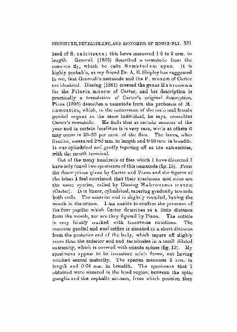


![Title page [image] - ie-net€¦ · 24/11/2015 2 DOMESTIC FLY - MUSCA DOMESTICA 1000 eggs/10 weeks - 12 generations/year Tuesday, 24 November 2015 4 FLIES Tuesday, 24 November 2015](https://static.documents.pub/doc/80x56/5f110c4d04c74d46ad14ac3a/title-page-image-ie-net-24112015-2-domestic-fly-musca-domestica-1000-eggs10.jpg)


