Page 1
The structure of Ca2+-loaded S100A2 at 1.3-A resolutionMichael Koch1,* and Gunter Fritz2
1 Bayer Schering Pharma AG, Wuppertal, Germany
2 Department of Neuropathology, University of Freiburg, Neurozentrum, Germany
Keywords
calcium; EF-hand; p53; S100 protein; X-ray
Correspondence
G. Fritz, Department of Neuropathology,
University of Freiburg, Neurozentrum,
Breisacher Strasse 64, 79106 Freiburg,
Germany
Fax: +49 761 270 50500
Tel: +49 761 270 50780
E-mail: [email protected]
*Present address
Bayer Pharma AG, Global Drug Discovery –
Lead Discovery Wuppertal, Aprather Weg
18a, Geb. 456, 42096 Wuppertal, Germany
(Received 13 December 2011, revised 24
February 2012, accepted 1 March 2012)
doi:10.1111/j.1742-4658.2012.08556.x
S100A2 is an EF-hand calcium ion (Ca2+)-binding protein that activates
the tumour suppressor p53. In order to understand the molecular mecha-
nisms underlying the Ca2+-induced activation of S100A2, the structure of
Ca2+-bound S100A2 was determined at 1.3 A resolution by X-ray crystal-
lography. The structure was compared with Ca2+-free S100A2 and with
other S100 proteins. Binding of Ca2+ to S100A2 induces small structural
changes in the N-terminal EF-hand, but a large conformational change in
the C-terminal EF-hand, reorienting helix III by approximately 90�. Thismovement is accompanied by the exposure of a hydrophobic cavity
between helix III and helix IV that represents the target protein interaction
site. This molecular reorganization is associated with the breaking and new
formation of intramolecular hydrophobic contacts. The target binding site
exhibits unique features; in particular, the hydrophobic cavity is larger than
in other Ca2+-loaded S100 proteins. The structural data underline that the
shape and size of the hydrophobic cavity are major determinants for target
specificity of S100 proteins and suggest that the binding mode for S100A2
is different from that of other p53-interacting S100 proteins.
Database
Structural data are available in the Protein Data Bank database under the accession number
4DUQ
Structured digital abstractl S100A2 and S100A2 bind by x-ray crystallography (View interaction)
Introduction
The family of S100 proteins constitutes the largest sub-
group within the EF-hand calcium ion (Ca2+)-binding
protein superfamily. Currently, more than 21 different
members of this family are known in humans [1,2].
The S100 proteins are expressed in a cell- and tissue-
specific manner, and are involved in different processes
such as cell-cycle regulation, cell growth, differentia-
tion and motility. Several S100 proteins occur in the
extracellular space where they act as pro-inflammatory
cytokines by activating the receptor for advanced gly-
cation end-products (RAGE) [3–5] and toll-like recep-
tor 4 (TLR-4) [6,7] or exert bacteriostatic function as
part of the innate immune system [8,9]. Accordingly,
dysregulation of S100 protein expression is very often
associated with a variety of severe diseases [10], among
them chronic inflammatory [11], neurodegenerative [12]
and cardiovascular [13,14] disorders, as well as cancer
[15–17]. The members of the S100 family are small
(10–12 kDa molecular mass), acidic proteins and, with
the exception of S100G (calbindinD9j), most S100 pro-
teins form homodimers and heterodimers under physi-
ological conditions. Several higher oligomers, ranging
from tetramers to octamers, have been reported for
S100B [4], S100A4 [18], S100A8 ⁄A9 [19] and S100A12
[20]. These larger assemblies appear to be important
for their extracellular cytokine-like function. Recent
Abbreviations
Ca2+, calcium ion; Na+, sodium ion; NDR kinase, nuclear Dbf2-related kinase; PEG, polyethylene glycol.
FEBS Journal 279 (2012) 1799–1810 ª 2012 The Authors Journal compilation ª 2012 FEBS 1799
Page 2
findings suggest that such oligomers might represent
precursors for amyloids formed by S100 proteins [21].
Each S100 protomer is composed of two different
EF-hands: an S100-specific EF-hand is located at the
N terminus, followed by the classical EF-hand at the C
terminus [17,22]. The classical EF-hand motif comprises
a typical sequence signature of 12 residues and is present
in other Ca2+-binding EF-hand proteins such as parval-
bumin, calmodulin or troponin C [23,24]. The S100-spe-
cific motif comprises 14 residues and is unique to the
S100 proteins and to a few domains that resemble S100
proteins [17]. In both EF-hands Ca2+ is coordinated in
a pentagonal bipyramidal configuration. The six posi-
tions in the coordination sphere are denoted by X, Y, Z,
-X, -Y and -Z. Five coordinating residues are provided
by the protein and are arranged in the following motifs:
X**Y*Z**-Y****-Z in the N-terminal EF-hand and X*
Y*Z*-Y****-Z in the C-terminal EF-hand (the asterisks
represent intervening residues; see also Fig. S1). The
sixth -X position is occupied by a water molecule. An
invariant glutamate at position -Z provides a carboxy-
late group for bidentate coordination of Ca2+.
S100 proteins do not exert their biological function
alone, but interact with various target proteins to regu-
late cellular processes [25,26]. While some S100 pro-
tein–protein interactions occur in the absence of Ca2+
[27,28], a calcium-dependent conformational change is
typically required to bind effector molecules and elicit
a biological response [26].
S100A2 is expressed at high levels in different organs
such as lung, kidney, liver, heart and skeletal muscle
[29]. In the cell, S100A2 is localized almost exclusively
in the nucleus, a feature that is unique in the S100 pro-
tein family [30]. Lower levels of S100A2 are detected
in the cytoplasm where S100A2 is reported to interact
with tropomyosin and modulate the organization of
the actin cytoskeleton [31]. Its function in the nucleus
remained unknown for several years although it has
been recognized that S100A2 is involved in cell cycle
regulation [32]. S100A2 was identified first as a major
tumour suppressor in human mammary epithelial cells
[33]. Drastic down-regulation of S100A2 was also
observed in prostate adenocarcinoma [34], lung cancer
[35] and breast carcinoma [36]. A direct molecular link
between the tumour-suppressor activity and the
nuclear localization of S100A2 was established when it
was shown that S100A2 binds and activates p53 in a
Ca2+-dependent manner [37]. The results suggest a
positive regulation of p53 through S100A2. Intrigu-
ingly, other studies report an oncogenic involvement of
S100A2 in the tumorigenesis of different squamous cell
carcinomas [38,39]. This might be a result of interac-
tions of S100A2 with DNp63, which has oncogenic
and growth-stimulating activities in the development of
tumours [40]. Altogether, the studies show that
S100A2 is a potent regulator of the cell cycle, and dys-
regulation of S100A2 can lead to cancer.
Besides Ca2+, S100A2 binds zinc ion (Zn2+) with
high affinity (Kd = 25 nM) [41]. S100A2 contains two
different Zn2+-binding sites, involving cysteine residues
for coordination. We have shown that Zn2+ is a nega-
tive modulator of S100A2; binding of Zn2+ decreases
dramatically the Ca2+ affinity and thereby inhibits the
response to intracellular Ca2+ signals [41]. These find-
ings suggest that S100A2 is regulated by both Ca2+
and Zn2+ metal ions. Our objective was to understand
how the activity of S100A2 is controlled by the differ-
ent metal ions at an atomic level. High-resolution
X-ray structures provide a snapshot of the different
states and are an excellent starting point for further
site-specific mutation and computational studies. We
have already determined the X-ray structure of S100A2
in its Ca2+-free state, showing the protein in its ‘closed’
inactive state [42]. Here, we present the high-resolution
structure of the active, Ca2+-bound form of S100A2 at
1.3 A resolution, and describe the conformational
changes involved when progressing from the Ca2+-free
state to the Ca2+-bound active state.
S100A2 undergoes a large conformational change
upon Ca2+ binding. Major structural changes occur at
the C-terminal EF-hand where helix III moves out-
wards from the molecule, resulting in a reorientation
of 92� compared with Ca2+-free S100A2 [42]. This
drastic molecular reorganization is associated with the
breakage and formation of new intramolecular hydro-
phobic interactions. The movement of helix III opens
a hydrophobic cavity between helices III and IV,
which constitutes the binding site for target proteins
such as p53. This binding site exhibits unique features
compared with other Ca2+-bound S100 proteins.
Results and Discussion
Overall structure of Ca2+-bound S100A2
The final model of Ca2+-bound S100A2 consists of
two protein chains (subunit A and subunit B), each
containing residues A2-A89 and B2-B91, four Ca2+
ions, 173 water molecules and two poly(ethylene gly-
col) 400 (PEG 400) molecules. Using fully anisotropic
displacement parameters in refinement resulted in a
final Rcryst to 13.6% and Rfree to 16.8%. The stereo-
chemistry was validated by PROCHECK [43], showing
that 98% of the residues are located in the most
favoured regions of the Ramachandran plot and 2%
of the residues reside in additional allowed regions.
X-ray structure of Ca2+-loaded S100A2 M. Koch and G. Fritz
1800 FEBS Journal 279 (2012) 1799–1810 ª 2012 The Authors Journal compilation ª 2012 FEBS
Page 3
Each subunit comprises 97 residues. However, the
first N-terminal methionine residue in each chain,
PheA90 to ProA97 and GlyB92 to ProB97 were not
resolved in the electron density. Similarly, the positions
of these C-terminal residues could not be resolved in
the Ca2+-free structure of S100A2 [42]. This might be
a result of structural heterogeneity caused by cis-trans
isomerization of the proline residues Pro94 and Pro97,
as indicated by an NMR study on Ca2+-free S100A2
[44].
The overall structure of Ca2+-bound S100A2 is
depicted in Fig. 1. Comparison of the two subunits
shows that the backbones adopt an almost identical
conformation, as illustrated by an rmsd value of
0.65 A for 88 Ca atoms. Strikingly, the majority of the
side chains also show almost identical orientations in
both subunits. Slight differences occur in the confor-
mation of the C-terminal EF-hand, where helix III and
helix IV adopt an interhelical angle of 119� in subunit
A and 114� in subunit B, respectively (Table 2). As a
result of this high similarity, the following description
of the structure applies for both subunits.
As in other S100 proteins, one S100A2 protom-
er consists of a pair of EF-hands linked by the
so-called hinge region [22] (Fig. 1). The N-terminal,
S100-specific EF-hand comprises helix I (4–20) fol-
lowed by the Ca2+-binding loop (Ser21-Ser30) and
helix II (31–41). Within the hinge region (42–51), resi-
dues Pro43 to Val46 form a short a-helix. Such a short
helix is also observed in other Ca2+-loaded S100 pro-
teins, for example S100A6 [protein data bank (PDB)
entry 1K96], S100A8 (PDB entry 1MR8) or S100A9
(PDB entry 1IRJ) [45–47]. The C-terminal, classical
EF-hand consists of helix III (52–62), the Ca2+-bind-
ing loop (63–71) and helix IV (72–89).
During the refinement process the electron-density
difference maps showed the presence of two PEG 400
molecules per asymmetric unit, both located at the
interface of two S100A2 dimers in the crystal. One
PEG molecule resides between the Ca2+-binding loop
of the N-terminal EF-hand from subunit B and the
Ca2+-binding loop of the C-terminal EF-hand and
helix III of subunit A from the neighbouring molecule.
The other PEG molecule is located between helix IV
from each of the subunits A of these two neighbouring
molecules. By applying crystallographic symmetry
operations, the second PEG 400 molecule was found
to consist of four carbon and three oxygen atoms,
whereby the central oxygen atom is located within the
001 face of the unit cell.
Calcium coordination
S100A2 binds four Ca2+ per dimer. The Ca2+-binding
site in the N-terminal EF-hand is formed by the back-
bone oxygen atoms of Ser20, Glu23, Asp25 and Lys28
and the carboxylate group of Glu33. An additional
water molecule completes the pentagonal bipyramidal
coordination preferred by Ca2+ ions (Fig. 2A,B). All
of these residues are conserved in S100A1 and S100B
(Fig. S1). The C-terminal classical EF-hand represents
the major target interaction site of the Ca2+-sensor
S100 proteins. Ca2+ is coordinated by Od1 of Asp63,
Od1 of Asn65, Od1 of Asp67, the backbone carbonyl
oxygen of Gln69, the carboxylate group of Glu74 and
a water molecule (Fig. 2B). The Ca2+–oxygen dis-
tances are summarized in Table 1. A similar Ca2+
coordination is also observed in the structures of other
S100 proteins [17,22].
Conformational changes upon Ca2+ binding
Recently, we have determined the structure of Ca2+-
free S100A2 [42], representing the inactive form of
S100A2. The comparison of the Ca2+-free and Ca2+-
loaded structures of S100A2 highlight the conforma-
tional changes that are required for the activation of
S100A2. In the Ca2+-free state the interhelical angle
Fig. 1. Stereo view of the Ca2+-bound S100A2 dimer. The N-terminal
S100-specific EF-hands (EF-1) from both subunits are shown in blue
and red, the C-terminal classical EF-hands (EF-2) are shown in cyan
and orange, respectively. The Ca2+ ions are depicted as green
spheres. HI, helix I; HII, helix II; HIII, helix III; HIV, helix IV.
Table 1. Distance (d ) between the Ca2+ ion and the coordinating
ligand in the two EF-hands in S100A2.
N-terminal C-terminal
EF-hand ligand d (A) EF-hand ligand d (A)
Ser20O 2.35 Asp63Od1 2.33
Glu23O 2.39 Asn65Od1 2.27
Asp25O 2.32 Asp67Od1 2.39
Lys28O 2.39 Gln69O 2.35
Glu33Oe1 2.46 Glu74Oe1 2.45
Glu33Oe2 2.53 Glu74Oe2 2.58
Water5 2.38 Water8 2.39
M. Koch and G. Fritz X-ray structure of Ca2+-loaded S100A2
FEBS Journal 279 (2012) 1799–1810 ª 2012 The Authors Journal compilation ª 2012 FEBS 1801
Page 4
between helix I and helix II is 126�. The orientation
of helices I and II changed upon Ca2+ binding, result-
ing in an interhelical angle of 137� (Table 2). Interest-
ingly, the N-terminal EF-hand of Ca2+-free S100A2
harbours an Na+ ion (Fig. 2C) [42]. Na+ prefers an
octahedral coordination sphere, in contrast to the pen-
tagonal bipyramidal coordination preferred by Ca2+
ions (Fig. 2B,C) [48,49]. Nevertheless, the coordination
scheme in the EF-hand is very similar for Na+ and
Ca2+. Both ions are bound by the backbone oxygen
atoms of Ser20, Glu23, Asp25, Lys28 and a water
molecule (Fig. 2B,C). A major difference is the coordi-
nation of Ca2+ by the carboxylate group of Glu33,
whereas Na+ is coordinated by a further water mole-
cule. The binding of Glu33 to Ca2+ pulls helix II
closer to the Ca2+-binding site, shifting the entire
helix slightly (Fig. 2A). This movement reorients sev-
eral residues of helix II, which subsequently form new
hydrophobic contacts with residues on helix III.
In contrast to the small conformational changes in
the N-terminal EF-hand (Fig. 2A) the classical C-ter-
minal EF-hand undergoes a large molecular rearrange-
ment upon Ca2+ binding (Fig. 2D,E). Unlike the
almost preformed N-terminal EF-hand, several resi-
dues in the C-terminal EF-hand undergo major reposi-
tioning upon Ca2+ coordination (Fig. 2D). The side
chain of Glu74 and the backbone carbonyl group of
Gln69 display some considerable movements to bind
Fig. 2. Conformational changes in the EF-hands upon Ca2+ binding. (A) Stereo view of the N-terminal EF-hand of Ca2+-free S100A2 (blue)
and Ca2+-bound S100A2 (yellow). Only a small conformational change is observed; this involves mainly a movement of helix II. (B) Details of
Ca2+ coordination in the N-terminal EF-hand. The Ca2+ ion (green) is coordinated by backbone carbonyls, the side chain of Glu33 and a water
molecule. (C) In Ca2+-free S100A2 an Na+ ion (magenta) is bound in the N-terminal EF-hand. Like Ca2+, the bound Na+ is coordinated by
backbone carbonyls; however, the side chain of Glu33 is not directly involved and is replaced by a water molecule. (D) Structural alignment
of the C-terminal EF-hand of Ca2+-bound S100A2 (yellow) and Ca2+-free S100A2 (blue). Upon Ca2+ binding, helix III undergoes a movement
of 92�. (E) In the C-terminal EF-hand, Ca2+ is coordinated mainly by side-chain oxygens (red).
X-ray structure of Ca2+-loaded S100A2 M. Koch and G. Fritz
1802 FEBS Journal 279 (2012) 1799–1810 ª 2012 The Authors Journal compilation ª 2012 FEBS
Page 5
Ca2+, and large reorientations of backbone and of
side chains are observed for residues 63–68 to accom-
modate the Ca2+ (Fig. 2D,E). Moreover, helix III has
to unwind by two residues (Leu62 and Asp63) to rear-
range Asp63-Od1 by 14 A for Ca2+ coordination.
Ca2+ binding finally causes a 92� movement of helix -
III, a feature also observed in other Ca2+-bound S100
proteins. In other EF-hand Ca2+-sensor proteins, such
as calmodulin [50] or troponin C [51], the exiting
helix IV of the EF-hand undergoes a large movement
upon Ca2+ binding instead of entering helix III. This
is readily explained by the fact that S100 proteins
form stable dimers where helix IV is involved in the
dimer interface and cannot undergo large conforma-
tional changes.
In order to describe the structural consequences of
helix reorientation and helix repacking we performed
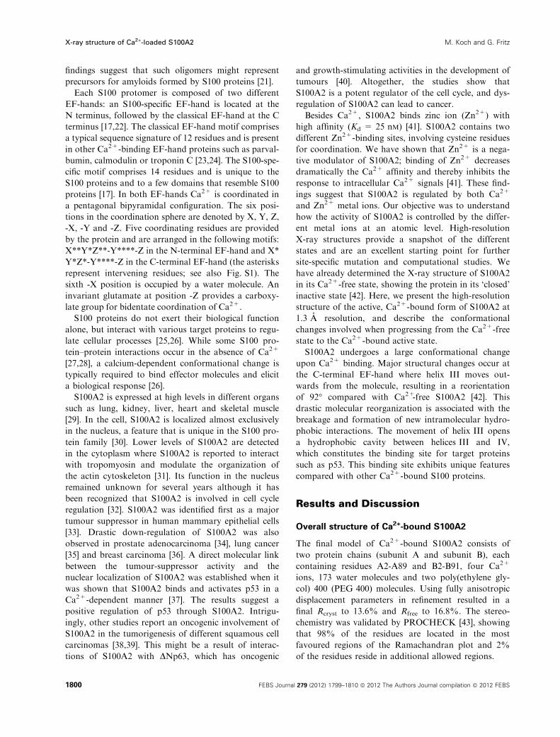
an analysis of the changes in hydrophobic interactions
during Ca2+ binding, similarly to an analysis for
Ca2+-bound S100A1 [52]. The conformational changes
break several hydrophobic contacts of helix III with
helix IV, whereas new hydrophobic contacts between
helix III and helix II are formed (Fig. 3). In Ca2+-free
S100A2, residues Leu55, Leu58 and Leu62 of helix III
and residues Phe78, Ile82 and Met85 of helix IV form
a hydrophobic core that stabilizes the antiparallel
packing of these helices (Fig. 3A), whereas hydropho-
bic contacts between helix III and helix II are blocked
in Ca2+-free S100A2 by the side chains of Lys31 and
Lys35 from helix III (Fig. 3E). Upon Ca2+ binding,
Leu55, Ile82 and Met85 do not contribute further to
helix III–helix IV packing (Fig. 3B). Interestingly,
Ca2+ binding to the N-terminal EF-hand causes a 10�rotation of helix II, moving Lys31 and Lys35 out of
the helix II–helix III interface (Fig. 3E,F). This rota-
tion enables Leu55 of helix III to form new contacts
with Cb and Cc of Lys31 and Lys35, as well as with
Met34 of helix II (Fig. 3D). Similarly, Leu58 is
involved in Ca2+-free S100A2 in helix III–helix IV
packing. Upon Ca2+ binding the contacts to
Phe78 and Ile82 break as a result of the movement of
helix III; however, new hydrophobic contacts are
formed with Met34 ⁄Leu38 from helix II (Fig. 3D,F).
Residues Ile82 and Met85, which had contributed to
helix III–helix IV packing in Ca2+-free S100A2, are
exposed to the solvent in Ca2+-bound S100A2
(Fig. 3B) forming part of the target interaction site.
The change in the solvent-accessible surface of these
two residues illustrates readily the loss of this hydro-
phobic core upon Ca2+ binding. In Ca2+-free S100A2,
Ile82 and Met85 have solvent-accessible surfaces of
2 A2 and 17.4 A2, respectively. In stark contrast, in the
Ca2+-loaded state these residues display solvent-
exposed surfaces of 22.9 A2 and 62.8 A2, respectively.
In summary, the subtle reorientation of helix II and
the movement of helix III are crucial for the tight
helix II–helix III packing in the Ca2+-bound state. It
is likely that these interactions are essential to stabilize
the open conformation of Ca2+-bound S100A2 and
might very well explain the positive cooperativity
observed for Ca2+ binding to the two EF-hands [41].
As several of these residues are conserved among the
S100 proteins, we wondered whether the conforma-
tional changes described here for S100A2 and the
resulting hydrophobic contacts also occur in other
S100 proteins. There are structures available for
S100A1, S100A4, S100A6 and S100B in the Ca2+-free
and Ca2+-loaded states [4,18,47,52–60]. Interestingly,
the Ca2+-free structures exhibit a highly similar
helix III–helix IV packing, with Leu62 (S100A2 num-
bering) strictly conserved among these four proteins.
As in S100A2, the charged side chains of Lys31 and
Lys35 (S100A2 numbering) sterically hinder the
Table 2. Interhelical angles in EF-hands in the Ca2+-free and Ca2+-
bound states. The values represent the average between subunit A
and subunit B. In the case of S100A6, there was only one subunit
in the asymmetrical unit, and the values for S100A1 represent the
average overall models of the NMR ensemble.
Interhelical angle (�)
I to II II to III III to IV
Apo-S100A2 126 ± 1 )139 ± 3 )152 ± 1
Ca-S100A2 137 ± 1 110 ± 1 116 ± 3
Apo-S100A6 127 )142 )153
Ca-S100A6 134 114 117
Apo-S100B 133 ± 1 )149 ± 1 )166 ± 1
Ca-S100B 135.2 ± 0.2 100 ± 1 103 ± 2
Apo-S100A1 120 ± 3 )148 ± 2 )150 ± 1
Ca-S100A1 132 ± 1 125 ± 2 121 ± 2
Table 3. Spatial properties of the hydrophobic cavities of S100A2,
S100A6 and S100B.
Protein
Atoms defining
the plane
Atoms residing
in the base of
the cavity
Depth of
cavity (A)
Volume of
the central
cavity (A3)
S100A2 Phe45O Leu42Cc 7.1 99.7
Leu58Cd1
Met85Ce
S100A6 Ile44Cd1 Ile83Cd1 4.6 52.5
Leu56Cd1
Ile83Cd1
S100B Leu44Cd2 Ile36Cd1 3.9 49.5
Val56Cc2
Met79Sd
M. Koch and G. Fritz X-ray structure of Ca2+-loaded S100A2
FEBS Journal 279 (2012) 1799–1810 ª 2012 The Authors Journal compilation ª 2012 FEBS 1803
Page 6
interaction of helices II and III in the Ca2+-free state
(Fig. 3E). An exception is S100B; here, the side chain
of Lys31 already points towards the solvent in the
Ca2+-free form (i.e. no rotation of helix II is required
for the formation of hydrophobic interactions). Indeed,
the binding of Ca2+ to S100B causes an axial rotation
of only 2� of helix II, in contrast to S100A2 where
helix II rotates by about 10�.In summary, the analysis of the structures of
S100A2, S100A1, S100A4, S100A6 and S100B revealed
that the Ca2+-induced movement of Lys31 and Lys35
out of the helix II–helix III interface and subsequent
hydrophobic contacts between these helices are con-
served features in the activation of S100 proteins
(Fig. 3E,F). Sequence comparison between all S100
proteins showed that the hydrophobic residues at posi-
tions 34 and 38 (S100A2 numbering) are strictly con-
served, underlining their role in S100 protein
activation (Fig. S1).
Target binding to S100A2
Sequence alignments of S100A2 with other S100 pro-
teins show that S100A2 shares high sequence homo-
logy with S100A4 (61% identity), followed by S100A1
(52%), S100A6 (47%) and S100B (45%). The five
S100 proteins have a very similar structural organiza-
tion. However, the sequence homology is not fully
reflected in structural resemblance. Although S100B
displays the lowest sequence identity, the structural
alignment of the Ca2+-bound protomers reveals that it
is most similar to S100A2 with an rmsd of 0.86 A for
75 Ca atoms excluding the Ca of the hinge region (resi-
dues 41–52). Further comparison with the subunits of
S100A6, S100A4 and S100A1 yielded rmsd values of
1.07 A (75 Ca atoms), 1.35 A (75 Ca atoms) and
1.59 A (64 Ca atoms), respectively. Major structural
differences occur in the hinge region and at the C-ter-
minal helix IV (Fig. 4). These areas represent the tar-
get protein-binding sites and the observed differences
may reflect the divergence in target molecules and
binding modes among these S100 proteins. For S100B,
several structures with peptides derived from different
target proteins have been determined [61–64]. The pep-
tide comprising the negative regulatory domain of p53
(367–392) bound to S100B adopts a helical conforma-
tion and interacts with residues from helix III, from
the hinge region and from helix IV [61]. A peptide
Fig. 3. Helix packing for Ca2+-free S100A2
(A, C, E) versus Ca2+-bound S100A2 (B, D,
F) and comparison with other S100 proteins
(E, F). In (A) and (B) the interactions
between helix III and helix IV of S100A2 are
shown, and in (C) and (D) the interactions
between helix II and helix III of S100A2 are
shown. The residues forming hydrophobic
contacts are shown in stick-type and trans-
parent van-der-Waals spheres. (E, F) Overlay
of S100A2 (cyan), S100A4 (3C1V, red)
S100A6 (1K9P, green) and S100B (2H61,
magenta). (E) Helix II and helix III in
Ca2+-free proteins (F) Helix II and helix III in
Ca2+-bound proteins. Upon Ca2+ binding,
helix II rotates slightly, by about 10�. This
rotation (counterclockwise in the figure)
removes Lys35 from the helix interface and
enables a hydrophobic contact to be made
between Leu38 and Met59 (S100A2
numbering) in the Ca2+-bound state (F).
X-ray structure of Ca2+-loaded S100A2 M. Koch and G. Fritz
1804 FEBS Journal 279 (2012) 1799–1810 ª 2012 The Authors Journal compilation ª 2012 FEBS
Page 7
representing the N-terminal regulatory domain of
nuclear Dbf2-related (NDR) kinase also adopts a heli-
cal conformation in complex with S100B, and several
residues of S100B important for p53 binding are
involved in complex formation. However, the orienta-
tion of both helical peptides is noticeably different
[62]. Further variations of the binding modes are
reported for the peptide TRTK12 derived from CapZ,
where a short helix [63] and a random coil conforma-
tion were observed [64]. Similarly to S100B, S100A1
binds peptides in the same region [65,66]. A different
interaction mode was described for the complex of
S100A6 with a peptide representing the binding site of
Siah-1-interacting protein [67], where the peptide binds
to the hydrophobic region between helix III and helix
IV as well as to a region on top of the dimer interface.
S100A2, S100A6 and S100B bind to p53 [37,68–70]
in a Ca2+-dependent manner. We therefore compared
the molecular surfaces of these three proteins with
regard to electrostatic potential, shape and distribution
of hydrophobic areas (Fig. 5). As a common feature all
three proteins exhibit a net-negative surface charge.
Nevertheless, there are clear differences between these
proteins with regard to the distribution of electrostatic
surface potential. Whereas S100A2 and S100A6 display
patches of negative surface potential, S100B exhibits
areas with continuous negative potential (Fig. 5A).
Further differences in charge distribution among the
three proteins are observed around the hydrophobic
target binding pocket. Whereas S100A6 displays only a
few charged patches, continuous areas of negative
charge are observed for S100A2 and S100B. The differ-
ences in surface properties are not unexpected and
reflect different target selectivity of the three S100 pro-
teins. S100A2, S100A6 and S100B bind to p53 but each
S100 protein recognizes further target proteins (e.g.
S100A2 binds specifically to p63 [40], S100A6 to annex-
in [71] and S100B to NDR kinase [62]).
S100B recognizes a region in the tetramerization
domain of p53 (residues 325–355) as well as the nega-
tive regulatory domain of p53 (residues 362–373)
[61,72]. Binding of S100B to p53 inhibits its transcrip-
tional activation [68] by sterically blocking tetrameriza-
tion of p53 or phosphorylation ⁄ acetylation sites
essential for activation [61]. Like S100B, S100A2 recog-
nizes the C terminus of p53 [37]. In contrast to S100B,
S100A2 activates p53 [37]. Van Dieck et al. attributed
these different activities to the stabilization of either a
monomeric or a tetrameric state by S100B and S100A2
[70]. The different effect of the two S100 proteins on
p53 should be reflected in a different binding mode of
p53 in the binding pocket. We therefore characterized
depth, size and solvent accessibility of binding pockets
in greater detail. This analysis revealed that S100A2
adopts an outstanding position among these three
proteins. The cavity of S100A2, with a depth of 7 A, is
Fig. 4. Structural alignment of one protomer of S100A2 (cyan),
S100A4 (red), S100A6 (green) and S100B (magenta). Ca traces of
all four proteins are shown. Overall, the fold of the protomers of
the different S100 proteins is very similar. Noticeable deviations
occur at the hinge region and at the C terminus, which form the
target protein-binding site. Further differences are observed in
interhelical angles for the helix III–helix IV pair (Table 2). HI, helix I;
HII, helix II; HIII, helix III; HIV, helix IV.
Fig. 5. Electrostatic surface potential (A) and distribution of hydro-
phobic residues (B) for S100A2, S100A6 and S100B. (A) Negatively
charged regions are depicted in red and positively charged regions
are depicted in blue, ranging from )10 kBT ⁄ e (red) to +10 kBT ⁄ e(blue) (e, elementary charge; kB, Boltzmann constant; T, 298 K.) (B)
Residues with a positive hydropathy index, according to Kyte and
Doolittle [88], are shown in green for S100A2, in yellow for S100A6
and in purple for S100B. Molecules in (A) and (B) have the same
orientation. Cross-sections through the hydrophobic cavity of the
three S100 proteins are shown in (C). The location of the cross-sec-
tion is indicated in (B) by a black bar. (C) Left: the contours of the
cavity cross-sections of S100A6 (yellow) and S100B (purple) are
overlaid with the cross-section through the cavity of S100A2. Obvi-
ously, the cavity of S100A2 is deeper and larger than the cavity of
S100A6. The cavity of S100B appears wider, but is much shallower
than the cavities of S100A2 and S100A6.
M. Koch and G. Fritz X-ray structure of Ca2+-loaded S100A2
FEBS Journal 279 (2012) 1799–1810 ª 2012 The Authors Journal compilation ª 2012 FEBS 1805
Page 8
about 60–70% deeper than those of S100A6 or S100B,
respectively (Table 3). Moreover, the cavity of S100A2
is noticeably larger than in the two other proteins
(Fig. 5C). The central cavity of S100A2 exhibits a vol-
ume of 99.7 A3, whereas the cavities in S100A6 and
S100B display volumes of 52.2 A3 and 49.5 A3, respec-
tively. Notably, the cavity of S100B is much wider and
shallower than in S100A2 and S100A6, perhaps
enabling S100B to recognize a larger number of differ-
ent targets [26]. Clearly, all three S100 proteins differ
with regard to charge and size of the target protein-
binding sites located between helix III and helix IV.
Altogether we conclude that these differences are
responsible for the different effects of the S100 proteins
on the activity of p53. Future studies on a p53362–
393 ⁄S100A2 complex might reveal a new binding mode
and will help to elucidate how S100A2 activates p53.
Materials and methods
Cloning, expression and purification
As wild-type S100A2 is prone to oxidation in air [41,73], an
S100A2-C2S-C21S-C86S-C93S variant was used for crystal-
lization experiments. S100A2-C2S-C21S-C86S-C93S was
obtained by site-directed mutagenesis and subsequently
cloned into the bacterial expression vector pMW172, as
described elsewhere [74]. Expression in Escherichia coli BL21
(DE3) and purification were carried out as described previ-
ously [73,75]. This variant is not sensitive to air oxidation.
Crystallization and data collection
Before crystallization experiments, S100A2 was loaded with
Ca2+ by passage over a NAP5 desalting column (GE
Healthcare, Munich, Germany) equilibrated with 10 mM
Tris ⁄HCl, 20 mM CaCl2, pH 7.6. The protein concentration
was 15 mgÆmL)1. Crystals were grown using the vapour dif-
fusion hanging-drop method (protein ⁄precipitant,1 lL : 1 lL). The precipitant solution was 0.1 M sodium
acetate, pH 4.0 containing 40% PEG 400. Crystals grew
within 5–10 days with dimensions up to 50 lm · 150 lm ·180 lm. The high PEG 400 concentration was sufficient as
cryoprotectant and the crystals were flash frozen in mother
liquor in the cryonitrogen stream at 100 K. Data collection
was carried out with a Swiss Light Source (PSI, Villigen,
Switzerland) at beamline X06SA using a mar225 mosaic
CCD detector (Mar Research, Norderstedt, Germany).
Data statistics are summarized in Table S1.
Structure determination and refinement
The structure of Ca2+-loaded S100A2 was determined
based on the anomalous scattering provided by 10 sulfur
atoms from methionine residues and four calcium ions present
in the structure [75]. The refinement of the model was car-
ried out with data to 1.3 A resolution using Refmac5 (ver-
sion 5.7) including anisotropic temperature factors [76,77].
Hydrogen atoms were included in refinement in riding
positions. Manual model building was performed with the
programs O [78] and Coot [79]. Data-collection and refine-
ment statistics are summarized in Table S1. Secondary
structure analysis was calculated using DSSP [80]. Struc-
ture validation was performed using SFCHECK [81] and
PROCHECK [43]. Electrostatic surface calculations were
performed using the program Adaptive Poisson-Boltzmann
Solver (APBS) [82]. Charges for amino-acid residues were
assigned using PDB 2PQR. Amber force-field parameters
for Ca2+ and Na+ were taken from Aqvist [83] for calcu-
lations of electrostatic potential. The structures were
aligned using LSQMAN [84], solvent-accessible volumes of
the cavities were calculated with CASTp using a probe
radius of 1.4 A [85] and solvent-accessible areas were
calculated using AREAIMOL [86]. For determination of
the depth of each cavity, a plane was defined by three
atoms at the border of the central cavity and the atom
inside the cavity with the largest distance to this plane
served as a reference point to define the distance between
the plane and the base of the cavity. Interhelical angles
were calculated using the INTERHLX software (K. Yap, Uni-
versity of Toronto, Toronto, Canada). Figures were pre-
pared using PyMol [87]. Structure factors and coordinates
have been deposited at the PDB database with accession
code 4DUQ.
Acknowledgements
G.F. is supported by a Heisenberg fellowship of the
Deutsche Forschungsgemeinschaft (FR 1488 ⁄3-1). Thiswork was further supported by a grant of the Deutsche
Forschungsgemeinschaft (FR 1488 ⁄ 5-1). We thank the
staff at beamline X06SA at Swiss Light Source for
excellent support.
References
1 Marenholz I, Lovering RC & Heizmann CW (2006) An
update of the S100 nomenclature. Biochim Biophys Acta
1763, 1282–1283.
2 Marenholz I, Heizmann CW & Fritz G (2004) S100
proteins in mouse and man: from evolution to func-
tion and pathology (including an update of the
nomenclature). Biochem Biophys Res Commun 322,
1111–1122.
3 Fritz G (2011) RAGE: a single receptor fits multiple
ligands. Trends Biochem Sci 36, 625–632.
4 Ostendorp T, Leclerc E, Galichet A, Koch M, Demling
N, Weigle B, Heizmann CW, Kroneck PM & Fritz G
X-ray structure of Ca2+-loaded S100A2 M. Koch and G. Fritz
1806 FEBS Journal 279 (2012) 1799–1810 ª 2012 The Authors Journal compilation ª 2012 FEBS
Page 9
(2007) Structural and functional insights into RAGE
activation by multimeric S100B. EMBO J 26, 3868–
3878.
5 Hofmann MA, Drury S, Fu C, Qu W, Taguchi A, Lu
Y, Avila C, Kambham N, Bierhaus A, Nawroth P et al.
(1999) RAGE mediates a novel proinflammatory axis:
a central cell surface receptor for S100 ⁄ calgranulin poly-
peptides. Cell 97, 889–901.
6 Bjork P, Bjork A, Vogl T, Stenstrom M, Liberg D,
Olsson A, Roth J, Ivars F & Leanderson T (2009)
Identification of human S100A9 as a novel target for
treatment of autoimmune disease via binding to
quinoline-3-carboxamides. PLoS Biol 7, e97.
7 Vogl T, Tenbrock K, Ludwig S, Leukert N, Ehrhardt
C, van Zoelen MA, Nacken W, Foell D, van der Poll
T, Sorg C et al. (2007) Mrp8 and Mrp14 are
endogenous activators of Toll-like receptor 4,
promoting lethal, endotoxin-induced shock. Nat Med
13, 1042–1049.
8 Kehl-Fie TE, Chitayat S, Hood MI, Damo S, Restrepo
N, Garcia C, Munro KA, Chazin WJ & Skaar EP
(2011) Nutrient metal sequestration by calprotectin
inhibits bacterial superoxide defense, enhancing neutro-
phil killing of Staphylococcus aureus. Cell Host Microbe
10, 158–164.
9 Corbin BD, Seeley EH, Raab A, Feldmann J, Miller
MR, Torres VJ, Anderson KL, Dattilo BM, Dunman
PM, Gerads R et al. (2008) Metal chelation and inhibi-
tion of bacterial growth in tissue abscesses. Science 319,
962–965.
10 Donato R, Sorci G, Riuzzi F, Arcuri C, Bianchi R,
Brozzi F, Tubaro C & Giambanco I (2009) S100B’s
double life: intracellular regulator and extracellular
signal. Biochim Biophys Acta 1793, 1008–1022.
11 Goyette J & Geczy CL (2010) Inflammation-associated
S100 proteins: new mechanisms that regulate function.
Amino Acids 41, 821–842.
12 Mori T, Koyama N, Arendash GW, Horikoshi-Sakuraba
Y, Tan J & Town T (2010) Overexpression of human
S100B exacerbates cerebral amyloidosis and gliosis in the
Tg2576 mouse model of Alzheimer’s disease. Glia 58,
300–314.
13 Most P, Remppis A, Pleger ST, Loffler E, Ehlermann
P, Bernotat J, Kleuss C, Heierhorst J, Ruiz P, Witt H
et al. (2003) Transgenic overexpression of the Ca2+-
binding protein S100A1 in the heart leads to increased
in vivo myocardial contractile performance. J Biol Chem
278, 33809–33817.
14 Schaub MC & Heizmann CW (2008) Calcium, tropo-
nin, calmodulin, S100 proteins: from myocardial basics
to new therapeutic strategies. Biochem Biophys Res
Commun 369, 247–264.
15 Wolf S, Haase-Kohn C & Pietzsch J (2011) S100A2
in cancerogenesis: a friend or a foe? Amino Acids 41,
849–861.
16 Gebhardt C, Nemeth J, Angel P & Hess J (2006)
S100A8 and S100A9 in inflammation and cancer.
Biochem Pharmacol 72, 1622–1631.
17 Heizmann CW, Fritz G & Schafer BW (2002) S100 pro-
teins: structure, functions and pathology. Front Biosci 7,
d1356–d1368.
18 Malashkevich VN, Dulyaninova NG, Ramagopal UA,
Liriano MA, Varney KM, Knight D, Brenowitz M,
Weber DJ, Almo SC & Bresnick AR (2010) Phenothia-
zines inhibit S100A4 function by inducing protein oligo-
merization. Proc Natl Acad Sci USA 107, 8605–8610.
19 Leukert N, Vogl T, Strupat K, Reichelt R, Sorg C &
Roth J (2006) Calcium-dependent tetramer formation of
S100A8 and S100A9 is essential for biological activity.
J Mol Biol 359, 961–972.
20 Moroz OV, Antson AA, Dodson EJ, Burrell HJ, Grist
SJ, Lloyd RM, Maitland NJ, Dodson GG, Wilson KS,
Lukanidin E et al. (2002) The structure of S100A12 in a
hexameric form and its proposed role in receptor signal-
ling. Acta Crystallogr D Biol Crystallogr 58, 407–413.
21 Fritz G, Botelho HM, Morozova-Roche LA & Gomes
CM (2010) Natural and amyloid self-assembly of S100
proteins: structural basis of functional diversity. FEBS
J 277, 4578–4590.
22 Fritz G & Heizmann CW (2004) D structures of the cal-
cium and zinc binding S100 proteins. In Handbook of
Metalloprotein (Messerschmidt A, Bode W & Cygler M
eds), pp. 529–540. John Wiley & Sons, Inc., Chichester.
23 Yap KL, Ames JB, Swindells MB & Ikura M (1999)
Diversity of conformational states and changes within
the EF-hand protein superfamily. Proteins 37, 499–507.
24 Ikura M (1996) Calcium binding and conformational
response in EF-hand proteins. Trends Biochem Sci 21,
14–17.
25 Bhattacharya S, Bunick CG & Chazin WJ (2004) Target
selectivity in EF-hand calcium binding proteins. Biochim
Biophys Acta 1742, 69–79.
26 Santamaria-Kisiel L, Rintala-Dempsey AC & Shaw GS
(2006) Calcium-dependent and -independent interactions
of the S100 protein family. Biochem J 396, 201–214.
27 Rety S, Sopkova J, Renouard M, Osterloh D, Gerke V,
Tabaries S, Russo-Marie F & Lewit-Bentley A (1999)
The crystal structure of a complex of p11 with the ann-
exin II N-terminal peptide. Nat Struct Biol 6, 89–95.
28 Landar A, Rustandi RR, Weber DJ & Zimmer DB
(1998) S100A1 utilizes different mechanisms for inter-
acting with calcium-dependent and calcium-independent
target proteins. Biochemistry 37, 17429–17438.
29 Glenney JR Jr, Kindy MS & Zokas L (1989) Isolation
of a new member of the S100 protein family: amino
acid sequence, tissue, and subcellular distribution. J Cell
Biol 108, 569–578.
30 Mandinova A, Atar D, Schafer BW, Spiess M, Aebi U
& Heizmann CW (1998) Distinct subcellular localization
of calcium binding S100 proteins in human smooth
M. Koch and G. Fritz X-ray structure of Ca2+-loaded S100A2
FEBS Journal 279 (2012) 1799–1810 ª 2012 The Authors Journal compilation ª 2012 FEBS 1807
Page 10
muscle cells and their relocation in response to rises in
intracellular calcium. J Cell Sci 111(Pt 14), 2043–2054.
31 Gimona M, Lando Z, Dolginov Y, Vandekerckhove J,
Kobayashi R, Sobieszek A & Helfman DM (1997)
Ca2+-dependent interaction of S100A2 with muscle and
nonmuscle tropomyosins. J Cell Sci 110, 611–621.
32 Maelandsmo GM, Florenes VA, Mellingsaeter T, Hovig
E, Kerbel RS & Fodstad O (1997) Differential expres-
sion patterns of S100A2, S100A4 and S100A6 during
progression of human malignant melanoma. Int J Can-
cer 74, 464–469.
33 Lee SW, Tomasetto C, Swisshelm K, Keyomarsi K &
Sager R (1992) Down-regulation of a member of the
S100 gene family in mammary carcinoma cells and
reexpression by azadeoxycytidine treatment. Proc Natl
Acad Sci USA 89, 2504–2508.
34 Gupta S, Hussain T, MacLennan GT, Fu P, Patel J &
Mukhtar H (2003) Differential expression of S100A2
and S100A4 during progression of human prostate ade-
nocarcinoma. J Clin Oncol 21, 106–112.
35 Feng G, Xu X, Youssef EM & Lotan R (2001) Dimin-
ished expression of S100A2, a putative tumor suppres-
sor, at early stage of human lung carcinogenesis. Cancer
Res 61, 7999–8004.
36 Wicki R, Franz C, Scholl FA, Heizmann CW & Schafer
BW (1997) Repression of the candidate tumor suppres-
sor gene S100A2 in breast cancer is mediated by site-
specific hypermethylation. Cell Calcium 22, 243–254.
37 Mueller A, Schafer BW, Ferrari S, Weibel M, Makek
M, Hochli M & Heizmann CW (2005) The calcium-
binding protein S100A2 interacts with p53 and modu-
lates its transcriptional activity. J Biol Chem 280,
29186–29193.
38 Villaret DB, Wang T, Dillon D, Xu J, Sivam D,
Cheever MA & Reed SG (2000) Identification of genes
overexpressed in head and neck squamous cell carci-
noma using a combination of complementary DNA
subtraction and microarray analysis. Laryngoscope 110,
374–381.
39 Xia L, Stoll SW, Liebert M, Ethier SP, Carey T,
Esclamado R, Carroll W, Johnson TM & Elder JT (1997)
CaN19 expression in benign and malignant hyperplasias
of the skin and oral mucosa: evidence for a role in
regenerative differentiation. Cancer Res 57, 3055–3062.
40 Hibi K, Fujitake S, Takase T, Kodera Y, Ito K,
Akiyama S, Shirane M & Nakao A (2003) Identification
of S100A2 as a target of the DeltaNp63 oncogenic
pathway. Clin Cancer Res 9, 4282–4285.
41 Koch M, Bhattacharya S, Kehl T, Gimona M, Vasak
M, Chazin W, Heizmann CW, Kroneck PM & Fritz G
(2007) Implications on zinc binding to S100A2. Biochim
Biophys Acta 1773, 457–470.
42 Koch M, Diez J & Fritz G (2008) Crystal structure of
Ca2+ -free S100A2 at 1.6 A resolution. J Mol Biol 378,
933–942.
43 Laskowski RA, MacArthur MW, Moss DS & Thornton
JM (1993) PROCHECK: a program to check stereo-
chemical quality of protein structures. J Appl Cryst 26,
283–291.
44 Randazzo A, Acklin C, Schafer BW, Heizmann CW &
Chazin WJ (2001) Structural insight into human Zn2+-
bound S100A2 from NMR and homology modeling.
Biochem Biophys Res Commun 288, 462–467.
45 Itou H, Yao M, Fujita I, Watanabe N, Suzuki M,
Nishihira J & Tanaka I (2002) The crystal structure of
human MRP14 (S100A9), a Ca2+-dependent regulator
protein in inflammatory process. J Mol Biol 316, 265–
276.
46 Ishikawa K, Nakagawa A, Tanaka I, Suzuki M &
Nishihira J (2000) The structure of human MRP8, a
member of the S100 calcium-binding protein family, by
MAD phasing at 1.9 A resolution. Acta Crystallogr D
Biol Crystallogr 56, 559–566.
47 Otterbein LR, Kordowska J, Witte-Hoffmann C, Wang
CL & Dominguez R (2002) Crystal structures of
S100A6 in the Ca2+-free and Ca2+-bound states: the
calcium sensor mechanism of S100 proteins revealed at
atomic resolution. Structure 10, 557–567.
48 Harding MM (2002) Metal-ligand geometry relevant to
proteins and in proteins: sodium and potassium. Acta
Crystallogr D Biol Crystallogr 58, 872–874.
49 Harding MM (2006) Small revisions to predicted dis-
tances around metal sites in proteins. Acta Crystallogr
D Biol Crystallogr 62, 678–682.
50 Wilson MA & Brunger AT (2000) The 1.0 A crystal
structure of Ca2+-bound calmodulin: an analysis of
disorder and implications for functionally relevant
plasticity. J Mol Biol 301, 1237–1256.
51 Houdusse A, Love ML, Dominguez R, Grabarek Z &
Cohen C (1997) Structures of four Ca2+-bound tropo-
nin C at 2.0 A resolution: further insights into the
Ca2+-switch in the calmodulin superfamily. Structure 5,
1695–1711.
52 Wright NT, Varney KM, Ellis KC, Markowitz J, Gitti
RK, Zimmer DB & Weber DJ (2005) The three-dimen-
sional solution structure of Ca2+-bound S100A1 as
determined by NMR spectroscopy. J Mol Biol 353,
410–426.
53 Rustandi RR, Baldisseri DM, Inman KG, Nizner P,
Hamilton SM, Landar A, Zimmer DB & Weber DJ
(2002) Three-dimensional solution structure of the cal-
cium-signaling protein apo-S100A1 as determined by
NMR. Biochemistry 41, 788–796.
54 Vallely KM, Rustandi RR, Ellis KC, Varlamova O,
Bresnick AR & Weber DJ (2002) Solution structure of
human Mts1 (S100A4) as determined by NMR
spectroscopy. Biochemistry 41, 12670–12680.
55 Pathuri P, Vogeley L & Luecke H (2008) Crystal struc-
ture of metastasis-associated protein S100A4 in the
active calcium-bound form. J Mol Biol 383, 62–77.
X-ray structure of Ca2+-loaded S100A2 M. Koch and G. Fritz
1808 FEBS Journal 279 (2012) 1799–1810 ª 2012 The Authors Journal compilation ª 2012 FEBS
Page 11
56 GingrasAR, Basran J, PrescottA,KriajevskaM,Bagshaw
CR&Barsukov IL (2008) Crystal structure of the Ca2+-
form andCa2+-binding kinetics ofmetastasis-associated
protein, S100A4. FEBSLett 582, 1651–1656.
57 Maler L, Potts BC & Chazin WJ (1999) High resolution
solution structure of apo calcyclin and structural varia-
tions in the S100 family of calcium-binding proteins.
J Biomol NMR 13, 233–247.
58 Charpentier TH, Wilder PT, Liriano MA, Varney KM,
Pozharski E, MacKerell AD Jr, Coop A, Toth EA &
Weber DJ (2008) Divalent metal ion complexes of
S100B in the absence and presence of pentamidine.
J Mol Biol 382, 56–73.
59 Drohat AC, Amburgey JC, Abildgaard F, Starich MR,
Baldisseri D & Weber DJ (1996) Solution structure of
rat apo-S100B(bb) as determined by NMR spectros-
copy. Biochemistry 35, 11577–11588.
60 Drohat AC, Baldisseri DM, Rustandi RR & Weber DJ
(1998) Solution structure of calcium-bound rat
S100B(bb) as determined by nuclear magnetic resonance
spectroscopy. Biochemistry 37, 2729–2740.
61 Rustandi RR, Baldisseri DM & Weber DJ (2000) Struc-
ture of the negative regulatory domain of p53 bound to
S100B(bb). Nat Struct Biol 7, 570–574.
62 Bhattacharya S, Large E, Heizmann CW, Hemmings B
& Chazin WJ (2003) Structure of the
Ca2+ ⁄ S100B ⁄NDR kinase peptide complex: insights
into S100 target specificity and activation of the kinase.
Biochemistry 42, 14416–14426.
63 Inman KG, Yang R, Rustandi RR, Miller KE,
Baldisseri DM & Weber DJ (2002) Solution NMR
structure of S100B bound to the high-affinity target
peptide TRTK-12. J Mol Biol 324, 1003–1014.
64 McClintock KA & Shaw GS (2003) A novel S100 target
conformation is revealed by the solution structure of
the Ca2+-S100B-TRTK-12 complex. J Biol Chem 278,
6251–6257.
65 Wright NT, Cannon BR, Wilder PT, Morgan MT,
Varney KM, Zimmer DB & Weber DJ (2009) Solution
structure of S100A1 bound to the CapZ peptide
(TRTK12). J Mol Biol 386, 1265–1277.
66 Wright NT, Prosser BL, Varney KM, Zimmer DB,
Schneider MF & Weber DJ (2008) S100A1 and calmod-
ulin compete for the same binding site on ryanodine
receptor. J Biol Chem 283, 26676–26683.
67 Lee YT, Dimitrova YN, Schneider G, Ridenour WB,
Bhattacharya S, Soss SE, Caprioli RM, Filipek A &
Chazin WJ (2008) Structure of the S100A6 complex
with a fragment from the C-terminal domain of Siah-1
interacting protein: a novel mode for S100 protein tar-
get recognition. Biochemistry 47, 10921–10932.
68 Lin J, Blake M, Tang C, Zimmer D, Rustandi RR,
Weber DJ & Carrier F (2001) Inhibition of p53 tran-
scriptional activity by the S100B calcium-binding pro-
tein. J Biol Chem 276, 35037–35041.
69 Slomnicki LP, Nawrot B & Lesniak W (2009) S100A6
binds p53 and affects its activity. Int J Biochem Cell
Biol 41, 784–790.
70 van Dieck J, Fernandez-Fernandez MR, Veprintsev DB
& Fersht AR (2009) Modulation of the oligomerization
state of p53 by differential binding of proteins of the
S100 family to p53 monomers and tetramers. J Biol
Chem 284, 13804–13811.
71 Nedjadi T, Kitteringham N, Campbell F, Jenkins RE,
Park BK, Navarro P, Ashcroft F, Tepikin A, Neoptole-
mos JP & Costello E (2009) S100A6 binds to annexin 2
in pancreatic cancer cells and promotes pancreatic can-
cer cell motility. Br J Cancer 101, 1145–1154.
72 Wilder PT, Rustandi RR, Drohat AC & Weber DJ
(1998) S100B(bb) inhibits the protein kinase C-depen-
dent phosphorylation of a peptide derived from p53 in
a Ca2+-dependent manner. Protein Sci 7, 794–798.
73 Koch M, Diez J & Fritz G (2006) Purification and crys-
tallization of the human EF-hand tumour suppressor
protein S100A2. Acta Crystallograph Sect F Struct Biol
Cryst Commun 62, 1120–1123.
74 Stradal TB, Troxler H, Heizmann CW & Gimona M
(2000) Mapping the zinc ligands of S100A2 by site-
directed mutagenesis. J Biol Chem 275, 13219–13227.
75 Koch M, Diez J, Wagner A & Fritz G (2010) Crystalli-
zation and calcium ⁄ sulfur SAD phasing of the human
EF-hand protein S100A2. Acta Crystallogr Sect F
Struct Biol Cryst Commun 66, 1032–1036.
76 Murshudov GN, Vagin AA & Dodson EJ (1997)
Refinement of macromolecular structures by the maxi-
mum-likelihood method. Acta Crystallogr D Biol Crys-
tallogr 53, 240–255.
77 Murshudov GN, Skubak P, Lebedev AA, Pannu NS,
Steiner RA, Nicholls RA, Winn MD, Long F & Vagin
AA (2011) REFMAC5 for the refinement of macromo-
lecular crystal structures. Acta Crystallogr D Biol Crys-
tallogr 67, 355–367.
78 Jones TA, Zou JY, Cowan SW & Kjeldgaard M (1991)
Improved methods for building protein models in elec-
tron density maps and the location of errors in these
models. Acta Crystallogr A 47(Pt 2), 110–119.
79 Emsley P & Cowtan K (2004) Coot: model-building
tools for molecular graphics. Acta Crystallogr D Biol
Crystallogr 60, 2126–2132.
80 Kabsch W & Sander C (1983) How good are predictions
of protein secondary structure? FEBS Lett 155, 179–182.
81 Vaguine AA, Richelle J & Wodak SJ (1999)
SFCHECK: a unified set of procedures for evaluating
the quality of macromolecular structure-factor data and
their agreement with the atomic model. Acta Crystallogr
D Biol Crystallogr 55, 191–205.
82 Baker NA, Sept D, Joseph S, Holst MJ & McCammon
JA (2001) Electrostatics of nanosystems: application to
microtubules and the ribosome. Proc Natl Acad Sci
USA 98, 10037–10041.
M. Koch and G. Fritz X-ray structure of Ca2+-loaded S100A2
FEBS Journal 279 (2012) 1799–1810 ª 2012 The Authors Journal compilation ª 2012 FEBS 1809
Page 12
83 Aqvist J (1990) Ion-water interaction potentials derived
from free energy perturbation simulations. J Phys
Chem, 94, 8021–8024.
84 Kleywegt GJ (1996) Use of non-crystallographic sym-
metry in protein structure refinement. Acta Crystallogr
D Biol Crystallogr 52, 842–857.
85 Dundas J, Ouyang Z, Tseng J, Binkowski A, Turpaz Y
& Liang J (2006) CASTp: computed atlas of surface
topography of proteins with structural and topographi-
cal mapping of functionally annotated residues. Nucleic
Acids Res 34, W116–W118.
86 Winn MD, Ballard CC, Cowtan KD, Dodson EJ,
Emsley P, Evans PR, Keegan RM, Krissinel EB, Leslie
AG, McCoy A et al. (2011) Overview of the CCP4 suite
and current developments. Acta Crystallogr D Biol
Crystallogr 67, 235–242.
87 Delano WL (2002) The PyMol Molecular Graphics
System. Schrodinger, LL. (http://www.pymol.org)
88 Kyte J & Doolittle RF (1982) A simple method for dis-
playing the hydropathic character of a protein. J Mol
Biol 157, 105–132.
Supporting information
The following supplementary material is available:
Fig. S1. Multiple sequence alignment of human S100
proteins. The secondary structure of S100A2 is indi-
cated below. The Ca2+-coordinating residues are high-
lighted in red (coordination by side chains) and
magenta (coordination by backbone carbonyl). Hydro-
phobic residues at the dimer interface are highlighted
in green. All sequences were obtained from the
SWISS-PROT protein sequence database [2].
Table S1. Data collection and refinement statistics.
Please note: As a service to our authors and readers,
this journal provides supporting information supplied
by the authors. Such materials are peer-reviewed and
may be re-organized for online delivery, but are not
copy-edited or typeset. Technical support issues arising
from supporting information (other than missing files)
should be addressed to the authors.
X-ray structure of Ca2+-loaded S100A2 M. Koch and G. Fritz
1810 FEBS Journal 279 (2012) 1799–1810 ª 2012 The Authors Journal compilation ª 2012 FEBS