I \ T.P.R. PRINT \ ' ,The · Sub-order Cypbophtbalmi Simon tn ew Zealand. \ . hy .R. R. FORSTER Museum. I . Dominion Museum Records in Entomology.' Vol. l. No. 7. W New Zealand. October lst. 1948.
Transcript
I
\
T.P.R. PRINT \ '
,The · Sub-order Cypbophtbalmi
Simon tn ew Zealand. \ .
hy .R. R. FORSTER
D~minion Museum.
I .
Dominion Museum Records in Entomology.'
Vol. l. No. 7.
W ell~ngton, New Zealand.
October lst. 1948.
The Sub .. order Cyphophthalnti Simon in New Zealand.
By R. R. FORSTER, Dominion Museum.
, This paper is not a complete survey of the Sub-order Cyphophthalmi in New Zealand, as the northernmost part of the North Island, and the West Coast of the South Island have yet to be explored for these animals'; but it serves to place on record those collected up to the present and correlate the descriptions of those previously described.
Following are described thirteen new forms, for three of which it has been necessary to erect a new genus. Two of the previously described species, Rakcda antipodiana Hirst and R. dorothea Phillipps and Grimmett~ are redescribed and figured.
The Cyphophalmids are small mite-like Opiliones, with each stinkgland opening at the apex of a cone, set on the cephalothoracic carapace, above the intervals between the second and third legs, and with the genital opening noLcoved with a movable operculum. All the New Zealand species are blind.
Secondary sexual characters are of a uniform nature throughout the New Zealand species. Tarsus IV of the male is armed with a dorsal spqr, associated with a duct, which opens apically in the case of R. stewartiensis but sub-apically in the remaining species. This gland leads down to a branching glandular mass, limited to the tarsus. In Neopurcelli'a, with two tarsal segments it is limited to the proximal segment. The purpose of this structure is not as yet understood, and its function has not been observed. A further constant sexual· character is found in the modification of the posterior tergites, from a simple deepening of the median tergal groove as seen in R. inerma, to the modification of tergite VIII
80 DOMINION MUSEUM RECORDS IN ENTOMOLOGY VoL. I.
into a pair of rounded tubercles as in R. healyi. It is of interest to find a similar series developed in Pu1·cellia Hansen and Sorensen (Lawrence 1939), the dominant South African genus of this group.
The Cyphophthalmids live in the damp leaf-mould found in the nmjority of our indigenous forest areas, where their small size and retiring habits render them extremely difficult to collect by normal methods. The majority of the specimens described below were extracted from leafmould by the use of Salmon's Apparatus (Salmon 1946), which was e:x. tremely successful when used for this purpose.
The first Cyphophthalmid recorded from New Zealand was by Hii·::;t (1925), who erected the genus Rakaia for his species R. anUpoclictna, col~ lected by Mr. T. Hall from Mount Algidus, Rakaia Gorge. After studyiPg a far greater collection than was available to Hirst I am able to amplify his original generic description. Phillipps and Grimmett (1932) de~ scribed another species of the above-mentioned genus, from Wellington, but placed it in the South African genus Pu1·cellia Hansen and Sorensen, from which I have transferred it. In a recent paper Roewer (1942) describes R. collaTis, from a single female specimen collected from Akaroa. However as the females show none of the distinctive specific characters seen in males, positive determination is often dependant on obtaining male specimens in association. There are no constant characters on which to separate the females of the two genera Rakaict and N eopu1~cellia, although the species can usually be separated by the shape of the basal chelicera! segment and the corona analis. It is therefore not possible to place Roewer's species until further specimens, including males, are collected. Lawrence (1939) noted a. similar condition when dealing with the South African species of Purcellia Hans. and Sor.
I have examined a single immature male specimen from a collection made by Mr. R. W. Balham and Mr. W. H. Pollock, from Campbell Island. It is probable that the specimen belongs to Rakaia or some allied genus, but cannot be described until further adult specimens are obtained.
The Sub-order Cyphophthalmi consists of only one family, Sironidae Simon, divided into two sub-familiies as follows:-
OCTOBER 1ST, 1948. THE SUB-ORDER CYPHOPHTHALMI SIMON 81
Coxae I movable, II, III, and IV immovable .............................. Sub-family Styllocellinae
Coxae I and II movable, III and V immovable .... ........... ........ Sub-family Sironinae
All the described New Zealand species belong to the Sub-family Sironinae. Hinton (1938) in his keys to the Sub-order Cyphophthalmi erroneously places Rakaia Hirst in the Sub-family Stylloceilinae, although in his generic description Hirst states, "First and second coxae slightly movable, not being coalesced with the posterior coxae," which statement I have verified by examination of specimens collected from the type locality.
KEY TO THE NEW ZEALAND GENERA OF THE SIRONINAE.
Tarsus IV of the male with a single segment .... Rakaia Hirst
Tarsus IV of the male with two segments .... N eapurcellia n. gen.
FAMILY SIRONIDAE Simon
SUB-FAMILY SIRONINAE Hansen and Sorensen.
Genus Rakaia Hirst, 1925.
Body granulate or coriacious. Cephalothoracic carapace widest at base, where it is usually wider than the abdomen. Stink-glands each opening from a conical tubercle, about as wide as high; set between one and two times their diameter from the lateral margin of the carapace. Eyes absent. Tergites of the male with a median longitudinal groove, which is deepened posteriorly to incise the posterior tergites. Both segments of the chelicerae approximately equal in length; proximal segment granulate, with a strong transverse dorsal ridge and a ventral swelling second segment smooth. Pedipalp slender; trochanter with or without an inferior process; tarsus of approximately equal length to ,the tibia. Coxa I wider than II and III; coxa IV much wider than coxa I. Maxillary lobes of coxa II longer than wide. Sternum 'small or absent. All segments of legs except tarsus granulate. Leg formula 4.1.2.3. Tarsus of both males and females single-jointed, approximately twice as long as metatarsus. Tarsus I flattened lateraHy and deeper than others, and with a ventral pad of short setae.
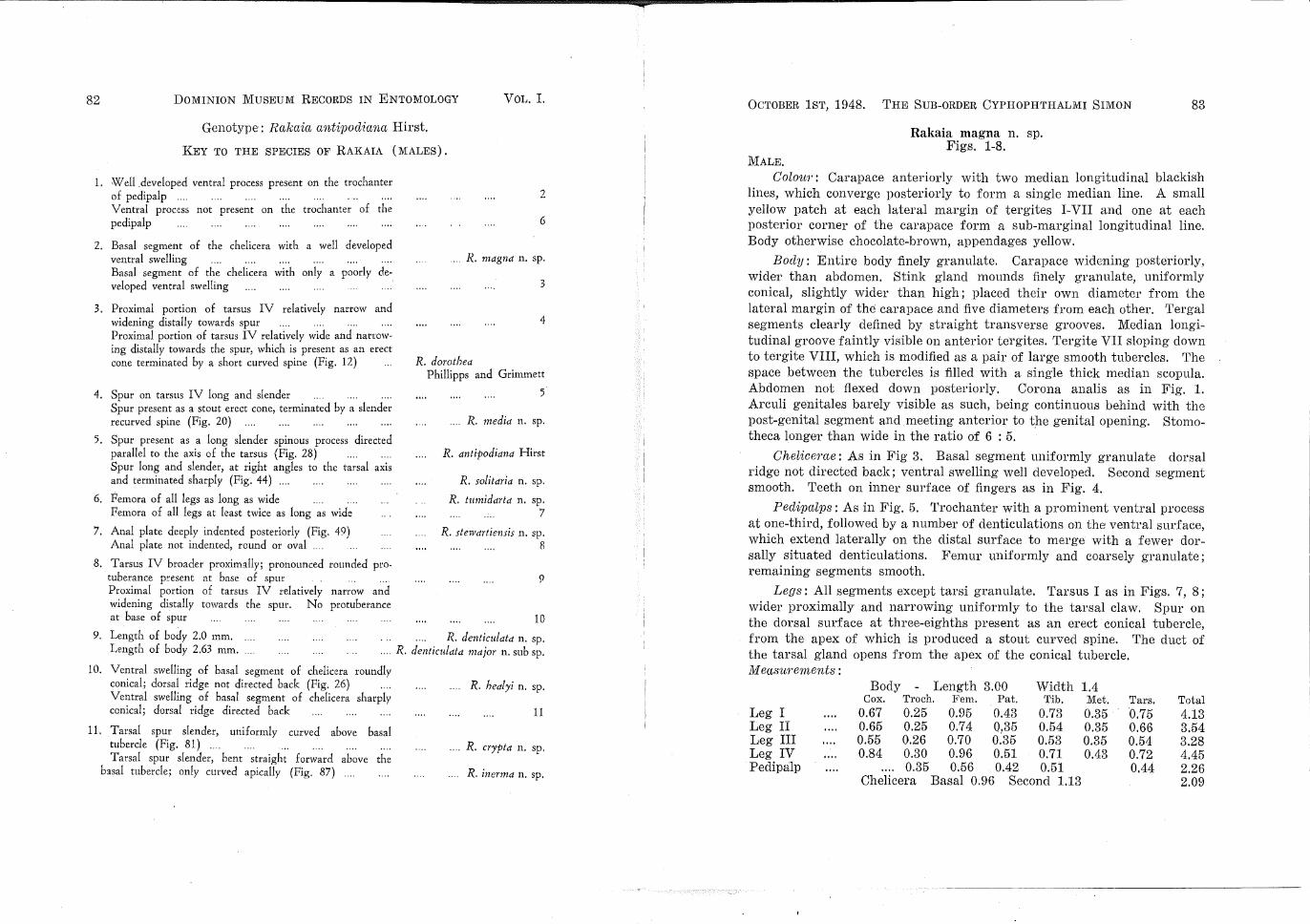
1. Well .developed ventral process present on the trochanter of pedipalp .... Ventral process not present on the trochanter of the pedipalp
2. Basal segment of the chelicera with a well developed ventral swelling Basal segment of the chelicera with only a poorly developed ventral swelling
3. Proximal portion of tarsus IV relatively narrow and widening distally towards spur Proximal portion of tarsus IV relatively wide and narrowing distally towards the spur, which is present as an erect cone terminated by a short curved spine (Fig. 12)
4. Spur on tarsus IV long and slender Spur present as a stout erect cone, terminated by a slender recurved spine (Fig. 20)
5. Spur present as a long slender spinous process directed parallel to the axis of the tarsus (Fig. 28) Spur long a11d slender, at right angles to the tarsal axis and terminated sharply (Fig. 44) ....
6. Femora of all legs as long as wide Femora of all legs at least twice as long as wide
7. Anal plate deeply indented posteriorly (Fig. 49) Anal plate not indented, round or oval
8. Tarsus IV broader proximally; pronounced rounded protuberance present at base of spur ... Proximal portion of tarsus IV relatively narrow and widening distally towards the spur. No protuberance at base of spur
2
6
.... R... magna n. sp.
3
4
R. dorothea Phillipps and Grimmett
5
R. media n. sp.
R. antipodiana Hirst
R. solitaria n. sp.
R. tumidarta n. sp. 7
R. stewartiensis n. sp. 8
9
10
9. Length of body 2.0 mm. Length of body 2.63 mm .....
R. denticulata n. sp. . ... R. denticulata major n. sub sp.
10. Ventral swelling of basal segment of chelicera roundlv conical; dorsal ridge not directed back (Fig. 26) Ventral swelling of basal segment of chelicera sharply conical; dorsal ridge directed back
11. Tarsal spur slender, uniform! y curved above basal tubercle (Fig. 81) .. . .. Tarsal spur slender, bent straight forward above the
basal tubercle; only curved apically (Fig. 87)
R. healyi n. sp.
11
.... R. crypta n. sp.
R. inerma n. sp.
OCTOBER 1ST, 1948. THE SUB-ORDER CYPHOPHTHALMI SIMON 83
Rakaia magna n. sp. Figs. 1-8.
MALE.
Colour: Carapace anteriorly with two median longitudinal blackish lines, which converge posteriorly to form a single median line. A small yellow patch at each lateral ma.rgin of tergites I-VII and one at each posterior corner of the carapace form a sub-marginal longitudinal line. Body otherwise chocolate-brown, appendages yellow.
Bocly: Entire body finely granulate. Carapace widening posteriorly, wider than abdomen. Stink gland mounds finely granulate, uniformly conical, slightly wider than high; placed their own diameter from the lateral margin of the carapace and five diameters from each other. Tergal segments clearly defined by straight transverse grooves. Median longitudinal groove faintly visible on anterior tergites. Tergite VII sloping down to tergite VIII, which is modified as a pair of large smooth tubercles. The space between the tubercles is filled with a single thick median scapula. Abdomen not flexed down posteriorly. Corona analis as in Fig. 1. Arculi genitales barely visible as such, being continuous behind with the post-genital segment and meeting anterior to the genital opening. Stomatheca longer than wide in the ratio of 6 : 5.
Chelicerne: As in Fig 3. Basal segment uniformly granulate dorsal ridge not directed back; ventral swelling well developed. Second segment smooth. Teeth on inner surface of fingers as in Fig. 4.
Peclipnlps: As in Fig. 5. Trochanter with a prominent ventral process at one-third, followed by a number of denticulations on the ventral surface, which extend laterally on the distal surface to merge with a fewer dorsally situated denticulations. Femur uniformly and coarsely granulate; remaining segments smooth.
Legs: All segments except tarsi granulate. Tarsus I as in Figs. 7, 8; wider proximally and narrowing uniformly to the tarsal claw. Spur on the dorsal surface at three-eighths present as an erect conical tubercle, from the apex of which is produced a stout curved spine. The duct of the tarsal gland opens from the apex of the conical tubercle. M ec~surements :
Body Length 3.00 Width 1.4 Cox. Troch. Fem. Pat. Tib. Met. Tars. Total
Leg I 0.67 0.25 0.95 0.43 0.73 0.35 0.75 4.13 Leg II 0.65 0.25 0.74 0.35 0.54 0.35 0.66 3.54 Leg III 0.55 0.26 0.70 o:35 0.53 0.35 0.54 3.28 Leg IV 0.84 0.30 0.96 0.51 0.71 0.43 0.72 4.45 Pedipalp 0.35 0.56 0.42 0.51 0.44 2.26
Chelicera Basal 0.96 Second 1.13 2.09
84 DoMINION MUSEUM RECORDS IN ENTOMOLOGY VoL. I.
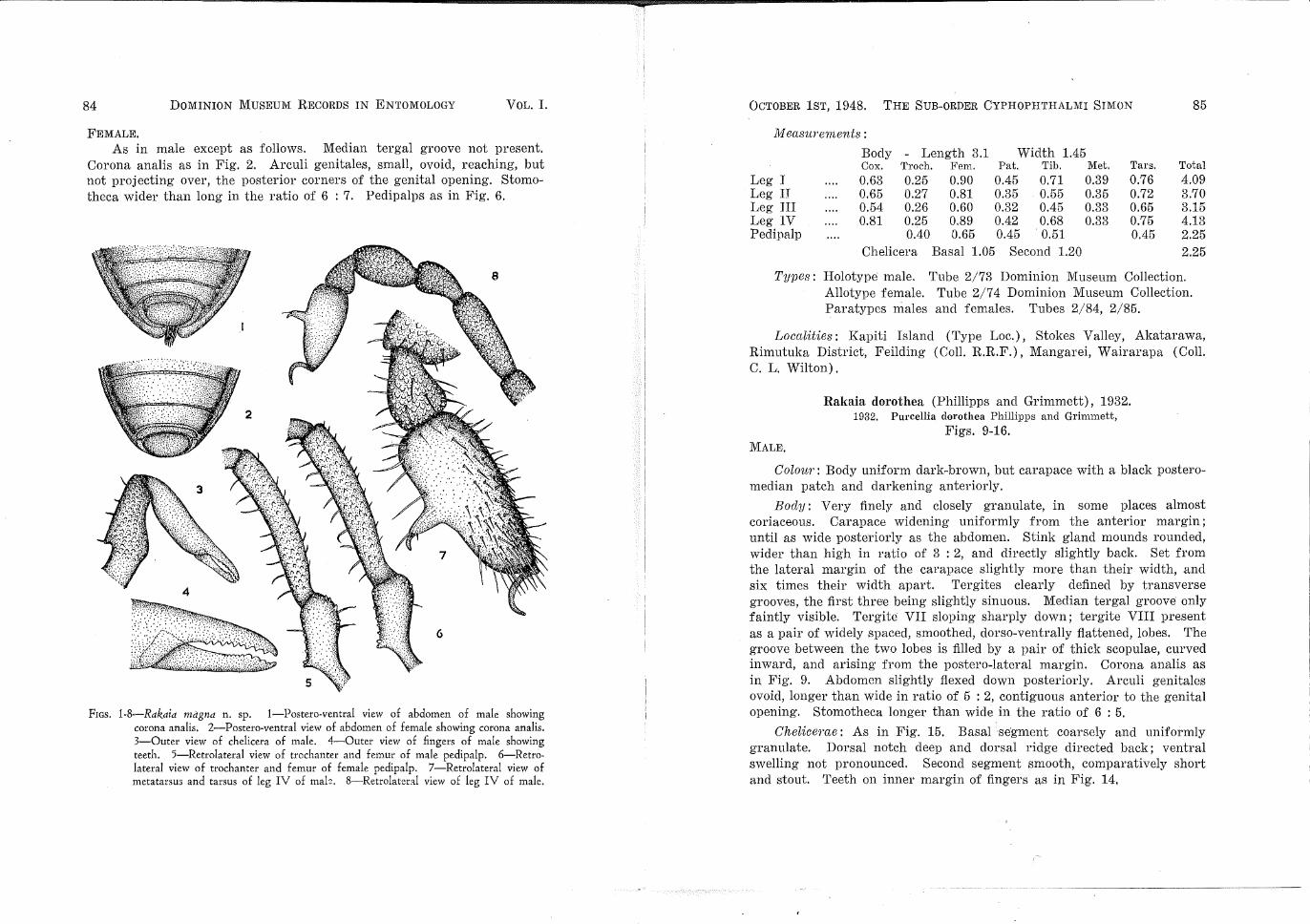
FEMALE. As in male except as follows. Median tergal groove not present.
Corona analis as in Fig. 2. Arculi genitales, small, ovoid, reaching, but not projecting over, the posterior corners of the genital opening. Stomatheca wider than long in the ratio of 6 : 7. Pedipalps as in Fig. 6.
FIGs. 1-8-Rakaia magna n. sp. 1-Postero-ventral view of abdomen of male showing corona analis. 2-Postero-ventral view of abdomen of female showing corona analis. 3-0uter view of chelicera of male. 4-0uter view of fingers of male showing teeth. 5-Retrolateral view of trochanter and femur of male pedipalp. 6-Retrolateral view of trochanter and femur of female pedipalp. 7-Retrolateral view of metatarsus and tarsus of leg IV of mak 8-Retrolater:tl view of leg IV of male.
OCTOBER 1ST, 1948. THE SUB-ORDER CYPHOPHTHALMI SIMON 85
Measurements :
Body - Length 3.1 Width 1.45 Cox. Troch. Fem. Pat. Tib. Met. Tars. Total
Leg I 0.63 0.25 0.90 0.45 0.71 0.39 0.76 4.09 Leg II 0.65 0.27 0.81 0.35 0.55 0.35 0.72 3.70 Leg III 0.54 0.26 0.60 0.32 0.45 0.33 0.65 3.15 Leg IV 0.81 0.25 0.89 0.42 0.68 0.33 0.75 4.13 Pedipalp 0.40 0.65 0.45 . 0.51 0.45 2.25
Chelicera Basal 1.05 Second 1.20 2.25
Types: Holotype male. Tube 2/73 Dominion Museum Collection. Allotype female. Tube 2/7 4 Dominion Museum Collection. Paratypes males and females. Tubes 2/84, 2/85.
Localities: Kapiti Island (Type Loc.), Stokes Valley, Akatarawa, Rimutuka District, Feilding (Coll. R.R.F.), Mangarei, Wairarapa (Coli. C. L. Wilton).
MALE.
Rakaia dorothea (Phillipps and Grimmett), 1932. 1932. Purcellia dorothea Phillipps and Grimmett,
Figs. 9-16.
Colour: Body uniform dark-brown, but carapace with a black posteromedian patch and darkening anteriorly.
Body: Very finely and closely granulate, in some places almost coriaceous. Carapace widening uniformly from the anterior margin; until as wide posteriorly as the abdomen. Stink gland mounds rounded, wider than high in ratio of 3 : 2, and directly slightly back. Set from the lateral margin of the carapace slightly more than their width, and six times their width apart. Tergites clearly defined by transverse grooves, the first three being slightly sinuous. Median tergal groove only faintly visible. Tergite VII sloping sharply down; tergite VIII present as a pair of widely spaced, smoothed, dorso-ventrally flattened, lobes. The groove between the two lobes is filled by a pair of thick scapulae, curved inward, and arising from the postero-lateral margin. Corona analis as in Fig. 9. Abdomen slightly flexed down posteriorly. Arculi genitales ovoid, longer than wide in ratio of 5 : 2, contiguous anterior to the genital opening. Stomotheca longer than wide in the ratio of 6 : 5.
Chelicerae: As in Fig, 15. Basal segment coarsely and uniformly granulate. Dorsal notch deep and dorsal ridge directed back; ventral swelling not pronounced. Second segment smooth, comparatively short and stout. Teeth on inner margin of fingers as in Fig. 14,
8G DOMINION MUSEUM RECORDS IN ENTC~VIOL08Y VoL. I.
PeclipcLlps: As in Fig. 15. Trochanter with a strong sharp ventral process, followed by a number of ventral and disto-dorsal denticulations; femur with a number of ecnttered denticulations, those on the ventral surface being stronger. Remaining segments without denticulations.
Legs: All segments except tarsus uniformly granulate. Tarsus IV (a~ in Figs. 11, 12) relatively wide proximary, but narrowing distally towards the spur, which is present as an erect cone and terminated by a short curved spinous process. The duct of the tarsal gland opens from the apex of the cone below the spinous process.
16
FIGs. 9-16-Rakaia dorothea Phillipps and Grimmett. 9-Postero-ventral view of abdomen of male showing co:·ona analis. 10-Posterc-ventral view of abdomen of female showing corona analis. 11-Retrolateral view of leg IV of male. 12-Retrolateral view of metatarsus and tarsus of leg IV of male. 13-0uter view of ch;\icera of male. 14-0uter view of fingers of m:1!e. showing teeth. 15-Retrolateral view of trochanter and femur of pedip:1lp of male. 16-R~trolateral view of trcchant~r and femur of pedipalp of female. -
OCTOBER 1ST, 1948. THE SUB-ORDER CYPHOPHTHALMI SIMON 87
M ecLsurements :
Body Length 2.05 Width 0.85 Cox. Troch. Fem. Pat. Tib. Met. Tars. Total
Leg I 0.38 0.15 0.55 0.25 0.40 0.21 0.45 2.39 Leg II 0.50 0.15 0.45 0.25 0.33 0.20 0.42 2.30 Leg III 0.50 0.15 0.42 0.21 0.32 0.20 0.34 2.14 Leg IV 0.65 0.25 0.50 0.25 0.30 0.15 0.41 2.51 Pedipalp 0.21 0.40 0.22 0.25 0.25 1.33
Chelicera Basal 0.42 Second 0.59 1.01
FEMALE.
As in male except as follows. Median tergal groove faintly visible. Tergites all defined by straight transverse grooves and not modified posteriorly. Corona analis as in Fig. 10. Arculi genitales sub-ovoid, projecting well over the posterior corners of the genital opening. Stomotheca longer than wide in the ratio of 3 : 2. Trochanter and femur of pedipalp as in Fig. 16.
M easu?'ements :
Body - Length 2.15 Width 1.35 Cox. Tro·ch. Fem. Pat. Tib. Met. Tars. Total
Leg I 0.40 0.15 0.51 0.32 0.40 0.25 0.41 2.44 Leg II 0.45 0.18 0.35 0.20 0.25 0.15 0.35 1.93 Leg III 0.45 0.19 0.42 0.15 0.25 0.15 0.30 1.91 Leg IV 0.73 0.21 0.50 0.23 0.30 0.15 0.35 2.47 Pedipalp 0.23 0.39 0.22 0.25 0.25 1.34
Chelicera Basal 0.42 Second 0.60 1.02
Types: Holotype male. Tube 2/5, Dominion Museum Collection. Allotype female. Tube 2/98, Dominion Museum Collection.
Paratypes male and female. Tube 2/6, Dominion Museum Collection.
Localities: Wellington (Type Loc.) (Coli. R.E.R. Grimmett and R.R.F.), Gollans Valley, Kapiti Island (Coli. R.R.F.), Wainui Valley (Coli.
I R. K. Dell). ·
Remarks: The type specimens were ki,ndly presented to the Dominion Museum by Mr. R. E. R. Grimmett. This species alone is found in the patches of native forest in the immediate vicinity of Wellington City, while on Kapiti Island and at Stokes Valley it' is found in association with R. magna n. sp. Further to the north, in the Akatawara, Rimutaka District it is entirely replaced by R. magna.
-- ---!
88 DOMINION MUSEUM RECORDS IN ENTOMOLOGY VOL. I.
Rakaia media n. sp. Figs. 17-24.
MALE. ,Colou1·: Body dark yellow-brown, with two longitudinal black lines
ronverging into a blackish patch on the postero-median surface of the carapace. Posterior margins of the sternites blackish-brown. Appendages light yellow-brown.
19
FIGs. 17-24-Rakaia media n. sp. 17-Postero-ventral view of abdomen of male showing corona analis. 18-Postero-ventral view of abdomen of female showbg corona analis. 19-Retrolateral view of leg IV of male. 20-Retrolateral view of metatarsus and tarsus of leg IV of male. 21-0uter view of chelicera of male. 22-0uter view of fingers of male showing teeth. 23-Retrolateral view of trochanter and femur of male pedipalp. 24-Retrolateral view of trochanter and femur of female pedipalp.
OCTOBER 1ST, 1948. THE SUB-ORDER CYPHOPHTHALMI SIMON 89
Body: Entire surface finely and evenly granulated. Carapace widening posteriorly, slightly wider than abdomen. Stink gland cones wider than high in ratio of 5 : 4; directed slightly back; set less than one and a half times their diameter from the lateral margin of the carapace and six diameters apart. Median tergal groove well defined. Tergites distinguished by straight transverse grooves; tergite VII sloping sharply posteriorly; tergite VIII flattened and divided medially so as to appear from above as a pair of granulated lobes, directed slight!y inward and upward. From the posterior margin of the anal plate arises a small scapula of fine hairs projecting between the lobes. Corona analis as in Fig. 17. Arculi genitales longer than wide in the ratio of 3 : 1, not meeting anterior to the genital opening. Stomotheca longer than wide in the ratio of 8 : 7.
Chelicerae: As in Fig. 21. Ventral swelling of the basal segment, poorly developed; dorsal ridge directed slightly b0ck; anterior half of segment uniformly granulate. Second segment 1smooth and relatively stout. Teeth on inner surface of fingers as in Fig. 22.
Peclipc~lps: As in Fig. 23. Trochanter with a prominent basal process at almost half-way, followed by numerous ventral and disto-dorsal denticulations. Femur uniformly but sparsely denticulate. Remaining segments smooth.
Legs: All segments except tarsus finely granulated. Femora II curved, remainer normal and straight. Tarsus IV as in Figs. 19, 20, widening uniformly to one third, where the dorsal surface is produced into a stout conical spur, directed slightly forward and terminated by a short, sharp spine, curved down to the tarsal segment. The duct of the tarsal gland opens from beneath, at the base of the terminal spine.
M ec~su1·ements :
Body - Length 2.32 Width 1.29 Cox. Troch. Fem, Pat, Tib. Met. Tars. Total
Leg I 0.45 0.21 0.59 0.33 0.45 0.23 0.53 2.59 Leg II 0.41 0.19 0.54 0.21 0.37 0.21 0.48 2.41 Leg III 0.45 0.18 0.52 0.21 0.33 0.21 0.45 2.35 Leg IV 0.55 0.29 0.54 0.27 0.39 0.33 0.54 2.91 Pedipalp 0.25 0.37 0.25 0.33 0.33 1.53
Chelicera Basal 0.87 Second 0.75 1.62 FEMALE.
As in male, except as follows. Median tergal groove faintly visible on tergites I-VI. Tergites VII and VIII entire. Corona analis as in Fig.
90 DOMINION MUSEUM RECORDS IN ENTOMOLOGY VOL. I.
18. Arculi genitales pyriform, obtuse and projecting over posterior cot·· ners of genital opening. Stomotheca longer than wide in the ratio of 6:7. Trochanter and femur of pedipalp as in Fig. 24.
FIGs. 25-32-Rakaia antipodiana Hirst. 25-Postero-ven~ral vtew of abdomen of. male showing corona analis. 26-Pos::ero-ventral view of abdomen of female showing corona analis. 27-Retrolateral view of leg IV of male. 28-Retrolateral view of metatarsus and tarsm of leg IV of male. 30-Retrolateral view of trochanter and femur of male pedipalp. 3 0-Retrolateral view of trochanter and femur of female pedipalp. 31-0uter view of fingers of male chelicera showing t:oeth. 32-0uter view of male chelicera.
OCTOBER 1ST, 1948. THE SUB-ORDER CYPHOPHTHALMI SIMON 91
Types: Holotype male. Tube 2/101, Dominion Museum Collection. I
Allotype fema~e. Tube 2/102, Dominion Museum Collection. Paratypes. Tubes 2/103-106, Dominion Museum Colledion.
Localities: Mamaku (Type Loc. Call. J. T. Salmon), Ohingaita, Upper Rangitikei, (Coli. J. Ramsay), Lake Waikaremoana (Coli. R.R.F.).
Remarks: The distribution of this species coincides approximately with that of R. inerma n. sp. The two species both being pre~ent at Waikaremoana, where howeveT they are not found living together, R. inerma being found to the north of Lake Waikaremoana and .R. meclict about the Lake and to the south.
Rakaia antipodiana Hirst, 1925. Figs. 25-32.
MALE.
Colo~w: Body uniform dark chocolate-brown. Carapace with a median longitudinal black area. CheliceTae dark-brown, remaining appendages yellow-brown.
Body: Entire surface closely granulate. Carapace wider and broadly rounded behind, as wide as abdomen. Stink gland mounds low, rounded, twice as wide as high, set their diameter from the lateral margin of the carapace and almost six diameters apart. Median tergal groove only faintly visible. Tergite VII with a broad median indentation and sloping sharply down posteriorly. Tergite VIII comp~etely divided and, whell vi.ewed from above, seen as a pair of well-developed, rounded, granulated tubercles, directed slightly towards one another. Posterior portion of the median groove filled with a scapula of fine hairs originating as three bundle~, one from each lateral margin of the groove and a median bunch from the posterior margin of the anal plate. Posterior margin of the abdomen not flexed down. Corona analis as in Fig. 25. Arculi genitales ovoid, longer than wide in ratio 9 : 5, contiguous immediately anterior to the genital opening. Stomotheca longer than wide in ratio of 11 : 10.
Cheliceme: As in Fig. 32. Basal segment uniformly granulate, dorsal ridge sharp and directed back, basal swelling only poorly developed. Second segment smooth and comparatively ~tout. Teeth on inner face of fingers as in Fig. 31.
Pecli1JCLlps: As in Fig. 29. Trochanter produced below into a strong precess. Disto-dorsal and ventral surfaces sparsely denticulate. Femora ~parsely but uniformly dent~culate. Remaining segments free from dentlculations.
92 DOMINION MUSEUM RECORDS IN ENTOMOLOGY VOL. I.
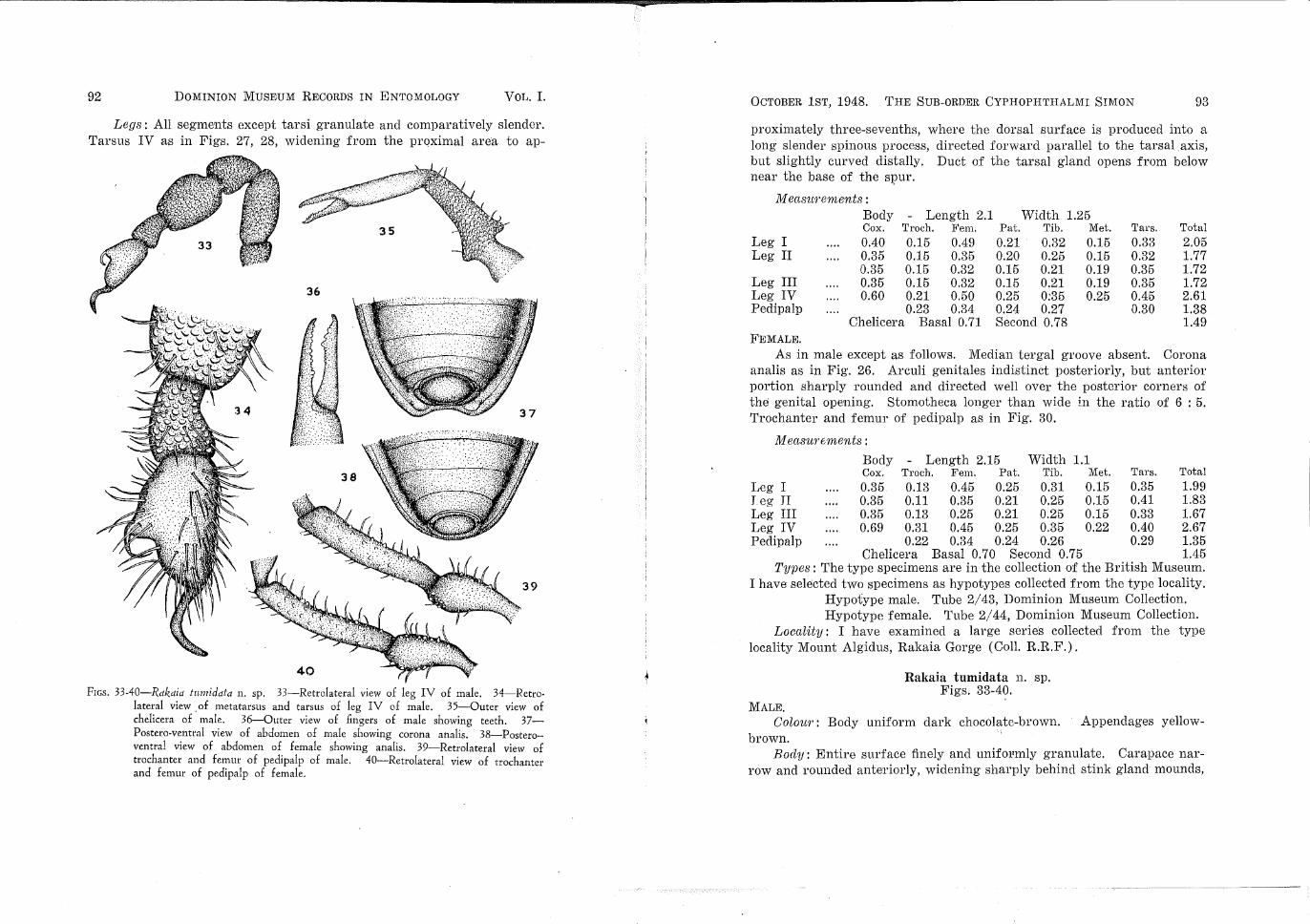
Legs: All segments except tarsi granulate and comparatively slender. Tarsus IV as in Figs. 27, 28, widening from the proximal area to ap-
36
FrGs. 33-40-Rakaia tumidata n. sp. 33-Retrolateral view of leg IV of male. 34-Retrolateral view . of metatarsus and tarsus of leg IV of male. 3 5-0uter view of chelicera of male. 36-0uter view of fingers of male showing teeth. 37-Postero-ventral view of abdomen of male showing corona analis. 38-Postero-ventral view of abdomen of female showing analis. 39-Retrolateral view of trochanter and femur of pedipalp of male. 40-Retrolateral view of trochanter and femur of pedipalp of female.
OCTOBER 1ST, 1948. THE SUB-ORDER CYPHOPHTHALl\11 SIMON 93
proximately three-sevenths, where the dorsal surface is produced into a long slender spinous process, directed forward parallel to the tarsal axis, but slightly curved distally. Duct of the tarsal gland opens from below near the base of the SJ?Ul'.
M ensurements : Body - Length 2.1 Width 1.25 Cox. Troch. Fem. Pat. Tib. Met. Tars. Total
Leg I 0.40 0.15 0.49 0.21 0.32 0.15 0.33 2.05 Leg II 0.35 0.15 0.35 0.20 0.25 0.15 0.32 1.77
0.35 0.15 0.32 0.15 0.21 0.19 0.35 1.72 Leg III 0.35 0.15 0.32 0.15 0.21 0.19 0.35 1.72 Leg IV 0.60 0.21 0.50 0.25 0:35 0.25 0.45 2.61 Pedipalp 0.23 0.34 0.24 0.27 0.30 1.38
Chelicera Basal 0.71 Second 0.78 1.49 FEMALE.
As in male except as follows. Median tergal groove absent. Corona analis as in Fig. 26. Arculi genitales indistinct posteriorly, but anterior portion sharply rounded and directed well over the posterior corners of the genital opening. Stomotheca longer than wide i.n the ratio of 6 : 5. Trochanter and femur of pedipalp as in Fig. 30.
Chelicera Basal 0.70 Second 0.75 1.45 Types: The type specimens are in the collection of the British Museum.
I have selected two specimens as hypotypes collected from the type locality. Hypotype male. Tube 2/43, Dominion Museum Collection. Hypotype female. Tube 2/44, Dominion Museum Collection.
Locnlity: I have examined a large series collected from the type locality Mount Algid us, Rakaia Gorge (Coli. R.R.F.).
Rakaia tumidata n. sp. Figs. 33-40.
MALE. Colour: Body uniform dark chocolate-brown. Appendages yellow-
brown. Body: Entire surface finely and unifo1•mly granulate. Carapace nar
row and rounded anteriorly, widening sharply behind stink gland mounds,
94 DOMINION MUSEUM RECORDS IN ENTOMOLOGY VoL. I.
considerably wider than abdomen. Stink gland cones low and rounded, twice as high as wide, set their diameter from the lateral margin of the carapace and six diameters apart. Tergal segments not clearly distinguished by straight transverse shallow grooves. Median groove present on o1~ly tergites VI-VIII. Tergite VIII not modified as a pair of tubercles and scapulae absent. Abdomen slightly flexed clown posteriorly. Arculi genitales present as a ridge bounding the margins of the genital opening and meeting anteriorly. Corona analis as in Fig. 37.
Chelicerae: As in Fig. 35. Both segments slender. Basal segment evenly granulate, dorsal ridge well developed and directed back, ventral swelling strong and roundE:cl. Second segment smooth. Teeth on inner surface of fingers as in Fig. 36.
Peclipalps: As in Fig. 39. Ventral process absent, but with numerous ventral and disto-dorsal clenticulations. Femur uniformly but sparsely denticulate. Otherwise free from clenticulations.
Legs: All segments except tarsi uniformly and strongly granulate. All segments stout, tibia greatly so, as wide as long. Tarsus IV as in Figs. 33, 34, gradually widening on the dorsal surface to the spur at approximately half-way. Spur present as a broad conical tubercle from the apex of which is produced a strong spinous process directed parallel to the tarsal axis for half of its length and then sharply curved to the tarsal segment. The duct of the tarsal gland passes up the spinous process and opens from the under surface immediately before it bends towards the tarsus.
M casurements :
Body Length 2.25 Width 1.15 Cox. Troch. Fem. PRt. Tib. Met. Tars. Total
Leg I 0.51 0.15 0.55 0.30 0.45 0.15 0.40 2.51 Leg II 0.50 0.15 0.35 0.25 0.35 0.19 0.35 2.14 Leg III 0.50 0.15 0.25 0.25 0.32 0.20 0.32 1.99 Leg IV 0.71 0.25 0.51 0.30 0.45 0.25 0.35 2.82 Pedipalp 0.26 0.41 0.29 0.34 0.33 1.63
Chelicera Basal 0.86 Second 1.00 1.86
FEMALE.
As in male except as follows. Median longitudinal groove faintly discernible on all tergites. Corona analis as in Fig. 38. Arculi genitales triangular, apex directed over posterior corners of the genital opening. Stomotheca wider than long in the ratio of 9 : 7. Pedipalp as in Fig. 40.
OCTOBER 1ST, 1948. THE SUB-ORDER CYPHOPHTHALMI SIMON
M eas~wements :
l.eg I Leg II Leg III Leg IV Pedipalp
Body Length 2.5 Cox. Troch. Fem. 0.50 0.50 0.45 0.81
Types: Holotype male. Tube 2/50, Dominion Museum Collection. Allotype female. Tube 2/52, Dominion Museum Collection. Paratypes. Tube 2/51, Dominion Museum Collection.
Locality: Cuvier Island, Hauraki Gulf, collected from under stones and logs (Coli. R.R.F.).
Ratmia solitaria n. sp. Figs. 41-48.
MALE,
Colaw·: As in R. magna, but median longitudinal groove along entire length of abdomen blackish-brown; inter-tergal grooves blackish.
Body: Dorsal surface finely granulate, but more strongly on the posterior tergites. Ventral surface uniformly and finely granulate. Cephalothoracic carapace smoothly rounded behind, slightly wider than abdomen. Stink gland mounds bluntly conical and sloping back; slightly wider than high; set almost twice their diameter from the lateral margin of the carapace and five diameters apart. Inter-tergal grooves only faintly visible, straight, accentuated by the transverse pigment lines. Median longitudinal groove well defined. Tergite VII sharply sloping down and, when viewed from above, with a shallow median V-shaped groove. Tergite VIII modified to form a pair of smooth lobes, directed inward at their tips; the space between being filled by a thick median scopula of fine white hairs. Posterior portion of abdomen flexed down. Corona analis as in Fig. 41. Arculi genitales pyriform, obtuse and reaching anterior to the genital opening but not contiguous. Sternum minute, visible between coxae II. Stomotheca wider than long in ratio of 11 : 10.
Chel·ice·me: As in Fig. 47. Basal segment uniformly granulate beyond dorsal notch; ventral swelling almost absent; dorsal ridge rounded. Second segment stout and smooth. Teeth on inner margin of fingers as in Fig. 48.
Peclipalps: As in· Fig. 46. A strong ventral process is present on the trochanter at slightly less than half-way, followed by a number of basal
96 DOMINION MUSEUM RECORDS IN ENTOMOLOGY VoL. I.
42
FIGs. 41-48-Rakaia solitaria n. sp. 41--Postero-ventral view of abdomen of m~le showing corona analis. 42-Postero-ventral view of abdomen of female showmg corona analis. 43-:-Retrolateral view of leg IV of male. 44-Retrolateral view of metatarsus and. tarsus of leg IV of male. 45-Retrolateral view of trochanter and femur of female pedipalp. 45-Retrolateral view of trochanter a.nd femur of male pedipalp. 47-0uter view of chelicera of male. 48-0uter vtew of fingers of male showing teeth.
OCTOBER 1ST, 1948. THE SUB-ORDER CYPHOPHTHALMI SIMON 97
and a few disto-dorsal denticulations. Femur uniformly but sparsely covered with small rounded denticulations. Remaining segments smooth.
Legs: All segments except tarsi uniformly granulate. Tarsus IV as in Figs. 43, 44, widening dorso-ventrally to the spur at one third, distal to which the segment narrows uniformly. Spur present as a long slender spinous process rising at right angles to the tarsal axis, curved forward at the distal extremity. The duct of the tarsal gland passes up the spur and opens from the under surface at two thirds.
Leg I 0.45 0.21 0.58 0.25 0.37 0.25 0.50 2.61 Leg II 0.50 0.12 0.41 0.25 0.33 0.21 0.45 2.27 Leg III 0.47 0.16 0.37 0.21 0.29 0.25 0.41 2.10 Leg IV 0.66 0.16 0.50 0.33 0.45 0.25 0.50 2.75 Pedipalp 0.25 0.39 0.23 0.37 0.37 1.61
Chelicera Basal 0.79 Second 0.83 1.62 FEMALE.
Colour: As in male. Body: As in male except as follows. Median tergal groove present only
on tergites I-VI. Corona analis as in Fig. 42. Arculi genitales longer than wide in ratio of 8 : 3, projecting over the posterior corners of the genital opening·. Stomotheca longer than wide in ratio of 10 : 9. Trochanter and femur of pedipalp as in Fig. 45. Pedipalp 0.26 0.41 0.29 0.34 0.33 1.63
M eas~wements : Body - Length 2.33 Width 1.20 Cox. Troch. Fem. Pat. Tib. Met. Tars. Total
Leg I 0.45 0.20 0.58 0.29 0.41 0.23 0.50 2.66 Leg II 0.45 0.16 0.35 0.25 0.29 0.16 0.29 1.95 Leg III 0.41 0.16 0.37 0.16 0.29 0.21 0.29 1.89 Leg IV 0.62 0.21 0.45 0.33 0.50 0.22 0.50 2.83 Pedipalp 0.29 0.37 0.25 0.33 0.37 1.61
Chelicera Basal 0.75 Second 0.91 1.66 Types: Holotype male. Tube 2/107, Dominion Museum Collection.
Allotype female. Tube 2/108, Dominion Museum Collection. Paratypes males and females. · Tube 2/109, Dominion Museum
Collection.
Locality: A large series were extracted from leaf-mould collected in a small patch of Beech forest at Opouawa Gully, West Wairarapa. (Coli. R.R.F.).
98 DOMINION MUSEUM RECORDS IN ENTOMOLOGY VoL. I.
FIGs. 49-56-Rakaia stewartiensis n sp. 49-Postero-ventral view of abdomen of male showing corona analis. 50-Postero--ventral view of abdomen of female showing corona analis. 51-0uter view of chelicera of male. 52-0uter view of fingers of male showing teeth. 53-Retrolateral view of trochanter and femur of female pedipa:p. 54-Retrolateral view of trochanter and femur of male pedipalp. 55-Retrolateral view of leg IV of male. 56--Retrolateral view of metatarsus and tarsus of leg IV of male.
I
~
-r
OCTOBER 1ST, 1948. THE SUB-ORDER CYPHOPHTHALMI SIMON 99
Ralmia stewartiensis n. sp. Figs. 49-56.
MALE. Colour: Body uniform dark brown, carapace with a distinct median
V-shaped black mark narrowing posteriorly. Appendages yellow-brown. Body: Entire surface granulate, being more prominent on the pos
terior tergites. Carapace widening behind the stink gland mounds, much wider than the abdomen. Stink gland mounds conical, but slightly truncate; wider than high in the ratio of 4 : 3; set almost twice their diameter from the lateral margin of the carapace and five diameters apart. Tergites clearly distinguished by straight transverse grooves. Median longitudinal groove only faintly discernible. Tergite VII strongly granulate, divided by a broad median V-shaped groove. Tergite VIII divided by a deep median cleft to appear from below as in Fig. 49. A pair of small scapulae are present, one on each side of the posterior margin of the cleft. Anal plate deeply indented posteriorly into a halfmoon, the inner surface of which is filled with fine closely placed hairs. Abdomen flexed down posteriorly. Corona analis as in Fig. 49. Arculi genitales large·, ovoid, but slightly truncate anteriorly, not meeting in front of the genital opening. Stomotheca longer than wide in the ratio of 7 : 6.
Cheliceme: As in Fig. 51. Basal segment strongly and evenly granulate; transverse ridge low and rounded; ventral swelling well developed. second segment smooth. Teeth on inner surface of fingers as in Fig. 52.
Pedipalps: As in Fig. 54. Ti·ochanter smooth except for several small ventral and disto-lateral denticulations. Femur with a few ventral denticulations; pedipalp otherwise smooth.
Legs: All segments except tarsi uniformly granulate. Tarsus IV as in Figs. 55, 56. Proximal portion sub-parallel; spur at two-fifths present as a strong even process directed along the tarsal axis and unevenly bifurcate distally, the basal fork being longer. The duct of the tarsal gland passes through the spur to open distally. The shape of the distal extremity of the spur at first glance appears to be due to the tip being broken, however it is found to be identical in all specimens.
M ec~su1~ements : Body Length 2.58 Width 1.5 Cox. Troch. Fem. Pat. Tib. Met. Tars. Total
Leg I 0.60 0.25 0.59 0.31 0.45 0.25 0.52 2.97
Leg II 0.60 0.22 0.51 0.30 0.52 0.25 0.45 2.85
Leg III 0.60 0.21 0.55 0 .. 25 0.49 0.21 0.50 2.81
FEMALE. As in male except as follows. Median longitudinal groove only faintly
visible. Corona analis as in Fig. 50. Arculi genitales pyrifm·m and truncated anteriorly where they project well over the posterior corners of the
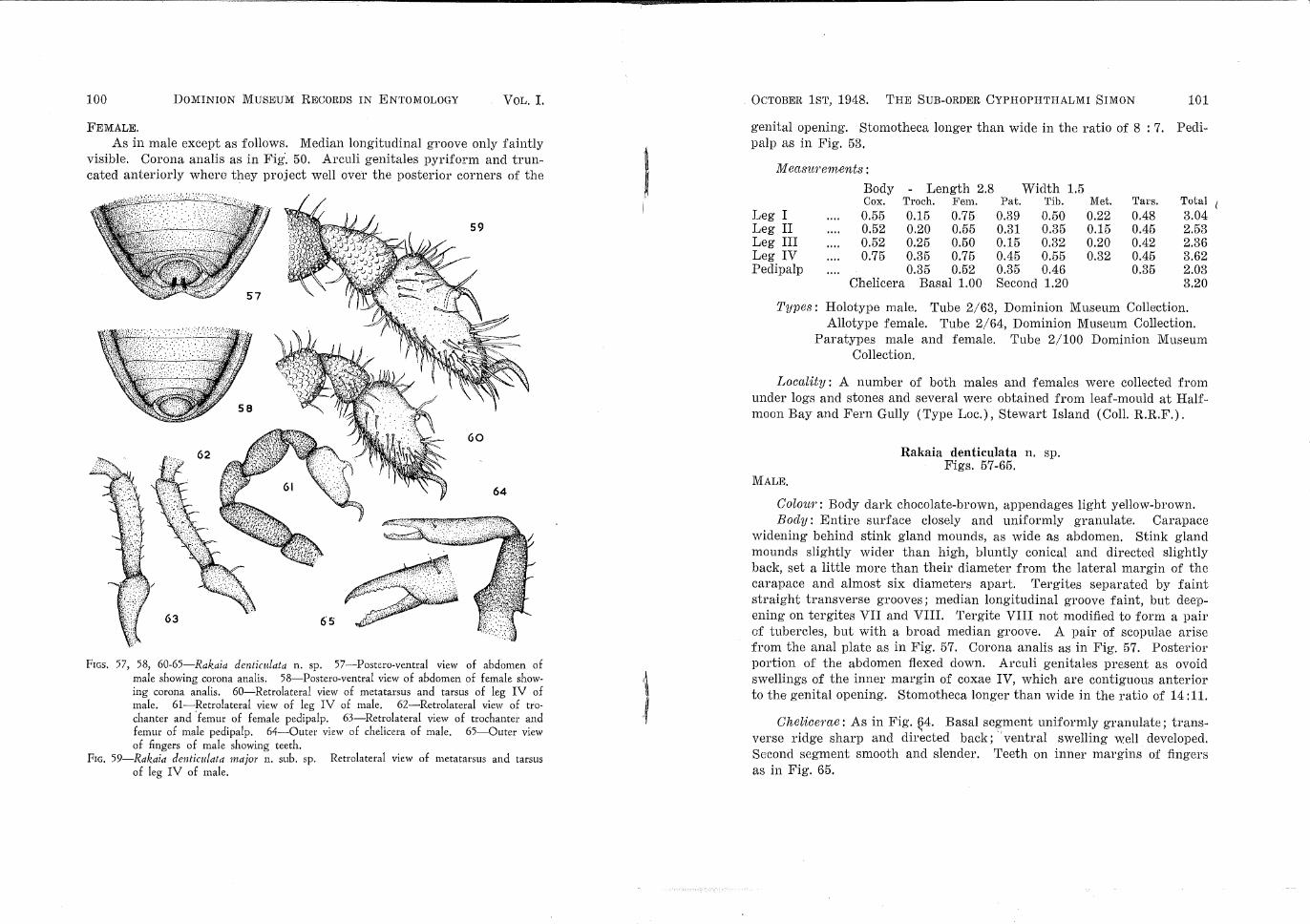
FIGs. 57, 58, 60-65-Rakaia denticulata n. sp. 57--Postero-ventral view of abdomen of male showing corona analis. 58-Postero-ventral view of abdomen of female showing corona analis. 60-Retrolateral view of metatarsus and tarsus of leg IV of male. 61-Retrolateral view of leg IV of male. 62-Retrolateral view of trochanter and 'femur of female pedipalp. 63-Retrolateral view of trochanter and femur of male pedipalp. 64-0uter view of chelicera of male. 65-0uter view of fingers of male showing teeth.
FrG. 59-Rakaia denticulata major n. sub. sp. Retrolateral view of metatqrsus and tarsus of leg IV of male.
I . OCTOBER 1ST, 1948. THE SUB-ORDER CYPHOPHTHALMI SIMON 101
genital opening. Stomotheca longer than wide in the ratio of 8 : 7. Peclipalp as in Fig. 53.
M easu1·em.ents :
Body Length 2.8 Width 1.5 Cox. Troch. Fem. Pat. Tib. Met. Tars. Total
Leg I 0.55 0.15 0.75 0.39 0.50 0.22 0.48 3.04 Leg II 0.52 0.20 0.55 0.31 0.35 0.15 0.45 2.53 Leg III 0.52 0.25 0.50 0.15 0.32 0.20 0.42 2.36 Leg IV 0.75 0.35 0.75 0.45 0.55 0.32 0.45 3.62 Peclipalp 0.35 0.52 0.35 0.46 0.35 2.03
Chelicera Basal 1.00 Second 1.20 3.20
Types: Holotype male. Tube 2/63, Dominion Museum Collection. Allotype female. Tube 2/64, Dominion Museum Collection.
Paratypes male and female. Tube 2/100 Dominion Museum Collection.
Locality: A number of both males and females were collected from under logs and stones and several were obtained from leaf-mould at Halfmoon Bay and Fern Gully (Type Loc.), Stewart Island (Coli. R.R.F.).
Rakaia denticulata n. sp. Figs. 57-65.
MALE.
Colou1': Body clark chocolate-brown, appendages light yellow-brown. Body: Entire surface closely and uniformly granulate. Carapace
widening behind stink gland mounds, as wide as abdomen. Stink gland mounds slightly wider than high, bluntly conical and directed slightly back, set a little more than their diameter from the lateral margin of the carapace and almost six diameters apart. Tergites separated by faint straight transverse grooves; median longitudinal groove faint, but deepening on tergites VII and VIII. Tergite VIII not modified to form a pair of tubercles, but with a broad median groove. A pair of scapulae arise from the anal plate as in Fig. 57. Corona analis as in Fig. 57. Posterior portion of the abdomen flexed clown. Arculi genitales present as ovoid swellings of the inner margin of coxae IV, which are contiguous anterior to the genital opening. Stomotheca longer than wide in the ratio of 14:11.
Cheliceme: As in Fig. ~4. Basal segment uniformly granulate; transverse ridge sharp and directed back; ·ventral swelling well developed. Second segment smooth and slender. Teeth on inner margins of fingers as in Fig. 65.
102 DOMINION MUSEUM RECORDS IN ENTOMOLOGY VOL. I..
Pedipal1Js: As in Fig. 63. Ventral process absent from trochanter, but a number of disto-dorsal and disto-ventral denticulations are present. Femur sparsely denticulate. Pedipalp otherwise smooth.
Legs: All segments except tarsi uniformly granulate. Tarsus IV as in Figs. 60, 61, wider proximally and narrowing to the spur, which is present as a strong, forwardly directed, spine; the cuticle on the anterior portion at the base of the spur is distended to form a rounded tubercle. The duct of the tarsal gland opens a little way above this tubercle on the under surface of the spur.
M ecw~wements : Body - Length 2.00 Width 1.12 Cox. Troeh, Fem. Pat. Tib. Met. Tal'S. Total
Leg I 0.45 0.15 0.49 0.25 0.34 0.16 0.36 2.20 Leg II 0.48 0.15 0.36 0.21 0.32 0.16 0.35 2.03 Leg III 0.48 0.15 0.32 0.21 0.31 0.15 0.25 1.87 Leg IV 1.20 0.25 0.45 0.26 0.31 0.19 0.38 3.04 Pedipalp 0.25 0.35 0.26 0.32 0.26 1.44
Chelicera Basal 0.85 Second 0.86 1.71
FEMALE.
Differing from the male as follows. Median tergal groove absent. Arculi genitales present as small ovoid ridges not separated from coxae IV, but with the anterior portion projecting over the posterior corners of the genital opening. Corona analis as in Fig. 58. Stomotheca as long as wide. Pedipalp as in Fig. 62.
M eas~wements : Body - Length 2.10 Width 1.15 Cox. Troch. Fem. Pat. Tib. Met. Tal's. Total
Leg I 0.45 0.15 0.48 0.26 0.35 0.19 0.44 2.32 Leg II 0.45 0.11 0.41 0.22 0.28 0.15 0.41 2.03 Leg III 0.45 0.15 0.25 0.21 0.25 0.17 0.30 1.78 Leg IV 1.31 0.26 0.44 0.26 0.34 0.20 0.35 3.16 Pedipalp 0.25 0.36 0.24 0.24 0.25 1.34
Chelicera Basal 0.75 Second 0.85 1.60
Types: Holotype male. Tube 2/53, Dominion Museum Collection. Allotype female. Tube 2/54, Dominion Museum Collection. Paratypes males and females. Tube 2/99, Dominion Museum
Collection.
Localities: Starvation Ridge, (Type Loc. Coli. J. T. Salmon), Leslie Valley, (CoiL R.R.F.).
OCTOBER 1ST, 1948. THE SUB-ORDER CYPHOPHTHALMI SIM0::--.1
Rakaia denticulata major n. sub. sp.
103
Fig. 59. MALE.
With the majority of the characters of R. clenticulatct, but differing as follows. Entire animal much larger, the length of the body being 2.63 mm. as compared with 2.00 mm.; legs I and IV almost equal in length, whereas in R. clenticulata leg I is much longer than leg IV. Tarsus IV as in Fig. 59, broad proximally.
Only two male specimens were available, collected by Dr. J. G. Myers from Arthurs Pass. Both are identical with regard to size as well as other characters. A characteristic noted during the study of N.Z. Cyphophthalmids is the extremely slight variation in size shown within a species and hence I have no hesitation in naming the above as a sub-species.
Body - Length 2.63 Width 1.50 Cox. Troch. Fem. Pat. Tib. Met. Tars. Total
Leg I 0.54 0.21 0.83 0.37 0.55 0.29 0.55 3.34 Leg II 0.45 0.25 0.62 0.25 0.45 0.24 0.45 2.71 Leg III 0.45 0.21 0.61 0.21 0.45 0.25 0.46 2.64 Leg IV 0.62 0.33 0.62 0.37 0.56 0.33 0.55 3.38 Pedipalp 0.35 0.52 0.36 0.41 0.41 2.05
Chelicera Basal 1.09 Second 1.24 3.33
Ty1Jes: Holotype male. Tube 2/44, Dominion Museum Collection. Paratype male. Tube 2/110, Dominion Museum Collection.
Locnlity: Two male specimens were collected by Dr. J. G. Myers from Arthurs Pass (Grimmett Collection).
Rakaia healyi n. sp. Figs. 66-73.
MALE. Colour: Carapace with a blackish area anterior to the stink glands
extending back along the middle line to the posterior margin. Tergite VIII light yellow. Remainder of body and coxae deep reddish-brown. Appendages light yellow.
Bocly: Entire surface finely and closely granulate. Carapace sharply widening posteriorly, wider than abdomen. Stink gland mounds blunt~y conical in shape, slightly higher than wide, placed nearly twice their diameter from the lateral margin of the• carapace and six diameters from each other. Tergites clearly defined by straight transverse grooves. Median groove only faintly discernible on tergites I-V, but deepening on
104 DOMINION MUSEUM RECORDS IN ENTOMOLOG"Y VoL. I.
tergites VI and VII. Tergite VII slopes steeply down to tergite VIII, which is present as a pair of large smooth rounded protuberances. The area between these is filled by two pairs of scapulae, one pair originating from the lateral margins, the other from the posterior margin of the anal
72
FrGs. 66-73-Rakaia healyi n. sp. 66-Postero-ventral view of abdomen of male showing corona analis., 67-Postero-ventral view of abdome11 of female showing corona analis. 68-Retrolateral view of leg IV of male. 69-Retrolateral view of metatarsus and tarstw of leg IV of male. 70-Retrolateral view of trochanter and femur of male pedipalp. 71-Retrolateral view of trochanter and femur of female pedipalp. 72-0uter view of chelicera of male. 73-0uter view of fingers of male showing teeth.
0C'f0BER 1ST, 1948. THE SUB·-ORDER CYPHOPHTHALMI SIMON 105
plate. (Fig. 66). Corona anal is as in Fig. 66. Arculi genitales ovoid, twice as long as wide, not meeting distally. Stomotheca longer than wide in ratio of 9 : 8.
Chelicerae: As in Fig. 72. Basal segment uniformly granulate, dorsal transverse ridge rounded; ventral swelling rounded but well developed. Second segment smooth, rather slender. Teeth on cutting edges of fingers as in Fig. 73.
Pedipalps: As in Fig. 70. Trochanter without a ventral process, but with a number of denticulations on the ventral and distal surface. Femur uniformly but sparsely denticulate. Denticulations not present on other segments.
Legs: All segments except tarsi closely granulate. Tarsus IV, as in Figs. 68, 69, ovoid in general shape. Spur present in the form of a wide tubercle, the apex of which is produced along the tarsal axis as a spinous process, which is sharply curved at two thirds to pass down the retrolateral margin of the tarsus. The duct of the tarsal gland opens from below at approximately half-way along the spinous process.
M easu1·ements : Body - Length 2.2 Width 1.25 Cox. Troch. Fem. Pat. Tib. Met. Tars. Total
Leg I 0.45 0.16 0.59 0.24 0.43 0.21 0.48 2.56 Leg II 0.45 0.13 0.52 0.19 0.31 0.15 0.36 2.11 Leg III 0.45 0.15 0.41 0.15 0.35 0.20 0.26 1.97 Leg IV 0.65 0.25 0.68 0.24 0.46 0.29 0.47 3.04 Pedipalp 0.21 0.44 0.26 0.33 0.32 1.56
Chelicera Basal 1.05 Second 1.2 2.25
FEMALE. Colour as in male, but with a white patch on each side of the median
black a~·ea immediately behind the stink gland mounds.
Body as in male, but median tergal groove absent. Corona analis as Fig. 67. Arculi genitales ovoid, twice as long as wide, not projecting over the genital opening. Stomotheca longer than wide in the ratio of 11 : 8. Pedipalps as in Fig. 71.
Measurements : Body - Length 2.25 Width 1.25 Cox. Troch. Fem. Pat. Tib. Met. TaTs. Total
Leg I 0.45 0.21 0.76 0.34 0.49 0.26 0.50 3.01 Leg II 0.42 0.15 0.64 0.24 0.39 0.15 0.34 2.33 Leg III 0.42 0.13 0.35 '0.24 0.38 0.17 0.35 2.04 Leg IV 0.65 0.25 0.62 0.36 0.44 0.29 0.47 3.08 Pedipalp 0.52 0.45 0.24 0.28 0.28 1.77
FIGs. 74-81-Rakaia crypt a n. sp' 74-Retrolateral view of trochanter and femur of pedipalp of male. 75-Retrolateral view of trochanter and femur of pedipalp of female. 76-Retrolateral view of leg IV of male. 77-0uter view of chelicera o~ male. 78-;-0uter view of fingers of male showing teeth. 79-Postero-ventral v1ew of abdomen of male showing corona analis. 80-Postero-ventral view of abdomen of female showing corona analis. 81-Retrolateral view of metatarsu3 and tarsus of male.
OCTOBER 1ST, 1948. THE SUB-ORDER CYPHOPHTHALMI SIMON 107
Types: Holotype male. Tube 2/65, Dominion Museum Collection. Allotype female. Tube 2/66, Dominion Museum Collection.
Locality: A large number of specimens were extracted from leaf-mould collected from Ships Cove (Coll. A. J. Healy) and Endeavour Inlet (Coll. J. Barnard), Queen Charlotte Sounds.
Rakaia crypta n. sp. Figs. 7 4-81.
MALE,
Colml1': Carapace dark-brown, with a median pair of longitudinal yellow streaks immediately behind the level of the stink glands; posterior margin yellow-brown. Inter-tergal grooves and lateral margins of. the tergites dark-brown, otherwise tergites yellow-brown. Ventral surface of body yellow-brown, but with a dark a1·ea on the lateral margins of each sternite. · Appendages yellow-brown.
Body: Squat, entire surface finely and evenly granulate. Carapace widening evenly posteriorly until slightly wider than abdomen. Stink gland mounds sharply conical, directed back, as high as wide, set a little more than their diameter from the lateral margin of the carapace and six diameters apart. Tergal segments defined by shallow sinuate grooves, median groove only faintly visible. Tergite VIII modified to form two small, smooth, rounded tubercles, widely separated, with the space between them filled by two scopulae of fine curved hairs, which originate from the inner basal margin of each tubercle. Abdomen flexed down posteriorly. Corona anaEs as in Fig. 79. Arculi genitales curved round the genital opening to meet on a broadly truncate surface, which extends to coxae III. Stomotheca wider than long in ratio of 7 : 5.
Cheliceme: As in Fig. 77. Basal segment uniformly granulate, dorsal ridge directed back, ventral swelling well ·developed and terminated sharply. Second segment smooth and slender. Teeth on inner surface of fingers as in Fig. 78.
Pedipalps : As in Fig. 7 4. Ventral trochanteral process absent, but trochanter with a few small denticulations on both the ventral and distodorsal surface. Femur denticulate ventrally, remaining segn1ents smooth.
Legs: All segments except tarsi finely granulate. Tarsus IV as in Figs. 76, 81, dorsal surface widening evenly to two-fifths, where the spur is present in the form of steep cone, the apex of which is produced forward as a strong, spinous process evenly' curved so that the distal quarter is directed to the tarsal segment. The duct of the tarsal gland passt,., along the spinous process to open from beneath at approximatelv half-w~y.
108 DOMINION MUSEUM RECORDS IN ENTOMOLOGY VoL. I.
82
FIGs. 82-89. Rakaia inerma n. sp. 82-Postero-ventral view of abdomen of male showing corona analis. 83-Postero-ventral view of abdomen of female showing corona analis. 84-0uter view of chelicera of male. 85-0uter view of fingers of male showing teeth. 68-Retrolateral view of leg IV of male. 87-Retrolateral view of metatarsus and tarsus of male. 88-Retrolateral view of trochanter and femur of female pedipalp. 89-Retrolateral view of trochanter and femur of male pedipalp.
OCTOBER 1ST, 1948. THE SUB-ORDER CYPHOPHTHALMI SIMON 109
Leg I 0.48 0.21 0.75 0.41 0.55 0.53 0.71 3.64 Leg II 0.53 0.21 0.53 0.37 0.53 0.33 0.69 3.19 Leg III 0.55 0.21 0.48 0.41 0.45 0.25 0.55 2.90 Leg IV 0.96 0.33 0.55 0.41 0.53 0.53 0.63 3.94 Pepipalp 0.37 0.48 0.3R 0.41 0.37 1.96
Chelicera· Basal 1.17 Second 1. 29 2.4G
FEMALE. As in male except as follows. Median tergal groove clearly visible,
tergite VIII distinctly notched, but not so as to form a pair of tubercles. Corona analis as in Fig. 80. Arculi genitales triangular, the apex directed across the posterior corner of the genital opening. Stomotheca wider than long in the ratio of 6: 5. Pedipalp as in Fig. 75.
M ectSU1'ements :
Body Length 2)m Width 1.61 Cox. Troch. Fem. Pat. Tib. Met. Tars. Tot a
Leg I 0.45 0.18 0.58 0.37 0.53 0.29 0.61 3.01 Leg II 0.53 0.21 0.5$ 0.33 0.41 0.29 0.48 2.78 Leg III 0.55 0.18 0.45 0.29 0.37 0.25 0.41 2.50 Leg IV 0.87 0.37 0.55 0.37 0.48 0.37 0.63 3.64 Pedipalp 0.29 0.48 0.33 0.41 0.39 1.90
Chelicera Basal 1.17 Second 1.27 2.24
Types: Holotype male. Tube 2/86, Dominion Museum Collection. Allotype female. Tube 2/87, Dominion Museum Collection. Paratypes female. Tubes 2/88, 2/97, Dominion Museum
Collection.
Locctlity: A number of both males and females were extracted from leaf-mould collected by Dr. J. T. Salmon from Te Aroha Mountain, 3000 ft.
posteriorly until distinctly wider than the abdomen. Stink gland mounds sharply conical, as wide as high and directed slightly posteriorly, set a little more than their diameter from the lateral margin of the carapace and more than six diameters apart. Tergites distinguished by faint sinu-
110 DOMINION MUSEUM RECORDS IN ENTOMOLOGY VoL. I.
ous grooves. Median groove visible on all segments, but shallow on tergites I-VI, deeper and broader on tergites VII and VIII, which are not modified to form tubercles. 'rwo lateral and one median scapulae present as in Fig. 82. Arculi genitales ovoid, almost as wide as long, not meeting anterior to the genital opening. Stomotheca longer than wide in the ratio of 15 : 11.
Cheliceme: As in Fig. 84. Basal segment relatively narrow and uniformly granulate; dorsal ridge prominent but not directed back; ventral swelling well developed and terminating sharply. Second segment slender and smooth. Teeth on inner margin of fingers as in Fig, 85.
Peclipalps: As in Fig. 89. Trochanter with a few ventral and distodorsal denticulations, but ventral process absent. Remaining segments, including femur; smooth.
Legs: All segments except tarsus uniformly granulate. Tarsus IV as in Figs. 86, 87, slender, the spur at one third, present as a small rounded tubercle produced apically into a long slender spinous process, directed at an angle from the tarsus and the distal third bent sharply down to the tarsus. The duct of the tarsal gland passes through the spinous process to open from the under surface at almost half-way.
M ecLSU1'ements : Body - Length 2.35 Width 1.45
Cox. Troch. Fem. Pat. Tib. Met. Tars. Total Leg I 0.51 0.22 0.76 0.32 0.41 0.24 0.59 3.05 Leg II 0.56 0.23 0.56 0.26 0.32 0.21 0.53 2.61 Leg III 0.51 0.21 0.43 0.24 0.35 0.24 0.43 2.41 Leg IV 0.75 0.36 0.60 0.35 0.46 0.36 0.56 3.44 Pedipalp 0.31 0.43 0.32 0.36 0.36 1.78
Chelicera Basal 0.95 Second 1.00 1.95
FEMALE. As in male except as follows. Tergal groove visible on all except
tergite VIII. Arculi genitales ovoid, projecting over the posterior corners of the genital opening. Corona analis as in Fig. 83. Stomotheca longer than wide in the ratio of 3 : 2. Pelipalp as in Fig. 88.
M easm·ements : Body Length 2.75 Width 1.45 Cox. Troch. Fem. Pat. Tib. Met. Tars. Total
Leg I 0.45 0.21 0.54 0.36 0.54 0.15 0.62 2.88 Leg II 0.43 0.20 0.52 0.25 0.38 0.24 0.43 2.45 Leg III 0.43 0.21 0.44 0.23 0.40 0.24 0.36 2.31 Leg IV 0.81 0.32 0.59 0.31 0.49 0.28 0.53 3.33 Pedipalp 0.31 0.46 0.29 0.34 0.32 1.72
Chelicera Basal 0.95 Second 0.95 1.90
OCTOBER 1ST, 1948. THE SUB-ORDER CYPHOPHTHALMI SIMON 111
Types: Holotype male. Tube 2/45, Dominion Museum Collection. Allotype female. Tube 2/47, Dominion Museum Collection. Paratype3 males and females. Tube 2/46, Dominion Museum
Collection.
Localities: Lake Wai.kare-iti (Type Loc.), from leaf-mould collected by P. N. Wilton and R.R.F., Desert Road, Waiouru, from leaf-mould collected by Dr. J. T. Salmon.
Genus Neopurcellia nov. Body granulate. Cephalothoracic carapace widening behind, wider
than abdomen. Stink glands each opening from the apex of a conical tuberc}e, set from one to two times their diameter from the lateral margin of tho carapace. EyeE absent. Posterior tergi.tes of the male modified, tergite VIII present as a pair of rounded tubercles. Both segments of chelicera approximately equal in length, basal segment granulate, with both dorsal ridge and ventral swelling present. Second segment smooth. Pedipalp slender; ventral process of trochanter present or absent; tarsus approximately the same length as tibia; claw small, simple, almost straight. Maxillary lobes of coxae II longer than wide. Coxae I-III approximately equal in width, coxae IV twice as wide. Sternum if present minute. Tarsus I deeper than II and III, with a ventral pad of short E:etae (Fig. 98). Tarsus IV of male bisegmented, both segments almost equal in length, spur present on dorsal ~urface of proximal segment. Remaining tarsi unsegmented. Tarsal claws simple.
Genotype: N eopur.cellia salmoni n. sp. N eopu1·cellia is undoubtedly closely related to the South African
genus Purcellia Hans. and Sor., with which it shares the development, as a secondary E:exual character, of two approximately equal segments to tarsus IV. The only other genus recorded with two tarsal segments is SpeleosiTo Lawrence, in which the distal segment is twice as long as the proximal. In all these genera the spur and associated gland are found on the proximal segment.
KEY TO THE SPECIES OF NEOPURCELLIA (MALES). 1. Trochanter of pedipalp with a ventral process. Body very small, no more than I .7 mm. in length Trochanter of pedipalp without ventral process. Body no less than 2.2 mm. in length
2. Two scapulae present, one origi11ating from each inner margin of the posterior tergal tubercles .... · , A single median scapula present, originating from the posterior margin of the anal plate .
N. minutissima n. sp.
2
N. salmoni n. sp.
N. florensis n. sp.
112 DOMINION MUSEUM RECORDS IN ENTOMOLOGY VoL. I.
FIGs. 90-98-Neopurcellia salmoni 11. sp. 90-Postero-ventral view of abdomen of male showing corona analis. 91-Postero-ventral view of abdomen of female showing corona analis. ' 92-0uter view of chelicera of male. 93-0uter view of fingers of male showing teeth. 94-Retrolateral view of trochanter and femur of female pedipalp. 95-Retrolateral view of trochanter and femur of male pedipalp. 96-Retrolateral view of leg IV of male. 97-Retrolateral view of tarsm IV of male. 98-Retrolateral view of metatarsus a11d tarsus of leg 1 of female.
OCTOBER 1ST, 1948. THE SUB-ORDER CYPHOPHTHALMI SIMON 113
N eopurcellia salmoni n. sp. Figs. 90-98.
MALE. Colou1>: Carapace blackish-brown anteriorly, but body otherwise uni
form dark-brown. Appendages yellow-brown; Body: Posterior tergites coarsely granulate, but otherwise body uni
formly and finely granulate. Carapace widening behind stink gland mounds. much wider than abdomen. Stink gland mounds bluntly conical, directed slightly back, one and a half times as high as wide, set one and a half times their diameter from the lateral margin of the carapace and seven diameters apart. Tergal segmentation clearly distinguished by shallow, straight, transverse grooves. Median tergal groove shallow, but clearly defined on all tergites. Tergite VI sloping steeply down to tergites VII and VIII, which are fused and present as a pair of widely spaced, smooth, rounded tubercles. Basal area between the two tubercles filled by a pair of thick scapulae, which originate from the posterior margin of the corona analis. Corona analis as in Fig. 90. Arculi genitales ovoid, meeting distally, but not quite extending to coxae III. Stomotheca longer than wide in the ratio of 7 : 5.
Chelicerae: As in Fig. 92. Basal segment uniformly granulate; dorsal ridge rounded, directed back; ventral swelling well developed. Second segment slender and smooth. Teeth on inner margin of fingers as in Fig. 93.
Pedipctlps: As in Fig. 95. Trochanter without basal process, but with a strong ventral tooth at approximately half-way, followed by a number of smaller rounded denticulations on the ventral surface and several sharp denticulations on the disto-clorsal surface. Femur uniformly but sparsely covered with small denticulations. Remaining segments smooth.
Legs: All segments except tarsi uniformly granulate. Tarsus IV as in Figs. 96, 97; proximal segment longer than wide in ratio of 6 : 5, slightly longer than distal segment. Spur present on disto-dorsal segment of proximal segment, strong, and, directed slightly forward; the apical third sharply curved along the tarsal axis, and terminating sharply. The duct of the tarsal gland opens from below at about one third.
FEMALE. As in male except as follows. Body uniformly and finely granulate.
Median tergal groove absent. Corona analis as in Fig. 91. Arculi genita!es ovoid, almost twice as long as wide, projecting over the posterior corners of the genital opening. Stomotheca longer than wide in the ratio of 4 : 3. Pedipalp as in Fig. 94.
FJGs. 99-105-Neopurcellia minutzsszma n. sp. 99-Pmtero-ventral view of abdomen of male showing coro1n analis. 100-Postero-vcntral view of ~bdomen of female showing corona analis. 101-0uter view of chelicera of male. 102-Retrolateral view of trochanter and femur of femab pedipalp. 103-Retrolateral view of trochanter and femur of male pedipalp. 104-Retrolate~al view of tarsus IV of male. 105-Retrolateral view of leg IV of male.
OCTOBER 1ST, 1948. THE SUB-ORDER CYPHOPHTHALMI SIMON 115
Jill easU?'emro nts :
Body Length 2.25 Width 1.25 Cox. Troch. Fem. Pat. Tib. Met. Tal's. Total
Leg I 0.42 0.21 0.69 0.34 0.55 0.32 0.54 3.07 Leg II 0.41 0.15 0.52 0.25 0.46 0.15 0.43 2.37 Leg III 0.40 0.15 0.34 0.25 0.33 0.22 0.32 2.01 Leg IV 0.63 0.15 0.68 0.35 0.48 0.25 0.56 3.10 Peclipalp 0.25 0.44 0.26 0.33 0.34 1.62
Chelicera Basal 1.10 Second 1.21 2.31
FEMALE. Types: Holotype male. Tube 2/48, Dominion Museum Collection.
Allotype female. Tube 2/49, Dominion Museum Collection.
Locc~lities: Anita Bay, Milford Sound (Co E. J. T. Salmon), Falls Creek, Homer District, Lake Howden (Coll. R.R.F.). This species was first extracted from leaf-mould collected from the type locality, Anita Bay, by Dr. J. T. Salmon, after whom I have pleasure in naming it.
Neopurcellia minutissima n. sp. Figs. 99-105.
MALE. Colour: Median dorsal and inter-tergal grooves dark-brown, small
round yellow patch present on the antero-lateral margin of each tergite. Body otherwise yellow-brown. Appendages light-yellow.
Bod?!: Small. Entire body finely and evenly granulate. Carapace widening posteriorly sli.ghtly wider than abdomen. Stink gland mounds conical, as wide as high, directed slightly back; set twice their diameter from the lateral margin of the carapace and seven diameters apart. Tergal segmentation clearly defined by straight transverse grooves; median longitudinal groove present on all tergites. Tergite VII deeply incised and sloping steeply down to the posterior tergite. Tergite VIII modified to form a pair of smooth rounded tubercles, the space between which, is filled by a thick median scopula, originating from the posterior margin of the anal plate. Abdomen strongly flexed clown posteriorly. Corona analis as ;.n Fig·. 99. Arculi genitales ovoid, almost twice as long as wide, meeting anterior to the genital opening and extending to coxae III. Stomotheca longer than wide in the ratio of 9 : 8.
Chelicerae: As in Fig. 101. Basal segment granulate on the dorsal surface distal to the dorsal notch; dorsal ridge directed back; basal swelling well developed. Second segment smooth, with several small transverse ridges on the outer distal surface. Teeth on i.nner face of fingers as in
Fig. 101.
116 DOMINION MUSEUM RECORDS IN ENTOMOLOGY VoL. I.
Peclipalps: As in Fig. 103. Strong ventral proce8s present on the trochanter at almost half-way, followed by a number of ventral and distodorsal denticulations. Femur with a few peg-like teeth on the ventral surface. All segments otherwise smooth.
Legs: All segments except tarsus uniformly granulate. Tarsus IV as in Figs, 104, 105; proximal segment as long as wide, slightly longer than distal segment. Spur on the disto-dorsal surface of the proximal segment strong, directed at right angles to the axis of the tarsus for a third of its length, and then constricted and evenly curved forward so that the distal extremity is pointing to the tarsal segment. The duct of the tarsal gland passes through the spur to open at two-thirds.
As in male except as follows. The median tergal groove is more shallow and not present on the posterior tergites. Corona analis as in Fig. 100. Arculi genitales very small, forming the posterior corners of the genital opening. Stomotheca as wide as long. Pedipalp as in Fig. 102.
M easu?'ements : Body - Length 1.70 Width 0.79 Cox. Troch. Fem. Pat. Tib. Met. Tars. Total
Leg I 0.24 0.11 0.42 0.21 0.26 0.17 0.32 1.73 Leg II 0.31 0.16 0.31 0.16 0.25 0.12 0.22 1.53 Leg III 0.28 0.12 0.28 0.16 0.21 0.15 0.25 1.45 Leg IV 0.41 0.16 0.38 0.17 0.32 0.15 0.21 1.80 Pedipalp 0.15 0.31 0.19 0.19 0.22 1.06
Chelicera Basal 0.55 ·Second 0.60 1.15
Types: Holotype male. Tube 2/76, Dominion Museum Collection. Allotype female. Tube 2/77, Dominion Museum Collection. Paratypes males and females. Tubes 2/78, 2/89-91, Dominion
Museum Collection.
Localities: Marokopa River, near Piripiri (Type loc.), Taumatatotora, Raglan, collected by Mr. A. J. Healy.
OCTOBER 1ST, 1948. THE SUB-ORDER CYPHOPHTHALMI SIMON 117
Neopurcellia florensis n. sp. Figs. 106-113.
MALE,
Colour: Each tergite with an orange-patch at the lateral margins. Body otherwise a uniform dark chocolate-brown. Appendages yellowbrown.
Body: Large. Entire body surface finely and evenly granulate. Carapace widening behind and flattening to form a lateral ridge, wider than the abdomen. Stink gland mounds slightly wider than high, evenly conical; set almost twice their diameter from the lateral margin of the carapace and six diameters apart. Tergites clearly defined by straight transverse grooves. Median tergal groove faint, tetgite VII deeply indented, each lateral portion smoothly rounded. Tergite VIII modified to form a pair smooth rounded lobes. A thick scapula originating from the posterior margin of the anal plate fills the basal area between the two lobes. Corona analis as in Fig. 113. Arculi genitales ovoid, widely separated above the genital opening. Stomotheca longer than wide in the ratio of 11 : 10.
Chelicera: As in Fig. 108. Basal segment uniformly granulate; dorsal ridge sharp, not directed back; ventral swelling well developed. Second segment slender and smooth. Teeth on inner surface of fingers as in Fig. 109. I "
Peclipalps: As in Fig. 110. Trochanter without a basal process, but with a large tooth on the ventral surface at one third, followed by a number of smaller teeth on the ventral and disto-dorsal surfaces. Ventral surface of the femur uniformly denticulate. Remaining segments smooth.
Legs: All segments except tarsus uniformly granulate. Femur I-III curved, femur IV normal. Tarsus IV as in Figs. 100, 107. Basal segment longer than wide in the ratio of 4 : 3, of same length as distal segment. Dorsal spur present at three-quarters, erect, curved forward apically and terminated obtusely. The duct of the tarsal gland passes directly through the spur to open sub-distally.
FEMALE. As in male except as follows. Median longitudinal tergal groove
absent. Corona analis as in Fig. 112. Arculi genitales ovoid, directed over the posterior corners of the genital opening. Stomotheca longer than
FIGS. 106-113-Neopurcellia flormsis n. sp. 106-Retrolateral view of tarsus IV of male. 107-Retrolateral view of leg IV of male. 108-0uter view of chelicera of male. 109-0uter view of fingers showing teeth. 110---Retrolateral view of trochanter and femur of male pedipalp. 111-Retrolateral view of trochanter and femur of female pedipalp. ·112-Postero-ventral view of abdomen showing corona · analis of female. 113-Postero-ventral view of abdomen showing corona analis of male.
OCTOBER 1ST, 1948. THE SUB-ORDER CYPHOPHTHALMI SIMON 119
wide in the ratio of 17 : 16. Pedipalp as in Fig. 111. Femora of leg·s I-III not decidedly curved as in male.
M easw·em.ents : Body Length 3.25 Width 1.75 Cox. Troch. Fem. Pat. Tib. Met. Tars. Total
Leg I 0.53 0.25 0.87 0.37 0.55 0.33 0.55 3.45 Leg II 0.53 0.21 0.75. 0.37 0.52 0.29 0.55 3.22 Leg III 0.55 0.21 0.63 0.29 0.45 0.29 0.52 2.94 Leg IV 0.79 0.33 0.79 0.41 0.55 0.29 0.63 3.79 Pedipalp 0.33 0.52 0.37 0.45 0.44 2.11
Chelicera Basal 1.17 Second 1.29 i 2.46
Types: Holotype male. Tube 2/111, Dominion Museum Collection. Allotype female. Tube 2/112, Dominion Museum Collection. Paratypes males and females. Tube 2/113, Dominion Museum
Collection.
Locality : Numerous specimens were collected from under logs and extracted from leaf-mould collected from Flora Camp (Call. R.R.F.). This species was only found localised in this area, its place being taken in the other nearby localities investigated, Leslie Valley and Starvation Ridge, by Rcdccda clenticulata.
LITERATURE.
HANSEN and SoRENSEN, 1904. On Two Orders of Arachnida, Cambridge University Press.
HINTON, H. E., 1938. A Key to the Sub-order Cyphophthalmi, with a description and figures of Neogoyea immsi gen. et. sp. n. Ann. Mag. Nat. Hist., (2), 2, pp. 331-338.
HIRsT, S., 1925. Some New Genera of Arachnida. Proc. Zoo!. Soc., Lond., 1925, pp. 1271-1280. The Harvest-spiders (Opiliones) of South Africa.
LAWRENCE, R. F. 1932. The Harvest-spiders (Opiliones) of South Africa. Ann. S. Afr. Mus., 29, (2), pp. 341-508.
---, 1939. A contribution to the Opilionid Fauna of Natal and Zululand. Ann. Natal Mus., 9, (2), pp. 225-243.
PHILLIPPS, W. J., and GRIMMETT, R. E. R., 1932. Some New Opiliones from New Zeala11d. Proc. Zoo!. Soc., Land., 1932, (3), pp. 731,740.
RoEWER, C. Fr., 1923. Die Weberknechte Der Erde. Jena. ---, 1942. Einige neue Arachniden I, Veroff. deuts. Kolon.-Ubersee-Mus., 3, (3),
pp. 277-280. SALMON, J. T., 1946. A Portable Apparatus for the Extraction from Leaf Mould of
Collembola and other Minute Organisms. Dom. Mus. Rec. Ent., 1, (2), pp. 13-18.