Page 1
Review Article
* Corresponding author: Dr.Dinesh Francis Swamy, Department of Pedodontics & Preventive Dentistry,Goa Dental College & Hospital,Bambolim,Goa
Journal of Applied Dental and Medical Sciences
NLM ID: 101671413 ISSN:2454-2288
Volume 3 Issue2 April-June 2017
The Talon Cusp Anomaly – Review of The Literature And Treatment Options
Dinesh Francis Swamy1, Elaine Savia Barretto2 , Sapna Sada Raut Dessai3 , Kathleen Manuela
D’Souza4, Kennedy Mascarenhas5, Sheen Ann Alex6
1.2 Department of Pedodontics & Preventive Dentistry,Goa Dental College & Hospital, Bambolim,Goa
3Department of Oral Medicine and Radiology,Goa Dental College & Hospital,Bambolim,Goa 4.5Department of Prosthodontics, Goa Dental College & Hospital,Bambolim,Goa
6Department of Pedodontics & Preventive Dentistry,PMS College of Dental Science & Research,Thiruvananthapuram,Kerala
A R T I C L E I N F O
Keywords:
Tooth,Abnormalities/*pathology/
therapy,Malocclusion/*complications/
therapy,Pulp Capping and Pulpectomy
Agents/therapeutic use,Occlusal
Adjustment,Humans
A B S T R A C T
Talon cusp is a developmental anomaly which occurs as a result of disturbances in the complex process of
tooth development. The clinical problems caused by the anomaly may be diagnostic, functional, aesthetic
or pathological. Treatment should be planned depending on the configuration and size of the talon cusp
and during tongue of occlusal interferences, irritation associated clinical problems such as attrition,
speech or mastication, development of dental caries and it ranges from nil treatment, conservative
treatment such as cusp reduction and restoration, to intentional endodontics. This paper reviews the
literature associated with the anomaly and their conservative management.
Introduction
Aberrations occurring in the morpho-differentiation
stage of tooth development can result in unique
manifestations such as mulberry molars, peg laterals,
and also, talon cusps. The Talon cusp anomaly,
according to Hattab[1],
may originate as a result of
outward folding of inner-enamel epithelial cells
(precursor of ameloblasts) and/or transient focal
hyperplasia of the mesenchymal dental papilla
(precursor of odontoblasts).
This phenomenon was first described by Mitchell in
1892[2]
and the term “Talon cusp” was coined by
Mellor and Ripa in 1970[3]
because of its resemblance
to an eagle‟s talon. The accessory cusp, if connected to
the incisal ridge may form a „T‟ shape, or if attached
more cervically, a „Y‟ shape may result. Varying
degrees of the anomaly exist and have attracted
various names – „enlarged/prominent cingula‟ for
lesser cusp-like formations in the cingulum area,
„hyperplastic cingulum‟, „cusped cingulum‟, Leong‟s
Page 2
TALON CUSP ANOMALY: LITERATURE-REVIEW AND TREATMENT OPTIONS3(2);2017 183
Journal Of Applied Dental and Medical Sciences 3(2);2017
Fig 1: Different degrees of Talon Cusp: A – Type 1/Major Talon; B – Type 2/Minor Talon; C – Type 3/Trace Talon (from Mull P,
2016[63])
premolar, occlusal pearl, evaginatus odontoma and
„supernumerary lingual tubercle‟. Electronic literature
search of the MEDLINE database via Pubmed was done
for scientific papers published in English from 1990 to
2016. Eligible studies were Case Reports, Case Series
and Epidemiological studies describing the management
of talon cusp. The search equation combined free text
words and controlled vocabulary pertaining to the
condition (Talon Cusp, Dens Evaginatus, talon AND
cusp). The search yielded 99 articles. Manual checking
of the reference list was done for relevant studies. This
article attempts to reviews the literature associated with
the anomaly and discusses the various clinical problems
encountered and their conservative management.
Prevalence:
Talon cusps occur more frequently in the permanent
dentition than the primary dentition – three times more
according to a 1996 study by Hattab.[4]
Numerous studies
have attempted to estimate the prevalence of the talon
cusp anomaly. Prevalence ranging from 0.06% in
Mexico, 0.17% in the United States, 5.2% in Malaysia,
1-2.4% in Pakistan and 7.7% in a north-Indian
population have been reported.[5],[6],[7],[8],[9]
However, race
seems to influence the incidence of Talon cusp
considerably, with greater incidence seen in Mongoloid
populations.[10]
The gender distribution shows more
frequent occurrence in males than in females, with a ratio
of 1.8:1 in the permanent dentition and 3.5:1 in the
primary dentition.[11]
Literature reports describing the
occurrence of the anomaly indicate that the site-
distribution involves the maxilla predominantly, with the
maxillary lateral incisors most frequently involved
(67%), followed by the maxillary central incisors (24%)
and canines (9%).[11]
About one fifth of all cases are
bilateral[12]
.
In the literature, numerous conservative or radical
treatment modalities have been advocated to manage
Talon cusps. The chief objective of treatment is the
avoidance of occlusal interference, and the main
treatment consideration is the avoidance of damage to
the dentine-pulp complex. This paper reviews the
literature associated with the anomaly and discusses the
various clinical problems encountered and the various
modalities of management of Talon Cusp anomaly.
Classification
Since the beginning, there has been significant
heterogeneity in the description of the talon cusp
anomaly. Some authors believe that the term „dens
evaginatus‟ can be used in place of the Talon anomaly –
as it is more descriptive of the pathology; while others
believe its use should be exclusively confined to similar
features affecting the posterior dentition.[13],[14],[15]
The anomaly has been more consistently described
following the use of classification systems. With the
Page 3
TALON CUSP ANOMALY: LITERATURE-REVIEW AND TREATMENT OPTIONS3(2);2017 184
Journal Of Applied Dental and Medical Sciences 3(2);2017
recognition of talon cusp occurrence on facial surfaces,
in addition to lingual surfaces, the older classification of
Hattab and co-workers was modified by Hsu & co-
workers based on the degree of their formation and
extension as:[4],[16],[17],[18]
Type 1 – Major talon: A morphologically well-defined
additional cusp that projects from the facial or
palatal/lingual surface of an anterior tooth and extends
at least half the distance from the cemento-enamel
junction to the incisal edge. (Fig 1A)
Type 2 – Minor talon: A morphologically well-defined
additional cusp that projects from the facial or
palatal/lingual surface of an anterior tooth and extends
more than one fourth, but less than half the distance
from the cemento-enamel junction to the incisal edge.
(Fig 1B)
Type 3 – Trace talon: Enlarged or prominent cingula
and their variations which occupy less than one fourth
the distance from the cemento-enamel junction to the
incisal edge. (Fig 1C)
Etiopathogenesis
Cusp morphogenesis is controlled initially, by the
primary enamel knot during bud-stage, and thereafter, by
the secondary enamel knot during late-bell stage, through
expression of certain signalling proteins.[19]
These
specific molecules diffuse from the mesenchymal cells
and interact with various cell types and are thought to be
responsible for orderly formation of the various parts of
the tooth. Hence, defects occurring at various points in
this developmental sequence can be implicated in the
genesis of anomalies of cusp number and size.[20]
The talon cusp anomaly has been reported in patients
with certain syndromes; in non-syndromic patients along
with additional dental malformations; and, in an isolated
manner in normal individuals with no associated
abnormalities – with the latter constituting the usual
mode of occurrence. Cases of Talon cusps have been
reported in patients with Rubinstein-Taybi syndrome,
Mohr syndrome, Sturge-Weber syndrome, Ellis van
Creveld Syndrome.[21],[22],[23],[24]
Given the fact however,
it has not been conclusively proven that an association
between such syndromes and talon cusp exists, and talon
cusp is not reported as an integral part of any syndrome.
Similarly, occurrence along with other dental
malformations such as peg-shaped lateral incisors,
impacted mesiodens, complex odontoma, supernumerary
teeth, megadont, dens invaginatus, shovel shaped maxilla
incisors, bifid cingula, exaggerated cusps of Carabelli
have been reported in the
literature.[4],[14],[15],[25],[26],[27],[28],[29]
These facts suggest that genetics may be a major
causative factor. However, the usual sporadic
occurrences of the abnormality are speculated to be
caused by trauma, local pressure effects from adjacent
tooth germs, or other localized insults affecting the tooth
germ.[12],[30],[31]
Notwithstanding the exact underlying
mechanism, there is undoubtedly an abnormal folding of
a region of the inner-enamel epithelium and adjacent
ectomesenchymal cells of the dental papilla into the
stellate reticulum of the enamel during the bell-stage. It
is thought that the high incidence of occurrence in the
lateral incisors is due to compression of the tooth germ
during the morpho-differentiation stage between the
adjacent central incisors and canine, which develop
several months earlier.[18]
The focal outfolding of the
inner-enamel epithelium does not affect the architecture
of the rest of the tooth and there is no alteration in the
root canal system of the tooth. Also, there is no
communication between the pulp and the oral cavity
unless concomitantly involved with other invaginating
anomalies.
Page 4
TALON CUSP ANOMALY: LITERATURE-REVIEW AND TREATMENT OPTIONS3(2);2017 185
Journal Of Applied Dental and Medical Sciences 3(2);2017
Complications
The complications of talon cusp are diagnostic,
functional, aesthetic and pathological. These are
elaborated below. Additionally a summary of the
literature is presented. (Table 1)
1. Diagnostic complications:
The outline of a talon cusp can mimic a supernumerary
tooth, and since most supernumeraries occur in the
maxillary anterior region it is essential to differentiate
between the two. Reports on misidentification of talon
cusp as supernumerary teeth, treated by accidental
extraction serve to highlight the need for meticulous
pre-operative assessment.[18],[32]
In the clinic, it has
been suggested that this differentiation may be done by
employing a shift-cone technique, whereby the talon
cusp presents as incisally-converging radio-opacity in
multiple angulations, whereas the supernumerary
presents as separate images of teeth and follicles.[23]
The presence of a horn of pulpal tissue within the talon
cusp has clinical implications for any operative
procedures, but the reliable presence of such pulpal
extensions are not evident in several histologic studies
of extracted talon cusp teeth.[14],[33]
To further
complicate clinical-planning, radiographic tracing of
the pulpal configuration inside the talon cusp is
inherently difficult as the cusp is superimposed over
the affected tooth crown, making accurate pre-
operative estimation of the extent of permissible cusp
removal rarely possible.[33]
This can be overcome with
the use of newer imaging techniques such as Cone
Beam Computed Tomography that offer higher
resolution, greater sharpness and permit volume
rendering.[34],[35]
2. Functional complications:
Occlusal interference when present can produce
numerous functional complications affecting both the
hard and soft tissues. The goal of treatment is therefore
to reduce the offending cusp, while preserving the
normal vitality and continued-growth of the tooth and
managing any complications encountered.
Functional complications to soft tissues include
irritation to the tongue or compromised tongue space,
speech disturbances or difficulty in feeding.[1],[36],[37],[38]
The prominent cusp may also come into premature
contact during occlusion – such hyper-occlusion can
result in damage to the cusp itself – resulting in
varying degrees of trauma, ranging from attrition to
cusp-fracture, to temporomandibular joint disorders
due to the deflecting premature contact.[39]
Hyper-
occlusion may also cause damage to the opposing
tooth, causing attrition and primary trauma from
occlusion, and subsequently damage to the
periodontium (i.e. secondary trauma from occlusion)
accompanied by tenderness, mobility and attachment
loss.[40]
3. Aesthetic complications:
Unlike the evaginatus-anomaly on posterior teeth, the
talon cusp is much less likely to encounter crushing
masticatory-forces. Rather than resulting in cusp-
fracture, the oblique vector of forces acting on both the
talon cusp-affected tooth and its opposing counterpart
are more likely to produce their displacement and/or
malocclusion.[41]
Talon cusp could also present an
obstacle for mandibular growth, holding the
mandible back and causing and causing Class II
malocclusions.[37]
Such malocclusions are treated by a
removal of the interfering part/entire Talon cusp
followed by appropriate appliance therapy, either
removable or fixed depending on the
malocclusion.[42],[43]
4. Other pathological complications:
Page 5
TALON CUSP ANOMALY: LITERATURE-REVIEW AND TREATMENT OPTIONS3(2);2017 186
Journal Of Applied Dental and Medical Sciences 3(2);2017
Developmental grooves are susceptible to plaque
accumulation and predispose to dental caries.[12]
Their
extension onto the root can also lead to gingival
inflammation and/or attachment loss or pocketing
necessitating periodontal and restorative
treatment.[1],[44]
Management:
Management of talon cusp ranges from nil-intervention
to simple cusp-reduction to cusp-reduction with pulp-
therapy. The degree of pulpal involvement and status of
the pulp would influence the choice of either
conservative pulp procedures such as pulp-capping or
pulpotomy, or more radical pulp procedures. The various
variables to consider while planning treatment are
elaborated.[37],[45],[46],[47]
1. Extent of cusp-reduction:
Cusp reduction is carried out with a high-speed
handpiece using abundant coolant. In performing the
reduction, it is essential to check the contact in
maximum intercuspation and also in protrusive
movements. The extent of cusp reduction that is
permissible varies according to literature. Pitts and
Hall[43]
have reported the removal up to 3mm of
anomalous cusp in one visit, without pulp exposure,
while Hattab and co-workers have reported the
removal of 1-1.5mm of cusp in several
instances.[1],[33],[36]
A systematic review found that in
56% of case reports analysed complete reduction was
done in a single appointment; periodic reduction in
26% of cases; abstention in 13% and extraction in
5%.[48]
However, since the pulp-horn may infrequently
extend up to the dentino-enamel junction such
techniques may not be relied upon exclusively, or
without adequate imaging.
Excessive grinding may result in inadequate
remaining-dentine-thickness placing the pulp at risk of
insult and subsequent necrosis. In such cases, the
layering of an acid-etched flowable light-cured resin or
glass ionomer applied after cusp-reduction on any
exposed dentin is indicated. Reports exists where
cusp-reduction without subsequent restoration has
resulted in early pulpal involvement.[49]
2. Interval between cusp-reduction appointments:
Studies have shown that the rate of tertiary-dentin
formation varies from about 0.74μm to 3.5μm daily
(Average daily reparative dentin formation has been
reported to be 2.85μm for primary and 1.5 μm for
permanent teeth.[31]
Mechanical stimulation that
recruits the maximum number of odontoblasts is
desirable. Since the odontoblasts are oriented along the
length of the cusp, reduction done on the side of the
cusp, rather than at the tip, is likely to yield maximum
reactionary dentin deposition.[50]
It is important to note
however that process produces a „narrowing‟ of the
pulp horn, rather than a „recession‟ of the pulp. Ideally,
after every instance of cusp-reduction, a
sensitizing/remineralizing agent should be applied to
occlude open dentinal tubules. The interval of 6-8
weeks in between appointments allows sufficient time
for tertiary dentine deposition, and additional time-
interval produces further pulp-horn obliteration.
Extensive reduction in excess of 2/3rd
of talon cusp
over a prolonged treatment period of 5yrs (four-month
recall intervals) has been reported in literature.[51]
Levitan[52]
has suggested that this technique be
employed at 6-month intervals when the tooth in
question has a mature apex and a normal (non-
inflamed) pulp, and at 3- or 4-month intervals in teeth
with immature apices. However, in cases where there
the pulp is inflamed or necrotic, radical pulp therapy is
Page 6
TALON CUSP ANOMALY: LITERATURE-REVIEW AND TREATMENT OPTIONS3(2);2017 187
Journal Of Applied Dental and Medical Sciences 3(2);2017
recommended. This entails performing conventional
root-canal treatment in teeth with mature apices, or
apexification procedure in cases with immature apices,
using Mineral Tri-Oxide Aggregate (MTA) or Calcium
Hydroxide (CaOH).[23],[47],[53]
In obtaining access to the
root canal, the access-cavity outline can involve the
talon cusp so that the offending cusp is removed
during the course of root-canal treatment. Also, since
the anomaly is a localized enamel outfolding, the
subsequent procedure of the root-canal is uneventful
and routine in term of canal negotiation, irrigation and
obturation.
Between these two often used endodontic
medicaments, MTA has proven to be superior to
CaOH when employed for pulp-capping, pulpotomy
and apexification procedures. It has largely overcome
the concerns of CaOH use i.e. time-duration for barrier
induction, CaOH-related weakening of dentin
structure, vascular inclusions in barrier, and formation
of zones sterile pulp necrosis. MTA shows a more
bacteria-tight seal by induction of a hard tissue bridge
with lesser defects or porosities, superior adhesion to
dentin, and greater radicular-dentine
reinforcement.[54],[55]
Also, MTA has a faster rate of
induction and is more biocompatible. In a study by
Tziafas[56]
, MTA was able to induce crystalline
directly at the pulp-MTA interface without any
intervening zone of necrosis within only 1 week, and
could induce cellular dentin within 3-4 weeks.
Additionally, the success rates of MTA are superior to
those of CaOH in clinical studies – in pulpotomies
(CaOH – 95% avg. success-rate vs. MTA – 100% avg.
success-rate), pulp-capping‟s (MTA 98% success-
rate), and apexifications (CaOH – 95% avg. success-
rate Vs. MTA – 89% avg. success-rate). In cases of
Talon cusp, the use of MTA over CaOH has been
suggested by Koh et al.[57]
Regarding the choice of material after occlusal
grinding and pulp-medicament, incremental layers of
micro-hybrid and micro-fill flowable light-cured
resin materials offer the best abrasion resistance, with
ease of applicability. Resin modified glass ionomers
also offer similar benefits. In cases where
exposure/near-exposure is not a concern, acid-etching
is also thought to accelerate the rate of tertiary-dentin
formation.[31]
Prophylactic Maintenance:
Maintenance of oral hygiene, even in trained patients,
is challenging owing to the grooves present on either
side of the junction of the talon cusp and palatal (or
labial) surface of the tooth. These sites are predisposed
to plaque build-up, staining and have higher caries
susceptibility. Patients benefit from emphasis on
plaque-control and counselling on periodic check-ups,
and the application of fluoride and/or sealants is
justified in patients whose etiologic factors and disease
determinants are not well controlled. When required,
pre-prophylaxis with an abrasive slurry and/or invasive
sealant (with fissurotomy/enameloplasty) application
may also be employed.[42]
The application of an agent such as fluoride or GC
Tooth Mousse™ (GC Corporation, Tokyo, Japan),
promotes remineralisation, reduces sensitivity,
stimulates reparative dentin formation, and increases
the local resistance of the enamel to acid dissolution.
In cases where the talon cusp is small, not
compromising aesthetics, not interfering with
occlusion or contributing to malocclusion or soft-
tissue irritation, and the site is deemed sufficiently
maintainable by the patient, then „no-intervention‟ is
perfectly acceptable.
Page 7
TALON CUSP ANOMALY: LITERATURE-REVIEW AND TREATMENT OPTIONS3(2);2017 188
Journal Of Applied Dental and Medical Sciences 3(2);2017
CONCLUSION
The aim of this case report was to illustrate the
conservative management of a case of Talons cusp, to
avoid complications related with this condition. A
review of the literature is also presented to further an
understanding of the anomaly, its associated pathoses
and their management. The patient‟s degree of
compliance must also be taken into account during
treatment planning as conservative management
Table 1: Summary review of literature with problems reported and treatments proposed
Problems reported in cases of Talon Cusp Treatments proposed in Literature
Small, inconsequential No treatment[12],[36]
Radiographic misinterpretation (as supernumerary,
compound odontoma, dens in dente)[13]
Care in diagnosis[18]
Susceptibility to Caries Prophylactic cleaning of grooves[1] + Sealant[11],[39],[58]
Placement of Sealants alone[18]
Compromised aesthetics[1] Aesthetic Restorations
Dental Caries[12],[58] Enameloplasty and Restoration:[58]
o With GIC[1],[39]
o With RMGIC[59]
o With Composite[1]
o With Full-coverage Crown[60]
Occlusal interferences causing:
o Attrition[1]
o Pathological mobility/Traumatic
occlusion[40]
o Tongue irritation/Compromised tongue
space[1],[36],[61]
o Cusp fracture[52]
o Speech disturbances[37]
o TMJ Disorders due to premature contact,
deflecting contact[1],[39]
Occlusal grinding:
o Sequential Grinding alone[11]
o Sequential Grinding + Fluoride-
application/desensitizing-
agent[1],[11],[33],[37],[39],[42]
o Grinding followed by restoration with:
GIC
Composite[37]
Risk-of or Actual Pulp-exposure, Pulpal Irritation
(Pulpitis) or Necrotic Pulp
o Secondary to Attrition/Traumatic
occlusion[3],[52]
o Secondary to Dental Caries
Pulp Capping
o Hard-setting CaOH-based medicaments[37]
o MTA[45]
Partial Pulpotomy (When immature)
o CaOH Pulpotomy[23],[47],[53]
o MTA[46]
RCT (When mature)[3],[52]
Apexification (When immature)
o CaOH/MTA[52]
Malocclusion & Displacement of opposing/affected
teeth
Orthodontic correction with:
o Removable appliance[18],[42],[43]
Page 8
TALON CUSP ANOMALY: LITERATURE-REVIEW AND TREATMENT OPTIONS3(2);2017 189
Journal Of Applied Dental and Medical Sciences 3(2);2017
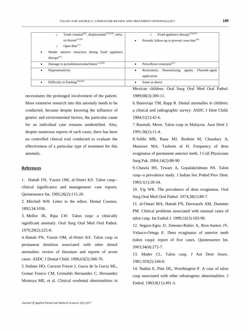
o Tooth rotation[62], displacement[37],[58], infra-
occlusion[1],[18]
o Open Bite[11]
Hinder anterior retraction during fixed appliance
therapy[41]
o Fixed appliance therapy[33],[43]
Periodic follow-up to prevent cross-bite[58]
Damage to periodontium/attachment [1],[44] Perio/Resto treatment[33]
Hypersensitivity Restoration, Desensitizing agents, Fluoride-agent
application
Difficulty in Feeding[36],[38] Same as above
necessitates the prolonged involvement of the patient.
More extensive research into this anomaly needs to be
conducted, because despite knowing the influence of
genetic and environmental factors, the particular cause
for an individual case remains unidentified. Also,
despite numerous reports of such cases, there has been
no controlled clinical trial conducted to evaluate the
effectiveness of a particular type of treatment for this
anomaly.
References
1. Hattab FN, Yassin OM, al-Nimri KS. Talon cusp--
clinical significance and management: case reports.
Quintessence Int. 1995;26(2):115-20.
2. Mitchell WH. Letter to the editor. Dental Cosmos.
1892;34:1036.
3. Mellor JK, Ripa LW. Talon cusp: a clinically
significant anomaly. Oral Surg Oral Med Oral Pathol.
1970;29(2):225-8.
4. Hattab FN, Yassin OM, al-Nimri KS. Talon cusp in
permanent dentition associated with other dental
anomalies: review of literature and reports of seven
cases. ASDC J Dental Child. 1996;63(5):368-76.
5. Sedano HO, Carreon Freyre I, Garza de la Garza ML,
Gomar Franco CM, Grimaldo Hernandez C, Hernandez
Montoya ME, et al. Clinical orodental abnormalities in
Mexican children. Oral Surg Oral Med Oral Pathol.
1989;68(3):300-11.
6. Buenviaje TM, Rapp R. Dental anomalies in children:
a clinical and radiographic survey. ASDC J Dent Child.
1984;51(1):42-6.
7. Rusmah, Meon. Talon cusp in Malaysia. Aust Dent J.
1991;36(1):11-4.
8. Sobhi MB, Rana MJ, Ibrahim M, Chaudary A,
Manzoor MA, Tasleem ul H. Frequency of dens
evaginatus of permanent anterior teeth. J Coll Physicians
Surg Pak. 2004;14(2):88-90.
9. Chawla HS, Tewari A, Gopalakrishnan NS. Talon
cusp--a prevalence study. J Indian Soc Pedod Prev Dent.
1983;1(1):28-34.
10. Yip WK. The prevalence of dens evaginatus. Oral
Surg Oral Med Oral Pathol. 1974;38(1):80-7.
11. al-Omari MA, Hattab FN, Darwazeh AM, Dummer
PM. Clinical problems associated with unusual cases of
talon cusp. Int Endod J. 1999;32(3):183-90.
12. Segura-Egea JJ, Jimenez-Rubio A, Rios-Santos JV,
Velasco-Ortega E. Dens evaginatus of anterior teeth
(talon cusp): report of five cases. Quintessence Int.
2003;34(4):272-7.
13. Mader CL. Talon cusp. J Am Dent Assoc.
1981;103(2):244-6.
14. Natkin E, Pitts DL, Worthington P. A case of talon
cusp associated with other odontogenic abnormalities. J
Endod. 1983;9(11):491-5.
Page 9
TALON CUSP ANOMALY: LITERATURE-REVIEW AND TREATMENT OPTIONS3(2);2017 183
Journal Of Applied Dental and Medical Sciences 3(2);2017
15. Sharma A. Dens evaginatus of anterior teeth (talon
cusp) associated with other odontogenic anomalies. J
Indian Soc Pedod Prev Dent. 2006;24 Suppl 1:S41-3.
16. Jowharji N, Noonan RG, Tylka JA. An unusual case
of dental anomaly: a "facial" talon cusp. ASDC J Dent
Child. 1992;59(2):156-8.
17. Tsutsumi T, Oguchi H. Labial talon cusp in a child
with incontinentia pigmenti achromians: case report.
Pediatr Dent. 1991;13(4):236-7.
18. Hsu Chin-Ying S, Girija V, Fei YJ. Bilateral talon
cusps in primary teeth: clinical significance and
treatment. ASDC J Dent Child. 2001;68(4):239-43, 28.
19. Thesleff I. Epithelial-mesenchymal signalling
regulating tooth morphogenesis. J Cell Sci.
2003;116(9):1647-8.
20. Sarkar S, Misra J, Das G. "Talon cusp-heredity
origin" – a case report. J Indian Soc Pedod Prev Dent.
1999;17(4):126-8.
21. Gardner DG, Girgis SS. Talon cusps: a dental
anomaly in the Rubinstein-Taybi syndrome. Oral Surg
Oral Med Oral Pathol. 1979;47(6):519-21.
22. Goldstein E, Medina JL. Mohr syndrome or oral-
facial-digital II: report of two cases. J Am Dent Assoc.
1974;89(2):377-82.
23. Chen RJ, Chen HS. Talon cusp in primary dentition.
Oral Surg Oral Med Oral Pathol. 1986;62(1):67-72.
24. Hattab FN, Yassin OM, Sasa IS. Oral manifestations
of Ellis-van Creveld syndrome: report of two siblings
with unusual dental anomalies. J Clin Pediatr Dent.
1998;22(2):159-65.
25. Lehl GK. Talon cusp associated with other dental
anomalies – a case report. J Indian Soc Pedod Prev Dent.
1999;17(1):13-4.
26. Hegde S, Jain M, Shubha AB. A Rare Bilateral
Presentation of Multiple Dens Invaginatus, Shovel-
Shaped Incisor and Talon Cusp With Mesiodens.
Kathmandu Univ Med J. 2014;12(48):292-5.
27. Dharmani U, Rajput A, Chaudhary S, Talwar S,
Verma M. Type III talon cusp and Type III B dens
invaginatus occurring simultaneously in a mandibular
lateral incisor. Gen Dent. 2014;62(5):e16-21.
28. Colak H, Yilmaz C, Keklik H, Colak T. Talon cusps
occurring concurrently with dens invaginatus on a
permanent maxillary lateral incisor: a case report and
literature review. Gen Dent. 2014;62(3):e14-8.
29. Tewari T, Ather A, Ather H. Endodontic
involvement of a geminated tooth with talon cusp: a rare
occurrence. J Mass Dent Soc. 2013;62(2):46-7.
30. Lomcali G, Hazar S, Altinbulak H. Talon cusp:
report of five cases. Quintessence Int. 1994;25(6):431-3.
31. Hargreaves KM, Goodis HE, Seltzer S. Seltzer and
Bender's dental pulp: Quintessence Pub. Co.; 2002.
32. Gosselin ML, Doyle T, MacLellan J, Anderson RD,
Dyment H. A talon cusp mistaken for a mesiodens: case
report. J Can Dent Assoc. 2012;78:c6.
33. Hattab FN, Hazza'a AM. An unusual case of talon
cusp on geminated tooth. J Can Dent Assoc.
2001;67(5):263-6.
34. Raut Dessai SS, Barretto ES, Swamy DF. Cone
Beam Computed Tomography: A Comprehensive
Review. Research Review Journal of Medical Health
Sciences. 2014;3(2S):1-10.
35. Esmaeilzadeh M, Donyavi Z, Shokri A. Cone-Beam
Computed Tomography Study Of Crown Dilaceration
With a Talon Cusp in an Unerupted Permanent Maxillary
Tooth. J Craniofac Surg. 2016;27(2):e170-2.
36. Hattab FN, Yassin OM. Bilateral talon cusps on
primary central incisors: a case report. Int J Paediatr
Dent. 1996;6(3):191-5.
Page 10
TALON CUSP ANOMALY: LITERATURE-REVIEW AND TREATMENT OPTIONS3(2);2017 184
Journal Of Applied Dental and Medical Sciences 3(2);2017
37. Bansal AV, Choudhary P, Kulkamrni VK, Bansal A,
Shashikiran ND. Talon cusps: conservative management.
J Clin Pediatr Dent. 2011;35(4):345-8.
38. Gungor HC, Altay N, Kaymaz FF. Pulpal tissue in
bilateral talon cusps of primary central incisors: report of
a case. Oral Surg Oral Med Oral Pathol Oral Radiol
Endod. 2000;89(2):231-5.
39. Kumar Rao P, Ram Shetty S, Prabhu R. V, Veena
KM, Chatra L, Shenai P. Talon cusps in mandibular
incisors: an unusual presentation in a child patient. J
Dent Res Dent Clinics Dent Prospects. 2011;5(1):37-9.
40. Davis PJ, Brook AH. The presentation of talon cusp:
diagnosis, clinical features, associations and possible
aetiology. Br Dent J. 1986;160(3):84-8.
41. Sharma P, Arora A, Valiathan A, Urala A, Acharya
SR. Gradual grinding of a talon cusp during orthodontic
treatment. J Clin Orthod. 2012;46(2):111-4.
42. Bolan M, Gerent Petry Nunes AC, de Carvalho
Rocha MJ, De Luca Canto G. Talon cusp: report of a
case. Quintessence Int. 2006;37(7):509-14.
43. Pitts DL, Hall SH. Talon-cusp management:
orthodontic-endodontic considerations. ASDC J Dent
Child. 1983;50(5):364-8.
44. Fabra-Campos H. Failure of endodontic treatment
due to a palatal gingival groove in a maxillary lateral
incisor with talon cusp and two root canals. J Endod.
1990;16(7):342-5.
45. Shetty P, Xavier AM. Management of a talon cusp
using mineral trioxide aggregate. Int Endod J.
2011;44(11):1061-8.
46. Kumar V, Chawla A, Logani A, Shah N. Mineral
trioxide aggregate pulpotomy: An ideal treatment option
for management of talon cusp. Contem Clin Dent.
2012;3(4):491-3.
47. Pledger DM, Roberts GJ. Talon cusp: report of a
case. Br Dent J. 1989;167(5):171-3.
48. Smail-Faugeron V, Picou Rollin J, Muller Bolla M,
Courson F. Management of non-syndromic dens
evaginatus affecting permanent maxillary central
incisors: a systematic review. BMJ Case Rep.
2016;2016.
49. de Sousa SM, Tavano SM, Bramante CM. Unusual
case of bilateral talon cusp associated with dens
invaginatus. Int Endod J. 1999;32(6):494-8.
50. Oehlers FA, Lee KW, Lee EC. Dens evaginatus
(evaginated odontome). Its structure and responses to
external stimuli. Dent Pract Dent Rec. 1967;17(7):239-
44.
51. Myers CL. Treatment of a talon-cusp incisor: report
of case. ASDC J Dent Child. 1980;47(2):119-21.
52. Levitan ME, Himel VT. Dens evaginatus: literature
review, pathophysiology, and comprehensive treatment
regimen. J Endod. 2006;32(1):1-9.
53. Asadi SG, Asadi ZG. Presentation and management
of talon cusp. J Pak Med Assoc. 1998;48(10):314-5.
54. Andreasen JO, Munksgaard EC, Bakland LK.
Comparison of fracture resistance in root canals of
immature sheep teeth after filling with calcium
hydroxide or MTA. Dent Traumatol. 2006;22(3):154-6.
55. Bakland LK, Andreasen JO. Will mineral trioxide
aggregate replace calcium hydroxide in treating pulpal
and periodontal healing complications subsequent to
dental trauma – A review. Dent Traumatol.
2012;28(1):25-32.
56. Tziafas D, Pantelidou O, Alvanou A, Belibasakis G,
Papadimitriou S. The dentinogenic effect of mineral
trioxide aggregate (MTA) in short-term capping
experiments. Int Endod J. 2002;35(3):245-54.
57. Koh ET, Ford TR, Kariyawasam SP, Chen NN,
Torabinejad M. Prophylactic treatment of dens
evaginatus using mineral trioxide aggregate. J Endod.
2001;27(8):540-2.
Page 11
TALON CUSP ANOMALY: LITERATURE-REVIEW AND TREATMENT OPTIONS3(2);2017 185
Journal Of Applied Dental and Medical Sciences 3(2);2017
58. Liu JF, Chen LR. Talon cusp affecting the primary
maxillary central incisors in two sets of female twins:
report of two cases. Pediatr Dent. 1995;17(5):362-4.
59. Soares AB, de Araujo JJ, de Sousa SM, Veronezi
MC. Bilateral talon cusp: case report. Quintessence Int.
2001;32(4):283-6.
60. Meon R. Talon cusp in primary dentition--case
report. Singapore Dent J. 1990;15(1):32-4.
61. Sarkar S, Misra J. Talon cusp in the deciduous
dentition: a case report. J Indian Soc Pedod Prev Dent.
2000;18(4):151-2.
62. Rayen R, Muthu MS, Sivakumar N. Aberrant talon
cusps: report of two cases. J Indian Soc Pedod Prev
Dent. 2006;24 Suppl 1:S7-10.
63. Mull JP, Nabeel S, Hegde U, Danish G. Multiple
Bilateral Talon Cusps: A Report Of Three Cases. Indian
J Dent Sci. 2012;4(2).