Psychological Review 2005, 112(1) XXX-XXX Final accepted version See published version for page numbers, exact quotes The Temporal Context Model in spatial navigation and relational learning: Toward a common explanation of medial temporal lobe function across domains Marc W. Howard, Mrigankka S. Fotedar, Aditya V. Datey Department of Psychology Syracuse University Michael E. Hasselmo Department of Psychology, Program in Neuroscience, and Center for Memory and the Brain Boston University Accepted March 23, 2004 The medial temporal lobe (MTL) has been studied extensively at all levels of analysis, yet its function remains unclear. Theory regarding the cognitive function of the MTL has centered along 3 themes. Different authors have emphasized the role of the MTL in episodic recall, spa- tial navigation, or relational memory. Starting with the temporal context model (M. W. Howard and M. J. Kahana, 2002), a distributed memory model that has been applied to benchmark data from episodic recall tasks, the authors propose that the entorhinal cortex supports a gradually changing representation of temporal context and the hippocampus proper enables retrieval of these contextual states. Simulation studies show this hypothesis explains the firing of place cells in the entorhinal cortex and the behavioral effects of hippocampal lesion in relational memory tasks. These results constitute a first step towards a unified computational theory of MTL function that integrates neurophysiological, neuropsychological and cognitive findings. The medial temporal lobe (MTL) is a region that includes the hippocampus proper, the subicular complex and parahip- pocampal cortical regions, including entorhinal, perirhinal, and parahippocampal/postrhinal cortices. A great deal of data from neuropsychology (e.g. Eichenbaum & Cohen, 2001; Scoville & Milner, 1957; Squire, 1992) and functional imaging (e.g. Fernandez, Effern, Grunwald, et al., 1999; Stern, Corkin, Gonzalez, et al., 1996; Wagner et al., 1998) has converged on the idea that the MTL is important in learn- ing and memory. In order to bridge the gap between cogni- tion and cellular-level physiology, we need a mechanistic, mesoscopic description of MTL computational function. We already have several successful verbally-formulated theories Michael Kahana developed the first versions of TCM and has collaborated closely on its development as a model of episodic re- call. He also made numerous helpful comments on an earlier draft of the ms. Thanks are due to Loren Frank for generously shar- ing and patiently explaining positional and derived data from the W-maze, and to Neil Burgess and Colin Lever for sharing posi- tional data from the open field. The early development of the ideas about the place code benefited from discussions with Larry Abbott and John Lisman. Supported by Conte Center grant NIMH P50 MH60450 (principal investigator Joseph Coyle), R01 MH61492 and MH60013 to MEH, 2-R01MH55687 (principal investigator Michael Kahana), F32 MH65841 to MWH, and the College of Arts and Sciences of Syracuse University. Correspondence con- cerning this article should be addressed to [email protected]. Marc Howard, Syracuse University, Department of Psychology, 430 Huntington Hall, Syracuse, NY 13244-2340. of the cognitive function of the MTL. These are described in turn in the following subsections. This paper will attempt to draw these multiple verbal theories together into a single computational framework that is consistent with known neu- rophysiological and neuroanatomical data. Episodic recall All of the diverse skills and facts that differentiate an adult from an infant must be some form of memory—we say that one learns to ride a bike, or remembers the alphabet. In the early part of this century, memory theory strove to describe general laws that would presumably apply to all these dif- ferent types of learning (e.g. Estes, 1950; Osgood, 1949). Recent decades have seen this unitary approach to memory fragment into the categorization of multiple types of mem- ory, typically with separable neural substrates for each (e.g. Eichenbaum & Cohen, 2001; Nadel & Moscovitch, 1997; Tulving & Schacter, 1991). One of the most fruitful of these distinctions has been that of episodic memory. Episodic memory refers to the ability to remember spe- cific events from one’s personal experience (Tulving, 1983, 2002). For instance, one might have an episodic memory of having eaten a banana at breakfast. The memory for this episode, perhaps with details about the other objects and peo- ple present at breakfast, with the taste of the banana, the sounds and smells that were present in the room, is in prin- ciple quite distinct from other types of memory one might have for bananas. For instance, one could remember many things about bananas—that they are yellow, that they are good to eat, that people like to eat them at breakfast—without memory for any specific experience with a banana. Recent 1
Transcript
Psychological Review2005, 112(1) XXX-XXX
Final accepted versionSee published version for page numbers, exact quotes
The Temporal Context Model in spatial navigation and relationallearning: Toward a common explanation of medial temporal lobe
function across domains
Marc W. Howard, Mrigankka S. Fotedar,Aditya V. Datey
Department of PsychologySyracuse University
Michael E. HasselmoDepartment of Psychology, Program in Neuroscience, and
Center for Memory and the BrainBoston University
Accepted March 23, 2004
The medial temporal lobe (MTL) has been studied extensively at all levels of analysis, yet itsfunction remains unclear. Theory regarding the cognitive function of the MTL has centeredalong 3 themes. Different authors have emphasized the role of the MTL in episodic recall, spa-tial navigation, or relational memory. Starting with the temporal context model (M. W. Howardand M. J. Kahana, 2002), a distributed memory model that has been applied to benchmark datafrom episodic recall tasks, the authors propose that the entorhinal cortex supports a graduallychanging representation of temporal context and the hippocampus proper enables retrieval ofthese contextual states. Simulation studies show this hypothesis explains the firing of placecells in the entorhinal cortex and the behavioral effects of hippocampal lesion in relationalmemory tasks. These results constitute a first step towards a unified computational theory ofMTL function that integrates neurophysiological, neuropsychological and cognitive findings.
The medial temporal lobe (MTL) is a region that includesthe hippocampus proper, the subicular complex and parahip-pocampal cortical regions, including entorhinal, perirhinal,and parahippocampal/postrhinal cortices. A great deal ofdata from neuropsychology (e.g. Eichenbaum & Cohen,2001; Scoville & Milner, 1957; Squire, 1992) and functionalimaging (e.g. Fernandez, Effern, Grunwald, et al., 1999;Stern, Corkin, Gonzalez, et al., 1996; Wagner et al., 1998)has converged on the idea that the MTL is important in learn-ing and memory. In order to bridge the gap between cogni-tion and cellular-level physiology, we need a mechanistic,mesoscopic description of MTL computational function. Wealready have several successful verbally-formulated theories
Michael Kahana developed the first versions of TCM and hascollaborated closely on its development as a model of episodic re-call. He also made numerous helpful comments on an earlier draftof the ms. Thanks are due to Loren Frank for generously shar-ing and patiently explaining positional and derived data from theW-maze, and to Neil Burgess and Colin Lever for sharing posi-tional data from the open field. The early development of the ideasabout the place code benefited from discussions with Larry Abbottand John Lisman. Supported by Conte Center grant NIMH P50MH60450 (principal investigator Joseph Coyle), R01 MH61492and MH60013 to MEH, 2-R01MH55687 (principal investigatorMichael Kahana), F32 MH65841 to MWH, and the College ofArts and Sciences of Syracuse University. Correspondence con-cerning this article should be addressed to [email protected] Howard, Syracuse University, Department of Psychology, 430Huntington Hall, Syracuse, NY 13244-2340.
of the cognitive function of the MTL. These are describedin turn in the following subsections. This paper will attemptto draw these multiple verbal theories together into a singlecomputational framework that is consistent with known neu-rophysiological and neuroanatomical data.
Episodic recall
All of the diverse skills and facts that differentiate an adultfrom an infant must be some form of memory—we say thatone learns to ride a bike, or remembers the alphabet. In theearly part of this century, memory theory strove to describegeneral laws that would presumably apply to all these dif-ferent types of learning (e.g. Estes, 1950; Osgood, 1949).Recent decades have seen this unitary approach to memoryfragment into the categorization of multiple types of mem-ory, typically with separable neural substrates for each (e.g.Eichenbaum & Cohen, 2001; Nadel & Moscovitch, 1997;Tulving & Schacter, 1991). One of the most fruitful of thesedistinctions has been that of episodic memory.
Episodic memory refers to the ability to remember spe-cific events from one’s personal experience (Tulving, 1983,2002). For instance, one might have an episodic memoryof having eaten a banana at breakfast. The memory for thisepisode, perhaps with details about the other objects and peo-ple present at breakfast, with the taste of the banana, thesounds and smells that were present in the room, is in prin-ciple quite distinct from other types of memory one mighthave for bananas. For instance, one could remember manythings about bananas—that they are yellow, that they aregood to eat, that people like to eat them at breakfast—withoutmemory for any specific experience with a banana. Recent
1
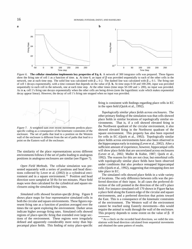
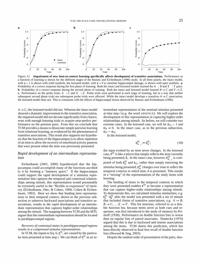
2 HOWARD, FOTEDAR, DATEY, AND HASSELMO
work has argued that episodic memory relies on the MTL,in particular the hippocampus (Nadel & Moscovitch, 1997;O’Keefe & Nadel, 1978; Tulving & Markowitsch, 1998).
A number of behavioral tasks test episodic memory. Forexample, in the free recall task, the subject is presented witha list of stimuli, typically words. The task is to recall asmany words as possible from the list, with the subject freeto determine the order of recall. Free recall is an episodictask in that performance requires that the subject recall thewords presented in a particular episodic setting. Free recallis sufficiently sensitive to MTL damage that it can be used asa diagnostic tool for MTL damage in clinical settings (Graf,Squire, & Mandler, 1984).
Spatial navigation
O’Keefe and Nadel (1978) proposed that the primaryfunction of the hippocampus is to construct and read out“cognitive maps.” In the following years, however, this theo-retical approach has focused on the role of the hippocampusand related structures in learning and navigating through spa-tial environments. The most remarkable piece of evidencesupporting this view is the existence of place cells (O’Keefe& Dostrovsky, 1971). Pyramidal cells within the hippocam-pus, recorded from rats moving throughout an environment,fire selectively when the animal is in one particular regionof the environment. In open environments, this doesn’t de-pend on the direction the animal is facing (Muller, Bostock,Taube, & Kubie, 1994), and firing persists in the dark (Quirk,Muller, & Kubie, 1990), ruling out an explanation based onsimple visual stimuli correlated with place.
There is an extensive literature describing characteristicsof place cells in dorsal CA1 (e.g. Muller & Kubie, 1987;O’Keefe & Burgess, 1996; O’Keefe & Dostrovsky, 1971;Wilson & McNaughton, 1993). Less is known about theplace code in other MTL structures. It is known that thereare place cells in the entorhinal cortex (EC, Barnes, Mc-Naughton, Mizumori, Leonard, & Lin, 1990; Frank, Brown,& Wilson, 2000; Quirk, Muller, Kubie, & Ranck, 1992), a re-gion of cortex that provides input to the hippocampus proper.The place response in EC differs in some respects from theplace code observed in CA1, indicating that the hippocam-pus performs significant computations on the incoming placerepresentation. Nonetheless, it is clear that we can’t have ameaningful understanding of the function of the hippocam-pus performs until we have a correct understanding of thenature of the entorhinal place code.
Transitive associations and relational memory
Data from olfactory learning in the rat (Bunsey & Eichen-baum, 1996; Dusek & Eichenbaum, 1997) has been usedto argue that the hippocampus, the central structure of theMTL, enables transitive associations, a function believed tobe important in relational memory. In these experiments, ratslearned associations or relationships between arbitrary stim-uli. For instance, in the study of Bunsey and Eichenbaum(1996), rats with hippocampal lesions were able to learn as-sociations between odors A and B, and between B and C.
Unlike normal rats, however, lesioned rats did not show atransitive generalization for the association A � C. Althoughthe lesioned animals were able to learn simple associationsbetween the stimuli, Bunsey and Eichenbaum (1996) arguedthat they did not learn the relationships among stimuli thatweren’t presented together (see also Dusek & Eichenbaum,1997).
The mnemonic deficit exhibited by hippocampal-lesionedanimals cannot apparently be described as a deficit in the de-velopment of simple stimulus-response associations. How-ever, when complex relationships between stimuli must belearned, the MTL, and the hippocampus in particular, ap-pear to be critically involved. This emphasis on relationalmemory is not at all contradictory to a role for the MTL inepisodic memory. After all, memory for an episode involvesdrawing together the many different stimuli present withinthe episode, in a unique configuration.
Toward a unified framework
These three theoretical approaches to MTL function,episodic recall, spatial navigation and relational memory, arenot mutually contradictory. As mentioned previously, mem-ory for an episode should include memory for the configu-ration of stimuli present in that episode. Similarly, O’Keefeand Nadel (1978) pointed out that a cognitive map could beused to encode the relationships between non-spatial sets ofstimuli, resulting in binding items to a temporal-spatial con-text, supporting episodic memory (ch 14 O’Keefe & Nadel,1978). Because the neurobiology of the MTL is such an in-tensely studied subject, there is a tremendous incentive toconstruct a model that can address questions from all threedomains.
The goal of the present paper is to present the begin-nings of a theoretical framework that begins to draw togetherthese three disparate approaches. This will be accomplishedwithin the structure provided by the Temporal Context Model(TCM, Howard & Kahana, 2002a), developed to explain ex-perimental findings from free recall, an episodic recall task.TCM describes a set of rules that govern the behavior of adistributed representation of temporal context. We will showthat the equation governing contextual drift, taken as a modelof temporal-spatial context, can explain the primary featuresof the entorhinal place code, a phenomenon central to theMTL’s support for spatial function. We will then demon-strate that the equation governing retrieved temporal con-text, a kind of plasticity postulated to explain properties ofepisodic association, can support a more general function inextracting the temporal structure of experience. This pro-vides a framework for modeling the dissociation between re-lational learning and simple pairwise association.
Recency and Contiguity: TCMand Fundamental Properties of
Episodic Recall
TCM was developed to describe two fundamental proper-ties of episodic memory. The recency effect (Bjork & Whit-ten, 1974; Howard & Kahana, 1999; Murdock, 1963b; Rat-
TCM, THE PLACE CODE AND RELATIONAL MEMORY 3
cliff & Murdock, 1976) is the tendency for more recent itemsto be recalled better than less recent items. Associative ef-fects (Howard & Kahana, 1999, 2002b; Kahana, 1996) de-scribe the development of episodically-formed connectionsbetween items. This section will first review prior workon TCM, describing the structure and reasoning behind themodel. Following this, we will describe a linking hypothesisbetween TCM and the brain, with a special emphasis on themedial temporal lobe.
An episodic representation
Context, in one form or another, has long been an impor-tant component of models of episodic memory performance(e.g. Anderson & Bower, 1972; Raaijmakers & Shiffrin,1980; Mensink & Raaijmakers, 1988; Yntema & Trask,1963). The basic approach of TCM has been to take a partic-ular formulation of context, referred to as temporal contextand use it as the sole cue for recall of item representations.Because context changes gradually over time TCM can pre-dict forgetting over long time scales. Unlike some prior for-mulations (e.g. Mensink & Raaijmakers, 1988), however,TCM also explicitly models context that changes graduallywithin a list of items. This assumption enables a descriptionof recency effects within lists, an effect which has often beenattributed to short-term memory (e.g. Atkinson & Shiffrin,1968; Raaijmakers & Shiffrin, 1980). The most radical pointof departure of TCM from prior models of episodic recall,however, is the assumption that context serves as the solecue for episodic recall. In TCM, observed episodic associa-tions between items are a consequence of effects items haveon context, eliminating the need for direct item-to-item asso-ciations in describing episodically-formed associations. Wewill describe TCM in more detail in the following subsec-tions. This treatment reviews prior work (Howard & Kahana,2002a; Howard, Wingfield, & Kahana, In revision; Howard,2004). Readers already familiar with TCM as a model ofepisodic recall may wish to advance to the subsection enti-tled “A mapping between TCM and the MTL.”
Temporal context and items. The central assumption ofTCM is that there is a distinction between temporal contextand to-be-recalled items. The current state of temporal con-text at time step i is referred to as ti. We assume that ti isa vector in a high-dimensional space; typically an infinite-dimensional space for simplicity. The item presented at timestep i is referred to as fi. We assume that the item repre-sentations f are vectors in a separate high-dimensional space,typically infinite for simplicity. We assume that item repre-sentations do not change over the course of a typical recallexperiment and that they are orthonormal. That is, we as-sume that there is no overlap between item representationsand that the length of each item vector is one.
Activation of an item representation corresponds to per-ception. The current state of the item vector corresponds tothe item currently being experienced. For instance, an itemrepresentation may be activated on the basis of external stim-uli during presentation of a list of items. Similarly, an item
representation may be activated by means of an “internalstimulus” during the recall process. No matter the source,the consequence of activating an item representation is theperception of the corresponding item. Howard and Kahana(2002a) assumed that only one item representation could beactivated at any one time. Although not a fundamental as-sumption of TCM, we will also assume that at most one itemrepresentation is active at a time throughout the current ms.
Context cues items. In TCM, the current state of context,ti, is used to cue recall of items in semantic memory. Eachitem in semantic memory is activated by a state of contextto the extent that that state of context resembles the contextsin which it was presented. This can be implemented using aHebbian outer product matrix connecting states of context twith patterns in semantic memory, fi
MT Fi � MT F
i � 1�
fit �i � (1)
where the prime denotes the transpose. When MT F is multi-plied from the right with a context vector, t, this results in asuperposition of patterns in semantic memory, each weightedby their similarity between their context and the cue context.That is
MT Ft j � ∑i
�fi � t �it j �� � (2)
which follows immediately from the definition of MT F
(Eq. 1) and basic properties of vector arithmetic. The keyhere is the t �it j term. The transpose of a vector multiplied byanother vector is a scalar referred to as the inner product. Forthe present purposes, this is the same as the dot product andcan also be written ti t j.1 We can see that when the itemlayer is cued by a state of context, the result is a combinationof item representations. A particular item enters this combi-nation in a way that is proportional to the similarity (quanti-fied by the dot product) of the contexts it has been presentedin to the cue state of context.
Mapping activations onto probability of recall. Howardand Kahana (2002a) assumed that this combination of itemrepresentations was unstable. Due to attractor dynamics, thesuperposition of item representations that results from cuingwith a state of context would collapse into one particular itemrepresentation (or perhaps a null state in which all elementsof the vector went to zero). Let us define the activation of aparticular item i by a particular state of context t as
ai : � f �iMT F t � (3)
1 For the present purposes, we can define the inner, or dot productas
v � w ∑i
�v � i � w � i �
where the� � � i operator refers to the ith element of the vector taken
as its argument. The dot product is positive if the two vectors pointin similar directions (if they are correlated). It is negative if theypoint in opposite direction. Importantly, the dot product is zero ifthe two vectors are orthogonal.
4 HOWARD, FOTEDAR, DATEY, AND HASSELMO
Using this definition (and the assumption that the item repre-sentations are orthonormal), then the scalar ai just measuresthe extent to which the superposition points in the directionof the word corresponding to fi. The probability of recallingitem i given t can be given by the Luce choice rule:
P � fi � t � � exp � 2aiτ �
∑ j exp � 2a jτ � � (4)
This can be conceived of as the probability of the superpo-sition collapsing to a particular state. Howard and Kahana(2002a) took the sum in the denominator of Eq. 4 to be overpotential recalls in the list. This equation is not a fundamen-tal part of TCM. The important properties of this equationare simply that it provides a non-linear mapping between ac-tivations and recall, and that it is a competitive recall rule.That is, the probability of recalling item i depends not onlyon the activation ai, but also the activation of the other itemsa j. This makes it a useful equation for describing situationsin which we are interested in the relative probability of re-calling an item.
Items retrieve context. In much the same way that tempo-ral context can be used to provide an input to the item-space,items provide the input to the context-space. Howard andKahana (2002a) proposed that a matrix MFT provides a con-nection such that the input to the context layer at time step i,tINi , is a consequence of the item presented at time step i:
tINi � MFT
i � 1fi � (5)
The vector tINi will sometimes be referred to as the “context
retrieved by item i” to emphasize the effect of item represen-tations on contextual states. The form of MFT was derived insuch a way to implement a functional rule that will be intro-duced later (Eq. 9 below). The form of MFT is rather compli-cated and probably does not correspond simply to any singlestructure in the brain. For this reason we will not discuss itfurther here, but rather treat the functional rule as the basicdescription of retrieved context for the present ms. However,we strongly assert the central point of retrieved context thatitems cause contextual input patterns.
Recency across time scales
At each time step, the state of context at time step i, tiis formed from the prior state of context ti � 1 and an inputvector tIN
i according to:
ti � ρiti � 1� βtIN
i � ρi : ��� ti ��� � 1 (6)
When applied to list-learning applications, we have previ-ously assumed that the time-steps correspond to the times atwhich list items are presented. We will assume (for conve-nience) that the input vectors, tIN
i , are always of unit length( ��� tIN
i ��� � 1 � for all i). The vector tINi is weighted by the scalar
β. This parameter is generally estimated from the data and isconstrained such that 0 � β � 1. We can see that Eq. 6 adds
input vectors to the state of t. To ensure that the length of tidoes not grow without bound, we assume that the scalar ρi ischosen to ensure that the length of ti remains unity: ��� ti ��� � 1.This constraint means that ti changes as a function of inputto the system, rather than the passage of time per se (Waugh& Norman, 1965). This can be seen clearly if one assumesthat at some time step i, the input vector is empty, ��� tIN
i ��� � 0.In this case
ti � ρiti � 1� β0 (7)
requires that ρi � 1. This is consistent with the findings ofBaddeley and Hitch (1977), who argued that the recency ef-fect was unaffected by addition of an unfilled delay at the endof the list.
If the system is presented with an infinitely long seriesof orthonormal tIN ’s, then the value of ρi will stabilize atρ : �� 1 β2.2 Under these circumstances, it becomes pos-sible to concisely describe the similarity relationships be-tween ti and the state of context at some other time, j, t j:
ti t j � ρ � i � j � � (8)
From this it is clear that t changes gradually over time. Anyparticular component of ti decays exponentially as long asorthonormal inputs are presented.
In sum, contextual evolution in TCM is characterized byseveral important properties:
1. For a given value of β, when given a series of orthogo-nal inputs, the similarity of the current state to an initial statedecays exponentially.
2. The change in context depends on the input.3. When no input is given, the state of context does not
change.Because ti is the functional cue for recall, and ti is an ef-fective cue for recall of item j to the extent that ti over-laps with t j, the property that ti decays gradually naturallyprovides a basis for the the principle of recency (Howard& Kahana, 2002a), which is observed in all of the majorepisodic memory paradigms (Howard & Kahana, 1999; Mur-dock, 1962, 1963b; Neath, 1993; Ratcliff & Murdock, 1976).Appendix A illustrates this principle with a worked examplethat demonstrates the recency effect.
Long-term Recency and Buffer Models of Short-termMemory. For many years, the conventional wisdom was thatthe recency effect in free recall reflected the operation of a
2 Under these circumstance, tINi � ti � 1 0 and �
ti � 2 ρ2
i � ti � 1
� 2 � β2 � tINi � 2 �
Because � tINi
� 1 by assumption and � ti � 1
� 1 because of thecondition on ρi � 1, we find that the condition that
� ti � 1 implies
that1 ρ2
i� β2 �
which implies that ρi �� 1 � β2 . More generally, when tINi � ti � 1 �
0, a quadratic equation in ρi is obtained, which can be solved byelementary methods.
TCM, THE PLACE CODE AND RELATIONAL MEMORY 5
short-term memory buffer (Atkinson & Shiffrin, 1968; Raai-jmakers & Shiffrin, 1980). Indeed, detailed search modelsbased on a short-term memory buffer can describe standardfree recall in considerable detail (Kahana, 1996; Raaijmak-ers & Shiffrin, 1980, 1981; Sirotin, Kimball, & Kahana,submitted). The recency effect in immediate free recall iseliminated by a distractor at the end of the list (Glanzer &Cunitz, 1966; Postman & Phillips, 1965), presumably be-cause the distractor removes items from the end of the listfrom STS. However, when a distractor is also presented be-tween each list item, this results in an increased recency ef-fect over delayed free recall (Bjork & Whitten, 1974; Glen-berg et al., 1980; Glenberg, Bradley, Kraus, & Renzaglia,1983; Howard & Kahana, 1999; Nairne, Neath, Serra, &Byun, 1997; Thapar & Greene, 1993; Watkins, Neath, &Sechler, 1989). This presentation schedule is referred to ascontinuous-distractor free recall; the recency effect observedin continuous-distractor free recall is referred to as the long-term recency effect. Howard and Kahana (2002a) fit TCMto the probability of first recall, a sensitive measure of therecency effect (Howard & Kahana, 1999; Laming, 1999), todata from immediate, delayed and continuous-distractor freerecall (see Figure 1).3 TCM accurately predicts the existenceof a recency effect in immediate free recall, the disruption ofrecency in delayed free recall and the recovery of recency incontinuous-distractor free recall.
Although contextual drift in TCM can account for muchof the function of STS in free recall, there is of course muchmore to the concept of short-term memory than a rehearsalbuffer. Atkinson and Shiffrin (1968) emphasized the im-portance of control processes in strategically manipulatingthe information in the buffer. This theme has persisted notonly in the emphasis of the working memory framework in-troduced by Baddeley and Hitch (1974) on executive func-tion, but also in more recent models of executive functioningin prefrontal cortex (e.g Rougier & O’Reilly, 2002; Braveret al., 2001, for an integrative review, see Miller & Cohen,2001). Although we argue that ti captures the critical storageprocesses of short-term memory essential for generation ofthe recency effect, we make no claim whatever that it de-scribes control processes or executive function—these func-tions clearly require something external to TCM.
Retrieved temporal context and episodic associa-tion
In free recall, the canonical episodic memory task, sub-jects recall multiple words from a list without concern toword order. A great deal of evidence indicates that the orderin which the items are recalled reflects the associative struc-ture of memory. For instance, when a list of words from dif-ferent natural categories is presented, words from the samecategory will tend to be recalled together, even if presenta-tion order is randomized (e.g. Bousfeld, 1953; Pollio, Kass-chau, & DeNise, 1968). This tendency for adjacent recallsto come from the same category can be taken as a measureof stronger associations between words from the same cate-gory than between words from different semantic categories.
In this case, output order in free recall presumably revealssomething about the structure of semantic memory. In ad-dition to semantic, or structural sources of association, asso-ciations can also be formed rapidly among items presentedin temporal proximity. If free recall is indeed a consequenceof an episodic representation, then temporally-defined out-put order relationships should reveal the properties of thisepisodic representation.
We can define the association between two items func-tionally as the tendency of one item to cause productionof the other. To measure associations in episodic memoryKahana (1996) developed conditional response probability(CRP) curves. CRP curves measure the probability of mak-ing a transition from one item to another in free recall as afunction of the distance between them in the list. CRPs havenow been computed for data collected under a wide variety ofsituations (Howard & Kahana, 1999; Kahana, 1996; Kahana& Caplan, 2002; Kahana, Howard, Zaromb, & Wingfield,2002; Klein, Addis, & Kahana, In press; Ward, Woodward,Stevens, & Stinson, 2003). Consideration of these data con-firm two very general properties of episodically-formed as-sociations among items in a series:
1. Contiguity. Stronger associations are formed betweenstimuli that occur near each other in time than between thosethat are separated by a greater interval.
2. Asymmetry. Forward associations are stronger thanbackward associations.Both of these properties have been observed in immediate(Howard & Kahana, 1999; Kahana, 1996; Ward et al., 2003),delayed (Howard & Kahana, 1999; Kahana et al., 2002) andcontinuous-distractor free recall (Howard & Kahana, 1999),as well as serial recall (Kahana & Caplan, 2002; Raskin &Cook, 1937).
Because the current state of context is always the cue forepisodic recall, associative effects in TCM are mediated bythe effects items have on the state of context. This is possi-ble because a central postulate of TCM is that the input toEq. 6 is caused by the presentation of items.4 In TCM itemscause an input, tIN
i , that is part of ti. Because t is the cue forepisodic recall, associative effects between items are medi-ated by the effect they have on t—by the contextual inputsthose items evoke, and the similarity of those inputs to statesof t in which other items were encoded. TCM produces con-tiguity effects because items retrieve contextual elements thatwere present when the items were initially presented. Be-cause context changes gradually (Eq. 6), these contextual el-
3 Details of the procedure can be found in Howard and Kahana(2002a).
4 Although this might not seem such a radical assumption, sev-eral memory models have included mechanisms of contextual driftin which the change in contextual elements is assumed to be a con-sequence of stochastic fluctuations that are not under experimen-tal control (Estes, 1955; Mensink & Raaijmakers, 1988; Murdock,Smith, & Bai, 2001). Another set of models developed to explainperformance in short-term serial recall tasks have explicitly decou-pled contextual representations from item recall, while not neces-sarily assuming that context fluctuates randomly (Brown, Preece, &Hulme, 2000; Burgess & Hitch, 1992, 1999; Henson, 1998).
6 HOWARD, FOTEDAR, DATEY, AND HASSELMO
1 2 3 4 5 6 7 8 9 10 11 12
0
.1
.2
.3
.4
.5
.6
Pro
babi
lity
of F
irst R
ecal
l ExperimentalPredicted
Immediate
1 2 3 4 5 6 7 8 9 10 11 12Serial Position
Delayed
1 2 3 4 5 6 7 8 9 10 11 12
Continuous Distractor
Figure 1. TCM describes the recency effect in immediate, delayed and continuous-distractor free recall. Experimental and predictedvalues of the probability of first recall, a sensitive measure of the recency effect across delay schedules. a. In immediate free recall, the recalltest follows immediately after the presentation of the last item. b. In the delayed condition, sixteen seconds of a distractor task intervenedbetween presentation of the last list item and the recall test. Accordingly, the recency effect, the advantage for recall of the last items in thelist, was greatly reduced. c. In continuous-distractor free recall, sixteen seconds of distractor intervened between the last item of the list andthe recall test, but also in between each item of the list, effectively “stretching out” the list while preserving the relative temporal spacingof the list. Under these circumstances, the recency effect was much larger than that observed in delayed recall. Because information thatenters ti decays gradually, TCM, when coupled with a competitive retrieval rule, can describe the persistence of the recency effect acrosstime scales. Model results are from Howard and Kahana (2002a). The experimental data is taken from Howard and Kahana (1999).
ements will tend to overlap with “nearby” states of context.Because a state of context cues a given item for recall to theextent that it overlaps with the context(s) in which the itemwas presented (Eqs. 1, 3), these retrieved contextual elementswill favor recall of nearby items. TCM predicts asymmetrybecause of the detailed assumptions about the nature of theseretrieved contextual elements.
Two components of contextual retrieval. Because re-trieved context provides the basis for associations betweenitems, the form of MFT is clearly very important. Howardand Kahana (2002a) hypothesized that retrieved contextshould be a combination of prior contextual states and thecontext initially retrieved by an item. Let us refer to the ithtime step at which stimulus A is presented as Ai. The inputcaused by stimulus A changes from presentation to presenta-tion according to
tINAi � 1 � αOtIN
Ai
� αNtAi (9)
where αO determines the level of retrieval of old contextualassociations and αN determines the level of new item-to-context associations.5 This is a critical further assumptionbeyond Eq. 5 that allowed the specification of a learning rulefor MFT (Howard & Kahana, 2002a).6 The values of αOand αN are calculated on each learning trial such that thelength of the retrieved context vector on subsequent presen-tations of A will be one (see Appendix B for details). Howardand Kahana (2002a) derived a learning rule for MFT to allowthe model to simultaneously satisfy Eqs. 5 and 9. The ma-trix MFT probably does not correspond simply to a singlebrain structure, so here we will simply take the functionaldescription of contextual retrieval, Eq. 9, as the basic levelof description for changes in contextual retrieval. Equation 9states that when item A, initially presented at time Ai is re-peated later on at time Ai � 1, the input to Eq. 6, tIN
Ai � 1will be a
combination of two components:
1. The input from the original presentation, tINAi
, weightedby αO.
2. The context, tAi , that was present when the item wasinitially presented, weighted by αN .The ratio of these two components is controlled by a freeparameter γ : � αN
�αO. These two components give rise to
qualitatively different associative effects.
Two components describe episodic association. TCM de-scribes asymmetric associations between stimuli in episodicrecall (Howard & Kahana, 1999; Kahana, 1996; Kahana &Caplan, 2002) as a consequence of the combined effects ofthe two components of Eq. 9. One component, tIN
Ai, is the
same input pattern that was evoked by A when it was ini-tially presented. Because tIN
Aidoes not contribute to contex-
tual states that preceded Ai, but does contribute to subsequentstates of context (see Eq. 6), tIN
Aiprovides an asymmetric cue
that favors forward recalls. The other retrieved context com-ponent, tAi , is the context that was present when A was pre-sented previously. Because each state of context in a list ofnon-repeated items is as similar to its predecessor as it is tothe states that follow, tAi provides a symmetric retrieval cuethat favors nearby list items in both the forward and back-ward directions (see Eq. 8). In concert, these two retrievalcues provide an asymmetric retrieval cue that favors recall of
5 The notation used here is slightly different from that used inHoward and Kahana (2002a). There αO was referred to as Ai andαN was referred to as Bi. The notation used here is consistent withthat used in Howard et al. (In revision).
6 In treating the effect of normal aging on episodic association,Howard et al. (In revision) introduced a third component, a noisevector weighted by a parameter η to Eq. 9. The function of thisterm was to provide an ineffectual retrieval cue that could trade offwith the other two components to model the age-related deficit inassociative processes. The interested reader should be aware thatthe version of Eq. 9 used here is not the most general case.
Figure 2. TCM provides a natural explanation of asymmetric association in free recall. a. In TCM there are two sources of associativeeffects. One source relies on the ability to retrieve contextual elements consistently from presentation to presentation of an item. The cuestrength derived from these “old” item-to-context associations provides an asymmetric cue that only helps recall items forward in the list.The other source is the ability of an item to retrieve contextual elements that were already present when the item is presented. The cuestrength derived from these “new” item-to-context associations provides a symmetric cue that helps both forward and backward recalls. Thecombination of these two cues leads to the characteristic shape of the CRP. After Howard and Kahana (2002a). b. The combination of anasymmetric retrieval cue and a symmetric retrieval cue is an asymmetric retrieval cue. This results in good quantitative fits to observed CRPcurves. The left panel shows data from a delayed free recall study of younger adults along with predicted data from TCM. The right panelshows analogous curves from older adults. The decrease in associative tendencies for older adults was modeled as a result of including anoise term in Eq. 9. This data was originally presented in Kahana, et al (2002). The modeling of the older adults’ data is explained in greaterdetail in Howard et al (in revision).
nearby items.
Figure 2a shows a plot of the cue strength from the twocomponents of context retrieved by an item at the center ofthe curve to its neighbors. The curve labeled “Old” showsthe cue strength of the old context tIN
Aito the neighbors of
A. The cue strength is large for items that immediately fol-lowed A, and falls off with temporal distance. The old cuestrength is zero for items that preceded A. This, combinedwith the non-zero cue strength to items that followed A leadsto an associative asymmetry. The curve labeled “New” inFigure 2a shows the cue strength from the new context com-ponent tAi . This component provides a cue strength that con-tributes to both forward and backward recalls. Combiningthese two components in an appropriate ratio shows a strongcorrespondence to the shape of observed CRPs, a measure oftemporally-defined associations observed in free recall (seeFigure 2b). Appendix C shows a worked example of a sim-ple calculation of episodically-formed associations that mayhelp to illustrate in more detail why these properties arisefrom the model.
By varying the relative contributions of αO and αN to tIN ,we can modulate the directionality of association. Whenγ � 0, tIN does not change from presentation to presentation.Under these circumstances, αO � 1 and αN � 0 at each timestep. There is a strong forward association and no backwardassociation. Of particular interest here is the fact that thebackward association is completely dependent on the valueof αN . If we were somehow able to selectively disrupt newitem-to-context learning so that αN � 0, we would observetemporally-defined associations with a form like the curvelabeled “Old” in Figure 2a. This ability to dissociate forwardfrom backward associations is consistent with neuropsycho-logical results. Bunsey and Eichenbaum (1996) found that
rats with hippocampal damage were able to learn forwardassociations as well as control rats, but did not generalize toa backward association.
We saw in the previous subsection that TCM can describethe long-term recency effect. This is a consequence of a grad-ually decaying strength provided by a contextual cue and acompetitive retrieval process. If recency effects and associa-tive effects came from a common source, this would predictthat associative effects, like recency effects, should persistacross time scales. In a continuous-distractor experimentwith great care taken to avoid inter-item rehearsal, Howardand Kahana (1999) observed no reliable change in the shapeor magnitude of the CRP functions used to describe associ-ations in free recall with inter-item distractor intervals up to16 s. Howard and Kahana (2002a) showed that TCM pre-dicts the persistence of both contiguity and asymmetry asthe length of the inter-item distractor interval is increased.Howard (2004) provides a more complete set of quantitativepredictions for the behavior of TCM coupled with Eq. 4 forcalculating probability of recall as the time scale is increased.
A mapping between TCM and the MTL
TCM has been shown to describe fundamental propertiesof episodic recall performance. MTL damage is known toaffect episodic recall (Graf et al., 1984). If TCM providesa realistic description of episodic recall performance, then itought to be possible to make a mapping of TCM onto theanatomy of the MTL. In this subsection we present a coarsepicture of such a mapping. The remainder of this paper eval-uates this mapping by examining the ability of TCM with thislinking hypothesis to explain the entorhinal place code andconsequences of hippocampal lesions on relational memoryperformance in rats. It should be noted that the results in
8 HOWARD, FOTEDAR, DATEY, AND HASSELMO
these later sections provide much of the justification for theparticular mapping proposed here.
Three stages of processing relevant to the functioning ofthe MTL. Here we briefly summarize the large-scale orga-nization of the MTL and related structures. This presenta-tion draws heavily on reviews by Burwell (2000) and Suzukiand Eichenbaum (2000). The hippocampus proper consistsof the CA sub-fields and the dentate gyrus. The hippocam-pus receives subcortical input from the medial septum via thefornix. This input from the septum is essential for the nor-mal operation of theta oscillations, which has an extremelyimportant effect on the normal functioning of the hippocam-pus (e.g. Holscher, Anwyl, & Rowan, 1997; Huerta & Lis-man, 1993; Wyble, Linster, & Hasselmo, 2000). We willnot explicitly model theta here, although theta is almost cer-tainly essential for a detailed physiological description ofmany of the phenomena discussed here (Hasselmo, Bodelon,& Wyble, 2002; Hasselmo, Hay, Ilyn, & Gorchetchnikov,2002). However, the septo-hippocampal pathway is not be-lieved to carry detailed information about to-be-rememberedstimuli. Detailed stimulus representations are believed to beconveyed to the hippocampus via the perforant path fromEC, which provides the primary informational input to thehippocampus proper.
The entorhinal cortex is reciprocally connected to perirhi-nal and postrhinal/parahippocampal cortex.7 These three re-gions, collectively referred to as the parahippocampal region,provide the cortical inputs to the hippocampus proper, andare, in turn, reciprocally connected to a wide variety of neo-cortical association areas. These neocortical association ar-eas draw on every sensory system of the brain as well ashigher-order multimodal association areas.
In summary, there are three stages of information process-ing relevant to the large-scale structure of the MTL. Corti-cal association areas gather higher-order information fromthe cortex and provide input to the MTL via parahippocam-pal regions. Parahippocampal regions, including entorhi-nal, perirhinal and postrhinal (parahippocampal) cortices arereciprocally connected and provide input to the hippocam-pus proper, primarily through EC. The hippocampus proper,then, receives input from essentially the entire brain in asmall number of synapses.
Mapping TCM onto the three stages. We will argue thatthe three large-scale stages described above correspond tostructures and functions within TCM. We will argue that itemrepresentations, f, correspond to cortical association areas,that the context vector, ti, resides in parahippocampal re-gions, including in particular EC, and that a function of thehippocampus proper is to affect new item-to-context learn-ing, corresponding to a nonzero value of αN in Eq. 9. Thiscorresponds to a reconstruction of patterns of activity in ECthat were present when an item was initially presented.
Item representations are activated when an item is per-ceived, whether as a result of external stimulation or recallof an item by means of connections from the context layer.General perception and cognition is generally not affected by
even extensive MTL lesions (see Corkin, 2002, for a recentreview). This leads us to hypothesize that the item repre-sentations, the f vectors, reside outside of the MTL, in thecortical association areas that project to the parahippocampalregion.
In this ms we advance the hypothesis that ti resides inparahippocampal regions. Before laying our the reasoningfor this hypothesis, we first consider the evidence for the al-ternative hypothesis that ti resides in the prefrontal cortex.Changes in the context vector ti are associated with the re-cency effect, the recency effect is associated with short-termmemory (e.g Atkinson & Shiffrin, 1968). Short-term mem-ory is associated with working memory (Baddeley, 1986;Baddeley & Hitch, 1974) and working memory is associ-ated with prefrontal cortex (PFC). There is indeed ample evi-dence that the PFC is involved in working memory tasks (e.gGoldman-Rakic, 1996; Rypma & D’Esposito, 1999; Smith& Jonides, 1999). Working memory involves a great manycognitive functions beyond those necessary to support a re-cency effect, notably executive and attentional functions. Al-though frontal regions participate in encoding and retrievalinto episodic memory (for recent reviews see Rugg, Otten, &Henson, 2002; Simons & Spiers, 2003), this does not implythat the locus of ti is in PFC, even if one grants that TCM isan accurate description of episodic recall. For instance, en-coding and retrieval related activations in PFC may reflecta gating function allowing selective access to ti. Indeed,a number of computational models have emphasized theexecutive and organizational properties of PFC in workingmemory tasks (Becker & Lim, in press; Botvinick, Braver,Barch, Carter, & Cohen, 2001; Dehaene & Changeux, 1997;Rougier & O’Reilly, 2002).
There is good evidence (beyond the simulations of en-torhinal place cells that will be reported in the followingsection) to support the hypothesis that ti resides in parahip-pocampal regions, including EC. As discussed above, tifunctions very much like a short-term memory store innon-spatial tasks. There is strong evidence that extra-hippocampal MTL structures, including EC, have propertiesconsistent with a role in non-spatial memory over the scaleof tens of seconds. Given that animals cannot do free recallof words, the best analogue of the recency effect in free re-call is the forgetting observed with recognition of non-spatialstimuli over tens of seconds.
There is evidence for a role of parahippocampal regionsin such tasks from both single-unit and lesion studies. Ina delayed match to sample (DMS) task using odor stim-uli in the rat, Young, Otto, Fox, and Eichenbaum (1997)showed that responses of parahippocampal neurons, includ-ing those in the lateral EC, exhibited stimulus-specific fir-ing that persisted into the delay interval. Suzuki, Miller, andDesimone (1997) extended this result to demonstrate that thisstimulus-specific firing persisted across multiple interveningstimuli. Buffalo, Reber, and Squire (1998) showed that peo-
7 The nomenclature postrhinal cortex is used in rats, whereasthe homologous region is referred to as parahippocampal cortex inmonkeys.
TCM, THE PLACE CODE AND RELATIONAL MEMORY 9
ple with lesions to the perirhinal cortex showed deficits ofrecognition memory over delays as short as 6 s. Mumbyand Pinel (1994) showed that rats with damage to entorhinaland perirhinal cortex were impaired on delayed non-matchto sample (DNMS) of trial-unique object at delays as shortas 15 s. Otto and Eichenbaum (1992) showed no deficit in acontinuous delayed non-match task from fornix transection,but showed a deficit from combined perirhinal/entorhinal le-sions at delays of 30 s. This not only points to a role forthe parahippocampal regions in memory on the time scaleof the recency effect in free recall, but argues against a roleof the hippocampus in such processes. Murray and Mishkin(1998), showed that lesions to the amygdala and hippocam-pus that spared rhinal cortex did not have an effect on DNMSperformance, whereas a comparable study showed a severeimpairment from rhinal cortex lesions at delays as short astens of seconds (Meunier, Bachevalier, Mishkin, & Murray,1993).
States of context ti also include contextual input patternstINi (see Eq. 6). The hypothesis that ti resides in parahip-
pocampal regions brings with it the corollary that tINi also
resides in parahippocampal regions. As we have seen, tINi
is caused by the particular item presented to the network attime i (Eq. 5). In this way, we can think of tIN
i as a higher-order stimulus representation driven by item presentation.The newly-activated contextual elements tIN
i would dependon the item presented and its prior history. These elementswould be present in a background of activity ti � 1 that in turndepends on the prior items presented and their history.
If ti resides in parahippocampal regions, then what is thefunction of the hippocampus proper? The first suggestioncomes from the finding that hippocampal damage is associ-ated with a disruption of memory for items from the earlypart of the serial position curve. Studies of epileptic pa-tients who received anterior temporal lobectomies that in-cluded hippocampal resection show a deficit in memory thatis largest for items from early and middle serial positions(Hermann, Seidenberg, Wyler, et al., 1996; Jones-Gotman,1986). These studies both suggested that damage to thehippocampus itself was responsible for the deficit. Jones-Gotman (1986) showed that performance was related to theextent of the damage to the right hippocampus in memory forvisual materials. Hermann et al. (1996) showed that memoryfor verbal materials was more affected by the lobectomy inpatients who did not have hippocampal sclerosis in the lefthippocampus, suggesting that the non-sclerotic hippocam-pus was contributing to recall of pre-recency items prior tothe operation. Lesion studies in rats also support the viewthat memory for the early and middle items in a list dependson an intact hippocampus (Kesner, Crutcher, & Beers, 1988;Kesner & Novak, 1982). Although it is not as clear that thehippocampus in particular is implicated, studies of humanamnesics have also argued for a dissociation between the re-cency portion and pre-recency portions of the serial positioncurve (Baddeley & Warrington, 1970; Carlesimo, Marfia,Loasses, & Caltagirone, 1996).
In TCM, recall of items from the end of the list is predom-
inantly a result of the recency effect caused by using end-of-list context as a cue. In contrast, recall of non-recency itemsis predominantly a consequence of contextual retrieval giv-ing rise to temporally-defined associations. Indeed Kahanaet al. (2002) showed that the mnemonic deficit in normal ag-ing, which may be associated with MTL dysfunction (Gradyet al., 1995) results in normal recency effects, accompaniedby reduced temporally-defined associations, which can beexplained within TCM as a disruption of the process of con-textual retrieval (Howard et al., In revision).
If damage to the hippocampus proper resulted in a disrup-tion of contextual retrieval, this would manifest as a deficitfor pre-recency items. However, a complete disruption ofcontextual retrieval, with say tIN
i � 0, would result in a dis-ruption of the recency effect as well, because the rate of con-textual drift depends on the amount of input provided. Inany event, the state of temporal context ti in parahippocam-pal regions should be able to be affected by input from itemrepresentations in neocortical association areas. These con-siderations lead us to hypothesize that the hippocampus is re-sponsible for a more subtle aspect of contextual retrieval. Inthis manuscript we explore the hypothesis that the hippocam-pus is responsible for learning new item-to-context associa-tions. Hippocampal lesions will be modeled by setting αN tozero. More concretely, we hypothesize that the hippocampusfunctions to recover the state active in EC when an item waspreviously presented (Figure 3).
The hypothesis that the hippocampus affects associativememory by recovering states of activity in EC is consistentwith the finding that hippocampal damage results in a deficitfor backward associations. In the Bunsey and Eichenbaum(1996) experiment, rats learned something like a paired as-sociate task. In a cue phase, the animals were presented withan odor. In a choice phase, they had to select which of twoscented cups contained a food reward. The odor presentedin the cue phase of the trial predicted which of the scentscontained the reward. Correct performance depended on theformation of some sort of association between each cue odorand the correct choice odor. Animals with hippocampal dam-age were able to perform the choice as well as unlesionedanimals. In a second phase of the experiment, animals weretested on their generalization to the backward association. Inthis phase, the odors from the choice phase were presented ascues to select among. Control-lesioned rats selected the odorconsistent with the presence of a backward association. Thatis, after learning to choose B when cued with A, control ratschose A when cued with B. Despite their ability to learn theforward association as well as control rats, the hippocampal-lesioned rats showed no development of a backward associa-tion. In TCM, this finding of impaired backward associationsand intact forward associations is what one would expect ifthe hippocampus was necessary to make αN � 0. If αN � 0,then this “lesioned” model would be able to make forwardassociations, but would not support backward association;the lesioned model would show associations like the curvelabeled “old” in Figure 2a.
The mapping between TCM and the MTL describes a pro-cess of memory encoding and retrieval. Item presentation
10 HOWARD, FOTEDAR, DATEY, AND HASSELMO
a b cEncoding (intact) Retrieval (lesioned) Retrieval (intact)
SM
PH
H
SM
PH
H
SM
PH
H
Figure 3. A linking hypothesis between TCM and the MTL a. “Items” are patterns of activity in semantic memory (SM), which ispresumed to reside in cortical association areas. These areas project to parahippocampal (PH) regions, including at least EC, which supporta state of context ti which serves as the cue for episodic recall. Presentation of an item in semantic memory calls up a set of elements tIN
iin PH. The state of context also includes patterns activated by previous item presentations (the red and green patterns). The set of elementsactivated by the item causes a set of elements in the hippocampus (H) to be activated, perhaps biased by the other contextual elements activein EC and/or the prior state of activation in H. Hebbian association (indicated by the thin solid lines) takes place between the state of contextin PH and the state in semantic memory to allow contextual states to cue the item in semantic memory. b. Repetition of the item in semanticmemory reactivates the stimulus-specific elements in PH. Because the stimulus-specific elements remained active in PH following the initialpresentation of the stimulus, their reactivation serves as a cue for items that followed the initial presentation. c. The proposed function of thehippocampus is to allow retrieval of contextual states upon re-presentation of an item. In this case, when the item is re-presented in semanticmemory, it again activates the set of stimulus-selective elements in PH, as in b. However, H functions to reinstate the entire contextual statethat obtained when the stimulus was originally presented. Because this state includes elements derived from items presented prior to theoriginal item presentation, this “retrieved context” functions as a symmetric cue for recall of other stimuli.
corresponds to activation of an appropriate pattern in corticalassociation areas. These provide an input to EC and otherparahippocampal regions. These newly-active patterns of ac-tivity decay over time as new items are presented, activat-ing other patterns of input. At any time, the state of activityin parahippocampal regions is the cue for episodic retrieval.Repeating an item representation has an effect on the patternof activity in parahippocampal regions. If the hippocampusis functioning properly, it enables repetition to result in therecovery of other patterns of activity that were present whenthe item was initially presented. Disruption of hippocam-pal function does not prevent an item from activating a pat-tern in parahippocampal regions. However, it does preventitem presentation from reconstructing other patterns of ac-tivity in parahippocampal regions. Figure 3 attempts to illus-trate these properties. In this view, the hippocampus does not“contain” memories per se. Rather, it operates to change thepattern of activity in EC, which cues cortical regions. Suc-cessfully activation of cortical regions corresponds to the actof remembering. Insofar as the function of the hippocam-pus and MTL is to draw together different transiently activecortical representations it bears a strong resemblance to hip-pocampal indexing theory (Teyler & DiScenna, 1985, 1986).
Preview. In the remainder of this ms, we will explore thevalue of the linking hypothesis described above by arguingthat TCM describes location-specific firing characteristics ofcells in EC and by showing that disrupting contextual learn-ing can describe characteristic effects from relational learn-
Table 1Parameters used in simulations. The parameters β and γare intrinsic to TCM. β controls the rate of contextual drift(Eq. 6). γ controls the ratio of new to old retrieved context(the ratio αN
�αO, Eq. 9). In the relational memory simu-
lations, β was set to � 1 ρ2, with ρ � 0 � 9. The large dif-ference between β in the spatial applications and the non-spatial applications is appropriate given the different timescale of contextual evolution (see text for details). σ is spe-cific to the spatial applications and determines the width ofthe tuning curves for the head direction inputs to the placecells. The value of π
�6 was taken to be coarsely consistent
with experimental findings for the head direction system. τ isused in the transitivity simulations and determines the sensi-tivity of the recall rule (Eq. 4).
ParameterIntrinsic Application-specific
Simulation β γ σ τOpen field 0.01 0 π
�6
W-maze 0.01 0 π�6
Transitivity 0.435 0/1 1Memory space 0.435 0/1
ing experiments. Table 1 summarizes the values of the pa-rameters used in the simulations. TCM itself contributestwo parameters. The value of β, from Eq. 6, determines
TCM, THE PLACE CODE AND RELATIONAL MEMORY 11
how rapidly context changes given a particular set of inputs.Larger values of β mean that context changes rapidly; smallervalues mean a more slowly-changing ti. The difference be-tween the values of β across applications should not be tootroubling given the difference in the time-steps. That is, β de-termines the change between time step i 1 and time step i.In the spatial applications, the time steps come at 50 Hz (forthe open field) and 30 Hz (for the W-maze data). In contrast,the time difference between ti and ti � 1 in the relational mem-ory applications is much slower, corresponding to the timebetween sampling of odors, on the scale of seconds.8 Thevalue of γ is just the ratio αN
�αO; this determines the rate of
change of tIN across different presentations of the same item.The value of γ is different in the spatial compared to the non-spatial applications. This reasons for this are rather subtleand are discussed extensively in the General Discussion. Theother two parameters are specific to the subject areas coveredin this ms. The spatial applications include a parameter σ thatcontrols the width of the tuning curves of simulated head di-rection cells. The value of this parameter was taken to beroughly consistent with published properties of actual cells(Taube, 1998). The parameter τ (Eq. 4) is necessary to mapactivity onto probability of recall. This was used previouslyin modeling free recall (Howard & Kahana, 2002a) and isused here in the simulation of transitive associations.
The Entorhinal Place Code andContextual Drift
The most striking piece of data implicating the MTL inspatial navigation is the finding that cells in the hippocam-pus fire in response to the animal’s location within an envi-ronment. This phenomenon was first reported by O’Keefeand Dostrovsky (1971) and has subsequently been exploredextensively by numerous researchers. This research has cen-tered on the responses of cells within subfield CA1 of thedorsal hippocampus (e.g. Muller & Kubie, 1987; O’Keefe& Burgess, 1996; Shapiro, Tanila, & Eichenbaum, 1997;Thompson & Best, 1989; Wilson & McNaughton, 1993),although other subfields and MTL regions have also beenexplored (Barnes et al., 1990; Frank et al., 2000; Gothard,Hoffman, Battaglia, & McNaughton, 2001; Jung, Wiener,& McNaughton, 1994; Phillips & Eichenbaum, 1998; Quirket al., 1992; Sharp & Green, 1994; Skaggs, McNaughton,Wilson, & Barnes, 1996). Given the importance of the hip-pocampus in learning and memory and the replicability ofthe place cell phenomena, there have been several attemptsto model the computational origin of the place code (e.g.Burgess & O’Keefe, 1996; Doboli, Minai, & Best, 2000;Hartley, Burgess, Lever, Cacucci, & O’Keefe, 2000; Het-herington & Shapiro, 1993; Kali & Dayan, 2000; O’Keefe,1991; Redish, 1999; Samsonovich & McNaughton, 1997;Sharp, 1991; Sharp, Blair, & Brown, 1996; Touretzky & Re-dish, 1996; Zipser, 1985, 1986). One obvious reason, how-ever, that there is a place code in the hippocampus is that itreceives input from the EC, which itself shows place-specificfiring. The computational/cognitive origin of the hippocam-pal place code is apparently not in the hippocampus. If we
find a satisfactory explanation of the activity of EC cells, wewill be one step closer to understanding the origin of the hip-pocampal place code.
EC place-specific activity in the open field. Cells in ECexhibit several properties that are not shared with hippocam-pal place cells. Hippocampal place cells typically show verycompact, distinct place fields. Cells that fire robustly ( � 10Hz) in one location within the open field will typically becompletely silent when the animal is outside the place field(Thompson & Best, 1989). In contrast, EC place cells typ-ically fire throughout open environments. Firing for theseentorhinal cells is reliably modulated by the animal’s posi-tion (Quirk et al., 1992), but in a much more noisy way thanhippocampal cells. In addition to this quantitative difference,qualitative differences are observed in the firing propertiesof entorhinal vs hippocampal place cells. After repeatedexposure to multiple environments (Lever, Wills, Cacucci,Burgess, & O’Keefe, 2002), the hippocampal place code“remaps” from one environment to another. If an animal isobserved after extensive experience in two distinct spatial en-vironments, say, a cylindrical enclosure and a square enclo-sure, the place fields observed in the one environment willbe uncorrelated with the place fields observed in the otherenvironment. That is, if a particular hippocampal place cellshows a place field in the Northwest corner of the square en-closure, this does not predict its responsiveness in the cylin-drical enclosure; in the cylindrical enclosure it may have aplace field in a completely different location or stop firingaltogether (Muller & Kubie, 1987). During the initial ex-posures to unfamiliar environments, the hippocampal placecode, like the entorhinal place code, shows similar firing inboth environments. In contrast, EC place cells show corre-lated firing across environments that persists even after ex-tensive training (Quirk et al., 1992). That is, an EC placecell that is more likely to fire in the Northwest quadrant ofthe square enclosure will also be more likely to fire in theNorthwest quadrant of the cylindrical enclosure.
EC place-specific activity on the linear track. A key fea-ture of Eq. 6 is that ti is sensitive to the history of inputsleading up to time step i. To make this point more concretely,it is clear from Eq. 6 that ti includes tIN
i and ti � 1. However,because ti � 1 contains tIN
i � 1, this means that ti also containstINi � 1. We can continuing this process of “unwinding” indefi-
nitely. In this way we find that the context vector ti dependson the history of stimulus presentations leading up to timestep i. Recent evidence from place cell studies indicates thatthe entorhinal place code also exhibits history-dependence.Frank et al. (2000) recorded from place cells in EC and CA1while animals traversed a W-shaped maze. The animals’ taskwas to repeatedly visit the arms of the maze in sequence (seeFigure 4a). Of particular interest here is a phenomenon calledretrospective coding.
8 Given the definition of β, it is also reasonable to assume thatsome classes of inputs, like odors, might produce a stronger re-sponse in EC cells than others.
12 HOWARD, FOTEDAR, DATEY, AND HASSELMO
a b
2
3 614
5
7
8
9
11
12 10
0 .2 .4 .6 .8 1ρ
0
.2
.4
.6
Ret
rosp
ectiv
e E
ncod
ing
Figure 4. Retrospective encoding requires an imperfect spatial representation. a. Simple schematic model of the paths taken by theanimal in the W-maze. The animal repeatedly traveled the path 1-2-3-4-. . . -10-11-12-1-2-3. . . . Initially the animal traveled from the centerarm to the left arm, a center-left trip (steps labeled 1-3), followed by a left-center trip (4-6), followed by a center-right trip (7-9) and aright-center trip (10-12). A different representation on step 6 compared to step 12 is evidence for retrospective coding. b. A simplifiedversion of the TCM context evolution equation was presented with velocity vectors corresponding to the series of movements to generate apositional representation p. We defined retrospective encoding as 1 ��� p6 � p12 � . This reflects the degree to which p6 and p12 are differentfrom each other. Retrospective coding is plotted as a function of ρ in a general integration scheme, where pi ρpi � 1
� vi. When ρ 0,p is just the most recent movement and the model provides a “pure head direction” representation. When ρ 1, p reflects the sequence ofall prior movements and the model provides a perfect place representation. At both of these extremes, the model fails to show evidence forretrospective coding. In contrast, for intermediate values of ρ, the model shows retrospective coding, as seen in EC and the hippocampus(Frank, et al, 2000). Although this is an imperfect representation of Euclidean space, it is in some sense superior to a perfect representation,in that it discriminates different episodes that happen in the same location (Wood et al, 2000).
In the W-maze, the animal visits the middle arm followingvisits to either the left arm or the right arm (steps 6 and 12in Figure 4a). In these situations, the animal’s location, andheading, as well as all available visual cues are presumablyidentical. These visits differ, however, in the history of move-ments leading up to them. This provides us an opportunity todistinguish between a “pure place code,” which would pre-dict that cells should not distinguish between 6 and 12 and a“history-dependent pseudo-place code,” which would. Franket al. (2000) found that some cells in EC reliably differenti-ated these visits, a phenomenon they referred to as retrospec-tive coding. Wood, Dudchenko, Robitsek, and Eichenbaum(2000) observed a similar phenomenon. In their task, the an-imal repeatedly ran in a figure-8 pattern around an elevatedtrack. As the animal ran up the central stem of the maze, thefiring of some hippocampal cells depended on whether theanimal was about to turn onto the left or the right arm. Thisfinding provides clear evidence that “place cells” respond tovariables other than physical location in the environment.In particular, this result shows that the hippocampal placecode distinguishes among separable episodes occurring at thesame location—a property that would certainly serve it wellin memory more generally (Eichenbaum, 2001; Wood et al.,2000). However, because the animal always alternated be-tween “loops” of the 8, it was unclear from the task whetherthe cells were coding for the sequence of prior movementsor the sequence of future movements in that experiment. In-terestingly, Frank et al. (2000) observed retrospective codingin cells in superficial EC, which provides input to, but doesnot receive output from the hippocampus. This suggests thatthis history-dependence in the entorhinal place code does notdepend on the functioning of the hippocampus proper.9 In
contrast, cells showing prospective coding that showed dif-ferential activity based on where the animal was going togo on trips up the center arm (see Figure 4a) were mostrobustly observed in deep layers of EC, that receive inputfrom the hippocampus. Retrospective and prospective cellswere further differentiated by the spatial distribution of dif-ferential firing. Retrospective cells were found that distin-guished the prior history of movements along the length ofthe center arm. In contrast, prospective coding was most fre-quently seen close to the choice point where the paths di-verged (Frank et al., 2000). This suggests that perhaps somesort of postural realignment in preparation of a turn con-tributes to prospective coding. Recent studies have furtherillustrated the somewhat controversial relationship betweenretrospective and prospective coding (Lenck-Santini, Save,& Poucet, 2001; Ferbinteanu & Shapiro, 2003).
In this section we demonstrate that Eq. 6 is sufficient to de-scribe key features of the entorhinal place code, given strictlya velocity, i.e. speed plus allocentric head direction, as in-put. In this section we will demonstrate that in the open fieldEq. 6, when provided with velocity vectors as input, givesrise to simulated cells with noisy place fields that are con-sistent from environment to environment, in correspondencewith available data (Quirk et al., 1992). We will also demon-strate that this minimal model is sufficient to describe keyfeatures of the entorhinal place code in the W-maze, includ-ing the history-dependence illustrated by the phenomenon ofretrospective coding. We start with some broad theoreticalconsiderations before presenting a cellular simulation imple-
9 It is of course possible that superficial layers of EC acquirethese properties as a consequence of indirect connections from thehippocampus.
TCM, THE PLACE CODE AND RELATIONAL MEMORY 13
menting the important properties of Eq. 6.
An imperfect integrator and retrospective coding. How dowe keep track of our location as we move around our environ-ment? One way might be to continuously update our positionby orienting ourselves relative to salient landmarks. This isundoubtedly one way in which we, and other animals, knowour position. But what about when no suitable landmarks areavailable. What if we are at sea on a cloudy night? Underthese circumstances, we might, as ancient sailors did, adopta strategy of dead reckoning.
Dead reckoning refers to the strategy of figuring out wherewe are based on the movements we have made. If we startout in a specific location and then make some movement wecan figure out where we are after the movement if we addthe movement to our initial location using vector addition.For instance, if we start out at some location p0, and movedue East along some vector v1, defined in allocentric space,then our location after the movement is just p1 � p0
�v1.
If we make another movement along some other vector v2,then our new location is p2 � p1
�v2. In general, denoting
the movement taken at time i as vi, and the position at theconclusion of that movement as pi, we can keep track of ourposition using
pi � pi � 1�
vi � (10)
In this way, we can always keep track of our location relativeto our starting point p0. Although the precise form of ourplace representation will depend on the choice of starting lo-cation, the key feature is that the spatial relationships amongthe p’s is perfectly preserved.10
Comparing the contextual evolution equation (Eq. 6) withthe dead reckoning equation (Eq. 10), we see that the con-textual evolution equation is also integrating its inputs, tIN
i ;the evolution equation, however, is performing an imperfect,leaky, integration. Because ρi is typically less than one, thecontextual evolution equation gradually “forgets” inputs asmore information is presented. For the sake of the follow-ing illustration, let us write an integrator equation similar toEq. 6:
pi � ρpi � 1�
vi � (11)
This is similar to the context evolution equation (Eq. 6) ex-cept that ρ does not change from time-step to time-step toenable normalization and there is no β to parameterize themagnitude of the input. Let’s consider the behavior of thismodel with various values of ρ. If ρ � 1, this model givesrise to the perfect path integrator described above. If ρ � 0,on the other hand, then the representation p is identical tothe current velocity vector: pi � vi. In this case, p is morelike a representation of head direction, if one ignores vari-ation in the speed of movement. As ρ increases from zero,not only the current velocity vector contributes to pi, but pre-vious velocity vectors contribute as well. That is, when ρis intermediate between zero and one, p is not the result ofpath integration, nor is it a representation of head direction.It lies somewhere in between, a weighted sum over recentmovements, something more like a trajectory. These trajec-tories should be sensitive to the head direction of the current
movement, as well as to the direction at preceding time steps.A weighted sum over recent movements is ideal for de-
scribing the phenomena of trajectory coding and retrospec-tive coding, whereas neither a perfect path integrator (ρ � 1)nor a representation of head direction (ρ � 0) can accom-plish this. To demonstrate this property Figure 4b shows theresult of a simple calculation. Equation 11 was repeatedlypresented with velocity vectors corresponding to the appro-priate stage of the path through the W-maze. For instance, v1was the same as v4 and reflected a movement to the North.We assumed that the velocity vectors were orthogonal to eachother. To get an intuition as to what this means, we assumedthat “South” is not the opposite of “North,” but rather an en-tirely different direction. The same holds true for East andWest. After presenting many circuits around the maze, v1,v2, . . . v12, the similarity matrix of the p vectors correspond-ing to the different stages of the path was constructed. Wethen took 1 �� p6 p12 � as a measure of retrospective coding.This reflects the degree to which p6 and p12 are differentfrom each other. Figure 4b shows this quantity as a func-tion of ρ. Although there is no retrospective coding for theextremes where ρ � 0 or ρ � 1, there is retrospective codingfor intermediate values of ρ.11 We conclude from this thatthat a leaky, or “pseudo”-integrator is more appropriate fordescribing the phenomenon of retrospective coding than is aperfect integrator.
In spatial navigation tasks, we will assume that the dom-inant source of input to Eq. 6 is provided by information re-lated to movement. Specifically, we will assume that the in-put to Eq. 6 consists exclusively of velocity vectors derivedfrom the head direction system, modulated by the animal’sspeed
tINi � vi (12)
where vi is the velocity vector at time step i. We implementthis model using a cellular-level simulation that we will nowdescribe.
Mechanisms of contextual evolution in EC
Anatomical and electrophysiological data indicate that EChas everything it would need to implement the source of theentorhinal place code postulated here. Three major compo-nents are necessary to accomplish this; access to a represen-tation of velocity, the means to add vectors using vector ad-dition, and a mechanism to normalize the context vector.
10 Actually, an additional necessity for “perfect” path integrationis the presence of an additive inverse on the v’s. Let’s supposeyou start at position pstart. You make an easterly movement of oneunit followed by a westerly movement. You end up in the sameposition. Now, what would a perfect path integrator model predict?Well, your position after the movements is pend pstart
� vE� vW .
The integration is only successful if vE � vW . This need not bethe case, as, for instance, in the simulations below.
11 The values of ρ plotted in Figure 4b should not be directly com-pared to values of � 1 � β2 taken from the value of β used in thecellular simulation, later. The difference between the time steps ofthe cellular simulation is several orders of magnitude smaller thanthe time step between steps on the W-maze as defined by Figure 4a.
14 HOWARD, FOTEDAR, DATEY, AND HASSELMO
Head direction system. The MTL has access to a repre-sentation of heading from the head direction system (Taube,1998). Cells in the head direction system respond preferen-tially when the animal’s head is pointed in a particular direc-tion in allocentric space. For instance, one head direction cellmight respond best when the animal is pointed toward theNorth end of the room, independent of the animal’s location.Another head direction cell might respond best when the ani-mal is pointed toward the Southeast. A large number of suchcells would provide very precise information about the ani-mal’s heading. If the inputs of these cells to the MTL weregated by information about running speed12 this would pro-vide the necessary velocity signal as input for Eq. 6. Thereis ample evidence to suggest that the head direction systemcontributes to the maintenance of the place code. Perhapsmost compellingly, disruption of the vestibular sense disruptsthe head direction system and also has a profound effect onthe hippocampal place code (Russell, Horii, Smith, Darling-ton, & Bilkey, 2003; Stackman, Clark, & Taube, 2002).
Integrator cells. Cells in EC have precisely the elec-trophysiological properties necessary to implement Eq. 6.Egorov, Hamam, Fransen, Hasselmo, and Alonso (2002) ob-served cells in EC layer V that performed an integration ontheir inputs. These cells were able to adopt a stable firing ratein the absence of external inputs. In the absence of externalinputs (tIN
i � 0), t remains constant. In this case, ρi � 1and ti � ti � 1. The existence of a stable firing rate in theabsence of input observed by Egorov et al. (2002) providesthe capability to implement this property. Further, Egorovet al. (2002) observed that these cells respond to subsequentsuprathreshold inputs by adopting a new stable firing rate(see Figure 5). Depolarizing (positive) inputs resulted in ahigher stable firing rate. Hyperpolarizing (negative) inputsresulted in a lower stable firing rate. This would enable thecells to perform the vector addition necessary to implementEq. 6 when tIN
i is of non-zero length. It is worth notingthat neuroanatomical studies have demonstrated that the pre-subiculum, which contains head-direction cells, projects toEC layer V cells (Haeften, Wouterlood, & Witter, 2000).
Normalizing gain modulation. The other main property ofEq. 6, is an exponential decay in the presence of additionalinputs. This would require that the firing rate of a decay-ing cell be multiplied by a scalar less than one at each timestep. This amounts to a gain control on the internal currentthat allows integrator cells to sustain firing in the slice. Gainmodulation has been widely observed in diverse cortical sys-tems (for a review, see Salinas & Thier, 2000). This in itself,however is not sufficient to enable us to implement the im-portant properties of Eq. 6. A constant gain would cause ti todecay even when there was no input provided, in contrast toone of the main properties of Eq. 6. To implement Eq. 6, thegain should be inversely related to the total network activity.That is, when the network is more active, the gain shouldbe lower; when the network is less active, the gain shouldbe higher. Chance, Abbott, and Reyes (2002) measured thegain of cultured somatosensory cells. They injected a con-
Figure 5. Cells in layer V EC integrate their inputs. Recordingswere made from slices bathed in a solution including low concen-trations of the cholinergic agonist muscarine. a. Cells from layer V,when presented with a depolarizing input, began firing at a stablerate (epoch 1). As subsequent depolarizing inputs were presented,the cell adopted a new, higher, stable firing rate. c. Analogously,when the cell is at a high firing rate, hyperpolarizing inputs causethe cell to adopt a lower, stable firing rate. b. Shows power spectrafor the epochs labeled in a (left) and b (right). From Egorov, et al(2002).
stant amount of current and measured the cell’s firing rate.They took the slope of output firing rate to input current asa measure of the cell’s gain. In addition to the driving cur-rent Chance et al. (2002) also injected a current designed tomimic synaptic currents from some number of other cells.These inputs balanced excitatory and inhibitory input, so thatthe net current was zero. As the number of simulated synap-tic inputs was increased, simulating a higher level of overallnetwork activity, the target cells’ gain factor was reduced.
12 One candidate for this “movement gating signal” is the hip-pocampal theta rhythm. The presence or absence of type I thetaduring navigation is closely yoked to the animal’s movement. Thesignal formed from movement direction information derived fromthe head direction system, coupled with theta-derived speed infor-mation would provide a representation of velocity. In fact, Vertes,Albo, and Viana Di Prisco (2001) have pointed out that the regionswith head direction cells are always adjacent to regions which con-tain theta firing cells. Vertes et al. (2001) note further that thesepopulations don’t appear to have reciprocal connections, as if theirfunction was to cooperatively represent velocity to downstream re-gions.
TCM, THE PLACE CODE AND RELATIONAL MEMORY 15
With a population of integrator cells, gain modulation of thistype could cause the network to maintain a stable level ofactivity, implementing something like normalization.13 Inaddition to providing new insight into a basic principle ofcortical information processing, the result of Chance et al.(2002) provides evidence for a process that should enable usto implement the key properties of Eq. 6.
Cellular Simulation Methods
Here we will introduce methods for the cellular simula-tion, which will be applied to the open field and the W-maze.Thus far we have used subscripts to refer to the time step.For instance, we have used ti to refer to the temporal contextvector at time step i. It is necessary to introduce some newnotation in order to talk about individual cells, analogous tothe elements of a vector. In these settings, we will denote thetime step s as an argument and use the subscript to refer tothe cell number. Using this notation, the firing rate of eachsimulated cell at time step s was calculated as
ti � s � � ρ � s � � ti � s 1 � � βt INi � s � � � (13)
where t INi � s � is the input to cell i at time s. The form of the
input will be discussed below. The quantity ρ � s � is here again modulation factor. We have assumed that ρ � s � is a func-tion of the total network activity, not too dissimilar to the invitro results of Chance et al. (2002). At each time step ρ � s �was calculated according to
ρ � s � ��∑
i � ti � s 1 ��� 2 � � 1 � 2� (14)
Inclusion of the factor ρ � s � constitutes a form of divisive in-hibition (e.g. Chance & Abbott, 2000). Equations 13 and 14bear more than a passing resemblance to Eq. 6. As in Eq. 6,ρ � s � functions to keep the length of ti (nearly) constant.
The cellular-level simulation captures key properties ofEq. 6. This can be seen in Figure 6. Figure 6a shows thefiring rate of one cell as a function of time step. After time 0,each of the other cells in the network was turned on one at atime with an input of β for one time step each. The activityof the cell decays exponentially as a function of time. Thetime constant of this decay depends on β. Figure 6b showsexplicitly that the amount of decay depends on the input tothe network. When no input is given to the network, there isno decay (time-steps 50-100 and after time-step 200).
In the simulations reported here, we used 220 integratorcells. Each cell received an input given by a Gaussian func-tion representing a head direction cell with a preferred di-rection φi and standard deviation σ, weighted by β. To dothis we first took the minimum absolute difference betweenthe actual head direction at time s, φ � s � , and the preferreddirection of cell i, φi:
φdiffi � s � : ��� � φ � s � φi � � � φ � s � φi �� π
2π � φ � s � φi � � � φ � s � φi � π � (15)
This defines the minimum angular distance between the ac-tual head direction and the cell’s preferred direction. Pre-ferred directions for the different cells were evenly spacedeach 2π
�220 radians. We can write an expression for the
input to cell i at time s:
t INi � s � � ��� p � s � 1 � p � s � ��� 1
σ � 2πexp
�φdiff
i � s � � 2
2σ2 � (16)
where p � s � is the rat’s observed position at time s and��� p � s � 1 � p � s � ��� is just the distance the rat moved betweensuccessive observations. The value of σ was set to π
�6 for
each cell. This value was chosen to be roughly consistentwith observed head direction cells, which have been shown tohave a tuning curve that falls to baseline levels with a widthof about 100 � (Taube, 1998). The Gaussian expression gen-erates a tuning curve for each cell as a function of that cell’spreferred direction φi. This direct input from head directioncells predicts that entorhinal place cells should show selec-tivity in the open field. Across cells this input is sensitive tospeed and head direction in such a way that it can be referredto as a velocity vector. In this way, the cellular simulation canbe said to implement Eq. 6 with velocity vectors provided asinput (Eq. 12).
In the following two subsections, we will use the cellularsimulation to demonstrate that Eq. 6, coupled with a veloc-ity vector as input (Eq. 12), is able to describe characteristicproperties of the entorhinal place code observed in two broaddomains of experiments. First we will treat the properties ofentorhinal place cells in the open field. After that, we willtreat phenomena observed on the W-maze.
Applications: The open field
Entorhinal cells exhibit several key properties while ani-mals move through the open field. First, EC cells do showplace-specific firing, although the place-modulation is con-siderably weaker than hippocampal cells in comparable tasks(Quirk et al., 1992). The place-modulated firing of cells inEC is also comparable across similar environments. Thismeans that if an entorhinal cell tends to fire in, say, the North-west corner of a square enclosure, it will also tend to fire inthe Northwest quadrant of a circular enclosure (Quirk et al.,1992). 14
Equation 6 with Eq. 12 predicts both of these properties.The existence of a place code is a consequence of the factthat the set of paths the animal takes to get to a given placeshould depend on where the animal is within an enclosure.
13 Gain control like that described in Chance et al (2002) operat-ing on a set of integrator cells should keep some measure of networkactivity nearly constant over time. However, precise Euclidean nor-malization would require a very specific relationship between gainand inputs.
14 This property is also observed for hippocampal cells early inthe animal’s experience with different environments (Lever et al.,2002), although with a sufficient amount of experience firing be-comes uncorrelated across enclosures of different type (Muller &Kubie, 1987).
16 HOWARD, FOTEDAR, DATEY, AND HASSELMO
a b
-50 0 50 100 150 200 250Time step
0
.02
.04
.06
Firi
ng R
ate
0 50 100 150 200 250 Time Step
10-1
1
Firi
ng R
ate
Figure 6. The cellular simulation implements key properties of Eq. 6. A network of 300 integrator cells was prepared. These figuresshow the firing rate of cell 1 as a function of time. a. At time 0, an input of β was provided sequentially to each of the other cells in thenetwork, one at each time step. The solid line was calculated with β 0 � 2. The dashed line was calculated with β 0 � 1. The firing rateof cell 1 decays exponentially, with a time constant that depends on the value of β. b. At time steps 0-50 and 100-200, input was providedsequentially to each cell in the network, one at each time step. At the other times (time steps 50-100 and � 200), no input was provided.As in a, cell 1’s firing rate decays exponentially when the other cells are being driven (note the logarithmic scale which makes exponentialdecay appear linear). However, the decay of cell 1’s firing rate stopped when no input was provided.
Figure 7. A weighted sum over recent movements predicts place-specific coding as a consequence of the kinematic constraints of theenclosure. The set of paths that lead to a position on the Westernwall of the enclosure is different from the set of paths that lead to apoint on the Eastern wall of the enclosure.
The similarity of the place representations across differentenvironments follows if the set of paths leading to analogouspositions in analogous enclosures are similar (see Figure 7).
Open Field Methods. The cellular simulation was pre-sented separately with a series of positions and head direc-tions collected by Lever et al. (2002) in a cylindrical envi-ronment and in a square environment.15 Position and headdirection were sampled at 50 Hz for ten minutes. Place fieldmaps were then calculated for the cylindrical and square en-closures using the simulated firing rates.
Simulated cells showed location-specific firing. Figure 8shows place maps for four representative simulated cells inboth the circular and square environments. These figures rep-resent firing rate as a function of position averaged over thetimes the rat spent exploring the environment. Darker areasindicate higher average firing rates. Simulated cells showedregions of place-specific firing that extended over large sec-tions of the environment. These regions were irregularlydefined and apparently considerably more noisy than hip-pocampal place fields. This finding of noisy place-specific
firing is consistent with findings regarding place cells in ECin the open field (Quirk et al., 1992).
Topologically similar place fields across enclosures. Theother primary finding of the simulation was that cells showedplace fields in similar locations of topologically similar en-vironments. That is, if a cell showed elevated firing inthe Northwest quadrant of the circular environment, it alsoshowed elevated firing in the Northwest quadrant of thesquare environment. This property has also been reportedfor cells in EC (Quirk et al., 1992). Topologically similarplace fields across environments have also been observed inthe hippocampus early in training (Lever et al., 2002). After asufficient amount of experience, however, hippocampal cellswill show place fields that are uncorrelated across enclosures(Lever et al., 2002; Muller & Kubie, 1987; Quirk et al.,1992). The reasons for this are not clear, but entorhinal cellswith topologically similar place fields have been observedunder conditions that also produce remapped hippocampalcells (Quirk et al., 1992), suggesting that remapping does nottake place in EC.
The simulated cells showed place fields in a wide varietyof locations. The only difference between cells was the pre-ferred direction of their input. In all cases the preferred di-rection of the cell pointed in the direction of the cell’s placefield. For instance simulated cell 170 shown in Figure 8a hasa place field along the Eastern edge of the circular and squareenvironments, and a preferred direction that points towardthe East. This is a consequence of the kinematic constraintsof the environment. The Western wall of the environmentcannot be reached using Easterly movements—this wouldrequire the animal to walk through the wall of the enclosure.This property depends to some extent on the value of β. If
15 As a check on the recorded head directions, we redid the sim-ulations with head direction calculated from sequential movementsand obtained the same pattern of results.
TCM, THE PLACE CODE AND RELATIONAL MEMORY 17
a b
-40 -20 0 20 40X (cm)
-40
-20
0
20
40
Y (
cm)
-40 -20 0 20 40X (cm)
-40
-20
0
20
40
Y (
cm)
0
.04
.08
.12
-40 -20 0 20 40X (cm)
-40
-20
0
20
40
Y (
cm)
-40 -20 0 20 40X (cm)
-40
-20
0
20
40
Y (
cm)
0
.05
.10
.15
.20
c d
-40 -20 0 20 40X (cm)
-40
-20
0
20
40
Y (
cm)
-40 -20 0 20 40X (cm)
-40
-20
0
20
40
Y (
cm)
0
.05
.10
.15
.20
-40 -20 0 20 40X (cm)
-40
-20
0
20
40
Y (
cm)
-40 -20 0 20 40X (cm)
-40
-20
0
20
40
Y (
cm)
0
.05
.10
.15
.20
Figure 8. The cellular simulation shows place fields that are topologically similar in similar environments. Four representativecells from the simulation of motion in the open field. Paths were generated using positions and head directions from an experimentalsession presented in Lever et al (2002). The vast majority of cells showed apparent location-specific firing in both the circular and squareenclosures. Like cells in EC described by Quirk et al (1992), the simulated cells showed large irregular place fields with a definite spatialcorrelate. Further, the simulated cells, like entorhinal cells observed by Quirk et al (1992), showed a high correlation between the placefields observed in the circular environment and in the square environment. The location of the place field is determined by the preferreddirection of the input to the cell and the movements taken within the environment. a. Simulated cell 170 (preferred direction East). This cellfires preferentially in the East of the circular and square environments. b. Simulated cell 48 (W) fired preferentially in the Western edge inboth environments. c. Simulated cell 20 (SW) fired in the southwest of both environments. d. Simulated cell 75 (NW) fired in the northwestof both environments.
β goes to one, the model should behave like a set of headdirection cells. In this case the cells would fire preferentiallyin whatever location the animal assumed a particular head di-rection. Presumably, the animal would be less likely to pointtoward the East when positioned along the Eastern wall.
Applications: The W-maze
In the previous subsection, we saw that a representationof temporal context (Eq. 6), if driven by self-motion infor-mation as inputs, can capture key properties of the entorhinalplace code in the open field. The “place code” derived fromEq. 6 did not directly represent place, per se, but rather re-flected a sensitivity to the sequence of movements leadingup to the current position. This treatment of the open fieldleads to strong predictions when the sequence of movementsleading to a particular position is carefully controlled. Thesepredictions can be readily tested within the maze paradigmused by Frank et al. (2000).
The W-maze (see Figure 4a) enables one to examine sit-uations where location (and heading) are controlled but thesequence of movements leading up to that position are var-ied. Under these circumstances, a large proportion of en-torhinal cells show retrospective coding, differentiating thesetwo cases. It is also possible to compare situations in whicha similar series of movements occur in different spatial lo-cations. A sizable proportion of entorhinal cells exhibit tra-
jectory coding, showing similar firing in response to similarsequences of movements. Both of these effects are consis-tent with an entorhinal representation that responds to thesequence of movements leading up to the present location,rather than place, per se. Here we make explicit that theseexperimentally observed phenomena are indeed predictionsof Eq. 6.
W-maze Methods. The cellular simulation was drivenwith positions and head directions from a segment of datalasting a little over twenty minutes, sampled at 30 Hz.16 Thisdata was included as part of the study of Frank et al. (2000).After the simulation was completed, we calculated the fir-ing rate maps separately for four types of trips named on thebasis of the arm of the W-maze the trip started and finishedon: center-left, left-center, right-center and center-right. Tripidentity was provided at each time step, so that the valueswere identical to those used in Frank et al. (2000). Three tripsin which the animal started on the center arm and crossedover to the wrong arm before reversing were eliminated fromthe path analyses (although not from the simulation itself).Two center-left trips and one center-right trip were excluded
16 This sampling rate is different than that used in the open fielddata. Although we might have down-sampled the open field data toequalize the sampling rate across simulations, this is not a concernbecause the change in the context (place) vector is driven by theanimal’s movements, rather than time per se.
18 HOWARD, FOTEDAR, DATEY, AND HASSELMO
Center-left Left-center Right-center Center-righta b c d
.05
.10
.15
e f g h
.05
.10
Figure 9. Trajectory coding in the cellular simulation. Average firing rate as a function of position for different trips in the W-maze.The trip each map corresponded to is indicated at the top of each column. Positions the animal did not visit on a particular trip are coloredgrey. a-d. Simulated cell 64 (preferred direction east) fired preferentially on left-center and center-right trips. The scale bar for this cell isshown to the right of d. e-h. Simulated cell 155 (preferred direction Northwest) fired preferentially on right-center and center-left trips. Thescale bar for this cell is shown to the right of h.
in this way. In addition, there were some small gaps (typi-cally one or two samples) in the position record. These werefilled in with linear extrapolation of both position and headdirection.
Trajectory coding. Numerous simulated cells showed ev-idence for trajectory coding. Figure 9 shows two examples.The figure shows firing rate maps separately for four dif-ferent trips. The cell in the top row (a-d) had a preferreddirection close to due East, such that it fired on left-centerand center-right trips. The cell on the bottom (e-h) had apreferred direction toward the Southwest and fired on right-center and center-left trips. In general, almost all the cellsobserved showed some type of trajectory coding. Cells withpreferred directions toward the North or South showed placefields that extended along the length of the arms on appro-priate paths. This finding is consistent with the observa-tion that place fields observed in EC are longer than thosein the hippocampus (Frank et al., 2000), and that elongatedfields tended to be observed on the long arms of the W-maze(L. Frank, personal communication).
Retrospective/prospective coding. To determine if themodel showed retrospective and prospective coding in a waythat is comparable to the available data, we also undertook
an analysis closely analogous to that used by Frank et al.(2000) in classifying cells as retrospective or prospective. Inaddition to actual position, we were provided with positionprojected onto a linear path along the track. We first made afiring rate map with bins of 6 cm as a function of linear po-sition, using the firing rates from the actual navigation data.We then plotted each mean firing rate as a function of dis-tance from the start of the trip. In accordance with methodsused in Frank et al. (2000), we constructed an analogue of theFrank et al. (2000) study’s Figure 4. That study used a Gaus-sian kernel with a standard deviation of one bin to smooththe curves. We smoothed using a moving window of 4 bins.Figure 10 shows representative retrospective and prospectivecells from this analysis. All of the cells that showed retro-spective or prospective coding also showed evidence for tra-jectory coding.
Many cells showed retrospective coding. Many of thesecells only showed a difference in firing in regions of the cen-ter arm near the choice point. Figure 10a shows an exam-ple of such a cell. This cell had a tuning curve with a pre-ferred direction that pointed toward the South East. The ani-mal’s head frequently pointed in this direction just before thechoice point on the left-center path (top). This elevated fir-ing persists along the center arm because of the exponentialdecay of activity. The tuning curve of this cell was such that
TCM, THE PLACE CODE AND RELATIONAL MEMORY 19
Inbound Outbounda b
0 100 200 300Distance
0
.1
Firi
ng R
ate right arm
CP
0
.1
Firi
ng R
ate left arm
0 100 200 300Distance
0
.1
.2
Firi
ng R
ate right arm
CP
0
.1
.2
Firi
ng R
ate left arm
c d
0 100 200 300Distance
0
.1
Firi
ng R
ate right arm
CP
0
.1
Firi
ng R
ate left arm
0 100 200 300Distance
0
.1
.2
Firi
ng R
ate right arm
CP
0
.1
.2
Firi
ng R
ate left arm
Figure 10. The cellular simulation showed retrospective and prospective cells. Firing rate is shown as a function of distance fromthe end of the center arm for different trips in the W-maze. The label “CP” indicates the location of the choice point defined by Frank etal (2000). Panels a and c show retrospective cells. Panels b andd show prospective cells. a. Simulated cell 30 (preferred direction E) showeda peak in firing just after the choice point for left-center trips. This cell showed differential firing after the choice point, but comparablefiring toward the end of the center arm. b. Simulated cell 130 (NNW) showed elevated firing along most of the length of the center arm onboth journeys to the right arm and journeys to the left arm. On center-left trips the cell showed elevated firing that began shortly before thechoice point. In contrast, the cell showed depressed firing just before the choice point on center-right trips. The curves diverge about 10 cmbefore the choice point. c. Simulated cell 50 (ESE) showed a peak firing rate shortly before the choice point for left-center paths. Elevatedfiring lasted most of the length of the center arm. d. Simulated cell 83 (NNE) showed a pattern comparable to cell 130, except the elevationin firing came on center-right trips.
it overlapped slightly with the head direction associated with“Southward” travel down the center arm. This resulted in agradual buildup of firing rate on the right-center path (bot-tom). As a consequence, the firing rate was similar acrosspaths toward the food end of the center arm (toward the rightend of the figure). The cell in Figure 10b shows a cell thatalso showed retrospective coding, but with a somewhat dif-ferent profile. The preferred direction of this cell was typi-cally obtained slightly further from the choice point than thecell in Figure 10a. As a consequence, there was much lessoverlap with the head directions typically obtained on thecenter arm, so that there was no visible elevation in firing onthe right-center paths. As a consequence, this cell showed ahigher firing rate for left-center paths than right-center pathsover most of the length of the center arm. A smaller numberof cells showed this type of retrospective coding comparedto the pattern illustrated in Figure 10a. We observed compa-rable numbers of retrospective cells that preferred left-centertrips and right-center trips. This was predictable given theeven spacing of preferred directions across cells.
A small number of cells also showed some evidence forprospective coding. The cells shown in Figure 10c-d are par-ticularly strong examples of these cells. These cells show
a peak in firing along one of the two arms after the choicepoint. However, the increase in firing leading to the peakstarts reliably before the choice point when approaching thearea of the peak, and decreases reliably when approach-ing the arm without the peak in firing. We only observedprospective coding immediately prior to the choice point.
Prospective coding is something of a misnomer for thismodel. The TCM evolution equation (Eq. 6) contains no in-formation about future events. It is somewhat paradoxicalthat these cells can show firing that diverges based on what isabout to happen. These simulated cells are actually respond-ing to small variations in head direction that happen shortlybefore the choice point. These simulated cells have preferreddirections that point along the “curve” in the path betweenarms. This is why the peak of firing is observed on one arm.As can be seen in these cells, there is also firing on the centerarm for both paths. This is so because the tuning curve over-laps somewhat with the “due north” direction associated withmoving up the center arm. Relatively small changes in headdirection prior to the choice point come in a part of the tun-ing curve that is relatively steep, resulting in relatively largechanges in simulated cellular activity from small changes inhead direction.
20 HOWARD, FOTEDAR, DATEY, AND HASSELMO
Trajectory coding and retrospective coding were robustlyobserved in the simulation. In contrast, the prospective cod-ing we observed was considerably more fragile and eventhe best prospective cells we found (Figure 10c-d) were notnearly as impressive as the striking prospective coding shownby the deep EC cell shown in Figure 4 of the Frank et al.(2000) paper. A larger β resulted in more cells showingprospective coding, as β controls the time constant for therise in firing as well as the decay. Also, cells with morerobust prospective coding were observed when the tuningcurves were made more narrow. This was not adopted tokeep with experimental findings about the width of the tuningcurves in head direction cells (Taube, 1998). However, somesort of lateral inhibition process could result in a sharper ef-fective tuning curve for entorhinal cells. If so, this wouldamplify the “prospective” coding generated by this purelyretrospective model.
Discussion
We showed that the same equations that govern the evolu-tion of temporal context in a model of human episodic mem-ory performance also describe the activity of cells in EC dur-ing spatial navigation. We showed a consistency betweenthe simulated cells and entorhinal cells during navigationthrough the open field and in the W-maze. In the open field,simulated cells had large, noisy place fields that were con-sistent across topologically similar environments (Figure 8).This is consistent with what is known about entorhinal cellsin the open field (Quirk et al., 1992). In the W-maze, simu-lated cells showed evidence for trajectory coding (Figure 9),as well as retrospective and prospective coding (Figure 10).These are consistent with observations of entorhinal cells inthe W-maze (Frank et al., 2000).
This theoretical connection between human memory andthe place cell literature is especially timely in light of recentfindings that suggest place cells exist in humans. Ekstromet al. (2003) examined the activity of single units at variouslocations in patients being treated for pharmacologically re-sistant epilepsy during performance of a virtual navigationtask. A number of cells showed virtual-place-specific firing.Notably, these cells were clustered in the hippocampus andthe rhinal cortex. These findings support the view that thereis more than a computational similarity between the EC func-tion of rats and humans.
Ways in which the simulation could be more realistic. Thecellular simulation is remarkably simple—it only includesenough detail to implement the important properties of Eq. 6given velocity vectors as input. Nonetheless, it is apparentlysufficiently rich to robustly describe the major phenomenademonstrated for entorhinal cells during navigation throughspatial environments. Here we discuss some straightforwardadditions that would make the simulation more realistic.
Our implementation of integrator cells was limited in atleast two respects. First, Egorov et al. (2002) showed thattheir integrator cells did not initiate sustained firing with asufficiently small input. Similarly, if sufficiently hyperpolar-ized, integrator cells shut off and remained off. The simu-
lated integrator cells used here did not include this type ofthresholding behavior. If thresholding were included in thesimulations it would add to the realism of the model by pre-venting situations where a long-lasting period of low acti-vation was produced (e.g. the firing on the center arm inFigure 10c-d). Another aspect of the Egorov et al. (2002)study that we neglected was the rate of change of firing ratein response to an input. Here we assumed that the response ofthe integrator cells to an input was essentially instantaneous.While this probably would not have had a negative effect onthe open field results, or on the phenomena of trajectory orretrospective coding, this could have had a negative effect onthe ability of the model to generate prospective coding.
Prospective coding could be a consequence of hippocam-pal inputs to EC, which were completely neglected in thecurrent treatment. Consistent with this view, Frank et al.(2000) suggested that prospective coding was more frequentin EC layer V (which receives hippocampal input) than insuperficial layers (which do not). Indeed, Muller and Kubie(1989) argued that the hippocampus does not actually codefor the animal’s current position, but rather its position ap-proximately 120 ms in the future (but see Breese, Hampson,& Deadwyler, 1989). Prospective coding could have beeneasily and robustly implemented if the inputs to the simulatedentorhinal cells included information from cortical areas thatcontained information about future movements.
Another simplification in the current simulation was thelack of any inputs other than velocity information. It isclearly within the framework of the current model that non-spatial stimuli should contribute to firing in EC. For instance,we assume that ti is driven by non-spatial inputs tIN
i in bothepisodic applications (Howard & Kahana, 2002a; Howardet al., In revision; Howard, 2004) and in the relational mem-ory simulations presented in the next section of this ms. Al-though the fact that the current, highly simplified, treatmentof the EC did remarkably well in describing the basic phe-nomena of the entorhinal place code, there are several situa-tions where including other types of stimuli could have im-proved the model’s performance. For instance, the simulatedtrajectory coding cells occasionally showed elevated firingnear the food wells (e.g. Figure 9). Although not explicitlyaddressed in Frank et al. (2000), none of the representativeplace field maps presented in that study showed such an ef-fect. In the model, firing near the food well is a consequenceof the wide range of directions the animal assumes as it turnsaround. This elevated firing rate would have been attenuatedif we had included cells tuned for proximity to food reward.These “chocolate milk cells” would have been strongly ac-tivated and inhibited (divisively) other cells receiving headdirection input when near the food wells. Along these lines,Gothard, Skaggs, and McNaughton (1996) reported “goalbox cells” in the hippocampus that fired as the rat approacheda movable goal location on a linear track.
Another, extremely important aspect of the functioningthat we have ignored here is the basis of the head direc-tion system. The model’s description of topologically sim-ilar place fields in circular and square environments is onlyvalid insofar as cells in the head direction system maintain
TCM, THE PLACE CODE AND RELATIONAL MEMORY 21
the same preferred direction across enclosures. The empiri-cal situation with regards to this is somewhat unclear. Taube,Muller, and Ranck (1990) reported that 3/8 head directioncells studied in both the cylinder and the square enclosuresshowed a change in their preferred direction of more than48 � across enclosures. Golob and Taube (1997) reported thatonly 2/11 cells (in animals with lesions to the hippocampus)reported a change of greater than 18 � , and concluded thatin general minimal changes take place across cylindrical andsquare enclosures, a conclusion that they regarded as consis-tent with the Taube et al. (1990) results. In any event, thepresent treatment of the place code predicts that the entorhi-nal place code should rotate in register with the head direc-tion cells that provide its input.
For the present model to make any predictions at all, itis necessary to first specify the activity of the head directionsystem. This requires that the head direction system accu-rately integrates from moment to moment within an environ-ment, a process believed to rely on attractor dynamics (Re-dish, Elga, & Touretzky, 1996; Zhang, 1996). This is in sharpcontrast to the “pseudo” integrator proposed here to supportthe entorhinal place code. Further, the head direction systemis reoriented by manipulations of visual stimuli (Taube et al.,1990), a process believed to result from a sufficiently diver-gent visual input causing a reset of the attractor network intoa state distant from its predecessor (Skaggs, Knierim, Kudri-moti, & McNaughton, 1995, see chapter 5 of Redish, 1999,for a review and discussion).
Deep vs superficial cortical layers. We implemented thecellular simulation of Eq. 6 using known properties of cellsin EC layer V (Egorov et al., 2002), the deep cortical layer.These cells receive input from, but do not project to, the hip-pocampus proper. In addition to layer V cells, EC also con-tains principal cells in layers II/III, the superficial layers ofEC. Layer II cells project directly to the hippocampus. Al-though they do not receive input from the hippocampus di-rectly, they do receive input from layer V cells in EC, so theyare indirectly connected to hippocampus.
The cells reported in the open field by Quirk et al. (1992)were identified as being from superficial layers of EC. In thatstudy, a small number of layer V cells from deep layers ofEC were observed, but the spatial firing characteristics ofthese cells were not described. There are several issues thathave bearing on the validity of “mixing layers” across exper-iments. There is no published data on EC layer V cells inthe open field. However, Frank et al. (2000) showed similarqualitative properties for deep and superficial layers of EC,although superficial cells showed less positional information(were noisier) and may have shown less prospective codingthan deep cells. This qualitative similarity suggests that per-haps similar properties would obtain for deep and superficialcells in the open field as well.
Similarities in the firing properties of deep and superfi-cial cells could reflect a direct physiological connection or aparallel computational function. As pointed out earlier, EClayers II/III receive input from layer V, so perhaps this is theorigin of the spatial properties of superficial EC. Although
cells in the superficial layers of EC do not show the strikingintegrator cell behavior observed in layer V in vitro, cells insuperficial EC do show plateau potentials in response to in-puts that persist for a relatively long time (Klink & Alonso,1997). These plateau potentials have been argued to sup-port cellular responses observed in DNMS tasks (Fransen,Alonso, & Hasselmo, 2002) and could be sufficient to sup-port something sufficiently similar to Eq. 6 to result in similarplace-specific activity. There are may also be other mecha-nisms by which a leaky integrator could be implemented insuperficial EC.
Can Eq. 6 support the hippocampal place code?. The hip-pocampus is said to support a representation of place insofaras cells in the hippocampus correlate with the animal’s lo-cation. If the activity of these cells correlated perfectly withthe animal’s location in allocentric space, then this represen-tation could be said to be a perfect representation of place.The representational scheme pursued here is not a “perfect”representation of place, but then again neither is the hip-pocampal place code. Directional firing of cells on the lineartrack (McNaughton, Barnes, & O’Keefe, 1983) are a clearexample of a situation in which place cells’ responses aredifferent despite the animal being in the same place. Path-dependence (Frank et al., 2000; Wood et al., 2000), includingretrospective coding, is another such example of a situationwhere position is not sufficient to predict the firing of “placecells.” Similarly, the finding that the responses of place cellsdepend on the behavioral context (Markus, Qin, Leonard,et al., 1995) and the finding that similar responses take placein different environments (Lever et al., 2002) argue against aperfectly accurate hippocampal place code.
Having said that, the hippocampus can show remarkablespatial precision. The location of the animal in a famil-iar open environment can be reconstructed from examiningplace cell activity to a precision comparable to the errorin recording the animal’s position (Wilson & McNaughton,1993). Exceptionally good positional reconstruction canbe found when recording from cells during navigation onthe linear track (Jensen & Lisman, 2000). Can the pre-cision of the hippocampal place code be derived from thesystematically-imperfect representation of place that resultsfrom Eq. 6? A definitive answer must await further exper-imental and theoretical investigation. However, the presenttreatment of the entorhinal place code predicts that it shouldbe possible to reconstruct position to sufficient precision us-ing the history-dependent firing scheme presented here. Ona linear track where movements are relatively constrained, itshould be possible to get very good precision. In the openfield, very good reconstruction is theoretically possible if thedecay of velocity information is sufficiently slow.
Although position is a correlate of the cells in the simula-tion presented here, it would be fair to say that Eq. 6 doesn’treally support a positional representation at all. The weightedsum over recent movements of Eq. 6 should retain sensitivityto head direction specifically, and trajectory more generallyin the open field. To be explicit, the present model predictsthat the firing of entorhinal place cells should be modulated
22 HOWARD, FOTEDAR, DATEY, AND HASSELMO
by not only the head direction, but preceding head directionsas well. At the values of β used here, we also observedthat cells’ preferred directions point in the direction of theirplace fields. This would be a marker of a history-dependentpseudo-place code like the one we have hypothesized residesin entorhinal cortex.
A number of models of hippocampal function assume thatentorhinal place cells should be directional in the open field(e.g. Brunel & Trullier, 1998; Kali & Dayan, 2000). Theseproperties for entorhinal place cells would need to be recon-ciled with the lack of a strong directionally selective signalin hippocampal place cells in the open field. Although hip-pocampal place cells show directional selectivity (e.g. Sk-aggs, McNaughton, Gothard, & Markus, 1993), this can beaccounted for by taking into account the different amount oftime the animal spends in different locations with differenthead directions (Muller et al., 1994). One possibility is thatthe hippocampus transforms directional inputs in such a waythat it shows omnidirectional place fields in the open field.Mechanisms for this have been proposed by other authors(Brunel & Trullier, 1998; Kali & Dayan, 2000; Sharp, 1991).
An intriguing possibility is that the hippocampal placecode really is dependent on head direction, but that it is notreflected in firing rate. Directionality could be retained at theensemble level if theta phase is taken into account. Thetaphase precession (O’Keefe & Recce, 1993) has been ob-served in the open field (Skaggs et al., 1996). Phase preces-sion refers to the finding that when the animal initially entersa cell’s place field, it fires at a later phase relative to the hip-pocampal theta rhythm than it does as it moves through theplace field. This can, in principle at least, be used to recon-struct velocity as well as position, as the following thoughtexperiment will illustrate.
Imagine two hippocampal cells with place fields in anopen enclosure. Cell A and cell B have symmetric overlap-ping place fields. The center of field A is due west of field B.Burgess, Recce, and O’Keefe (1994) showed that place cellsin the open field fire at a late theta phase when the cells’field center is in front of the rat, and at early phases whenthe cells’ field center is behind the rat. Let’s assume that theanimal moves West to East on a path that crosses through thecenter of field A and then the center of field B. Consider thetheta phase of A and B at the halfway point. Cell A shouldfire at an early phase, because the center of its place field isbehind the animal. On the other hand, cell B should fire at alate phase because its field is in front of the animal. What ifthe animal makes the trip in the opposite direction? Now, themoves from East to West, passing first through the center offield B and then the center of field A. In this case the phase ofthe cells at the midpoint will be reversed. Now, cell A shouldfire at a late phase because the center of field A is in front ofthe animal, whereas B should fire at an early phase becausethe center of its field is behind the animal. The phase of firingof these cells is reversed relative to the situation in which theanimal moved West to East, despite the fact that the animalis in the exact same position. What differs in these two casesis the animal’s velocity. We conclude that theta phase couldin principle be used to reconstruct velocity in the open field.
It should be pointed out, however, that the mechanism bywhich integrated head direction inputs in entorhinal cortexcould give rise to theta phase coding of movement directionis not at all clear at this time.
We mentioned previously that the model presented herepredicts that firing of entorhinal place cells should depend onthe recent history of movements in the open field. An anal-ogous prediction can be made regarding non-spatial stimuli.In a homogeneous list of to-be-remembered non-spatial stim-uli, firing of entorhinal cells should depend not only on thecurrent stimulus, but also on prior stimuli as well. In fact,Suzuki et al. (1997) showed that stimulus-specific entorhi-nal cells fired across several intervening stimuli in a work-ing memory task, supporting at least the general thrust of theprediction. The predictions of the model, however, can bequantified and extend to experimental situations in which thestimulus in question would not be expected to be activelymaintained in working memory.
The role of the hippocampus inrelational memory
The previous section argued that a component of TCM,Eq. 6, describes a key computational function of the en-torhinal cortex, and perhaps other extra-hippocampal MTLstructures as well. In this section we argue that new item-to-context learning is supported by the hippocampus. As men-tioned earlier, this process results in reinstatement of patternsin parahippocampal regions in response to the item being re-peated (Figure 3). We will see that disrupting new item-to-context learning predicts neuropsychological dissociationsobserved with hippocampal damage. In the model, new item-to-context learning also causes representational changes thathave been directly observed in extrahippocampal MTL areasand that may result from hippocampal function. We will dis-cuss the utility of representations that result from new item-to-context learning in capturing relationships between tem-porally disparate stimuli. This corresponds to the develop-ment of a higher-order stimulus representation in parahip-pocampal regions.
Modeling transitive association using TCM
Eichenbaum and colleagues have argued that the hip-pocampus supports relational memory (Cohen et al., 1997;Eichenbaum, 2001). In contrast to extrahippocampal areasthat are said to be capable of forming simple pairwise as-sociations, the hippocampus supports the ability to discoverand encode higher-order relationships among stimuli. Thecanonical example of this proposed hippocampal function isthe formation of transitive associations between items thatwere never paired during training (Bunsey & Eichenbaum,1996).
Bunsey and Eichenbaum (1996) examined the effect ofhippocampal damage on transitive associations. In their task,animals were first presented with a cue odor. The identityof the cue odor predicted which of two choice odors wouldbe paired with reward. There were two cue odors, each ofwhich predicted reward for one of the two choice odors (see
TCM, THE PLACE CODE AND RELATIONAL MEMORY 23
B Y
A
B
C Z
B Y
X
C Z
Y
?A
C ZFigure 11. Schematic of the transitive association experiment usedby Bunsey and Eichenbaum (1996). In an initial learning phase, an-imals learned to choose between choice odors (B and Y) dependingon which cue odor was presented. The effect of this training wasto form an association within each of the pairs, indicated by thearrows. In a second learning phase, the choice odors from the firstlearning phase became cues used to discriminate between anotherpair of odors. In a third phase, the animals were tested for theirgeneralization across learning phases. In this transfer phase, ani-mals were given cues from the first stage and choices from the sec-ond stage. Animals were tested, in the absence of reward, for theirpreference of the choice that would result if they formed a transitiveassociation across phases (arrow with question mark). Although an-imals with hippocampal damage learned as well as controls on eachof the learning phases, they were impaired at the transfer stage.
Figure 11). Two associations, A � B and X � Y were thussimultaneously established during the first stage of learning.Following the first stage of learning, the choice odors be-came cue odors for a second pair of associations. In thissecond stage, associations B � C and Y � Z were trained.In a final stage, transitive association was probed; the an-imals were presented with a cue from the first stage, andtested for their preference for the choices from the secondphase. This probe phase tested for the existence of an asso-ciation that “bridged” across B from A to C. Although ratswith hippocampal damage learned each of the premise pairs,A � B and B � C, they showed no evidence for a transi-tive association from A � C. This is consistent with the hy-pothesis that the hippocampus, while not required for simplepairwise associations, is required for higher-order transitiveassociations. The hippocampus was apparently important inlearning the relationship between A and C, which were neveractually presented together, but were presented in the sametemporal context, B.
Here we will show that using the theoretical frameworkoffered by TCM, transitive associations can be selectivelyimpaired, while leaving the ability to learn pairwise associa-tions intact. This is accomplished by disrupting the ability ofthe model to bind items to their temporal context; by setting
αN to 0.Of key interest is the effect of the relative contribution of
old and new context to Eq. 9. We will examine two extremevalues for the ratio γ : � αN
�αO. In the “intact” case, γ � 1.
For the intact case, old and new retrieved contextual compo-nents contribute equally to tIN . This is in the range of valuesthat have been used in the past to describe human episodicrecall data.17 In the “lesioned” case, representing the hy-pothesized effect of hippocampal lesions, γ � 0. Althoughthe magnitude of tIN is the same in both cases, they differin that the intact case allows new item-to-context learning(αN � 0), whereas the impaired case does not (αN � 0).
Previously we argued that simulating a hippocampal le-sion by setting αN � 0 would selectively impair backwardassociations (see Fig. 2a). In fact Bunsey and Eichenbaum(1996) found that hippocampal lesions do selectively impairbackward associations. In this section, we are interested inthe ability of the model to develop and utilize transitive as-sociations. To ensure that neither recency effects nor across-pair temporal associations enter into these analyses and sim-ulations, we will assume that an infinitely long delay inter-venes between pairs, and between study and test, effectivelyisolating the pairs from the rest of experience.
Consider the case in which a pair of stimuli A � B is pre-sented, then a pair B � C is presented. If αN is greater thanzero, then when B is presented the second time, it will re-trieve elements of the context retrieved by A. As a conse-quence, when learning B � C, the model is also in effect learn-ing A � C as well. If αN � 0, however, tIN
A can still be associ-ated to B, and tIN
B can be associated to C. However, there willbe no transitive association between A and C. Appendix Dexplicitly derives the cue strength between A and C whenA is presented during a recall test after presentation of A � Band B � C. From Appendix D we find that after both stages oflearning the cue strength to item C given A is given by:
aC � αNρ2β � αOβ � αN � � (17)
We can see from this expression that the cue strength is zeroif αN is zero. The transitive association from A to C, itemsthat were never presented together, depends on a non-zerovalue of αN , which we hypothesize corresponds to an intacthippocampus. In contrast, as derived in Appendix D, we findthat the cue strengths from A to B and from B to C do notdepend on a non-zero value of αN . This is possible becauseforward associations do not depend on new item-to-contextlearning (see the curve labeled “old” in Figure 2a).
As a complement to the derivation presented in Ap-pendix D, we also carried out a simulation. The goals ofthe simulation are to demonstrate the ability of this theoreti-cal framework to describe the dissociation between learningof pair-wise associations from transitive associations moredetail and under more realistic conditions.
17 In some cases, γ has been fixed at one (Howard & Kahana,2002a), whereas in other cases, γ has been allowed to vary as a freeparameter (Howard et al., In revision).
24 HOWARD, FOTEDAR, DATEY, AND HASSELMO
Transitivity Simulation Methods.The equations for ti and tIN
i are typically assumed to de-scribe infinite-dimensional vectors. How should we go aboutimplementing an infinite-dimensional vector space? On theone hand, we might have chosen some large number torepresent the dimensionality of the space and chosen ran-dom vectors to describe the tIN
i ’s when items are first pre-sented. These vectors would have been asymptotically or-thogonal if the number of dimensions had been much largerthan the number of vectors. To eliminate any concerns thatmight arise from random variability in choosing patterns, weadopted an alternative approach that has been used in pre-vious simulations applying TCM to human data (Howard &Kahana, 2002a; Howard et al., In revision). The true dimen-sionality of the space is the dimensionality of the actual inputvectors, which can be infinite. However, if the initial inputvectors tIN
i are orthogonal, then they can be used as basisvectors to span the relevant parts of the space. In the simu-lations, we express ti and tIN
i as vectors of coefficients of thebasis vectors. This greatly reduces the dimensionality of thesimulations. It also makes it particularly easy to introduce aninfinite delay. To introduce an infinite delay, all that needs tobe done is set ti to one times a basis vector that has not yetbeen used.
Matrices corresponding to MT F and MFT were main-tained. The matrix MT F was updated when a particular itemwas presented simply by adding the current state of ti to theappropriate column of MT F . The matrix MFT was some-what more complicated. First αN and αO were calculatedaccording to the procedure in Appendix B. Then, after ti wascalculated MFT was updated according to
for ( i = 0 ; i < currentdim ; i++){MFT[item][i] = aold * MFT[item][i] + anew * t[i];
}
where “item” is the index of the stimulus presented, “cur-rentdim” is the number of basis vectors that have been pre-sented up to that time and “anew” and “aold” are calculatedaccording to the assumptions of the simulation and the con-straint that the Euclidean length of “MFT[item]” should beone after the updating. This enables MFT to implementEq. 9. Note that “synapses” that do not connect to the currentitem are unaffected.
Learning. The model was presented with two phases oflearning. During the first phase, A or X were presented ran-domly, and the model had to choose either B or Y as a re-sponse. If A was the cue stimulus, then B was consideredthe correct response. Similarly, if X was the cue stimulus,then Y was considered the correct response. Although onlyone cup was baited with a food reward, Bunsey and Eichen-baum (1996) allowed the animal to dig in the other cup ifit initially dug in the incorrect cup (although the first cupwas counted as the response for that trial). Using a similarprocedure, if the model made the correct choice, then thestimulus corresponding to the correct choice was presentedto the model and the trial ended. If the model made an incor-rect choice, then the stimulus corresponding to that choice
was presented to the model. Before the trial ended, however,the correct choice stimulus was presented to the model. Thissimulates the experimental method that allowed the animalto dig in the correct cup after choosing incorrectly (Bunsey& Eichenbaum, 1996). For A trials, there are therefore twopossibilities. If the animal chose correctly, A was presented,followed by B. If the animal chose incorrectly, A was pre-sented, followed by Y and then B. In both cases, there is anincrement to the cue strength from A to B, but only whenthere is an incorrect response is there an increment to the cuestrength from A to Y . As a consequence, as long as the animalchooses more or less randomly during the initial of learningtrials, A � B develops more strongly than A � Y . Similarreasoning describes the development of X � Y over X � B,and also applies to the second stage of learning.
Choice situations. The model was presented with choicesduring each trial of learning and during probe trials. At eachchoice, the probability of recalling the choice stimuli wascalculated using Eq. 4. The sum in the denominator wentover the two choice stimuli. The two choices were B and Yin the first stage of learning and C and Z in the second phaseand in the probe trials.
Probe trials. After each ten learning trials in phase two,ten probe trials were presented. In these probe trials, tIN
was calculated given either A or X as a stimulus. That is,t was reset with an infinite delay and then updated with tIN
set to MFT fA or MFT fX , as appropriate. A choice was thenmade between C and Z. However, neither of the associativematrices, MT F nor MFT , were updated either when the cuestimulus was presented, nor when the response was selected.In this way, a probe trial would not affect either subsequentlearning of the premise pairs, nor subsequent probe trials.Nonetheless, we could observe the process of learning in thissituation, rather than just recording a single value at the endof each simulation run.
Results.For each set of parameters, we repeated the simulation for
1000 random presentation orders. There was no systematicsearch of the parameter space. Rather, an informal searchwas undertaken to find a set of values that showed reasonablelearning curves for A � B and B � C. When this conditionwas met, the intact model always outperformed the lesionedmodel on A � C and the lesioned model never deviated sig-nificantly from chance. If τ was set too low, the model re-membered whatever choice it happened to make on the firsttrial, even if it was incorrect. The parameter values used thesimulation were listed in Table 1. Figure 12 shows resultsof the simulation. Figure 12a-b shows performance for theintact and lesioned model on first and second stage learn-ing. Both the intact and the impaired model showed goodlearning on the premise pairs, A � B and B � C, with re-sponses tending toward perfect performance for both stagesand both models. Figure 12c shows performance in the probetrials. Whereas the intact model showed generalization to
TCM, THE PLACE CODE AND RELATIONAL MEMORY 25
a b c
0 20 40 60 80 100Trials
.5
.6
.7
.8
.9
1.0
Pro
babi
lity
Cor
rect
IntactLesioned
0 20 40 60 80 100Trials
.5
.6
.7
.8
.9
1.0
Pro
babi
lity
Cor
rect
0 20 40 60 80 100Trials
.5
.6
.7
.8
.9
1.0
Pro
babi
lity
Cor
rect
Figure 12. Impairment of new item-to-context learning specifically affects development of transitive associations. Performance asa function of learning is shown for the different stages of the Bunsey and Eichenbaum (1996) study. In all three panels, the intact model,with γ 1 is shown with solid symbols; the lesioned model, with γ 0 to simulate hippocampal damage, is shown with open symbols. a.Probability of a correct response during the first phase of learning. Both the intact and lesioned models learned the A � B and X � Y pairs.b. Probability of a correct response during the second phase of training. Both the intact and lesioned model learned B � C and Y � Z.c. Performance on the probe trials, A � C and X � Z. Probe trials were performed at each stage of learning, but in a way that neithersubsequent second phase trials nor subsequent probe trials were affected. While the intact model develops a transitive A � C association,the lesioned model does not. This is consistent with the effects of hippocampal lesion observed by Bunsey and Eichenbaum (1996).
A � C, the lesioned model did not. Whereas the intact modelshowed a dramatic improvement in the transitive association,the impaired model did not deviate significantly from chance,even with enough learning trials to acquire near-perfect per-formance on the premise pairs. From this we conclude thatTCM provides a means to dissociate simple pairwise learningfrom relational learning, as evidenced by the phenomenon oftransitive associations. This result also supports our hypothe-sis that the function of the hippocampus is to allow repetitionof an item to allow the recovery of entorhinal activity patternsthat were present when the item was previously presented.
Rapid development of an intermediate representa-tion
Eichenbaum (2001, 2000) hypothesized that the hip-pocampus could accomplish many of the functions ascribedto it by forming a “memory space.” If the hippocampuscould support the rapid development of a stimulus repre-sentation that captures the temporal and contextual relation-ships among stimuli, this representation would presumablybe extremely useful in the “flexible re-expression” of mem-ory (Eichenbaum, Otto, & Cohen, 1994; Cohen & Eichen-baum, 1993). Here we show that binding item representa-tions to their temporal context, shown in the previous sub-section to subserve backward associations and transitive as-sociations, results in the rapid development of an interme-diate representation that captures higher-order relationshipsamong the stimuli. The mapping between TCM and the MTLargues that this intermediate representation should be locatedin parahippocampal regions.
Recovery of contextual states in parahippocampal regionsresults in a compressed stimulus representation.
In TCM, the inputs to Eq. 6, tINi , are caused by the particu-
lar item presented at time step i. We can think of tINi as an in-
termediate representation of the nominal stimulus presentedat time step i (e.g. the word ABSENCE). We will explore thedevelopment of this representation in capturing higher-orderrelationships among stimuli. As before, we will consider twoextreme cases. In the lesioned case, we will let αO � 1 andαN � 0. In the intact case, as in the previous subsection,αO � αN .
In the lesioned model,
tINAi � 1 � tIN
Ai; (18)
the input evoked by an item never changes. In the lesionedcase, tIN is like a mirror that simply reflects the item currentlybeing presented, fi. In the intact case, however, tIN
Ai � 1is com-
posed of both tINAi
and tAi ; rather than simply mirroring thestimulus being presented, tIN
Aichanges over time to reflect the
temporal contexts in which item A is presented. This resultsin a “mixing” of the representations of the study items withlearning.
The binding of items to the temporal contexts in whichthey were presented enables tIN to become a representationthat can capture higher-order relationships among stimuli.To demonstrate this, we calculated stimulus similarities, e.g.tINA tIN
B after the model was presented with a set of stimulithat included chains of transitive associations, e.g. A � B,B � C, . . . E � F . This list structure, referred to as a dou-ble function list, because items serve as both cues and re-sponses, was first introduced to the study of memory by Pri-moff (1938). Performance on double function lists is worsethan on regular lists of paired associates. Slamecka (1976)argued that this is due to backward and remote associationsamong the items. TCM shares this prediction, which hasbeen directly observed in final free recall of double functionlists (Howard & Jing, 2003).
Despite the random order of presentation of the pairs, dou-
26 HOWARD, FOTEDAR, DATEY, AND HASSELMO
Before Learning One trial . . . Five TrialsIntact
A B C D E F
A
B
C
D
E
F
A B C D E F
A
B
C
D
E
F
A B C D E F
A
B
C
D
E
F
Lesioned
A B C D E F
A
B
C
D
E
F
A B C D E F
A
B
C
D
E
F
A B C D E F
A
B
C
D
E
F
Figure 13. Rapid development of a “memory space” in TCM with an intact hippocampus. The network was presented with thedouble function pairs A � B, B � C etc in 1,000 different random orders, intermixed with a parallel series of X � Y , Y � Z, etc, pairs.Each panel shows the similarity matrix among tIN
A , tINB , etc., at various stages of learning. For instance, the color of cell D � F indicates the
value of tIND � tIN
F . Black indicates a value of one, white indicates a value of zero. The top row of matrices is for the “intact” model. Thebottom row is for the lesioned model. Parameter values are the same used in Figure 12. The intact and impaired model both start with anorthonormal representation of the tIN ’s. This can be seen by the value of one for all the cells along the diagonal and zeros for all off-diagonalcells on the left-most panels. With learning, however, the intact model develops a similarity structure that comes to reflect the “distance”within the double function list. This can be seen by the development of non-zero off-diagonal cells whose magnitude falls off with distancefrom the diagonal. This is what enables the intact model to “generalize” associations to pairs that were never presented together. In contrast,the lesioned model always evokes the same tIN in response to each stimulus. This prevents the lesioned model from generalizing, althoughthe orthonormal tIN representation may be associated to any presented stimulus to support a forward association.
ble function lists induce a higher-order structure:
A � B � C � D � E � F � (19)
In this structure, B is closer to D than it is to E. If the tINshave come to capture this higher order structure, then afterlearning we should observe that
tINB tIN
D � tINB tIN
E � (20)
In general, if tIN is a representation that reflects higher-orderrelationships among the stimuli, then the similarity betweenthe tIN s evoked by any two stimuli ought to be inverselyproportional to their distance in the structure illustrated byEq. 19.
Memory space simulation methods. We examined the ef-fect of learning on the similarity relationships among 5 pairsstructured according to Eq. 19. The pairs were presentedin a random order, with presentation of another parallel se-ries of pairs (i.e. an X � Y series) interspersed randomly.For each level of learning, 1000 replications with a differ-ent random presentation order were averaged. The value ofβ was the same as those used previously in the simulationshown in Figure 12. Both the lesioned model and the intactmodel were run for 1-5 trials. In both cases, we assumedthat initially the tINs were orthonormal prior to learning:tINA0 tIN
B0 � δAB.
Results. The stimulus similarities for the intact and le-sioned model at various stages of learning are illustrated in
Figure 13. On the left, we can see that before learning boththe lesioned model and the intact model start with an or-thonormal stimulus representation. This is just an expressionof our assumptions about the initial conditions used in thesimulation. With repeated presentations of the linked lists,the lesioned model does not change its stimulus representa-tion. This is a consequence of Eq. 18; the similarity relation-ships among the tINs do not change for the lesioned modelbecause tIN
Ai � tINAi � 1
. The intact model, however shows a moreinteresting pattern of results. First, we note that the stimulusrepresentation of members of the same pair become similarto each other; although tIN
C and tIND are initially completely
dissimilar, they quickly come to have some similarity. Com-paring the rightmost panel with the middle panel, we see thatthis similarity increases with subsequent learning for the in-tact model.
Moreover, the intact model develops a stimulus represen-tation that reflects the higher order structure of the linkedlist. Looking at the right of the figure, we see that after fivelearning trials the similarity of tIN
B to tIND is higher than it
was at the start of learning. Stimuli B and D were neverpresented together, but were both presented with C. Themodel shows stimulus generalization among arbitrary stim-uli as a function of the similarity of the temporal contexts inwhich they were presented. This stimulus generalization isthe property that allows the development of transitive associ-ations seen in the simulations of the Bunsey and Eichenbaum(1996) experiment (Figure 12). In addition to allowing asso-ciations between stimuli that were never presented together,this stimulus generalization also comes to reflect the higher-order structure of the list. For example tIN
B and tIND are more
TCM, THE PLACE CODE AND RELATIONAL MEMORY 27
similar to each other than are tINB and tIN
E . Similarly, tINB and
tINE are more similar to each other than are tIN
B and tINF . The
similarity between any two input patterns comes to reflecttheir “distance” in the linked-list structure.
Discussion
Transitive associations link items that were not actuallypaired together during study, but rather are associated bymeans of having been presented in the context of some other,common element. We showed that one component of re-trieved context, weighted by αN , is responsible for backwardand transitive association, hallmarks of relational learning(Figure 12). We also showed that in TCM this ability is aconsequence of the development of an intermediate stimulusrepresentation that comes to reflect the temporal context inwhich items were presented.
TCM developed a two-component account of associationsto describe the characteristic shape of CRP curves (Figure 2).It is striking that this two-component account also turns outto provide an account of the dissociations between transi-tive and pairwise associations that result from hippocampaldamage (Bunsey & Eichenbaum, 1996). The two-componentaccount also predicts that hippocampal function is importantin proper development of intermediate representations nec-essary for relational learning.
TCM and “transitive inference”.If you tell a school age child that Alexander is taller than
Betsy, and Betsy is taller than Catherine, that child should beable to tell you, without being explicitly instructed so, thatAlexander is also taller than Catherine. In this example oftransitive inference, the child is able to infer from her ex-perience with the world that the property of height obeys atransitive relationship; if A � B and B � C, then A � C. Thecognitive process that enables one to reach the conclusionthat A � C is referred to as a transitive inference.
In the animal cognition and neuropsychology literaturethere has been considerable attention paid to a related task,in which animals learn preference relations between arbitrarystimuli. This has become an issue in describing hippocam-pal function because of the finding that MTL damage selec-tively disrupts “transitive inference.” Dusek and Eichenbaum(1997) trained rats on a series of conditional discriminations.When presented with a pair of odors A and B, one of theodors, A, was always paired with reward and the other wasnot. To receive a food reward, the animal would choose Awhen presented with the pair A � B. Several such pairs, e.g.B � C and so on, up to D � E were presented with the stim-ulus with the label appearing earlier in the alphabet pairedwith reward. After learning all of these premise pairs, theanimals were tested on novel stimulus pairings. The novelend-anchored pairing A � E should be relatively easy; A wasalways rewarded and E never was rewarded. However, thepairing of B � D cannot be solved simply on the basis of re-ward valence. Control animals preferred B when presentedwith the B � D pairing, as if they had learned relationshipslike A � B, B � C and so on from the premise pairs and
performed a transitive inference when presented with B � D.Interestingly, Dusek and Eichenbaum (1997) found that an-imals with lesions intended to disrupt hippocampal function(either fornix lesions or entorhinal lesions intended to deaf-ferent the hippocampus) learned the pairwise discriminationsas well as intact animals. Lesioned animals also selected Aas often as control animals when presented with the end-anchored pairs. However, unlike the control animals, theyselected B and D equally often when presented with B � D.Hippocampal lesions specifically disrupted performance onthe novel stimulus pairings that were presumably solved bymeans of transitive inference.
Referring to performance on the B � D pair as an inferencemay be something of a misnomer; it is not necessary to as-sume that the animal has actually performed a logical infer-ence to explain this behavior as the task can be performed ona purely associative basis. Recently Van Elzakker, O’Reilly,and Rudy (2003) did an experiment that they argued contra-dicted an inferential explanation of the transitive inferencefindings of Dusek and Eichenbaum (1997). Rather than pre-senting four pairs, as in the study of Dusek and Eichenbaum(1997), they presented five pairs, referring to the additionalpair as E � F . This enabled them to compare transitive choiceswhen the animal was presented with novel pair combinationsof differing lags. For instance, only one item intervenes be-tween B and D, whereas two items intervene between B andE. The logic of their experiment was that if the choice onnovel pairs was made on the basis of a logical inference,then B � D should be easier than B � E, because fewer premisesmust be combined to make the judgment. In fact, Van Elza-kker et al. (2003) found that performance was better on B � Ethan on B � D. This finding is consistent with an associativeaccount. In the experiment of Van Elzakker et al. (2003),stimulus A was always rewarded, whereas F never was. If astimulus similarity gradient is established (as in Figure 13),then stimuli closer in the chain to A would be more stronglyassociated to food than items further away in the chain.
The finding that hippocampal damage selectively disruptsperformance on novel stimulus pairings that could be solvedon the basis of a transitive inference has been extensivelycovered recently by models of hippocampal function (Frank,Rudy, & O’Reilly, 2003; O’Reilly & Rudy, 2001; Levy,1996; Wu & Levy, 1998, 2001). For the most part, models ofthe role of the hippocampus in transitive inference hypothe-size that the hippocampus supports overlapping stimulus rep-resentations that can be used to perform the task.18 This is arole that is wholly consistent with the role for the hippocam-pus proposed here. Frank et al. (2003) hypothesized thatthere were two stages in making a response when presentedwith a pair of stimuli in a choice situation. In a first stage, theanimal selected which of the two odors to approach based onan associative gradient from reward to each of the stimuli.After selecting a stimulus to approach, the animal then ei-
18 Although it has not been directly applied to the transitive in-ference task, this property is also shared by the Gluck and Myers(1993) model of hippocampal associative learning (see Gluck &Myers, 1997, for a review).
28 HOWARD, FOTEDAR, DATEY, AND HASSELMO
ther selected that odor on the basis of a recall-like processor it switched to the other odor. Transitive inferences werea consequence of an associative gradient across the stimulito the “dig” response. The Complementary Learning Sys-tems model (O’Reilly & Rudy, 2001) postulates that transi-tive performance is a consequence of overlapping hippocam-pal stimulus representations. In that model, however, correctresponding depends on network dynamics to affect patterncompletion. As a consequence, transitive performance is sen-sitive to the detailed structure of the learning episode. Sim-ilarly, in the Wu and Levy (1998, 2001) model of transitiveinference performance, the extent to which the hippocampalrepresentation evoked by the B � D probe overlaps with therepresentation evoked by C corresponds with network per-formance on the transitive inference problem (Wu & Levy,1998). This apparently supports a representation that cap-tures the “distance” between the stimuli in the higher orderstructure, resulting in the network showing a symbolic dis-tance effect (Wu & Levy, 2001).
An intermediate stimulus representation like that de-scribed here could be used to construct an associative gra-dient to perform the transitive inference task, in much thesame way that the Frank et al. (2003) model did. However,the intermediate stimulus representation does not necessarilyimply a purely associative account of the transitive inferencetask. Quite the contrary, if an intermediate stimulus represen-tation is developed that places the stimuli in order along a rel-evant, albeit abstract, dimension, then this information couldbe used to inform a logical inference, in much the same waythat an inference about a physical dimension, like locationor height, can be performed. For instance, a different levelsof association between stimuli and a food reward could beused to generate an abstract dimension like “foodliness.” Itis clear from Figure 13 that the similarity of items nearby inthe higher-order structure is higher than for items far apart inthe higher-order structure. From this it is clear that this repre-sentation has extracted a dimension analogous to “position”from the higher-order list structure. This could, in principleat least, be used as the basis for a non-associative, logicaldecision.
The hippocampus and semantic learning.In contrast to episodic memory, semantic memory refers
to general knowledge about the world without reference forspecific events. For instance our knowledge about bananasmust have been learned as a result of some instruction orexperience, but it is not necessary to remember any one ofthose learning events to remember that bananas are yellow,or that they are good to eat. The default hypothesis, untilquite recently, has been that semantic memory depends onepisodic memory. The idea is that we experience a numberof specific episodes pertaining to the same subject (bananasin this case). Perhaps the brain manages to gradually buildup a representation that extracts the commonalities of theseexperiences so that it no longer requires any of the individ-ual episodes (e.g. Marr, 1971; McClelland, McNaughton, &O’Reilly, 1995).
The belief that learning of semantic memory depends
on episodic memory is consistent with findings showingthat some MTL amnesics have not learned the meanings ofwords that entered the lexicon after the incident that causedtheir amnesia (Ostergaard, 1987; Gabrieli, Cohen, & Corkin,1988). More recently, the dependence of semantic memoryon episodic memory has been cast into doubt by the find-ing that patients with substantial hippocampal damage ac-quired at a very early age show no evidence for any episodicmemory, but nonetheless have acquired enough semanticmemory to perform at a normal level in school (Vargha-Khadem et al., 1997). Subsequent studies have purported toshow some acquisition of post-morbid vocabulary in adultamnesics (Kitchener, Hodges, & McCarthy, 1998; Lindenet al., 2001; Schmolck, Kensinger, Corkin, & Squire, 2002).These findings have led some to propose alternative relation-ships between episodic and semantic memory (Tulving &Markowitsch, 1998; Vargha-Khadem, Gadian, & Mishkin,2001). Others have argued that, even if the data is to be takenat face value, the observed semantic knowledge of these pa-tients is a consequence of some preserved episodic memory,or is perhaps the result of some reorganization available tothe developing brain that does not reflect normal adult func-tion (e.g. Squire & Zola, 1998). This position is supportedby evidence that severe damage limited to the hippocampusresults in measurable deficits in post-morbid vocabulary ac-quisition (Cipolotti et al., 2001; Nadel & Moscovitch, 2001;Spiers, Maguire, & Burgess, 2001; Verfaellie, Koseff, &Alexander, 2000). Others note that while MTL amnesics canacquire familiarity for new words, and even learn to recitetheir definitions, their semantic knowledge for these materi-als lacks the inter-related richness of normal subjects (West-macott & Moscovitch, 2001).
Vocabulary acquisition can be seen as a special case ofsemantic learning. A dictionary describes the meaning ofeach word simply in terms of other words. Learning themeaning of a word can in some sense be described as a pro-cess of placing the word in the proper relationship to theother words in the lexicon. TCM describes episodic associa-tion and transitive associations on the basis of retrieved con-text. Recent models of vocabulary acquisition using realisticdatabases of naturally occurring text describe semantic rela-tionships among words by extracting information about thewords’ contextual relationships (Griffiths & Steyvers, 2002;Landauer & Dumais, 1997). In much the way that associ-ations in TCM can be seen as a retrieved context model ofepisodic association, these models can be seen as retrievedcontext models of semantic association.
Latent Semantic Analysis (LSA Landauer & Dumais,1997) is a well-studied computational model that has beenshown to describe something of human vocabulary acquisi-tion. It expresses a representation of the semantic structure ofthe language by extracting useful information from the tem-poral co-occurrence properties of the language, as measuredby large bodies of naturally-occurring text (for instance, anencyclopedia). This is possible because of regularities in theuse of language. Words that are similar to each other tendto occur in the same context. For instance, words that referto similar objects, like “table” and “chair,” will tend to occur
TCM, THE PLACE CODE AND RELATIONAL MEMORY 29
together in discussions of, say, seating arrangements or fur-niture. It is easy to extract this type of information—it canbe accessed using a simple co-occurrence matrix. This infor-mation is analogous to pairwise associations between wordsin a transitive association experiment. But LSA goes further.Words that refer to the same object are not necessarily likelyto occur in the same context, but will tend to appear in similarcontexts. This is often the case with synonyms. If “sofa” and“couch” mean very nearly the same thing, an author is likelyto choose one or the other, but not both, for a given passage.LSA is able to extract the similarity that can be inferred inthis way by means of dimensional reduction. This process isanalogous to the transitive associations described here. Theend result of these computations is that the representation ofthe words in the corpus comes to reflect with some fidelitythe semantic structure of English. As evidence for this claim,LSA can achieve a passing score on the Test of English as aForeign Language (TOEFL Landauer & Dumais, 1997).
To summarize, LSA provides a description of semanticrelationships that relies on two processes: one process thatassociates items based on their temporal co-occurrence and asecond process that discovers transitive associations betweenitems based on the contexts in which they occur. These areanalogous to the two components giving rise to associationsin TCM. One process can support associations between itemsthat actually co-occur, like A � B in the Bunsey and Eichen-baum (1996) experiment. The other can support transitiveassociations between items that never occurred together, butthat occurred in similar contexts, like A and C in the Bunseyand Eichenbaum (1996) experiment. If TCM can providea description of semantic learning, and if the mapping be-tween hippocampal function and TCM is the way we havehypothesized here, then this suggests a way to reconcile theconflicting data regarding hippocampal involvement in newsemantic learning. Perhaps the preserved semantic learningwith hippocampal damage can be described largely by a se-ries of pairwise relationships.
Physiological evidence for a stimulus representation thatreflects temporal context.
In this ms we have argued that the hippocampus functionsto reconstruct the state of activity in entorhinal cortex whenan item is repeated (Figure 3). We have shown that this abil-ity to make new item-to-context associations leads to an in-termediate stimulus representation that reflects the temporalcontexts in which an item is presented (Figure 13) and ar-gued that this representation can support transitive associa-tions (Figure 12). We have argued that this representationshould result from hippocampal function and should be lo-cated in parahippocampal regions. There is strong physio-logical evidence that the MTL in fact does in fact support thedevelopment of an intermediate stimulus representation thatcomes to reflect temporal context with learning.
Miyashita (1988) used abstract visual patterns as stimuliin a delayed match to sample (DMS) experiment. In his ex-periment, monkeys were presented with many learning ses-sions. In each session, the order of sample stimuli remainedconstant. The sample stimuli evoked sustained firing in some
1 5 thneighbor10
.2
.1
.0
Cor
rela
tion
Coe
ffici
ent
Figure 14. The stimulus representation of arbitrary stimulicomes to reflect temporal context in the inferior temporal cortexof monkeys. Single units were recorded from area TE of the infe-rior temporal cortex of macaque monkeys while they performed adelayed match to sample (DMS) task using abstract visual stimuli.The y-axis shows the correlation coefficient calculated for pairs ofstimuli. High values of the correlation coefficient mean that neuronstended to fire selectively in response to both stimuli. The correla-tion coefficient, then, provides a measure of the overlap betweenthe patterns of neural activity corresponding to different stimuli.The cues constituting the DMS task were presented in a fixed or-der. The filled symbols show the correlation coefficients for stimulias a function of their distance within a “list” of DMS trials that waspresented many times. The open symbols are for an unfamiliar list.The correlation coefficient falls off with distance for the familiarlist such that remote pairs are no more correlated than by chance,or for pairs from the new list. For stimuli that were presented manytimes, the representation of stimuli that were presented in similartemporal contexts becomes more similar. Graph based on data fromMiyashita (1988).
subset of the neurons in area TE, an inferotemporal area re-ciprocally connected to the perirhinal cortex, an extrahip-pocampal MTL region. According to the mapping betweenTCM and the MTL set out at the beginning of this ms, TEcould be part of an item representation. Miyashita (1988)found that after many sessions of learning, but not after asingle session, neurons that responded to the ith sample inthe session also tended to respond to respond to samples thatwere presented at nearby positions in the session (see Fig-ure 14). Subsequent experimental work extended this find-ing to show “pair-selective” neurons that responded to bothmembers of a pair of stimuli that were repeatedly presentedtogether in an analogue of a paired-associate task (Sakai &Miyashita, 1991).
There is good evidence that this effect, first observed inTE, is in fact a consequence of MTL functioning. Pair-coding neurons are observed in perirhinal cortex (Erick-son & Desimone, 1999; Messinger, Squire, Zola, & Al-bright, 2001), which, like the entorhinal cortex is an extra-hippocampal MTL area. Further, the time course of activityfollowing an individual stimulus presentation shows associa-tive effects in perirhinal cortex about 100 ms earlier than inTE (Naya, Yoshida, & Miyashita, 2001). Naya, Yoshida, andMiyashita (2003) showed that pair-coding neurons are moreprevalent in perirhinal cortex. These data suggest that thetemporal stimulus generalization effect observed in TE is ac-
30 HOWARD, FOTEDAR, DATEY, AND HASSELMO
tually a consequence of MTL functioning.The finding of pair-selective cells is perfectly consistent
with the results for the intact model (αN � 0) shown in Fig-ure 13 (top). Similarly the lesioned model would not showsuch an effect (Figure 13, bottom). Higuchi and Miyashita(1996) trained monkeys on a set of paired associates to a cri-terion. The pairs were each presented several hundred times.After training, the monkeys received ibotenic lesions to theentorhinal and perirhinal cortices, disconnecting TE from thebackward signal from the MTL. After the lesion the mon-keys were trained on a new set of stimuli. Pair coding wasabolished in TE for both the old and new stimuli after thelesion, while general firing properties of the neurons wereunchanged. Similar results have been found in another study(Miyashita, Kameyama, Hasegawa, & Fukushima, 1998).This result, in conjunction with the data reviewed above, ar-gues strongly that the pair-coding phenomenon depends oninput from MTL. The fact that pair-coding was abolishedby lesion, even after several hundred trials suggests that thepair-coding phenomenon does not result from a change inthe item representation per se, but rather from direct inputsfrom an activated MTL representation. That is, the observedpair coding in TE could result from input analogous to themixture of item representations that results from MT F t. Thismapping predicts that pair-coding should be dependent onhippocampal lesions, and the pair-coding should be observedin parahippocampal MTL regions after relatively little train-ing compared to extra-MTL regions.
General Discussion
TCM describes a distributed representation of temporalcontext that was argued to mediate performance in free re-call, an episodic memory task. By demonstrating that thesame equation used for contextual drift, Eq. 6, can be usedto describe the entorhinal place code when provided with ap-propriate inputs, the model becomes one of a joint temporal-spatial context. Indeed, if episodic memory is defined to bememory that refers to a specific event in time and place, itis reasonable to hypothesize that a joint representation oftemporal-spatial context contributes to this cognitive func-tion.
A key component of TCM (Howard & Kahana, 2002a) isa form of short-term memory, ti, that varies according to asimple equation (Eq. 6). We implemented the key features ofEq. 6 (Figure 6) using a model intended to represent EC. Thesimulation was populated of integrator cells modeled afterthose in EC layer V (Egorov et al., 2002) and provided withinput from the head direction system (Taube, 1998), whichare known to synapse on EC layer V (Haeften et al., 2000).Normalization of the integrator cell population was accom-plished by means of a gain modulation where the gain var-ied inversely with the activity in the network (Chance et al.,2002).
This cellular simulation was essentially just Eq. 6 with in-put from velocity movements. This simple model describedmuch of the place code observed in EC. In the open field,these features include a representation that correlated with
spatial position and was consistent across different environ-ments (Figure 8). In the W-maze, we showed that this repre-sentation naturally accounts for history-dependent phenom-ena, including retrospective (Figure 10) and trajectory coding(Figure 8), observed in entorhinal place cells. This close cor-respondence between the predictions of Eq. 6 and the activityof entorhinal cells during spatial navigation is consistent withthe hypothesis that ti resides in parahippocampal regions, in-cluding EC.
We explored the ability of TCM to organically explainneuropsychological dissociations associated with hippocam-pal damage. We hypothesized that a primary function of thehippocampus was to allow repetition of an item to reconstructthe state of ti in EC that was present when that item was ini-tially presented (Figure 3). In TCM, a parameter, αN , de-scribes this ability. We showed that setting αN to zero, corre-sponding to no reconstruction, prevents transitive and back-ward associations while pair-wise associations remain intact(Figure 12). These dissociations have been reported with hip-pocampal damage (Bunsey & Eichenbaum, 1996), and havebeen taken to be hallmarks of relational memory. We thenillustrated that the ability to reconstruct states of ti in ECallows the development of an intermediate stimulus repre-sentation that captures the higher-order structure of the stim-uli, consistent with the “memory space” idea advanced byEichenbaum (2000, 2001) (Figure 13). We also argued thata memory space could be useful in describing performancein so-called transitive inference tasks in a way broadly con-sistent with existing models of the hippocampus and transi-tive inference performance. Neurophysiological results fromprimate studies have shown direct evidence for a stimulusrepresentation that comes to reflect the temporal context inwhich items were presented (Erickson & Desimone, 1999;Messinger et al., 2001; Miyashita, 1988; Sakai & Miyashita,1991). The development of this intermediate stimulus repre-sentation is also known to be a consequence of MTL function(Higuchi & Miyashita, 1996; Miyashita et al., 1998; Nayaet al., 2001).
Our hypotheses regarding the entorhinal place code andrelational memory were both supported by substantive phys-iological evidence. We argued that EC supports a leaky in-tegrator functioning like short-term memory (Eq. 6). We ar-gued for the plausibility of this hypothesis using detailed in-tracellular experiments (Egorov et al., 2002), neuroanatomy(Haeften et al., 2000), and physiology (Chance et al., 2002).Using this implementation we demonstrated a close corre-spondence between simulated neurons and data from singleentorhinal units during spatial navigation (Quirk et al., 1992;Frank et al., 2000). In treating relational memory, we arguedthat the MTL, in particular the hippocampus proper, causesthe development of an intermediate stimulus representationthat reflects temporal context (Eq. 9). There is considerableevidence for just this phenomenon in the primate (Higuchi& Miyashita, 1996; Messinger et al., 2001; Miyashita, 1988;Naya et al., 2001).
TCM, THE PLACE CODE AND RELATIONAL MEMORY 31
Relationship to other work
The present work draws together thought on MTL func-tion in apparently disparate domains. In doing so, it buildson extensive work in each of these domains. The relation-ship of the model presented here to other models of rela-tional memory, in particular models developed to describethe effect of hippocampal lesions on the so-called transitiveinference task, was discussed above. We discuss the relation-ship of TCM to other models of episodic memory and placecells here.
Retrieved context models of memory. In the domain ofepisodic recall TCM can be seen as a descendant of thestimulus-sampling model (Estes, 1950, 1955), which wassubsequently cast as a model of temporal effects and forget-ting in paired associate learning (Mensink & Raaijmakers,1988, 1989). The important difference between TCM andthese prior models is the nature of contextual drift. Whereasthose other works assumed that contextual drift was a randomprocess, TCM assumes that contextual drift is a consequenceof elements retrieved by the nominal stimuli presented duringlearning.
TCM’s focus on contextual processing in describingepisodic recall has parallels in other aspects of memory re-search. As mentioned earlier, retrieved context models havealso made considerable headway in describing the structureof semantic memory (Griffiths & Steyvers, 2002; Landauer& Dumais, 1997). Retrieved context has also been proposedas the basis for episodic recognition decisions (Dennis &Humphreys, 2001). When presented with a probe item, Den-nis and Humphreys (2001) proposed that it is used to retrievea superposition of context vectors corresponding to the stateof contexts in which the item was previously presented. Thisretrieved context is then compared to a representation of listcontext. This approach, which successfully explains the bulkof the extant recognition memory data, represents a depar-ture from many previous models of recognition memory (e.gMurdock, 1982; Shiffrin & Steyvers, 1997). Recent yearshave seen the development of a neuroanatomical model oftwo-process recognition memory in which the hippocampusproper is responsible for episodic recollection, whereas cor-tical regions within the MTL are responsible for a scalar fa-miliarity signal (Davachi, Mitchell, & Wagner, 2003; Nor-man & O’Reilly, 2003; Rugg & Yonelinas, 2003; Yonelinas,Kroll, Dobbins, Lazzara, & Knight, 1998; Yonelinas et al.,2002). This view of the hippocampus in recognition mem-ory is quite consistent with the view expressed here—thatthe hippocampus is responsible for reconstructing patterns ofcontext present in entorhinal cortex. Reconstruction of thesepatterns is a plausible candidate for recollection (Polyn, Nor-man, & Cohen, 2002). If this is the case, and context changesgradually in entorhinal cortex, as hypothesized here, then onewould expect to see associative effects as a consequence ofsuccessful recollection during a recognition test.
It is striking that retrieved context has been proposedin the cognitive literature, more or less independently, asa mechanism for performance in three diverse classes of
tasks: episodic recall (Howard & Kahana, 2002a), recog-nition memory (Dennis & Humphreys, 2001), and seman-tic learning (Griffiths & Steyvers, 2002; Landauer & Du-mais, 1997). The similarities of these three classes of modelsrepresent a unique opportunity for theoretical convergence.The present work suggests that a unification would have rel-evance for understanding the function of the medial temporallobe.
Path integration models of the place code. TCM describesthe entorhinal place code as a joint expression of temporal-spatial context. That this might provide an explanation ofthe MTL’s importance in both episodic memory and spatialnavigation has been proposed by other authors (e.g. Levy,1989). Our emphasis on inputs corresponding to informationabout physical motion in space places the present treatmentin the tradition of “path integration” models of the place code(McNaughton, Barnes, Gerrard, et al., 1996; Samsonovich &McNaughton, 1997; Redish & Touretzky, 1997). Much likethe present treatment, these models postulate that the placecode results from updating a representation of position byoperating on input from the head direction system. In partic-ular, the treatment of Redish and Touretzky (1997, see alsoRedish, 1999) postulated that path integration takes place inthe EC. In the present treatment, we have argued that a leaky,“pseudo” integrator resides in EC.
The most obvious difference between prior path integra-tion place cell models and the present treatment is the levelof neural sophistication those models brought to bear on theproblem. The relative simplicity of the present treatment isa consequence of several factors. One is the relatively lim-ited scope of of the current treatment, restricting our attentionto the properties of the entorhinal place code and neglectingsuch important factors as the means of operation of the headdirection system and the hippocampal place code. Anotheris the recent discovery of “integrator cells” in the EC thatintegrate their inputs in the absence of synaptic connections(Egorov et al., 2002). This remarkable finding simplifiesconsiderably the neural hardware required to implement anintegrator. Of course the intracellular machinery that sup-ports the properties of these cells is of tremendous interest(Fransen, Egorov, Hasselmo, & Alonso, 2003).
On a computational level, the current treatment differsfrom prior work on path integration models of place cellformation by postulating that path integration is “leaky”—ρi is less than one (see Figure 4), meaning that integrationis not perfect. In contrast, prior models hypothesized thatintegration was not leaky, but perfect. The “leakiness,” orforgetting, in the current treatment was originally introducedto TCM as a way of modeling recency and contiguity effectsin episodic memory performance. However, the assumptionof forgetting in dead reckoning simplifies considerably thecomputational requirements of the system.
In dead reckoning, the current position is derived fromthe prior position combined with the current movement. Ifthere is any error in the estimation of the current movement,this will lead to an error in the subsequent estimate of posi-tion. This error will accumulate in a perfect integrator—with
32 HOWARD, FOTEDAR, DATEY, AND HASSELMO
more movements, the amount of uncertainty in position willgrow without bound as more and more movements are inte-grated. Previous path integration models have devoted con-siderable effort to error-correcting mechanisms to counter-act this tendency (e.g Redish & Touretzky, 1997). However,when ρ � 1, t is not subject to cumulative error. The amountof “error,” while decidedly non-zero, is stable with time. Itis an open question whether the systematic discrepancies be-tween the model’s representation and a perfect representationof place are reasonable given the observed data. At least inthe case of retrospective coding, an “error-free” representa-tion of place is unable to describe the observed data (Fig-ure 4).
Receptive field place cell models. The other large classof models of place cell formation can be referred to as “re-ceptive field models” (Brunel & Trullier, 1998; Burgess &O’Keefe, 1996; Hartley et al., 2000; Kali & Dayan, 2000;Sharp, 1991; Sharp et al., 1996). These models make twobroad assumptions about the basis of the place code. One isthat the hippocampus receives inputs from the EC that havea spatial-geometric character. The second assumption is thatthe hippocampus supports a conjunctive coding of these in-puts, resulting in a sharper, more focused spatial representa-tion.
In one popular theory (Burgess & O’Keefe, 1996; Hartleyet al., 2000), entorhinal cells are assumed to code for thedistance to a particular landmark, such as a wall, within theenvironment. Hippocampal cells receive input from a num-ber of entorhinal cells, resulting in a relatively focused placefield. For instance, one entorhinal cell might respond prefer-entially whenever the animal is 10 cm from the Eastern wallof an enclosure, resulting in a place field shaped like a “strip”running North-South 10 cm from the Eastern wall. Anotherentorhinal cell might respond preferentially whenever the an-imal is 8 cm from the Northern wall of the enclosure. In othertreatments (Brunel & Trullier, 1998; Kali & Dayan, 2000),the entorhinal inputs are assumed to retain directionality, aswell as sensitivity to the distance of landmarks. The inclu-sion of directionality is consistent with an encoding of “localview” information.
Receptive field models rely on a conjunctive code of en-torhinal representations. For instance, Brunel and Trullier(1998) and Kali and Dayan (2000) showed that by meansof conjunctive coding, broad, directionally-sensitive placefields in EC can give rise to focused, non-directional fieldsin the hippocampus. Conjunctive coding from multiple, non-specific entorhinal cells can give rise to a more specific hip-pocampal representation. To use the example above, a hip-pocampal cell might receive input from these two entorhinalcells and have a place field that is in the North-East quad-rant of the enclosure, 10 cm from the Eastern wall and 8 cmfrom the Northern wall. In this example, the hippocampusprovides a more focused spatial representation than EC bymeans of a conjunctive representation. This is quite con-sistent with recent findings of Anderson and Jeffery (2003)that some hippocampal place fields were modulated by thepresence of non-spatial environmental stimuli in a conjunc-
tive fashion. The combination of spatial-geometric input andconjunctive encoding leads to some very specific predictions.For instance, if the inputs to hippocampal place cells are cod-ing for distances to the boundary in an environment, thenhippocampal place cells should deform in a very specificway as the environment is stretched. These predictions havebeen directly observed in quite dramatic fashion (O’Keefe &Burgess, 1996).
The present treatment, in focusing exclusively on the en-torhinal place code, is completely mute on the issue ofwhether or not the hippocampus generates a conjunctive codeof its inputs. The other main assumption of receptive fieldmodels of the hippocampus is that cells in EC provide aspatial-geometric code as input to the hippocampus. At firstglance, it might seem that this is in direct contrast to theweighted sum over recent movements explored here. Thiscontrast could be more apparent than real. It is possiblethat the weighted sum over recent movements postulatedhere approximates the spatial assumptions of the receptivefield models sufficiently closely to result in comparable pre-dictions if similar assumptions about the hippocampus aremade. For instance, a weighted sum over recent movementsshould weight recent movements strongly, resulting in a di-rectional selectivity, as assumed by some recent receptivefield models (Brunel & Trullier, 1998; Kali & Dayan, 2000).Similarly, a weighted sum over recent movements might beable to approximate the specification that entorhinal cellscode for distance to a wall of an enclosure.
This paper has tried to explain the entorhinal place codeusing solely self-motion information as input to Eq. 6. Thisshould not be taken as a statement that tIN
i should containonly self-motion information in spatial applications. Becauseti reflects a temporal-spatial integrator, a joint representationof temporal-spatial context, we would expect that exposureto salient non-spatial stimuli during exploration would con-tribute to ti. There is therefore no fundamental difficultyin modeling “receptive fields” defined by a relationship toa landmark. Inputs tIN
i corresponding to landmark stimulishould be able to be “dropped in” to ti in the same way asretrieved temporal context from words are. In this way, ar-guments advanced in the current ms is not necessarily incon-sistent with hippocampal place cells that appear to be boundto landmarks or conjunctions of landmarks (e.g. Gothard,Skaggs, Moore, & McNaughton, 1996).
Contextual retrieval in spatial navigation
In treating relational memory we emphasized the impor-tance of new item-to-context learning in establishing an in-termediate stimulus representation. We argued that a non-zero value of αN meant that the hippocampus was function-ing normally and allowed an item to reconstruct the state ofcontext in EC that was present when the item was initiallypresented. In that section, we argued that setting αN to zeroprovided a good model of hippocampal lesions in transitiveassociation (Figure 12). In contrast, when we were treatingthe entorhinal place code, we set αN to zero throughout. Howis it that the activity of cells in EC during spatial navigation,
TCM, THE PLACE CODE AND RELATIONAL MEMORY 33
believed to be the most characteristically hippocampal func-tion (O’Keefe & Nadel, 1978) can be described under cir-cumstances that corresponded to a hippocampal lesion in ourtreatment of relational memory?
Although we initially set γ to zero in treating the entorhi-nal place code out of convenience, it is clear that includingitem-to-context learning in spatial navigation would requiresome elaboration of the model. What would happen if γ wasset to a non-zero value in the spatial navigation applications?The first decision that needs to be made is what constitutesan “item” to define item-to-context learning (as in Eq. 9). Ifwe simply define each head direction as an “item” this leadsto a very interesting, but suboptimal situation. A thoughtexperiment should suffice to illustrate.
A thought experiment in which head directions retrievecontext. Consider the situation in which we have a series offour movements that repeat in sequence as the animal runsaround a square maze. We have four orthonormal “items,”v0, vπ � 2, vπ and v3π � 2 corresponding to movements in thefour cardinal directions. These are repeatedly presented inorder. We can then describe the behavior of t in terms ofthese four basis vectors. If there is no item-to-context learn-ing, there will be something like a place code—ti will bedifferent on the four sides of the square. Let us denote theactivity on the ith segment of the Nth traversal of the maze astiN . Let us “turn on” new item-to-context learning with γ � 1and consider the asymptotic behavior as N gets large. Aftera sufficiently long time, tIN
iN should no longer change with N,so that tIN
i � N � 1 � � tINiN . For this to be the case, substituting into
Eq. 9 tells us that
tINi � N � 1 � ∝ tiN
�tINi � N � 1 � � (21)
For this to be true, tiN must lie in the same direction astINi � N � 1 � . But tiN includes a term tIN� i � 1 � N . This means that
tIN� i � 1 � N , the input vector from the previous direction has to lie
in the same direction as tINiN . The steady state of this system is
for the t vectors corresponding to all four stages of the pathand all four input vectors tIN to point in the same direction.19
This means that the space spanned by ti after learning hascollapsed into a single point. There is no longer any place-specific firing under these circumstances. After sufficient ex-perience, the place field for every simulated cell would coverthe entire maze. We conclude from this thought experimentthat self-motion information can not retrieve context in thesame way that non-spatial items do in Eq. 6.
Elaborations of the contextual retrieval rule. We just sawthat it is insufficient to treat velocity vectors as “items” inengaging the contextual retrieval rule (Eq. 9). How mightTCM be elaborated to account for hippocampal function dur-ing spatial navigation? One possibility is that for an “item” toengage the new item-to-context learning rule (Eq. 9) it musthave certain properties that are not met by input from thehead direction system, but that are met by words in a ran-domly assembled list and other non-spatial stimuli. There
are several properties that distinguish these classes of stim-uli. For instance, it is possible that the anatomy and/or phys-iology of the MTL is such that head direction inputs can-not engage new item-to-context learning whereas non-spatialstimuli can. It is possible that to engage new item-to-contextlearning it is necessary to have a rapid change in the itemrepresentation. This is more plausible for non-spatial stimulithan for head direction—physics and the inherent overlap inthe tuning curves of head direction cells means that you can’t“turn on” one particular head direction all at once. If this isthe case, then a high-pass filter at the input end of the hip-pocampus (perhaps the dentate gyrus) could accomplish thistask. Another possibility is that new item-to-context learningcould only be engaged by items with sufficiently low fre-quency. The head direction system is active more or less allthe time, whereas the types of non-spatial stimuli typicallyused in memory experiments are infrequently encountered.
If the hippocampus does not associate head directionsto positional representations during spatial navigation, thenwhat does it do? Redish (1999) has suggested that the hip-pocampus plays a role in spatial navigation by retrieving con-text to help orient the animal when it enters a new environ-ment. Another possibility is that the hippocampus does per-form new item-to-context learning during spatial navigation,but that this process is restricted to salient environmentalstimuli. This could be important in associating non-spatialstimuli to spatial locations (Burgess, Maguire, & O’Keefe,2002; Gilbert & Kesner, 2002). Recently, Burgess and col-leagues (Burgess, 2002; Burgess et al., 2002) have hypoth-esized that when presented with items encountered in a vir-tual environment, the hippocampus plays a key role in re-trieving the spatial context the item was learned in. This hy-pothesis is supported by both neuropsychology (Spiers et al.,2001) and functional imaging (Burgess, Maguire, Spiers,& O’Keefe, 2001). Contextual retrieval of salient stimulicould also be important in supporting behavioral path inte-gration. To return to the home cage, the rat must presum-ably recover the spatial representation of the home cage’slocation. If presenting the item “home cage” as a probe,then retrieved context would be the location of that object.This interpretation is consistent with lesion studies of be-havioral path integration, which show that animals with hip-pocampal damage cannot return directly to their starting po-sition (Maaswinkel, Jarrard, & Whishaw, 1999; Whishaw,McKenna, & Maaswinkel, 1997; Whishaw & Maaswinkel,1998 but see Alyan & McNaughton, 1999). The finding thathippocampal place fields in blind rats only become alignedafter the first experience with a distinctive landmark object(Save, Cressant, Thinus-Blanc, & Poucet, 1998) is also quiteconsistent with the idea that contextual learning and retrievalonly engages sufficiently distinctive stimuli.
At the very least the mesoscopic computational approachtaken here has enabled us to frame the question of hippocam-pal function in a way that, if satisfied, will be simultaneouslyconsistent with considerations from multiple domains. If one
19 If we have four orthonormal input vectors initially, the steadystate is the vector with all four components set to 1 � 2.
34 HOWARD, FOTEDAR, DATEY, AND HASSELMO
can model hippocampal place cell behavior in a way that en-ables the hippocampus to reconstruct the state of EC whenpresented with a repeated non-spatial stimulus, then the re-sulting physiological model would be able to explain datafrom a broad variety of cognitive memory tasks.
Conclusions
The Temporal Context Model (TCM), developed to de-scribe essential properties of episodic recall, captures keyproperties of both the entorhinal place code and relationalmemory. It does so by proposing the existence of a leakyintegrator, and changes in stimulus representations, respec-tively. Both mechanisms are consistent with observedcellular-level data. TCM can address data across a wide va-riety of tasks, providing a first step toward a unified compu-tational account of MTL function.
ReferencesAlyan, S., & McNaughton, B. L. (1999). Hippocampectomized rats
are capable of homing by path integration. Behavioral Neuro-science, 113(1), 19-31.
Anderson, J. R., & Bower, G. H. (1972). Recognition and retrievalprocesses in free recall. Psychological Review, 79(2), 97-123.
Anderson, M. I., & Jeffery, K. J. (2003). Heterogeneous mod-ulation of place cell firing by changes in context. Journal ofNeuroscience, 23(26), 8827-35.
Atkinson, R. C., & Shiffrin, R. M. (1968). Human memory: Aproposed system and its control processes. In K. W. Spence &J. T. Spence (Eds.), The psychology of learning and motivation(Vol. 2, p. 89-105). New York: Academic Press.
Baddeley, A. D. (1986). Working memory. Oxford, England:Clarendon Press.
Baddeley, A. D., & Hitch, G. J. (1974). Working memory. InG. H. Bower (Ed.), The psychology of learning and motivation:Advances in research and theory (Vol. 8, p. 47-90). New York:Academic Press.
Baddeley, A. D., & Hitch, G. J. (1977). Recency reexamined.In S. Dornic (Ed.), Attention and performance VI (p. 647-667).Hillsdale, NJ: Erlbaum.
Baddeley, A. D., & Warrington, E. K. (1970). Amnesia and thedistinction between long- and short-term memory. Journal ofVerbal Learning and Verbal Behavior, 9, 176-189.
Barnes, C. A., McNaughton, B. L., Mizumori, S. J., Leonard, B. W.,& Lin, L. H. (1990). Comparison of spatial and temporal charac-teristics of neuronal activity in sequential stages of hippocampalprocessing. Progress in Brain Research, 83, 287-300.
Becker, S., & Lim, J. (in press). A computational model of pre-frontal control in free recall: strategic memory use in the Califor-nia Verbal Learning Task. Journal of Cognitive Neuroscience,15(6).
Bjork, R. A., & Whitten, W. B. (1974). Recency-sensitive retrievalprocesses in long-term free recall. Cognitive Psychology, 6, 173-189.
Botvinick, M. M., Braver, T. S., Barch, D. M., Carter, C. S., &Cohen, J. D. (2001). Conflict monitoring and cognitive control.Psychological Review, 108(3), 624-52.
Bousfeld, W. A. (1953). The occurrence of clustering in the recall ofrandomly arranged associates. Journal of General Psychology,49, 229-240.
Braver, T. S., Barch, D. M., Keys, B. A., Carter, C. S., Cohen, J. D.,Kaye, J. A., Janowsky, J. S., Taylor, S. F., Yesavage, J. A., Mu-menthaler, M. S., Jagust, W. J., & Reed, B. R. (2001). Con-text processing in older adults: evidence for a theory relatingcognitive control to neurobiology in healthy aging. Journal ofExperimental Psychology: General, 130(4), 746-63.
Breese, C. R., Hampson, R. E., & Deadwyler, S. A. (1989). Hip-pocampal place cells: stereotypy and plasticity. Journal of Neu-roscience, 9(4), 1097-111.
Brown, G. D. A., Preece, T., & Hulme, C. (2000). Oscillator-basedmemory for serial order. Psychological Review, 107(1), 127-181.
Brunel, N., & Trullier, O. (1998). Plasticity of directional placefields in a model of rodent CA3. Hippocampus, 8(6), 651-665.
Buffalo, E. A., Reber, P. J., & Squire, L. R. (1998). The humanperirhinal cortex and recognition memory. Hippocampus., 8(4),330-9.
Bunsey, M., & Eichenbaum, H. B. (1996). Conservation of hip-pocampal memory function in rats and humans. Nature (Lon-don), 379(6562), 255-257.
Burgess, N. (2002). The hippocampus, space, and viewpoints inepisodic memory. Quarterly Journal of Experimental Psychol-ogy, 55(4), 1057-80.
Burgess, N., & Hitch, G. J. (1992). Toward a network model ofarticulatory loop. Journal of Memory and Language, 31, 429-460.
Burgess, N., & Hitch, G. J. (1999). Memory for serial order: Anetwork model of the phonological loop and its timing. Psycho-logical Review, 106(3), 551-581.
Burgess, N., Maguire, E. A., & O’Keefe, J. (2002). The humanhippocampus and spatial and episodic memory. Neuron, 35(4),625-41.
Burgess, N., Maguire, E. A., Spiers, H. J., & O’Keefe, J. (2001). Atemporoparietal and prefrontal network for retrieving the spatialcontext of lifelike events. NeuroImage, 14(2), 439-53.
Burgess, N., & O’Keefe, J. (1996). Neuronal computations un-derlying the firing of place cells and their role in navigation.Hippocampus., 6(6), 749-62.
Burgess, N., Recce, M., & O’Keefe, J. (1994). A model of hip-pocampal function. Neural Networks, 7(6-7), 1065-1081.
Burwell, R. D. (2000). The parahippocampal region: corticocorticalconnectivity. Annals of the New York Academy of Sciences, 911,25-42.
Carlesimo, G. A., Marfia, G. A., Loasses, A., & Caltagirone, C.(1996). Recency effect in anterograde amnesia: Evidence fordistinct memory stores underlying enhanced retrieval of termi-nal items in immediate and delayed recall paradigms. Neuropsy-chologia, 34, 177-184.
Chance, F. S., & Abbott, L. F. (2000). Divisive inhibition in recur-rent networks. Network, 11(2), 119-29.
Chance, F. S., Abbott, L. F., & Reyes, A. D. (2002). Gain modula-tion from background synaptic input. Neuron, 35(4), 773-82.
Cipolotti, L., Shallice, T., Chan, D., Fox, N., Scahill, R., Harrison,G., Stevens, J., & Rudge, P. (2001). Long-term retrograde am-nesia...the crucial role of the hippocampus. Neuropsychologia,39(2), 151-72.
Cohen, J. D., Perlstein, W. M., Braver, T. S., Nystrom, L. E., Noll,D. C., Jonides, J., & Smith, E. E. (1997). Temporal dynam-ics of brain activation during a working memory task. Nature(London), 386(6625), 604-608.
TCM, THE PLACE CODE AND RELATIONAL MEMORY 35
Cohen, N. J., & Eichenbaum, H. (1993). Memory, amnesia, and thehippocampal system. Cambridge, MA: The MIT Press.
Corkin, S. (2002). What’s new with the amnesic patient H.M.?Nature Reviews, Neuroscience, 3(2), 153-60.
Davachi, L., Mitchell, J. P., & Wagner, A. D. (2003). Multi-ple routes to memory: distinct medial temporal lobe processesbuild item and source memories. Proceedings of the NationalAcademy of Science, USA, 100(4), 2157-62.
Dehaene, S., & Changeux, J. P. (1997). A hierarchical neuronalnetwork for planning behavior. Proceedings of the NationalAcademy of Science, USA, 94(24), 13293-8.
Dennis, S., & Humphreys, M. S. (2001). A context noise model ofepisodic word recognition. Psychological Review, 108(2), 452-78.
Doboli, S., Minai, A. A., & Best, P. J. (2000). Latent attractors:a model for context-dependent place representations in the hip-pocampus. Neural Computation, 12(5), 1009-1043.
Dusek, J. A., & Eichenbaum, H. (1997). The hippocampus andmemory for orderly stimulus relations. Proceedings of the Na-tional Academy of Science, USA, 94(13), 7109-7114.
Egorov, A. V., Hamam, B. N., Fransen, E., Hasselmo, M. E., &Alonso, A. A. (2002). Graded persistent activity in entorhinalcortex neurons. Nature (London), 420(6912), 173-8.
Eichenbaum, H. (2000). A cortical-hippocampal system for declar-ative memory. Nature Reviews, Neuroscience, 1(1), 41-50.
Eichenbaum, H. (2001). The hippocampus and declarative mem-ory: cognitive mechanisms and neural codes. Behavioural BrainResearch, 127(1-2), 199-207.
Eichenbaum, H., & Cohen, N. J. (2001). From conditioning toconscious recollection: Memory systems of the brain. London:Oxford University Press.
Eichenbaum, H., Otto, T., & Cohen, N. J. (1994). Two functionalcomponents of the hippocampal memory system. Behavioraland Brain Sciences, 17, 449-518.
Ekstrom, A. D., Kahana, M. J., Caplan, J. B., Fields, T. A., Isham,E. A., Newman, E. L., & Fried, I. (2003). Cellular networks un-derlying human spatial navigation. Nature (London), 425(6954),184-8.
Erickson, C. A., & Desimone, R. (1999). Responses of macaqueperirhinal neurons during and after visual stimulus associationlearning. Journal of Neuroscience, 19(23), 10404-10416.
Estes, W. K. (1950). Toward a statistical theory of learning. Psy-chological Review, 57, 94-107.
Estes, W. K. (1955). Statistical theory of spontaneous recovery andregression. Psychological Review, 62, 145-154.
Ferbinteanu, J., & Shapiro, M. L. (2003). Prospective and retro-spective memory coding in the hippocampus. Neuron, 40(6),1227-39.
Fernandez, G., Effern, A., Grunwald, T., et al.. (1999). Real-timetracking of memory formation in the human rhinal cortex andhippocampus. Science, 285, 1582-1585.
Frank, L. M., Brown, E. N., & Wilson, M. (2000). Trajectoryencoding in the hippocampus and entorhinal cortex. Neuron,27(1), 169-178.
Frank, M. J., Rudy, J. W., & O’Reilly, R. C. (2003). Transitivity,flexibility, conjunctive representations, and the hippocampus. ii.a computational analysis. Hippocampus, 13(3), 341-54.
Fransen, E., Alonso, A. A., & Hasselmo, M. E. (2002). Simu-lations of the role of the muscarinic-activated calcium-sensitive
nonspecific cation current INCM in entorhinal neuronal activityduring delayed matching tasks. Journal of Neuroscience, 22(3),1081-1097.
Fransen, E., Egorov, A., Hasselmo, M., & Alonso, A. (2003).Model of graded persistent activity in entorhinal cortex neurons.Society for Neuroscience Abstracts, 557.6.
Gabrieli, J. D., Cohen, N. J., & Corkin, S. (1988). The im-paired learning of semantic knowledge following bilateral me-dial temporal-lobe resection. Brain and Cognition, 7(2), 157-77.
Gilbert, P. E., & Kesner, R. P. (2002). Role of the rodent hippocam-pus in paired-associate learning involving associations betweena stimulus and a spatial location. Behavioral Neuroscience,116(1), 63-71.
Glanzer, M., & Cunitz, A. R. (1966). Two storage mechanisms infree recall. Journal of Verbal Learning and Verbal Behavior, 5,351-360.
Glenberg, A. M., Bradley, M. M., Kraus, T. A., & Renzaglia, G. J.(1983). Studies of the long-term recency effect: Support for acontextually guided retrieval theory. Journal of ExperimentalPsychology : Learning, Memory, and Cognition, 12, 413-418.
Glenberg, A. M., Bradley, M. M., Stevenson, J. A., Kraus, T. A.,Tkachuk, M. J., & Gretz, A. L. (1980). A two-process accountof long-term serial position effects. Journal of ExperimentalPsychology: Human Learning and Memory, 6, 355-369.
Gluck, M. A., & Myers, C. E. (1993). Hippocampal mediation ofstimulus representation: a computational theory. Hippocampus,3(4), 491-516.
Gluck, M. A., & Myers, C. E. (1997). Psychobiological models ofhippocampal function in learning and memory. Annual Reviewof Psychology, 48, 481-514.
Goldman-Rakic, P. S. (1996). Regional and cellular fractionationof working memory. Proceedings of the National Academy ofScience, USA, 93(24), 13473-13480.
Golob, E. J., & Taube, J. S. (1997). Head direction cells andepisodic spatial information in rats without a hippocampus. Pro-ceedings of the National Academy of Science, USA, 94(14),7645-50.
Gothard, K. M., Hoffman, K. L., Battaglia, F. P., & McNaughton,B. L. (2001). Dentate gyrus and ca1 ensemble activity duringspatial reference frame shifts in the presence and absence of vi-sual input. Journal of Neuroscience, 21(18), 7284-92.
Gothard, K. M., Skaggs, W. E., & McNaughton, B. L. (1996).Dynamics of mismatch correction in the hippocampal ensemblecode for space: interaction between path integration and envi-ronmental cues. Journal of Neuroscience, 16(24), 8027-40.
Gothard, K. M., Skaggs, W. E., Moore, K. M., & McNaughton,B. L. (1996). Binding of hippocampal CA1 neural activity tomultiple reference frames in a landmark-based navigation task.Journal of Neuroscience, 16(2), 823-35.
Grady, C. L., McIntosh, A. R., Horwitz, B., Maisog, J. M., Unger-leider, L. G., Mentis, M. J., Pietrini, P., Schapiro, M. B., &Haxby, J. V. (1995). Age-related reductions in human recog-nition memory due to impaired encoding. Science, 269(5221),218-21.
Graf, P., Squire, L. R., & Mandler, G. (1984). The informationthat amnesic patients do not forget. Journal of ExperimentalPsychology : Learning, Memory, and Cognition, 10(1), 164-78.
Griffiths, T., & Steyvers, M. (2002). A probabilistic approach tosemantic representation. Proceedings of the 24th Annual Con-ference of the Cognitive Science Society.
36 HOWARD, FOTEDAR, DATEY, AND HASSELMO
Haeften, T. van, Wouterlood, F. G., & Witter, M. P. (2000). Pre-subicular input to the dendrites of layer-v entorhinal neurons inthe rat. Annals of the New York Academy of Science, 911, 471-3.
Hartley, T., Burgess, N., Lever, C., Cacucci, F., & O’Keefe, J.(2000). Modeling place fields in terms of the cortical inputsto the hippocampus. Hippocampus, 10(4), 369-79.
Hasselmo, M. E., Bodelon, C., & Wyble, B. P. (2002). A proposedfunction for hippocampal theta rhythm: Separate phases of en-coding and retrieval enhance reversal of prior learning. NeuralComputation, 14, 793-817.
Hasselmo, M. E., Hay, J., Ilyn, M., & Gorchetchnikov, A. (2002).Neuromodulation, theta rhythm and rat spatial navigation. Neu-ral Networks, 15(4-6), 689-707.
Henson, R. N. A. (1998). Short-term memory for serial order: Thestart-end model. Cognitive Psychology, 36, 73-137.
Hermann, B. P., Seidenberg, M., Wyler, A., et al.. (1996). Theeffects of human hippocampal resection on the serial positioncurve. Cortex, 32, 323-334.
Hetherington, P. A., & Shapiro, M. L. (1993). A simple net-work model simulates hippocampal place fields: Ii. computinggoal-directed trajectories and memory fields. Behavioral Neuro-science, 107(3), 434-43.
Higuchi, S., & Miyashita, Y. (1996). Formation of mnemonic neu-ronal responses to visual paired associates in inferotemporal cor-tex is impaired by perirhinal and entorhinal lesions. Proceedingsof the National Academy of Science, USA, 93(2), 739-743.
Holscher, C., Anwyl, R., & Rowan, M. J. (1997). Stimulation on thepositive phase of hippocampal theta rhythm induces long-termpotentiation that can be depotentiated by stimulation on the neg-ative phase in area CA1 in vivo. The Journal of Neuroscience,17, 6470-6477.
Howard, M. W. (2004). Scaling behavior in the temporal contextmodel. Journal of Mathematical Psychology, 48, 230-238.
Howard, M. W., & Jing, B. (2003). Mediated associations in pairedassociate learning. Poster presented at 44th annual meeting ofthe Psychonomic Society.
Howard, M. W., & Kahana, M. J. (1999). Contextual variabilityand serial position effects in free recall. Journal of ExperimentalPsychology : Learning, Memory, and Cognition, 25, 923-941.
Howard, M. W., & Kahana, M. J. (2002a). A distributed representa-tion of temporal context. Journal of Mathematical Psychology,46(3), 269-299.
Howard, M. W., & Kahana, M. J. (2002b). When does seman-tic similarity help episodic retrieval? Journal of Memory andLanguage, 46(1), 85-98.
Howard, M. W., Wingfield, A., & Kahana, M. J. (In revision).Dissociations between recency and association in the TemporalContext Model: A description of the mnemonic deficit observedin cognitive aging.
Huerta, P. T., & Lisman, J. E. (1993). Heightened synaptic plasticityof hippocampal CA1 neurons during a cholinergically inducedrhythmic state. Nature, 364(6439), 723-725.
Jensen, O., & Lisman, J. E. (2000). Position reconstruction froman ensemble of hippocampal place cells: contribution of thetaphase coding. Journal of Neurophysiology, 83, 2602-2609.
Jones-Gotman, M. (1986). Right hippocampal excision impairslearning and recall of a list of abstract designs. Neuropsycholo-gia, 24(5), 659-70.
Jung, M. W., Wiener, S. I., & McNaughton, B. L. (1994). Compar-ison of spatial firing characteristics of units in dorsal and ventral
hippocampus of the rat. Journal of Neuroscience, 14(12), 7347-56.
Kahana, M. J. (1996). Associative retrieval processes in free recall.Memory & Cognition, 24, 103-109.
Kahana, M. J., & Caplan, J. B. (2002). Associative asymmetry inprobed recall of serial lists. Memory & Cognition, 30(6), 841-9.
Kahana, M. J., Howard, M. W., Zaromb, F., & Wingfield, A. (2002).Age dissociates recency and lag-recency effects in free recall.Journal of Experimental Psychology : Learning, Memory, andCognition, 28, 530-540.
Kali, S., & Dayan, P. (2000). The involvement of recurrent connec-tions in area CA3 in establishing the properties of place fields: amodel. Journal of Neuroscience, 20(19), 7463-7477.
Kesner, R. P., Crutcher, K., & Beers, D. R. (1988). Serial positioncurves for item (spatial location) information: role of the dor-sal hippocampal formation and medial septum. Brain Research,454(1-2), 219-26.
Kesner, R. P., & Novak, J. M. (1982). Serial position curve in rats:role of the dorsal hippocampus. Science, 218(4568), 173-5.
Kitchener, E. G., Hodges, J. R., & McCarthy, R. (1998). Acquisi-tion of post-morbid vocabulary and semantic facts in the absenceof episodic memory. Brain, 121(7), 1313-27.
Klein, K. A., Addis, K. M., & Kahana, M. J. (In press). A compar-ative analysis of serial and free recall. Memory & Cognition.
Klink, R., & Alonso, A. (1997). Muscarinic modulation of theoscillatory and repetitive firing properties of entorhinal cortexlayer II neurons. Journal of Neurophysiology, 77(4), 1813-1828.
Laming, D. (1999). Testing the idea of distinct storage mechanismsin memory. International Journal of Psychology, 34(5/6), 419-426.
Landauer, T. K., & Dumais, S. T. (1997). Solution to Plato’s prob-lem : The latent semantic analysis theory of acquisition, induc-tion, and representation of knowledge. Psychological Review,104, 211-240.
Lenck-Santini, P. P., Save, E., & Poucet, B. (2001). Place-cell firingdoes not depend on the direction of turn in a y-maze alternationtask. The European Journal of Neuroscience, 13(5), 1055-8.
Levy, W. B. (1989). A computational approach to hippocampalfunction. In R. D. Hawkins & G. H. Bower (Eds.), Computa-tional models of learning in simple neural systems (p. 243-305).New York: Academic Press.
Levy, W. B. (1996). A sequence predicting CA3 is a flexible as-sociator that learns and uses context to solve hippocampal-liketasks. Hippocampus, 6, 579-590.
Linden, M. Van der, Cornil, V., Meulemans, T., Ivanoiu, A.,Salmon, E., & Coyette, F. (2001). Acquisition of a novel vo-cabulary in an amnesic patient. Neurocase, 7(4), 283-93.
Maaswinkel, H., Jarrard, L. E., & Whishaw, I. Q. (1999). Hip-pocampectomized rats are impaired in homing by path integra-tion. Hippocampus, 9(5), 553-561.
Markus, E. J., Qin, Y. L., Leonard, B., et al.. (1995). Interactionsbetween location and task affect the spatial and directional fir-ing of hippocampal neurons. Journal of Neuroscience, 15(11),7079-7094.
Marr, D. (1971). Simple memory: a theory for archicortex. Philo-sophical Transactions of the Royal Society B, 262(841), 23-81.
TCM, THE PLACE CODE AND RELATIONAL MEMORY 37
McClelland, J. L., McNaughton, B. L., & O’Reilly, R. C. (1995).Why there are complementary learning systems in the hip-pocampus and neocortex: insights from the successes and fail-ures of connectionist models of learning and memory. Psycho-logical Review, 102(3), 419-57.
McNaughton, B. L., Barnes, C. A., Gerrard, J. L., et al.. (1996). De-ciphering the hippocampal polyglot: the hippocampus as a pathintegration system. Journal of Experimental Biology, 199(1),173-85.
McNaughton, B. L., Barnes, C. A., & O’Keefe, J. (1983). Thecontributions of position, direction, and velocity to single unitactivity in the hippocampus of freely-moving rats. ExperimentalBrain Research, 52(1), 41-9.
Mensink, G.-J. M., & Raaijmakers, J. G. W. (1988). A model forinterference and forgetting. Psychological Review, 95, 434-55.
Mensink, G.-J. M., & Raaijmakers, J. G. W. (1989). A model forcontextual fluctuation. Journal of Mathematical Psychology, 33,172-186.
Messinger, A., Squire, L. R., Zola, S. M., & Albright, T. D. (2001).Neuronal representations of stimulus associations develop in thetemporal lobe during learning. Proceedings of the NationalAcademy of Science, USA, 98(21), 12239-12244.
Meunier, M., Bachevalier, J., Mishkin, M., & Murray, E. A. (1993).Effects on visual recognition of combined and separate ablationsof the entorhinal and perirhinal cortex in rhesus monkeys. Jour-nal of Neuroscience, 13(12), 5418-32.
Miller, E. K., & Cohen, J. D. (2001). An integrative theory ofprefrontal cortex function. Annual Review of Neuroscience, 24,167-202.
Miyashita, Y. (1988). Neuronal correlate of visual associative long-term memory in the primate temporal cortex. Nature (London),335(6193), 817-820.
Miyashita, Y., Kameyama, M., Hasegawa, I. I., & Fukushima, T.(1998). Consolidation of visual associative long-term memoryin the temporal cortex of primates. Neurobiology of Learningand Memory, 70(1/2), 197-211.
Muller, R. U., Bostock, E., Taube, J. S., & Kubie, J. L. (1994).On the directional firing properties of hippocampal place cells.Journal of Neuroscience, 14(12), 7235-51.
Muller, R. U., & Kubie, J. L. (1987). The effects of changes in theenvironment on the spatial firing of hippocampal complex-spikecells. Journal of Neuroscience, 7(7), 1951-68.
Muller, R. U., & Kubie, J. L. (1989). The firing of hippocam-pal place cells predicts the future position of freely moving rats.Journal of Neuroscience, 9(12), 4101-10.
Mumby, D. G., & Pinel, J. P. (1994). Rhinal cortex lesions andobject recognition in rats. Behavioral Neuroscience, 108(1), 11-8.
Murdock, B., Smith, D., & Bai, J. (2001). Judgments of frequencyand recency in a distributed memory model. Journal of Mathe-matical Psychology, 45(4), 564-602.
Murdock, B. B. (1962). The serial position curve in free recall.Journal of Experimental Psychology, 64, 482-488.
Murdock, B. B. (1963a). Interpolated recall in short-term memory.Journal of Experimental Psychology, 66, 525-532.
Murdock, B. B. (1963b). Short-term memory and paired-associatelearning. Journal of Verbal Learning and Verbal Behavior, 2,320-328.
Murdock, B. B. (1963c). Short-term retention of single paired as-sociates. Journal of Experimental Psychology, 65, 433-443.
Murdock, B. B. (1982). A theory for the storage and retrievalof item and associative information. Psychological Review, 89,609-626.
Murray, E. A., & Mishkin, M. (1998). Object recognition and loca-tion memory in monkeys with excitotoxic lesions of the amyg-dala and hippocampus. Journal of Neuroscience, 18(16), 6568-82.
Nadel, L., & Moscovitch, M. (1997). Memory consolidation, retro-grade amnesia and the hippocampal complex. Current Opinionin Neurobiology, 7(2), 217-27.
Nadel, L., & Moscovitch, M. (2001). The hippocampal complexand long-term memory revisited. Trends in Cognitive Science,5(6), 228-230.
Nairne, J. S., Neath, I., Serra, M., & Byun, E. (1997). Positionaldistinctiveness and the ratio rule in free recall. Journal of Mem-ory and Language, 37, 155-166.
Naya, Y., Yoshida, M., & Miyashita, Y. (2001). Backward spread-ing of memory-retrieval signal in the primate temporal cortex.Science, 291(5504), 661-664.
Naya, Y., Yoshida, M., & Miyashita, Y. (2003). Forward process-ing of long-term associative memory in monkey inferotemporalcortex. Journal of Neuroscience, 23(7), 2861-71.
Neath, I. (1993). Distinctiveness and serial position effects in recog-nition. Memory & Cognition, 21, 689-698.
Norman, K. A., & O’Reilly, R. C. (2003). Modeling hippocam-pal and neocortical contributions to recognition memory: acomplementary-learning-systems approach. Psychological Re-view, 110(4), 611-46.
O’Keefe, J. (1991). An allocentric spatial model for the hippocam-pal cognitive map. Hippocampus., 1(3), 230-5.
O’Keefe, J., & Burgess, N. (1996). Geometric determinants ofthe place fields of hippocampal neurons. Nature (London),381(6581), 425-428.
O’Keefe, J., & Dostrovsky, J. (1971). The hippocampus as a spa-tial map. preliminary evidence from unit activity in the freely-moving rat. Brain Research, 34, 171-175.
O’Keefe, J., & Nadel, L. (1978). The hippocampus as a cognitivemap. New York: Oxford University Press.
O’Keefe, J., & Recce, M. L. (1993). Phase relationship betweenhippocampal place units and the EEG theta rhythm. Hippocam-pus, 3, 317-30.
O’Reilly, R. C., & Rudy, J. W. (2001). Conjunctive representationsin learning and memory: principles of cortical and hippocampalfunction. Psychological Review, 108(2), 311-345.
Osgood, C. E. (1949). The similarity paradox in human learning.Psychological Review, 56, 132-143.
Ostergaard, A. L. (1987). Episodic, semantic and procedural mem-ory in a case of amnesia at an early age. Neuropsychologia,25(2), 341-57.
Otto, T., & Eichenbaum, H. (1992). Complementary roles of the or-bital prefrontal cortex and the perirhinal-entorhinal cortices in anodor-guided delayed-nonmatching-to-sample task. BehavioralNeuroscience, 106(5), 762-75.
Phillips, R. G., & Eichenbaum, H. (1998). Comparison of ventralsubicular and hippocampal neuron spatial firing patterns in com-plex and simplified environments. Behavioral Neuroscience,112(3), 707-13.
Pollio, H. R., Kasschau, R. A., & DeNise, H. E. (1968). Associativestructure and the temporal characteristics of free recall. Journalof Verbal Learning and Verbal Behavior, 10, 190-197.
38 HOWARD, FOTEDAR, DATEY, AND HASSELMO
Polyn, S. M., Norman, K. A., & Cohen, J. D. (2002). Connection-ist modeling of source memory phenomena. Society for Neuro-science Abstracts, 582.5.
Postman, L., & Phillips, L. W. (1965). Short-term temporal changesin free recall. Quarterly Journal of Experimental Psychology,17, 132-138.
Primoff, E. (1938). Backward and forward associations as an orga-nizing act in serial and in paired-associate learning. Journal ofPsychology, 5, 375-395.
Quirk, G. J., Muller, R. U., & Kubie, J. L. (1990). The firing ofhippocampal place cells in the dark depends on the rat’s recentexperience. Journal of Neuroscience, 10(6), 2008-17.
Quirk, G. J., Muller, R. U., Kubie, J. L., & Ranck, J. B., Jr. (1992).The positional firing properties of medial entorhinal neurons:Description and comparison with hippocampal place cells. Jour-nal of Neuroscience, 12(5), 1945-1963.
Raaijmakers, J. G. W., & Shiffrin, R. M. (1980). SAM: A theoryof probabilistic search of associative memory. In G. H. Bower(Ed.), The psychology of learning and motivation: Advances inresearch and theory (Vol. 14, p. 207-262). New York: AcademicPress.
Raaijmakers, J. G. W., & Shiffrin, R. M. (1981). Search of associa-tive memory. Psychological Review, 88, 93-134.
Raskin, E., & Cook, S. W. (1937). The strength and direction of as-sociations formed in the learning of nonsense syllables. Journalof Experimental Psychology, 20, 381-395.
Ratcliff, R., & Murdock, B. B. (1976). Retrieval processes in recog-nition memory. Psychological Review, 83(3), 190-214.
Redish, A. (1999). Beyond the cognitive map: From place cells toepisodic memory. Cambridge, MA: MIT Press.
Redish, A. D., Elga, A. N., & Touretzky, D. S. (1996). A coupledattractor model of the rodent head direction system. Network:Computation in Neural Systems, 7(4), 671-685.
Redish, A. D., & Touretzky, D. S. (1997). Cognitive maps beyondthe hippocampus. Hippocampus, 7, 15-35.
Rougier, N. P., & O’Reilly, R. C. (2002). Learning representationsin a gated prefrontal cortex model of dynamic task switching.Cognitive Science, 26(4), 503-520.
Rugg, M. D., Otten, L. J., & Henson, R. N. (2002). The neural basisof episodic memory: evidence from functional neuroimaging.Philosophical Transactions of the Royal Society B, 357(1424),1097-110.
Rugg, M. D., & Yonelinas, A. P. (2003). Human recognition mem-ory: a cognitive neuroscience perspective. Trends in CognitiveScience, 7(7), 313-319.
Russell, N. A., Horii, A., Smith, P. F., Darlington, C. L., & Bilkey,D. K. (2003). Long-term effects of permanent vestibular lesionson hippocampal spatial firing. Journal of Neuroscience, 23(16),6490-6498.
Rypma, B., & D’Esposito, M. (1999). The roles of prefrontal brainregions in components of working memory: effects of memoryload and individual differences. Proceedings of the NationalAcademy of Science, USA, 96, 6558-6563.
Sakai, K., & Miyashita, Y. (1991). Neural organization for the long-term memory of paired associates. Nature (London), 354(6349),152-155.
Salinas, E., & Thier, P. (2000). Gain modulation: a major compu-tational principle of the central nervous system. Neuron, 27(1),15-21.
Samsonovich, A., & McNaughton, B. L. (1997). Path integrationand cognitive mapping in a continuous attractor neural networkmodel. Journal of Neuroscience, 17, 5900-5920.
Save, E., Cressant, A., Thinus-Blanc, C., & Poucet, B. (1998).Spatial firing of hippocampal place cells in blind rats. Journalof Neuroscience, 18(5), 1818-1826.
Schmolck, H., Kensinger, E. A., Corkin, S., & Squire, L. R. (2002).Semantic knowledge in patient h.m. and other patients with bi-lateral medial and lateral temporal lobe lesions. Hippocampus.,12(4), 520-33.
Scoville, W. B., & Milner, B. (1957). Loss of recent memory af-ter bilateral hippocampal lesions. Journal of Neurology, Neuro-surgery, and Psychiatry, 20, 11-21.
Shapiro, M. L., Tanila, H., & Eichenbaum, H. (1997). Cues thathippocampal place cells encode: dynamic and hierarchical rep-resentation of local and distal stimuli. Hippocampus, 7(6), 624-42.
Sharp, P. E. (1991). Computer simulation of hippocampal placecells. Psychobiology, 19(2), 103-115.
Sharp, P. E., Blair, H. T., & Brown, M. (1996). Neural networkmodeling of the hippocampal formation spatial signals and theirpossible role in navigation: a modular approach. Hippocampus,6(6), 720-34.
Sharp, P. E., & Green, C. (1994). Spatial correlates of firing patternsof single cells in the subiculum of the freely moving rat. Journalof Neuroscience, 14(4), 2339-2356.
Shiffrin, R. M., & Steyvers, M. (1997). A model for recognitionmemory: REM — retrieving effectively from memory. Psycho-nomic Bulletin and Review, 4, 145.
Simons, J. S., & Spiers, H. J. (2003). Prefrontal and medial tem-poral lobe interactions in long-term memory. Nature ReviewsNeuroscience, 4, 637-648.
Sirotin, Y. B., Kimball, D. R., & Kahana, M. J. (submitted). Goingbeyond a single list: Modeling the effects of prior experience onepisodic free recall.
Skaggs, W. E., Knierim, J. J., Kudrimoti, H. S., & McNaughton,B. L. (1995). A model of the neural basis of the rat’s sense ofdirection. Advances in Neural Information Processing Systems,7, 173-80.
Skaggs, W. E., McNaughton, B. L., Gothard, K., & Markus, E. J.(1993). An information-theoretic approach to deciphering thehippocampal code. In S. J. Hanson, J. D. Cowan, & C. L. Giles(Eds.), Advances in neural information processing 5 (p. 1030-1037). San Mateo, CA: Morgan Kaufman.
Skaggs, W. E., McNaughton, B. L., Wilson, M. A., & Barnes, C. A.(1996). Theta phase precession in hippocampal neuronal popu-lations and the compression of temporal sequences. Hippocam-pus, 6, 149-172.
Slamecka, N. J. (1976). An analysis of double-function lists. Mem-ory & Cognition, 4, 581-585.
Smith, E. E., & Jonides, J. (1999). Storage and executive processesin the frontal lobes. Science, 283, 1657-1661.
Spiers, H. J., Burgess, N., Maguire, E. A., Baxendale, S. A., Hartley,T., Thompson, P. J., & O’Keefe, J. (2001). Unilateral temporallobectomy patients show lateralized topographical and episodicmemory deficits in a virtual town. Brain, 124(12), 2476-89.
Spiers, H. J., Maguire, E. A., & Burgess, N. (2001). Hippocampalamnesia. Neurocase, 7(5), 357-82.
Squire, L. R. (1992). Memory and the hippocampus: A synthesisfrom findings with rats, monkeys, and humans. PsychologicalReview, 99, 195-231.
TCM, THE PLACE CODE AND RELATIONAL MEMORY 39
Squire, L. S., & Zola, S. M. (1998). Episodic memory, semanticmemory and amnesia. Hippocampus, 8, 205-211.
Stackman, R. W., Clark, A. S., & Taube, J. S. (2002). Hippocampalspatial representations require vestibular input. Hippocampus,12, 291-303.
Stern, C. E., Corkin, S., Gonzalez, R. G., et al.. (1996). The hip-pocampal formation participates in novel picture encoding: ev-idence from functional magnetic resonance imaging. Proceed-ings of the National Academy of Science, USA, 93(16), 8660-8665.
Suzuki, W. A., & Eichenbaum, H. (2000). The neurophysiologyof memory. Annals of the New York Academy of Science, 911,175-91.
Suzuki, W. A., Miller, E. K., & Desimone, R. (1997). Object andplace memory in the macaque entorhinal cortex. Journal of Neu-rophysiology, 78(2), 1062-1081.
Taube, J. S. (1998). Head direction cells and the neurophysiologicalbasis for a sense of direction. Progress in Neurobiology, 55(3),225-256.
Taube, J. S., Muller, R. U., & Ranck, J. B. (1990). Head-directioncells recorded from the postsubiculum in freely moving rats.ii. effects of environmental manipulations. Journal of Neuro-science, 10(2), 436-47.
Teyler, T. J., & DiScenna, P. (1985). The role of hippocampusin memory: a hypothesis. Neuroscience and Biobehavioral Re-views, 9(3), 377-89.
Teyler, T. J., & DiScenna, P. (1986). The hippocampal memoryindexing theory. Behavioral Neuroscience, 100(2), 147-54.
Thapar, A., & Greene, R. L. (1993). Evidence against a short-term- store account of long-term recency effects. Memory &Cognition, 21, 329-337.
Thompson, L. T., & Best, P. J. (1989). Place cells and silent cellsin the hippocampus of freely-behaving rats. Journal of Neuro-science, 9, 2382-90.
Touretzky, D. S., & Redish, A. D. (1996). Theory of rodent navi-gation based on interacting representations of space. Hippocam-pus, 6(3), 247-70.
Tulving, E. (1983). Elements of episodic memory. New York:Oxford.
Tulving, E. (2002). Episodic memory: from mind to brain. AnnualReview of Psychology, 53, 1-25.
Tulving, E., & Markowitsch, H. J. (1998). Episodic and declarativememory: Role of the hippocampus. Hippocampus, 8, 198-204.
Tulving, E., & Schacter, D. L. (1991). Priming and human memorysystems. Science, 247, 301-305.
Van Elzakker, M., O’Reilly, R. C., & Rudy, J. W. (2003). Transi-tivity, flexibility, conjunctive representations, and the hippocam-pus. i. an empirical analysis. Hippocampus, 13(3), 334-40.
Vargha-Khadem, F., Gadian, D. G., & Mishkin, M. (2001). Dis-sociations in cognitive memory: the syndrome of developmen-tal amnesia. Philosophical transactions of the Royal Society ofLondon. Series B: Biological sciences., 356(1413), 1435-40.
Vargha-Khadem, F., Gadian, D. G., Watkins, K. E., Connely, A.,Van Paesschen, W., & Mishkin, M. (1997). Differential effectsof early hippocampal pathology on episodic and semantic mem-ory. Science, 277, 376-380.
Verfaellie, M., Koseff, P., & Alexander, M. P. (2000). Acquisi-tion of novel semantic information in amnesia: effects of lesionlocation. Neuropsychologia, 38(4), 484-92.
Vertes, R. P., Albo, Z., & Viana Di Prisco, G. (2001). Theta-rhythmically firing neurons in the anterior thalamus: implica-tions for mnemonic functions of Papez’s circuit. Neuroscience,104(3), 619-25.
Wagner, A. D., Schacter, D. L., Rotte, M., Koutstaal, W., Maril, A.,Dale, A. M., Rosen, B. R., & Buckner, R. L. (1998). Buildingmemories: remembering and forgetting of verbal experiences aspredicted by brain activity. Science, 281, 1188-91.
Ward, G., Woodward, G., Stevens, A., & Stinson, C. (2003). Usingovert rehearsals to explain word frequency effects in free recall.Journal of Experimental Psychology : Learning, Memory, andCognition, 29(2), 186-210.
Watkins, M. J., Neath, I., & Sechler, E. S. (1989). Recency ef-fect in recall of a word list when an immediate memory task isperformed after each word presentation. American Journal ofPsychology, 102, 265-270.
Waugh, N. C., & Norman, D. (1965). Primary memory. Psycho-logical Review, 72, 89-104.
Westmacott, R., & Moscovitch, M. (2001). Names and words with-out meaning: incidental postmorbid semantic learning in a per-son with extensive bilateral medial temporal damage. Neuropsy-chology, 15(4), 586-96.
Whishaw, I. Q., & Maaswinkel, H. (1998). Rats with fimbria-fornix lesions are impaired in path integration: a role for thehippocampus in “sense of direction”. Journal of Neuroscience,18(8), 3050-3058.
Whishaw, I. Q., McKenna, J. E., & Maaswinkel, H. (1997). Hip-pocampal lesions and path integration. Current Opinion in Neu-robiology, 7, 228-234.
Wilson, M. A., & McNaughton, B. L. (1993). Dynamics of thehippocampal ensemble code for space. Science, 261, 1055-8.
Wood, E. R., Dudchenko, P. A., Robitsek, R. J., & Eichenbaum, H.(2000). Hippocampal neurons encode information about differ-ent types of memory episodes occurring in the same location.Neuron, 27(3), 623-33.
Wu, X., & Levy, W. B. (2001). Simulating symbolic distance ef-fects in the transitive inference problem. Neurocomputing, 38-40, 1603-1610.
Wu, X. B., & Levy, W. B. (1998). A hippocampal-like neuralnetwork model solves the transitive inference problem. In J. M.Bower (Ed.), Computational neuroscience: Trends in research(p. 567-572). New York: Plenum Press.
Wyble, B. P., Linster, C., & Hasselmo, M. E. (2000). Size of CA1-evoked synaptic potentials is related to theta rhythm phase in rathippocampus. Journal of Neurophysiology, 83(4), 2138-44.
Yntema, D. B., & Trask, F. P. (1963). Recall as a search process.Journal of Verbal Learning and Verbal Behavior, 2, 65-74.
Yonelinas, A. P., Kroll, N. E., Quamme, J. R., Lazzara, M. M.,Sauve, M. J., Widaman, K. F., & Knight, R. T. (2002). Effects ofextensive temporal lobe damage or mild hypoxia on recollectionand familiarity. Nature Neuroscience, 5(11), 1236-41.
Yonelinas, A. P., Kroll, N. E. A., Dobbins, I. G., Lazzara, M., &Knight, R. T. (1998). Recollection and familiarity deficits inamnesia: Convergence of remember-know, process dissociation,and receiver operating characteristic data. Neuropsychology, 12,323-339.
Young, B. J., Otto, T., Fox, G. D., & Eichenbaum, H. (1997). Mem-ory representation within the parahippocampal region. Journalof Neuroscience, 17(13), 5183-5195.
40 HOWARD, FOTEDAR, DATEY, AND HASSELMO
Zhang, K. (1996). Representation of spatial orientation by the in-trinsic dynamics of the head-direction cell ensemble: a theory.Journal of Neuroscience, 16(6), 2112-26.
Zipser, D. (1985). A computational model of hippocampal placefields. Behavioral Neuroscience, 99(5), 1006-1018.
Zipser, D. (1986). Biologically plausigble models of place recog-nition and goal location. In D. E. Rumelhart & J. L. McClleland(Eds.), Parallel distributed processing: Explorations in the mi-crostructure of cognition (Vol. 2, p. 423-470). Cambridge, MA:MIT Press.
Appendix A: A worked exampleillustrating the recency effect
Table 2 shows a worked example that illustrates how con-textual drift results in recency in TCM. In this example, itemsA, B and C were presented at times 1, 2, and 3, respectively.In the example an immediate recall test was presented attime-step 4. In this case, there was no item presented andthus no contextual drift so that tT � t3.20 At the end of thelist,
MT FT � MT F
0�
fAt �1 �fBt �2 �
fCt �3 � (22)
where MT F0 is the state of the matrix before the list is pre-
sented. We will assume for simplicity that there are no termsin MT F
0 involving the item representations of the items in thelist.21 Let us explicitly calculate the cue strength betweentT and item B. This cue strength is fB � MT F
T tT . First, let uscalculate fB � MT F
T :
fB � MT FT � fB � � MT F
0�
fAt �1 �fBt �2 �
fCt �3 �� fB � MT F
0�
fB � fAt �1 �fB � fBt �2 �
fB � fCt �3� 0
� � 0 � t �1 � � 1 � t �2 � � 0 � t �3� t �2 (23)
The third line follows from our assumption that the item rep-resentations are orthonormal. Multiplying MT F from the leftwith the item representation fB � has “picked out” only theterms in MT F involving fB. From this, we see that ai for itemB is just t �2tT . This is illustrates the statement made earlierthat the cue strength between an item and a state of context isthe similarity of the cue context to the states of context thatobtained when the item was presented—in this case tB.
Let us explicitly calculate this quantity, using the fact thattT in this example is just t3:
aB � t2 t3 � t2 � ρt2� βtIN
C1 �� ρt2 t2
� βt2 tINC1
� ρ � 1� β � 0
Where the last line follows from the constraint that ��� ti ��� � 1for all i and the assumption that initially all the input vec-tors tIN
i from a random word list are orthonormal. The lastcolumn in Table 2 gives the probability that the first item re-called with tT as a cue will be A, B or C. These values add upto one, which is consistent with the definition of the probabil-ity of first recall (Laming, 1999; Howard & Kahana, 1999).
It is important to note that while TCM has been applied ex-tensively to free recall, it does not contain any of the sam-pling and recovery rules that would be necessary to producea complete description of the task (such as those specified bySAM Raaijmakers & Shiffrin, 1980, 1981).
Appendix B: Calculation of αOand αN on each trial
When items are presented in contexts that are similar tothe input patterns they evoke (i.e.tIN
i ti � 1 � 0), the constraintthat ��� tIN
r � 1 ��� requires that αO and αN be different on suchtrials compared to trials where there is no such similarity. IfαO and αN were not able to change value from trial to trial toenforce the condition that ��� tIN
i ��� � 1, then Eq. 9 could enable��� tIN ��� to grow without bound, or decay to zero with repeateditem presentations. Each time an item is presented at timestep i, the constraint that the length of the input pattern whenthat item is repeated at time step r is unity, ��� tIN
r ��� � 1, leadsto the equation,
α2O � 1
γ2 �2γti tIN
i�
1� (24)
If there is no similarity between tINi and ti � 1, then ti tIN
i � βfrom Eq. 6. The value of αO can be determined from thisequation, given γ. When γ � 0, αO � 1 for all presentations.When γ � 0, as in the intact case, the value of αO depends onthe similarity of the input pattern tIN
i to the contextual patternti. Once a value for αO is calculated, αN is then determinedfrom the definition of γ. We assume that the initial inputsevoked by the stimuli are orthonormal, tIN
A1 tINB1 � δAB, where
δAB is one if A � B and 0 otherwise.
Appendix C: A worked exampleillustrating associative effects
Table 3 shows a worked example illustrating the associa-tive effects attributed to the two components. In this exam-ple, five items, A through E are presented in sequence at timesteps 1 through 5. We assume that an infinitely long delay in-tervenes before the recall test, such that tT � 1 ti � 0 for all theitems in the list. To illustrate the associative effects causedby retrieved context, at the time of test T , we present itemC as a cue for recall of the other items in the list. In treat-ing free recall, previous studies (Howard & Kahana, 2002a;Howard et al., In revision; Howard, 2004) have presented ajust-recalled item to the network as a cue to retrieve otheritems to generate a CRP function. Equation 9 tells us that thecontext retrieved by C when it is presented the second time,as a cue, will be a combination of the state initially evoked,plus the state of context that obtained when C was initiallypresented:
tINC2 � αOtIN
C1
� αNtC1 � (25)
20 Under some circumstances, it might be desirable to considerthat the “recall signal” itself causes some degree of contextual drift.
21 We could also assume that there was no overlap between thepre-list contexts and the test context.
TCM, THE PLACE CODE AND RELATIONAL MEMORY 41
Table 2Worked example showing the values of various quantities necessary to calculate probability of recall for several items. In thisexample, items A, B and C are presented at times 1, 2, and 3 respectively. The recall test was given at time T . For convenience,τ has been set to 2. The last column shows the probability of recalling each item first if ρ � 0 � 7.i fi ti ∆MT F ai P � i � tT �0 0 t0 01 fA ρt0
� βtINA1
fAt �1 t1 t3 � ρ2 0.262 fB ρt1
� βtINB1
fBt �2 t2 t3 � ρ 0.323 fC ρt2
� βtINC1
fCt �3 t3 t3 � 1 0.43T 0 t3 0
Table 3Worked example showing the values of various quantities necessary to calculate probability of recall for several items. In thisexample, items A, B, C, D and E are presented at times 1, 2, and 3 respectively. After a very long delay, item C was presentedas a cue for a recall test at time T . For convenience, τ has been set to 2, and γ set to one. The last column shows the probabilityof recalling each item in response to C as a cue for ρ � 0 � 7.
i fi ti ∆MT F ai P � i � tT �0 0 t0 01 fA ρt0
� βtINA1
fAt �1 t1 tT � βαNρ2 0.222 fB ρt1
� βtINB1
fBt �2 t2 tT � βαNρ 0.233 fC ρt2
� βtINC1
fCt �34 fD ρt3
� βtIND1
fCt �3 t4 tT � βρ � αN� βαO � 0.29
5 fE ρt4� βtIN
E1fCt �3 t5 tT � βρ2 � αN
� βαO � 0.26...T fC ρtT � 1
� βtINC2
fCt �T
For this example, we assume that γ � 1, meaning that αO �αN . We also assume that all of the initial inputs are orthonor-mal, and orthogonal to t0 and tT � 1. Now, the state of contextused to cue recall of A, B, D and E is just
tT � ρtT � 1� βtIN
C2
� ρtT � 1� β � αOtIN
C1
� αNtC1 �First, let’s explicitly calculate the cue strength of tT to B:
aB � tB1 tT
� tB1 � ρtT � 1� βtIN
C2 �� tB1 � ρtT � 1
� β � αOtINC1
� αNtC1 � �� ρtB1 tT � 1
� βαOtB1 tINC1
� βαNtB1 tC1
� ρ � 0� βαO � 0
� βαNtB1 � ρtB1
� βtINC1 �
� βαNρ
In particular, the last line takes advantage of the fact thattB1 tIN
C1 � 0. Note that if αN � 0, then aB � 0—the cuestrength of the item immediately preceding the cue goes tozero. Now let’s calculate the cue strength of tT to A. Pickingthe derivation up further in than the last one, we find
aA � tA1 tT
� tA1 β � αOtINC1
� αNtC1 �
� βαOtB1 tINC1
� βαNtA1 tC1
� βαO � 0� βαNtA1 � ρtB1
� βtINC1 �
� βαNtA1 � ρ � ρtA1
� βtINB1 � � βtIN
C1 �� βαN � ρ2 � 1
� βρ � 0� β � 0 �
We see from this that the cue strength of A, the item two be-fore the cue, is also zero if αN � 0. When not zero, it lowerthan the cue strength for B, because it includes an extra factorof ρ. This illustrates the contiguity effect—items closer tothe cue have a higher cue strength (and are thus more likelyto be recalled).
In the forward direction, not only does the tC1 term fromtINC2
contribute to the cue strength, but so does the tINC1
term.This is so because the context from items that followed C1include a term with tIN
C1. For instance,
tD1 � ρtC1
� βtIND1
� ρ � ρtB1
� βtINC1 � � βtIN
D1�
Where the second line follows from the first by expandingtC1 using Eq. 6. From this we can see that tD1 tIN
C1 � ρβ. Asa consequence,
aD � βtD1 tINC2
� βtD1 � αOtINC1
� αNtC1 �
42 HOWARD, FOTEDAR, DATEY, AND HASSELMO
� β � αOtD1 tINC1
� αNtD1 tC1 �� β � αOρβ � ραN �� βρ � αOβ � αN �
We can see that this is greater than aB if αO � 0. This imple-ments associative asymmetry. The cue strength to E is justthis expression with an additional factor of ρ:
aE � ρaD � (26)
showing evidence for contiguity.
Appendix D: Derivation oftransitive associations
The following derivation assumes that in a first stage oflearning, A � B is presented, followed by B � C in a secondstage of learning. Each pair is presented just once in thisderivation. We will refer to the state of context prior to pre-sentation of A in the first stage of learning as t1 and the stateprior to the presentation of B in the second stage as t2. Weassume that the delay between phases of learning is infinitelylong so that t1 t2 � 0. So, item A is presented at time step A1,item B is presented at time step B1, and then later at time stepB2, and item C is presented at time step C2. We assume thatthe initial inputs from each item (tIN
A1, tIN
B1and tIN
C2, but not tIN
B2)
are orthonormal (meaning, orthogonal and of unit length), aswell as orthogonal to the initial contexts t1 and t2. We denotethe time of test, when one of the items is repeated as a cue astime step r. We assume that there is an infinite delay prior totest so that tr � 1 tC2 � 0.22
For the first stage of learning we have:
tA1 � ρt1� βtIN
A1
tB1 � ρtA1
� βtINB1
�During the second stage of learning we have:
tB2 � ρt2� βtIN
B2
tC2 � ρtB2
� βtINC2
�The state of context at time of test, tr, serves as the re-
trieval cue. This will include the input from the cue item(for instance, tIN
Arif item A is the cue), as well as the state of
context tr � 1 prior to presentation of the cue. This componentcould be responsible for a recency effect (Murdock, 1963b,1963c, 1963a), but we have assumed that there is an infinitedelay so that tr � 1 is not an effective retrieval cue for any ofthe stimuli. In this case, the cue strength is solely determinedby the input pattern (e.g. tIN
Ar) retrieved by the cue item. If
item A is presented as a cue at time-step Ar, then the cuestrength of item B is
βtINAr � tB1
�tB2 � � (27)
Similarly, the cue strength from A to C after learning is just
βtINAr tC2 � (28)
The cue pattern tINAr
will be a function of tINA1
and tA1 , accord-ing to Eq. 9. To determine the value for each of these cuestrengths, we just need to expand the t’s far enough usingEqs. 6 and 9 so that their relationship to tIN
A1and tA1 is made
clear.First we will show that when A is presented as a cue, the
cue strength to B is non-zero even when αN is zero. The cuestrength from A to B is proportional to
tINAr � tB1
�tB2 � � � αOtIN
A1
� αNtA1 ���� ρtA1
� βtINB1
� ρt2� βtIN
B2 �� � αOtIN
A1
� αNtA1 ����ρtA1
� βtINB1
� ρt2� β � αOtIN
B1
� αNtB1 ��� αOρtIN
A1 tA1
� αNρtA1 tA1
� α2NβtA1 tB1
� ρ � αOβ � αN� α2
Nβ � �This value is non-zero even when αN � 0. Similarly, the cuestrength from B to C is proportional to:
tINBr tC2 � � αOtIN
B2
� αNtB2 � tC2
� αOβρ � αNρ
Again, this is non-zero even if αN � 0. Learning of thepremise pairs can proceed even in the absence of new item-to-context learning.
In contrast, a non-zero transitive association between Aand C depends completely on the existence of new item-to-context learning. The cue strength from A to C is given by:
tINAr tC2 � � αOtIN
A1
� αNtA1 � � � ρtB2
� βtINC2 �
� � αOtINA1
� αNtA1 ��� �ρ � ρt2
� βtINB2 � � βtIN
C2 �� � αOtIN
A1
� αNtA1 � ��ρ
�ρt2
� β � αOtINB1
� αNtB1 �� � βtINC2 �
� αOαNρβ � tINA1 tB1 � � α2
Nρβ � tA1 tB1 �� αOαNρ2β2 � α2
Nρ2β
� αNρ2β � αOβ � αN �The last line is Eq. 17 from the main text. Clearly, this cuestrength goes to zero if αN � 0, demonstrating that transitiveassociations depend on new item-to-context learning.
Within this framework, transitive associations develop be-cause of the context retrieved during the second presentationof B. When B becomes bound to contextual elements fromA, these elements form part of the contextual representationassociated with C, leading ultimately to the transitive associ-ation. To make this explicit, when B is presented the second
22 These simplifying assumptions enable us to avoid changes inαO and αN that would occur as a consequence of the assumptionthat
� tINi � . If there is similarity between the input tIN
i and the priorcontext ti, then αO and αN must be adjusted. See the discussion ofnormalization in the description of the simulation for more infor-mation.
TCM, THE PLACE CODE AND RELATIONAL MEMORY 43
time as part of B � C,
tINB2 � αOtIN
B1
� αNtB1 � (29)
This second term, tB1 overlaps considerably with the contex-tual elements retrieved by A:
tB1 � ρtA1
� βtINB1
(30)
The contextual state associated with C, tC2 , includes tINB2
.When αN � 0, the context retrieved by B on its second pre-sentation includes tA1 and tIN
A1. In the presence of new item-
to-context learning, contextual elements originating from Aare associated to C. In the absence of item-to-context learn-ing (i.e. when αN � 0), then only a stimulus-specific repre-sentation from B contributes to C’s context. In this case therewill be no A � C association.