Page 1
Illinois State University Illinois State University
ISU ReD: Research and eData ISU ReD: Research and eData
Theses and Dissertations
3-9-2017
The Therapeutic Effects Of Dry Cupping On Iliotibial Band The Therapeutic Effects Of Dry Cupping On Iliotibial Band
Tightness Tightness
Madeline Marie Biehl Illinois State University, [email protected]
Follow this and additional works at: https://ir.library.illinoisstate.edu/etd
Part of the Kinesiology Commons
Recommended Citation Recommended Citation Biehl, Madeline Marie, "The Therapeutic Effects Of Dry Cupping On Iliotibial Band Tightness" (2017). Theses and Dissertations. 728. https://ir.library.illinoisstate.edu/etd/728
This Thesis is brought to you for free and open access by ISU ReD: Research and eData. It has been accepted for inclusion in Theses and Dissertations by an authorized administrator of ISU ReD: Research and eData. For more information, please contact [email protected] .
Page 2
THE THERAPEUTIC EFFECTS OF DRY CUPPING ON ILIOTIBIAL BAND TIGHTNESS
Madeline Marie Biehl
29 Pages
Context: The iliotibial band (ITB) is a muscle-like structure on the outside of the thigh and
plays a vital role in movement and can become tight if overused. This tightness can lead to a
wide array of injuries in an otherwise healthy individual. Previous research has proved the need
to treat a tight ITB, but techniques previously researched have not proven to be effective in
reducing ITB tightness. Dry cupping is a method in which suction is generated from a vacuum-
sealed cup using a pump in order to alleviate pain, reduce tightness, and promote healing.
Recently, cupping has been used as a therapeutic treatment to relieve muscular tightness.
Currently, there is no research on the effectiveness of dry cupping in relieving lower extremity
tightness, particularly ITB tightness
Objective: To determine if dry cupping is an effective treatment intervention in releasing ITB
tightness and increasing hip and knee range of motion in a physically active population.
Design: Controlled laboratory study with randomization and clinician and participant blindness.
Setting: Athletic Training Laboratory
Patients or Other Participants: Forty healthy participants (17 males, 23 females; age: 21 ± 1.8
years; height: 170.94 ± 10.81 cm; weight: 74.20 ± 13.67 kg) with ITB tightness as determined by
a positive Ober’s test (-12.79 ± 6.86 degrees). Participants were excluded if there was current
pain and/or injury to the leg in the past year, had an intervention for ITB tightness in the past
Page 3
three months, blood flow dysfunctions, hemorrhagic disorders, cancer, or a possibility of
pregnancy.
Intervention(s): Dry cupping or sham cupping. Participants were placed in a side-lying position
with pillow between slightly bent knees. Four stationary cups were placed along the ITB for
seven minutes. Cup placement was determined based off a scanning technique used to identify
areas with adhesions.
Main Outcome Measure(s): Angle of hip adduction achieved during Ober’s test, active and
passive hip flexion, and active and passive knee flexion.
Results: There were no significant differences between groups for any measure (p >.305).
However, after running effect sizes between groups for immediate post and 24 hours after
intervention, some variables had strong effects, namely Ober’s (immediate post: -.66 (-1.3- -.03);
24-hours post: -.67 (-1.3- -.03)) and active and passive hip flexion (active 24-hours post: .67
(.03- 1.3); passive 24-hours post: .66 (.02- 1.29)).
Conclusions: Our findings indicate that a single intervention of dry cupping is probably
effective in reducing ITB tightness and increasing hip flexion ranges of motion. These changes
can be observed 24 hours post intervention in addition to immediately after the intervention. This
study supports the assumption that dry cupping may be a safe and effective treatment option to
combat ITB tightness seen by clinicians.
KEYWORDS: ober’s test; hip flexion; knee flexion; cupping
Page 4
THE THERAPEUTIC EFFECTS OF DRY CUPPING ON ILIOTIBIAL BAND TIGHTNESS
MADELINE MARIE BIEHL
A Thesis Submitted in Partial Fulfillment of the Requirements
for the Degree of
MASTER OF SCIENCE
School of Kinesiology and Recreation
ILLINOIS STATE UNIVERSITY
2017
Page 5
© 2017 Madeline Marie Biehl
Page 6
THE THERAPEUTIC EFFECTS OF DRY CUPPING ON ILIOTIBIAL BAND TIGHTNESS
MADELINE MARIE BIEHL
COMMITTEE MEMBERS:
Noelle M. Selkow, Chair
Rebecca Begalle
Page 7
i
ACKNOWLEDGMENTS
I would like to acknowledge those who peaked my interest in athletic training and manual
therapy specifically. Without them I would not be where I am today. I would also like to thank
my family and friends for being the most supportive, both through life and through this process.
Their constant words of encouragement fueled me to reach my goal. I also want to express
thanks to my thesis committee for supporting me through the thesis process and allowing me to
produce this work. Most of all, I would like to thank Paddy Hardy for being the best research
assistant I could ask for. Paddy, you know I could not have done this without you; thanks for
getting us through all the late nights and early mornings.
M. M. B.
Page 8
ii
CONTENTS
Page
ACKNOWLEDGMENTS i
CONTENTS ii
TABLES iv
FIGURES v
CHAPTER I: INTRODUCTION 1
CHAPTER II: REVIEW OF LITERATURE 3
Anatomy 3
Skin 3
Bone/Joint 4
Muscle 5
Fascia 6
Bursa 8
ITB Tightness 9
Causes 9
Injury Risk 9
Inflammatory Process 10
Ober's Test 10
Treatment Difficulties 11
Manual Therapy 11
Theories of Manual Therapy 11
Benefits of Manual Therapy 13
Page 9
iii
Potential Side Effects and Contraindications of Manual Therapy 13
Cupping 13
Background/History of Cupping 13
Benefits of Cupping 14
Theories of Cupping 14
Potential Side Effects and Contraindications of Cupping 15
CHAPTER III: METHODS 16
Study Design 16
Participants 16
Instrumentation 16
Procedures 17
Data Reduction 21
Statistical Analysis 21
CHAPTER IV: RESULTS 22
CHAPTER V: DISCUSSION 24
REFERENCES 28
Page 10
iv
TABLES
Table Page
1. Descriptive statistics on Ober’s test 22
2. Descriptive statistics on hip flexion ranges of motion 22
3. Descriptive statistics on knee flexion ranges of motion 23
Page 11
v
FIGURES
Figure Page
1. Ober’s test 18
2. Knee Flexion PROM 19
3. Hip Flexion PROM 19
Page 12
1
CHAPTER I: INTRODUCTION
The iliotibial band (ITB) is an anatomical structure composed of nonelastic, collagen
fascia on the lateral aspect of the upper leg.1 Fascia can be defined as a mass of dense connective
tissue surrounding muscles, nerves, and blood vessels.2 The fascial fibers of the ITB, beginning
at the iliac crest and ending at the lateral patella, fibular head, and Gerdy’s tubercle, are
interwoven with collagen, ground substance, and nerve fibers.1,3 These substances within fascia
play a role in the cause of tightness. Excessive fibroblast formation can lead to a lack of mobility
of the fascia.4 Moreover, the collagen within fascia can contain cross links and form adhesions in
the ITB causing tightness of the structure.4
The ITB has both a passive and dynamic role at the patellofemoral and hip joints.1,5 The
ITB provides stability by increasing tension and altering loading mechanics at these two joints.6,7
Due to its origin and insertion, the ITB plays a vital role in movement, specifically during the
stance and swing phase of running mechanics.1 During these motions, the ITB allows for fluid
movements and maintains proper biomechanics.1,7 The ITB acts as a synergist to the muscles
involved in hip and knee flexion.7 Therefore, a tight ITB may alter biomechanics and increase
the risk of injury.8 For example, tightness at the patellar insertion of the ITB may lead to a lateral
tracking patella, resulting in patellofemoral dysfunction or ITB friction syndrome.6,7,9
Additionally, since the ITB acts as a restraint to anterior tibial translation, tightness may result in
an anterior pull of the tibia,7 increasing the risk for anterior cruciate ligament injuries.5,10-12
ITB tightness is a common ailment among athletes and is especially common in long
distance sports such as running and cycling12 from repetitive hip and knee flexion and extension.
Ober’s test is the accepted method by clinicians to determine if ITB tightness is present. Ferber
et al.13 established normative data for Ober’s test and range of motion (ROM) for the ITB.
Page 13
2
During the test, the leg would need to drop 23.16° or less from the horizontal starting position to
be positive for tightness.13
Since fascial tightness is difficult to treat due to the tissue being non-contractile, several
forms of instrument assisted techniques have been developed to combat this tightness, including
cupping therapy. Dry cupping is a method in which suction is generated from a vacuum-sealed
cup using a pump.14-19 The purpose of creating this suction is to promote blood flow to the area.
The blood circulating in the treatment area is theorized to alleviate pain and promote healing.15-20
Recently, cupping has been used as a therapeutic treatment to relieve muscular tightness,14,15,19
however, there are no known mechanisms for how cupping may relieve fascial tightness. One
explanation is that the mechanism is similar to other manual therapies in that the layers of fascia
and muscle separate to reduce adhesions.3 There have been no studies performed on dry cupping
in relation to ITB tightness.
Therefore, the purpose of this study is to determine if dry cupping is an effective
treatment option in releasing ITB tightness and increasing ROM in a healthy population that
present with a positive Ober’s test. We hypothesize that cupping will be effective in reducing
ITB tightness and increasing ROM compared to sham cupping.
Page 14
3
CHAPTER II: REVIEW OF LITERATURE
Anatomy
Skin
Skin is one of the most versatile organs in the body and is vital in maintaining
homeostasis in the body.2 The skin is composed of two primary layers, the epidermis and dermis,
There is also the hypodermis that provides some of the protective functions of skin.2
The epidermis is the outermost layer of the body and is a characterized as keratinized
stratified squamous epithelium.2 The main purpose of the epidermis is to be a protective shield
for the underlying tissues. Within the epidermis are cells that allow for sensation, protection, and
pigmentation.2 Nutrients reach the epidermis by diffusing through the blood vessels of the
dermal layer of skin.2
The dermis is the second layer of skin and lies just deep to the epidermis. The dermis is
the only layer of skin that is vascularized.2 The dermis makes up the bulk of the skin, as it is
comprised mostly of dense, irregular connective tissue. The dermis has a rich supply of blood
vessels, nerve fibers, and lymphatic vessels.2 The nerve fibers carry motor impulses to the
dermal muscles and sensory impulses away from the sensory receptors.2 Cutaneous receptors,
glands, and hair follicles also reside within the dermis. The deepest layer of skin is the
hypodermis.2
The hypodermis, also knows as the subcutaneous layer of skin, lays deep to the dermis.2
This layer is superficial to the fascia of the skeletal muscles and is composed mostly of adipose.
This fatty composition allows the skin to act as both a shock absorber and an insulator reducing
the amount of heat lost from the body.2 The hypodermis also anchors the skin to the underlying
Page 15
4
muscle and structures. This attachment is loose enough to allow sliding of the skin, which serves
a protective mechanism from blows taken by the body.2
Bone/Joint
The hip joint is the articulation of the pelvis and the femur. The femoral head sits in the
acetabulum of the pelvis to create stability at this joint.2 The femoral head is held in the
acetabulum with the help of the ligamentum teres, which prevents inferior translation of the
femur.2 Other surrounding hip ligaments help to stabilize the hip joint, as does the acetabular
labrum.2 The femur is the single bone of the thigh. Along the shaft of the femur is the linea
aspera in which the ITB attaches to.7,9 At the distal portion of the femur are two bony protrusions
called epicondyles.2 The lateral epicondyle acts as an insertion point for the ITB.7,9 Just distal to
the epicondyles, the femoral condyles from a groove for the patella to articulate with.2 The
patella is a sesamoid bone that allows for normal biomechanics at the knee.
The knee joint is formed by the distal femur, the patella, and the tibia.2 The ITB crosses
this joint to create additional stability.6,7,9 The ITB spans across the lateral aspect of the patella
and morphs with the lateral retinaculum to attach to the patella.7,9 The proximal tibia articulates
with the condyles of the femur to create the knee joint.2 Along the anterolateral surface of the
tibia is a bony prominence called Gerdy’s tubercle just distal to the lateral condyle of the tibia.7,9
This tubercle is a primary attachment site for the ITB.6,7,9 The wide insertion of this attachment
site encompasses the head of the fibula where it articulates with the tibia.2,6 Though some
stability of the knee joint comes from bony congruency, a large portion of stability is due to the
menisci and ligaments within the joint.2 The lateral patellar retinaculum and the patellar ligament
merge in conjuction with the ITB along the lateral patella and tibia to provide additional support
and restraint of lateral patellar movement.2 The anterior cruciate ligament (ACL) is an
Page 16
5
instracapsular ligament that runs from the posteromedial aspect of the lateral femoral condyle to
the anterior intercondylar aspect of the tibia.2 The ACL is an important knee stabilizer as it
prevents anterior translation of the tibia and hyperextension of the knee.2
Muscle
Due to both the size and the number of attachment sites of the ITB, a lot of muscles have
a relationship with the ITB. The ITB joins the gluteus maximus and tensor fasciae lata (TFL) just
distal to the greater trochanter of the femur. 6,7,9 The ITB articulates with the vastus lateralis and
the short head of the biceps femoris as well.2,7 In the lower leg, the ITB is associated with the
tibialis anterior, peroneus longus, and the extensor digitorum longus.2
The gluteus maximus originates on the posterior third of the ilium and attaches to the
femur and ITB posteriorly, and to the greater trochanter laterally.6 The inferior gluteal nerve
innervates this large muscle. The bloody supply is from the superior and inferior gluteal arteries
and the first penetrating branch of the profundus femoris artery.6 The gluteus maximus is a
strong hip joint extensor.6
The TFL originates along the iliac crest and inserts onto the ITB. The nerve supply of the
TFL is from the superior gluteal nerve.6 The TFL’s blood supply comes from an ascending
branch of the lateral femoral circumflex artery.6 The anterior fibers of the TFL flex the hip, while
the posterior fibers abduct and internally rotate the hip.6 While the TFL produces many
movements at the hip, there is relatively low force production in these movements.6
The vastus lateralis, which forms the lateral aspect of the thigh, originates at the linea
apera, intertrochanteric line, and the greater trochanter of the femur and inserts on the patella and
tibial tuberosity via the patellar ligament.2 The femoral nerve and artery supply the nerve and
Page 17
6
bloody supply of this muscle.2 The actions of the vastus lateralis are to stabilize the leg and
extend the knee.2
The biceps femoris is the most lateral muscle of the hamstring group and separates into
two heads, the long head and the short head.2 The short head of the biceps femoris has similar
origins and insertions of the ITB. The short head originates at the linea aspera, lateral
supracondylar line, and the distal femur and inserts on the lateral condyle of the tibia and the
head of the fibula.2 The common fibular nerve and sciatic nerve innervate this muscle.2 The main
actions of the biceps femoris are hip extension and knee flexion.2
The tibialis anterior is located on the lateral aspect of the lower leg. This muscle
originates at the lateral condyle and upper two thirds of the tibia and inserts on the medial
cuneiform and metatarsal.2 The deep fibular nerve innervates the tibialis anterior.2 The tibialis
anterior primarily performs dorsiflexion and also assists in foot inversion.2
The peroneus longus runs along the lateral aspect of the lower leg, originating at the head
of the fibula and inserting on the inferior aspect of the first metatarsal and medial cuneiform.2
The superficial fibular nerve innervates this muscle.2 The peroneus longus performs both plantar
flexion and eversion of the foot.2
The extensor digitorum longus is just lateral to the tibialis anterior.2 The extensor
digitorum longus originates on the lateral condyle of the tibia and inserts at the middle and distal
phalanges on toes two through five.2 The extensor digitorum longus is innervated by the deep
fibular nerve and performs toe extension as well as assisting with dorsiflexion of the foot.2
Fascia
Fascia can be defined as a mass of dense connective tissue surrounding muscles, nerves,
and blood vessels.2 The three main components of connective tissues are ground substance,
Page 18
7
fibers, and cells.2 Ground substance consists of fluid that fills the space between fibers and cells.
This fluid acts as a medium for diffusion between cells and makes the whole connective tissue
more pliable.2 The fibers in connective tissue are made up of collagen fibers, elastic fibers, and
reticular fibers. In fascia, the collagen fibers provide tensile strength and the elastic fibers
provide the tissue with its stretch and recoil function.2 The connective tissue cells within fascia
are: fibroblasts/fibrocytes, fat cells, white blood cells, mast cells, and macrophages. Fibroblasts
are an immature form of fibrocytes; in this form fibroblasts secrete ground substance and fibers
of the tissue matrix.2 Once in the mature form of the cell, fibrocytes maintain matrix health.2
Upon tissue injury, fibrocytes can revert back to fibroblasts to help repair and regenerate the
matrix.2 Fascia has an interwoven fiber arrangement to allow for the functions of fascia.21 The
ITB is a fascia directly stemming from skeletal muscle fibers. As one of the densest tissue bands
in the entire body, the ITB can be separated into different layers of fascia.21
The superficial layer of the ITB consists of the aponeuroses of the vastus lateralis and the
biceps femoris muscles and the superficial oblique retinaculum, consisting of the fascial fibers
covering the patella and patellar tendon.7 Anteriorly, the superficial layer is formed by the patella
and lateral patellar ligament.7 The posterior border of this layer comes to meet the biceps muscle
of the leg.7 This layer of the ITB fascia has a wide insertion over Gerdy’s tubercle.6,7,9 When the
superficial layer is dissected and flapped away, the deep layer of the ITB can be seen.7 This layer
spans from the linea aspera of the femur to the patella and Gerdy’s tubercle, where it fuses itself
to the superficial layer of the ITB.7 The capsular-osseous layer of the ITB originates at the lateral
epicondyle and inserts on Gerdy’s tubercle.7 This layer of the ITB has fibers that extend to the
patella and form the lateral femoropatellar ligament.7 All three layers of the ITB act together to
perform the functions of the ITB.
Page 19
8
The ITB acts as a stabilizer, restraint, and a synergist for the surrounding structures.1,6,7,9
At both the hip and knee, the ITB stabilizes the joint and acts as a restraint for abnormal motion.
The added pressure as the ITB tenses acts as a centralizing force to stabilize the hip joint and
reduce the load placed on the hip.6 The distal portion of the ITB acts as a restraint to lateral
patellar movement, stabilizing that joint.7,9 The stabilizing of these two joints is an important
factor for proper biomechanics during the stance phase of gait.1 The ITB also acts as a synergist
for the surrounding musculature to allow for fluid movement during both the swing phase of
motion.1,7 As the swing phase is initiated the ITB tenses throughout to assist in both hip and knee
flexion.1,7 In addition to acting as a synergist for surrounding musculature, the ITB also acts as a
synergist for the ACL.7,9 The tension created from the ITB during knee flexion pulls at the tibial
insertion in turn pulling the tibia more posteriorly, reducing the amount of anterior tibial
translation.7,9
Bursa
A bursa is a synovial fluid filled sac that reduces frictions during movement.2 ITB
tightness can cause irritation at the greater trochanteric bursa and the bursa between the distal
ITB and the lateral femoral epicondyle.22 The trochanteric bursa allows for fluid motion of the
gluteus maximus passing over the greater trochanter.22 The bursa near the lateral femoral
epicondyle is not considered a primary bursa, but instead a continuation of the capsule of the
knee.22 The bursa here lubricates where the ITB passes over the lateral femoral condyle.22 This
bursa can become inflamed secondary to overuse of the ITB.22
Page 20
9
ITB Tightness
Causes
Tightness of the ITB often occurs over a length of time due to repeated stress placed on
the structure.1 The fascia of the ITB tightens and creates fascial adhesions leading to ITB
tightness. These adhesions can form in a number of ways. If the inflammation process is initiated
in a region of the ITB, the enhanced hydration of the matrix from the vasodilation may lead to
stiffening of the dense connective tissue and harden the fascia, creating an adhesion in that
location and restricting range of motion.23 During the inflammatory response, excessive
fibroblast formation can lead to decreased mobility of a fascia.4 Adhesions also form when cross
links are created in fascia. Cross links form whenever there is a change in the collagen formation
of the fascia.4
Injury risk
ITB tightness is commonly found in athletes with repetitive hip and knee flexion and
knee extension, such as long distance runners and cyclists.1,12 When existing fascial adhesions
lead to an overall tightness and range of motion restrictions, pulling or overcrowding of the ITB
fibers can occur and lead to altered biomechanics which increase the risk of injury.24 Due to the
lateral patellar insertion, ITB tightness can result in a lateral pull of the patella (Genu varum)
leading to patellofemoral dysfunction and ITB friction syndrome.6,7,9,22 ITB tightness can also
cause an anterior pull on the tibia, at the ITB attachment site, and increase the risk of ACL
injuries.5,10-12 Tightness of the ITB limits passive adduction of the affected limb potentially
altering biomechanics in the swing phase of gait.6
Page 21
10
Inflammatory Process
Inflammation is the body’s response to an abnormal mechanical stress or injury. The
inflammatory process has three main phases: inflammation phase, regeneration and repair phase,
and remodeling phase.2 Upon trauma, inflammatory chemicals are released causing capillaries in
and around the area to dilate and increase vascular permeability.2 The vasodilation aspect of the
inflammatory phase allows for more oxygen to travel to the injury site and aid in the healing
process.2 The excess permeability also allows for healing substances, such as white blood cells
and clotting proteins, to enter the tissue and begin forming a blood clot.2
During the regeneration and repair phase the blood clot begins to be replaced with new
tissue.2 This new tissue has a high amount of capillaries which allows for more nutrition and
oxygen to come to the injured tissue area.2 The new collagen being laid down is created by
fibroblasts within the tissue.2 Typically, this tissue repair is messy and causes scar tissue to form
which can contribute to tightness of the tissue.2 The collagen fibers of the new tissue can
remodel for up to a year after the initial injury. This remodeling phase is when the collagen fibers
shorten to create a tight scar and restore the function of the tissue.2 The remodeling of the
collagen also improves the tensile strength of the tissue.2
Ober’s Test
Ober’s test is an accepted special test used to assess ITB tightness. The test is performed
with the subject lying on their non-affected limb side.10,13 The testing position is in a side lying
position with the pelvis and shoulders aligned along a vertical plane and the knee flexed to 90
degrees.10,13 A clinician stabilizes the pelvis with one hand while the other hand moves the tested
limb into hip flexion, abduction, and extension and then lowers the limb into adduction until the
descending limb stops due to either soft tissue restriction or a posterior rotation of the pelvis.10,13
Page 22
11
A positive Ober’s test is when the descending leg remains in an abducted position while the
correlating muscles are relaxed.10,13 A positive test is indicative of ITB tightness.10,13
Ferber et al.13 has established critical criteria to determine the exact amount of leg
adduction to dictate a positive Ober’s test. During the test, the affected limb would need to drop
23.16° or less from the starting position to be considered a positive test.13 This exact degree was
established by including 1 standard deviation both above and below the mean of the limbs that
tested positive.13 A 1.56° overlap was used to include those whose degree fell within the overlap
between positive and negative Ober test.13
Treatment Difficulties
The anatomy and physiology of the ITB suggests that it is not a structure that can be
stretched.9,25,26 Because of both the attachments and the length of the ITB, physiological
lengthening of the fascia is limited.26 Since fascia is a non-contractile tissue, tightness is difficult
to treat, especially in a thick fascia like the ITB.27 Previous studies conducted on stretching the
ITB have revealed that the fascial aspect of the ITB itself does not lengthen, but rather the
surrounding musculature.26 This has been theorized to be due to the large amount of collagen
components versus elasticity of fascia.2,26,27 Stretching does not produce enough mechanical
stress on the fascia to cause actual deformation and elongation of the tissue.26,27 Therefore, other
therapies need to be introduced to address and treat ITB tightness.
Manual Therapy
Theories of Manual Therapy
Manual therapy is a physical treatment intervention for musculoskeletal dysfunction
and/or pain applied by a clinician, often involving the use of the hands. In addition, instrumented
assisted intervention methods may also be incorporated to target deeper tissues. There are two
Page 23
12
main indications for the use of manual therapy: pain and restricted range of motion.3 There are
different theories explaining how manual therapy attains these goals.
One accepted theory of manual therapy is the disruption of crosslinks to break up
adhesions formed in fascia.3,28 It is theorized that manual therapy loosens the crosslinks formed
in adhesions and elongates the tissue treated.28 The breaking of crosslinks initiates the
inflammatory response and restores interstitial fluid levels.3,23,28 The use of an instrument in
manual therapy techniques is to purposefully initiate the inflammatory response through direct
tissue damage.3 The goals of doing so are to speed the healing process and lead to normal
frictional resistance of fascia and mobilize the connective tissue.3,28 This mobilization allows for
an increased sliding of the fascial layers to restore range of motion deficits.4
Another theory behind the effectiveness of manual therapy is eliciting a
neurophysiological response in the body.23,29,30The provocation manual therapy creates is
theorized to activate the central and autonomic nervous systems through mechanoreceptor
stimulation. Upon mechanoreceptor activation, vasodilation occurs and alters local blood
supply, which in turn changes the tonus of motor units.23,30 This change in tonus and blood
supply is thought to relax the surrounding tissue and release the fascial adhesion.23,29,30
An additional theory that attempts to explain the cause of the positive effects manual
therapy creates is the piezoelectric effect.23,24 Manual therapy is believed to increase the amount
of ground substance and fibroblasts that makeup fascia.23 The piezoelectric effect is the idea of
adapting the amount of ground substance through a mechanical force, one that is produced when
performing manual therapy.24 This alteration is possible do to the change of a mechanical stress
to an electrical energy, eliciting ground substance alteration and elongating taut tissue to relieve
tightness.24
Page 24
13
Benefits of Manual Therapy
Manual therapy has been shown to have positive effects on musculoskeletal injuries and
tightness. The primary benefits of manual therapy are reducing pain and increasing range of
motion.29 Manual therapy can also be beneficial by improving circulation, inducing relaxation of
tissues, breaking apart adhesions, and releasing muscle spasms.29 These benefits come about
through the theories and mechanisms mentioned previously.
Potential Side Effects and Contraindications of Manual Therapy
As with any treatment intervention, manual therapy has the possibility for side effects and
cases in which the therapy should not be utilized. Recorded side effects of manual therapy
include mild soreness in the area treated and possible light-headedness if doing an aggressive
treatment.31 Contraindications of manual therapy include open wounds, sutures, hematomas,
infections, anticoagulant therapies, advanced diabetes, skin hypersensitivity, aneurisms,
osteomyelitis, and any circulatory conditions.31
Cupping
Background/History of Cupping
The origins of cupping therapy are thought to have been in Ancient China. Cupping
therapy has been dated back to 3000 B.C.E with the earliest recorded treatment of cupping in
1500 B.C.E in Egypt.17,32 Since then, cupping has been widely utilized in Eastern cultures for a
wide array of medical ailments.14,17 Traditionally, cupping was thought of healing in an Eastern
manner of correcting the flow of Qi in the body.14,20,33
The two manners in which cupping can be performed are known as dry cupping and wet
cupping. Dry cupping involves placing a cup, traditionally made of glass, but more commonly
plastic, on the skin and suctioning the air from within the cup. This can be done using heat or a
Page 25
14
safer method of a suction pump. Wet cupping is the same process but involves blood letting prior
to placing the cup on the body.34,35 The suction created is strong enough to separate and lift the
skin and fascia from the muscle beneath. Research in this area does not reveal a standardized
approach for treatment guidelines (ie number of treatment sessions, time between treatment
sessions, etc.) for dry cupping. Recently, Western cultures have been interested in the healing
abilities of cupping therapy and studies have shown the positive effects dry cupping can have on
musculoskeletal injuries.14-20,33,36
Benefits of Cupping
Research on cupping has brought three primary benefits to light: decreases in pain
disability/health questionnaire scores, decreases in pain levels, and increases in range of motion
(ROM).15-19 Numerous studies using cupping as an intervention for tight musculature have found
that the intervention leads to a decrease in both disability questionnaires and general health
questionnaires.15-19 The participants’ scores on the pain scales post cupping intervention were
significantly lower than those in control groups.15-19 These decreases in pain levels were seen as
soon as 24 hours15,18 and lasted as long as 12 weeks.17 ROM was used as an outcome
measurement in two of the studies. In each study, ROM in the respective body part (cervical
motions and lumbar spine motions) increased in the cupping intervention group of the study. 15,19
Theories of Cupping
There is no proven physiological mechanism for the effectiveness of cupping. One
commonly inferred theory of cupping is the result of increasing blood flow to an area assisting in
the healing process.15-18,20,36 The tensile stress created from the suctioning of the skin is thought
to be enough to rupture blood vessels and increase bloodflow.33 Another proposed theory states
that cupping has a similar mechanism to that of acupressure in which the nervous system is
Page 26
15
activated.20 Like acupressure, cupping is thought to release chemical transmitters to block pain
messages and activate the gate control theory of pain. This is theorized to occur from the
vasoconstriction/dilation occurring under the cup surface.20
Potential Side Effects and Contraindications of Cupping
As with any form of therapy, cupping has the potential for side effects. To date, the only
reported side effects from a properly conducted cupping session have been ecchymosis, edema,
and erythema.15-19 These effects lasted anywhere from several days to several weeks depending
on the individual body’s response of the patient.20 For this reason cupping is considered a safe
treatment with minimal side effects.19,20 These side effects that have been reported are a direct
result of the mechanism of cupping.33
There are instances in which cupping should not be used as a viable treatment on a
patient. As outlined in previous studies cupping should not be performed if a patient has cancer,
a hemorrhagic disorder, blood flow dysfunctions, or the possibility of pregnancy.15-17,19,20
Page 27
16
CHAPTER III: METHODS
Study Design
The design of this study was a controlled laboratory study with randomization and
clinician and participant blindness. The primary investigator was blinded to all measurement
outcomes taken and the participant was blinded to the intervention. The independent variables
were the intervention condition (dry cupping or sham cupping) and time (pre, immediate post,
and 24 hours post intervention). The dependent variables were the angle of hip adduction
achieved during Ober’s test, active and passive hip flexion, and active and passive knee flexion.
Participants
Participants were recruited through classes and flyers posted throughout a Division I
campus. A healthy population of 40 participants (17 males, 23 females; Age: 21 ± 1.8 years;
Height: 170.94 ± 10.81 cm; Weight: 74.20 ± 13.67 kg), with ITB tightness as determined by a
positive Ober’s test (-12.79 ± 6.86 degrees), was used for this study. The participant was
excluded if there was current pain and/or injury to the leg in the past year, had an intervention for
ITB tightness in the past three months, blood flow dysfunctions, hemorrhagic disorders, cancer,
or a possibility of pregnancy.14,16
Instrumentation
Biomagnetic Chinese Cupping Therapy Cups (Kangzhu 24-Cup Set, ISO9001 quality
certified) were used for the cupping intervention. A set of sham cups were made using the same
brand, make, and model of cups, but a hole was created near the top of the cup to reduce the
suctioning effect.37 The hole was created by heating a 1mm acupuncture needle and piercing it
through the top of the cup.37 A previous study has validated the use of these sham cups.37 Three
Page 28
17
pumps were delivered to each cup, regardless if real or sham, on the participant. Baby Oil
(Johnson & Johnson) was used as the medium between the skin and the cup for the intervention.
Hip and knee flexion AROM and PROM, as well as the Ober’s test were measured with
a digital inclinometer (Spitronic, Pro 3600). The clinician established intrarater reliability among
repetitions prior to data collection for all dependent variables. ICC3, k/1 were above .88.
Procedures
When each participant arrived for screening, they were wearing loose fitting athletic
shorts that could expose the entire length of the ITB. The participants were informed of the
protocol before signing an Institutional Review Board-approved consent form. Anthropometric
measurements were then taken for each participant (age, gender, height and mass). To determine
if participants were eligible for this study, a research assistant performed an Ober’s test on the
dominant leg, which was defined as the leg the participant kicked a soccer ball with.
The participant was placed in a side lying position with the pelvis and shoulders aligned
along a vertical plane and the knee flexed to 90 degrees.13 The research assistant stabilized the
pelvis with one hand while the other hand moved the tested limb into hip flexion, abduction, and
extension and then lowered the limb into adduction until the descending limb stopped due to
either soft tissue restriction or a posterior rotation of the pelvis.13 A positive test, measured by a
digital inclinometer, was when the descending leg dropped 23.16 degrees or less into
adduction.13
A digital inclinometer was placed at the midpoint between the anterior superior iliac
spine and the patella along the longitudinal axis of the lateral aspect of the thigh.13 The clinician
placed the digital inclinometer, while the research assistant read and recorded the degree to
ensure the clinician was blinded to the measurement. The participants with a negative Ober’s test
Page 29
18
(greater than 23.16 degree leg drop) were thanked for their participation and excluded from this
study.
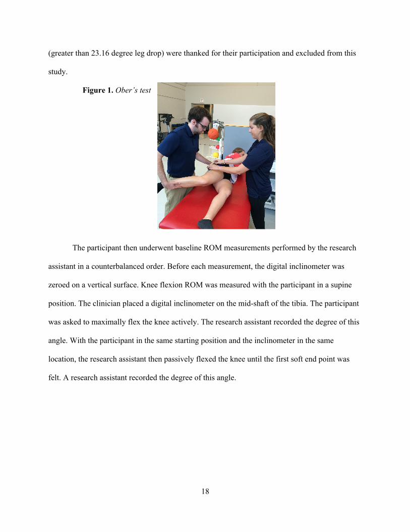
Figure 1. Ober’s test
The participant then underwent baseline ROM measurements performed by the research
assistant in a counterbalanced order. Before each measurement, the digital inclinometer was
zeroed on a vertical surface. Knee flexion ROM was measured with the participant in a supine
position. The clinician placed a digital inclinometer on the mid-shaft of the tibia. The participant
was asked to maximally flex the knee actively. The research assistant recorded the degree of this
angle. With the participant in the same starting position and the inclinometer in the same
location, the research assistant then passively flexed the knee until the first soft end point was
felt. A research assistant recorded the degree of this angle.
Page 30
19
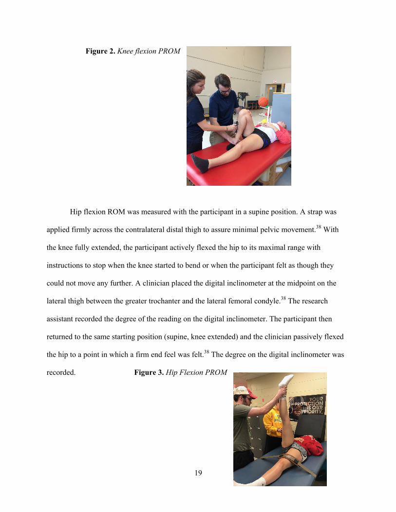
Figure 2. Knee flexion PROM
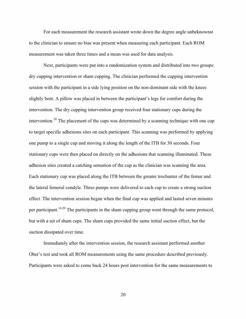
Hip flexion ROM was measured with the participant in a supine position. A strap was
applied firmly across the contralateral distal thigh to assure minimal pelvic movement.38 With
the knee fully extended, the participant actively flexed the hip to its maximal range with
instructions to stop when the knee started to bend or when the participant felt as though they
could not move any further. A clinician placed the digital inclinometer at the midpoint on the
lateral thigh between the greater trochanter and the lateral femoral condyle.38 The research
assistant recorded the degree of the reading on the digital inclinometer. The participant then
returned to the same starting position (supine, knee extended) and the clinician passively flexed
the hip to a point in which a firm end feel was felt.38 The degree on the digital inclinometer was
recorded. Figure 3. Hip Flexion PROM
Page 31
20
For each measurement the research assistant wrote down the degree angle unbeknownst
to the clinician to ensure no bias was present when measuring each participant. Each ROM
measurement was taken three times and a mean was used for data analysis.
Next, participants were put into a randomization system and distributed into two groups:
dry cupping intervention or sham cupping. The clinician performed the cupping intervention
session with the participant in a side lying position on the non-dominant side with the knees
slightly bent. A pillow was placed in between the participant’s legs for comfort during the
intervention. The dry cupping intervention group received four stationary cups during the
intervention.20 The placement of the cups was determined by a scanning technique with one cup
to target specific adhesions sites on each participant. This scanning was performed by applying
one pump to a single cup and moving it along the length of the ITB for 30 seconds. Four
stationary cups were then placed on directly on the adhesions that scanning illuminated. These
adhesion sites created a catching sensation of the cup as the clinician was scanning the area.
Each stationary cup was placed along the ITB between the greater trochanter of the femur and
the lateral femoral condyle. Three pumps were delivered to each cup to create a strong suction
effect. The intervention session began when the final cup was applied and lasted seven minutes
per participant.14,20 The participants in the sham cupping group went through the same protocol,
but with a set of sham cups. The sham cups provided the same initial suction effect, but the
suction dissipated over time.
Immediately after the intervention session, the research assistant performed another
Ober’s test and took all ROM measurements using the same procedure described previously.
Participants were asked to come back 24 hours post intervention for the same measurements to
Page 32
21
be taken. The same research assistant measured participants at each time point. The research
assistant was blinded to the participant’s intervention group.
Data Reduction
All ROM measurements, including Ober’s, were noted in Excel and the mean of three
trials was used for data analysis. Each individual degree was then adjusted to reflect measures
greater or less than 90.
Statistical Analysis
A 2 x 3 mixed model analysis of variance (ANOVA) with one fixed factor at two levels
(group: cupping, sham) and one repeated measure at three levels (time: baseline, immediate, 24-
hr post) was performed to evaluate the effect of dry cupping versus sham cupping on all outcome
variables. Alpha level was set a priori at p ≤ 0.05. All statistical analyses were performed using
SPSS (IBM SPSS Statistics for Windows, version 21.0; IBM Corp, Armonk, NY).
Page 33
22
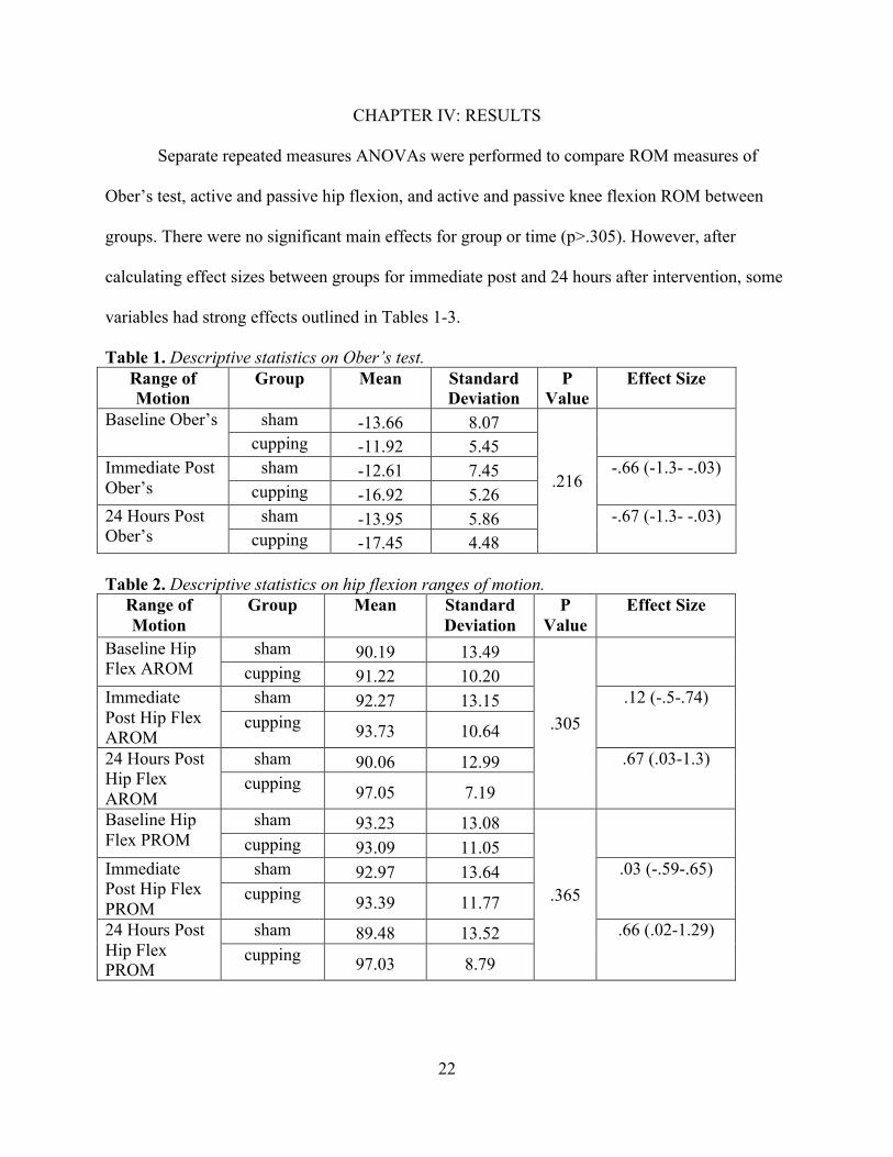
CHAPTER IV: RESULTS
Separate repeated measures ANOVAs were performed to compare ROM measures of
Ober’s test, active and passive hip flexion, and active and passive knee flexion ROM between
groups. There were no significant main effects for group or time (p>.305). However, after
calculating effect sizes between groups for immediate post and 24 hours after intervention, some
variables had strong effects outlined in Tables 1-3.
Table 1. Descriptive statistics on Ober’s test. Range of Motion
Group Mean Standard Deviation
P Value
Effect Size
Baseline Ober’s sham -13.66 8.07
.216
cupping -11.92 5.45
Immediate Post Ober’s
sham -12.61 7.45 -.66 (-1.3- -.03) cupping -16.92 5.26
24 Hours Post Ober’s
sham -13.95 5.86 -.67 (-1.3- -.03) cupping -17.45 4.48
Table 2. Descriptive statistics on hip flexion ranges of motion.
Range of Motion
Group Mean Standard Deviation
P Value
Effect Size
Baseline Hip Flex AROM
sham 90.19 13.49
.305
cupping 91.22 10.20
Immediate Post Hip Flex AROM
sham 92.27 13.15 .12 (-.5-.74) cupping 93.73 10.64
24 Hours Post Hip Flex AROM
sham 90.06 12.99 .67 (.03-1.3) cupping 97.05 7.19
Baseline Hip Flex PROM
sham 93.23 13.08
.365
cupping 93.09 11.05
Immediate Post Hip Flex PROM
sham 92.97 13.64 .03 (-.59-.65) cupping 93.39 11.77
24 Hours Post Hip Flex PROM
sham 89.48 13.52 .66 (.02-1.29) cupping 97.03 8.79
Page 34
23
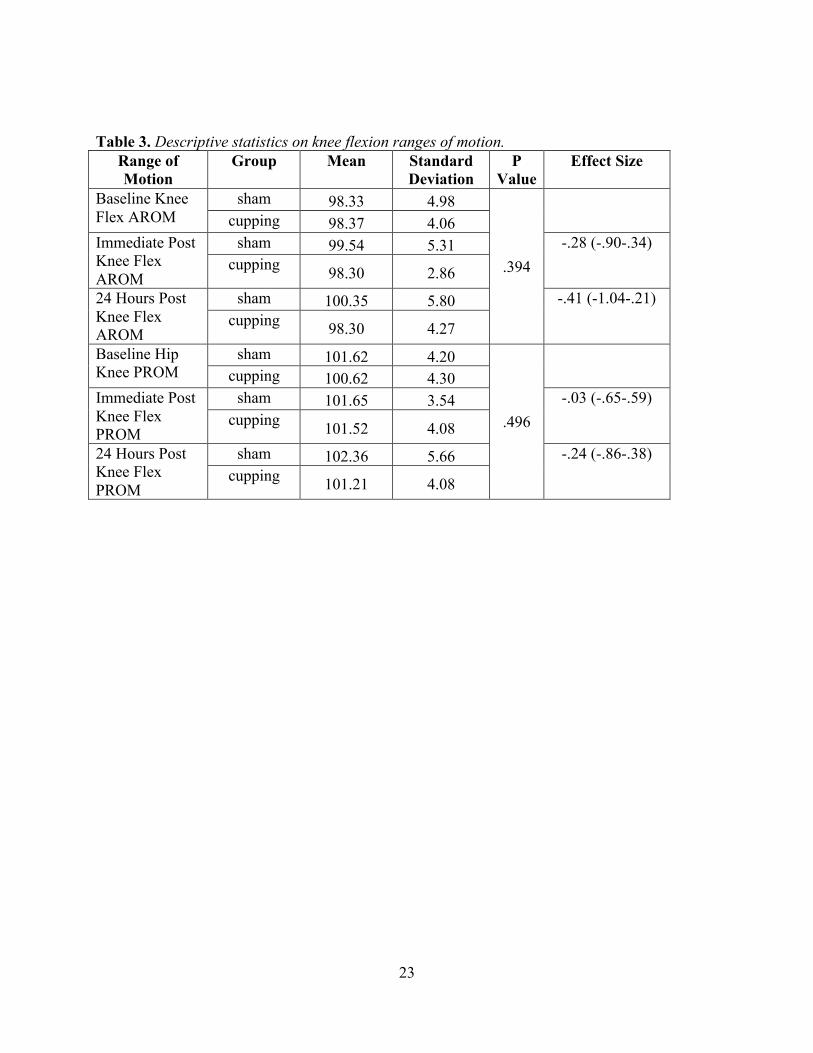
Table 3. Descriptive statistics on knee flexion ranges of motion. Range of Motion
Group Mean Standard Deviation
P Value
Effect Size
Baseline Knee Flex AROM
sham 98.33 4.98
.394
cupping 98.37 4.06
Immediate Post Knee Flex AROM
sham 99.54 5.31 -.28 (-.90-.34) cupping 98.30 2.86
24 Hours Post Knee Flex AROM
sham 100.35 5.80 -.41 (-1.04-.21) cupping 98.30 4.27
Baseline Hip Knee PROM
sham 101.62 4.20
.496
cupping 100.62 4.30
Immediate Post Knee Flex PROM
sham 101.65 3.54 -.03 (-.65-.59) cupping 101.52 4.08
24 Hours Post Knee Flex PROM
sham 102.36 5.66 -.24 (-.86-.38) cupping 101.21 4.08
Page 35
24
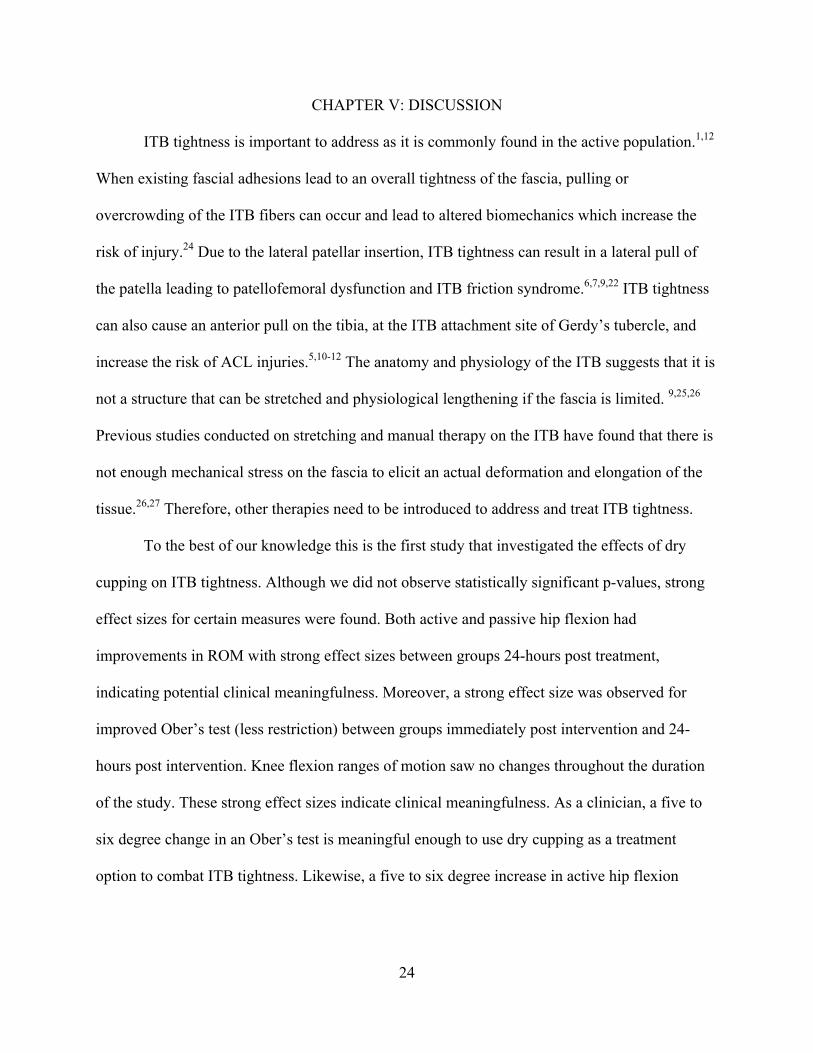
CHAPTER V: DISCUSSION
ITB tightness is important to address as it is commonly found in the active population.1,12
When existing fascial adhesions lead to an overall tightness of the fascia, pulling or
overcrowding of the ITB fibers can occur and lead to altered biomechanics which increase the
risk of injury.24 Due to the lateral patellar insertion, ITB tightness can result in a lateral pull of
the patella leading to patellofemoral dysfunction and ITB friction syndrome.6,7,9,22 ITB tightness
can also cause an anterior pull on the tibia, at the ITB attachment site of Gerdy’s tubercle, and
increase the risk of ACL injuries.5,10-12 The anatomy and physiology of the ITB suggests that it is
not a structure that can be stretched and physiological lengthening if the fascia is limited. 9,25,26
Previous studies conducted on stretching and manual therapy on the ITB have found that there is
not enough mechanical stress on the fascia to elicit an actual deformation and elongation of the
tissue.26,27 Therefore, other therapies need to be introduced to address and treat ITB tightness.
To the best of our knowledge this is the first study that investigated the effects of dry
cupping on ITB tightness. Although we did not observe statistically significant p-values, strong
effect sizes for certain measures were found. Both active and passive hip flexion had
improvements in ROM with strong effect sizes between groups 24-hours post treatment,
indicating potential clinical meaningfulness. Moreover, a strong effect size was observed for
improved Ober’s test (less restriction) between groups immediately post intervention and 24-
hours post intervention. Knee flexion ranges of motion saw no changes throughout the duration
of the study. These strong effect sizes indicate clinical meaningfulness. As a clinician, a five to
six degree change in an Ober’s test is meaningful enough to use dry cupping as a treatment
option to combat ITB tightness. Likewise, a five to six degree increase in active hip flexion
Page 36
25
ROM is a significant enough change for clinicians to see cupping as an effective treatment
option.
The positive changes in measurements indicate a single cupping intervention session
increases hip flexion ranges of motion and leads to a relief in tightness of the ITB, with the
changes being meaningful to a clinician. The results of this study support those of other research
studies in which cupping has resulted in an increase of ranges of motion and decrease of
tightness.15-19 There is no proven physiological mechanism for the effectiveness of cupping. One
commonly inferred theory of cupping is the result of increasing blood flow to an area assisting in
the healing process.15-18,20,36 The tensile stress created from the suctioning of the skin is thought
to be enough to rupture blood vessels and increase blood flow.33 Another proposed theory states
that cupping releases chemical transmitters to block pain messages and activate the gate control
theory of pain. This is theorized to occur from the vasoconstriction/dilation occurring under the
surface of the cup.20
The researchers have attributed the lack of increase of knee flexion to the positioning of
the participant when performing knee flexion; the gastrocnemius would come into contact with
posterior thigh limiting the amount of knee flexion to be obtained. Deficits in knee flexion were
also not observed, creating a ceiling effect for the measurement, where there was no room from
improvement even if the treatment was effective.
As with any research, this study has limitations. The small sample size utilized in this
study may have played a role in the lack of statistically significant p-values. If the number of
participants was larger the standard deviations may have been smaller leading to more statistical
significance. However, the strong effects sizes did indicate clinical meaningfulness for dry
cupping on ITB tightness. Another limitation in this study was the lack of multiple ranges of
Page 37
26
motion deficits for inclusion criteria. Had the researchers included hip and knee flexion deficits
in the inclusion criteria there may have been more evident increases in these ranges of motions
after the cupping intervention.
Future research should aim to standardize dry cupping intervention sessions. The
researchers chose the dry cupping guidelines based off the limited research available and past
clinical experience with the intervention. As of now, there are no specific guidelines to a cupping
intervention; everything from treatment time to the placement of cups varies. Research focusing
on standardizing a cupping intervention session could improve future research and utilization of
dry cupping in the clinical setting. Research on dry cupping should continue to be able to suggest
a mechanism for the effectiveness of this therapy. Existing hypotheses on the physiological
mechanism should be investigated. Additionally, research should evaluate the duration of the
effects seen from a single cupping intervention session. The positive effects on tightness and
ranges of motion observed after 24 hours may have just been the beginning stages of the relief on
ITB tightness. Perhaps more significant changes could have been seen after a greater lapse of
time post intervention. Lastly, future research on dry cupping should aim to include the use of
subjective measures as outcome variables. Participants in the experimental group reported
feelings of increased flexibility and analyzing those findings may have further supported the
effectiveness of dry cupping.
In conclusion, our findings indicate that a single intervention of dry cupping is probably
effective in reducing ITB tightness and increasing hip flexion ranges of motion. These changes
can be observed 24 hours post intervention in addition to immediately after the intervention.
Further studies are needed to confirm these results and to further evaluate the effectiveness of
Page 38
27
dry cupping. However, this study supports the assumption that dry cupping may be a safe and
effective treatment option to combat ITB tightness seen by clinicians.
Page 39
28
REFERENCES
1. Gose JC, Schweizer P. Iliotibial band tightness. J Orthop Sports Phys Ther. 1989;10(10):399-407.
2. Marieb E, N., Hoehn K. Human Anatomy & Physiology. Tenth ed. Prentice Hall, Inc.2016.
3. Simmonds N, Miller P, Gemmell H. A theoretical framework for the role of fascia in manual therapy. J Bodyw Mov Ther. 2012;16(1):83-93.
4. Tozzi P. Selected Fascial Aspects of Osteopathic Practice. Journal of Bodywork and Movement Therapies. 2012;16:22.
5. Hudson Z, Darthuy E. Iliotibial band tightness and patellofemoral pain syndrome: a case-control study. Man Ther. 2009;14(2):147-151.
6. Beals RK. The Iliotibial Tract: A review. Current Orthopaedic Practice. 2009;20(1):5. 7. Vieira EL, Vieira EA, da Silva RT, Berlfein PA, Abdalla RJ, Cohen M. An anatomic
study of the iliotibial tract. Arthroscopy. 2007;23(3):269-274. 8. Krivickas LS, Feinberg JH. Lower extremity injuries in college athletes: relation between
ligamentous laxity and lower extremity muscle tightness. Arch Phys Med Rehabil. 1996;77(11):1139-1143.
9. Fairclough J, Hayashi K, Toumi H, et al. The functional anatomy of the iliotibial band during flexion and extension of the knee: implications for understanding iliotibial band syndrome. J Anat. 2006;208(3):309-316.
10. Herrington L, Rivett N, Munro S. The relationship between patella position and length of the iliotibial band as assessed using Ober's test. Man Ther. 2006;11(3):182-186.
11. Puniello MS. Iliotibial band tightness and medial patellar glide in patients with patellofemoral dysfunction. J Orthop Sports Phys Ther. 1993;17(3):144-148.
12. Khaund R, Flynn SH. Iliotibial band syndrome: a common source of knee pain. Am Fam Physician. 2005;71(8):1545-1550.
13. Ferber R, Kendall KD, McElroy L. Normative and critical criteria for iliotibial band and iliopsoas muscle flexibility. J Athl Train. 2010;45(4):344-348.
14. Mehta P, Dhapte V. Cupping therapy: A prudent remedy for a plethora of medical ailments. J Tradit Complement Med. 2015;5(3):127-134.
15. Kim TH, Kang JW, Kim KH, et al. Cupping for treating neck pain in video display terminal (VDT) users: a randomized controlled pilot trial. J Occup Health. 2012;54(6):416-426.
16. Lauche R, Cramer H, Choi KE, et al. The influence of a series of five dry cupping treatments on pain and mechanical thresholds in patients with chronic non-specific neck pain--a randomised controlled pilot study. BMC Complement Altern Med. 2011;11:63.
17. Teut M, Kaiser S, Ortiz M, et al. Pulsatile dry cupping in patients with osteoarthritis of the knee - a randomized controlled exploratory trial. BMC Complement Altern Med. 2012;12:184.
18. Akbarzadeh M, Ghaemmaghami M, Yazdanpanahi Z, Zare N, Azizi A, Mohagheghzadeh A. The Effect Dry Cupping Therapy at Acupoint BL23 on the Intensity of Postpartum Low Back Pain in Primiparous Women Based on Two Types of Questionnaires, 2012; A Randomized Clinical Trial. Int J Community Based Nurs Midwifery. 2014;2(2):112-120.
19. Markowski A, Sanford S, Pikowski J, Fauvell D, Cimino D, Caplan S. A pilot study analyzing the effects of Chinese cupping as an adjunct treatment for patients with
Page 40
29
subacute low back pain on relieving pain, improving range of motion, and improving function. J Altern Complement Med. 2014;20(2):113-117.
20. Rozenfeld E, Kalichman L. New is the well-forgotten old: The use of dry cupping in musculoskeletal medicine. J Bodyw Mov Ther. 2016;20(1):173-178.
21. Schleip R, Jäger H, Klingler W. What is 'fascia'? A review of different nomenclatures. J Bodyw Mov Ther. 2012;16(4):496-502.
22. Starkey C, Brown S, Ryan J. Examination of Orthopedic and Athletic Injuries. 3 ed. Philadelphia, PA: F.A. Davis Company; 2010.
23. Schleip R. Fascial Plasticity - A New Neurobiological Explanation: Part 1. Journal of Bodywork and Movement Therapies. 2003;7(1):9.
24. Barnes M. The Basic Science of Myofascial Release: Morphologic Changes in Connective Tissue. Journal of Bodywork and Movement Therapies. 1997;1(4):8.
25. Fairclough J, Hayashi K, Toumi H, et al. Is iliotibial band syndrome really a friction syndrome? J Sci Med Sport. 2007;10(2):74-76; discussion 77-78.
26. Falvey EC, Clark RA, Franklyn-Miller A, Bryant AL, Briggs C, McCrory PR. Iliotibial band syndrome: an examination of the evidence behind a number of treatment options. Scand J Med Sci Sports. 2010;20(4):580-587.
27. Chaudhry H, Schleip R, Ji Z, Bukiet B, Maney M, Findley T. Three-dimensional mathematical model for deformation of human fasciae in manual therapy. J Am Osteopath Assoc. 2008;108(8):379-390.
28. Threlkeld AJ. The effects of manual therapy on connective tissue. Phys Ther. 1992;72(12):893-902.
29. Bialosky JE, Bishop MD, Price DD, Robinson ME, George SZ. The mechanisms of manual therapy in the treatment of musculoskeletal pain: a comprehensive model. Man Ther. 2009;14(5):531-538.
30. Schleip R. Fascial Plasticity - A New Neurobiological Explanation: Part 2. Journal of Bodywork and Movement Therapies. 2003;7(2):13.
31. Ramsey SM. Holistic manual therapy techniques. Prim Care. 1997;24(4):759-786. 32. Nickel JC. Management of urinary tract infections: historical perspective and current
strategies: Part 2--Modern management. J Urol. 2005;173(1):27-32. 33. Tham LM, Lee HP, Lu C. Cupping: from a biomechanical perspective. J Biomech.
2006;39(12):2183-2193. 34. Cramer H, Lauche R, Hohmann C, et al. Randomized controlled trial of pulsating
cupping (pneumatic pulsation therapy) for chronic neck pain. Forsch Komplementmed. 2011;18(6):327-334.
35. Kim JI, Kim TH, Lee MS, et al. Evaluation of wet-cupping therapy for persistent non-specific low back pain: a randomised, waiting-list controlled, open-label, parallel-group pilot trial. Trials. 2011;12:146.
36. Kim JI, Lee MS, Lee DH, Boddy K, Ernst E. Cupping for treating pain: a systematic review. Evid Based Complement Alternat Med. 2011;2011:467014.
37. Lee MS, Kim JI, Kong JC, Lee DH, Shin BC. Developing and validating a sham cupping device. Acupunct Med. 2010;28(4):200-204.
38. Pua YH, Wrigley TV, Wrigley TW, Cowan SM, Bennell KL. Intrarater test-retest reliability of hip range of motion and hip muscle strength measurements in persons with hip osteoarthritis. Arch Phys Med Rehabil. 2008;89(6):1146-1154.