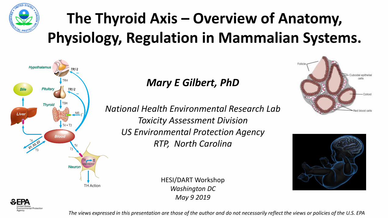
The Thyroid Axis – Overview of Anatomy, Physiology, Regulation in Mammalian Systems. The views expressed in this presentation are those of the author and do not necessarily reflect the views or policies of the U.S. EPA Mary E Gilbert, PhD National Health Environmental Research Lab Toxicity Assessment Division US Environmental Protection Agency RTP, North Carolina HESI/DART Workshop Washington DC May 9 2019
Transcript
The Thyroid Axis – Overview of Anatomy, Physiology, Regulation in Mammalian Systems.
The views expressed in this presentation are those of the author and do not necessarily reflect the views or policies of the U.S. EPA
Mary E Gilbert, PhD
National Health Environmental Research LabToxicity Assessment Division
US Environmental Protection AgencyRTP, North Carolina
HESI/DART WorkshopWashington DC
May 9 2019
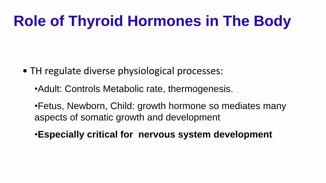
Role of Thyroid Hormones in The Body
• TH regulate diverse physiological processes:
•Adult: Controls Metabolic rate, thermogenesis.
•Fetus, Newborn, Child: growth hormone so mediates many
aspects of somatic growth and development
•Especially critical for nervous system development
Gilbert et al. 2012 Neurotoxicology 33: 842-852Adapted from Boas et al., 2006
OUTLINE• Synthesis in the Gland• Regulatory Feedback Control• Carrier Proteins• Metabolism in Liver• Deiodinases• Transporters• TH Action• Neurodevelopmental Consequences
• Humans and Rodent Models
Thyroid System
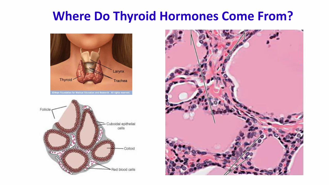
Where Do Thyroid Hormones Come From?
I I I I
Tri-iodothyronineT3
I
Tetra-iodothyronine or ThyroxineT4
I I
What are Thyroid Hormones?
• Major circulating form is T4 – 80:20 ratio in human
• T4 considered ‘prohormone’ as T3 the ‘active’ hormone, but this view is changing – non-genomic effects mediated by T4
TPO
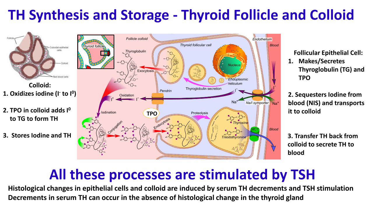
TH Synthesis and Storage - Thyroid Follicle and Colloid
All these processes are stimulated by TSHHistological changes in epithelial cells and colloid are induced by serum TH decrements and TSH stimulationDecrements in serum TH can occur in the absence of histological change in the thyroid gland
Colloid:1. Oxidizes iodine (I- to I0)
2. TPO in colloid adds I0
to TG to form TH
3. Stores Iodine and TH
Follicular Epithelial Cell:1. Makes/Secretes
Thyroglobulin (TG) and TPO
2. Sequesters Iodine from blood (NIS) and transports it to colloid
3. Transfer TH back from colloid to secrete TH to blood
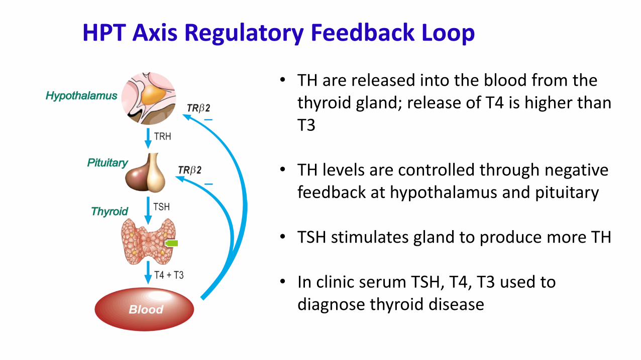
HPT Axis Regulatory Feedback Loop
Kidney
• TH are released into the blood from the thyroid gland; release of T4 is higher than T3
• TH levels are controlled through negative feedback at hypothalamus and pituitary
• TSH stimulates gland to produce more TH
• In clinic serum TSH, T4, T3 used to diagnose thyroid disease
TH drops during pregnancy and recovers postnatally
T4 – low levels in fetus
GD20 Fetal Serum - LCMS
Dose of PTU
0.00 0.10 0.50 1.00 2.00 3.00
Se
rum
T4
(ng/m
l)
0.0
0.5
1.0
1.5
2.0
2.5
3.0
3.5
4.0Pup Serum T3 and T4
Pup Age
PN2 PN5 PN10 PN14 PN18
Seru
m T
3
an
d T
4
0
10
20
30
40
50
60
T4 (ng/ml)
T3 (ng/dl)
TH increases with age in neonate
From Dohler (1979)
From Hassan et al., Tox Sci 2017; O’Shaughnessy et al., Tox Sci, 2018
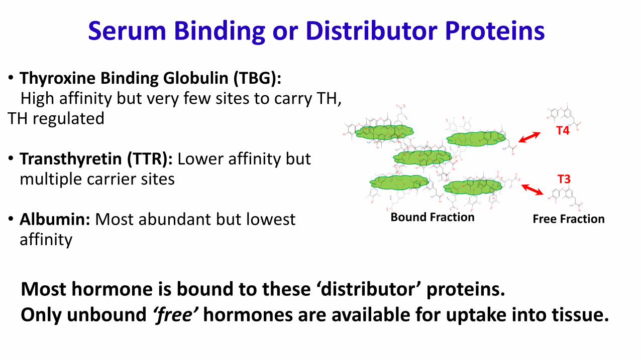
Serum Binding or Distributor Proteins
• Thyroxine Binding Globulin (TBG):High affinity but very few sites to carry TH,
TH regulated
• Transthyretin (TTR): Lower affinity but multiple carrier sites
• Albumin: Most abundant but lowest affinity
T4
T3
Bound Fraction Free Fraction
Most hormone is bound to these ‘distributor’ proteins.Only unbound ‘free’ hormones are available for uptake into tissue.
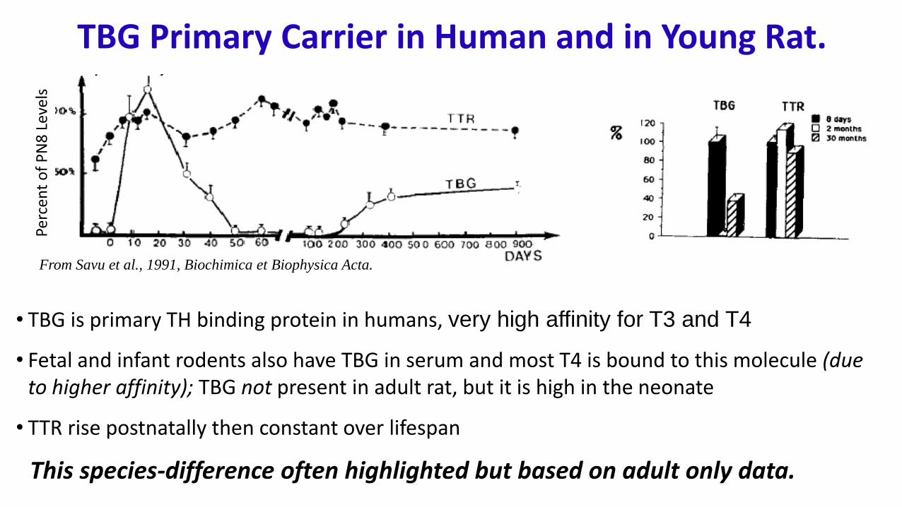
• TBG is primary TH binding protein in humans, very high affinity for T3 and T4
• Fetal and infant rodents also have TBG in serum and most T4 is bound to this molecule (due to higher affinity); TBG not present in adult rat, but it is high in the neonate
• TTR rise postnatally then constant over lifespan
TBG Primary Carrier in Human and in Young Rat.Pe
rcen
t o
f P
N8
Lev
els
From Savu et al., 1991, Biochimica et Biophysica Acta.
This species-difference often highlighted but based on adult only data.
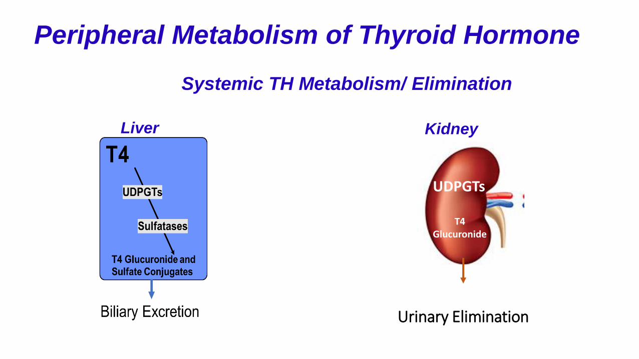
Peripheral Metabolism of Thyroid Hormone
UDPGTs
T4 Glucuronide
Systemic TH Metabolism/ Elimination
Liver
Urinary Elimination
Kidney
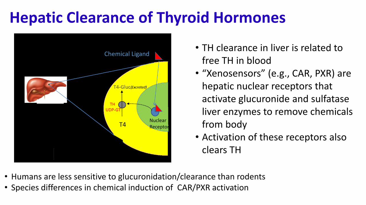
Hepatic Clearance of Thyroid Hormones
• TH clearance in liver is related to free TH in blood
• “Xenosensors” (e.g., CAR, PXR) are hepatic nuclear receptors that activate glucuronide and sulfatase liver enzymes to remove chemicals from body
• Activation of these receptors also clears TH
T4
Chemical Ligand
NuclearReceptor
• Humans are less sensitive to glucuronidation/clearance than rodents• Species differences in chemical induction of CAR/PXR activation
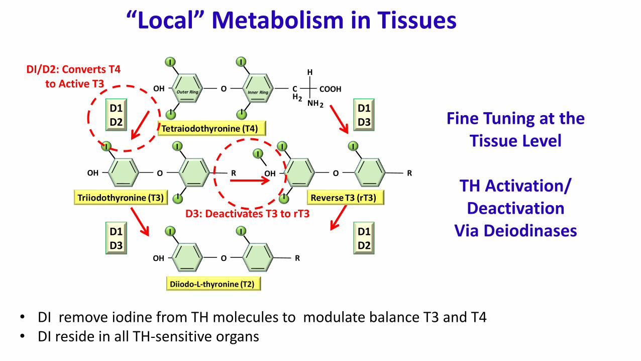
“Local” Metabolism in Tissues
• DI remove iodine from TH molecules to modulate balance T3 and T4• DI reside in all TH-sensitive organs
OH
OH
O
I I
R
O
I I
IR
D1D3
D1D2
D1D2
D1D3
OH O CH2
COOH
NH 2
HI I
I I
Tetraiodothyronine (T4)
Diiodo-L-thyronine (T2)
Triiodothyronine (T3) Reverse T3 (rT3)
O
I I
ROHI
Outer Ring Inner Ring
I
Fine Tuning at the Tissue Level
TH Activation/Deactivation
Via Deiodinases
DI/D2: Converts T4 to Active T3
D3: Deactivates T3 to rT3
Local TH Activation/Deactivation Via Deiodinases
•Tissue, region, age-dependent expression•Essential function is to provide fine resolution over T3 for gene transcription/TH action
DIO-1 DIO-2 DIO-3
Primary TH Analyte Substrate T4 T4 T3 and T4
FunctionGenerates T3
from T4Generates T3
from T4Inactivates
T3
Tissue TypeLiver
KidneyThyroid Pituitary
Brain Placenta Brain Placenta
Liver
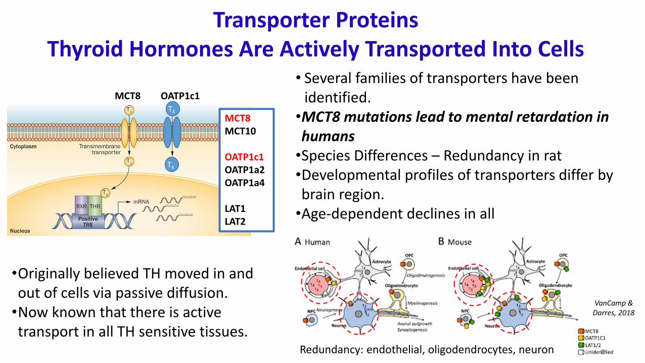
Transporter ProteinsThyroid Hormones Are Actively Transported Into Cells
MCT8MCT10
OATP1c1OATP1a2OATP1a4
LAT1LAT2
• Several families of transporters have been identified.
•MCT8 mutations lead to mental retardation in humans
•Species Differences – Redundancy in rat•Developmental profiles of transporters differ by brain region.
•Age-dependent declines in all
•Originally believed TH moved in and out of cells via passive diffusion.
•Now known that there is active transport in all TH sensitive tissues.
Redundancy: endothelial, oligodendrocytes, neuron
VanCamp & Darres, 2018
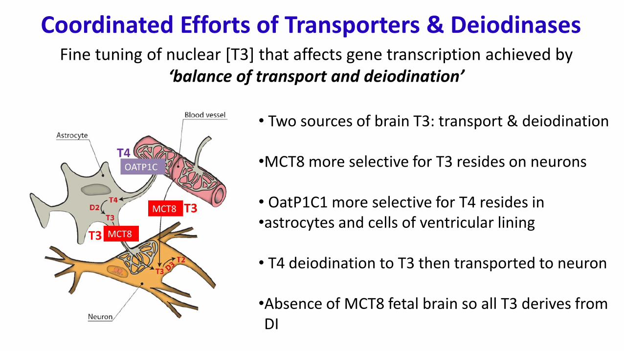
T4
T4
MCT8 OATP1c1
• Two sources of brain T3: transport & deiodination
•MCT8 more selective for T3 resides on neurons
• OatP1C1 more selective for T4 resides in •astrocytes and cells of ventricular lining
• T4 deiodination to T3 then transported to neuron
•Absence of MCT8 fetal brain so all T3 derives from DI
Coordinated Efforts of Transporters & DeiodinasesFine tuning of nuclear [T3] that affects gene transcription achieved by
‘balance of transport and deiodination’
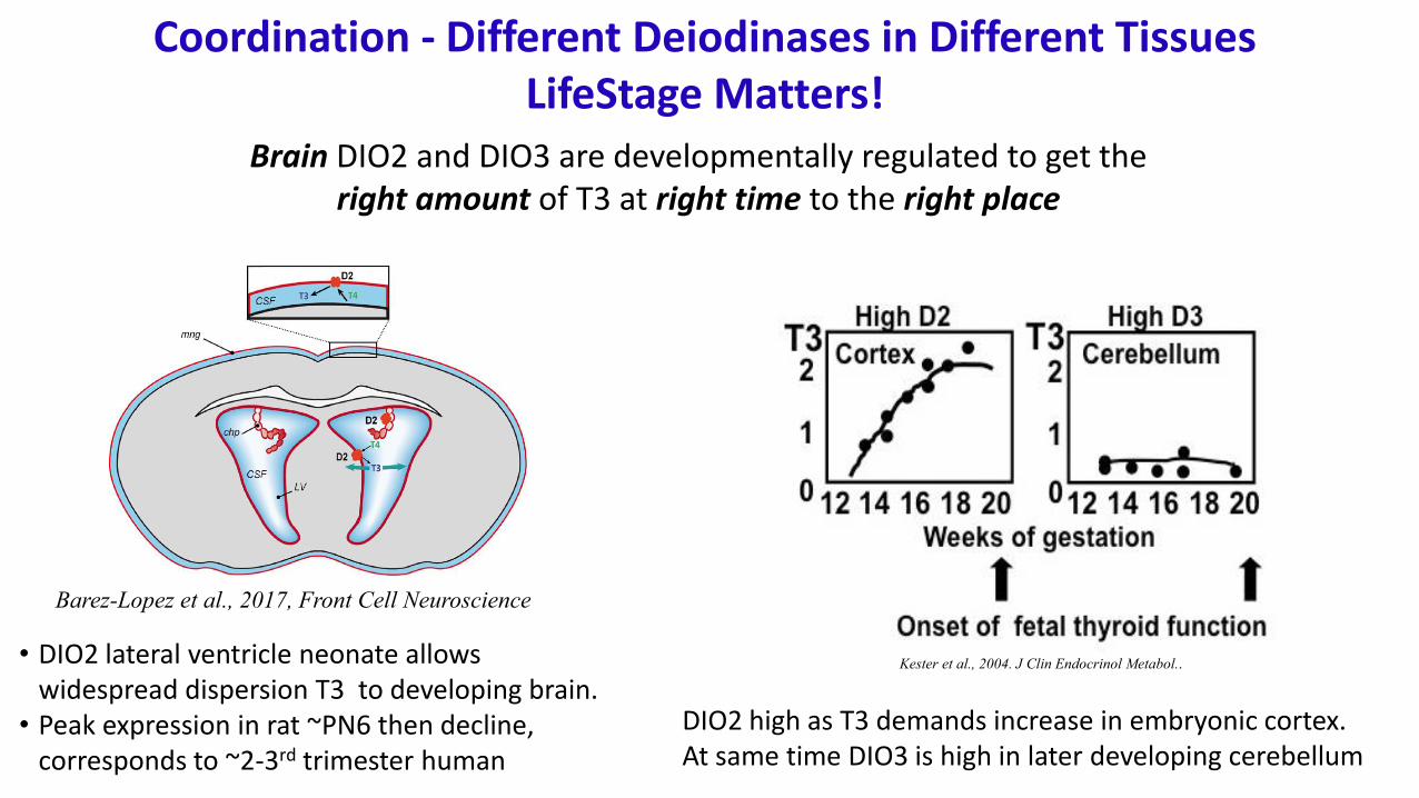
Coordination - Different Deiodinases in Different Tissues LifeStage Matters!
Kester et al., 2004. J Clin Endocrinol Metabol..
Brain DIO2 and DIO3 are developmentally regulated to get the right amount of T3 at right time to the right place
• DIO2 lateral ventricle neonate allows widespread dispersion T3 to developing brain.
• Peak expression in rat ~PN6 then decline, corresponds to ~2-3rd trimester human
DIO2 high as T3 demands increase in embryonic cortex. At same time DIO3 is high in later developing cerebellum
Barez-Lopez et al., 2017, Front Cell Neuroscience
Brain TH and TH ActionCoordinated Temporal and Spatial Control of TH Essential
Primary action T3 is to Regulate Gene Transcription that Modulate Brain Development
Other ‘nongenomic’ actions have been identified for membrane bound T4 receptors
Protein
mRNA
Nucleus
Cytoplasm
Modified from Williams and Bassett, 2011
Bernal, 2012
TH Action Hr
Dose of PTU (ppm)
0 1 2 3 10
Fo
ld C
ha
ng
e
0.0
0.2
0.4
0.6
0.8
1.0
1.2
**
*
Parv
Dose of PTU (ppm)
0 1 2 3 10
Fo
ld C
ha
ng
e
0.0
0.2
0.4
0.6
0.8
1.0
1.2
**
*
*
Many of These Genes Modulate Brain Development - Dose, Region, and Time-DependentLimited Dose-Response Data Available, largely limited to model chemicals – PTU, MMILimited data on temporal and spatial resolution
O’Shaughnessy et al., 2018
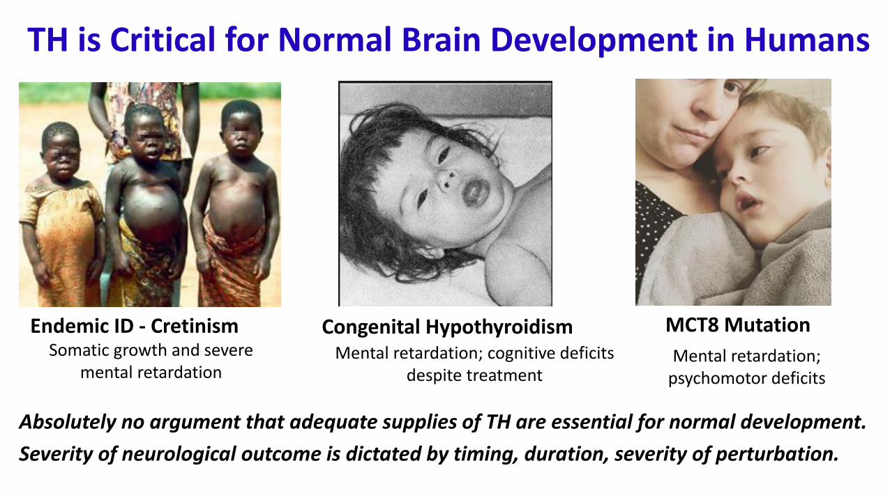
Absolutely no argument that adequate supplies of TH are essential for normal development.
Severity of neurological outcome is dictated by timing, duration, severity of perturbation.
Endemic ID - Cretinism Congenital Hypothyroidism
TH is Critical for Normal Brain Development in Humans
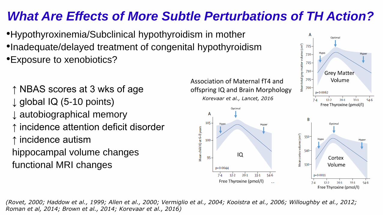
What Are Effects of More Subtle Perturbations of TH Action?
(Rovet, 2000; Haddow et al., 1999; Allen et al., 2000; Vermiglio et al., 2004; Kooistra et al., 2006; Willoughby et al., 2012; Roman et al, 2014; Brown et al., 2014; Korevaar et al., 2016)
•Hypothyroxinemia/Subclinical hypothyroidism in mother
•Inadequate/delayed treatment of congenital hypothyroidism
•Exposure to xenobiotics?
Korevaar et al., Lancet, 2016
Association of Maternal fT4 and offspring IQ and Brain Morphology
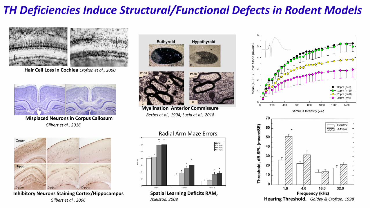
Myelination Anterior CommissureBerbel et al., 1994; Lucia et al., 2018
Hair Cell Loss in Cochlea Crofton et al., 2000
0 ppm 3 ppm 10 ppm
Hippo
Cortex
Inhibitory Neurons Staining Cortex/HippocampusGilbert et al., 2006
TH Deficiencies Induce Structural/Functional Defects in Rodent Models
Control HypothyroidHeterotopia In Corpus Callosum
Misplaced Neurons in Corpus CallosumGilbert et al., 2016
EPSP Slope
Stimulus Intensity (
0 200 400 600 800 1000 1200 1400
Mean (
+/-
SE
) E
PS
P S
lope (
mv/m
s)
0
1
2
3
4
5
6
0ppm (n=7)
1ppm (n=10)
2ppm (n=10)
3ppm (n=8)
Hearing Threshold, Goldey & Crofton, 1998
Radial Arm Maze Errors
Spatial Learning Deficits RAM, Axelstad, 2008
Rodent ModelsThyroid and Brain
Most of what we know has derived from Rodent Models Basic Components of Thyroid Synthesis and Regulation
Ontogeny of Thyroid Function in Fetus/Neonate Metabolism Mom and Fetus
Presence of Placental and Brain BarriersRodent models mimic human neurological phenotype
Human PregnancyThyroid and Brain
FetalThyroid Gland
Maternal Thyroid Gland
Maternal Metabolism
Placenta
FetalThyroid Gland
Fetal Metabolism
Brain Barriers(BBB CSF)
Rodent Models Mimic The Complexities of Thyroid Biology During Development
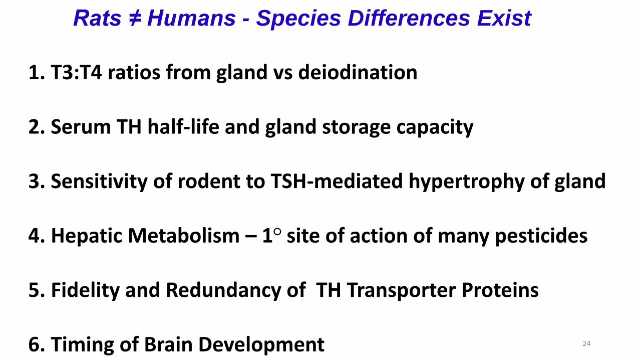
Rats ≠ Humans - Species Differences Exist
1. T3:T4 ratios from gland vs deiodination
2. Serum TH half-life and gland storage capacity
3. Sensitivity of rodent to TSH-mediated hypertrophy of gland
4. Hepatic Metabolism – 1○ site of action of many pesticides
5. Fidelity and Redundancy of TH Transporter Proteins
6. Timing of Brain Development 24
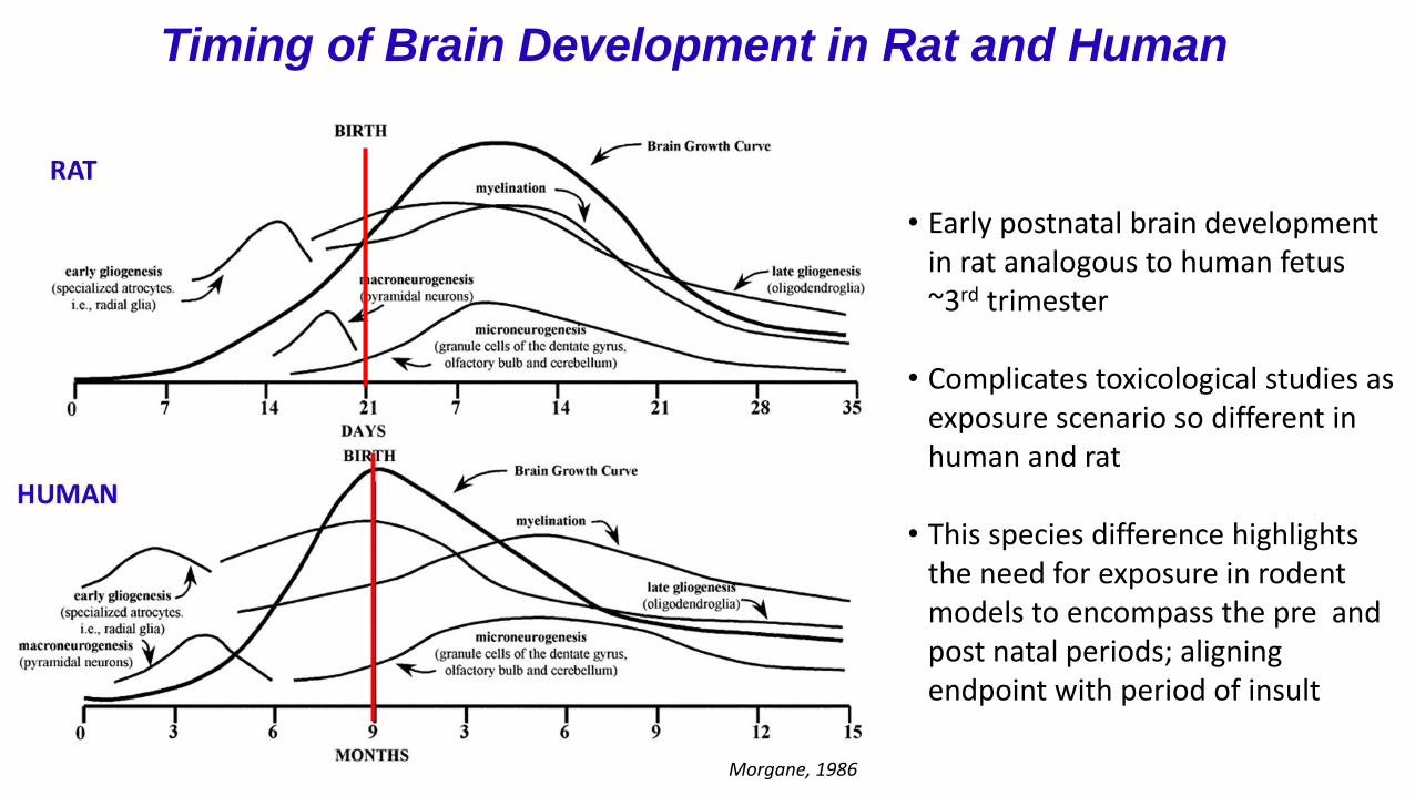
Timing of Brain Development in Rat and Human
• Early postnatal brain development in rat analogous to human fetus ~3rd trimester
• Complicates toxicological studies as exposure scenario so different in human and rat
• This species difference highlights the need for exposure in rodent models to encompass the pre and post natal periods; aligning endpoint with period of insult
Morgane, 1986
Gilbert et al., 2011, NeurotoxicologyAdapted from Boas et al., 2006
Challenges in Assessing Risk Thyroid Disrupting Chemicals
Thyroid System- It is Complex!
Thyroid Disruption and Brain - Different consequences at different life stages; limited simple brain readouts of disruption
Serum TH: Clinical tool to diagnose thyroid disease; flag for xenobiotics that disrupt thyroid signaling and may impact brain dev’t
Many Potential Sites of Chemical Disruption: High Throughput Assays Developed to Target These Sites