Draft The treatment effects of flaxseed-derived secoisolariciresinol diglycoside and its metabolite enterolactone on benign prostatic hyperplasia involve the G protein-coupled estrogen receptor 1 Journal: Applied Physiology, Nutrition, and Metabolism Manuscript ID apnm-2016-0332.R2 Manuscript Type: Article Date Submitted by the Author: 12-Sep-2016 Complete List of Authors: Ren, Guan-Yu; Soochow University Chen, Chun-Yang; Soochow University Chen, Wei-Guo; Soochow University Huang, Ya; Soochow University Qin, Li-Qiang; Soochow University Chen, Li-Hua; School of Public Health, Soochow University; , Department of Nutrition and Food Hygiene Keyword: Flaxseed, Lignan, Benign prostatic hyperplasia, G Protein-Coupled Estrogen Receptor 1 https://mc06.manuscriptcentral.com/apnm-pubs Applied Physiology, Nutrition, and Metabolism
Transcript
Draft
The treatment effects of flaxseed-derived
secoisolariciresinol diglycoside and its metabolite enterolactone on benign prostatic hyperplasia involve the G
protein-coupled estrogen receptor 1
Journal: Applied Physiology, Nutrition, and Metabolism
Manuscript ID apnm-2016-0332.R2
Manuscript Type: Article
Date Submitted by the Author: 12-Sep-2016
Complete List of Authors: Ren, Guan-Yu; Soochow University Chen, Chun-Yang; Soochow University Chen, Wei-Guo; Soochow University Huang, Ya; Soochow University Qin, Li-Qiang; Soochow University Chen, Li-Hua; School of Public Health, Soochow University; , Department of Nutrition and Food Hygiene
Keyword: Flaxseed, Lignan, Benign prostatic hyperplasia, G Protein-Coupled Estrogen
Receptor 1
https://mc06.manuscriptcentral.com/apnm-pubs
Applied Physiology, Nutrition, and Metabolism
Draft
1
The treatment effects of flaxseed-derived secoisolariciresinol diglycoside and its
metabolite enterolactone on benign prostatic hyperplasia involve the G
protein-coupled estrogen receptor 1
Guan-Yu Ren1,2,#
, Chun-Yang Chen1,2,#
, Wei-Guo Chen2, Ya Huang
3, Li-Qiang Qin
1,
Li-Hua Chen1,
*
1 Jiangsu Key Laboratory of Preventive and Translational Medicine for Geriatric
Diseases, Department of Nutrition and Food Hygiene, School of Public Health,
Hammes, S. R. and Levin, E. R. 2007. Extranuclear steroid receptors: nature and
actions. Endocr. Rev. 28(7):726-741.
Isaacs, J. T. 1994. Etiology of benign prostatic hyperplasia. Eur. Urol. 25 Suppl
1:6-9.
Lappano, R., Rosano, C., Santolla, M. F., Pupo, M., De Francesco, E. M., De Marco,
P., Ponassi, M., Spallarossa, A., Ranise, A. and Maggiolini, M. 2012. Two
novel GPER agonists induce gene expression changes and growth effects in
cancer cells. Curr. Cancer. Drug. Targets. 12(5):531-542.
Marshall, C. J. 1995. Specificity of receptor tyrosine kinase signaling: transient
versus sustained extracellular signal-regulated kinase activation. Cell.
80(2):179-185.
Meyer, M. R., Fredette, N. C., Howard, T. A., Hu, C., Ramesh, C., Daniel, C.,
Amann, K., Arterburn, J. B., Barton, M. and Prossnitz, E. R. 2014. G
protein-coupled estrogen receptor protects from atherosclerosis. Sci. Rep.
4:7564.
Pan, A., Yu, D., Demark-Wahnefried, W., Franco, O. H. and Lin, X. 2009.
Meta-analysis of the effects of flaxseed interventions on blood lipids. Am. J.
Clin. Nutr. 90(2):288-297.
Prenzel, N., Zwick, E., Daub, H., Leserer, M., Abraham, R., Wallasch, C. and Ullrich,
A. 1999. EGF receptor transactivation by G-protein-coupled receptors
requires metalloproteinase cleavage of proHB-EGF. Nature.
Page 24 of 34
https://mc06.manuscriptcentral.com/apnm-pubs
Applied Physiology, Nutrition, and Metabolism
Draft
25
402(6764):884-888.
Ren, G. Y., Chen, C. Y., Chen, G. C., Chen, W. G., Pan, A., Pan, C. W., Zhang, Y. H.,
Qin, L. Q. and Chen, L. H. 2016. Effect of Flaxseed Intervention on
Inflammatory Marker C-Reactive Protein: A Systematic Review and
Meta-Analysis of Randomized Controlled Trials. Nutrients. 8(3):136.
Royuela, M., de Miguel, M. P., Bethencourt, F. R., Sanchez-Chapado, M., Fraile, B.,
Arenas, M. I. and Paniagua, R. 2001. Estrogen receptors alpha and beta in the
normal, hyperplastic and carcinomatous human prostate. J. Endocrinol.
168(3):447-454.
Simons, R., Sonawane, N., Verbruggen, M. and Chaudhary, J. 2015. Efficacy and
safety of a flaxseed hull extract in the symptomatic management of benign
prostatic hyperplasia: a parallel, randomized, double-blind,
placebo-controlled, pilot study. J. Med. Food. 18(2):233-240.
Smith, L. C., Ralston-Hooper, K. J., Ferguson, P. L. and Sabo-Attwood, T. 2016.
THE G Protein-Coupled Estrogen Receptor Agonist G-1 Inhibits Nuclear
Estrogen Receptor Activity and Stimulates Novel Phosphoproteomic
Signatures. Toxicol. Sci. 151(2):434-446
Tang, D., Wu, D., Hirao, A., Lahti, J. M., Liu, L., Mazza, B., Kidd, V. J., Mak, T. W.
and Ingram, A. J. 2002. ERK activation mediates cell cycle arrest and
apoptosis after DNA damage independently of p53. J. Biol. Chem.
277(15):12710-7.
Tashiro, E., Tsuchiya, A. and Imoto, M. 2007. Functions of cyclin D1 as an oncogene
Page 25 of 34
https://mc06.manuscriptcentral.com/apnm-pubs
Applied Physiology, Nutrition, and Metabolism
Draft
26
and regulation of cyclin D1 expression. Cancer. Sci. 98(5):629-635.
Thompson, L. U. 1998. Experimental studies on lignans and cancer. Baillieres. Clin.
Endocrinol. Metab. 12(4):691-705.
Ventura, S., Oliver, V., White, C. W., Xie, J. H., Haynes, J. M. and Exintaris, B. 2011.
Novel drug targets for the pharmacotherapy of benign prostatic hyperplasia
(BPH). Br. J. Pharmacol. 163(5):891-907.
Wang, C., Du, X., Yang, R., Liu, J., Xu, D., Shi, J., Chen, L., Shao, R., Fan, G., Gao,
X. and others. 2015. The prevention and treatment effects of tanshinone IIA
on oestrogen/androgen-induced benign prostatic hyperplasia in rats. J.
Steroid. Biochem. Mol. Biol. 145:28-37.
Wu, G., Robertson, D. H., Brooks, C. L., 3rd and Vieth, M. 2003. Detailed analysis
of grid-based molecular docking: A case study of CDOCKER-A
CHARMm-based MD docking algorithm. J. Comput. Chem.
24(13):1549-1562.
Wu, H., Pan, A., Yu, Z., Qi, Q., Lu, L., Zhang, G., Yu, D., Zong, G., Zhou, Y., Chen,
X. and others. 2010. Lifestyle counseling and supplementation with flaxseed
or walnuts influence the management of metabolic syndrome. J. Nutr.
140(11):1937-42.
Yu, Y., Song, J., Song, Y., Guo, X., Han, Y. and Wei, J. 2013. Characterization of
catalytic activity and structure of selenocysteine-containing hGSTZ1c-1c
based on site-directed mutagenesis and computational analysis. IUBMB. Life.
65(2):163-70.
Page 26 of 34
https://mc06.manuscriptcentral.com/apnm-pubs
Applied Physiology, Nutrition, and Metabolism
Draft
27
Zhang, W., Wang, X., Liu, Y., Tian, H., Flickinger, B., Empie, M. W. and Sun, S. Z.
2008. Effects of dietary flaxseed lignan extract on symptoms of benign
prostatic hyperplasia. J. Med. Food. 11(2):207-214.
Zhao, L., Sun, Y., Hou, Y., Peng, Q., Wang, L., Luo, H., Tang, X., Zeng, Z. and Liu,
M. 2012. MiRNA expression analysis of cancer-associated fibroblasts and
normal fibroblasts in breast cancer. Int. J. Biochem. Cell. Biol.
44(11):2051-2059.
Page 27 of 34
https://mc06.manuscriptcentral.com/apnm-pubs
Applied Physiology, Nutrition, and Metabolism
Draft
28
Figure legends
Figure 1. Schematic figure for induction of BPH and treatments.
Figure 2. GPER docked with G1 and ENL. (A-C). chemical structure of SDG, ENL
and G1. (D) residues involved in the interactions with G1. (E) residues involved in
the interactions with ENL.
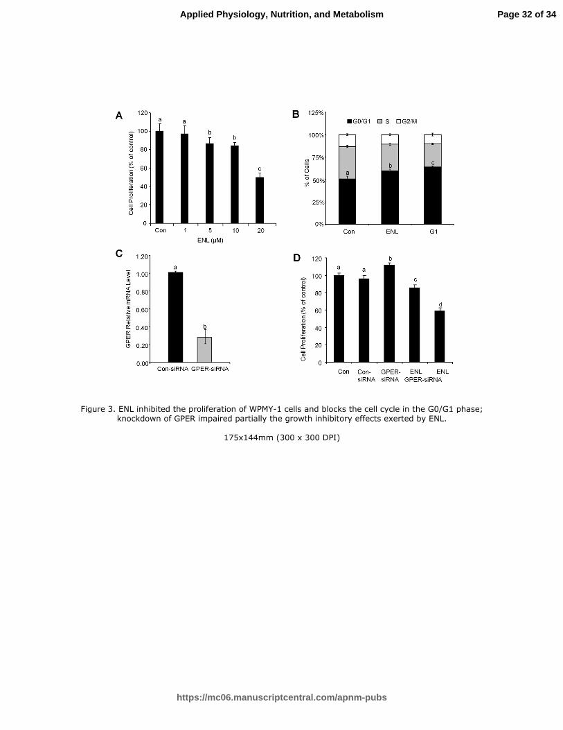
Figure 3. ENL inhibited the proliferation of WPMY-1 cells and blocks the cell cycle
in the G0/G1 phase; knockdown of GPER impaired partially the growth inhibitory
effects exerted by ENL. (A) the cells were incubated on a 96-well plate overnight.
Then indicated doses of ENL were supplemented for 24 h and cell proliferation was
checked by MTT assay. (B) the cells were cultured the same as (A); cell cycle were
checked by flow cytometry assay. ENL (20 µM), G1 (1 µM). (C) after transfection,
the cells were kept for incubation for 24 h followed by real-time PCR. (D) the
transfected cells were incubated 24 h and distributed on 96-well plates. ENL (20 µM)
was supplemented and the incubation continued for 24 h. Then, cell proliferation
was checked by MTT assay. The untreated controls were set to 100%. Values are
means ± SE, n=6. Means without a common letter differ, P < 0.05.
Figure 4. ENL increased the expression of GPER, its downstream target ERK, and
changed the expression of cell cycle related proteins. WPMY-1 cells were treated
with ENL (20 µM) and G1 (1 µM) for 24 h. Total proteins were extracted and target
Page 28 of 34
https://mc06.manuscriptcentral.com/apnm-pubs
Applied Physiology, Nutrition, and Metabolism
Draft
29
proteins were examined by western-blot. (B) Quantified data of immunoblotting.
The densitometry data of GPER, p-ERK, P53, P21 and Cyclin D1 obtained in (A)
were normalized to Tublin or Actin levels and those of the control (lane 1). Values
are means ± SE, n=4. Means without a common letter differ, P < 0.05.
Figure 5. ENL effectively inhibited TP-induced BPH in rats. (A) the total body
weight of the rat prostates in each group after 28 days of treatment. (B) the wet
weight of the rat prostates in each group. n = 8. (C) quantitative analysis of the rat
prostatic index of each group. n = 8. (D-G) representative photomicrographs for
H&E staining of rat prostate tissues (magnification: 200), Scale bars: 50 µm. D for
control group, E for model group, F for Fin treated group and G for SDG treated
group. (H) real-time PCR for GPER. n=4. (I-K) representative photomicrographs for
immunofluorescence staining for GPER of rat prostate tissues (magnification: 400),
Scale bars: 50 µm. I for untreated control group (Normal), J for model group, K for
SDG treated group.
Values are means ± SE. Means without a common letter differ, P < 0.05.
Page 29 of 34
https://mc06.manuscriptcentral.com/apnm-pubs
Applied Physiology, Nutrition, and Metabolism
Draft
Figure 1. Schematic figure for induction of BPH and treatments.
150x20mm (300 x 300 DPI)
Page 30 of 34
https://mc06.manuscriptcentral.com/apnm-pubs
Applied Physiology, Nutrition, and Metabolism
Draft
Figure 2. GPER docked with G1 and ENL.
170x169mm (300 x 300 DPI)
Page 31 of 34
https://mc06.manuscriptcentral.com/apnm-pubs
Applied Physiology, Nutrition, and Metabolism
Draft
Figure 3. ENL inhibited the proliferation of WPMY-1 cells and blocks the cell cycle in the G0/G1 phase; knockdown of GPER impaired partially the growth inhibitory effects exerted by ENL.
175x144mm (300 x 300 DPI)
Page 32 of 34
https://mc06.manuscriptcentral.com/apnm-pubs
Applied Physiology, Nutrition, and Metabolism
Draft
Figure 4. ENL increased the expression of GPER, its downstream target ERK, and changed the expression of cell cycle related proteins.
172x81mm (300 x 300 DPI)
Page 33 of 34
https://mc06.manuscriptcentral.com/apnm-pubs
Applied Physiology, Nutrition, and Metabolism
Draft
Figure 5. ENL effectively inhibited TP-induced BPH in rats.


![Correlation between Prostatitis, Benign Prostatic ...75 identified in the past decades [7-11]. Unexpectedly, prostatitis and benign prostatic hyperplasia 76 (BPH) were listed among](https://static.documents.pub/doc/80x56/5e51c595ede02257ee0a1ee7/correlation-between-prostatitis-benign-prostatic-75-identified-in-the-past.jpg)


