IntroductionVirulence associated with several Gram-negative bacterial patho-gens requires the translocation of “effector” proteins from bacte-ria into host cells through a dedicated protein translocation apparatus termed the type III secretion system (TTSS) (Galan and Collmer, 1999; Cornelis and Van Gijsegem, 2000). Many bacterial effector proteins possess a specialized activity required to limit anti-microbial immune response and promote bacterial growth and dissemination during pathogenesis. We are particu-larly interested in the mechanisms of type III effector proteins
found in the attaching and effacing (A/E) pathogen group includ-ing enteropathogenic Escherichia coli (EPEC) and its close rela-tives, enterohaemorrhagic Escherichia coli (EHEC 0157:H7) and Citrobacter rodentium that cause severe gastrointestinal disease. Although the coordinated actions of several type III effector proteins including Tir, Map, EspF, EspG, EspH, EspI, EspJ, EspK, EspZ, and NleA-F are required for virulence asso-ciated with A/E pathogens, the biochemical activities of most effectors are poorly de! ned.
Microbial pathogens may hijack the actin cytoskeleton machinery to perform a variety of functions that include actin-based motility, cellular invasion, and intracellular traf! cking through the endocytic pathway (Gruenheid and Finlay, 2003). A key regulator of the actin cytoskeleton is neuronal Wiskott-Aldrich syndrome protein (N-WASP), a eukaryotic protein that initiates actin ! lament branching and assembly through the direct activation of the Arp2/3 complex. Due to its critical role in this process, N-WASP is tightly regulated by upstream sig-nals including phospholipids (PIP2), the small G-protein Cdc42,
The type III effector EspF coordinates membrane traffi cking by the spatiotemporal activation of two eukaryotic signaling pathwaysNeal M. Alto,1 Andrew W. Wefl en,3 Matthew J. Rardin,1 Defne Yarar,4 Cheri S. Lazar,1 Raffi Tonikian,5 Antonius Koller,2 Susan S. Taylor,2 Charles Boone,5 Sachdev S. Sidhu,6 Sandra L. Schmid,4 Gail A. Hecht,3 and Jack E. Dixon1,2
1Departments of Pharmacology, Cellular and Molecular Medicine, and Chemistry and Biochemistry and 2The Howard Hughes Medical Institute, University of California, San Diego, La Jolla, CA 92093
3Department of Medicine, Section of Digestive Diseases and Nutrition, University of Illinois at Chicago, Chicago, IL 606124Department of Cell Biology, The Scripps Research Institute, La Jolla, CA 920375Banting and Best Department of Medical Research and Department of Medical Genetics and Microbiology, University of Toronto, Toronto, Ontario M5S 3E1, Canada6Department of Protein Engineering, Genentech Inc., South San Francisco, CA 94080
Bacterial toxins and effector proteins hijack eukary-otic enzymes that are spatially localized and display rapid signaling kinetics. However, the molecular mech-
anisms by which virulence factors engage highly dynamic substrates in the host cell environment are poorly under-stood. Here, we demonstrate that the enteropathogenic Escherichia coli (EPEC) type III effector protein EspF nucleates a multiprotein signaling complex composed of eukaryotic sorting nexin 9 (SNX9) and neuronal Wiskott-Aldrich syndrome protein (N-WASP). We demonstrate that a specifi c and high affi nity association between EspF
and SNX9 induces membrane remodeling in host cells. These membrane-remodeling events are directly coupled to N-WASP/Arp2/3–mediated actin nucleation. In addition to providing a biochemical mechanism of EspF function, we fi nd that EspF dynamically localizes to membrane- traffi cking organelles in a spatiotemporal pattern that correlates with SNX9 and N-WASP activity in living cells. Thus, our fi ndings suggest that the EspF-dependent as-sembly of SNX9 and N-WASP represents a novel form of signaling mimicry used to promote EPEC pathogenesis and gastrointestinal disease.
A.W. Wefl en, M.J. Rardin, and D. Yarar contributed equally to this paper.Correspondence to Jack E. Dixon: [email protected]. Alto’s present address is Department of Microbiology, University of Texas Southwestern Medical Center, S323 Harry Hines Blvd., Dallas, TX 75390-9048.Abbreviations used in this paper: A/E, attaching and effacing; BAR, Bin/Amphiphysin/Rvs; CCP, clathrin-coated pit; CRIB, Cdc42/rac interactive binding; EHEC, enterohaemorrhagic Escherichia coli; EPEC, enteropathogenic Escherichia coli; N-WASP, neuronal Wiskott-Aldrich syndrome protein; PRR, proline-rich repeat; PX, phox; SH3, Src homology-3; SNX9, sorting nexin 9; TER, trans-epithelial electrical resistance; TTSS, type III secretion system.The online version of this article contains supplemental material.
JCB • VOLUME 178 • NUMBER 7 • 2007 1266
and Src homology-3 (SH3) adaptor proteins (Higgs and Pollard, 2001). Recent work also indicates that N-WASP is intimately asso-ciated with mechanisms of endocytosis (Merri! eld et al., 2004; Benesch et al., 2005; Kaksonen et al., 2006), a cellular process required for the uptake of extracellular components including microbial pathogens and viruses (Veiga and Cossart, 2006).
Membrane remodeling during endocytosis requires the spatiotemporal coordination of several phospholipids and F-actin binding proteins (Merri! eld et al., 2002; Kaksonen et al., 2005). As a speci! c example, sorting nexin 9 (SNX9) is dynamically recruited to clathrin-coated pits (CCPs) at the late stages of vesicle formation (Lundmark and Carlsson, 2003; Soulet et al., 2005). Although the exact role of SNX9 is not well understood, its domain architecture suggests that it links the plasma mem-brane to proteins associated with the cellular cortex. SNX9 pos-sesses two lipid interaction domains, a phospholipid-binding module termed the phox (PX) domain followed by a putative Bin/Amphiphysin/Rvs (BAR) lipid-binding domain. Interest-ingly, the BAR domain is a banana-shaped helical dimer that senses membrane curvature and can recon! gure lipid vesicles or sheets into membrane tubules (Peter et al., 2004). In addition to its lipid-binding properties, SNX9 also possesses an N-terminal Src homology-3 (SH3) protein interaction module that was recently shown to bind WASP (Badour et al., 2007) and to
functionally activate dynamin at CCPs (Soulet et al., 2005). Thus, SNX9 is uniquely suited to regulate the membrane/cytoskeletal interface during clathrin-mediated endocytosis.
In this paper, we found that the type III effector EspF co-ordinates membrane remodeling and F-actin polymerization during EPEC pathogenesis. Similar to previous results (Marches et al., 2006), we found that EspF binds the SH3 domain of SNX9 and we further demonstrated that this interaction induces membrane remodeling, a phenotype that is functionally coupled to N-WASP–dependent actin polymerization in eukaryotic cells. EspF activated both SNX9 and N-WASP in a coordinated spatiotemporal pattern at CCPs. Importantly, these data provide a molecular mechanism for EspF function in host cells and fur-ther suggest that the dynamic interplay between bacterial type III effector proteins and eukaryotic signaling pathways is a critical aspect of host–pathogen interactions.
ResultsIdentifi cation of SNX9 and N-WASP as an EspF-binding proteinDatabase searches reveal that EspF orthologues are found in an A/E pathogen group that includes EPEC, EHEC 0157:H7, and Citrobacter rodentium (Fig. 1, A and B). EspF is composed of
Figure 1. The EPEC effector EspF binds both SNX9 and N-WASP. (A) Schematic of EspF proline-rich repeat (PRR) domains from EPEC, EHEC 0157:H7, and Citrobacter rodentium. SNX9 (blue) and putative N-WASP (orange) binding sites are shown. (B) Clustal alignment comparing each PRR domains of EspF from EPEC (EP), EHEC 0157:H7 (EH), and Citrobacter rodentium (CR) to those of EspFu/TccP (EH). GenBank accession no. is listed. The SNX9 binding site is highlighted in blue; and putative N-WASP binding residues are shown in orange. (C) Schematic of SNX9 showing the Src homology-3 (SH3), p47 oxidase (PHOX), and Bin-Amphiphysin-RVS (BAR) domains. The cDNA clone (residues 1–111) that interacts with EspF is indicated. (D) Schematic of N-WASP show-ing the Enabled/VASP homology 1 (EVH1), the Cdc42/rac interactive binding (CRIB), the poly-proline (Proline), and the Verpolin/Cofi lin/Acidic (VCA) domain. The cDNA clone (residues 200–258) that interacts with EspF is indicated.
ESPF NUCLEATES A MEMBRANE-TRAFFICKING SIGNALING COMPLEX • ALTO ET AL. 1267
47 amino acid proline-rich repeats (PRRs) that are repeated sev-eral times throughout the coding sequence. Although the repeat numbers vary amongst EspF orthologues in A/E pathogens, there are no cases where the PRR sequence is present as a single copy. In addition, EHEC 0157:H7 possesses a second PRR do-main protein called EspFu/TccP that shares 35% homology with EspF, but has a distinct biological function (Campellone et al., 2004; Garmendia et al., 2004). To de! ne eukaryotic binding partners we screened EspF against a mouse embryo cDNA library using the yeast two-hybrid system. Screening of 8 million yeast transformants yielded ! ve positives. Two cDNAs encoded the SH3 domain of SNX9 (Fig. 1 C, residues 1–111) and the Cdc42/rac interactive binding (CRIB) domain of N-WASP (Fig. 1 D, residues 200–258).
EspF binds SNX9 through a highly evolved SH3 ligand motifOur yeast two-hybrid clone spanned residues 1–111 of SNX9, a region encompassing its SH3 domain. This interaction was con! rmed using [35S]-methionine labeled SNX9 1–111 (SH3 domain) and recombinant GST-EspF fusion protein, which se-lectively interacted in pull-down assays (Fig. 2 A). Grb2, Nck1, and Nck2, three SH3 domain–containing proteins with broad ligand speci! city, could not be copuri! ed with EspF (Fig. 2 A). Similar binding speci! city was also observed in vivo by co-immunoprecipitation assays from transfected Hek293A cells.
These data demonstrate that EspF interacts with full-length SNX9 but not Grb2, Nck1, or Nck2 in cells (Fig. 2 B).
In general the !300 SH3 domains in the human genome bind ligands with a canonical PxxP motif. It was surprising that only one SH3 protein was identi! ed in our high coverage yeast two-hybrid screen. To better understand this apparent speci! city, we used phage-displayed peptide libraries to explore the binding speci! city pro! les of the SH3 domain of SNX9. The SH3 domain was screened against a library composed of 1010 completely random dodecapeptides. After four rounds of af! nity selec-tion, 150 individual binding-phage were sequenced. Remarkably, only 13 unique peptides were identi! ed from the 150 sequences, suggesting that SNX9 recognizes a highly conserved sequence motif (Fig. 2 C). Homology alignment was used to derive a SNX9 speci! city pro! le in the form of a preferred binding motif (Fig. 2 C). Notably, the speci! city pro! le con! rmed that the SH3 domain recognizes multiple features of the ligand sequence; an arginine was invariant at the "3 position, alanine was pre-ferred at the "1, and prolines were selected at the 0 and #3 positions of the PxxP motif. These data determine the preferred consensus SNX9 SH3 ligand motif as RxAPxxP.
Next, an in vitro–translated 35S-SNX9 was used to screen a solid phase library containing peptides of 15-mer residues (offset every three residues) spanning amino acids 1–166 in EspF (Fig. 2 D). By overlaying 35S-SNX9 onto the EspF peptide library, we found that the SH3 binding site in EspF was con! ned
Figure 2. Identifi cation of motifs required for direct EspF and SNX9 interactions. (A) Glutathione-Sepharose pull-down with 10 $g of GST-EspF (residues 48–206) mixed with the [35S]-methionine proteins indicated (left diagram). Autoradiograph of GST pulldown (left) and of 1/20th input of 35S-labeled NCK1, NCK2, SNX9 (residues 1–111), and GRB2 is shown (right). (B) HEK293A cells were cotransfected with EGFP-EspF and V5-tagged proteins indi-cated (left diagram). Anti-GFP immunoprecipitations (IP) were probed by V5 immunoblot (IB) (left). Cell lysates were probed by V5 or GFP immunoblot to show input levels. (C) Logos plot of the SNX9 binding consensus sequence derived by phage display experiments (left). Alignment of 13 unique SNX9 bind-ing sequences used to derive the consensus is shown. The invariant arginine (blue) and highly conserved residues (gray) are highlighted. (D) Peptide array analysis of the SNX9-binding sites on EspF. Top: diagram of EspF residues 1–166 used for the peptide scanning experiments. Middle: ultraviolet (UV) illumi-nation shows the qualitative amount of each peptide synthesized (top). Bottom: solid-phase binding of 35S-SNX9 to 15-mer EspF peptides was assessed by autoradiography. An alignment of EspF-binding peptides from two SNX9 binding series is shown. (E) Saturation binding curves were generated with increasing concentrations of GST-SNX9-SH3 (left) to a fi xed concentration of EspF peptide by fl uorescence polarization (see Materials and methods). (F) HEK293A cells were cotransfected with EGFP-EspF or triple mutant EGFP-EspF-D3 (top diagram) and V5-tagged SNX9. Anti-GFP immunoprecipitations (IP) were probed by V5 immunoblot (IB) (top panel). Cell lysates were probed by V5 or GFP immunoblot to show input levels (bottom two panels).
JCB • VOLUME 178 • NUMBER 7 • 2007 1268
to two distinct regions within residues 75–81 and 122–128 (Fig. 2 D). Remarkably, these regions in EspF both possess a com-mon RxAPxxP motif that conforms to the SNX9 SH3 consensus-binding motif identi! ed in our unbiased phage display. Because residues 75–81 and 122–128 are found at the N-terminal por-tion of PRR domain 1 and 2, respectively, and because homo-logous regions are found in all EspF orthologues, we can conclude that each PRR domain of EspF possesses a single SNX9 bind-ing site (see the alignment in Fig. 1). In support of this con-clusion, a dissociation constant of 2.2 $M was experimentally derived from an EspF peptide (RPAPPPP) bound to the SNX9-SH3 domain (Fig. 2 E), suggesting that these residues constitute the minimal sequence required for SNX9 interaction. In addi-tion, we substituted aspartic acids for the arginines in position 75, 122, and 169 (Arg75,122,169 Asp or EspF-D3) in all three PRR domains of EPEC EspF. Mutant EspF-D3 failed to bind SNX9 in both in vitro pull-down assays (unpublished data) and coimmunoprecipitation experiments (Fig. 2 F).
EspF induces Arp2/3-dependent actin assembly through the direct activation of N-WASPNext, we turned to the interaction between EspF and N-WASP. Full-length N-WASP interacted with EspF in coimmunoprecip-itation assays, but not with control EGFP (Fig. 3 A). This inter-action was comparable to the interaction between N-WASP and prophage-expressed EspFU/TccP gene that displays 35% iden-tity to EspF (Fig. 1 B) (Garmendia et al., 2006). These data sug-gest that the functionally distinct EspF and EspFU/TccP proteins both interact with N-WASP in eukaryotic cells.
We tested the ability of EspF to directly activate N-WASP by assaying Arp2/3 complex-dependent actin polymerization kinetics using a pyrene-actin assembly assay in vitro. For these assays, we used Arp2/3-complex puri! ed from bovine brain extracts and puri! ed N-WASP %EVH1 (residues 138–501), an N-WASP truncation mutant lacking the WH1/EVH1 domain that exhibits strong autoinhibition (Fig. 3 B) (Co et al., 2007). In addition, a puri! ed 6&-His tagged EspF protein possessing three intact PRR domains was used in these studies (EspF%47, resi-dues 48–206) (Fig. 3 B). In the absence of either N-WASP or the Arp2/3 complex, EspF did not alter F-actin assembly dynamics (Fig. 3 C). However, in the presence of both Arp2/3 and N-WASP %EVH1, EspF stimulated actin nucleation (Fig. 3 C). These data indicate that EspF directly activates N-WASP in vitro.
As demonstrated in Fig. 1 D, residues 200–258 encom-passing the Cdc42 binding CRIB domain of N-WASP consti-tuted the minimal interaction site with EspF, suggesting that EspF could functionally mimic this GTPase. We used an engi-neered “mini N-WASP” protein possessing the minimal Basic and CRIB regulatory domains linked to the Arp2/3 activating VCA domain to test this hypothesis (Fig. 3 B) (Prehoda et al., 2000). EspF potently stimulated mini-N-WASP in a dose-dependent manner and the half-maximal activity of mini-N-WASP occurred at an EspF concentration of 11.8 nM as determined by satura-tion binding experiments (Fig. 3 C). In total, these data indicate that EspF can relieve N-WASP autoinhibition through a direct binding interaction with the CRIB regulatory domain.
EspF nucleates an active complex composed of SNX9 and N-WASP in vitroBecause both N-WASP and SNX9 bind to residues within the PRR domains of EspF, it was important to determine if their binding sites were mutually exclusive. We found that the SNX9 binding de! cient mutant EspF-D3 induced mini-N-WASP–dependent actin polymerization to a similar extent as EspF, sug-gesting that the RxAPxxP motif did not play a role in N-WASP activation (Fig. 3 E). Moreover, addition of saturating concen-trations of GST-SNX9-SH3 (10 $M) to EspF had a minor affect on N-WASP activation (Fig. 3 E), suggesting the possibility that EspF can bind SNX9 and N-WASP through two distinct inter-action sites.
To directly test the ability of EspF to nucleate a functional SNX9 and N-WASP complex, we ! rst incubated recombinant EspF with GST-SNX9-SH3 immobilized to glutathione-Sepharose beads (Fig. 3 F). The formation of a stable protein complex be-tween the SH3 domain (34 kD) and EspF (26 kD) was con-! rmed by SDS-PAGE analysis (Fig. 3 F). Next, we added the stable SH3/EspF complex to mini-N-WASP and measured actin polymerization rates (Fig. 3 G). The SH3/EspF complex directly activated mini-N-WASP–mediated actin assembly kinetics (Fig. 3 G, blue). In control experiments, actin polymerization kinet-ics were not increased with SH3 beads alone (Fig. 3 G, gray), with mutant EspF-D3 that could not form a complex with the SH3 beads (Fig. 3 G, purple), or with GST control proteins (Fig. 3 G, black). In total, these data provide strong evidence that EspF coordinates the binding of SNX9 and the activation of N-WASP through two independent binding motifs found within its highly conserved PRR domains (see Fig. 1 B).
EspF dynamics observed in living cellsSNX9 and N-WASP participate in several membrane traf! cking events and localize to cellular membranes at CCPs and traf! ck-ing organelles (Merri! eld et al., 2002, 2005; Benesch et al., 2005; Soulet et al., 2005; Yarar et al., 2005). We used total inter-nal re" ection-" uorescence microscopy (TIR-FM), a technique that allows " uorophore-conjugated proteins to be visualized within 100 nm of the cell surface (Steyer and Almers, 2001), to determine if EspF also localized to the plasma membrane in living cells. EGFP-EspF was transfected into Swiss-3T3 cells stably expressing DsRed-Clathrin light chain- a (Clc-DsRed), a cellular marker of CCPs and the plasma membrane (Merri! eld et al., 2002). Several 10–12-min videos were recorded in which EGFP-EspF and Clc-DsRed " uorescence was simultaneously cap-tured by dual-color TIR-FM (Video 1, available at http://www.jcb.org/cgi/content/full/jcb.200705021/DC1).
Analysis of living cells indicated that EspF was highly dy-namic at the plasma membrane and it transiently accumulated into patches at the cell surface (Video 1). Interestingly, EspF partially colocalized with clathrin in still images (Fig. 4 A) and examination of time-resolved videos indicated that 92% (357 of 388 events from 8 cells) of clathrin structures transiently colocalized with EspF (Fig. 4 B and Video 2, available at http://www.jcb.org/cgi/content/full/jcb.200705021/DC1). In addition, larger EspF tu-bules could be found extending near the cell surface (Video 3). Expression of EspF did not affect normal clathrin dynamics, as the
ESPF NUCLEATES A MEMBRANE-TRAFFICKING SIGNALING COMPLEX • ALTO ET AL. 1269
lifetime of clathrin at the plasma membrane was nearly identical between untransfected cells (62 ' 4 s, n ( 60 events) and EGFP-EspF transfected cells (64 ' 5 s, n ( 58 events) (Fig. 4 C). These dynamics were similar to those previously reported for clathrin in Swiss 3T3 cells (Merri! eld et al., 2002).
We noticed that the spatial and temporal dynamics of EspF were exquisitely coordinated with clathrin-mediated endo-cytic events. To quantify these observations, the surface ki-netics of EspF was directly compared with clathrin dynamics.
As a reference index, the Clc-DsRed " uorescence intensity values of 25 clathrin structures were measured from 8 cells expressing EGFP-EspF. We analyzed events in which clathrin appeared at the plasma membrane, was stable for !60 s in the TIR-FM imaging field, and exponentially decayed from the plasma membrane (Fig. 4 D, red trace). These measurements were di-rectly compared with the EGFP-EspF " uorescence intensities at each CCP. By aligning all 25 EspF traces relative to clathrin membrane departure, it was clear that EspF displayed a tightly
Figure 3. EspF directly binds to and activates N-WASP. (A) HEK293A cells were cotransfected with EGFP-EspF, EGFP-EspFu/TccP, or control EGFP and V5-tagged N-WASP. Anti-GFP immunoprecipitations (IP) were probed by V5 immunoblot (IB) (top). Cell lysates were probed by V5 or GFP immunoblot to show input levels (bottom two panels). (B) A diagram depicting N-WASP %EVH1 and mini-N-WASP proteins used in actin polymerization experiments and a Coomassie-stained gel of purifi ed 6&His EspF%47 (residues 48–206) used in the actin polymerization experiments. (C) Pyrene-actin assembly assay demon-strating EspF activates N-WASP in vitro. The polymerization kinetics of actin alone (red) was not increased by addition of 500 nM EspF (gold). Polymerization curves of Arp2/3 and N-WASP%EVH1 (light green) compared with these components plus 500 nM EspF (blue) is shown. Unless otherwise stated, all assays contain 2.5 $M pyrene-actin, 40 nM Arp2/3 complex, and 100 nM N-WASP proteins. (D) EspF activates mini-N-WASP in vitro. A pyrene-actin assembly assay showing that EspF activated mini-N-WASP in a dose-dependent manner (blue). The rate of actin polymerization for each EspF concentration was deter-mined at 2 $M G-actin consumption (80%) and plotted against EspF protein concentration (right graph). (E) Pyrene actin assembly assays comparing EspF (100 nM) and mutant EspF-D3 (100 nM) activating mini-N-WASP. Addition of 10 $M GST-SNX9 (SH3) domain to EspF%47 had a negligible affect on mini-N-WASP activation. (F) Schematic depicting the experimental procedure for Fig. 3 G (left) and an SDS-PAGE of GST (1), GST-SNX9-SH3 (2), GST-SNX9-SH3 in complex with EspF%47 (3), and GST-SNX9-SH3 control that did not form a complex with mutant EspF-D3 (4). The mobility of the stable SH3/EspF%47 complex is indicated. Non-specifi c (NS) bands are indicated (*). (G) Mini-N-WASP actin polymerization assay on protein complexes described in Fig. 3 F.
JCB • VOLUME 178 • NUMBER 7 • 2007 1270
coordinated spatiotemporal localization pattern at the plasma membrane. First, EspF was recruited to the plasma membrane at preexisting CCPs (Fig. 4 D, green trace). The peak EspF " uorescence signal coincided with the exponential decay of clathrin signal from the plasma membrane (Fig. 4 D, arrow). Because these clathrin dynamics are indicative of clathrin coated-vesicle movement away from the plasma membrane and vesicle scission (Merri! eld et al., 2005), EspF is likely to associate with actively budding membrane domains. Second, EspF remained at the cell surface beyond the time of clathrin departure, sug-gesting that EspF was associated with the plasma membrane and not in CCPs (Fig. 4 D). Finally, EspF signal decayed in the TIR-FM image, indicating its retreat from the plasma membrane.
Next, we examined if EspF dynamics at the plasma membrane correlated with the protein interactions de! ned by our in vitro data. The triple-mutant EspF-D3 protein did not interact with SNX9, but maintained N-WASP binding activity
(Fig. 2 F and Fig. 3 E). Therefore, this mutant was used to de-termine the potential contributions of SNX9 binding on EspF dynamics. We recorded several TIR-FM videos from cells expressing EGFP-EspF-D3 (Fig. 4 E) and compared its dynam-ics to wild-type EspF (Fig. 4 F). EspF-D3 was recruited to 86% (112 of 130 events from 5 cells) of cell surface clathrin struc-tures. Unlike EspF, however, EspF-D3 departed from the plasma membrane coincident with or slightly before clathrin-mediated endocytosis (Fig. 4 F). The average lifetime of EspF-D3 asso-ciated with endocytic sites (12.3 ' 5 s, n ( 108 events) was approximately ! vefold shorter than wild-type EspF (68 ' 14.5 s, n ( 98 events) (Fig. 4 G). It was also clear that EspF-D3 did not exhibit the prolonged plasma membrane localization that wild-type EspF displayed after clathrin departure. These data are consistent with the ability of EspF to interact with SNX9 at the plasma membrane (Fig. 4 H); however, additional experi-mental approaches are necessary to directly establish this link.
Figure 4. EspF transiently associates with clathrin at endocytic sites. (A) Dual-color TIR-FM image of cells expressing EGFP-EspF and Clc-DsRed and merged image showing partial colocalization. (B) Time series of two stereotypical EspF puncta correlated with simultaneous Clc-DsRed dynamics using live cell TIR-FM. Arrows track a single clathrin endocytic event. (C) The cell surface lifetimes of Clc-DsRed in untransfected, EGFP-EspF, and EGFP-EspF-D3 transfected Swiss-3T3 cells. The average lifetime and SEM of CCPs from at least fi ve cells are shown. (D) Average fl uorescence traces of Clc-DsRed (red) or EGFP-EspF (green) from 25 individual CCPs. Arrow indicates the time of peak EGFP-EspF signal at the moment of clathrin departure. SEM of averaged traces is shown. Time points represent (1) the recruitment of EspF, (2) the assembly phase of EspF at the plasma membrane, and (3) the departure of EspF. (E and F) Analysis of individual EGFP-EspF (E) or EGFP-EspF-D3 (F) puncta by TIR-FM. Top: kymograph representation of single CCPs. Bottom: representative fl uorescence trace of single CCPs as described in D. (G) The cell surface lifetimes of EGFP-EspF or mutant EspF-D3 associated with endocytic sites are shown. The average life-time and SEM of !100 CCPs events from at least fi ve cells and three separate experiments are shown. (H) Diagram depicting EspF associated with highly curved membranes at single CCPs.
ESPF NUCLEATES A MEMBRANE-TRAFFICKING SIGNALING COMPLEX • ALTO ET AL. 1271
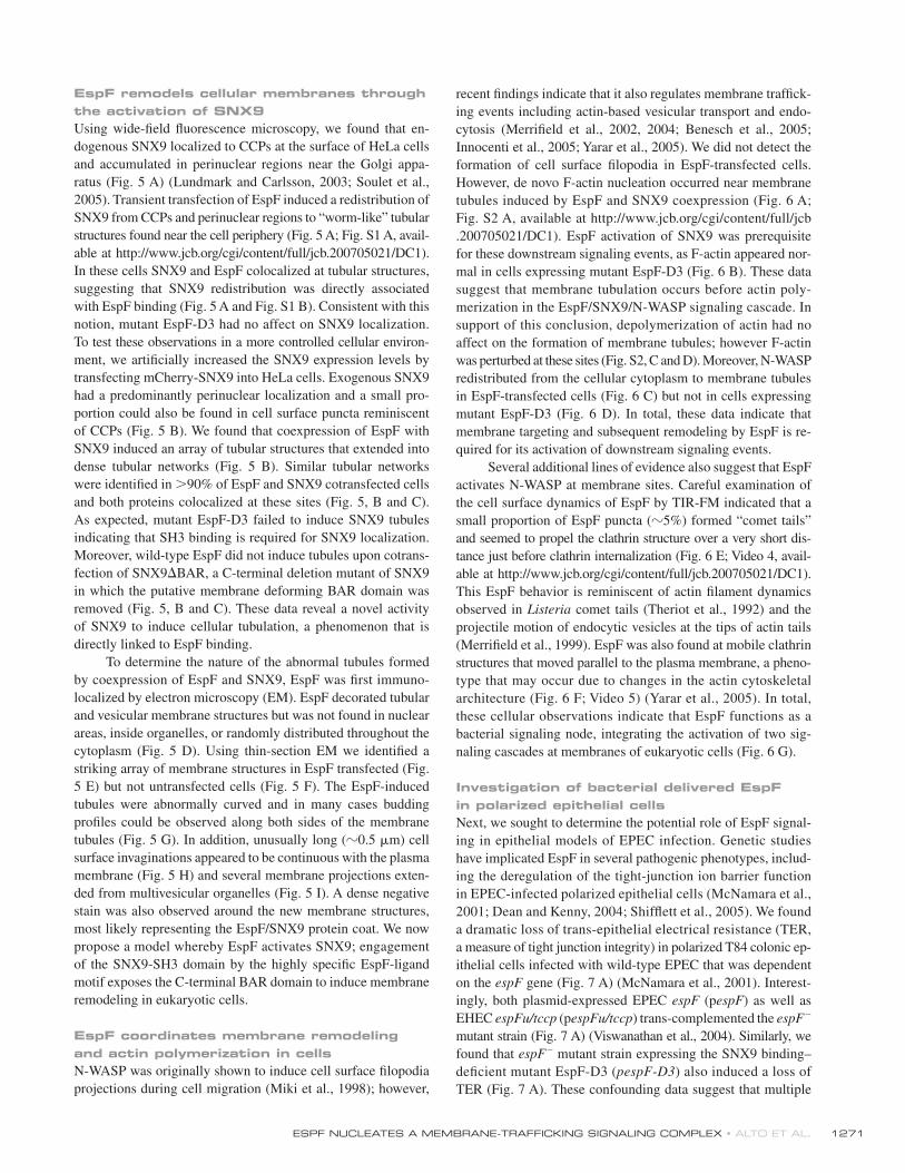
EspF remodels cellular membranes through the activation of SNX9Using wide-! eld " uorescence microscopy, we found that en-dogenous SNX9 localized to CCPs at the surface of HeLa cells and accumulated in perinuclear regions near the Golgi appa-ratus (Fig. 5 A) (Lundmark and Carlsson, 2003; Soulet et al., 2005). Transient transfection of EspF induced a redistribution of SNX9 from CCPs and perinuclear regions to “worm-like” tubular structures found near the cell periphery (Fig. 5 A; Fig. S1 A, avail-able at http://www.jcb.org/cgi/content/full/jcb.200705021/DC1). In these cells SNX9 and EspF colocalized at tubular structures, suggesting that SNX9 redistribution was directly associated with EspF binding (Fig. 5 A and Fig. S1 B). Consistent with this notion, mutant EspF-D3 had no affect on SNX9 localization. To test these observations in a more controlled cellular environ-ment, we arti! cially increased the SNX9 expression levels by transfecting mCherry-SNX9 into HeLa cells. Exogenous SNX9 had a predominantly perinuclear localization and a small pro-portion could also be found in cell surface puncta reminiscent of CCPs (Fig. 5 B). We found that coexpression of EspF with SNX9 induced an array of tubular structures that extended into dense tubular networks (Fig. 5 B). Similar tubular networks were identi! ed in >90% of EspF and SNX9 cotransfected cells and both proteins colocalized at these sites (Fig. 5, B and C). As expected, mutant EspF-D3 failed to induce SNX9 tubules indicating that SH3 binding is required for SNX9 localization. Moreover, wild-type EspF did not induce tubules upon cotrans-fection of SNX9%BAR, a C-terminal deletion mutant of SNX9 in which the putative membrane deforming BAR domain was removed (Fig. 5, B and C). These data reveal a novel activity of SNX9 to induce cellular tubulation, a phenomenon that is directly linked to EspF binding.
To determine the nature of the abnormal tubules formed by coexpression of EspF and SNX9, EspF was ! rst immuno- localized by electron microscopy (EM). EspF decorated tubular and vesicular membrane structures but was not found in nuclear areas, inside organelles, or randomly distributed throughout the cytoplasm (Fig. 5 D). Using thin-section EM we identi! ed a striking array of membrane structures in EspF transfected (Fig. 5 E) but not untransfected cells (Fig. 5 F). The EspF-induced tubules were abnormally curved and in many cases budding pro! les could be observed along both sides of the membrane tubules (Fig. 5 G). In addition, unusually long (!0.5 $m) cell surface invaginations appeared to be continuous with the plasma membrane (Fig. 5 H) and several membrane projections exten-ded from multivesicular organelles (Fig. 5 I). A dense negative stain was also observed around the new membrane structures, most likely representing the EspF/SNX9 protein coat. We now propose a model whereby EspF activates SNX9; engagement of the SNX9-SH3 domain by the highly speci! c EspF-ligand motif exposes the C-terminal BAR domain to induce membrane remodeling in eukaryotic cells.
EspF coordinates membrane remodeling and actin polymerization in cellsN-WASP was originally shown to induce cell surface ! lopodia projections during cell migration (Miki et al., 1998); however,
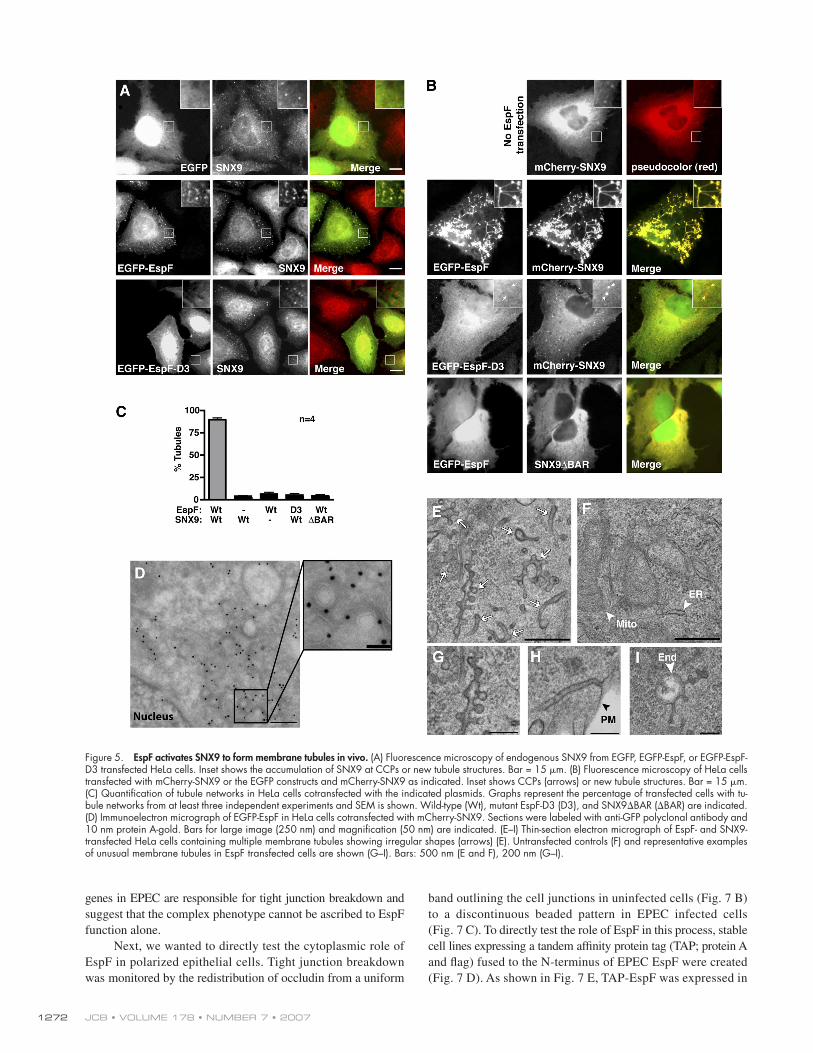
recent ! ndings indicate that it also regulates membrane traf! ck-ing events including actin-based vesicular transport and endo-cytosis (Merri! eld et al., 2002, 2004; Benesch et al., 2005; Innocenti et al., 2005; Yarar et al., 2005). We did not detect the formation of cell surface ! lopodia in EspF-transfected cells. However, de novo F-actin nucleation occurred near membrane tubules induced by EspF and SNX9 coexpression (Fig. 6 A; Fig. S2 A, available at http://www.jcb.org/cgi/content/full/jcb.200705021/DC1). EspF activation of SNX9 was prerequisite for these downstream signaling events, as F-actin appeared nor-mal in cells expressing mutant EspF-D3 (Fig. 6 B). These data suggest that membrane tubulation occurs before actin poly-merization in the EspF/SNX9/N-WASP signaling cascade. In support of this conclusion, depolymerization of actin had no affect on the formation of membrane tubules; however F-actin was perturbed at these sites (Fig. S2, C and D). Moreover, N-WASP redistributed from the cellular cytoplasm to membrane tubules in EspF-transfected cells (Fig. 6 C) but not in cells expressing mutant EspF-D3 (Fig. 6 D). In total, these data indicate that membrane targeting and subsequent remodeling by EspF is re-quired for its activation of downstream signaling events.
Several additional lines of evidence also suggest that EspF activates N-WASP at membrane sites. Careful examination of the cell surface dynamics of EspF by TIR-FM indicated that a small proportion of EspF puncta (!5%) formed “comet tails” and seemed to propel the clathrin structure over a very short dis-tance just before clathrin internalization (Fig. 6 E; Video 4, avail-able at http://www.jcb.org/cgi/content/full/jcb.200705021/DC1). This EspF behavior is reminiscent of actin ! lament dynamics observed in Listeria comet tails (Theriot et al., 1992) and the projectile motion of endocytic vesicles at the tips of actin tails (Merri! eld et al., 1999). EspF was also found at mobile clathrin structures that moved parallel to the plasma membrane, a pheno-type that may occur due to changes in the actin cytoskeletal architecture (Fig. 6 F; Video 5) (Yarar et al., 2005). In total, these cellular observations indicate that EspF functions as a bacterial signaling node, integrating the activation of two sig-naling cascades at membranes of eukaryotic cells (Fig. 6 G).
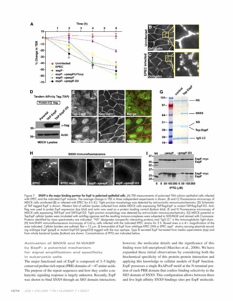
Investigation of bacterial delivered EspF in polarized epithelial cellsNext, we sought to determine the potential role of EspF signal-ing in epithelial models of EPEC infection. Genetic studies have implicated EspF in several pathogenic phenotypes, includ-ing the deregulation of the tight-junction ion barrier function in EPEC-infected polarized epithelial cells (McNamara et al., 2001; Dean and Kenny, 2004; Shif" ett et al., 2005). We found a dramatic loss of trans-epithelial electrical resistance (TER, a measure of tight junction integrity) in polarized T84 colonic ep-ithelial cells infected with wild-type EPEC that was dependent on the espF gene (Fig. 7 A) (McNamara et al., 2001). Interest-ingly, both plasmid-expressed EPEC espF (pespF) as well as EHEC espFu/tccp (pespFu/tccp) trans-complemented the espF" mutant strain (Fig. 7 A) (Viswanathan et al., 2004). Similarly, we found that espF" mutant strain expressing the SNX9 binding–de! cient mutant EspF-D3 (pespF-D3) also induced a loss of TER (Fig. 7 A). These confounding data suggest that multiple
JCB • VOLUME 178 • NUMBER 7 • 2007 1272
genes in EPEC are responsible for tight junction breakdown and suggest that the complex phenotype cannot be ascribed to EspF function alone.
Next, we wanted to directly test the cytoplasmic role of EspF in polarized epithelial cells. Tight junction breakdown was monitored by the redistribution of occludin from a uniform
band outlining the cell junctions in uninfected cells (Fig. 7 B) to a discontinuous beaded pattern in EPEC infected cells (Fig. 7 C). To directly test the role of EspF in this process, stable cell lines expressing a tandem af! nity protein tag (TAP; protein A and " ag) fused to the N-terminus of EPEC EspF were created (Fig. 7 D). As shown in Fig. 7 E, TAP-EspF was expressed in
Figure 5. EspF activates SNX9 to form membrane tubules in vivo. (A) Fluorescence microscopy of endogenous SNX9 from EGFP, EGFP-EspF, or EGFP-EspF-D3 transfected HeLa cells. Inset shows the accumulation of SNX9 at CCPs or new tubule structures. Bar = 15 $m. (B) Fluorescence microscopy of HeLa cells transfected with mCherry-SNX9 or the EGFP constructs and mCherry-SNX9 as indicated. Inset shows CCPs (arrows) or new tubule structures. Bar = 15 $m. (C) Quantifi cation of tubule networks in HeLa cells cotransfected with the indicated plasmids. Graphs represent the percentage of transfected cells with tu-bule networks from at least three independent experiments and SEM is shown. Wild-type (Wt), mutant EspF-D3 (D3), and SNX9%BAR (%BAR) are indicated. (D) Immunoelectron micrograph of EGFP-EspF in HeLa cells cotransfected with mCherry-SNX9. Sections were labeled with anti-GFP polyclonal antibody and 10 nm protein A-gold. Bars for large image (250 nm) and magnifi cation (50 nm) are indicated. (E–I) Thin-section electron micrograph of EspF- and SNX9-transfected HeLa cells containing multiple membrane tubules showing irregular shapes (arrows) (E). Untransfected controls (F) and representative examples of unusual membrane tubules in EspF transfected cells are shown (G–I). Bars: 500 nm (E and F), 200 nm (G–I).
ESPF NUCLEATES A MEMBRANE-TRAFFICKING SIGNALING COMPLEX • ALTO ET AL. 1273
every cell and localized to small puncta near the apical cell surface. SNX9 interaction seemed to be required for this localization as the SH3-binding mutant EspF-D3 was distributed throughout the cytoplasm and accumulated in nuclear regions (Fig. 7 F). Importantly, MDCK cells expressing either EspF or EspF-D3 formed normal tight junctions (Fig. 7, E and F), suggesting that EspF activity alone is not suf! cient to break down tight junc-tions. Next, we biochemically puri! ed EspF from MDCK cells as an unbiased approach to determine protein complexes asso-ciated with EspF in a polarized cell type (Fig. 7 G). TAP-puri! ed EspF or control parental cell lines were separated by SDS-PAGE and Coomassie stain indicated that the major EspF-binding pro-tein had a molecular weight of !70 kD (Fig. 7 G). This protein was unambiguously identi! ed as canine SNX9 (MW ( 71 kD) by tandem mass spectrometry from two independent experiments (see Materials and methods). Thus, SNX9 is the major target of cytoplasmic EspF in polarized epithelial cells.
Finally, we sought to determine if the localization and function of bacterial delivered EspF relies on interaction with SNX9 in polarized cell types. Human CaCo2 intestinal epithelial cells were infected with wild-type EPEC or the espF" mutant strain. Whereas wild-type EPEC induced SNX9 membrane tu-bules, this phenotype did not occur in espF" mutant EPEC- infected cells (Fig. 7 H). The wild-type phenotype was rescued by introducing a plasmid encoding the espF allele (pespF ) into the mutant espF" strain (Fig. 7 H). However, there was no pheno-type associated with mutant strains complemented with SNX9 binding–de! cient espF-D3 (pespF-D3) (Fig. 7 H). Similar results
were also found in EPEC-infected HeLa cells (unpublished data). Western blot analysis con! rmed the expression and secretion of the EspF proteins by wild-type and trans-complemented strains (Fig. 7 I). These data further indicate that the biochemical activ-ities of EspF that we have described in vitro are likely to occur during EPEC pathogenesis of intestinal epithelial cells.
DiscussionIn this study we have obtained evidence for a new model of EspF function during bacterial pathogenesis. Our data suggest that EspF physically links membrane-traf! cking proteins to the actin polymerization machinery in host cells. Several lines of evi-dence support this hypothesis. First, EspF interacts directly with SNX9 through a highly evolved SH3 domain–binding motif that is repeated several times throughout the EspF coding sequence. Second, EspF stimulates N-WASP/Arp2/3 actin polymerization in reconstitution experiments. Third, our live-cell imaging studies revealed that EspF dynamically localized to CCPs. This subcellu-lar localization is similar to those previously described for both SNX9 and N-WASP (Merri! eld et al., 2004; Benesch et al., 2005; Innocenti et al., 2005; Soulet et al., 2005). Fourth, EspF induced membrane remodeling through the activation of SNX9, and the actin polymerization pro! les at these sites correlated with the ability of EspF to interact with N-WASP in cells. Finally, the EspF-dependent redistribution of SNX9 during bacterial infection sug-gests that the biochemical and cellular activities that we have described are likely to occur during natural EPEC pathogenesis.
Figure 6. EspF induces N-WASP activation at membrane tubules. (A and B) Fluorescence microscopy images depicting mCherry-SNX9 and FITC-phalloidin (actin) in EspF (A) or EspF-D3 (B) transfected HeLa cells. Bar = 15 $m. (C and D) Fluorescence microscopy images of HeLa cells cotransfected with EGFP-EspF (C) or EGFP-EspF-D3 (D) with mCherry-SNX9 and N-WASP-V5. N-WASP was detected by anti-V5 immunocytochemistry. Bar = 15 $m. (E) Time series of EspF puncta (EGFP-EspF) exhibiting a “rocketing” phenotype (arrow) from live cell TIR-FM. Cartoon below. (F) Time series of EspF puncta (EGFP-EspF) exhibit-ing a mobile phenotype from live cell TIR-FM. Images are correlated with simultaneous Clc-DsRed dynamics showing the movement of a single CCP (arrow). (G) Schematic of EspF functioning as a node of signaling integration at eukaryotic membranes.
JCB • VOLUME 178 • NUMBER 7 • 2007 1274
Activation of SNX9 and N-WASP by EspF: a potential mechanism for signal amplifi cation and specifi city in eukaryotic cellsThe major functional unit of EspF is composed of 3–5 highly conserved proline-rich repeat (PRR) domains of !47 amino acids. The purpose of the repeat sequences and how they confer a eu-karyotic signaling response is largely unknown. Recently, EspF was shown to bind SNX9 through an SH3 domain interaction;
however, the molecular details and the signi! cance of this ! nding were left unexplored (Marches et al., 2006). We have expanded these initial observations by considering both the biochemical speci! city of this protein–protein interaction and applying this knowledge to cellular models of EspF function. EspF possesses a single RxAPxxP motif at the N-terminal por-tion of each PRR domain that confers binding selectivity to the SH3 domain of SNX9. This con! guration allows between three and ! ve high af! nity SNX9 bindings sites per EspF molecule.
Figure 7. SNX9 is the major binding partner for EspF in polarized epithelial cells. (A) TER measurements of polarized T84 colonic epithelial cells infected with EPEC and the indicated EspF mutants. The average change in TER in three independent experiments is shown. (B and C) Fluorescence microscopy of MDCK cells uninfected (B) or infected with EPEC for 4 h (C). Tight junction morphology was detected by anti-occludin immunocytochemistry. (D) Schematic of TAP tagged EspF is shown. Western blot of cellular lysates collected from stable MDCK cells expressing TAP-fl ag-EspF or mutant TAP-fl ag-EspF-D3. Anti-fl ag was used to probe EspF expression (top blot) and actin was used as a protein loading control (bottom blot). (E and F) Fluorescence microscopy of MDCK cells expressing TAP-EspF and TAP-EspF-D3. Tight junction morphology was detected by anti-occludin immunocytochemistry. (G) MDCK parental or Tap-EspF cellular lysates were incubated with anti-fl ag agarose and the resulting immuno-complexes were subjected to SDS-PAGE and stained with Coomassie. Proteins identifi ed by mass spectrometry are indicated. “NS” designates nonspecifi c interacting proteins and “IgG LC” is the immunoglobulin light chain. (H) Anti-SNX9 immunofl uorescence microscopy of CaCo2 cells infected with the indicated EPEC strains for 3 h. Boxed area is a 4& magnifi cation of the area indicated. Cellular borders are outlined. Bar = 15 $m. (I) Immunoblot of EspF from wild-type EPEC (Wt) or EPEC espF" strains carrying plasmids encod-ing wild-type EspF (pespf) or mutant EspF-D3 (pespf-D3) tagged with the myc epitope. Type III secreted EspF harvested from media supernatants (top) and from whole bacterial lysates (bottom) are shown. Concentrations of IPTG are indicated below.
ESPF NUCLEATES A MEMBRANE-TRAFFICKING SIGNALING COMPLEX • ALTO ET AL. 1275
Thus, the PRR domains may provide the bacterial effector a mechanism to increase protein avidity, or more interestingly, a means to amplify the SNX9 signal through oligomerization (Yarar et al., 2007). Consistent with these biochemical ! ndings, engagement of the SNX9 SH3 domain by EspF induced robust membrane tubulation in cells. We now propose a concerted model for EspF function; direct binding to the SH3 domain of SNX9 transmits a membrane-remodeling signal to the C-terminal BAR domain. Importantly, similar membrane tubules have been observed upon expression of other BAR domain proteins (Peter et al., 2004), and we found that EspF-induced membrane tubu-lation did not occur with a BAR domain deletion mutant of SNX9. The potential importance and mechanism of these intra-molecular interactions will be clari! ed as the EspF/SNX9 effec-tor system is analyzed in biophysical detail.
Although we have focused primarily on the mechanisms of SNX9 activation by EspF, we have also discovered that EspF regulates actin cytoskeletal dynamics. In the context of the pyrene-actin assembly assay, the PRR domains in EspF are suf-! cient to induce N-WASP/Arp2/3 actin nucleation. EspF binds directly to the CRIB regulatory domain in N-WASP, a phenom-enon similar to the reported mechanism of the Rho GTPase Cdc42. Similar ! ndings have been previously reported for the functionally distinct PRR protein EspFu/TccP (Garmendia et al., 2006), indicating that both EspF and EspFu/TccP may use similar residues to independently activate N-WASP (Fig. 1). In addition, we have extended these ! ndings by demonstrating that EspF nucleates a trimeric complex composed of SNX9 and N-WASP. This complex may provide EHEC 0157:H7 a potent mechanism for eukaryotic signal ampli! cation and downstream signaling speci! city during bacterial pathogenesis. For exam-ple, bacterial pedestals could be modi! ed by EspFu/TccP acti-vation of N-WASP (Campellone et al., 2004) while membrane traf! cking would be regulated by EspF and the SNX9/N-WASP complex. This idea is particularly intriguing in light of studies by Yarar et al. (2007) that demonstrate the importance of SNX9 oligomerization to activate N-WASP during multiple modes of eukaryotic endocytosis.
EspF mimics an endogenous actin/membrane-signaling module to regulate membrane traffi cking eventsGenetic studies have linked EspF activity to a wide range of biological activities, including mitochondrial apoptosis (Crane et al., 2001; Nougayrede and Donnenberg, 2004), epithelial barrier disruption (McNamara et al., 2001), apical microvilli effacement (Dean et al., 2006), and inhibition of macrophage phagocytosis (Quitard et al., 2006). We found EspF to be neces-sary but not suf! cient to regulate epithelial tight junction archi-tecture during bacterial pathogenesis (see Fig. 7), suggesting that the EspF phenotypes observed in bacterial genetic studies may result from the concerted actions of several type III effector proteins or additional bacterial signaling mechanisms (Dean et al., 2006). Taking an alternative approach to bacterial genetics, we found that puri! ed EspF binds directly to SNX9 in vitro and activates N-WASP in reconstitution assays. These activities could have direct and profound effects on membrane traf! cking
and the overall homeostasis of infected intestinal epithelial cells. For example, a recent report suggests that the Aquaporin (AQP) water channels are mislocalized during A/E pathogen infection, and this phenotype requires EspF expression (Guttman et al., 2007). In addition, EspF promotes the internalization of tight junction proteins in vivo (Guttman et al., 2006), poten-tially through a membrane-traf! cking phenotype. These possi-bilities are consistent with our observed localization of EspF at clathrin structures, as membrane traf! cking is required for epithelial tight junction maintenance and the formation of the apical/basolateral poles (Wells et al., 2006).
It is intriguing to note that the coordinated activation of SNX9 and N-WASP by EspF may actually represent a patho-genic strategy to mimic a natural host SNX9/N-WASP signaling complex. In earlier studies, our lab found that the Drosophila SNX9 (DSH3PX1) links axonal guidance receptors directly to N-WASP and the actin cytoskeleton (Worby et al., 2001). In ad-dition, more recent studies suggest that engagement of the T cell antigen receptor (TCR) combined with costimulation of CD28 evoked the formation of a SNX9/WASP complex at endocytic sites of T cells (Badour et al., 2007). Together, these data es-tablish the importance of an endogenous SNX9 and N-WASP signaling complex to regulate diverse eukaryotic traf! cking events. Importantly, the functional link between these proteins may extend beyond clathrin-mediated endocytosis. Yarar et al. (2007) have described a mechanism in which SNX9 directly ac-tivates N-WASP for the purpose of regulating dorsal membrane ruf" es, clathrin-independent " uid-phase endocytosis, and uptake of GPI-anchored proteins. Thus, our data suggest for the ! rst time that EspF hijacks an endogenous SNX9/N-WASP signaling complex involved in multiple host cellular regulatory pathways.
Materials and methodsPlasmidsThe espf gene from EHEC O157:H7 (GenBank accession no.#AP002566) and the EPEC E2348/69 espf were PCR cloned in-frame into pEGFP-C2 (CLONTECH Laboratories, Inc.). The espfu/tccp gene was cloned from a clinical isolate of EHEC 0157:H7 and has the sequence identical to GI: 15831969. The yeast two-hybrid clone of mouse SNX9 (NM_025664) residues 1–111, as well as full-length human SNX9, N-WASP, Grb2, NckI, and Nck2 were Topo cloned into pcDNA3.1-V5-His (Invitrogen). To produce mCherry-SNX9, the EGFP from pEGFP-C1 was replaced with mCherry by PCR subcloning. For bacterial expression, 47 amino acid N-terminal dele-tions (%N47) of EspF were PCR subcloned into pGEX-4T1 (GST-tag) (GE Healthcare) or pet30A (6xHis tag) (Novagen). EspF-D3 was generated with Multi-site QuikChange Site-Directed Mutagenesis (Stratagene) following the manufacturer’s instructions. For bacterial complementation, the EPEC espf or mutant espf-D3 was Topo cloned into pTrcHis2 with a c-myc epitope tag (Invitrogen). GST and His-tagged mini-N-WASP were obtained from Wendall Lim (University of California, San Francisco, San Francisco, CA; Prehoda et al., 2000). All constructs were verifi ed by DNA sequencing.
Yeast two-hybridThe yeast expression vector pLexA encoded a gene with N-terminal LexA binding domain and residues 1–248 of EHEC EspF. 250 $g of a d 9.5 and 10.5 mouse embryo library in VP16 were screened using the yeast two-hybrid system as previously described (Alto et al., 2006).
Cell culture, transfection, and wide-fi eld fl uorescence microscopyCell culture, transfections, and wide-fi eld fl uorescence microscopy was per-formed as previously described (Alto et al., 2006). Images were acquired on a microscope (Axiovert; Carl Zeiss Microimaging, Inc.) using a digital camera (MicroMax; Roper-Princeton Instruments) controlled by MetaFluor software
JCB • VOLUME 178 • NUMBER 7 • 2007 1276
(Universal Imaging, Corp.). Optical fi lters were obtained from Chroma Technologies and 40 or 63& objectives were used for image acquisition.
AntibodiesConcentrations of anti-GFP polyclonal and monoclonal (CLONTECH Laboratories, Inc.), anti-His (QIAGEN), anti-GST, anti-fl ag M2, anti-fl ag polyclonal (Sigma-Aldrich), anti-HA (Covance), and anti-occludin (Zymed Laboratories) were used for immunoprecipitation, immunocytochemistry, and immunoblotting as recommended by the manufacturer. Rabbit poly-clonal SNX9 antibody was used at 1:200 (Soulet et al., 2005). Rabbit polyclonal EspF was used at 1:10,000 (McNamara et al., 2001). Anti-tubulin antibody was used at 1:200 (Sigma-Aldrich), and Latrunculin-A at 1 $M (Calbiochem).
Recombinant protein production and GST pull-down assaysRecombinant 6&His EspF%47, mini-N-WASP (from Wendall Lim), N-WASP %EVH1 (from Jack Taunton; University of California, San Francisco, San Francisco, CA) and SNX9 were produced in BL-21/DE3 E. coli strain fol-lowing standard methods. Cells were lysed in either His buffer (50 mM Tris, pH 7.4, 300 mM NaCl, 10 mM imidazole, and 0.5% Triton-X 100) or GST buffer (phosphate-buffered saline) supplemented with protease cock-tail (Roche). Proteins were purifi ed with Nickel agarose (QIAGEN) or glutathione-Sepharose (GE Healthcare) following the manufacturer’s instructions. Proteins were dialyzed overnight in 2 l of lysis buffer at 4)C. Glycerol was added to 30% and aliquots were stored at "80)C. For GST pull-down assays, 10 $g of recombinant GST proteins immobilized to glutathione-Sepharose was incubated with 50 $l of [35S]-methionine (GE Healthcare) produced with TNT reaction (Promega) for 4 h at 4)C. Proteins were separated by SDS-PAGE, dried, and exposed by autoradiography.
Peptide array and SNX9 overlayImmobilized peptides were synthesized on cellulose paper using a Multi-pep Autospot synthesis robot following the manufacturer’s directions (Intavis AG). Membranes were blocked in 5% milk in TBS-Tween. 50 $l of [35S]-methionine SNX9 produced by TNT (Promega) was added to 5 ml of block solution and overlayed onto peptide membranes for 4 h at room temperature. Membranes were washed 3& 10 min in TBS-T and exposed by autoradiography.
Fluorescence polarizationFITC-labeled peptides (Cell Essentials) used for fl uorescence polarization include EspF residues 69–86 (FITC-A T S F T P S R P A P P P T S G Q A ) or control pep-tide (FITC-Q I A K R R R L S S L R A ). Peptides (5 nM) were suspended to working dilutions in PBS with 5 mg/ml BSA. Increasing concentrations of purifi ed GST-SNX9-SH3 or control GST proteins were mixed with peptides in 100 $l and incubated at room temperature for 10 min. Fluorescence polarization (FP) was measured on a GENios Pro (TECAN) fi tted with FP excitation and emission fi lters, 485/535 nm. Polarization values (mP) were determined at equilibrium and normalized to the highest value of saturation. Saturation binding curve was generated with PRISM software (Graph Pad) and disso-ciation constants (KD) were calculated from the nonlinear regression curve from averages of three independent experiments.
Phage displayA library of random dodecapeptides fused to the N terminus of the M13 gene-8 major coat protein was constructed and cycled through rounds of binding selections with the bacterially expressed SH3 domain immobilized on 96-well Maxisorp immunoplates (NUNC), as described previously (Sidhu et al., 2000; Laura et al., 2002). After four rounds of selection, individual phage were isolated and analyzed in a phage ELISA. Phage that bound to the SH3 domain was subjected to DNA sequence analysis.
Actin polymerization assays1:1 mix of pyrene actin (!40% labeled, fi nal is !20% labeled) to cold G-actin (Cytoskeleton, Inc.) were mixed in G-buffer (5 mM Tris, pH 8.0, 0.2 mM CaCl2, 0.2 mM ATP, and 1 mM DTT) and centrifuged at 100,000 g for 2 h. A fi nal of 2.5 $M pyrene actin mix was added to 270 $l of Arp2/3 buffer (20 mM Tris, pH 7.5, 25 mM KCl, 1 mM MgCl2, 0.1 mM ATP, and 1 mM ATP). 40 nm bovine Arp2/3 (a gift from Tom Pollard; Yale University, New Haven, CT) and 100 nM mini-N-WASP or N-WASP %EVH1 was mixed with various concentrations of GST, GST-EspF, or SNX9 constructs in 30 $l of 10& actin polymerization buffer (100 mM Tris, pH 7.5, 500 mM KCl, 20 mM MgCl2, and 10 mM ATP). 270 $l pyrene actin (2.5 $M) was mixed with 30 $l mini-N-WASP-Arp2/3 and actin-assembly kinetics was monitored by pyrene fl uorescence over a 10–20-min time interval. Peak fl uorescence
values were normalized to 2.4 $M actin consumption and graphed with PRISM Software.
Total internal refl ection fl uorescence microscopy (TIR-FM)TIR-FM imaging procedures and microscopic manipulations were con-ducted as previously reported (Yarar et al., 2005). Swiss 3T3 cells stably expressing clathrin-light chain DsRed (Merrifi eld et al., 2002) were grown to 70% confl uency in a 100-mm tissue culture dish in DME # 10% fetal calf serum and transfected with 20 $g EGFP-EspF or EGFP-EspF-D3 DNA using Lipofectamine 2000 (Invitrogen) according to the manufacturer’s instruc-tions. Cells were allowed to recover in fresh medium for 2 h, replated onto 22 & 22-mm, no. 1.5 glass coverslips (Corning; n ( 1.523), and fi lmed the following morning.
Total internal refl ection fl uorescence (TIRF) microscopy was performed using an inverted microscope (TE2000U; Nikon) custom-modifi ed to allow for through-the-objective multispectral total-internal refl ection fl uorescence micros-copy using a 100&, 1.45 NA objective (Nikon), as previously described (Yarar et al., 2005). TIRF images were taken for 10 min at 2–3-s intervals, with a 100–300-ms exposure time, depending on the intensity of the signal.
Image analysisLow expressing EGFP-EspF or EGFP-EspF-D3 cells stably expressing Clc-DsRed were chosen for !8–12 min time-lapse movies. Stacks of red (Clc-DsRed) and green (GFP-EspF) images were de-interleaved into separate stacks. Clc-DsRed stacks were analyzed for candidate clathrin structures that were stable for 30 s before disappearance and seemed likely to repre-sent single CCPs. Clathrin structures were excluded similar to the criteria described previously (Merrifi eld et al., 2002). We marked the position of each Clc-DsRed candidate with a !1–2-$m diameter circle and observed GFP-EspF dynamics at these sites. This procedure yielded the coordinates of the CCS in both the red and green channels so that red and green re-gions precisely centered on the CCS could be excised and stored as minis-tacks for analysis. All time and fl uorescence intensity measurements were based on these criteria. Image analysis was done with Metamorph (Universal Imaging Corp), Excel, and graphed using PRISM (Graph Pad).
To measure 25 CCPs, we fi rst chose 25 candidate CCPs from 8 dif-ferent cells. Each CCP appeared during the 10-min video series was stable for at least 30 s, disappeared from the evanescent fi eld, and did not re-appear for the remainder of the video series. The peak fl uorescence inten-sity measurements were collected for each individual EspF and clathrin image and averaged background values were subtracted. Clathrin results were normalized to the mean fl uorescence during the 21 s (7 frames) before the peak clathrin fl uorescence (Merrifi eld et al., 2002). The time of clathrin departure (time 0) was defi ned as the last frame in which the normalized fl uorescence value was within 90% of peak fl uorescence and the signal reached <5% over the following 45 s of imaging. Each clathrin event (n ( 25) was aligned to the time of departure and averaged. For EspF, the results of each event were normalized to the mean of three contiguous intensity values, including the peak value, then aligned to the time of clathrin depar-ture and fi nally averaged (Merrifi eld et al., 2002). Kymograph analysis was performed using MetaMorph software.
EPEC infection experimentsT84 colonic epithelial cells were cultured and TER experiments were per-formed as described previously (McNamara et al., 2001). EPEC and espF" mutant strains (McNamara and Donnenberg, 1998) were comple-mented with plasmid EspF subcloned into pTrcHis2 (Invitrogen). Plasmid EspF was expressed in EPEC by incubation with 100 $M IPTG for 3 h be-fore EPEC infections. T84 cells were infected with an MOI of 100 for the indicated time points. CaCo2 experiments were performed by infecting cells with EPEC or the indicated mutants for 4 h before fi xation and immuno-fl uorescence processing.
MDCK stable cell lines and mass spectrometryTAP-tagged EspF or mutant EspF-D3 were subcloned into pCDNA4T/O and MDCK stable cell lines were generated following the manufacturer’s instructions (Invitrogen). Clonal lines expressing TAP-EspF or TAP-EspF-D3 were selected with 500 $g/ml zeocin and confi rmed by Western blot and immunofl uorescence microscopy. Purifi cation of TAP-EspF was performed as follows: MDCK cells were lysed in 50 mM Hepes, pH 7.5, 150 mM NaCl, 5% glycerol, 0.5% TX-100, 1.5 mM MgCl2, and 1 mM EGTA and incu-bated with anti-fl ag M2 beads for 2 h. The beads were washed 3& 10 min with lysis buffer and eluted with LDS-PAGE buffer (Invitrogen). Immuno-complexes were run down SDS-PAGE and gels were stained with Coo-massie. Proteins were in-gel digested and analyzed with nano liquid chromatography tandem mass spectrometry. Acquired data was searched
ESPF NUCLEATES A MEMBRANE-TRAFFICKING SIGNALING COMPLEX • ALTO ET AL. 1277
with Inspect against a dog database (National Center for Biotechnology Information, Canis familiaris fasta protein database). Identifi ed peptides were manually verifi ed to be canine SNX9: aa 218–237, S A A P Y F K D S E S A E A G-G A Q R ; aa 313–327, S Y I E Y Q L T P T N T N R ; aa 346–359, F G S A I P I P S L P D K ; aa 515–529, T Y E E I A G L V A E Q P K ; aa 543–557, K.G F L G C F P D I I G A H K .G; and aa 605–613, IYDYNSVIR.
Online supplemental materialFig. S1 shows membrane tubule formation induced by EspF, and colocali-zation of endogenous SNX9 at these sites (Fig. S1, A and B). Fig. S2 dem-onstrates that actin is polymerized de novo at sites of EspF/SNX9 membrane tubules. Video 1 shows dynamics of EGFP-EspF and Clc-DsRed in living Swiss 3T3 cells captured by TIR-FM. Video 2 shows EspF and clathrin dynamics at a single CCP. Video 3 shows a dynamic membrane tubule induced by EspF expressing at the cell surface. Video 4 demon-strates a “rocketing” clathrin structures seemingly being propelled by EspF. Video 5 demonstrates a splitting clathrin structures with EspF localizing to the center of this event.
We would like to thank C.J. Allison, Timo Meerloo, and Ingrid Niesman for expert technical assistance. Critical reagents were provided by Wendall Lim and Tom Pollard, for which we are grateful. We would like to thank members of the Dixon lab for critical reading of this manuscript and for helpful scientifi c discussions.
This research was supported by Grants from the National Institutes of Health, the Walther Cancer Institute, the Ellison Foundation, and the Howard Hughes Medical Institute (to J.E. Dixon). N.M. Alto was supported by Hema-tology and Diabetes National Institutes of Health training grants. Grants from NCIC support C. Boone and R. Tonikian.
Submitted: 4 May 2007Accepted: 27 August 2007
ReferencesAlto, N.M., F. Shao, C.S. Lazar, R.L. Brost, G. Chua, S. Mattoo, S.A. McMahon,
P. Ghosh, T.R. Hughes, C. Boone, and J.E. Dixon. 2006. Identi! cation of a bacterial type III effector family with G protein mimicry functions. Cell. 124:133–145.
Badour, K., M.K. McGavin, J. Zhang, S. Freeman, C. Vieira, D. Filipp, M. Julius, G.B. Mills, and K.A. Siminovitch. 2007. Interaction of the Wiskott-Aldrich syndrome protein with sorting nexin 9 is required for CD28 endocytosis and cosignaling in T cells. Proc. Natl. Acad. Sci. USA. 104:1593–1598.
Benesch, S., S. Polo, F.P. Lai, K.I. Anderson, T.E. Stradal, J. Wehland, and K. Rottner. 2005. N-WASP de! ciency impairs EGF internalization and actin assembly at clathrin-coated pits. J. Cell Sci. 118:3103–3115.
Campellone, K.G., D. Robbins, and J.M. Leong. 2004. EspFU is a translocated EHEC effector that interacts with Tir and N-WASP and promotes Nck-independent actin assembly. Dev. Cell. 7:217–228.
Co, C., D.T. Wong, S. Gierke, V. Chang, and J. Taunton. 2007. Mechanism of actin network attachment to moving membranes: barbed end capture by N-WASP WH2 domains. Cell. 128:901–913.
Cornelis, G.R., and F. Van Gijsegem. 2000. Assembly and function of type III secretory systems. Annu. Rev. Microbiol. 54:735–774.
Crane, J.K., B.P. McNamara, and M.S. Donnenberg. 2001. Role of EspF in host cell death induced by enteropathogenic Escherichia coli. Cell. Microbiol. 3:197–211.
Dean, P., and B. Kenny. 2004. Intestinal barrier dysfunction by enteropathogenic Escherichia coli is mediated by two effector molecules and a bacterial surface protein. Mol. Microbiol. 54:665–675.
Dean, P., M. Maresca, S. Schuller, A.D. Phillips, and B. Kenny. 2006. Potent diarrheagenic mechanism mediated by the cooperative action of three enteropathogenic Escherichia coli-injected effector proteins. Proc. Natl. Acad. Sci. USA. 103:1876–1881.
Galan, J.E., and A. Collmer. 1999. Type III secretion machines: bacterial devices for protein delivery into host cells. Science. 284:1322–1328.
Garmendia, J., A.D. Phillips, M.F. Carlier, Y. Chong, S. Schuller, O. Marches, S. Dahan, E. Oswald, R.K. Shaw, S. Knutton, and G. Frankel. 2004. TccP is an enterohaemorrhagic Escherichia coli O157:H7 type III ef-fector protein that couples Tir to the actin-cytoskeleton. Cell. Microbiol. 6:1167–1183.
Garmendia, J., M.F. Carlier, C. Egile, D. Didry, and G. Frankel. 2006. Characterization of TccP-mediated N-WASP activation during entero-haemorrhagic Escherichia coli infection. Cell. Microbiol. 8:1444–1455.
Gruenheid, S., and B.B. Finlay. 2003. Microbial pathogenesis and cytoskeletal function. Nature. 422:775–781.
Guttman, J.A., F.N. Samji, Y. Li, A.W. Vogl, and B.B. Finlay. 2006. Evidence that tight junctions are disrupted due to intimate bacterial contact and not in" ammation during attaching and effacing pathogen infection in vivo. Infect. Immun. 74:6075–6084.
Guttman, J.A., F.N. Samji, Y. Li, W. Deng, A. Lin, and B.B. Finlay. 2007. Aquaporins contribute to diarrhoea caused by attaching and effacing bac-terial pathogens. Cell. Microbiol. 9:131–141.
Higgs, H.N., and T.D. Pollard. 2001. Regulation of actin ! lament network forma-tion through ARP2/3 complex: activation by a diverse array of proteins. Annu. Rev. Biochem. 70:649–676.
Innocenti, M., S. Gerboth, K. Rottner, F.P. Lai, M. Hertzog, T.E. Stradal, E. Frittoli, D. Didry, S. Polo, A. Disanza, et al. 2005. Abi1 regulates the activity of N-WASP and WAVE in distinct actin-based processes. Nat. Cell Biol. 7:969–976.
Kaksonen, M., C.P. Toret, and D.G. Drubin. 2005. A modular design for the clathrin- and actin-mediated endocytosis machinery. Cell. 123:305–320.
Kaksonen, M., C.P. Toret, and D.G. Drubin. 2006. Harnessing actin dynamics for clathrin-mediated endocytosis. Nat. Rev. Mol. Cell Biol. 7:404–414.
Laura, R.P., A.S. Witt, H.A. Held, R. Gerstner, K. Deshayes, M.F. Koehler, K.S. Kosik, S.S. Sidhu, and L.A. Lasky. 2002. The Erbin PDZ domain binds with high af! nity and speci! city to the carboxyl termini of delta-catenin and ARVCF. J. Biol. Chem. 277:12906–12914.
Lundmark, R., and S.R. Carlsson. 2003. Sorting nexin 9 participates in clathrin-mediated endocytosis through interactions with the core components. J. Biol. Chem. 278:46772–46781.
Marches, O., M. Batchelor, R.K. Shaw, A. Patel, N. Cummings, T. Nagai, C. Sasakawa, S.R. Carlsson, R. Lundmark, C. Cougoule, et al. 2006. EspF of enteropathogenic Escherichia coli binds sorting nexin 9. J. Bacteriol. 188:3110–3115.
McNamara, B.P., and M.S. Donnenberg. 1998. A novel proline-rich protein, EspF, is secreted from enteropathogenic Escherichia coli via the type III export pathway. FEMS Microbiol. Lett. 166:71–78.
McNamara, B.P., A. Koutsouris, C.B. O’Connell, J.P. Nougayrede, M.S. Donnenberg, and G. Hecht. 2001. Translocated EspF protein from entero-pathogenic Escherichia coli disrupts host intestinal barrier function. J. Clin. Invest. 107:621–629.
Merri! eld, C.J., S.E. Moss, C. Ballestrem, B.A. Imhof, G. Giese, I. Wunderlich, and W. Almers. 1999. Endocytic vesicles move at the tips of actin tails in cultured mast cells. Nat. Cell Biol. 1:72–74.
Merri! eld, C.J., M.E. Feldman, L. Wan, and W. Almers. 2002. Imaging actin and dynamin recruitment during invagination of single clathrin-coated pits. Nat. Cell Biol. 4:691–698.
Merri! eld, C.J., B. Qualmann, M.M. Kessels, and W. Almers. 2004. Neural Wiskott Aldrich Syndrome Protein (N-WASP) and the Arp2/3 complex are recruited to sites of clathrin-mediated endocytosis in cultured ! bro-blasts. Eur. J. Cell Biol. 83:13–18.
Merri! eld, C.J., D. Perrais, and D. Zenisek. 2005. Coupling between clathrin-coated-pit invagination, cortactin recruitment, and membrane scission observed in live cells. Cell. 121:593–606.
Miki, H., T. Sasaki, Y. Takai, and T. Takenawa. 1998. Induction of ! lopodium formation by a WASP-related actin-depolymerizing protein N-WASP. Nature. 391:93–96.
Nougayrede, J.P., and M.S. Donnenberg. 2004. Enteropathogenic Escherichia coli EspF is targeted to mitochondria and is required to initiate the mito-chondrial death pathway. Cell. Microbiol. 6:1097–1111.
Peter, B.J., H.M. Kent, I.G. Mills, Y. Vallis, P.J. Butler, P.R. Evans, and H.T. McMahon. 2004. BAR domains as sensors of membrane curvature: the amphiphysin BAR structure. Science. 303:495–499.
Prehoda, K.E., J.A. Scott, R.D. Mullins, and W.A. Lim. 2000. Integration of multiple signals through cooperative regulation of the N-WASP-Arp2/3 complex. Science. 290:801–806.
Quitard, S., P. Dean, M. Maresca, and B. Kenny. 2006. The enteropathogenic Escherichia coli EspF effector molecule inhibits PI-3 kinase-mediated uptake independently of mitochondrial targeting. Cell. Microbiol. 8:972–981.
Shif" ett, D.E., D.R. Clayburgh, A. Koutsouris, J.R. Turner, and G.A. Hecht. 2005. Enteropathogenic E. coli disrupts tight junction barrier function and structure in vivo. Lab. Invest. 85:1308–1324.
Sidhu, S.S., H.B. Lowman, B.C. Cunningham, and J.A. Wells. 2000. Phage display for selection of novel binding peptides. Methods Enzymol. 328:333–363.
Soulet, F., D. Yarar, M. Leonard, and S.L. Schmid. 2005. SNX9 regulates dyna-min assembly and is required for ef! cient clathrin-mediated endocytosis. Mol. Biol. Cell. 16:2058–2067.
Steyer, J.A., and W. Almers. 2001. A real-time view of life within 100 nm of the plasma membrane. Nat. Rev. Mol. Cell Biol. 2:268–275.
JCB • VOLUME 178 • NUMBER 7 • 2007 1278
Theriot, J.A., T.J. Mitchison, L.G. Tilney, and D.A. Portnoy. 1992. The rate of actin-based motility of intracellular Listeria monocytogenes equals the rate of actin polymerization. Nature. 357:257–260.
Veiga, E., and P. Cossart. 2006. The role of clathrin-dependent endocytosis in bacterial internalization. Trends Cell Biol. 16:499–504.
Viswanathan, V.K., A. Koutsouris, S. Lukic, M. Pilkinton, I. Simonovic, M. Simonovic, and G. Hecht. 2004. Comparative analysis of EspF from enteropathogenic and enterohemorrhagic Escherichia coli in alteration of epithelial barrier function. Infect. Immun. 72:3218–3227.
Wells, C.D., J.P. Fawcett, A. Traweger, Y. Yamanaka, M. Goudreault, K. Elder, S. Kulkarni, G. Gish, C. Virag, C. Lim, et al. 2006. A Rich1/Amot com-plex regulates the Cdc42 GTPase and apical-polarity proteins in epithelial cells. Cell. 125:535–548.
Worby, C.A., N. Simonson-Leff, J.C. Clemens, R.P. Kruger, M. Muda, and J.E. Dixon. 2001. The sorting nexin, DSH3PX1, connects the axonal guidance receptor, Dscam, to the actin cytoskeleton. J. Biol. Chem. 276:41782–41789.
Yarar, D., C.M. Waterman-Storer, and S.L. Schmid. 2005. A dynamic actin cyto-skeleton functions at multiple stages of clathrin-mediated endocytosis. Mol. Biol. Cell. 16:964–975.
Yarar, D., C.M. Waterman-Storer, and S.L. Schmid. 2007. SNX9 couples actin assembly to phosphoinositide signals and is required for membrane remodeling during endocytosis. Dev. Cell. 13:43–56.