+ All Categories
Home > Documents > The Uncontrollable Shaking Arm

The Uncontrollable Shaking Arm

Date post: 03-Apr-2018
Category:
Upload: paulina810
View: 225 times
Download: 0 times
Share this document with a friend
3
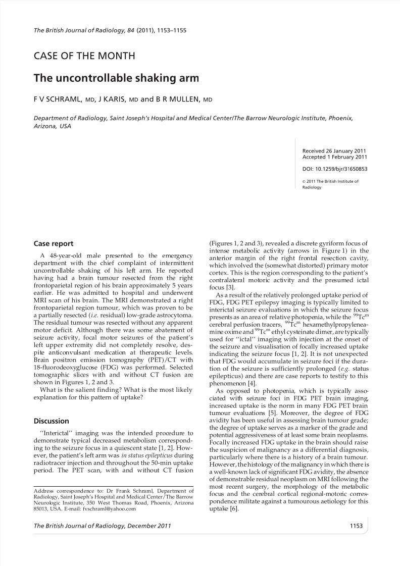
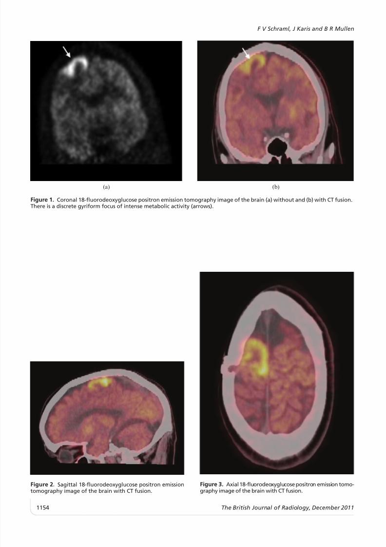
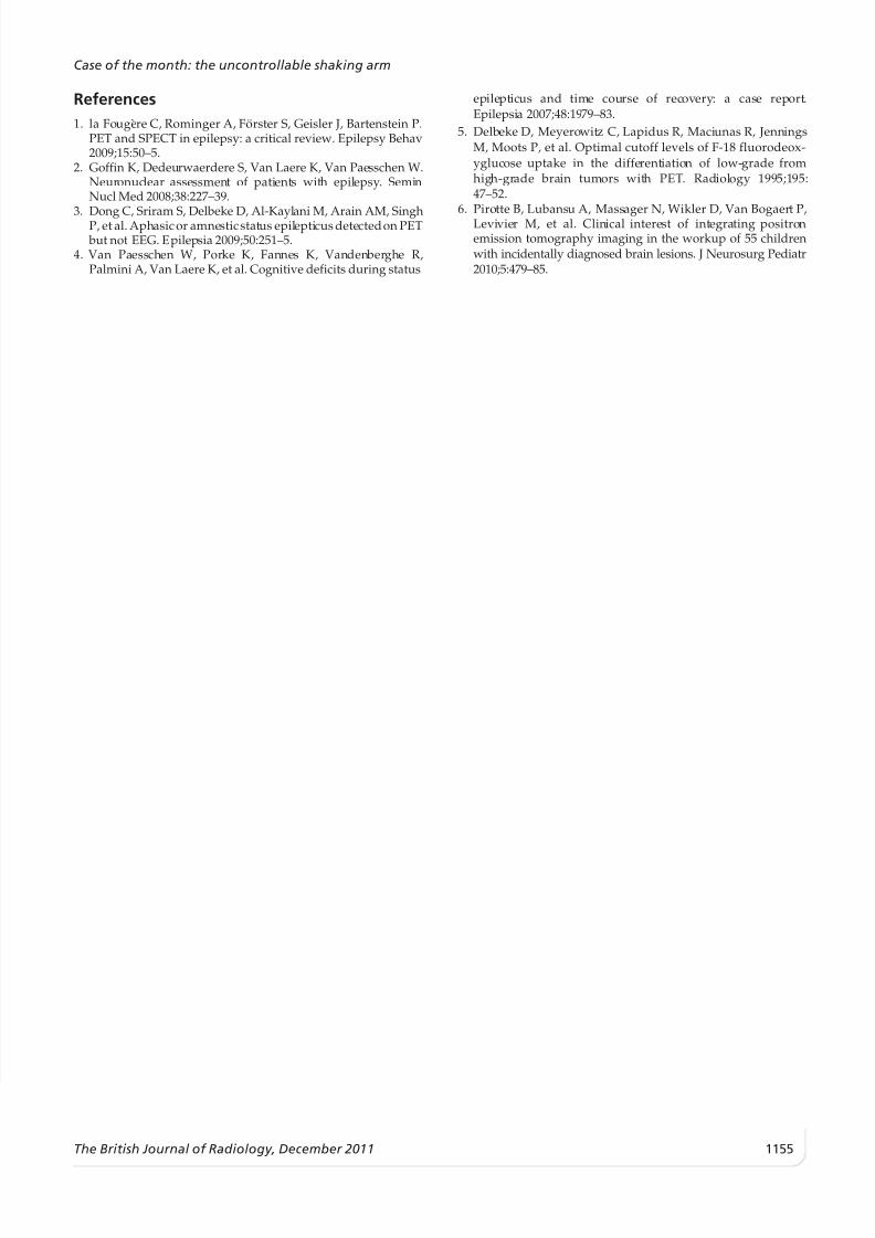
CASE OF THE MONTH The uncontrollable shaking arm F V SCHRAML, MD, J KARIS, MD and B R MULLEN, MD Department of Radiology, Saint Joseph’s Hospital and Medical Center/The Barrow Neurologic Institute, Phoenix,  Arizona, USA Received 26 January 2011 Accepted 1 February 2011 DOI: 10.1259/bjr/31 650853 2011 The British Institute of Radiology Case report A 48 -year- ol d ma le pr es ente d to the emer ge ncy dep art men t wit h the chi ef compla int of intermitt ent unc ont rol lab le sha king of his lef t arm. He repor ted havi ng ha d a brai n tumour rese ct ed from the right frontoparietal region of his brain approximately 5 years earlier. He was admitt ed to hos pit al and underwen t MRI scan of his brain. The MRI demonstrated a right frontoparietal region tumour, which was proven to be a partially resected ( i.e. residual) low-grade astrocytoma. The residual tumour was resected without any apparent motor deficit . Althou gh there was some abatement of seizure activi ty, foc al mot or seizures of the pat ien t’s left upper extremi ty did not complete ly resol ve, des- pit e ant iconvu lsa nt med ica tio n at the rapeutic levels. Brain pos itr on emi ssion tomography (PET)/CT with 18-fl uorod eoxy gluco se (FDG) was perf ormed. Sele cted tomogr aphi c slices wi th and wi thout CT fusion ar e shown in Figures 1, 2 and 3. What is the salient finding? What is the most likely explanation for this pattern of uptake? Discussion ‘‘Inte ricta l’’ imagi ng was the intended proc edur e to demonstrate typical decreased metabolism correspond- ing to the seizure focus in a quiescent state [1, 2]. How- ever, the patient’s left arm was in status epilepticus during radiotracer injection and throughout the 50-min uptake pe ri od. The PET sc an, wi th and wi thout CT fusi on (Figures 1, 2 and 3), revealed a discrete gyriform focus of int ense me taboli c act ivi ty (ar rows in Fig ure 1) in the ant er ior mar gin of the rig ht fro nta l resect ion cav ity , which involved the (somewhat distorted) primary motor cortex. This is the region corresponding to the patient’s con tra lat er al mot ori c activity and the pre sumed ict al focus [3]. As a result of the relatively prolonged uptake period of FDG, FDG PET epilepsy imaging is typically limited to interictal seizure evaluations in which the seizure focus presen ts as an area of relative photope nia, while the 99 Tc m cerebral perfusion tracers, 99 Tc m hexamethylpropylenea- min e oxi me and 99 Tc m ethy l cyst eina te dime r, are typi call y used for ‘‘ictal’’ imaging with injection at the onset of the seizure and visualisation of focally increased uptake indicating the seizure focus [1, 2]. It is not unexpected that FDG would accumulate in seizure foci if the dura- tion of the seizure is sufficiently prolonged ( e.g. status epilepticus) and there are case reports to testify to this phenomen on [4]. As opp ose d to photope nia , which is typi cally asso - ci ate d wit h seiz ur e foci in FDG PET brain imaging , increased uptake is the norm in many FDG PET brain tumour eva lua tion s [5]. Mor eover, the degree of FDG avidity has been useful in assessing brain tumour grade; the degree of uptake serves as a marker of the grade and potential aggressiveness of at least some brain neoplasms. Focally increased FDG uptake in the brain should raise the suspicion of malignancy as a differential diagnosis, particularly where there is a history of a brain tumour. Howe ve r, the his tol ogy of the ma li gnancy in whi ch the re is a well-known lack of significant FDG avidity, the absence of demonstrable residual neoplasm on MRI following the mos t rec ent sur ger y, the mor pho log y of the met abo lic focu s and the cer ebr al cort ical reg iona l-motoric cor res - pondence militate against a tumourous aetiology for this uptak e [6]. Address cor res ponden ce to: Dr Frank Schr aml , Department of Radiology, Saint Joseph’s Hospital and Medical Center/The Barrow Neurol ogic Institute, 350 West Thomas Road, Phoenix, Arizona 85013, USA. E-mail: [email protected] The British Journal of Radiology, 84 (2011), 1153–1155 The British Journal of Radiology, December 2011 1153
Transcript

7/28/2019 The Uncontrollable Shaking Arm

http://slidepdf.com/reader/full/the-uncontrollable-shaking-arm 1/3

CASE OF THE MONTH

The uncontrollable shaking arm

F V SCHRAML,MD, J KARIS, MD and B R MULLEN, MD

Department of Radiology, Saint Joseph’s Hospital and Medical Center/The Barrow Neurologic Institute, Phoenix, Arizona, USA

Received 26 January 2011Accepted 1 February 2011

DOI: 10.1259/bjr/31650853

’ 2011 The British Institute ofRadiology

Case report

A 48-year-old male presented to the emergencydepartment with the chief complaint of intermittentuncontrollable shaking of his left arm. He reportedhaving had a brain tumour resected from the rightfrontoparietal region of his brain approximately 5 yearsearlier. He was admitted to hospital and underwentMRI scan of his brain. The MRI demonstrated a rightfrontoparietal region tumour, which was proven to bea partially resected ( i.e. residual) low-grade astrocytoma.The residual tumour was resected without any apparentmotor deficit. Although there was some abatement of seizure activity, focal motor seizures of the patient’sleft upper extremity did not completely resolve, des-pite anticonvulsant medication at therapeutic levels.Brain positron emission tomography (PET)/CT with18-fluorodeoxyglucose (FDG) was performed. Selectedtomographic slices with and without CT fusion areshown in Figures 1, 2 and 3.

What is the salient finding? What is the most likelyexplanation for this pattern of uptake?

Discussion

‘‘Interictal’’ imaging was the intended procedure todemonstrate typical decreased metabolism correspond-ing to the seizure focus in a quiescent state [1, 2]. How-ever, the patient’s left arm was in status epilepticus duringradiotracer injection and throughout the 50-min uptakeperiod. The PET scan, with and without CT fusion

(Figures 1, 2 and 3), revealed a discrete gyriform focus of intense metabolic activity (arrows in Figure 1) in theanterior margin of the right frontal resection cavity,which involved the (somewhat distorted) primary motorcortex. This is the region corresponding to the patient’scontralateral motoric activity and the presumed ictalfocus [3].

As a result of the relatively prolonged uptake period of FDG, FDG PET epilepsy imaging is typically limited tointerictal seizure evaluations in which the seizure focuspresents as an area of relative photopenia, while the 99 Tcm

cerebral perfusion tracers, 99 Tcm hexamethylpropylenea-mine oxime and 99 Tcm ethyl cysteinate dimer, are typicallyused for ‘‘ictal’’ imaging with injection at the onset of the seizure and visualisation of focally increased uptakeindicating the seizure focus [1, 2]. It is not unexpectedthat FDG would accumulate in seizure foci if the dura-tion of the seizure is sufficiently prolonged ( e.g. statusepilepticus) and there are case reports to testify to thisphenomenon [4].

As opposed to photopenia, which is typically asso-ciated with seizure foci in FDG PET brain imaging,increased uptake is the norm in many FDG PET braintumour evaluations [5]. Moreover, the degree of FDGavidity has been useful in assessing brain tumour grade;the degree of uptake serves as a marker of the grade andpotential aggressiveness of at least some brain neoplasms.Focally increased FDG uptake in the brain should raisethe suspicion of malignancy as a differential diagnosis,particularly where there is a history of a brain tumour.However, thehistology of the malignancy in which there isa well-known lack of significant FDG avidity, the absenceof demonstrable residual neoplasm on MRI following themost recent surgery, the morphology of the metabolic

focus and the cerebral cortical regional-motoric corres-pondence militate against a tumourous aetiology for thisuptake [6].

Address correspondence to: Dr Frank Schraml, Department of Radiology, Saint Joseph’s Hospital and Medical Center/The BarrowNeurologic Institute, 350 West Thomas Road, Phoenix, Arizona85013, USA. E-mail: [email protected]

The British Journal of Radiology, 84 (2011), 1153–1155

The British Journal of Radiology, December 2011 1153

7/28/2019 The Uncontrollable Shaking Arm

http://slidepdf.com/reader/full/the-uncontrollable-shaking-arm 2/3

(a) (b)

Figure 1. Coronal 18-fluorodeoxyglucose positron emission tomography image of the brain (a) without and (b) with CT fusion.There is a discrete gyriform focus of intense metabolic activity (arrows).

Figure 2. Sagittal 18-fluorodeoxyglucose positron emissiontomography image of the brain with CT fusion.

Figure 3. Axial18-fluorodeoxyglucose positron emission tomo-graphy image of the brain with CT fusion.

F V Schraml, J Karis and B R Mullen

1154 The British Journal of Radiology, December 2011

7/28/2019 The Uncontrollable Shaking Arm

http://slidepdf.com/reader/full/the-uncontrollable-shaking-arm 3/3

References1. la Fougere C, Rominger A, Fo¨rster S, Geisler J, Bartenstein P.

PET and SPECT in epilepsy: a critical review. Epilepsy Behav2009;15:50–5.

2. Goffin K, Dedeurwaerdere S, Van Laere K, Van Paesschen W.Neuronuclear assessment of patients with epilepsy. SeminNucl Med 2008;38:227–39.

3. Dong C, Sriram S, Delbeke D, Al-Kaylani M, Arain AM, SinghP, et al. Aphasicor amnesticstatus epilepticus detectedon PET but not EEG. Epilepsia 2009;50:251–5.

4. Van Paesschen W, Porke K, Fannes K, Vandenberghe R,Palmini A, Van Laere K, et al. Cognitive deficits during status

epilepticus and time course of recovery: a case report.Epilepsia 2007;48:1979–83.

5. Delbeke D, Meyerowitz C, Lapidus R, Maciunas R, JenningsM, Moots P, et al. Optimal cutoff levels of F-18 fluorodeox-yglucose uptake in the differentiation of low-grade fromhigh-grade brain tumors with PET. Radiology 1995;195:47–52.

6. Pirotte B, Lubansu A, Massager N, Wikler D, Van Bogaert P,Levivier M, et al. Clinical interest of integrating positronemission tomography imaging in the workup of 55 childrenwith incidentally diagnosed brain lesions. J Neurosurg Pediatr2010;5:479–85.

Case of the month: the uncontrollable shaking arm

The British Journal of Radiology, December 2011 1155


Recommended