HAL Id: hal-00599231 https://hal.archives-ouvertes.fr/hal-00599231 Submitted on 9 Jun 2011 HAL is a multi-disciplinary open access archive for the deposit and dissemination of sci- entific research documents, whether they are pub- lished or not. The documents may come from teaching and research institutions in France or abroad, or from public or private research centers. L’archive ouverte pluridisciplinaire HAL, est destinée au dépôt et à la diffusion de documents scientifiques de niveau recherche, publiés ou non, émanant des établissements d’enseignement et de recherche français ou étrangers, des laboratoires publics ou privés. The use of CT derived solid modelling of the pelvis in planning cancer resections J. Bruns, C.R. Habermann, W. Rüther, D. Delling To cite this version: J. Bruns, C.R. Habermann, W. Rüther, D. Delling. The use of CT derived solid modelling of the pelvis in planning cancer resections. EJSO - European Journal of Surgical Oncology, WB Saunders, 2010, 36 (6), pp.594. 10.1016/j.ejso.2009.11.005. hal-00599231
Transcript
HAL Id: hal-00599231https://hal.archives-ouvertes.fr/hal-00599231
Submitted on 9 Jun 2011
HAL is a multi-disciplinary open accessarchive for the deposit and dissemination of sci-entific research documents, whether they are pub-lished or not. The documents may come fromteaching and research institutions in France orabroad, or from public or private research centers.
L’archive ouverte pluridisciplinaire HAL, estdestinée au dépôt et à la diffusion de documentsscientifiques de niveau recherche, publiés ou non,émanant des établissements d’enseignement et derecherche français ou étrangers, des laboratoirespublics ou privés.
The use of CT derived solid modelling of the pelvis inplanning cancer resections
J. Bruns, C.R. Habermann, W. Rüther, D. Delling
To cite this version:J. Bruns, C.R. Habermann, W. Rüther, D. Delling. The use of CT derived solid modelling of thepelvis in planning cancer resections. EJSO - European Journal of Surgical Oncology, WB Saunders,2010, 36 (6), pp.594. �10.1016/j.ejso.2009.11.005�. �hal-00599231�
Title: The use of CT derived solid modelling of the pelvis in planning cancerresections
Authors: J. Bruns, C.R. Habermann, W. Rüther, D. Delling
PII: S0748-7983(09)00513-7
DOI: 10.1016/j.ejso.2009.11.005
Reference: YEJSO 2916
To appear in: European Journal of Surgical Oncology
Received Date: 9 April 2009
Revised Date: 6November2009
Accepted Date: 9 November 2009
Please cite this article as: Bruns J, Habermann CR, Rüther W, Delling D. The use of CT derived solidmodelling of the pelvis in planning cancer resections, European Journal of Surgical Oncology (2009),doi: 10.1016/j.ejso.2009.11.005
This is a PDF file of an unedited manuscript that has been accepted for publication. As a service toour customers we are providing this early version of the manuscript. The manuscript will undergocopyediting, typesetting, and review of the resulting proof before it is published in its final form. Pleasenote that during the production process errors may be discovered which could affect the content, and alllegal disclaimers that apply to the journal pertain.
In principle, two different technological methods were employed:
MANUSCRIP
T
ACCEPTED
ARTICLE IN PRESS 6
The model was cut out of a block of polyurethane or was constructed from
a liquid polymer which had been hardened layer by layer under an
ultraviolet lamp.
Preoperative planning of the resection
The resection was preoperatively planned by means of CT and MRI
images. The bony line of resection was defined on the pelvic model using
the osseous anatomical landmarks such as the anterior superior or inferior
spine, the posterior superior or inferior spine, or the pubic symphysis. The
definite line of resection was than defined by measuring the distance from
the above-mentioned anatomical landmarks.
Depending on the anatomical exent of the tumor according to the
classification of Enneking & Dunham 1978 (12), in most of the cases a PI
or PII resection with a horizontal osteotomy in the iliac bone was
performed for the cranial osteotomies. In a few cases a vertical resection of
the os ilium according to a P I or PI/IV was necessary.
The osteotomies performed in other parts of the bony pelvis such as the
superior or inferior ramus of the os pubis or os ischium did not need to be
accurate in the millimeter dimension because these areas of the bony pelvis
were not needed for implantation of any part of the PPR.
MANUSCRIP
T
ACCEPTED
ARTICLE IN PRESS 7
Results
Patients data
During the period 1995 to 2004, 25 pelvic models were constructed
preoperatively for the planning of operations in 24 patients. The operations
were performed in the Department of Orthopaedic Surgery in the
University Hospital Hamburg by one surgeon (J.B.). Three patients refused
surgery. Thus, in 21 patients surgical resection of parts of the bony pelvis
was performed followed by either a partial pelvic replacement
(13 x PPR), hip transposition (5 x HTP), ilio-sacral resection (3 x ISR),
ilio-sacral revision and PPR exchange surgery (1 x).
In all patient who received a PPR the fit of the replacement was optimal, in
none of them a major unplanned resection was necessary (Fig.1). The same
was observed in all patients who received a HTP transposition or an ISR or
the revision surgery.
Oncological data
Oncologically, in most of the patients we achieved wide resection margins
(14x). In only 5 patients the margins were marginal (4x) or intralesionsal
(1x). In two cases the aim was a palliative resection because of a metastatic
disease or benign entity (1x).
In one of the patients, a second pelvic model had to be constructed
following an ilio-sacral resection and reconstruction using a fibula graft
MANUSCRIP
T
ACCEPTED
ARTICLE IN PRESS 8
and spine instrumentation system, this being necessitated by a fracture of
the primary fibular implant in order to facilitate better three-dimensional
orientation.
Almost all operations had been performed for oncological reasons and had
aimed at achieving a “wide resection”. Epidemiological data and the
diagnoses are presented in Table 1. In one patient, the indication for a PPR
was not related to a tumor but to material breakage ten years after an initial
PPR owing to a solitary metastasis. The implantation of a new PPR was
carried out.
The oncological resection areas and the achieved margins of resection are
listed in Table 2. Table 3 lists the intraoperative procedures: Out of 25
patients, 3 refused surgery, in the remaining 23 patients surgery was
performed.
Accuracy of the pelvic models
In all patients who received a PPR after an IHP (13 x), an optimal fit of the
pelvic implant into the remaining bone was achieved. This means, no major
unplanned additional bony resection was necessary. Even in the patients in
whom the exchange of the PPR was performed, the fit was optimal.
In the patients in whom a hip transposition procedure (HTP) (5 x) was
performed, the fit of the transposed proximal femur to the remaining part of
MANUSCRIP
T
ACCEPTED
ARTICLE IN PRESS 9
the pelvis was not a problem since this procedure does not need the
accuracy that a PPR requires.
The benefit of the pelvic model in the two patients with an ilio-sacral
resection (3 x ISR) was that it enabled better understanding of the line of
resection, particularly in the ilio-sacral region. For the reconstruction using
fibular grafts and spinal instrumentation systems, there was no need for an
optimal fit as with a PPR.
However, in one case with breakage of the instrumentation and the
implantted fibular grafts, a newly-produced pelvic model allowed more
insight into the ilio-sacral area even after tumor resection.
Quality of resection
Oncologically, in most of the patients, an adequate resection, i.e. a
resection with at least wide margins (14 x), was performed: e.g. in all
patients who had an IHP followed by a PPR. In two of the patients in
whom a HTP was carried out after an IHP, the resection was marginal and,
in one of those with an ilio-sacral resection, the tumor had already spread
into the spinal canal so that only an intralesional resection could be
achieved (Table 2). In 3 cases the resection was palliative because of the
metastatic character of the disease.
MANUSCRIP
T
ACCEPTED
ARTICLE IN PRESS 10
Discussion
History of pelvic surgery and partial pelvic replacement
When one considers the present level of technology for the planning and
performance of complex surgeries of the bony pelvis, it would seem
particularly heroic under what conditions the first of these IHP’s followed
by a PPR were carried out (8-11,13,14).
Without an MRI or pelvic model, it was only possible to roughly plan an
indivi-dual PPR based on X-rays and CT-scans. Although the use of pelvic
models had already been reported by Tonner & Engelbrecht (14), in the
first cases with a PPR and IHP, a fit to the bony line of resection was
achieved in stages since the modular technology did not exist. The optimal
fit of a custom-made prosthesis could be accomplished since, intraopera-
tively, the PPR was adapted step-by-step to the extent of the bony resection
and corrected by the orthopaedic workshop (9). At that time, a correct fit of
the prosthesis was not the main problem because fixation of the PPR was
achieved with horizontal screws which secured the fixation arms of the
PPR to the remaining part of the os ileum.
Thus, the oncological quality of a resection, whether radical or wide versus
marginal or even intralesional, was rather compromised (1,6,15,16).
In 1997 Abudu et al. (1) reported a series of 35 patients who received a
prosthesis. In the first 17 patients the operative procedure was performed in
two steps, in the remaining 18, a one-step-procedure was carried out. In the
MANUSCRIP
T
ACCEPTED
ARTICLE IN PRESS 11
two-step-procedure the resected specimen and a cast made of the cut iliac
surface were used as templates to manufacture the custom-made pelvic
prosthesis which was implanted six weeks later. In the one-step procedure,
the custom-made prosthesis was manufactured on the basis of radiographs
and 3-dimensional reconstructions.
The procedure was rapidly improved by means of a pelvic model (14,17,
18) but CT-technology was still in its early days. In 1999 Handels et al.
(19) reported on virtual three-dimensional models for the computer-assisted
planning and simulation of hip operations for oncological purposes.
The current technology used in producing an individual 3-D pelvic model
bases on CT-data from the bony pelvis. The line of resection can be
determined on such a model as described above. Thus, an individual PPR
with an acceptable accuracy needed for a good fit to the remaining part of
the pelvic bone can be produced.
Although the use of individual pelvic models had already been mentioned
by Burri and other authors et al. (8,14), to our knowledge, Guarino et al. in
2007 (20) were the first to mention the particular advantages of such
rapidly prototyped models in pediatric spinal and pelvic operations, e.g. the
improvement of preoperative planning, intraoperative orientation and
navigation and the reduction of operating time. But, the authors did not
give exact data regarding the accuracy and oncological results.
MANUSCRIP
T
ACCEPTED
ARTICLE IN PRESS 12
Thus, to our knowledge, our paper is the first to report on surgical and
oncological results regarding such pelvic models. We believe that the
results described here are very promising despite the generally high
complication rate of pelvic tumor resections (1,3,6,15,16).
Also, our oncological data are acceptable and are in the range of those
reported previously (1,3,6,15,16)or even better.
Current status of pelvic reconstruction
Meanwhile, the technology of reconstruction has changed towards a
modular PPR allowing a better intraoperative adaptation to improve the
position of the socket and thus improve the off-set for an optimal function
of the hip replacement. The construction of an individual pelvic model
using available CT data is the standard method used today in the planning
of oncological pelvic operations (3).
Completion of the model takes only a few days and the achieved accuracy
of the fit is optimal.
Despite this modern technology, the surgeon still has the intraoperative
problem of finding the anatomical landmarks that mark the starting-point of
the resection on the model. Until now, identification and marking of the
landmarks and mar-king of the resection line has been done with Kirschner
wires.
MANUSCRIP
T
ACCEPTED
ARTICLE IN PRESS 13
Aspects for the future
It is to be expected that the intraoperative navigation technology, which has
already been comprehensively employed in conventional total hip replace-
ments (21), in some trauma cases (22) and in a few tumor cases (23,24),
will bring about an improvement of tumor-resection procedures in the
pelvis.
Some first experiences regarding the osseous part of resection have already
been published (23,24). However, the navigation technology for determina-
tion of the oncologically correct line of resection of the soft-tissue part of
the tumor is still not advanced enough but is currently being improved (25).
Despite the new planning technology, this type of pelvic intervention is still
extremely complicated (1-3,6,16) and demands a high level of concentra-
tion, stamina and perseverance from the operating team. So, it is
recommended that such surgeries should only be performed in specialised
oncologiccal centres (1-3).
It is possible that the combined employment of pelvic models and
navigation technology will help to reduce the length of the operation and
thereby the high quotient of infections.
Conflict of interest
The authors state that they have no conflict of interest.
MANUSCRIP
T
ACCEPTED
ARTICLE IN PRESS 14
References:
1. Abudu A, Grimer RJ., Cannon SR., Carter SR., Sneath RS. Reconstruction of the hemipelvis after the excision of malignant tumours. J Bone Joint Surg (B) 1997;79-B:773-779. 2. Grimer RJ, Carter SR, Tillman RM, Spooner D, Mangham DC, Kabukcuoglu Y. Osteosarcoma of the pelvis. J Bone Joint Surg (B) 1999;81-B:796-802. 3. Tunn PU, Fehlberg S, Andreou D, Kettelhack C. Stellenwert der Becken-endoprothetik in der operativen Therapie von Tumoren des knöchernen Beckens. Z Orthop Unfall 2007;145:753-759. 4. Hoffmann C, Gosheger G, Gebert C, Jürgens H, Winkelmann, W. Functional Results and Quality of Life after Treatment of Pelvic Sarcomas Involving The Acetabulum. J Bone Joint Surg (A) 2006;88-A:575-582. 5. Matsuo T, Sugita T, Sato K, Hotta T, Tsuchiya H, Shimose S, Kubo T, Ochi M. Clinical outcome of 54 pelvic osteosarcomas registerd by Japanese musculoskeletal oncology group. Oncology 2005;68:375-381. 6. Ozaki T, Hillmann A, Lindner N, Blasius S, Winkelmann W. Chondsrosarcoma of the pelvis. Clin Orthop 1997;337:226-2239. 7. Ozaki TH, Hoffman C, Hillmann, A, Gosheger G, Lindner N, Winkelmann W. Implantation of Hemipelvic Prosthesis After Resection of Sarcoma. Clin Orthop 2002;396:197-205. 8. Burri C, Claes L, Gerngroß H, Mathys R jun. Total "Internal" Hemipelvectomy. Arch Orthop Trauma Surg 1979;94:219-226. 9. Dahmen G, Heise U. Alloplastischer Beckenteilersatz mit Hüftgelenk und proximalem Femur. Z. Orthop 1985;123:265-272. 10. Enneking WF. Local resection of malignant lesions of the hip and pelvis. J Bone Joint Surg (A) 1966;48-A:991-1007. 11. Steel HH. Partial or complete resection of the hemipelvis. An alternative to hindquarter amputation for periacetabular chondrosarcoma of the pelvis. J Bone Joint Surg (A) 1978;60-A:719-730. 12. Enneking WF, Dunham WK. Resection and reconstruction for primary neoplasms involving the innominate bone. J Bone Joint Surg (A) 1978;60-A:731-746. 13. Burri C, Schulte J. Beckentumoren. Langenbecks Arch Chir 1980;352:465-469. 14. Tonner HD, Engelbrecht H. A new method for the preparation of special allo-plastic implants for partial replacement of the pelvis. Fortschr Med 1979;26:781-783. 15. Capanna R, van Hoorn JR, Guernelli N, Briccoli A, Ruggieri P, Biagini R, Bettelli G, Campanacci M. Complications of pelvic resections. Arch Orthop Trauma Surg 1987;106:71-77. 16. Bruns J, Luessenhop SL, Dahmen G sen. Internal hemipelvectomy and endoprosthetic pelvic replacement: long-term follow-up results. Arch Orthop Trauma Surg 1997;116:27-31. 17. Gerngross H, Burri C, Claes L, Eckardt V. Possibilities of geometric X-ray examination of the pelvis for partial pelvis replacement. Z. Orthop 1980;118:331-336. 18. Gradinger R, Rechl H, Ascherl R, Plötz W, Hipp E. Endoprothetischer Teilersatz des Beckens bei malignen Tumoren. Orthopäde 1993;22:167-173. 19. Handels H, Ehrhardt J, Plötz W, Pöppl SJ. Computer-assisted planning and simulation of hip operations using virtual three-dimensional models. Stud Health Technol Inform 1999;68:686-689. 20. Guarino J, Tennyson S, McCain G, Bond L, Shea K, King H. Rapid prototyping technology for surgeries of the pediatric spine and pelvis: benefits analysis. J Pediatr Orthop 2007;27:955-960.
MANUSCRIP
T
ACCEPTED
ARTICLE IN PRESS 15
21. Kalteis T, Handel M, Bäthis H, Perlick L, Tingart M, Grifka J. Imageless navigation for insertion of the acetabular component in total hip arthroplasty: is it as accurate as CT-based navigation? J Bone Joint Surg (B) 2006;88-B:163-167. 22. Oszwald M, Citak M, Kendoff D, Kowal J, Amstutz C. Kirchhoff T, Nolte LP, Krettek C. Accuracy of navigated surgery of the pelvis after surface matching with an a-mode ultrasound probe. J Orthop Res 2008;26:860-864. 23. Hüfner T, Geerling J, Gänsslen A, Kendoff D, Citak C, Grützner P, Krettek C. Rechnergestütztes Operieren bei Beckenverletzungen. Chirurg 2004;75:961-966. 24. Wong KC, Kumta SM, Chiu KH, Antonio GE, Unwin P, Leung KS. Precision tumoour resection and reconstruction using image-guided computer navigation. J Bone Joint Surg (B) 2007;89-B:943-947. 25. Chopra SS, Hünerbein M, Eulenstein S, Lange T, Schlag PM, Beller S. Development and validation of a three dimensional ultrasound based navigation system for tumor resection. Europ J Surg Oncol 2008;34:456-461.
MANUSCRIP
T
ACCEPTED
ARTICLE IN PRESS 16
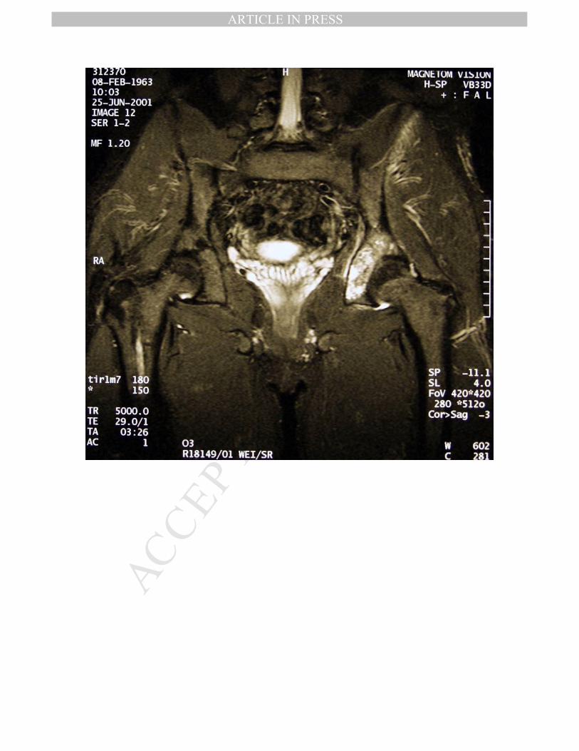
Legends of the figures: Fig.1a: MRI of the pelvis exhibiting the exact extend of the tumor Fig.1b: Pelvic model before planned resection Fig.1c: Pelvic model after after planned resection Fig.1d: Postop. X-ray
MANUSCRIP
T
ACCEPTED
ARTICLE IN PRESS
Indication for manufacturing the pelvic models:
24 patients with pelvic tumors 25 pelvic models
- 13 male, 11 female; Age: 57.4 yrs (25-69)
- 8 x Chondrosarcoma- 5 x Ewing-Sarcoma-Group- 4 x Osteosarcoma- 1 x Angiosarcoma- 2 x malignant fibrous histiocytoma (MFH)- 1 x malignant peripheral nerve-sheath-tumor (MPNST)- 1 x radiogenes sarcoma- 1 x benign nerve-sheath-tumor- 1 x breakage PPR (implanted 10 yrs before)
Table 1
MANUSCRIP
T
ACCEPTED
ARTICLE IN PRESS
23 surgeries
- 21x tumor surgeries, - 1x PPR-exchange- 1x revision surgery
Area of resection (Enneking):- 10x I-II- 8x II*- 4x II-III
Quality of resection (Enneking):- 14 x wide- 4 x marginal- 1 x intralesional (Angiosarkom, sacral spine)- 1 x palliativ (radiogenic sarcoma)- 1 x benign tumor
*: exchange of a 10 yrs previously implanted PPR
Table 2
MANUSCRIP
T
ACCEPTED
ARTICLE IN PRESS
18 patients with IHP + PPR or HTP (1 Surgeon)
- 13 x IHP + PPR: all optimal fit- 5 x IHP + HTTP: all optimal fit- 3 x IIio-sacral resection: wide (1) / intralesional res.
(1) - 1 x PPR exchange (no resection necessary):
optimal fit--------------------------
3 pats.: Refusal of surgery- 1 pat. (MFH) Tumor progress during manufacturing- 2 Pat. no surgery:
- 1 psychiatr. Disease, - 1 refusal of surgery duie to the bad prognosis