understood to recognise that its copyright rests with the author and that no quotation from
the thesis, nor any information derived there-from may be published without the author's
prior, written consent.
2
Declaration
This thesis is submitted to the University of East Anglia for the Degree of Doctor of
Philosophy and has not been previously submitted at this or any university for assessment
or for any other degree. Except where stated, and reference or acknowledgment is given,
this work is original and has been carried out by the author alone.
Michael James Stephenson
3
Acknowledgements
I would first of all like to thank my supervisors Prof. Mark Searcey, Dr Maria O’Connell and
Dr Steve Collingwood for all of the support and guidance they have given to me during the
course of my PhD studies. I feel privileged to have been afforded the opportunity to work
on such a fascinating family of natural products, and one which captured my interest as an
undergraduate (for which I must thank Mark again). I would also like to offer my deepest
thanks to Dr Lesley Howell for her additional support and training.
My thanks also go to Prof. Keith Fox for conducting the DNA thermal cleavage assay
reported in chapter three, to Dr Jenny Kingston for performing the supercritical fluid
chromatography reported in chapter two, and to Dr G. Richard Stephenson for helping me
perfom the DFT calculations reported in chapter three.
For their support and friendship, in and out of the lab, I thank Michael Austin, Sarah
Goffin, and Dr Richard Steel. My thanks also go to everyone I met during the course of my
time at UEA. I would also like to thank everyone I met during my placement at Novartis
for making me feel so welcome, particularly Dr Claire Adcock, Dr Richard Robinson, and
Dr Julia Hatto for their guidance and training, and the Barber family for welcoming me in to
their home.
I thank the School of Pharmacy and Novartis for funding, and the EPSRC mass
spectrometry service in Swansea for accurate mass analysis.
Finally I must thank my family for their support, and in particular I thank my partner Laura
for allowing me to pursue my passion for science, and for her unwavering support at
home.
4
Abstract
Duocarmycin SA is an archetypal member of an ultrapotent family of antitumor antibiotics.
The family is characterised by a common spirocyclopropylcyclohexadienone
pharmacophore. This unusual structural motif is responsible for exceptionally efficient
alkylation of adenine bases following activation through conformational changes induced
by non-covalent recognition of DNA’s minor groove.
This thesis describes the conception and multi-gram synthesis of a duocarmycin SA
alkylation subunit suitably substituted to serve as a building block for Fmoc based solid
phase synthesis and initial investigations into its application.
Chapter two describes the pilot and subsequent large scale racemic synthesis of the
desired duocarmycin building block, and its preparative chiral resolution by supercritical
fluid chromatography. The synthesis includes a novel route to an early indole
intermediate, and represents one of the shortest available strategies to access a
previously reported di-Boc-protected duocarmycin structure. The large scale synthesis
afforded over 8 g of the racemic Fmoc-protected building block, representing a 3 % overall
yield over 13 steps.
In chapter three, application of the building block to the conjugation of amino acids
through solid phase synthesis is explored. This work highlights the importance of careful
resin selection and the need to optimise cleavage conditions. A small library of
duocarmycin analogues was generated, and subsequent assays revealed the effects of C-
terminal amino acid substituents on biological activity. This work has demonstrated the
potential utility of this building block for the future development of novel peptide linked
antibody drug conjugates of duocarmycin SA.
Finally, chapter four explores incorporation of the building block into polypyrrole
structures, highlighting the potential for the direct solid phase synthesis of sequence
selective bifunctional hairpin polyamides which contain the alkylation subunit of
duocarmycin SA.
Published work within this thesis
Michael J Stephenson, Lesley A. Howell, Maria A. O'Connell, Keith R Fox, Claire Adcock, Jenny Kingston, Helen Sheldrake, Klaus Pors, Stephen P Collingwood, and Mark Searcey. The solid phase synthesis of duocarmycin analogues and the effect of C-terminal substitution on biological activity. Journal of Organic Chemistry, Just Accepted Manuscript. DOI: 10.1021/acs.joc.5b01373
5
Table of contents
TITLE PAGE ………………………………………………………………………………….…...1
DECLARATION …………………………………………………………………………………..2
ACKNOWLEDGEMENTS………………………………………………………………………..3
ABSTRACT ………………………………………………………………………………………..4
TABLE OF CONTENTS ………………………………………………………………...............5
Cancer is a condition arising from the aberrant proliferation of an individual’s own cells.
The term encompasses hundreds of individual diseases characterised by their cell type of
origin.1 It is now generally accepted that the transformation of a normal somatic cell to its
malignant counterpart is a progressive process. This progression has been likened to
Darwinian evolution in that successive genetic mutations, each conferring a specific
growth advantage, eventually leads to the presentation of a malignant phenotype.2, 3
The process is often segmented into three mechanistic phases.4 The first is tumour
initiation. In this phase, exposure to chemical or physical carcinogens produce DNA
damage. Mutations arising from this damage, resulting in the unrestrained activation of
oncogenes, or the ablation of tumour-suppressor genes, set the affected cells on their
path to malignancy. During the second phase, tumour promotion, these initiated cells
undergo a clonal expansion. The expansion is a result of disruption to regulatory circuits
which govern normal cell proliferation and tissue homoeostasis. This increase in
proliferation, coupled with genomic instability, can lead to the acquisition of yet more
mutations conferring further growth advantages. The final phase, tumour progression, is
characterised by the invasion of surrounding tissue, growth in tumour sizes, and eventual
metastasis. Again further tumour promoting mutations can drive this stage.
The vast array of different malignancies could be a problem for the field of cancer
research. It has led to a position of seemingly insurmountable complexity in the differing
pathophysiological changes and genomic presentations governing the development of
each individual disease. In 2000, Hanahan and Weinberg attempted to simplify the
situation. In their seminal review, ‘The Hallmarks of Cancer’, they outlined six
characteristic features of the malignant cell,5 which they believed likely to be common to
the majority, if not all cancers; essentially suggesting that the differences between
individual malignancies stem from the different mechanisms utilised to affect these
abilities and the order in which they are acquired. Although an essentially accurate model,
this view constrains consideration of the tumorigenic process to intrinsic properties of the
transforming cells alone. An increasing body of evidence suggests that this may be an
over simplification. It is suspected that normal cells play an important role in the promotion
of malignancy, particularly the inflammatory component of many tumour
microenvironments.6 Such interactions may serve to supplement the progression of
malignancy prior to the aberrant cell line acquiring particular abilities. This is reflected in
Hanahan and Weinberg follow up review published in 2011.7
For example, healthy cells require external signals to move from a state of quiescent to
that of proliferation. They are also subject to signals which inhibit growth. Malignant cells
23
must therefore acquire autonomy over proliferative control. This requires self-sufficiency in
growth promotion and insensitivity to inhibitory signals. In many cases, the former is a
result of oncogenic gene products providing autocrine growth signalling. However there is
also evidence for inflammatory cells providing the necessary stimuli. The evasion of
growth inhibition is often the result of mutations in tumour-suppressor genes which
disrupted inhibitory signalling cascades.5
Rate of proliferation is not the only factor governing tumour development. Many cancerous
tissues show a decrease in cellular attrition. Furthermore DNA damage inherent in tumour
initiation should in many cases result in the activation of regulatory machinery, and
subsequent repair or apoptosis. Thus the malignant cell must acquire mechanisms
allowing the evasion of apoptopic commands.5 Again this may be the result of mutations in
tumour-suppressor genes whose products are pivotal in implementing programmed cell
death, although anti-apoptopic NF-ΚB gene products activated through inflammatory
stimulus have also been implicated.6
Normal somatic cells possess an intrinsic tumour suppressing property; they are unable to
divide indefinitely.5 This is a consequence of the end-replication problem which causes the
gradual erosion of telomeres. Telomeres, which comprise non-coding regions at the end
of chromosomes, have a protective function. Once telomere erosion reaches a critical
point this protective effect is loss, and the cell enters senescence; a non-proliferatory
state. Malignant cells must acquire telomere extension mechanisms conferring them
limitless replicative capacity. In 85 % of cancers this is achieved by expression of the
dormant enzyme telomerase.8 This reverse transcriptase uses an endogenous RNA
template to extend the tandemly repeated telomere sequence, using the single stranded
3’ overhang as its primer.9
All cells, including cancer cells, require adequate perfusion from the vasculature to meet
their demand for oxygen and nutrients.5 The expansion of tumours appears to be initially
curtailed by an inability to stimulate the required level of angiogenesis. However this is
eventually overcome, in what has been described as an angiogenic switch;10 wherein the
aberrant malignant cells acquire the ability to alter the balance of pro and inhibitory factors
in favour of stimulating the require vessel growth.
The ability of primary tumours to spawn pioneer cells which can relocate and establish
secondary tumours, is perhaps the most lethal acquired ability of the malignant cell; being
responsible for 90 % of cancer deaths.11 The alteration of several protein classes,
especially those involved in the tethering of cells to extra cellular matrixes, have been
implicated in the mechanisms of metastasis.5
24
Despite the biological complexity of cancer, traditional therapy has remained relatively
simplistic. Surgery to excise the diseased tissue is by far the most effective treatment for
most solid tumours. However, radiotherapy and chemotherapy play important roles both
prior to excision, to decrease tumour mass, and as adjuvants following surgery, to prevent
reoccurrence from potential residual tumour cells.12
Cytotoxic drugs have long dominated pharmaceutical intervention in cancer therapy.13
Many such treatments target DNA directly, through both non-covalent and covalent
interactions, disrupting the cell’s replicatory machinery, and leading to apoptotic
responses. For example doxorubicin (figure 1.1) is widely prescribed in the treatment of
many cancers, including various carcinomas, sarcomas, and haematology malignancies.14
It is an intercalating agent, binding non-covalently with DNA through insertion of its planar
chromophore between two adjacent base pairs, and projecting its glyosidic structure in to
the minor groove.15
Cyclophosphamide is an alkylating agent, and an example of a treatment which targets
DNA through covalent interaction.16 It is arguably one of the most successful
antineoplastic drugs in the history of cancer chemotherapy. It is effective against a broad
spectrum of malignancies, and is still commonly included in many standard treatment
regimes,14 despite being first introduced over 50 years ago.16
Cyclophosphamide is an oxazaphosphorine prodrug of the original aliphatic nitrogen
mustards. Hepatic cytochrome P-450 metabolism leads to oxidation of the
oxazaphosphorine ring vincinal to the ring bound nitrogen. Tautomerization, and
subsequent β-elimination reveal the active mustard, which is free to form the highly
unstable aziridinium cation and ultimately alkylate DNA through nucleophilic attack by
guanine bases. The presence of two chloroethyl groups means inter and intrastrand
crosslinking is often observed (scheme 1.1).16, 17
Figure 1.1 Structure of doxorubicin.
25
As our understanding of the biology of cancer has evolved the scope of potential
treatment strategies has widened. It is therefore unsurprising that the draw of DNA as a
target has diminished over recent years, with many researchers turning their attention to
the disruption of aberrant signalling pathways specific to particular malignant cell lines.
Such approaches aim to produce more selective and therefore tolerable treatments. This
strategy has already yielded significant successes in the clinic.
Imatinib (figure 1.2), was one of the first such treatments to gain regulatory approval, and
has revolutionised the treatment of susceptible forms of chronic myeloid leukaemia (CML).
It is an orally administered synthetic tyrosine kinase inhibitor, which targets the BCR-ABL
oncogenic gene product;18 a fusion gene resulting from a chromosomal translocation
event which is common to the pathogenesis of the majority of CML cases. The BCR-ABL
protein is a permanently activated tyrosine kinase, whose activity results in the
uncontrolled proliferation of affected cells.19
Scheme 1.1 Structure and activation of cyclophosphamide.
26
The success of imatinib has been said to mark a paradigm shift in the treatment of
malignant disease,20 and since its introduction in 2001 many similar molecular targeted
therapies have gained regulatory approval.21 For example, trastuzumab, a monoclonal
antibody which targets the HER2 growth receptor,22 has had a major impact on the
treatment HER2 positive breast cancer.23, 24
It is likely that this field will contiune to grow and yield many more ground breaking
treatments in the future. Nevertheless, susceptibility to resistance, the implict narrow
spectrum of such treatments, and controversy surrounding the cost-effectivness of some
less successful additions to the clinic, mean a reliance on conventional cytotoxins may
remain for some time.25-27 It is therefore still of value to explore the development of novel
cytotoxic compounds, especially those with the necessary potency to be applied
successfully to tumour directed delivery and/or prodrug strategies. Such approaches also
hold the potential to yield more selective and tolerable treatments.
Perhaps contrary to public perception, particularly of those influenced by the market of
alternative therapy, modern medicine has long recognised the therapeutic potential of
natural products. However, unlike the pedlars of snake oils and folklore, this reasoned
world of scientific exploration, has always viewed nature’s rich pool of biologically active
secondary metabolites as leads; understanding the need to optimise these compounds for
uses to which they did not evolve. The selection pressures imposed upon bacteria have
resulted in a vast array of compounds with obvious applications in the treatment of
infectious and malignant disease. It was reported in 2010, that since the 1940s 51.5 % of
all anticancer agents introduced were either natural products or directly derived
analogues, many originally isolated from microbes.28 Indeed, the aforementioned
doxorubicin is a member of the anthracycline family of natural products; secondary
metabolites isolated from various strains of Streptomyces. 29, 30
Figure 1.2 Structure of imatinib.
27
Another pertinent example would be mitomycin C. Introduced in the 1960s,31 mitomycin C
is still commonly included in a number of treatment regimens.14 It is a member of the
aziridine family of antitumor antibiotics, isolated from Streptomyces caespitosus.32 The
cytotoxicity of mitomycin C is derived from its ability to crosslink DNA through a
bifunctional covalent interaction. This is thought to occur through a complex series of
intramolecular transformations, initiated from the bioreduction of mitomycin C to its
hydroquinone counterpart (scheme 1.2).33 Subsequent loss of the methoxy group, and
tautomerization of the resulting imine leads to opening of the aziridine ring by the
hydroquinone through the indole structure, and generation of the first alkylating species.
This is attacked by guanine bases by a conjugate addition. The resulting reformation of
Scheme 1.2 Structure and DNA crosslinking activity of mitomycin C.
28
the hydroquinone indole structure, allows formation of the second alkylating species, via
loss of the carbamate.
It is a pleasing coincidence for the medicinal chemist, that two of the most potent families
of antitumor antibiotics discovered to date also derive their biological activity from
interesting mechanistic chemistry. Perhaps the most potent is the family comprising the
enediyne antibiotics, such as calicheamicin originally isolated from Micromonospora
echinospora (scheme 1.3).34 Their cytotoxicity is developed from the diradical intermediate
of a biologically triggered Bergman cyclisation.35
The second family is that consisting of CC-1065, yatakemycin, and the duocarmycins,
whose structures are depicted in figure 1.3.36 It has now been over 30 years since the first
member of this family, CC-1065,37, 38 was reported, and despite substantial interest from
distinguished medicinal chemistry programmes, research is yet to yield a successful
clinical candidate. However this unrealised therapeutic potential only adds to the allure of
these already chemically pleasing entities.
Scheme 1.3 Structure and mechanism of cytotoxicity of calicheamicin γ1.
29
CC-1065, yatakemycin, and the duocarmycins all combine non-covalent recognition of AT
rich regions of DNA’s minor grove with exceptionally efficient alkylation of adenine,39 the
downstream effects of which result in their ultrapotent cytotoxic activity. The common
pharmacophore responsible for this alkylation consists of a cyclopropane vicinal to the α,
β unsaturated carbonyl of a cyclohexadienone. This spirocyclic system is flanked by the
fusion of a further two 5 membered nitrogen heterocycles. Alkylation proceeds through
nucleophilic attack of the cyclopropane by the N3 of adenine (Scheme 1.4). This occurs at
the least substituted carbon, resulting in ring opening of the cyclopropane and subsequent
development of an aromatic phenol from the conjugated and activating cyclohexadienone.
It is the control of this seemingly facile reaction, imposed by the subtle interplay of
molecular features contained within the compact structure of these compounds, that
Figure 1.3 Structures of CC-1065, yatakemycin, and the duocarmycins. Red = alkylation subunit. Blue = alkylation subunit precursor – requiring spirocyclisation prior to the alkylation event.
30
defines their characteristic and arguably most interesting feature: in situ activation.40 As
will be described this is a quite unique and elegant process; one which does not require
induction of chemical change, but simply a change in conformation induced from binding
to their target.
In solution these compounds demonstrate remarkable stability towards nucleophilic
attack. It is only when bound within the minor groove of DNA that the true reactivity of the
cyclopropane is revealed.41 This stability results from the electronic influence of a
vinylogous amide constructed from the carbonyl of the cyclohexadienone and the nitrogen
of the neighbouring heterocycle. This feature serves to reduce the electrophilicity of the
cyclohexadienone by conjugation of the nitrogen’s lone pair with the carbonyl and stabilise
the cyclopropane.
The magnitude of this effect has been implicitly demonstrated by comparison of the
reactivity of a carbocyclic analogue of the CBI alkylation subunit with its nitrogen
containing counterpart (figure 1.4).42 It was shown that 1 which lacks the vinylogous
amide, was 3200 times more reactive than 2 towards solvolysis at pH 3; and at pH 7
where 2 is stable, 1 was revealed to be 104 times more reactive. It is disruption of the
conjugation of this vinylogous amide which triggers these natural products in situ
activation.
Scheme 1.4 Alkylation of adenine by duocarmycin SA.
Figure 1.4 Structures of CBI analogues 1 and 2.
31
Disruption of this stabilising influence is made possible because the nitrogen of the
vinylogous amide also forms part of a linking amide, through which the alkylation subunits
of the natural products are connected to additional, non-alkylating, bi or polycyclic
systems, known to contribute non-covalently to minor groove recognition. As such, the
lone pair of this nitrogen is effectively cross conjugated with the carbonyls of both amides.
When the natural products bind to DNA they undergo a conformational change. This
change involves rotation around the linking amide so as to allow the adoption of a bent
conformation which is more complementary to the helical rise of the minor groove.
Structural studies of bound duocarmycin SA suggest that the bulk of this twist is carried by
the χ1 dihedral angle (22.4 o), as opposed to that of the amide χ2 (11.0 o) (see figure
1.5).43, 44 The result is a change in the balance of cross conjugation in favour of the linking
amide, as the nitrogen’s lone pair is no longer planner with the cyclohexadienone’s π
system.
Comparison of the activity of CBI analogues 3 and 4 (figure 1.6) clearly shows the
necessity of the linking amide in this process. While DNA alkylation is easily detectable
with 3, which possesses the linking amide, 4, in which this amide is replaced by a
methylene bridge is unable to alkylate DNA even under forcing conditions.45
Figure 1.5 Visual representation of the in situ activation of duocarmycin SA (see main text for description). NMR Structure images generated using ‘Chimera 1.10.1’, coordinates from protein
data bank (ID:1DSA).42
32
Comparison of the x-ray structures of a series of CBI analogues (figure 1.7), where the
ring size of the nitrogen heterocycle was increased from 5 through to 7 atoms, further
demonstrates this effect, and captures its incremental nature. As ring size increases the
χ1 angle widens. This is accompanied by a lengthening in the bond between the nitrogen
and cyclohexadienone, reflecting reduction in the conjugation of the vinylogous amide.
These structural features are mirrored by an expected progressive increase in reactivity
as the degree of vinylogous amide conjugation decreases. 46, 47
As disruption of conjugation of the vinylogous amide is not an absolute process, and the
degree of rotation induced at the χ1 angle can have an incremental effect on the reactivity
of the spirocyclopropylcyclohexadienone, the rigid length of compounds containing this
pharmacophore can have a significant impact on their relative DNA alkylation efficiencies.
Essentially, longer structures can amplify the twist imposed by the helical rise of the minor
groove, and thus increase the reactivity of the cyclopropane.
Figure 1.6 Structures of CBI analogues 3, and 4.
Figure 1.7 Structure of CBI analogues with increasing heterocycle size. Showing discussed crystallography and stability data.
33
This relationship is manifested in the observed importance of both the C6 methyl ester,
and C5’ methoxy substituent, on the DNA alkylation efficiency of duocarmycin SA (figure
1.8). Despite being shown to confer on the alkylation subunit greater stability towards
solvolysis, the absence of the C6 methyl ester reduces the relative rate of DNA alkylation
by 12 fold. Similarly, removal of the C5’ methoxy substituent produces a 20 fold decrease
in DNA alkylation rate. When both substituents are removed, a 250 fold decrease in DNA
alkylation rate is observed.48
These findings have been mirrored through investigation of the effects of substitution at
the C7, and C5 position of the CBI alkylation subunit (figure 1.9).49-51 It was observed that
C7 substituents, which extend along the floor of the minor groove, conveyed a rate
accelerating effect on DNA alkylation activity, and that this was independent of the
substituent’s electronic character, and hence corresponding effect on solvolytic stability of
the respective alkylation subunits. Conversely, addition of a methyl ester at the C5
Figure 1.9 Structure of discussed CBI analogues, highlighting the C7, and C5 substituents, and their respective positions relative to the full of the minor groove.
Figure 1.8 Structure of duocarmycin SA highlighting the C6 methyl ester, and C5' methoxy group.
34
position, which extends out of the minor groove, significantly enhanced the solvolytic
stability of the alkylation subunit, while also reducing the rate of DNA alkylation, in line
with its electronic influence. In the C5 position, the substituent does not increase the rigid
length of the structure relative to the minor groove, and hence its stabilising effect is not
countered by an increase in binding induce activation.
The relationship between the intrinsic stability of the alkylation subunit and its
corresponding biological activity is now well established. Many analogues of the
spirocyclopropylcyclohexadienone system have been generated ranging vastly in their
resistance towards solvolysis. A collative meta-analysis has revealed a strong parabolic
relationship between solvolytic stability and anti-proliferation activity (figure 1.10).52 This is
intuitively unsurprising; intrinsic stability towards nucleophilic attack confers a greater
chance that the alkylation subunit will reach its target intact, but this advantage is only
maintained if upon binding in the minor groove, the stability can be sufficiently disrupted
by in situ activation to produce an efficient alkylation event. Pleasingly, the two most
potent natural products in this family, yatakemycin and duocarmycin SA, are positioned at
the parabola of this trend. This beautifully highlights the power of natural selection to tailor
the properties of secondary metabolites through the direction of biosynthetic pathways in
microbes. Indeed, the S of duocarmycin SA literally stands for stable, and this ultrapotent
antitumor antibiotic has been referred to as nature’s prodrug. 53
Figure 1.10 Schematic representation of the observed parabolic trend between intrinsic stability and biological potency of the spirocyclopropylcyclohexadienone family.
35
However, duocarmycin SA is only a prodrug in the sense that it selectively alkylates DNA
over other biological nucleophiles. Like all classical alkylating agents, the natural products
of this family are incapable of discriminating malignant cells from those of healthy tissue.
They therefore have the potential to cause dose limiting toxicities apparent with all non-
selective cyctotoxins. The ultra-potent nature of this family has to date prohibited their
clinical use in the treatment of malignant disease. For example, the first member of this
family to be discovered, CC-1065, quickly moved into pre-clinical evaluation, following
identification of potent activity against a large range of malignant cell lines in vitro.54
However, it could not progress in to clinical trials, due to exhibiting severe hepatotoxicity,
which led to delayed death syndrome in mice at sub-therapeutic doses.55
This delayed death syndrome is not however a universal feature of the
spirocyclopropylcyclohexadienone pharmacophore, but appears to be specific to the
structure of CC-1065. This may be a consequence of the apparent irreversibility of the
alkylation event, or specific sequence selectivity of CC-1065. Indeed, analogues of CC-
1065 have been synthesised that do not exhibit the delayed death syndrome. Two such
clinical candidates, adozelesin, and it’s bifunctional analogue bizelesin (figure 1.11), have
progressed into early clinical trials, but have ultimately failed to reach the clinic due to
unacceptable toxicity profiles and limited efficacy at tolerated doses.56-62
Figure 1.11 Structures of adozelesin and bizelesin.
36
It would seem clear that some form of targeting mechanism is necessary in order to
harness the impressive antitumour activity of this unique family of natural products. One
advantage of pharmacophores which possess ultrapotent cytotoxicity mediated through
mechanistic chemistry, is that they provide the opportunity for the rational design of
prodrugs.
A common strategy employed with the spirocyclopropylcyclohexadienone pharmacophore
has been to inhibit the in vivo spirocyclisation of their seco precursors through biologically
labile protection of the phenolic oxygen. Carzelesin (figure 1.12), one of the few
compounds of this family to enter phase II clinical trials exploited this tactic.63-66 Protection
was achieved through a phenylurethane moiety which required serum dependent
hydrolysis for cleavage.67 More recent refinements of this approach have included
attempts at introducing phenol protection which is cleavable under tumour selective
conditions. The hypoxic nature of the tumour microenvironment has been key to this
strategy.
One example has been the development of N-acyl O-amino derivatives of the seco form
of the CBI subunit. These were designed with the anticipation that the weak N-O bond
could be cleaved by reducing nucleophiles of which a higher concentration can be found
in hypoxic tissue.68, 69 It was shown that the stability of this bond could be predictably
tuned by the electronic and steric nature of N-substitution. The most effective derivative
was a tert-Butyl carbamate, 5 (figure 1.13). It was shown to be incapable of alkylating
DNA in cell free systems, but demonstrated potency approaching that of the free drug in
functional cellular assays, with improved efficacy revealed from in vivo antitumor models.
This suggests preferential release of the free drug at tumour sites. Given the average life
span of the control mice and the length of the study, the number of long term survivors
detected suggests, that despite the absence of dose optimisation, cures were detected.
Figure 1.12 Structure of carzelesin.
37
This concept was later refined with the report of cyclic variants of 5. The oxazinone
prodrug 6 (figure 1.14),70 was designed to be cleaved by the same reductive mechanisms,
but to release no extraneous masking group as a by-product; a strategy that could simplify
downstream regulatory approval should this species progress as a clinical candidate.
6, was shown to exhibit hypoxia selective cytotoxicity in cellular assays, returning a 10 fold
increase in potency compared to its activity under aerobic conditions in a PC3/MM2 cell
line. Furthermore it was shown to demonstrate good stability in human plasma with a half-
life of one week. In addition, 6 was considerably more efficacious than a seco-CBI
equivalent in an in vivo murine model despite returning higher IC50 values in vitro. At a
dose of 2500 μg/kg, 6 produced six long-term survivors out of ten animals over a period of
one year. In comparison treatment with the seco-CBI equivalent control, resulted in
premature death due to toxicity in all animals at doses of between 100 and 500 μg/kg, and
only one long-term survivor at a lower 60 μg/kg dose. Again this suggests a benefit from
either hypoxia targeted or prolonged intercellular release of the free drug, most likely a
combination of both. It was also noted that 6 was better tolerated at the injection site, than
the seco-CBI equivalent.
Figure 1.13 Structure of N-acyl O-amino prodrug 5.
Figure 1.14 Structure of oxazinone prodrug 6.
38
Other ongoing attempts at hypoxia activated prodrugs of the seco-CBI alkylation subunit
include replacement of the phenolic hydroxyl with a nitro group.71-73 These compounds
were envisioned to undergo reductive metabolism to their aniline counterparts, which in
turn can undergo spirocyclisation to the active alkylating species through imine formation
analogous to that of the phenol compounds. This approach has proved to be far from
straight-forward. The initial simple nitro derivatives showed promising differential
cyctotoxicity in vitro, but low potency in vivo. This was reasoned to result from suboptimal
one electron reduction potential of the nitro group leading to insufficient bioreduction.
Introduction of electron-withdrawing ring substituents placed para or meta to the carbon
bearing the nitro group on the neighbouring ring were found to raise the reduction
potential of the nitro group, however this did not have a consistent effect on hypoxia
selective cytotoxicity. A clear correlation between increased reduction potential and
hypoxic selectively was only apparent for a subset of electron-withdrawing groups. These
were the sulfonamide and carboxamide substituents. The authors suggest this may result
from specific interactions of these groups with the reductases responsible for reduction
under hypoxic conditions, effectively suggesting that high reduction potential in
compounds not containing these reductase specific interactions, could lead to increased
oxic activation and reduced selectively. Despite inconsistencies in the structure activity
trends of these analogues, a promising hypoxia selective lead was identified. This
contained a meta SO2NH2 group. Further work has shown increased hypoxic selectivity by
introduction of sulfonate spirocyclisation leaving groups in place of the usual halogens.
Some of the best results were seen with analogues combining sterically demanding
sulfonate leaving groups, neutral DNA binding subunits, and meta sulfonamide or
carboxamide substituents (figure 1.15).
Figure 1.15 Annotated example of a nitro CBI prodrug structure.
39
Evidence of the over expression of particular isoforms of cyctochrome P450 oxidases in
tumour cells, has led to a novel take on the inhibition of spirocyclisation prodrug strategy.
Instead of masking this process by protection, the phenolic hydroxyl group was removed
completely.74, 75 The rationale being that the dehydroxylated CPI subunit (that of
duocarmycin SA minus the C6 ester) would provide a substrate for cyctochrome P450
metabolism. It was envisioned that site specific oxidation by this enzyme system would
reintroduce the hydroxyl group to reveal the active pharmacophore; a process
hypothesised to mimic the evolution of this family of antibiotics.
The compound reported, named ICT2700 (figure 1.16), consists of the dehydroxylated
seco-CPI alkylation subunit linked with the 5-methoxyindole DNA binding unit. It was
shown to be 1000 fold less cytotoxic than its hydroxylated counterpart in vitro. The cellular
distribution of the two compounds was shown to be comparable, thus confirming
successful deactivation of the pharmacophore by removal of the hydroxyl group.
Comparison of the cytotoxic effect of ICT2700 on CYP1A1 expressing and non-
expressing CHO cells, showed successful differential activation of this compound in
CYP1A1 positive cells, with an IC50 of <0.5 nM compared to >25000 nM in CYP1A1
negative cells.
An alternative, but not mutually exclusive approach for improving the therapeutic window
of the duocarmycins, is to add structural features which aid in the physical targeting of
tumour cells. One such strategy involves the refinement of sequence selectivity, so as to
preferentially bind in the minor groove at DNA sites which represent gene mutations
specific to the malignant genome; a concept which will be discussed in chapter four.
Another tactic is to separate the task of targeting and alkylation, and to consider the
alkylation subunit of the duocarmycins as a ‘payload’ for a cell selective delivery vehicle.
The impressive recognition properties of antibodies have seen these highly engineerable
proteins positioned as excellent candidates to serve as the delivery mechanism for
cytotoxins. The principle is simple; multiple units of a cytotoxic compound are tethered to
Figure 1.16 Structure and activation of ICT2700 by CYP1A1 oxidation.
40
an antibody which is engineered to recognise a tumour specific, or overexpressed
antigen. On binding to the antigen, which is presented on the surface of the malignant
cells, the antibody, together with its ‘payload’, is internalised via a process of receptor-
mediated endocytosis. Upon internalisation, the cytotoxic compound is cleaved via a labile
linker, or is released following lysosomal degradation of the antibody. 76
Early generations of antibody drug conjugates (ADCs), proved disappointing in the clinic.76
One reason for this was the common side effect of immunogenic responses to the mouse
derived antibodies that were used.77 This problem has largely been resolved through the
development of fully humanised monoclonal antibodies, which retain only the
complementarity determining region of the original murine protein (figure 1.17).78
A second problem was poor efficacy in vivo. ADCs can be very effective at selectively
targeting cytotoxins to malignant cells, however the actual physical number of cytotoxic
molecules which reach the interior of the targeted cell is thought to be small. The first
generations of ADCs employed commonly prescribed antitumor agents, such as
doxorubicin, as their payloads. These compounds simply did not possess the necessary
potency to mount an effective antitumor response when delivered in the amount possible
by antibodies.76 Therefore subsequent generations of ADCs have focused on the delivery
of ultrapotent cytotoxins, such as the enediyne antibiotics, maytansinoid and auristatin
derived antimitotic agents, and of course the duocarmycins.76
Figure 1.17 Schematic representation of the basic structure of a monoclonal antibody.77
41
One of the first ADCs reported which utilised the spirocyclopropylcyclohexadienone
pharmacophore was anti-B4-DC1 (figure 1.18).79 The ‘payload’ of anti-B4-DC1 was based
on the CC-1065 analogue adozelesin, but saw the replacement of the CPI alkylation
subunit with the more stable and easily synthesised seco-CBI structure. To allow
conjugation to the antibody via disulfide exchange, the benzofuran group of adozelesin
was also replaced by an indole unit terminally substituted with a reactive disulfide group.
Anti-B4-DC1 demonstrated low picomolar activity against CD19-positive cell lines in vitro,
and was effectivity nontoxic to antigen negative cells in the same assay. The selectivity of
cytotoxicity was also demonstrated by the abolishment of activity when competing with an
excess of unconjugated anti-B4 antibodies. Furthermore, the unconjugated payload was
equally cytotoxic towards antigen positive and negative cell lines. Anti-B4-DC1 proved to
be highly efficacious in murine human tumour models, significantly outperforming
doxorubicin, cyclophosphamide, vincristine, and etoposide when dosed at their respective
maximum tolerated levels.
Unfortunately, poor solubility of the anti-B4-DC1 ‘payload’ in aqueous conjugation reaction
solvents, and instability in physiological buffers, ultimately precluded its clinical
development. However, recent efforts by the same laboratory, has seen the development
of more soluble and stable phenolic phosphate prodrugs of the anti-B4-DC1 ‘payload’.80
Interestingly, antibody conjugation of these prodrugs, led to a 250 fold increase in in vitro
potency when compared to the unconjugated equivalents. This is suggestive of improved
cellular uptake of the conjugates, and that passive diffusion of the free prodrugs is
inhibited by the charged phosphate group. Again selective low picomolar activity against
CD19-positive cell lines was demonstrated. Furthermore in vitro potency was only
increased slightly by the co-incubation of phosphatases, demonstrating that the prodrugs
are effectively dephosphorylated by endogenous enzymes. Conjugate huB4-SPP−DC4
(figure 1.19), showed promising antitumor activity in in vivo models, where the
unconjugated prodrug was ineffective.80
Figure 1.18 Structure of the anti-B4-DCI ‘payload’ and linker.
42
MDX-1203 (figure 1.20), in development by Bristol-Myers Squibb, represents another
example of combining water solubilising phenolic protection of the CBI alkylation subunit
with antibody conjugation.81 Here, CBI is trapped in its seco form by a piperazino
carbamate, and is tethered to a CD70 targeting antibody via a dipeptide linker which
extends from substitution of a terminal aniline group. The N-terminus of the dipeptide
possesses a maleimide group through which conjugation to thiol modified sidechains of
antibody lysine residues form the attachment site.82 This prevents the need to disrupt
disulfide bonds between antibody cysteine residues, minimising disruption to protein
structure. Following antigen specific internalisation, the peptide is cleaved, releasing the
prodrug, which is ultimately activated by endogenous esterases. MDX-1203 has
completed phase I clinical trials for renal cell carcinoma or non-Hodgkin’s lymphoma.83
SYD985 (figure 1.21) is a novel HER2-targeting ADC which has recently entered pre-
clinical evaluation, and represents an alternative approach to antibody conjugation of the
spirocyclopropylcyclohexadienone pharmacophore.84, 85 In this strategy, the phenol group
of the seco-alkylation subunit forms the attachment site of the linking group. In this way
conjugation and seco-trapping phenol protection are combined functions of the same
Figure 1.19 Structure of the huB4-SPP−DC4 'payload' and linker.
Figure 1.20 Structure of the MDX-1203 ‘payload’ and linker.
43
structure. Linkage to the antibody is effected by conjugation of the linker’s terminal
maleimide group with cysteine thiols derived from intrachain disulphide bridges. The
remaining linker structure consists of a dipeptide, and two well characterised self-
eliminating spacers.86, 87 Upon internalisation, the dipeptide is cleaved revealing a p-
aminobenzyl alcohol from which the elimination cascade is initiated (scheme 1.5),
ultimately releasing the free seco form of the alkylation subunit to undergo activating
spontaneous spirocyclisation.
To date it would appear that ADCs incorporating the spirocyclopropylcyclohexadienone
pharmacophore, have been restricted to utilising derivatives of the CBI alkylation subunit,
with linkage extending from either the phenol, or N-terminal substituents. However, the
Figure 1.21 Colour coded structure of SYD985. Blue = dipeptide linker, Red = p-aminobenzyl alcohol spacer, Green = second spacer forming carbamate masking the phenol of the seco-
alklyation subunit.
Scheme 1.5 Mechanism of self-elimination initiated by release of p-aminobenzyl alcohol.
44
amine side chains of antibody lysine residues provide the opportunity for conjugation
through amide bonds. The terminal ester and amide functionality of the duocarmycin SA
alkylation subunit would seem to afford excellent potential for incorporation into polyamide
structures, including potentially short water solubilising peptides which could be applied as
C-terminal linkers for the development of novel ADCs. Fmoc based solid phase synthesis
is a convenient, and well established methodology for the construction of short peptides
on a laboratory scale (see chapter 3). This thesis describes the conception and multigram
synthesis of a duocarmycin SA alkylation subunit suitably substituted to serve as a
‘building block’ for Fmoc solid phase synthesis, and initial investigations in to its
application.
45
2 Chapter Two
Synthesis of the duocarmycin alkylation subunit suitably substituted to serve as a ‘building block’ for Fmoc based solid
phase synthesis.
46
2.1 Aims.
The work described in this chapter was carried out in order to address the following aims:
- To design an analogue of the duocarmycin SA alkylation subunit which is suitably
substituted to allow incorporation of this functionality in to polyamide structures via Fmoc
solid phase synthesis.
- To explore the synthesis of this target, and optimise a reliable route which could be
practically conducted on the scale necessary to access a sufficient quantity of the final
product to allow investigation of its use as a ‘building block’ for solid phase synthesis.
- To conduct the synthesis on a sufficient scale to access a multigram quantity of the
‘building block’.
47
Figure 2.1 Disconnection of duocarmycin SA to reveal amino acid functionality.
Scheme 2.1 Potential undesired by-product of resin cleavage by TFA.
2.2 Design of the ‘building block’.
As discussed in the introduction, the alkylation subunit of duocarmycin SA is essentially
an amino acid. The terminal ester and amide functionality conceal this, but clearly
highlight its potential for incorporation into polyamide structures (Figure 2.1).
Therefore it might be concluded that simply substituting the trimethoxyindole unit for
Fmoc-protection of the indoline nitrogen, and hydrolysis of the ester to give the free
carboxylic acid, would afford a suitable structure for application to solid phase synthesis.
However, this would ignore several foreseeable issues.
Firstly it has been shown that amide bond formation at the indoline nitrogen is inhibited
when the cyclopropane is in place.88 Thus, such a building block would suffer from poor
coupling efficiencies. The intact alkylation subunit introduces another problem. Commonly
used resins in Fmoc based solid phase synthesis are cleaved under acidic conditions,
typically with TFA.89 Under such conditions it is likely that ring opening of the
cyclopropane would be observed, and lead to the formation of undesired products such as
7 (scheme 2.1).
48
Scheme 2.2 Relevant step in Boger et al’s asymmetric synthesis of (+)-duocarmycin SA.
In addition to these synthetic concerns, use of the intact alkylation subunit represents a
considerable safety hazard. It was envisioned that the ‘building block’ would eventually be
synthesised on a multigram scale. Considering the formidable cytotoxicity of the
duocarmycins, handling and storing large quantities of active compound safely represents
a significant concern.
With these issues in mind, it was decided that a more suitable ‘building block’ would be
one trapped in a seco form of the cyclopropane by protection of the phenol (a standard
practice in duocarmycin synthesis). Different seco forms of the duocarmycin alkylation
subunit are possible, and would be dictated by the chosen synthetic strategy (see scheme
2.2, and scheme 2.3).
An elegant asymmetric route to the duocarmycin alkylation subunit has been reported by
Boger et al.90 The key step in this synthesis (Scheme 2.2) is the intramolecular epoxide
opening of 8. Metal-halogen exchange with i-PrMgCl affords the Grignard. Subsequent
transmetalation with CuI-PBu3 provides the cuprate, which in turn attacks the electrophilic
epoxide at the least substituted position. The resulting seco duocarmycin alkylation
subunit 9 is isolated with an impressive ee of 99 %. Although this strategy would remove
the need for chiral resolution, the structure of 9 may not be ideal to form the core of the
desired solid phase ‘building block’. This is because the free secondary alcohol would
need protecting to avoid acting as a competing nucleophile during couplings. Furthermore,
spirocyclisation by displacement of the alcohol requires Mitsunobu activation. The
chemistry could also prove challenging when performing the synthesis on a large scale.
An alternative to the epoxide opening is the radical 5-exo-trig cyclisation of a tethered
vinyl chloride (Scheme 2.3), first reported by Patel et al.91 in the synthesis of related
heterocycles, and employed by Boger et al.48, 49, 90, 92, 93 towards the racemic synthesis of
various duocarmycins. The halide seco form of the duocarmycin alkylation subunit 10,
was deemed more suitable than 9 to provide the core structure of the solid phase building
block. Not only does the chemistry appear more scalable, and the structure not contain a
49
Scheme 2.3 Racemic radical cyclisation.
Figure 2.2 Structure of the desired solid phase ‘building block’.
competing nucleophile, but the halide represents a better leaving group, and therefore
spirocyclisation can occur spontaneously in vivo after removal of phenol protection.
Indeed this has been shown not to be rate limiting, and halide seco duocarmcyins have
comparable activity to their spirocyclised counterparts.67, 94 Therefore this structure holds
the advantage that compounds of biological interest would be obtained in fewer synthetic
steps following cleavage from the solid phase.
To complete the design of the solid phase building block 11 (Figure 2.2) benzyl protection
of the phenol was chosen. This protection strategy is commonly used during synthesis of
duocarmycins. It is a robust group, unlikely to be lost unintentionally, and one which is
orthogonal to the conditions used during solid phase synthesis.95 This is an important
consideration, as were the phenol protection to be lost under the basic conditions of
Fmoc-deprotection, spirocyclisation could occur, and in turn this would reduce coupling
efficiencies, and introduce the cleavage problems already discussed.
50
Scheme 2.4 Tietze et al.’s synthesis of 10.
2.3 The pilot synthesis of 11.
It was envisioned that 11 would be easily accessible from the previously reported Boc-
protected seco duocarmycin alkylation subunit 10 (scheme 2.4).90, 96 This would be
achieved by hydrolysis of the methyl ester, exhaustive Boc-removal, and regioselective
protection of the indoline nitrogen with Fmoc-Cl.
One of the most concise routes to 10 is shown in scheme 2.4, and combines a Fischer
indole synthesis to give key nitro indole intermediate 15, with the radical 5-exo-trig
cyclisation to complete the indoline ring.96 However, the route contains an undesirable
switching of protecting groups from methyl ether 14 to benzyl ether 15. The authors
explain this as unintentional. Their initial plan was to retain methyl ether protection
throughout the synthesis, but that this proved incompatible with later steps. Previous
efforts in our lab to repeat the Fischer indole reaction with benzyl ether protection in place
from the start were unsuccessful. Furthermore attempts to repeat the published procedure
51
Scheme 2.5 Unpublished route to indole 15.
Scheme 2.6 Introduction of the benzyl ether.
have produced unsatisfactory yields. Therefore an alternative strategy to access the nitro
indole 15 was sought.
Personal communication with Dr Helen Sheldrake from the University of Bradford
suggested that the currently unpublished route shown in scheme 2.5 was worth exploring.
Here indole ring formation is achieved by a TBAF promoted annulation of the suitably
substituted aniline 26. The aniline is accessed by the Sonogashira coupling of propargyl
alcohol to 2-iodoaniline 23 with two subsequent oxidation steps to give the desired ester.
This route formed the basis of our initial strategy when beginning the pilot synthesis.
2.3.1 Introduction of the benzyl ether.
Cheap and readily available 2-amino-5-nitrophenol, was converted in to the benzyl ether
22 by treatment with K2CO3 and BnBr in DMF at room temperature (scheme 2.6). The
product was isolated in sufficient purity to be used without further purification, by
precipitation in a mixture of crushed ice and water, with near quantitative yield (99 %).
52
The product was confirmed by 1H NMR (see scheme 2.6 for atom numbering). A
distinctive aromatic splitting pattern characteristic of the substitution arrangement of the
aniline ring was observed. The hydrogen at the C-6 position produced a doublet at 6.66
ppm, with a coupling constant of 8.7 Hz, characteristic of ortho coupling to its
neighbouring hydrogen. The C-5 hydrogen itself gave rise to a doublet of doublets at 7.83
ppm with coupling constants of 8.7 Hz and 2.4 Hz, the smaller of these values
representing additional meta coupling to the C-3 hydrogen. The reciprocal doublet of the
C-3 hydrogen was observed at 7.77 ppm.
There is a dramatic difference in the chemical shift of the resonance signal for the C-6
hydrogen compared to that of the C-3 and C-5, with that of the C-6 being observed
significantly upfield relative to the other two. This can be explained through the mesomeric
effects of the ring substituents. The nitro group has an electron withdrawing effect which is
most pronounced at para or ortho positions. Thus the C-5 and C-3 hydrogens experience
greater deshielding relative to the C-6 hydrogen. This difference in shielding is amplified
by the donating effect of the amine, which increases the electron density at the C-6
position.
The presence of the benzyl ether is confirmed by the appearance of a further 5 aromatic
hydrogens signals overlapping to form a complex multiplet observed at 7.37-7.46 ppm,
and a singlet at 5.15 ppm corresponding to the two aliphatic hydrogens at the C-8
position. The downfield position of the C-8 hydrogens, represents the electron withdrawing
effect of the neighbouring oxygen atom and benzene ring. A broad singlet at 4.60 ppm,
integrating for the two exchangeable hydrogens of the amine, confirms the regioselectivity
of the reaction.
This reaction is a nucleophilic substitution (scheme 2.7). Deprotonation of the phenol by
addition of the basic K2CO3 generates the more nucleophilic phenoxide. This species then
most likely attacks the electrophilic centre of the polarised carbon bromine bond of BnBr
directly by a concerted SN2 pathway. Here the lone pair within the HOMO of the
phenoxide approaches the antibonding orbital at the rear of the carbon bromine bond. As
more electron density is donated to the antibonding orbital, the bromine carbon bond
increasingly weakens. This results in a hypervalent trigonal bipyramidal carbon transition
state, in which the carbon is partial bonded to both the oxygen and the bromine atom.
BnBr is particularly reactive via this mechanism as the transition state is stabilised by
orbital overlap of the neighbouring pi system.
53
Scheme 2.7 Benzyl ether formation by nucleophilic substitution predominantly via the SN2 mechanism.
It is possible for this reaction to also proceed via an SN1 mechanism. Bromine is a good
leaving group, and the resulting carbocation intermediate would be stabilised by
resonance with the pi system of the benzene ring. It is likely that both pathways occur to
some extent, but that the SN2 route dominates. The fact that the reaction is performed in
an aprotic polar solvent (DMF) also favours the SN2 pathway. This is due to preferential
solvation of the phenoxide’s counter ion, increasing the electron density of oxygen, and
hence reactivity. Polar protic solvents such as water would hydrogen bond with the
oxygen and reduce its electron density. At the same time they would also be able to
stabilise the carbocation of the SN1 route, favouring this mechanism. As the reaction is
achiral, which pathway is taken is of no consequence, because the same product is
reached.
The amine group of 2-amino-5-nitrophenol has the potential to act as a competing
nucleophile in this reaction. However its para position relative to the withdrawing nitro
group, means it is heavily deactivated. A small more hydrophobic side product was
observed by TLC, and this may represent the occurrence of some benzylation of the
amine, most likely occurring after formation of the ether, by reaction of the product with
54
Scheme 2.8 Iodination of 22.
the small excess of BnBr. It is possible that the potential for a SN1 reaction to occur,
allows the benzylation of this deactivated nucleophile to proceed. This side product was
present in very small quantities and did not precipitate with the product during the work
up.
2.3.2 Iodination of 22
Regioselective iodination of 22 provides the handle for subsequent Pd cross couplings.
The unpublished route achieves this using ICl in THF at reflux. ICl is a troublesome solid
to handle. It begins to melt at room temperature, and is sensitive to decomposition under
atmospheric conditions, producing toxic and corrosive fumes.97 With a view to the later
large scale synthesis, it was decided to substitute ICl, for an alternative and more easily
handled source of electrophilic iodine. Consequently iodination was achieved in this
synthesis, by treatment of 22 with NIS, and catalytic H2SO4 in THF at room temperature
overnight. Washing with sodium thiosulphate, and subsequent flash chromatography
afforded the product in a yield of 88 %.
The regioselectivity of iodination was confirmed by 1H NMR (see scheme 2.8 for
numbering). Disappearance of the ortho coupled doublet at 6.66 ppm corresponding to
the C-6 Hydrogen observed for 22 indicates that the desired position has been iodinated.
Furthermore the C-5 hydrogen which was a doublet of doublets, is now observed as
doublet with a coupling constant consistent with long range meta coupling to its C-3 ring
partner. The chemical shifts of the C-3, and C-5 hydrogen signals have also changed in
line with alteration in electronic character of the ring imposed by addition of the iodine
substituent. The electronegative nature of the iodine atom has decreased the electron
density of the ring. This is an inductive effect, and is most pronounced on the closest C-5
hydrogen. It has moved downfield from 7.83 to 8.29 ppm, due to decreased shielding. The
more distant C-3 hydrogen has not moved to a more downfield position. This can be
explained by mesomeric effects. Although the electronegativity of the iodine atom is
55
Scheme 2.9 Iodination of 22 by electrophillic aromatic substitution with NIS including the directing effect of the amine. In square brackets resonance structure of nitro benzene showing deactivation
of the ortho and para positions.
overall deactivating, its electronic configuration means it is able to donate electrons
through resonance, and therefore increases the relative electron density at ring positions
ortho and para to itself. Therefore the C-3 hydrogen benefits equally from this mesomeric
shielding effect as the C-5 hydrogen, but is least effected by the inductive deshielding
effect.
The reaction is an electrophilic aromatic substitution, with NIS serving as a source of
electrophilic iodine. The likely mechanism is depicted in scheme 2.9. NIS is activated by
the presence of the acid catalyst. Protonation of the carbonyl leads to heterolytic cleavage
of the nitrogen iodine bond to resolve the oxocarbenium ion. This can be as a result of
direct nucleophilic attack of the aniline ring as depicted in scheme 2.9, or through
spontaneous release of the iodine cation which is subsequently attacked by the aniline
ring. It is also possible that the released iodine cation is first captured by the bisulfate with
the resulting species acting as the actual source of electrophilic iodine.98 Scheme 2.9 also
shows the directing effect of the amine. Donation of the nitrogen’s lone pair increases
electron density at the ortho ring position, and hence the nucleophillicity of this position.
Attack of the electrophilic iodine results in the resonance stabilised iminium ion.
Abstraction of the hydrogen by the bisulfate restores aromaticity, resolving the iminium ion
56
Scheme 2.10 Introduction of the alkyne.
to give the desired product 23. This step also regenerates the acid catalyst. In addition to
the directing effect of the amine, regioselectivity is also promoted by the electron-
withdrawing effect of the nitro group which deactivates the remaining unsubstituted
positions of the aniline ring. Resonance structures of nitrobenzene which show the
deactivation of the ortho and para positions are shown in brackets in scheme 2.9.
2.3.3 Introduction of the alkyne.
With the iodine in place it provided a handle to investigate Pd cross couplings for
introduction of the alkyne. Initially it was decided to continue to follow the unpublished
route (scheme 2.5). In this route propargyl alcohol is introduced by Sonogashira
coupling,99 and subsequently oxidised in multiple steps to give the desired ester.
The Sonogashira coupling proceeded without issue. 23 was treated with Pd(PPh3)2Cl2 (5
mol %), CuI (10 mol %), and propargyl alcohol (1.1 equiv.) stirring overnight, at room
temperature, as a suspension in Et3N, under N2. Flash chromatography afforded the
desired alkyne 24 with a yield of 93 %. However this route had to be abandoned as
oxidation of the alcohol to the target ester could not be repeated.
57
Scheme 2.11 Mechanism of the Swern oxidation.
The first of these oxidations did work but at a very low yield of only 31 %. This step was a
Swern oxidation100 and proceeds via the mechanism shown in scheme 2.11.101 DMSO
reacts with the electrophilic oxalyl chloride forming a short lived intermediate which
decomposes to the reactive dimethylchlorosulfonium salt, with the release of carbon
dioxide and carbon monoxide. Attack of the alcohol yields the alkoxysulfonium salt.
Subsequent treatment with Et3N leads to deprotonation at one of the methyl groups
forming the sulfur yilde. This species then decomposes via an intramolecular β-elimination
to give the desired aldehyde and dimethylsulfide as a side product.
At first, TLC analysis suggested that the low yield was a result of the incomplete reaction
of the starting material. The Swern oxidation is a sensitive reaction and can be influenced
by the timing of addition of each reagent, and also by control of temperature. The reaction
is typically conducted at -78 oC. Cryogenic operating conditions are required for several
reasons. Firstly the reaction between DMSO and oxalyl chloride is extremely exothermic,
and dangerous at room temperature. The dimethylchlorosulfonium salt is also unstable at
elevated temperatures. Furthermore an alternative attack of the sulfur ylide can lead to a
58
Scheme 2.12 Pummerer rearrangement type side reaction.
Scheme 2.13 Possible but unlikely side product of Swern oxidation.
Pummerer type rearrangement giving rise to mixed thioacetal side products if the
temperature is not properly controlled (scheme 2.12).102
It is possible that addition of DMSO to the oxalyl chloride in THF was not slow enough and
thus temperatures rose leading to some decomposition of the dimethylchlorosulfonium
salt before it could react with the alcohol. Alternatively the delay of 20 mins prior to
addition of the alcohol may have been too long. The activation of DMSO with oxalyl
chloride is extremely facile even at -78 oC, thus addition of the alcohol after only 5 mins or
at the cessation of gas evolution, could have been more appropriate.
However, closer inspection of the TLC, revealed that the suspected unreacted starting
material may have been a side product, as although it co-eluted with the starting material
it produced a different colour spot under visualisation by staining with vanillin. This could
be as a result of the thioacetal formation as already discussed (scheme 2.12) if the
temperature was not sufficiently controlled.
59
Scheme 2.14 Potential but unlikely displacement reactions.
Swern oxidation conditions have the potential to oxidase amines into imines.103 However,
the amine in this compound is an aniline. Not only is it deactivated by the para nitro group,
but there is no hydrogen available for the necessary β-elimination. It is perhaps possible
for the resulting sulfur ylide to abstract the hydrogen from the nitrogen itself as depicted in
scheme 2.13, but this seems unlikely; although the resulting product could be stabilized by
resonance with the nitro group. Indeed it is also conceivable for the Pummerer type
rearrangement to occur here.
Another timing consideration is the delay between addition of the alcohol and addition of
the base. The alkoxysulfonium salt which exists prior to addition of the base is a potential
leaving group, which could promote displacement of the alcohol (Scheme 2.14). Here the
aniline could act as nucleophile. However this would have to be intermolecular, or involve
participation of the alkyne and both seem unlikely. Participation of the alkyne would lead
to a four membered heterocyclic ring and a cumulated diene so seems very unfavoured.
Another possible displacement could come from the chloride counter ion of the
alkoxysulfonium salt itself, leading to chlorination.
60
Scheme 2.15 Likely mechanism for direct oxidation of the alcohol to the ester by molecular iodine.
As this route was abandoned, the reaction was not repeated to isolate the side product
and determine at which stage the reaction was failing. In retrospect the complicated
operating conditions, and the notoriously malodourous dimethyl sulfide side product,
would have proved troublesome on scale up anyway.
Nevertheless, the small amount of product isolated was reacted on through the planned
route, but was unsuccessful. This reaction was a direct oxidation of the aldehyde to the
desired ester using NIS in MeOH with K2CO3. Starting material was consumed but led to
multiple products and the desired ester could not be isolated. To circumvent the Swern
oxidation a number of unsuccessful attempts were also made to oxidise the alcohol
directly to the ester by treatment with molecular iodine at reflux in MeOH with K2CO3.104
The likely mechanism of such reactions is shown in scheme 2.15.
As with the NIS oxidation of the aldehyde, this reaction was very unselective and led to
multiple products preventing isolation of the desired ester. The mechanism involves the
formation of unstable hypoiodite species. Alternative homolytic cleavage of these
intermediates would result in alkyoxy radicals which could be responsible for the
undesired side reactions. In retrospect conducting the reaction in the dark and under
anaerobic conditions would help to reduce this. The mechanism highlights another issue.
The aldehyde intermediate is stabilized by conjugation with the alkyne, and this must be
broken by subsequent attack of the MeOH to form the hemiacetal hypoiodite species. This
probably represents the rate limiting step in the reaction. Conjugation is restored by
formation of the ester. There is also the potential for competitive oxidation of the solvent.
This concern meant the reaction was conducted at high concentration. This proved
61
incompatible with refluxing, and the long reaction times required, leading to formation of a
slurry overnight. An attempt was made to substitute MeOH for 2,2,2-trifluroethanol.104 It
was expected that 2,2,2-trifluroethanol would be less susceptible to oxidation, and
therefore allow the reaction to be conducted at lower a concentration. However this
reaction also did not provide the desired product.
With the failure to find a reliable method to oxidase the propargyl alcohol, attention turned
to the possibility of introducing the ester directly by coupling of methyl propiolate. This was
initially attempted via the same Sonogashira conditions used to couple the alcohol.
However it soon became apparent that using Et3N as the solvent would not be successful.
Dilution of the methyl propiolate in Et3N prior to addition to the reaction mixture, led to the
instantaneous formation of a black precipitate. This was most likely the result of a Michael
type addition of the nucleophilic Et3N at the terminal alkyne of the methyl propiolate.
An alternative protocol was trialled using an inorganic base (K2CO3) and refluxing in
THF.105 However no reaction was observed. This was reasoned to be a potential
consequence of poor solubility of the base. Addition of a few drops of water as a co-
solvent had no effect. As a result the reaction was repeated in DMF. The change was
dramatic leading to full consumption of the starting material. Unfortunately the reaction
produced multiple products by TLC with none appearing to dominate.
It was decided to repeat the reaction again in DMF but substitute DIPEA for K2CO3. It was
anticipated that DIPEA, being a more hindered and therefore less nucleophilic organic
base, would not undergo the troublesome Michael addition suspected with Et3N,
particularly when only used at 4 equiv. as opposed to the large excess available if used as
the solvent. As a preliminary test methyl propiolate was added to a large excess of
DIPEA. Indeed the instantaneous reaction seen with Et3N was not apparent, and slow
discoloration was only observed after about an hour. The substitution of DIPEA for K2CO3
turned out to be the key to the success of this coupling, and the desired ester was
afforded with a yield of 79 % after flash chromatography. The reaction required 4 equiv. of
methyl propiolate; attempts to reduce this led to incomplete couplings.
Appearance in the 1H NMR spectra of a characteristic singlet at 3.86 ppm resulting from
the methyl group of the ester confirmed the successful coupling of methyl propiolate. The
doublet for the aromatic meta coupled C-5 hydrogen also shifted upfield from 8.29 ppm to
8.06 ppm (see scheme 2.10 for numbering). This is consistent with it no longer being so
deshielded by the inductive withdrawing effect of the iodine atom. The 13C NMR also
helped to confirm the correct product with the appearance of four additional carbon
62
Scheme 2.16 Mechanism of Sonogashira coupling.
signals. A DEPT-edited HSQC experiment also showed that only one of these new carbon
signals was correlated with protons, and that this was the CH3 of the methyl ester.
The exact mechanism of the Sonogashira coupling is not fully understood (scheme
2.16).106 It is believed to involve two independent catalytic cycles: that of the Pd and that
of the Cu co-catalyst. Although the protocol employed in this synthesis uses Pd(PPh3)2Cl2,
a Pd(II) species, it is actually a Pd(0) complex formed in situ that is believed to be the
active catalyst. It is suggested that this can be formed from the addition and subsequent
reductive elimination of two of the acetylene molecules, however there is also speculation
surrounding involvement of the amine in this reduction. This might explain why DIPEA
proved more effective than K2CO3. The first step of the Pd cycle is the oxidative addition
of the aryl halide and this is believed to be rate limiting. Transmetalation of the methyl
propiolate from the Cu cycle follows. The resulting Pd complex then undergoes cis/trans-
63
Scheme 2.17 Hiroya et al.’s Negishi coupling and in situ indole ring closure.
isomerization, followed by reductive elimination to give the final alkyne and regenerate the
Pd(0) complex. The formation of the copper acetylene, required for this transmetalation, is
promoted by deprotonation of methyl propiolate, which is catalysed by π-coordination of
the Cu. Transmetalation regenerates the CuI.
Unfortunately the yield of the Sonogashira coupling dropped considerable on modest
attempts to scale up the reaction, and although this ultimately provided enough material to
complete the pilot synthesis, it was decided to explore alternative Pd cross couplings with
a view to later large scale synthesis.
Electron deficient alkynes like methyl propiolate are known to be difficult substrates for
Sonogashira couplings, and standard conditions can often result in low yielding
reactions.107 A good alternative in such cases is the Negishi coupling.108 Both reactions
essentially employ the same Pd chemistry, but whereas the Sonogashira methodology
utilizes a copper co-catalyst, transmetalation in the Negishi coupling is effected directly
from an organozinc compound; typically in a heteroleptic form, such as R-Zn-X, where X
is Cl, or Br. In the standard Negishi coupling this species is formed prior to the coupling
reaction itself. However, in recent years Negishi et al. have reported successful Pd-
catalysed alkynylations via the in situ formation of the necessary alkynylzinc species, by
treatment with ZnBr2, and Et3N.109
This chemistry has since been utilised by Hiroya et al. towards the total synthesis of
duocarmycin SA, (scheme 2.17).110 They found that when the mono-mesylated iodoaniline
27 was subjected to Negishi coupling conditions, the indole was formed in situ. Previous
group members have tried to repeat this transformation but have found the reported yields
of upwards of 56 % difficult to replicate. In addition they report that the indole was difficult
to separate from uncyclised product. As following this route would also introduce two
additional synthetic steps (dimesylation and mono-demesylation), it was decided to
explore subjecting the already synthesised unprotected iodoaniline 23 directly to similar
Nesghi coupling conditions.
64
Here 23 was treated with Pd(PPh3)2Cl2 (5 mol %), ZnBr2 (4 equiv), methyl propiolate (4
equiv.), and DIPEA (4 equiv.), in THF at reflux under N2, overnight. The use of THF over
DMF led to a more convenient work up, as the increased volatility of this solvent allowed
the reaction mixture to be easily concentrated by rotary evaporation, and subjected
directly to flash chromatography. The desired coupled product 26 was isolated in a good
yield of 79 %. No evidence of cyclisation was observed. TLC analysis of the crude showed
only one UV active product and a baseline spot.
It is unclear why under our Negishi coupling conditions no indole formation is observed.
There are several differences between our reaction and that of Hiroya et al. Perhaps most
notable is the absence of the mesylate group. It is therefore possible that this protecting
group is necessary for ring closure to be promoted, but it is not obvious as to why this
should be the case. If the mechanism of ring closure requires some form of nucleophilic
attack of the aniline nitrogen, then the presence of a withdrawing mesylate group might be
seen to hinder rather than aid this process. However, Hiroya et al. claim this reaction is
catalyzed by a Pd species. They were not able to identify the active catalyst, but by
probing the ring closure in separate reactions to the coupling, they did ascertain that both
the Pd(PPh3)4, and methyl propiolate were necessary, and that although not essential
ZnBr2 did improve the efficiency of the reaction.111
They therefore speculate that a catalytic species formed from Pd(PPh3)4, and methyl
propiolate, the formation of which is accelerated by the presence of ZnBr2 is responsible
for this annulation. If this is the case then the presence of the mesylate group may provide
a handle for oxidative addition. However, this would seem inconsistent with the final
product which retains this protecting group.
Our conditions also differ from Hiroya et al. in the ratio of reagents used. For their
sequential coupling-cyclisation reactions Hiroya et al. typically employ 2 equiv. of methyl
propiolate, 3 equiv. of ZnBr2, and 6 equiv. of DIPEA. As a legacy from our previous
Sonogashira conditions, which worked best with 4 equiv. of methyl propiolate, 4 equiv. of
methyl propiolate, ZnBr2, and DIPEA were used in our Negishi coupling. This was in an
attempt to provide 4 equiv. of the alkynylzinc species for transmetalation. It is difficult to
reason why this would have a deleterious effect the formation of the cyclisation catalyst.
For example it seems unlikely that an excess of ZnBr2 over methyl propiolate, or an
excess of DIPEA over both is required.
The final difference is the starting form of the Pd species. Hiroya et al. used Pd(PPh3)4
while our conditions employ Pd(PPh3)2Cl2. Although Hiroya et al. state that the PPh3
ligand is required for cyclisation, Pd(PPh3)2Cl2 is likely to form the same catalytic species,
65
Scheme 2.18 Annulation to give the indole.
unless the additional two equiv. of this ligand are the important factor. However this
speculation can be ruled out, as our Negishi conditions were later trialled with Pd(PPh3)4,
and still no indole formation was observed.
Whatever the reason, it would seem to be an advantage to our purpose that no cyclisation
was seen. This is because Hiroya et al.’s conditions seem to lead to a mixture of the
indole and the uncyclised product, and a separate annulation step is preferable to a
difficult chromatography separation on scale. Nevertheless, a number of unsuccessful
attempts were made to promote cyclisation during the Negishi coupling.
Firstly, it was reasoned that increased temperature might drive ring closure. Therefore the
reaction was repeated in toluene at reflux. However this only served to produce faster
coupling at significantly reduced yield.
As treatment of the coupled product 26, with TBAF would later be used to successfully
form the indole, the use of TBAF over DIPEA as the base in the Negishi coupling was
trialed. However, although the starting material was consumed this did not lead to
formation of either the coupled product, or the indole. The product was not isolated in
sufficient quantity or purity to make a confident assignment, but the presence of alkene
signals in the 1H NMR lead to speculation that TBAF may have in fact promoted an aza
Michael type addition of the aniline to methyl propiolate.
2.3.4 Annulation to give the indole.
With the success of the direct Pd cross coupling of methyl propiolate, a reliable and
significantly shortened route to 26 had been secured. Therefore attention turned to the
formation of the indole as a separate step. The successful cyclisation of 2-ethynylaniline
derivatives using TBAF has been reported by Sakamoto et al.112 This chemistry is
employed in the unpublished duocarmycin synthesis, and was repeated here to promote
66
the desired annulation. 26 was treated with a stoichiometric excess of TBAF at reflux in
THF. The reaction was quick and selective, producing only one new UV active product
and a baseline impurity. Sadly, this step appears to be intrinsically low yielding, with the
best yield achieved being only 58 % after flash chromatography.
Successful annulation was confirmed by the product retaining the same mass, but
producing a different 1H NMR spectra. The changes were characteristic of the desired
indole structure. Perhaps most notable is the disappearance of the aniline hydrogen signal
at 5.32 ppm, which is now replaced by a broad singlet at 9.32 ppm integrating for the
single hydrogen of the indole nitrogen. Its downfield position reflects the fact that the
nitrogen atom’s lone pair is now formally contributing to the aromaticity of the indole.
The aromatic hydrogens of the benzyl group still appear as a complex multiplet, but 3
rather than 2 additional aromatic signals are now apparent, this resulting from the new
aromatic hydrogen signal at the C-3 position (see scheme 2.18 for numbering). The C-3,
C-4, and C-6, aromatic hydrogen signals all appear as narrow doublets with coupling
constants of around 2 Hz. This reflects long range coupling through the π system. The C-
4 hydrogen which is coupled to both the C-3, and C-6 hydrogen does not appear as a
doublet of doublets, this probably simply reflects the similarity in the coupling constant of
both long range couplings.
The new C-3 hydrogen signal adopts the most upfield position at 7.35 ppm. This is
consistent with this being the most nucleophilic position of the indole ring, receiving a
formal negative charge in resonance with the indole nitrogen’s lone pair.
Finally the C-18 methyl group hydrogen signal of the ester has shifted slightly downfield
from 3.86 ppm, to 3.96 ppm. This most likely reflects a reduction in electron density of the
ester carbonyl which is now conjugated with the larger aromatic indole system, as
opposed to the previous alkyne.
67
Scheme 2.19 Unlikely basic mechanism of ring closure.
How TBAF promotes the ring closure of 26 to the desired indole 15, is not clear.
Commercially available solutions of TBAF are known to be basic,113 and the ring closure
of 2-ethynylanilines via treatment with strong bases such as potassium tert-butoxide has
been reported.114 Such reactions presumably progress through the mechanism outlined in
scheme 2.19. Here the basic species deprotonates the aniline, and subsequent
protonation of the alkyne by the conjugate acid activates the alkyne for nucleophile attack.
However, it seems unlikely that the fluoride ion, as present in a TBAF solution, is
sufficiently basic to deprotonate the aniline. Although it is true that the acidity of the aniline
is increased by the withdrawing effect of the para nitro group.
Sakamoto et al. have explored the mechanism of similar TBAF promoted annulations of 2-
alkynylbenzyl alcohol and 2-alkynylbenzylamine derivatives.115 Their work clearly shows
that both the fluoride anion and the tetrabutylammonium cation are essential to promote
cyclisation. They also noticed the appearance of 1H NMR signals corresponding to the
formation of Bu3N during the reaction. With this evidence in mind the mechanism depicted
in scheme 2.20 seems more plausible.
In this proposal, Sakamoto et al. suggest that the alkyne is initially activated by
association with the tetrabutylammonium cation and fluoride anion. This leads to
polarization of the alkyne. The resulting isomerization brings the electrophilic end of this
species in closer proximity to the aniline leading to nucleophilic attack of the nitrogen’s
lone pair. The nucleophilic end of the isomerized alkyne abstracts a hydrogen from one of
the tert-butyl chains of the tetrabutylammonium cation via a Hoffmann type elimination.
68
Scheme 2.20 Possible reaction mechanism for TBAF promoted ring closure.
This results in a protonated intermediate of the desired indole, and the formation of Bu3N
observed by Sakamoto et al. Subsequent deprotonation by the fluoride anion, gives the
indole, and reaction of the Bu3N, butylene, and HF regenerates TBAF. However, the
resonance structure of the polarized alkyne proposed by Sakamoto et al. is very unusual.
It seems perhaps more likely that this process is concerted and progresses through a
transition state such as that also depicted in scheme 2.20.
Such a mechanism would suggest that the reaction might be possible with a catalytic
quantity of TBAF, and indeed Sakamoto et al. have shown that the annulation of 2-
alkynylbenzylamine derivatives is possible with as little as 10 mol % TBAF.
As noted previously the TBAF promoted cyclisation as used in this synthesis appears to
be intrinsically low yielding. It is not clear why, as the reaction is fast, and leads to only a
base line impurity by TLC. Considering the water content of THF, it is possible that TBAF
is sufficiently basic (via formation of hydroxide) under the reaction conditions to lead to
partial hydrolysis of the methyl ester, which is then lost as the baseline impurity. In
69
retrospect it would be interesting to see if the yield could be improved by using a catalytic
quantity of TBAF.
The annulation of 2-alkynylaniline derivatives is a common route to the formation of
indoles, and many methods exist. Considering that the low yield of the TBAF cyclisation
represented an early bottleneck in the synthesis, a number of alternative ring closures
were trialed. However none were successful.
As Cu(I) salts are sometimes employed in such annulations,116 the Sonogashira coupling
was briefly revisited, and trialled using a stoichiometric amount of CuI, to see if this would
induce in situ ring closure after coupling. Unfortunately this led to failure of the coupling
reaction. This is perhaps due to homo-coupling of the methyl propiolate.117
Lewis acids such as ZnBr2 have also seen success in similar ring closures,118 and
although no cyclisation was observed during the Negishi coupling, it was postulated that
this may be because the ZnBr2 is consumed in the formation of the alkynylzinc species.
Therefore, separate treatment of the coupled product, with a stoichiometric excess of
ZnBr2 at reflux in toluene was trialled. This resulted in no reaction.
Finally it was decided to briefly explore transition metal catalysis. As no cyclisation was
observed during the Negishi coupling, it was decided not to try separate treatment of the
coupled product with Pd(PPh3)2Cl2. Instead an attempt was made to cyclize 26 using Au
catalysis. Au(III) species are known to be excellent activators of alkynes for nucleophilic
attack, particularly for intramolecular hydroaminations such as this.119
Marinelli et al. report high yielding annulation of a wide range of 2-alkynylanilines, by
treatment with the Au(III) species, NaAuCl4.2H2O in EtOH at room temperature.120 Their
works showed promise, as one of their substrates contained a nitro group para to their
aniline. The deactivating effect on the aniline of this group has been suspected to possibly
contribute to the resistance of 26 to ring closure in this synthesis. They also use
unprotected anilines. Furthermore they state that their method has successfully cyclized
2-alkynylanilines which have proved unreactive towards Pd catalysis.
Therefore 26 was treated with NaAuCl4.2H2O in EtOH at room temperature. No reaction
was observed. However, it was noted that 26 appeared to be only partially soluble in
ethanol. The reaction was therefore heated to reflux, and although the starting material
was consumed the reaction produced multiple products none of which matched the Rf of
the desired indole by TLC. The reaction was repeated in THF, but no reaction was
observed at room temperature or reflux.
70
Scheme 2.21 Activation of alkyne by Au(III), disfavoured because of charge repulsion of the partially positively charged carbon of the neighbouring carbonyl.
The lack of reactivity at room temperature in ethanol is possibly due to poor solubility, but
this is not true for the lack of reactivity in THF. It is still possible that the deactivating effect
of the para nitro group is to blame, but as discussed one of Marinelli et al.’s substrates did
contain this ring configuration. Although, it is also true that this proved to be their lowest
yielding substrate.
It seems likely that the resistance of 26 to ring closure is in some way a consequence of
the carbonyl of the ester being vicinal to alkyne. Au(III) activates the alkyne of 2-
alkynylaniline derivatives for intramolecular hydroamination, by coordination as depicted in
scheme 2.21. For 26, this would lead to the formation of a positive charge on the carbon
of the alkyne which is neighbouring the already partially positive carbon of the carbonyl,
and this is likely unfavorable. It is true that this polarization of the alkyne is also required
for cyclisation by TBAF. It may be that it is the contribution of the fluoride anion that
makes that reaction possible, and why other annulation methods have failed.
The presence of the ester may introduce other problems for the Au catalyzed reaction. It
is possible to imagine that the Au could coordinate with both the alkyne and the carbonyl
oxygen leading to 5 membered metallic ring systems, or indeed that both oxygen atoms of
the ester could chelate the Au. However, such processes would surely lead to a change
by TLC analysis which was not observed.
As discussed, consumption of the starting material to form undesired products was
observed at reflux in ethanol. As this was not seen at reflux in THF, this must be a
consequence of the reactivity of ethanol, and may, or may not involve a contribution from
the NaAuCl4.2H2O.
71
Scheme 2.22 Boc-protection of the indole.
2.3.5 Boc protection of the indole.
Successful formation of indole 15, represents the convergence of this synthesis with the
previously published route (scheme 2.4).96 Here, Boc2O and a catalytic quantity of DMAP
is employed to introduce Boc-protection of the indole nitrogen. In this synthesis it was
decided, following from previous experience to instead use 1 equiv. of DMAP allowing
greatly reduced reaction times. The reaction was carried out in DCM and the reaction
mixture subjected directly to flash chromatography, affording 16 in a yield of 75 %.
The most characteristic change in the 1H NMR (see scheme 2.22 for numbering), was the
disappearance of the indole nitrogen hydrogen signal, and the emergence of an upfield
singlet at 1.47 ppm integrating for the 9 equivalent hydrogens of the Boc group (C-26, C-
27, and C-28 hydrogen signals).
Interestingly the appearance of the aromatic signals of the benzyl group also changed.
Where they appeared as single complex multiplet, they are now split in to 2 distinct
multiplets, the most upfield of which partially overlaps with the C-3 indole aromatic
Figure 2.3 Aromatic region of the DEPT-edited HSQC of 16 at 298 K.
72
Scheme 2.23 Mechanism of Boc-protection of the indole nitrogen using DMAP as an acyl transfer agent.
hydrogen signal at 7.33 ppm. Two dimensional DEPT-edited HSQC experiments
confirmed this, as the C-3 proton signal was clearly correlated with a separate CH carbon
distinct from those of the benzyl group, and one more similar in environment to the two
carbon signals correlated with the C-4 and C-6 hydrogen signals (see figure 2.3). It is not
obvious what causes this change. It may be a rotameric effect caused by steric clash
between the benzyl ether and the newly introduced Boc group.
The reaction is an acylation of the indole nitrogen by Boc2O to give the carbamate.
Although primary amines can react directly with Boc2O, the acylation of indole nitrogens
requires the use of a catalyst such as DMAP. The necessity for a catalyst is due to the
reduced nucleophilicity of the indole nitrogen, a consequence of its lone pair formally
contributing to the aromaticity of the indole ring system. The mechanism is outlined in
scheme 2.23. Here DMAP is acting as both a base and an acyl transfer agent.
The nucleophilic pyridine nitrogen of DMAP attacks the electrophilic centre of one of the
Boc2O carbonyls. This leads to formation of the tert-butyl carbamate pyridinium cation,
and a tert-butyl carbonate anion. DMAP also likely serves to deprotonate the indole. The
pyridinium species is attacked by the deprotonated indole. Here DMAP serves as a
leaving group, regenerating the catalyst to give the desired Boc-protected product.
73
2.3.6 Selective reduction of the nitro group and subsequent
Boc-protection of the amine.
The next step in the published route96 is the selective reduction of nitro indole 16 to the
amine 17, followed by Boc-protection to give 18. Tietze et al. achieved the reduction via a
heterogeneous Pd-catalysed hydrogenation. In order to prevent the competing benzyl
ether cleavage they use a poisoned Pd species in the form of the Lindlar catalyst.
It was decided not to use this procedure, as the need for H2 gas might prove troublesome
when later scaling up the synthesis. Instead, a dissolving metal reduction using Zn and
aqueous ammonium chloride, employed by Boger et al. for the same transformation,90
was explored. The Boger group used 5 equiv. of Zn and 10 equiv. of ammonium chloride,
stirring in a 5 to 1 mixture of acetone and water for 30 mins. This procedure was repeated
but the results were disappointing. Although the starting material was consumed very
quickly, many side products were observed by TLC, and the amine proved difficult to
isolate.
It was decided to repeat the reaction, and subject the crude product directly to Boc-
protection, with the expectation that the Boc-protected amine would be easier to isolate.
Indeed, this proved to be the case, and the protected amine was easily isolated by flash
chromatography. Sadly the yield was disappointing at 37 %. Boger et al. had reported 98
% for the reduction, and 95 % for the subsequent Boc-protection.
It was noted that during the chromatography, the silica column was left stained with a
large green band spreading from the base line. The side product was not isolated, but the
vivid colour led to speculation that it might be an azo type compound, formed from
intermediates during the reduction. This suggested that, although the starting material was
quickly consumed, formation of the amine might be slower than expected. It was noted on
closer inspection of the Boger procedure, that their method used Zn nanopowder. The
Scheme 2.24 Selective reduction of the nitro group and subsequent Boc-protection of the amine.
74
particle size of Zn nanopowder is 3 times smaller than the standard Zn powder employed
here. It therefore seemed possible that the larger surface area of the Zn nanopowder may
have led to the more efficient reduction reported by the Boger group.
As a result the reaction was repeated, with the quantity of Zn powder being increased
from 5 to 15 equiv. The product was again isolated as the Boc-protected amine by directly
treating the crude reduction product with Boc2O, and catalytic DMAP in THF. Pleasingly,
this led to a significant improvement in yield to 70 %. Through further refinement, it was
found that the process could be simplified, by trapping the forming amine via in situ Boc-
protection, without a reduction in yield. This provided a convenient one pot procedure. For
this process, THF was substituted for acetone. The change was made because THF was
the solvent previously used for the Boc-protection reaction. However, it provided further
unanticipated advantages. In acetone substantial aggregation of the zinc powder was
observed, while in THF this was less pronounced, and thus the stirring properties of the
suspension were improved. This was advantageous for scale up of the reaction, because
significant aggregation of the zinc at larger scales, could lead to difficulties in stirring the
reaction.
Successful nitro group reduction, and Boc-protection of the resulting amine can be
confirmed from the characteristic changes observed in the 1H NMR spectra of the product
(see scheme 2.24 for numbering). Two upfield singlets both integrating for 9 hydrogens,
observed at 1.51 ppm and 1.44 ppm, confirm the presence of the two Boc-protected
nitrogens. The single hydrogen of the Boc-protected amine produces a broad singlet at
6.83 ppm. Loss of the strongly withdrawing effect of the nitro group, has led to a large
upfield shift for the C-4 and C-6 aromatic hydrogen signals. Where they had appeared as
a pair of narrow meta coupled doublets at 8.26 ppm, and 7.67 ppm in the Boc-protected
nitro indole 16, they are now observed as a broad singlet at 6.38 ppm, and a sharp singlet
at 7.10 ppm respectively. The C-3 hydrogen signal remains relatively unchanged at 7.28
ppm and is still overlapping partially with the aromatic signals of the benzyl group. What
causes the broadening of the C-4 hydrogen is not obvious.
As already mentioned, the reduction of the nitro group using Zn and aqueous ammonium
chloride is termed a dissolving metal reduction. Here the bulk metal serves as a source of
electrons, and the mildly acidic ammonium chloride provides a source of protons. The
exact molecular mechanism of such reductions is not fully understood, however scheme
2.25 shows a plausible mechanism which is consistent with the known intermediates of
such reductions.121
75
Scheme 2.25 Plausible mechanism for nitro reduction.
76
Scheme 2.26 Possible condensation reaction to give azo side product.
The reduction is thought to progress through a series of single electron transfers and
protonations. For every two single electron transfers that occur, a Zn2+ cation can be lost
from the bulk metal, ultimately forming the soluble ZnCl2 salt. This gives rise to the name
‘dissolving metal reduction’.
The reduction is known to progress via a number of intermediates. First, the nitro group
receives 2 electrons and 3 protons. The resulting loss of water gives the protonated
nitroso. This species can be deprotonated to give the nitroso intermediate. Alternatively,
the protonated nitroso can receive a further two electrons and another proton to give the
hydroxylamine intermediate. The receipt of an additional 2 electrons and 2 protons, with
the resulting loss of water, gives the desired amine.
Such reductions commonly employ a strong acid such as HCl.122 However, the risk of
competing Boc-deprotection means this is not suitable for the reduction of 16. Fortunately,
ammonium chloride proves to be an acceptable source of protons. This is interesting
because the treatment of nitrobenzene with Zn and ammonium chloride is known to give
only the hydroxylamine.123 For nitrobenzene, ammonium chloride is not sufficiently acidic
to protonate the oxygen of the hydroxylamine over the nitrogen. The successful
generation of the amine in this case, may be a consequence of the decreased availability
of the nitrogen’s lone pair, as it is delocalised into the larger aromatic system of the indole.
Given the complexity of the reduction mechanism, the suspected azo impurity observed
when the reduction was carried out with only 5 equiv. of Zn powder, could have arisen in a
number of ways. For example, it is possible to envision the condensation of the nitroso
intermediate with the aniline (scheme 2.26).
77
Scheme 2.27 Iodination of the indole.
Boc-protection of the resulting amine, is achieved via the same acyl transfer mechanism
already described for the Boc-protection of the indole nitrogen in section 2.3.5. Trapping
the amine by in situ Boc-protection in the one pot procedure, likely further reduces the risk
of azo side products.
2.3.7 Iodination of the indole.
Successful nitro group reduction with in situ Boc-protection had provided a convenient one
pot procedure for the generation of 18. The next step in the published route96 is the
bromination of 18 to give 19 (scheme 2.4). Introduction of the halogen provides the handle
for the later radical 5-exo-trig cyclisation. In this synthesis, it was decided to again deviate
from this route, and instead to iodinate 18. The decision was made primarily due to
reagent availability; however the weaker carbon iodine bond in theory should provide a
more reactive site for initiation of the radical cyclisation.
In this pilot synthesis iodination was achieved by following Boger et al.’s method,90 via
treatment of 18 with NIS and catalytic acetic acid in toluene overnight. The reaction
mixture was subjected directly to flash chromatography, and the desired iodinated indole
29 isolated in a yield of 84 %.
Success of the iodination is evident in the 1H NMR spectra, by the disappearance of the
C-4 aromatic indole hydrogen signal (see scheme 2.26 for numbering). The withdrawing
effect of the electronegative iodine atom has also caused a pronounced downfield shift in
the C-3 hydrogen signal, which is now observed as broad singlet at 7.79 ppm. Again what
is causing the broadening of this signal is not obvious. The C-6 hydrogen signal remains
practically unchanged, and is still observed as a sharp singlet at 7.09 ppm. It is perhaps
less affected by the withdrawing effect of the iodine atom, due to increased donation from
the ether oxygen and Boc-protected amine.
78
The iodination occurs in an analogous fashion to that already described in section 2.3.2,
with the NIS serving as a source of electrophilic iodine. The regioselectivity of iodination is
likely a mixture of electronic and steric effects. The C-3 position (see scheme 2.26 for
numbering), which would be the most nucleophile position of an unsubstituted indole, is
deactivated by Boc-protection of the indole nitrogen and the vicinal ester. Furthermore,
the C-4 and C-6 position are activated by mesomeric electron denotation by the ether
oxygen, and also by the amine, although the amine’s directing effect is reduced by the
withdrawing affect of Boc-protection. It is not obvious that the C-4 position is more
nucleophilic than the C-6 position, but this does appear to be the case given its more
upfield resonance in the 1H NMR of 18. The decreased nucleophilicity of the C-6 position
might be a consequence of an inductive withdrawing effect due to its closer proximity to
the ether group. However preference for iodination at the C-4 position is most likely
augmented by steric blocking of the C-6 position by the benzyl ether, and Boc-protected
amine.
Confirmation of regioselectivity from the 1H NMR alone is difficult. The fact that the two
remaining indole hydrogen signals appear as singlets suggests a lack of meta coupling,
and implicates the C-4 position as the site of iodination. However, the broadening effect of
the C-3 hydrogen makes this difficult to rely on. This is especially true when considering
that doublets arising from meta coupling were not observed in the starting material, due to
broadening of the C-4 hydrogen signal in that case, and overlap of the C-3 hydrogen
signal with that of the benzyl hydrogen signals. However, coupling is also not evident in
COSY NMR analysis of 29, which might still be expected despite broadening if iodination
had occurred at the C-3 or C-6 position. The real confirmation of regioselectivity however,
is the success of later reactions.
79
Scheme 2.28 Introduction of the tethered vinyl chloride.
2.3.8 Introduction of the tethered vinyl chloride.
Successful iodination had provided a handle for the formation of an aryl radical, and thus
affords the starting point for the subsequent radical cyclisation to give the indoline ring.
Introduction of the vinyl chloride completes the substrate for this cascade reaction.
The vinyl chloride is introduced by the N-alkylation of 29 with 1,3-dichloropropene. In the
pilot synthesis this was achieved using the standard conditions of NaH in DMF.90, 96 Like
the formation of the benzyl ether discussed in section 2.2.1, this reaction is a nucleophilic
substitution. The carbon of the polarised sp3 hybridised carbon chlorine bond provides the
electrophilic centre, and the nitrogen of the carbamate acts as the nucleophile.
Deprotonation of the carbamate nitrogen by the NaH improves its nucleophilicity by
providing a formal negative charge.
As with the reaction of the phenol with benzyl bromide this reaction most likely proceeds
via a concerted SN2 mechanism, but again the sp2 hybridised nature of the vicinal carbon
means the carbocation formed via an SN1 mechanism could be stabilised by resonance
with the double bond. However the second chloride makes this less likely, as this
resonance structure would place a positive charge at the already partially positive carbon
(scheme 2.29).
The deprotonation was conducted at 0 oC due the reactivity of the hydride. Success is
evident by the evolution of H2 gas, prompting the addition of the 1,3-dichloropropene, at
which point the reaction was allowed to warm to room temperature. An excess of 3 equiv.
was used to ensure the reaction progressed to completion, as it proved difficult to monitor
by TLC. These conditions provided an acceptable yield of 60 % after purification by flash
chromatography.
80
Scheme 2.29 N-alkylation to introduce the vinyl chloride most likely proceeding via SN2 mechanism.
For economical reasons the 1,3-dichloropropene is used in the form of a mixture of E and
Z isomers. This is of no consequence for the next step of the synthesis as the double
bond is removed by the formation of the indoline ring. However, it does make assignment
of the 1H NMR of 30 difficult, as the product is also isolated as mixture of E and Z isomers.
Therefore the product was initially confirmed by accurate mass analysis, which showed a
mass consistent with the desired structure and the correct isotope pattern for a molecule
containing 1 Cl atom. Use of DEPT-edited HSQC NMR experiment allowed for some
interpretation of the 1H NMR (see scheme 2.28 for numbering).
The five aromatic hydrogen signals of the benzyl group produce a complex multiplet at
7.28-7.44 ppm. A sharp singlet at 7.18 ppm, and an apparent multiplet at 6.65-6.47 ppm
correspond to the two indole aromatic hydrogens at C-3 and C-6. DEPT-edited HSQC
clearly shows the multiplet is correlated with one indole carbon. At 6.00-5.80 ppm another
complex multiplet is seen. This integrates for two hydrogens. The HSQC shows that this is
correlated with 4 similar carbons, and the DEPT editing shows these to be CH carbons.
These must relate to the alkene hydrogen signals at C-36 and C-37, with the 4 carbon
correlations being a result of the two isomers (see figure 2.4 a).
81
At 5.17-5.28 ppm another apparent multiplet is observed. In the DEPT-edited HSQC this
is correlated with one CH2 carbon, and relates to the hydrogen signals at C-9 of the benzyl
group. The 2 hydrogens of the C-35 carbon are appearing as 4 distinct multiplets each
integrating to 0.5, corresponding to the geminal hydrogens at this position in each isomer.
At 3.93 ppm, the C-18 methyl hydrogens still appear as a narrow singlet (see figure 2.4 b).
Finally one Boc group is appearing as a singlet integrating for 9 hydrogens at 1.53 ppm.
The other is distorted and split into two peaks together integrating for 9, at 1.29 ppm &
1.27 ppm.
Figure 2.4 (a) Expansion of the 5.5 ppm to 8 ppm (F2) region of the DEPT-edited HSQC of 30 at 298 K. (b) Expansion of the 3.5 ppm to 5.5 ppm (F2) region of the DEPT-edited HSQC of 30 at
298 K. DEPT phasing: Blue = CH or CH3 carbon. Red = CH2.
82
2.3.9 Formation of the indoline ring.
Successful access to 30 provided the substrate for the 5-exo-trig radical cyclization which
would form the indoline ring, and afford the benzyl protected seco form of the di-Boc-
protected duocarmycin alkylation subunit 10.
The original chemistry pioneered by the Patel and Boger groups makes use of Bu3SnH to
propagate the radical reaction.90, 91 Here it was decided to follow the Tietze adaption which
employs TTMSS in place of the tin species, as it is reportedly easier to remove during
purification.96 The Tietze group conducts the reaction in benzene. Here it was decided to
substitute benzene for the less toxic toluene, as this was expected to be easier to handle
during the scale up of the synthesis.
This step proceeded without issue, using 25 mol % of AIBN, and 1.1 equiv. of TTMSS in
toluene at 90 oC. The presence of reactive O2 is known to be potential deleterious to the
success of radical reactions, therefore this step was performed under an atmosphere of
N2 and the solution degassed prior to heating. The reaction was carried out at a low
concentration of 0.03 M. This is to favour the intramolecular cyclisation, and lessen the
likelihood of any intermolecular side reactions. The product was easily isolated by
subjection of the reaction mixture directly to flash chromatography, in excellent yield (90
%).
Scheme 2.30 Formation of the indoline ring.
83
Scheme 2.31 Likely mechanism of the 5-exo-trig radical cyclisation.
As discussed the reaction is 5-exo-trig radical cyclisation, and is most likely to proceed via
the mechanism depicted in scheme 2.31. Here AIBN serves as a radical initiator. Upon
heating, the AIBN decomposes via the concerted homolytic cleavage of the two carbon
nitrogen bonds of the azo group. This is driven by the thermodynamically favourable
release of diatomic nitrogen gas. The process also forms two isobutyronitrile radicals.
These radicals initially existed as a cage pair. This is to say they are solvated as one
species via a ‘cage’ of surrounding solvent molecules. Some of the caged radical pairs
couple together to form tetramethylsuccinonitrile. Others escape the cage by rapid
diffusion and become available to initiate the reaction.
Although it is of course possible for the isobutyronitrile radicals to react directly with the
substrate, the main pathway involves the propagation of the reaction via the stoichiometric
excess of TTMSS. Here, the isobutyronitrile radical abstracts a hydrogen from the
relatively weak hydrogen silicone bond of the TTMSS, producing the silane radical.
Subsequently the silane radical attacks the weak iodine carbon bond of 30, abstracting
the halogen and providing the aryl radical. Formation of the aryl radical provides the
starting point of the intramolecular cyclisation, and the subsequent attack of this species
84
at the alkene forms the indoline ring. This process is likely augmented by the donating
effect of the para ether group, which increases the electron density at the radical centre.
The resulting alkyl radical is presumably reduced by either a molecule of unreacted
TTMSS, or the isobutyronitrile formed by the generation of the silane radical. Both routes
afford the desired product, and propagate the reaction by producing more radicals.
Various termination steps are possible, via the coupling of two radicals.
The reaction is selective for the 5-exo-trig cyclisation. No evidence of the completing 6-
endo-trig reaction was observed. This is consistent with Baldwin’s rules.124 Although, the
more substituted radical product of the 6-endo-trig cyclisation is often more stable, and
hence thermodynamically favoured; the dominance of the 5-exo-trig pathway is typically
rationalised as being more kinetically favourable, due to better overlap of the molecular
orbitals in the transition state.125
In fact, in the case of this substrate, the 5-exo-trig pathway might be both kinetically, and
thermodynamically favoured. This is because of the chlorine atom geminal to the radical
centre of the cyclisation product. Although this group is electronegative, and thus might be
seen to disfavour the electron deficient radical, it is important to remember that the alkyl
radical is best described as being sp2 hybridised. It is therefore conceivable, that the
singularly occupied p orbital of the radical could overlap with a lone pair containing p
orbital of the chloro group, and thus allow donation of electron density to the radical. This
may have a net stabilising effect which is greater than the inductive stabilisation of the
radical formed by the 6-endo-trig pathway.
As stated the substrate for this reaction is a mixture of E and Z isomers. This has no effect
on the product as the carbon bonding to the chlorine becomes sp3 hybridised and achiral
in the product. Chirality is however introduced at C-8 carbon (see scheme 2.30 for
numbering). The reaction is racemic, as the aryl radical can attack either face of the
alkene.
Introduction of the chiral centre produces diastereotopic effects in the 1H NMR. The
geminal hydrogens of the C-1 and C-9 carbons, are non-equivalent. This leads to complex
second order effects in the splitting patterns of the five hydrogens comprising the
substituted indoline ring. These appear as triplet integrating for one hydrogen at 4.13 ppm,
neighbouring a complex multiplet at 4.06-3.89 ppm integrating for the remaining 4
hydrogens.
A characterised correlation pattern is observed when 10 is analysed by DEPT-edited
HSQC experiment. The triplet at 4.13 ppm is correlated to a CH2 carbon at 52.3 ppm. This
85
carbon is also correlated with part of the neighbouring multiplet, approximately the 3.91
ppm region. These signals most likely correspond to the geminal diastereotopic pair of
hydrogens on the C-9 carbon. This assumption is made based on the downfield position
of the first hydrogen signal, as this pair is closest to the electronegative chlorine. Similarly
the 4.07 ppm, and 3.92 ppm regions of the multiplet, are both correlated with the same
CH2 carbon at 47.6 ppm. This represents the second geminal diasterotopic pair of the C-1
carbon. Finally the 3.99 ppm region of the multiplet is correlated to a CH carbon at 40.7
ppm, corresponding to the hydrogen of the C-8 carbon of the chiral centre (see figure 2.5).
Partially overlapping with the upfield edge of the indoline multiple is a strong singlet
integrating for the 3 hydrogens of the methyl ester, at 3.87 ppm. This correlated with a
CH3 carbon at 52.2 ppm.
The two Boc groups are now again seen as two singlets at 1.48 ppm, and 1.39 ppm. A
singlet at 5.27 ppm corresponds to the two alkyl hydrogens of the benzyl group. The
aromatic benzyl hydrogens signals are overlapping with one of the indole hydrogens to
form a complex multiplet 7.47-7.29 ppm, integrating for 6 hydrogens. This is again
confirmed by the HSQC experiment. The second indole hydrogen is observed as broad
singlet at 7.69 ppm. Again this signal was confirmed to correlate with a CH carbon at 97.4
ppm.
Figure 2.5 Expansion of the indoline region of the DEPT-edited HSQC of 10 at 298 K. DEPT phasing: Blue = CH or CH3 carbon. Red = CH2.
86
Scheme 2.32 Ester hydrolysis.
Scheme 2.33 Mechanism of methyl ester hydrolysis with LiOH.
2.3.10 Ester hydrolysis.
The successful 5-exo-trig radical cyclisation had provided the previously reported racemic
benzyl protected seco form of the di-Boc-protected duocarmycin alkylation subunit 10.96 It
was now envisioned that hydrolysis of the methyl ester, exhaustive Boc-removal, and
regioselective protection of the indoline nitrogen with Fmoc-Cl, would yield the desired
solid phase building block.
Although there was potential for the free carboxylic acid to interfere with Fmoc-protection
of the indoline nitrogen, it was decided that ester hydrolysis would be performed first. This
was because the Fmoc group would almost certainly be liable to cleavage under the basic
conditions planned to hydrolyse the methyl ester.
Hydrolysis was affected at room temperature in a 3:2:1 mixture of THF, MeOH and a
saturated aqueous solution of LiOH. This step proceeded without issue in a near
quantitative yield of 96 %.
87
The product was confirmed by the disappearance of the methyl hydrogen signal in the 1H
NMR, and the corresponding carbon signal in 13C NMR spectra of the HSQC. A broad
singlet integrating for the exchangeable hydrogen of the carboxylic acid, was also
observed at 13.43 ppm.
The mechanism of base promoted hydrolysis of the methyl ester is shown in scheme 2.33.
Here, the hydroxide ion attacks the electrophilic carbon of the carbonyl. This forms a new
α bond breaking the π bond, and forming an anionic tetrahedral intermediate. The anionic
oxygen acts as the driving force for collapse of this intermediate, resulting in reformation
of the carbonyl, and the loss of the alkoxide ion as a leaving group. This species
deprotonates the newly formed carboxylic acid to give the Li salt. The negative charge is
shared by both oxygen atoms of the carboxylate. Acidic work up gives the free carboxylic
acid.
2.3.11 Boc cleavage and introduction of Fmoc-protection.
Boc-deprotection of both nitrogens was affected simultaneously by treatment with 4 M HCl
in ethyl acetate overnight. This was followed by regioselective Fmoc-protection of the
crude product at the indoline nitrogen, using 1 equiv. of Fmoc-Cl and 3 equiv. of sodium
bicarbonate at 0 oC. The product was isolated in a yield of 78 % after column
chromatography.
Boc-deprotection of the indoline nitrogen under acidic conditions can be rationalised via
the mechanism depicted in scheme 2.35. Here protonation of the carbamate results in
decomposition of the Boc group to give the carbamic acid, and tert-butyl cation.
Subsequent decarboxylation of the unstable carbamic acid affords the secondary amine
driven by the release of carbon dioxide. Under the acidic conditions the amine is
Scheme 2.34 Boc cleavage and introduction of Fmoc-protection.
88
Scheme 2.35 Mechanism of acidic Boc-deprotection of the indoline nitrogen, and two possible fates of the tert-butyl cation.
protonated to give the chloride salt. Boc-deprotection of the indole nitrogen proceeds in an
analogous fashion.
The fate of the tert-butyl cation in anhydrous conditions is debatable. Deprotonation by the
chloride ion, and the subsequent release of isobutylene gas is one possibility, as is the
formation of tert-butyl chloride.
There is also a risk of alkylation of the nucleophilic indole scaffold by the t-butyl cation at
the electrophilic 3 position (standard indole numbering, C-7 in duocarmycin structure).
This may have occurred to a small extent, and it is possible that the yield maybe improved
by the addition of scavengers. However, it is also possible that the indole scaffold is
somewhat protected from alkylation under the strongly acidic conditions, due to
protonation of the indole. The aromatic nature of indole means the nitrogen is an
extremely weak base. However, protonation at the 3 position to give the indolyl ion is
possible with strong acids such as HCl (see scheme 2.36).126
89
Scheme 2.36 (a) Possible alkylation of the indole by the tert-butyl cation. (b) Possible protection from alkylation by protonation of the indole under the strongly acidic conditions.
Scheme 2.37 Mechanism of Fmoc-protection.
Fmoc-Cl is an acyl chloride, and Fmoc protection of the indoline nitrogen proceeds via a
nucleophilic addition-elimination mechanism (see scheme 2.37). The sodium bicarbonate
is added first to neutralise the chloride salts resulting from Boc-deprotection. Subsequent
addition of the Fmoc-Cl, leads to nucleophile attack of its carbonyl by the indoline
nitrogen. The resulting anionic tetrahedral intermediate collapses reforming the carbonyl,
with the chloride ion acting as the leaving group, and subsequently deprotonating the
carbamate, releasing HCl. This is neutralised by the sodium bicarbonate.
90
Scheme 2.38 Potential side reaction via formation of a mixed anhydride.
Fmoc-Cl is extremely reactive and the reaction is facile even at 0 oC. No excess of the
reagent is used in order to limit side reactions. Regioselectivity is achieved due to the
increased nucleophilicity of the indoline nitrogen, over both the indole and carboxylate.
Reaction at the carboxylate has the potential to lead to an interesting side reaction, as this
would form a mixed anhydride. Subsequent attack of the indoline nitrogen could then take
place at either carbonyl. This could lead to a second route to the desired product, or the
coupling of two indole units (see scheme 2.38). These side reactions did not prove to be a
problem. If they had, then use of the less reactive Fmoc-OSu in place of Fmoc-Cl would
likely resolve the issue.
91
A mass consistent with the structure of the desired product was observed by accurate
mass spectrometry, including the expected chlorine isotope peaks. However, success of
the reaction was not immediately evident from the 1H NMR spectrum, which contained
unexpected second order effects making assignment difficult. However, analysis by
DEPT-edited HSQC experiment confirmed the correct structure. The second order effects
were suspected to have resulted from the presence of rotamers. This was confirmed by
the observation of rotameric coalescence when 1H NMR analysis was performed at 333 K.
The rotamers most likely result from steric clash between the Fmoc and benzyl groups.
Figure 2.6a shows an overlay of the relevant region of the 1H NMR at 298 K and 333 K.
Figure 2.6b and c, show expansions of the DEPT-edited HSQC at 298 K for the aromatic
Figure 2.6 (a) Overlay of the 1H NMR of 11 at 298 K and 333 K. (b) Aromatic region of the DEPT-
edited HSQC of 11 at 298 K. (c) Aliphatic region of the DEPT-edited HSQC of 11 at 298 K. DEPT phasing: Blue = CH or CH3 carbon. Red = CH2.
92
and aliphatic regions respectively. Blue cross-peaks denote correlation with either CH or
CH3 carbons. Red cross-peaks denote correlation with CH2 carbons.
The peak labelled P7 integrates to 3, and corresponds to the overlapping signals of the C-
24 and C-25 aliphatic hydrogens of the Fmoc group (see scheme 2.34 for atom
numbering). This can clearly be seen by correlation with a CH and CH2 carbon in the
DEPT-edited HSQC. These signals separate 333 K, to give the expected triplet for the C-
25 Hydrogen, and an apparent broad quintet for the C-24 hydrogen where a doublet might
be expected. What is causing the apparent broad quintet is not obvious.
Peak 6 is a singlet and integrates for 2. It correlates with a CH2 carbon, and corresponds
to the C-12 aliphatic hydrogens of the benzyl group. At 298 K, Peak 6 is significantly and
unsymmetrically broadened, and this is also evident in the DEPT-edited HSQC.
Significant but not complete narrowing of the singlet is observed at 333 K. It maybe that
this is related to the apparent quintet observed at 333 K for the C-38 hydrogens of the
Fmoc group. Perhaps some rotameric effect is still present.
P8, P9, and P10 remain consistent at 298 K and 333 K. These peaks represent the
substituted indoline hydrogens. The characteristic diastereotopic pairs for the C-1 and C-9
hydrogens are again evident by the DEPT-edited-HSQC. P8 intergrates for 1 hydrogen.
Its relative downfield position, suggests it is one of the C-9 hydrogens, as this pair is
closest to the electronegative chlorine atom. P8 is correlated to a CH2 carbon which is
also correlated to part of the P9 signal, representing the second hydrogen of this
diastereotopic pair.
In total P9 integrates for 3 hydrogens. One of the additional signals corresponds to one of
the hydrogens of the second diastereotopic pair on the C-1 carbon. Again this is evident
from the DEPT-edited-HSQC, by correlation to a CH2 carbon, which is also correlated to
P10. P10 integrates for 1 hydrogen and is of course the second member of the C-1
diastereotopic pair. The remaining hydrogen signal of the P9 multiplet, is clearly correlated
with a CH carbon, and represents the single hydrogen of the C-8 carbon.
The aromatic region again shows significant peak sharping at 333 K, further suggesting
the rotameric effect is due to steric clash between the Fmoc and benzyl groups. P5 is a
narrow singlet integrating to 1, and is clearly correlated with an indole type carbon in the
DEPT-edited HSQC. It is most likely the C-3 Hydrogen. The C-7 indole hydrogen, is
observed as part of the P3 signal. Again this is due to correlation with an indole type
carbon. The remaining aromatic signals correspond to those of the Fmoc and benzyl
protecting groups.
93
Finally the hydrogen of the indole nitrogen and the carboxylic acid, are observed as a
narrow singlet at 11.90 ppm, and a broad singlet at 12.97 ppm respectively. Observation
of these peaks confirms the regioselectivity of the reaction.
As discussed, the unexpected second order effects at 298 K, are most likely rotameric in
nature, and caused by steric interaction between the Fmoc and benzyl groups. This is
apparent due to the signals from these groups being most effected. It is also possible that
it is an aggregation effect caused by pi-pi stacking interactions between molecules.
However, as the second order effects persist even when the sample is diluted, it is more
likely the intramolecular rotameric interaction discussed.
94
Scheme 2.39 The scale up synthesis.
2.4 The scale up synthesis.
Success of the pilot synthesis had provided a reliable synthetic route to the desired solid
phase building block 11. Although this work had afforded enough of the compound to
begin preliminary solid phase experiments, it was clear that in order to access a sufficient
quantity of 11, to allow significant solid phase work to be conducted, the synthesis would
need to be repeated, either several times, or on a much larger scale. It was decided that
95
one large scale synthesis would be a more time efficient option. The decision was made
to undertake this process during a 3 month placement at the laboratories of the author’s
industrial sponsor. This location would provide a superior infrastructure for large scale
synthetic work, including a purpose built scale up laboratory, and a team of experienced
process chemists who would be available to advise.
Scheme 2.39 depicts the scale up synthesis. It can be seen that some of the conditions
are different from those described for the pilot work. As will be discussed, these changes
were made for either process, or safety reasons.
Introduction of the benzyl ether was conducted as per the conditions used in the pilot
synthesis. Isolation by precipitation over crushed ice and water meant that 78.05 g of 22
was easily accessed in two batches with a mean yield of 98.5 %.
Iodination of 22 saw the first deviation from the pilot synthesis. This reaction was originally
conducted in THF, and isolated after extractive work up by column chromatography. It had
been observed that a thick and foaming emulsion initially formed on addition of the NIS
when the reaction was conducted in this solvent. What caused this emulsion was not
known, and it was deemed to be both, a practical, and safety concern for large scale work.
The reaction was therefore trialled on a small scale in DMF. In this solvent the foaming
emulsion was not observed. The reaction also proceeded significantly faster than in THF,
and at increased yield. Furthermore, isolation of the product was possible by precipitation,
thus avoiding large scale chromatography at this early stage. DMF was therefore chosen
as the solvent for the large scale reaction.
During the test reaction the internal temperature was monitored, and a small exotherm
was observed. As a precaution the large scale reaction was performed in a room
temperature water bath, and the internal temperature carefully monitored. The water bath
could be used to cool the reaction by the addition of ice if a large rise in temperature was
seen, although this proved to be unnecessary.
The improved conditions allowed the synthesis of 105.64 g of 23 in two batches with a
mean yield of 91.5 %.
For introduction of the alkyne, the more reliable Negishi coupling would be used in place
of the Sonogashira reaction. The Negishi coupling was seen to have the added advantage
of proceeding in THF. The increased volatility of this solvent over DMF, was
advantageous, because it allowed the reaction to be easily concentrated by rotary
evaporation, and the product isolated directly by short column chromatography. This was
96
Figure 2.7 HPLC of Negishi coupling test reactions after 3 hours. (a) THF and Pd(PPh3)2Cl2. (b) THF and Pd(PPh3)4. (c) DMF and Pd(PPh3)2Cl2. (d) DMF and Pd(PPh3)4. HPLC conditions:
Acquity CSH C18 50x2.1 mm column. Solvent A: Water + 0.1 % HCOOH. Solvent B: Acetonitrile +0.1 % HCOOH. Gradient, 0.0-0.2 min 5 % B, 0.2-1.8 min 5-98 % B, 1.8-2 min 98 % B. Flow rate
1 mL per min. Column temperature 50 oC.
important because extractive work up had proved impossible, as the interface was
obscured by the opaque black appearance of both phases.
Unfortunately, when the Negishi coupling was trialled on a modest 10 g scale in THF the
reaction stalled. As the Sonagashira coupling had not worked at all in THF, and was only
successful in DMF, it was questioned whether DMF could also serve as a better solvent
for the Negishi coupling. It was also questioned whether Pd(PPh3)4 might be a better
catalyst than Pd(PPh3)2Cl2, as this was the preference of Hiroya et al.
As a result, an array of four 100 mg scale reactions was set up to compare the different
reaction conditions. Monitoring by LC-MS showed the combination of DMF and
Pd(PPh3)2Cl2 to be superior to the other reaction conditions, proceeding faster and with
fewer side products, although all reactions went to completion overnight. Figure 2.7,
shows the HPLC trace for each set of conditions after 3 hrs. Figure 2.7c is the
combination of DMF and Pd(PPh3)2Cl2 and has clearly proceeded nearer to completion
after 3 hours than all other conditions.
The combination of DMF and Pd(PPh3)2Cl2, was clearly the most reliable choice. However
there were still concerns surrounding the work up. Anecdotal advice that DMF can often
97
be substituted for the more volatile DME, led to this solvent also being trialled. However,
the reaction in DME proved to be even slower than in THF.
These results led to the replacement of THF with DMF for the Negishi coupling. The
reaction was progressively repeated on increasing scales. First a 1 g scale reaction was
trialled. This went to completion overnight without issue. However, a subsequent attempt
to repeat the reaction on a 3.8 g scale led to the reaction failing to go to completion
overnight. The only difference between the 1 g and 3.8 g scale had been the
concentration. The 1 g scale reaction had been conducted at a concentration of 0.09 M. In
an attempt to reduce the quantity of DMF, the 3.8 g scale reaction had been conducted at
a concentration of 0.205 M. Repeating the 3.8 g scale reaction at the lower concentration
of 0.09 M was successful. This concentration was then trialled on a 10 g scale, and was
again successful. It therefore seemed clear that the reaction was concentration
dependent.
Before the new conditions could be used to convert the main batch of 23, the work up
would require optimisation. Advice from the process chemists in the scale up laboratory,
was that it would not be practical to concentrate the necessary volume of DMF by rotary
evaporation. As discussed, direct extractive work up was not possible due to an inability to
visually discrimination between the two phases. A number of alternative work ups were
therefore trialled to recover the product from the DMF.
First an attempt was made to subject a sample of the reaction mixture directly to short
column chromatography. Sadly, the product simply eluted with the DMF, and even after
this process, subsequent extractive work up was still not possible.
Next a sample of the reaction mixture was treated with a 1:1 mixture of MgSO4 and
activated charcoal. The resulting slurry was filtered through a column of diatomaceous
earth, before attempting the extractive work up. Again it still proved impossible to observe
the interface.
Finally a sample of the reaction mixture was poured over crushed ice and water. The
chocolate coloured precipitate was collected by filtration. LC-MS analysis showed the
precipitate to be crude product, and that the black filtrate contained no product. The
product could be isolated from the crude precipitate via short column chromatography.
The Negishi coupling was now ready to convert the remaining large batch of 23. However,
the scale of the reaction triggered an institutional safety protocol during the risk
assessment. The presence of the nitro and halide substituents of the aniline raised
98
concerns about the thermal stability of the starting material. No exotherm had been
detected during monitoring of the internal temperature of the small scale experiments.
However, the planned increase in scale was large, and therefore assessment of a safe
working temperature range was required to gain approval to conduct the reaction.
The assessment was made using heat flux differential scanning calorimetry (DSC). DSC
is an analytical technique which measures the energy required to maintain a constant
temperature between a sample and inert reference at a given temperature. In a heat flux
instrument, the sample and reference cell are contained within the same furnace.
Temperature differences between the sample and reference are measured as a function
of heat flow between the two cells.127 Figure 2.8 shows the DSC curve of 23, as observed
over a dynamic temperature range. Here the sample and reference were heated at a rate
of 4 oC per min, from 10 oC to 400 oC, under an atmosphere of nitrogen.
Melting of the solid is observed as a small endothermic event at 100 oC. The compound
then appears to be stable up to 182 oC, where the onset of a large exothermic event is
observed. This decomposition was well above the planned temperature of the reaction (66
oC). However, as an exothermic event was observed, a second precautionary experiment
was performed. This involved measuring the DSC curve of 23 at a constant temperature
of 130 oC for 12 hours, followed by a second dynamic run. No exothermic events were
observed at 130 oC over the course of the experiment, and the onset of decomposition in
Figure 2.8 Dynamic DSC curve of 23. Instrument: Mettler Toledo DSC 1. Pan type: 40µl gold high
pressure pan. Reference: empty pan. Sample size: 8 mg. Temperature: 10 oC to 400
oC at 4
oC per
min. Atmosphere: Nitrogen.
99
the following dynamic run was consistent with that of the initial experiment. These results
led to approval of the reaction conditions. In retrospect, it seems likely that the thermal
stability of the product of this reaction should also have been tested, as the alkyne is likely
to increase the explosive potential of 26 over 23.
In order to reduce the size of the necessary round bottom flask to 3 L, the large scale
reaction was conducted in two batches and the crude precipitates combined. Short
column chromatography afforded 62 g of 26 at a yield of 77 %. This was comparable to
the best yield obtained for the reaction in DMF on a small scale which was 84 %.
Due to the size of the glassware, in order to allow the heat source to be quickly removed
and the reaction cooled if exothermic run away was observed, the heating block was
placed on a jack. This would allow it to be lowered away from the clamped round bottom,
and replaced with an ice bath if necessary.
The reason for the reaction being concentration dependent is not known. The dark colour
of the reaction suggests the formation of Pd black.128 It is possible that this process is
accelerated at higher concentrations, reducing the efficiency of the reaction. Further
optimisation may be possible to allow the reaction to proceed at higher concentrations; for
example, lowering the catalyst loading. The reason why DMF is superior, is also not
known. This may simply be the improved solubility of all the reagents. Although it is also
possible that coordination of the polar aprotic solvent, is able to stabilise the Pd(0) species
thought to be responsible for the coupling reaction. 129
In the pilot synthesis, the next two products, the indole 15, and the Boc-protected indole
16, were both isolated by column chromatography. It was clear from the experience of
purifying the product of the Negishi coupling, that chromatography on a large scale, even
short column chromatography, is operationally demanding, and not a trivial step. It
requires large volumes of solvent which can be difficult to handle, and the process
requires significant planning.
It was therefore decided that an attempt should be made to see if Boc-protection of 15
could be effected without prior chromatographic isolation. Initially a one pot procedure was
trialled. Here, 26 was treated with 2 equiv. of TBAF in THF at reflux. On completion of the
reaction, as monitored by LC-MS, the mixture was cooled to room temperature, and 1
equiv. of DMAP, and 3 equiv. of Boc2O added. However, no reaction of the indole was
observed.
100
Inspection of the literature revealed that TBAF has been reported as an effective reagent
for the N-Boc deprotection, particularly of aromatic heterocyclics.130 This most likely
explains the failure of the one pot procedure. The authors postulate the mechanism
depicted in scheme 2.40 to be the most likely pathway of this reaction. Here the fluoride
anion acts as a nucleophile at the carbonyl of the carbamate, and the resulting tetrahedral
intermediate collapses with the indolic amide acting as the leaving group. The alkyl
fluorocarbonate generated is presumable hydrolysed, and the resulting HF protonates the
indolic amide.
It is therefore possible that the presence of TBAF interferes with the Boc-protection, either
by promoting the instantaneous deprotection of any product that does form, or perhaps
more likely by competing with the indole for reaction with the tert-butyl carbamate
pyridinium cation (scheme 2.41).
With this in mind, it was decided to see if Boc-protection would proceed, if the TBAF was
first removed by washing with water. Once the indole had formed, the THF was removed
by rotary evaporation. The residue was dissolved in ethyl acetate and washed 3 times
with water. After concentration of the ethyl acetate, the residue was dissolved in DCM,
Scheme 2.40 Mechanism of N-Boc-deprotection by TBAF.
Scheme 2.41 Possible inhibition of Boc-protection reaction by the fluoride ion of TBAF.
101
and treated with 1 equiv. of DMAP, and 3 equiv. of Boc2O. This proved successful and
Boc- protection proceeded without issue.
This procedure was repeated on the remaining large batch of 26, and afforded 31 g of the
Boc-protected indole 16 after column chromatography This represents a yield of 40 %
over the two steps. The low yield was expected, and is a result of the intrinsically low
yielding TBAF cyclisation step. It is not a consequence of the ‘telescoping procedure’ used
to avoid chromatographic isolation of indole 15. The yield over the 2 steps is comparable
to the mathematic product of the 2 best individual yields from small scale reactions (0.58 x
0.75 x 100 = 43 %).
The next steps were reduction of the nitro group and subsequent Boc-protection of the
amine. As discussed, a convenient one pot procedure had already been developed during
the pilot synthesis. It was decided that the reliability of the following iodination reaction
might allow avoidance of the prior chromatographic isolation of the Boc-protected amine
18. Therefore this was trialled on a small scale. A one pot procedure was not attempted
as the acidic catalyst required to activate NIS, seemed incompatible with the use of
DMAP. Instead crude 18 was subjected directly to iodination after aqueous work up. The
conditions for iodination were also altered from the pilot synthesis. Here toluene and
catalytic acetic acid, were replaced with DMF and catalytic H2SO4, as these conditions
had provided faster reaction times for iodination of the aniline. It was also hoped that the
product may be isolated by precipitation over crushed ice and water. Iodination proceeded
quickly under the new conditions. Precipitation of the product was observed when the
reaction mixture was poured over crushed ice and water, however it was very fine, thus in
order to maximise yield it was decided to recover it by extracting with ether.
The new conditions were repeated on the remaining large scale batch of 16. The
reduction/Boc-protection step was conducted in two batches to avoid the need for
mechanical stirring of the suspended zinc, and the crudes combined for iodination,
affording 26 g of 29 after column chromatography. This represents a yield of 59 % over
the 3 steps (nitro group reduction, Boc-protection, and iodination). In retrospect, it is likely
the chromatography of 29, could also have been avoided by collecting the precipitate by
filtration. This seemed possible when conducted on the large scale as the precipitate was
more substantial, however it was decided not to risk deviating from the trialled procedure
with the large batch. Also at this stage in the synthesis, the quantities of material were
now at a scale, where chromatography had become more practical. Although, still larger
than the typical scale of work being conducted in the research laboratories, it was now
102
possible to use the widely accessible standard equipment, and booking of the specialist
large scale flash chromatography instrument in the scale up laboratory was not required.
Alkylation of 29 by 1,3-dichloropropane, produced another hurdle. Although anticipated to
be straightforward, the conditions used in the pilot synthesis, could not be approved for
scaled up. The scale of the planned reaction, although considerably smaller than those at
the beginning of the synthesis, still fell within the institution’s classification of large scale
for the purpose of risk assessment. The problem was the combination of DMF and NaH.
This combination had recently (the previous week) been banned by the institution for work
classified as large scale, and was strongly discouraged on a small scale.
This combination has been documented to sometimes give rise to a self-accelerating and
uncontrollable exothermic reaction between the hydride and solvent.131 The reaction
results in the decomposition of the DMF, and produces CO and H2 gas. There is a
significant risk of explosion and fire if the runaway reaction occurs.
The combination is common in the literature,132 and often proceeds without issue on a
small scale. However, there have been several major incidents officially reported with this
combination on a plant scale. The risk is greatest if the reaction is heated above 50 oC,
however calorimetry data produced during an investigation after one industrial accident,
suggested the onset of this process can be as low as room temperature.133, 134
The probability of experiencing such an incident when using DMF and NaH, is difficult to
quantify, and probably very low particularly when conducted at low temperature. However,
there appears to be a consensus amongst process chemists that the risk associated with
this combination is real, dangerous, and unpredictable. There is argument the risk should
be better publicised, as the specific danger of this combination is not listed in the material
data safety sheets of either substance, and there are many published procedures which
use this combination, which can be repeated by other researchers without a full
appreciation of the risk.
To circumvent this problem an alternative method was sought. First, an attempt was made
to change the solvent. The choice was limited due to the poor solubility of the base. NaH
is often used as a suspension in THF.135 Therefore, the reaction was trialled on a small
scale in 2-MeTHF. Unlike THF and DMF, 2-MeTHF is immiscible in water, and this was
deemed to be advantageous for the work up. However no alkylation was observed by LC-
MS, although some hydrolysis of the ester was seen. Failure of the reaction is most likely
due to poor solubility of the deprotonated carbamate intermediate. As such, an attempt
was made to find an alternative base which could be used in DMF. It was found that 2
103
equiv. of potassium tert-butoxide could be used in place of 1 equiv. of NaH, and the
reaction proceeded with comparable yields, at room temperature. As such these
conditions were used to alkylate the remaining large batch 29, providing 18 g of 30, at a
62 % yield, after column chromatography.
Now attention turned to the radical cyclisation. Here the scale of the reaction again raised
concerns. AIBN is known to be potentially explosive. The use of DSC to assess the
danger is of course of no use, as the thermal decomposition of this reagent is the very
reason it is being used. An institutional operational protocol existed for the use of AIBN,
which limited the quantity of the reagent permitted within a single reaction. In order to
comply with this protocol the reaction was conducted in two batches. As a precaution the
reactions were performed behind a blast shield. The reaction is run under an atmosphere
of nitrogen. In order to achieve this and allow for the escape of pressure, a constant
steam of nitrogen was applied, and gas allowed to leave the system via a gas bubbler
fitted to the top of the reflux condenser. This also produced a weak point in the apparatus
which could disconnect in the event of a sudden increase in pressure, thus reducing the
risk of explosion of the round bottom flask.
This procedure afforded 10.28 g of 10 at a combined yield of 70 %, after column
chromatography. This is considerably lower than the 90 % yield achieved in the pilot
synthesis on a 2 g scale. It is not clear what caused the drop in yield. The concentration
was not increased on scale up, in order to retain the selectivity for the intramolecular ring
closure. It is conceivable that on a larger scale, the greater volume of solvent takes
longer to reach the desired temperature, and that this has a negative effect on yield. The
5-exo-trig cyclisation is known to be more efficient at higher temperatures.93 It may be that
the delay in heating from the temperature at which AIBN begins to decompose and yield
the first primary radicals, to the optimum temperature for 5-exo-trig cyclisation, leads to
more formation of side products, such as perhaps the reduction of the aryl radical before
cyclisation can occur. However such a side product was not isolated. It is possible that the
yield could have been improved by heating the reaction mixture to full temperature before
addition of the AIBN. Although, this would seem operationally risky, as the sudden heat
shock could promote explosion of this reagent. It may be that a higher concentration
would be more optimal at larger scales.
The remaining steps proceeded without issue. Some changes were made from the pilot
synthesis, but these were simply to improve the operational convince of the process.
Hydrolysis of the ester was achieved using the same reaction conditions as in the pilot
synthesis. However, the work up was simplified. Instead of extracting the free acid after
104
acidification of the Li salt, the resulting precipitate, which was more substantial on scale,
was collected by filtration. The organic solvents were also removed prior to acidification, to
avoid the risk of esterification by the MeOH, and to aid precipitation. This allowed for the
avoidance of chromatography, and afforded 10 g of 31 in quantitative yield.
Boc-deprotection was affected using 4 M HCl in dioxane. This was chosen over 4 M HCl
in ethyl acetate, used in the pilot synthesis, due to its commercial availability. This
removed the inconvenience of having to make the required volume of acidic solvent.
Subsequent Fmoc-protection was achieved as per the pilot synthesis, except for a minor
adjustment to the work up, where ethyl acetate was replaced with 2-MeTHF for extraction.
This was due to better solubility of the product in this solvent, making the extraction
process more efficient. After column chromatography, 8.3 g of racemic 11 was isolated at
a yield of 80 % over the 2 steps.
The synthesis had been successfully scaled up to provide 8.3 g of the racemate in an
overall yield of 3 % over the 13 steps. A better overall yield could be given for the route
itself, based on the best results achieved for each reaction over the course of both the
pilot and scale up synthesis. However, this figure feels artificial and less valid. The yield
given is a true result for the scale up synthesis itself, and should therefore be reproducible
at the same scale.
The synthesis also represents one of the most concise routes to the common
duocarmycin intermediate 10, at 9 steps, (or 10 if you consider the one pot reduction Boc-
protection to be two steps). Tietze et al.’s Fischer indole route, employs 11 steps to the
same intermediate (or 12 if you consider their one pot diazonium formation and reduction
to give the hydrazine to be two steps). Using the published data,96 their route afforded
0.855 g of 10 at an overall yield of 14 %. In comparison our route afforded 10.28 g of 10 at
an overall yield of 7 %. The published yield is superior despite involving more steps.
However, this route has not been proven to be practical on a large scale. Our route has
afforded 12 times the quantity of 10 at only a 2 fold decrease in yield.
105
Scheme 2.42 Boger et al.'s Hemetsberger-Rees indole synthesis route.
The same comparison cannot be made with Hiroya et al.’s similar Negishi coupling
strategy,110 as they do not go on to synthesise the same intermediate. However, despite
requiring a separate annulation step, our route is a step shorter to the first nitro indole
intermediate. Furthermore their indole is protected by a mesylate group, so an additional 2
steps to change the protecting group would be require to access 10. In addition the
incomplete in situ cyclisation to the indole, would require a demanding large scale
separation from the uncyclised product, which would seem to limit the scalability of this
route.
Another concise route to 10 which converges on the Tietze synthesis, is that of Boger et
al. which employs a Hemetsberger-Rees indole synthesis to reach the first indole
intermediate.90 This route is also longer than ours at 11 steps (or 12 if you count their one
pot nitro group displacement and benzyl ether protection as two steps). Again despite
more steps they appear to have accessed 10 with a superior overall yield of 30 %.
However, again this route has not been proven to be practical on a large scale. Boger et
al. report the synthesis of 0.025 g of 10. Therefore, based on this figure our synthesis has
afforded 411 times the quantity at only a 4 fold drop in yield. Furthermore, a number of
steps appear difficult to scale. Firstly, the temperature sensitive, and operationally
demanding early Swern oxidation, might be troublesome on a large scale. Also, the safety
of preforming the Hemetsberger-Rees step on a large scale might be questionable, as it
involves heating a compound which contains both an azide and an aromatic nitro group to
high temperatures (140 oC). This might present an unacceptable risk of explosion if this
compound proved to be thermally unstable by DSC. Similarly, it is likely a large quantity of
the explosive NaN3 would need to be handled, to provide the methyl azidoacetate used to
introduce this group.
106
Of course it is not possible to know for sure, that either Tietze’s Fischer, or Boger’s
Hemetsberger-Rees indole route, could not be scaled up, and maintain their superior
yields. However, our Negishi coupling strategy does at least provide a shorter route to the
first common indole intermediate, and has been proven to be scalable to provide a multi
gram quantity of 10. Furthermore, subtle changes in the later steps of the Tietze/Boger
route, have been made here which improved their scalability. Most significantly, the
employment of a one pot procedure for nitro group reduction and Boc-protection.
There are clearly significant areas where further improvement could be made in this
synthesis. Firstly, the intrinsically low yielding TBAF cyclisation represents a significant
early bottleneck. It seems this is a hindered annulation, most likely due to both, the
deactivating effect on the aniline of the para nitro group, and the necessity to polarise the
alkyne in such a way that places the electron deficient end vicinal to the carbonyl of the
ester. TBAF appears to possess specific properties which allow this ring closure to take
place, where for example, gold(III) catalysis has failed. It may be possible to improve the
yield of the TBAF method itself, for example by using a catalytic quantity of the reagent. A
wider such for alternative catalysts may also find a higher yielding reaction. It may be
possible to reduce the nitro group prior to the Negishi coupling, and if this was to improve
the favourability of ring closure, it might lead to in situ cyclisation. If this was the case the
indole and amine might be protectable in one step, reducing the length of the synthesis
dramatically. Of course di-Boc-protecting the amine may be a problem, and the aryl halide
may not be stable to reducing conditions.
Column chromatography has been avoided at several steps during the scale up synthesis.
However, to scale the synthesis further, more of the chromatographic steps would need to
be avoided. It seems likely that recrystallization could be used to replace the remaining
chromatography, but this has not been proven. Furthermore, it may be that the limits of
the 5-exo-trig radical cyclisation have been reached, as the yield dropped significantly
during the scale up synthesis. In addition, the use of the explosive AIBN is not ideal.
2.5 Chiral resolution of 11.
Scheme 2.43 Chiral resolution of 11.
107
The 8.3 g of 11, accessed from the large scale synthesis, was combined with a second
smaller batch (1.9 g), which was synthesised alongside the large batch to test scale up
processes. 9.8 g of the combined racemate was taken forward for chiral resolution. This
was achieved using preparative supercritical fluid chromatography.
A supercritical fluid, is a fluid state of matter which is distinct from both the liquid or gas
phases. Such a state exists when a substance is heated, and compressed, to a point
where the densities of the liquid and gas phases are equal. Above these critical
temperatures and pressures, the liquid and gas phases coalesce to form a single fluid with
no observable interface. Such fluids possess physical properties that lie somewhere
between those that would be expected for a particular substance in either its liquid or gas
phase. This typically results in fluids with liquid like densities, but viscosities that are more
comparable to a gas.136
Such properties mean that supercritical fluids can often serve as excellent solvents for
chromatographic separations. Their liquid like densities allow for the dissolution of solids,
while their gas like viscosities lead to improved mass transfer kinetics, and permit the use
of fast flow rates with high acuity columns. As such, highly efficient separations can be
achieved. This makes supercritical fluid chromatography particularly attractive for
preparative scale work, where the short analysis times, and low solvent consumption, are
particularly advantageous.137
In order to be useful as solvent for chromatography, a supercritical fluid must be
accessible at operationally feasible temperatures and pressures. The critical temperature
and pressure of CO2, is 304.12 K, and 73.74 bar respectively.138 A supercritical fluid of
CO2 is therefore easily attainable, and it is a very common solvent for supercritical fluid
chromatography. In addition it is relativity cheap, non-toxic, and non-flammable.
Supercritical CO2 is very non-polar, and it is seldom used as the sole component of the
mobile phase. Organic modifiers, such as polar co-solvents, are usually employed to
increase elution and solvation. Acidic additives such as TFA are also common. They can
be used to suppress ionisation of analytes and improve resolution. The critical
temperature and pressure of such binary or tertiary mixtures is likely to be higher than the
typical operating conditions of most separations. It has therefore been argued that the
majority of supercritical fluid chromatography takes place under subcritical conditions.
However, by convention, mobile phases which comprise a supercritical fluid as their major
component are generally referred to as supercritical fluid chromatography.
108
The application of supercritical fluid chromatography for the chiral resolution of racemic
mixtures is well established.138 Enantioselective retention is most commonly affected
using a chiral stationary phase, although chiral mobile phase additives are also used. The
low viscosity of the mobile phase, means that pack columns are typically favoured of open
tubular designs. Many stationary phases have been developed. Amongst the most
common are the polysaccharide stationary phases, comprising of various derivatives of
cellulose or amylose. The Chiralpak AD-H column is a typical example. Most columns in
use today have not been specifically designed for supercritical fluid chromatography, and
are also used in HPLC. However, in addition to the advantageous properties of
supercritical fluids already discussed, the small size of CO2 has been suggested to confer
a specific advantage in chiral separations. This is because it is easily displaced from the
hydrophobic pockets of chiral selectors, and leads to increased retention of the analyte.138
Here, the chiral resolution of 11 was affected using a Chiralpak AD-H column (250 x 30
mm, 5 micron), and an isocratic flow of 50 % CO2, and 50 % IPA containing 0.1 % TFA, at
45 mL per min. The back pressure was regulated at 10 MPa, and column temperature
controlled at 40 oC. The racemate of 11 (9.8 g) was dissolved in THF:MeOH 1:1 (100 mL),
and 1.25 mL (125 mg) injected every 9 mins. Fractions were combined and dried to afford
2.82 g of peak 1, and 3.1 g of peak 2, both as a cream solid. Figure 2.9 shows the
separation from one injection.
Peak 1 was assigned as the natural (S) enantiomer based on the sign of specific rotation
matching that of the well characterised seco Boc-protected derivative of the duocarmycin
alkylation subunit. Peak 1 [α]D –20 (c 0.05, DMF). Peak 2 [α]D +20 (c 0.05, DMF). This is
of course not proof of the absolute stereochemistry of 11, but it is a reasonable
assumption based on all previously reported seco-duocarmycin analogues exhibiting
small (-) rotation for the (S) enantiomer.
Figure 2.9 Chromatogram of preparative super critical fluid separation of racemic 11 monitored by UV at 220 nm. Method details are described in the main text.
109
Analytical supercritical fluid chromatography of peak 1, showed a 7 % impurity with a
similar retention time to peak 2. The mass of this peak suggested the loss of Cl as
opposed to racemization. NMR analysis showed no evidence of this impurity, with the 1H
NMR of both enantiomers being identical to that of the racemate, with the exception of a
small amount of residue IPA. We propose that the observed impurity may be an artefact of
supercritical fluid chromatography, and represent a transient alternative spirocyclisation
through the indole nitrogen; perhaps promoted by the pressure (scheme 2.44). Such a
spirocyclisation has been reported in limited cases, for seco forms of duocarmycin SA
when spirocyclisation through the phenol is blocked by benzyl ether protection.139
2.6 Conclusion
In summary, the pilot and scale up synthesis has realised a shorter route than previously
published to the common racemic duocarmycin intermediate 10, and one which has been
successfully conducted on a multigram scale. Conversion of 10 to the novel ‘building
block’ 11 was straightforward, and chiral resolution via supercritical fluid chromatography
provided both enantiomers to explore their application to the solid phase synthesis of
novel duocarmycin analogues.
Scheme 2.44 Possible transient alternative spirocyclistion during supercritical fluid chromatography.
110
3 Chapter Three
The first application of 11 towards the solid phase synthesis of duocarmycin analogues: coupling to resin bound amino acids.
111
3.1 Aims.
The work described in this chapter was carried out in order to address the following aims:
- To explore the utility of the newly synthesised 11 as a ‘building block’ for Fmoc solid
phase synthesis.
- To explore the effect of C-terminal amino acid substitution on the cytotoxic activity of the
seco-duocarmycin alkylation subunit.
This work was carried out with a view towards the potential future application of using
Fmoc solid phase synthesis to incorporate this functionality into water solubilising peptide
based linkers for antibody drug conjugation (see chapter one).
112
3.2 Sold phase peptide synthesis.
The introduction of solid phase synthesis in 1963140 significantly improved the synthetic
accessibility of peptides. Since its inception the technique has evolved considerably, and
has found far reaching applications beyond simply the construction of polyamide
targets.141 However, it is still within the field of peptide and oligonucleotide synthesis that
this method dominates. When first introduced, the method was received with unveiled
hostility by many within the peptide synthesis community.142 It certainly represented a
paradigm shift from the classical solution approach, and early work was not altogether
convincing. However, many initial challenges were soon overcome, and the method was
validated with results that only the most harden critic could deny as impressive.143 It is
now arguably the most common method for the synthesis of peptides on a laboratory
scale. The importance of this technique saw its inventor Bruce Merrifield later recognised
with the 1984 Nobel prize in chemistry.144
The basic concept is elegant in its simplicity.145 Synthetic transformations are performed
on a substrate which is covalently anchored to an insoluble support. The solid supported
product is isolated by filtration, allowing unbound side products and reagents to be simply
washed away. This operational simplicity allows for the use of large excesses of reagents
to drive reactions to completion. As multiple sequential reactions can be performed
without the need for intervening purification, this technique can significantly reduce the
time required to perform multistep syntheses. Furthermore, the use of reaction vessels
designed to allow in situ filtration, under pressure, or by the application of a vacuum,
mean multistep syntheses can effectivity be performed as one pot processes. This can be
particularly advantageous when working on a small scale, where in solution phase
chemistry the additive attrition of material during the work up and purification of individual
reactions can limit the effective starting scale of a long synthesis. The final product is
isolated by cleavage from the solid support.
The need for orthogonal cleavage conditions, and a cleavage product that introduces a
desired structural motif can limit the scope of solid phase synthesis. The repetitive nature
of peptide synthesis and the relative simplicity of the synthetic strategy means it is an
ideal area for application of solid phase methodology. It is therefore unsurprising that
peptides were the first target of solid phase synthesis, and represent its most common
application. The operational simplicity of solid phase peptide synthesis means automation
113
of the process was quickly introduced, and indeed peptide targets are now routinely
synthesised by commercially available automated peptide synthesisers.146
In solid phase peptide synthesis the target peptide is constructed in a stepwise fashion via
the sequential coupling of amino acid residues by amide bond formation. The first amino
acid is anchored to the solid support through its carboxylic acid. Each subsequent amino
acid is coupled with its amine protected. This prevents unintended homocoupling in
solution. Subsequent deprotection of the amine after coupling, allows the next amino acid
to be coupled, and so on, until the desired peptide is complete. Orthogonal side chain
protection is also required to prevent the formation of solid supported impurities. Side
chain protection is typical chosen that will be cleaved under the same conditions used to
release the completed peptide from the solid support. Linkage to the solid support is
usually, but not exclusively, designed to provide a free carboxylic acid after cleavage.
Solid supports typically take the form of cross-linked polymeric resins. Polystyrene based
resins are perhaps the most commonly used.145 The term solid phase synthesis is
arguably a misnomer, as reactions do not take place at the surface of a truly
heterogeneous system. Solvation, in fact, leads to swelling of the resin, and the formation
of a gel like matrix in which the reactions take place. Studies have shown that this
environment provides similar access to reagents, as if the individual polymers were free in
solution.147
The classical Merrifield approach utilises a Boc and benzyl-protecting strategy. The N-
terminal amino group of the growing peptide is protected by a Boc group. This is cleaved
prior to coupling by treatment with TFA. The side chains of trifunctional amino acids are
protected by benzyl derivatives, and the peptide anchored to the resin through a benzyl
ester. Treatment with anhydrous hydrofluoric acid yields the cleaved unprotected peptide.
In skilled hands this method has proved an extremely powerful technique. However, it
does suffer from several drawbacks. Firstly, the protection strategy is not truly orthogonal,
and relies on differing acid sensitivity between the Boc and benzyl-protecting groups. As
such some undesired loss of benzyl protection is often observed, which can result in the
accumulation of resin bound impurities. The strongly acidic conditions required for
cleavage can also limit the success of syntheses incorporating sensitive residues.
However, perhaps the biggest deterrent to the use of this method, is the hazardous nature
of hydrofluoric acid. This reagent is notoriously toxic, and requires demanding safety
protocols. It is also incompatible with common laboratory equipment.148
114
Although still in use, the Merrifield approach has been largely superseded by the
development of Fmoc solid phase peptide synthesis,149 which allows the use of milder
deprotection and cleavage conditions.150 This technique utilises the base sensitive Fmoc
group for N-terminal protection. As such, side chain protection and linkage to the resin can
be effected by protecting groups and linkers that are cleavable under mildly acidic
conditions.
Fmoc deprotection is typically achieved by treatment with piperidine (scheme 3.1). The
Fmoc group is base sensitive due to the acidic nature of the proton at the bridgehead of
the fluorenyl group. Acidity is a result of the aromatic stabilisation of the carbanion formed
by deprotonation. In the Fmoc group, deprotonation results in the intramolecular attack of
the anion at the neighbouring partially-positive carbon. Here the carbamic acid anion
serves as a leaving group, and subsequently decomposes releasing CO2 and the desired
amine. A large excess of piperidine is typically used to scavenge the reactive
dibenzofulvene by-product.
Many resins differing in both their linking strategy and polymeric construction have been
developed for use in Fmoc solid phase synthesis. As will be seen, this would prove
extremely useful in optimising the solid phase synthesis of duocarmycin analogues using
the new Fmoc-protected alkylation subunit 11.
Scheme 3.1 Mechanism of Fmoc-deprotection by treatment with piperidine.
115
3.3 The first solid phase experiment and resulting optimisation of cleavage conditions.
In order to assess the suitability of 11 for incorporation into polyamide structures through
the application of solid phase synthesis, a simple target compound was envisioned. This
consisted of the alkylation subunit of duocarmycin sandwiched between two alanine
residues (32, scheme 3.2). Standard conditions used routinely in our lab for peptide
synthesis were planned to prepare it (scheme 3.2). This preliminary work was undertaking
prior to completion of the scale up process with racemic 11 accessed from the pilot
synthesis.
The synthesis began from a commercially available preloaded alanine Wang resin. As
such, the manufacturer’s specification provided a substitution level with respect to the
Scheme 3.2 First solid phase experiment: synthesis of 32. Red structure = structure of the Wang linker.
116
quantity of alanine loaded. This allowed for fine control of the stoichiometric ratios of
reagents used for the following couplings.
The term Wang, refers to the structure of the linker, which anchors the alanine residue to
the polystyrene based resin.151 This is a very common linker for Fmoc solid phase peptide
synthesis. Its structure is depicted in red in scheme 3.2. The amino acid is attached via a
benzyl ester. However, unlike that used in the Merrifield approach, which requires
treatment with hydrofluoric acid to affect cleavage, the structure of the Wang linker, allows
cleavage by treatment with TFA. The increased acid sensitivity of the benzyl ester of the
Wang linker is the result of the para benzyl ether. This provides mesomeric electron
donation which stabilises the carbocation resulting from cleavage.
When conducting any solid phase synthesis. It is imperative that the resin is appropriately
swelled to allow efficient access to the individual polymer chains. This was achieved by
solvating the resin in DCM for 30 mins, followed by DMF for a further 30 mins.
As discussed one of the advantages of solid phase synthesis is the removal of by-
products and excess reagents by washing of the resin. This occurs between every step. In
the proceeding discussions, this process will not be highlighted, and unless otherwise
stated the resin was washed between treatments with an appropriate volume of DMF at
least 6 times.
The commercially available resin was supplied with Fmoc-protection of the loaded
alanine. This was removed by treatment with 40 % piperidine in DMF for 10 min, followed
by 20 % piperidine in DMF for 5 min twice. Separate treatments help to ensure full
deprotection, but has not been proven to be necessary. The mechanism of Fmoc-
deprotection by piperidine has already been discussed in section 3.1.
Next would follow the first coupling of 11, and to the best of our knowledge the first solid
phase amide coupling of a duocarmycin structure. Amide bond formation is typically
promoted by formation of an activated ester of the carboxyl group. Many coupling
reagents have been developed. Uronium salts are arguably amongst the most popular
choice for solid phase couplings, of which HBTU is a typical example. It is one of the older
uronium salts, and more efficient alternatives exist. However, it is relatively cheap when
compared to the newer generations of uronium salts, and thus is still commonly used as
cost effective option. Indeed, HBTU was the standard coupling reagent used in our lab for
solid phase peptide synthesis, and as such was considered first for the application of 11 to
the solid phase methodology.
117
The term uronium salt is misleading. This refers to the structure proposed when the
reagent was first introduced (33, scheme 3.3).152 However, more recent evidence
suggests it is more commonly found as the guanidinium isomer (34, scheme 3.3).153 The
likely mechanism of amide bond formation using HBTU is depicted in scheme 3.3. A
tertiary amine base, such as DIPEA is used to deprotonate the carboxylic acid. The
resulting carboxylate anion attacks the electrophilic sp2 hybridised carbon of the
guanidinium structure. Subsequent collapse of the tetrahedral intermediate, results in the
formation of the isouronium cation, and the oxybenzotriazole anion. These two species
then react to form oxybenzotriazole ester, with the release of tetramethylurea. Attack of
this activated ester by the amine gives the desired amide bond after deprotonation.
Uronium salts are used in an equimolar quantity to the carboxylic acid, and are typically
premixed before addition to the amine. This is to reduce the occurrence of competing
guanylation of the amine, which can lead to capping of the resin (scheme 3.4).145 When
using HBTU, it is also common to also use HOBt as an additive. This increases the
Scheme 3.3 Mechanism of amide bond formation using HBTU. Also shown are the different isomers of HBTU 33 and 34.
118
efficiency of the oxybenzotriazole ester formation. When coupling amino acids, this is also
said to act to supress potential racemization. As the isouronium ester, N-protected amino
acids can be particularly prone to base-induced racemization through either enolization or
oxazolone formation (Scheme 3.5).154 This is due to the increased acidity of the alpha
proton.
For coupling of 11, this is not a specific concern, but there is still the potential to benefit
from faster formation of the oxybenzotriazole ester with the use of HOBt as an additive.
Solid phase couplings are usually performed with a large access of the active ester, in
order to drive reactions to completion. Near quantitative conversions are important to
prevent the accumulation of resin bound deletion products. However, given the precious
nature of 11, it was decided that couplings should be first attempted with the smallest
excess possible.
Therefore, 1.1 equiv. of 11 was treated with an equimolar quantity of HBTU, and a twofold
excess of both HOBt and DIPEA, in DMF. After 30 seconds the mixture was added to the
resin. The resin was shaken for 2 hours. At this point a sample of the resin was taken, and
subjected to Kaiser testing.
Scheme 3.4 Potential capping of resin by guanylation.
119
The Kaiser test is a qualitative test used to detect the presence of primary amines.155 A
small sample of the washed resin is treated with a drop of three solutions. These are, 5%
w/v ninhydrin in ethanol, 80 % w/v phenol in ethanol, and a 2:98 mixture of aqueous 0.001
M KCN and neat pyridine. The sample is heated to 120 oC for 5 mins. Formation of a deep
blue colour indicates the presence of primary amines.
The Kaiser test is based on the reaction between ninhydrin and primary amines, which
results in the formation of the chromophoric compound ‘Ruhemann's Purple’. The
mechanism of this reaction has been the subject of some controversy. However it is now
Scheme 3.5 Potential base catalysed racemization, of the isouronium ester form of Fmoc-protected amino acids, by enolization or oxazolone formation during couplings.
120
generally accepted that the most likely mechanism (scheme 3.6),156 involves the
dehydration of ninhydrin to give 1,2,3-indantrione. This condenses with the amine to give
the Schiff base. Decomposition gives an intermediary amine derivative of ninhydrin, which
condenses with another molecule of 1,2,3-indantrione to give ‘Ruhemann's Purple’.
Unexpectedly, the Kaiser test performed after 2 hours, was negative, suggesting complete
coupling of 11, despite the modest excess of the reagent used. This was a surprise, as it
was anticipated that 1.1 equiv. of 11, would not be sufficient to affect a quantitative
coupling. The plan in the event of a positive Kaiser test would have been to perform
additional 2 hour couplings with 0.5 equiv. of 11 until a negative Kaiser test was observed.
This was deemed to be a good reagent conserving strategy for driving couplings to
completion. Fortunately this did not appear to be necessary.
Scheme 3.6 Mechanism of the Kaiser test.
121
Subsequently the indoline nitrogen was Fmoc-deprotected with piperidine as before, and
coupled to a final alanine residue. This was achieved using Fmoc-Ala-OH (5 equiv.),
HBTU (5 equiv.), and DIPEA (10 equiv.), in DMF. As before the reagents were premixed
for 30 secs prior to addition to the resin. Since a large excess of amino acid was used a
more standard 45 min reaction time was employed. Kasier testing was not employed here,
as the formation of ‘Ruhemann's Purple’, does not occur between the reaction of ninhydrin
and secondary amines. Other resin tests exist which are capable of detecting free
secondary amines. These are typically used to monitor couplings of proline. A good
example is the chloranil test.157 However this was not employed here due to the lack of
reagent availability. Therefore, it was decided to assume that the single coupling with a
large excess of amino acid was likely sufficient.
After removal of the N-terminal Fmoc group of the alanine residue, the resin was prepared
for cleavage by extensive washing with DMF, followed by DCM, and drying under a
stream of nitrogen. Cleavage was affected by treatment of the resin with a solution of 95
% TFA, 2.5 % TIPS, and 2.5 % water. After shaking for 2 hours, the cleavage mixture was
filtered and concentrated by rotary evaporation, followed by precipitation with the addition
of cold Et2O.
The crude product was analysed by reverse phase analytical HPLC. Initial results were
disappointing. Although an obvious product dominated the HPLC trace, with a strong peak
at 9.0 min, several significant side products were also observed (figure 3.1a). It seemed
unlikely that these could represent deletion products. Firstly there were more side
products than could be predicted to have resulted from incomplete couplings.
Furthermore, Kaiser testing had suggested that the first coupling was quantitative.
Incomplete Fmoc-deprotection also has the potential to result in deletion products,
however considering the reliability of this step, it too seemed unlikely to explain the
observed impurities.
HPLC analysis was monitored by UV absorbance at the 254 nm, and 214 nm
wavelengths. The side products showed greater intensity at 254 nm, and this strongly
suggested that they contained aromatic character derived from the duocarmycin residue.
It was possible that the large excess of activated alanine was able to react, not only with
the indoline nitrogen, but also lead to acylation of nucleophilic sites on the indole scaffold
of the duocarmycin residue. However, this seemed unlikely with activation of the alanine
by HBTU. Even if an acid chloride of alanine had been used, such a reaction would be
likely to require Friedel-Craft like conditions.158 Furthermore, the indole side chain of
tryptophan is not reported to be susceptible to acylation during peptide couplings.
122
As this synthesis had been conducted with racemic 11, it was briefly considered that
perhaps the impurities were the result of separation of diastereomeric deletion products.
The pattern of impurities certainly could be described as two pairs either side of the main
product. However, the main product would also be a mixture of diastereoismers, and no
separation of the peak at 9 min had been observed.
These explanations seemed unlikely, and therefore attention turned to the cleavage
conditions. The cleavage cocktail contained 95 % neat TFA, and it was possible that the
duocarmycin residue was not stable under these conditions. Therefore, small sample of
Figure 3.1 HPLC analysis of crude 32 after cleavage under varying conditions. a) 95 % TFA, 2.5 % TIPS, 2.5 % H2O. b) 50 % TFA, 50 % DCM. c) 95 % TFA, 5 % DCM. d) 47.5 % TFA, 47.5 % DCM, 2.5 % TIPS, 2.5 % H2O. 10 mg of dried resin was cleaved under either conditions a, b ,c, or d, with
5 mL of the respective cleavage cocktail for 2 hours. The cleavage mixture was filtered and evaporated to dryness. The crude was dissolved in 1 mL of MeOH and analysed by HPLC at 254 nm. Agilent Eclipse XDB-C18 column, 4.8 x 150 mm, 5 µm. Solvent A: [Water and 0.05 % TFA],
Solvent B: [ACN and 0.05 % TFA]. Gradient: 0% [B] to 95 % [B], from 0 min to 15 mins, 95 % [B] to 0 % [B] from 15 to 20 mins. Monitored UV 254 nm. Flow rate 1 mL/min. Column temperature 40
oC.
123
11 was treated with neat TFA. Monitoring by TLC showed decomposition of this reagent.
This suggested, that the unexpected impurities were the result of side reactions during
cleavage. As a result, the synthesis of 32 was repeated as before. On completion, the
dried resin was portioned into 10 mg samples, and exposed to varying cleavage
conditions. The crude products were again analysed by reverse phase analytical HPLC
(figure 3.1).
Reducing the TFA concentration to 50 % by dilution with DCM, and the omission of the
scavengers (TIPS and H2O), saw a dramatic improvement (figure 3.1b). The side products
that eluted before the main product at 9 min, which had been the most intense impurities
under the original conditions, were no longer present. However the two impurities at 9.5
and 9.7 min appeared to have grown in intensity when compared to the main product at 9
min. It seemed that the inclusion of scavengers protected against the formation of these
two impurities; but the question arose, was it the reduction in TFA concentration, or the
omission of scavengers which had protected against formation of the other side products?
It was at least conceivable, that the combination of TFA as proton donor, and the
trialkylsilane, as potential hydride donor, could lead to reduction of the indole scaffold to
an indoline.159 Indeed, the use of TES as a scavenger in the cleavage of tryptophan
containing peptides is known to cause reduction of the indole side chain.160 However,
TIPS is cited as a good alternative when this problem is encountered, as it is significantly
less prone to producing this side reaction.
In order to ascertain whether reducing the TFA concentration had derived a benefit. The
next sample of resin was subjected to cleavage with 95 % TFA and 5 % DCM (figure
3.1c). Here, it can clearly been seen that the high concentration of TFA has had a
detrimental effect. The main product peak previously observed at 9 mins, is no longer the
majority product, in fact it is arguably no longer observed at all. Furthermore, the two
impurities at 9.5 and 9.7 min now dominate the HPLC trace.
The logical extension of these findings was to combine a reduced concentration of TFA
with the inclusion of the scavengers. Thus, the next sample of resin was cleaved using
47.5 % TFA, 47.5 % DCM, 2.5 % TIPS, and 2.5 % H2O. This combination appeared to
produce a synergistic benefit (figure 3.1d). Under these conditions the main product at 9
min, was the only discernible peak. This was confirmed as the desired product by
accurate mass analysis.
Scavengers, are included in cleavage cocktails to protect potentially nucleophilic residues
from alkylation by cationic species derived from cleavage. In the case of peptides
containing trifunctional amino acids, this can include the tert-butyl cations derived from
124
simultaneous side chain deprotection. This is of course not applicable to the alanine
residues present in 32. However, another source of carbocations is the linker itself.
Scheme 3.7 Acidic cleavage of 32 from Wang resin. Production of p-quinone methide, and resin bound cations. Potential alkylation of indole scaffold leading to soluble and permanently resin
bound impurities.
125
Scheme 3.7 depicts the acidic cleavage of 32 from Wang resin. The Wang linker is
attached to the polystyrene resins via a benzyl ether, and is linked to the product by a
benzyl ester. As discussed previously, it is the electron-donating effect of the para-benzyl
ether, which increases the acid sensitively of the benzyl ester, and allows for complete
cleavage by treatment with TFA. However, an undesired consequence is the potential for
subsequent cleavage of the linker remnant itself. This liberates the highly reactive p-
quinone methide species, which is then available to alkylate sensitive residues;161 in this
case, the indole scaffold of the duocarmycin structure, by electrophilic aromatic
substitution. This would result in soluble side products, and is likely responsible for the
impurities seen at 9.5 and 9.7 min, which are reduced by the addition of scavengers.
It is likely that the side product at 9.5 min represents alkylation at the most nucleophilic 3
position of the indole, depicted in scheme 3.7, and the side product at 9.7 mins,
represents the additional alkylation of this product at the 6 position (standard indole
numbering) or at the indole nitrogen itself. Attack of the aromatic ring of the benzyl ether
protecting the phenol group seems less likely. A second route to this impurity can also be
envisioned, when p-quinone methide in not released. Here, the resin bound carbocation
generated reacts first with the indole scaffold, and this product is then cleaved from the
resin at the benzyl ether.
It can be seen in figure 3.1, that addition of TIPS and H2O protects against the formation
of these side products, as the intensity of their peaks grows with the omission of these
scavengers (figure 3.1a vs figure 3.1b, and figure 3.1c). Here H2O acts as a completing
nucleophile, and TIPS as a hydride donor. The synergy in protection observed between
reduction in TFA concentration and the addition of scavengers (figure 3.1a vs figure 3.1d),
most likely results from the reduced liberation of p-quinone methide. Thus, the smaller
quantity of this soluble reactive species, is more effectively quenched by the concentration
of scavengers. The resin bound-carbocation resulting from cleavage of the benzyl ester
still exists in the same quantity. However, this species can also be quenched by the
polystyrene resin itself, leading to increased crosslinking of the resin. This effect has been
used to explain the reduced swelling capacity of Wang resins post TFA cleavage.161
As can be seen in scheme 3.7, liberation of p-quinone methide also produces a second
carbocation on the polystyrene resin itself. This too can be envisaged to react with the
sensitive indole scaffold. The consequence here would be a so called ‘back alkylation’,
which permanently sequesters the product to the resin, reducing yields. Closer inspection
of figure 3.1 provides some tentative evidence for this effect. The test cleavages were
identical except for the composition of the cleavage cocktail, and were analysed by HPLC
126
after dissolution of the crude in the same volume of MeOH. Thus the intensity of the HPLC
peaks gives a qualitative indication of recovery. Under the most destructive cleavage
conditions of no scavengers and high TFA concentration, where back alkylation would be
greatest (figure 3.1c), the highest intensity peaks are those of the soluble alkylated side
products, at around 25 mAU. At reduced TFA concentration and with the presence of
scavengers (figure 3.1d) the only discernible peak is the desired product with much
greater intensity of around 140 mAU. This suggests a reduction in recovery of the product,
which is consistent with loss due to back alkylation. Furthermore, comparison of figure
3.1a, and figure 3.1c, demonstrates that at high TFA concentration but with the presences
of scavengers, which would protect against back alkylation, the desired product still
dominates with an intensity of around 60 mAU.
Further support for these suspected side reactions during cleavage, can be found in the
literature, concerning the cleavage of tryptophan containing peptides from Wang resin.
Side protects resulting from the alkylation of the indole side chain of tryptophan by
liberated p-quinone methide, have been isolated and characterised.162, 163 Furthermore,
Wang resin is reported to produce low yields of tryptophan containing peptides, even after
soluble side products have been taken into account, and this has been attributed to losses
incurred by back alkylation to the resin.162, 164 Considering that it is the more nucleophilic
3- position of the pyrrole ring of the duocarmycin indole which is unsubstituted, compared
to the 2-position in the tryptophan indole, syntheses incorporating 11, may be particularly
susceptible to such side reactions. Furthermore, the 6-position of the duocarmycin indole
is further activated over the same position in the tryptophan indole, by the ortho-benzyl
ether. Although this position is sterically shielded by the benzyl group.
It is reported that for tryptophan containing peptides such side reactions can be reduced
by the use of Boc side chain protection.165, 166 This reduces the nucleophilicity of the
indole. It is suggested that removal of this group during cleavage by TFA is retarded at the
carbamic acid intermediate,166, 167 which is lost during work up, offering protection
throughout cleavage. It may be that 11 would be an improved solid phase building block, if
it retained Boc-protection of the indole nitrogen.
It is also clear from the comparison of figure 3.1a, b, and c, reducing the TFA
concentration alone is also beneficial. Considering the observed instability of 11 itself to
neat TFA, this likely represents the decomposition of the duocarmycin residue at high
concentrations of TFA. It is not obvious what causes decomposition and it is likely to occur
via multiple routes. It is possible for example that some benzyl ether protection is lost. The
occurrence of 3-trifluoroacetylindoles has also been reported by the reaction of TFA with
127
indole-2-carboxylic acids, although this was under refluxing conditions.168 Furthermore the
authors state that acylation occurs after decarboxylation, and this should not be possible
once 11 is incorporated into a polyamide structure. However, they do report that the
reaction produces many other by-products which were not isolated.
This first set of experiments, had clearly demonstrated that 11, could be incorporated into
polyamide structures using Fmoc solid phase synthesis. Pleasingly, large excesses of 11
did not appear to be necessary to affect quantitative couplings to resin bound amino
acids. It is clear however that cleavage is not a trivial step, and care must be taken to
avoid degradation of the product. Reducing TFA concentration and the use of appropriate
scavengers effectively suppresses these side reactions, at least for short sequences
which do not contain side chain protected trifunctional amino acids.
3.4 The first attempt at a small library of amino acid-duocarmycin conjugates.
It was decided to attempt to use the solid phase synthesis and optimised cleavage
conditions employed in the first experiments, to access a small library of amino acid-
duocarmycin conjugates, and assess the effects of different amino acids on the
antiproliferative activity of the resulting analogues. This work predated the scale up
synthesis, and thus was conducted using racemic 11 from the pilot synthesis.
For this series of analogues the decision was made to acetylate the indoline nitrogen as
opposed to coupling a final amino acid residue. This would allow comparison of biological
activity with previously described, truncated duocarmycin analogues.169 Alanine, phenyl
alanine, serine, glutamic acid, and lysine were chosen as the C-terminal amino acids
(figure 3.2). These were chosen to represent non-polar, polar, and charged side chains. It
was considered particularly interesting to see if the positively charged side chain of lysine,
could both increase water solubility, and benefit minor grove binding, by association with
the polyanionic backbone of DNA.
Figure 3.2 Targets of the first attempt at a library of amino acid-duocarmycin conjugates.
128
The lab already had a large supply of unsubstituted Wang resin, and bulk quantities of the
required Fmoc-protected amino acids. Therefore, for economic reasons, it was decided
not to purchase preloaded resins, and instead load the first amino acid residues manually.
Loading of the first amino acid residue, requires esterification to the hydroxyl group of the
Wang linker. This was achieved using the symmetrical anhydride method.145 For the
preparation of each resin, the Fmoc-protected amino acid was converted to its anhydride
by treatment with DIC in anhydrous DCM. Typically a few drops of anhydrous DMF were
also required to aid dissolution of the amino acid. The mixture was stirred under N2 for 1
hour at 0 oC. During this time the Wang resin was swelled with anhydrous DCM for 30
min, followed by anhydrous DMF for a further 30 min. The anhydride was concentrated by
rotary evaporation, and the residue dissolved in anhydrous DMF, before being added to
the swelled resin. Addition of 10 mol % DMAP, was also added to the swelled resin to
promote esterification. The resin was shaken under N2 for 1 hour. At this point the resin
was washed thoroughly with anhydrous DMF, before being treated with a large excess of
acetic anhydride, again in anhydrous DMF under N2 for 1 hour. The acetic anhydride
treatment was use to acetylate unreacted hydroxyl groups. This was necessary because
the substitution level of the resin with respect to the Wang linker was higher than desired.
Therefore, sub-stoichiometric quantities of the amino acid anhydrides were used. The
lower substitution allowed larger quantities of resin to be used which were more
convenient to weigh. Following this treatment the resins were washed thoroughly with
DMF followed by DCM. The washed resins were then dried under a steam of N2, followed
by desiccation under vacuum overnight.
Scheme 3.8 Mechanism of anhydride formation using DIC.
129
Scheme 3.8 depicts the mechanism of anhydride formation. In brief, DIC promotes the
condensation of two molecules of the amino acid. First the carboxyl group of one amino
acid reacts with DIC to form the O-acylisourea intermediate. This is attacked by the
carboxylic acid group of a second amino acid to liberate the symmetrical anhydride. The
reaction of the symmetrical anhydride with the hydroxyl group of the Wang linker is
analogous to the formation of the t-butyl carbamates, described in section 2.2.5, with
DMAP acting as an acyl transfer agent.
Before the loaded resins could be used, an accurate estimation of the resin loading with
respect to the amino acids was required. This was especially important due to the small
excesses of 11 being used for the following couplings. Fortunately, this can be determined
by monitoring of the Fmoc-deprotection reaction of a sample of the resin. The relationship
between UV absorption and concentration of the fulvene-piperidine adduct is well-
characterised, and thus can be used to calculate the resin loading. 145
For each resin the following procedure was followed. Two accurately weighed 5 mg
samples of the thoroughly dried resin were treated with 3 mL of 20 % piperidine in DMF.
The samples were allowed to react for 3 hours with occasional agitation. The long reaction
time was to ensure complete deprotection, as the dried resin was not swelled prior to
addition of the piperidine solution. UV absorbance at 290 nm was then measured for each
solution against a blank of 20 % piperidine in DMF, in 1 cm quartz cuvettes. It is important
that the UV absorbance is within the linear range, and typically this required a series of 2
fold serial dilutions, until an absorbance reading below 1 was observed. Resin loading
was then calculated from the mean absorbance using the following equation,145 which is
based on the Beer-Lambert law (A = ԑcl), where A is absorbance at 290 nm, ԑ is the molar
extinction coefficient (M-1 cm-1), c is the concentration (M), and l is the path length (cm).
The 1.75 constant is derived from the extinction coefficient of the fulvene-piperidine
adduct (5253 M-1 cm-1) and a reaction volume of 3 mL, with a path length of 1 cm. The
dilution factor corrects for the dilutions needed to give an absorbance reading below 1
To validate the method, it was first used to estimate the resin loading of the commercially
available preloaded resin used in the first solid phase experiment. The results match the
manufacturer’s stated value.
130
The loaded resins were then used to conduct the solid phase synthesis of the small library
on a 0.038 mmol scale. Couplings of 11 were conducted as before, and after Fmoc-
removal, the indoline nitrogen was acetylated by treatment with acetyl chloride and DIPEA
in DMF. The analogues were cleaved using the optimised conditions described in section
3.2, and concentrated to dryness. HPLC analysis of the crude products confirmed the
successful cleavage, with all exhibiting just one major product peak. Figure 3.3 shows an
example HPLC trace of crude lysine analogue 39 after cleavage.
Mass spec analysis confirmed the desired products. This showed that the reduced TFA
concentration of the optimised cleavage conditions was still sufficiently acidic to affect
removal of tert-butyl side chain protection of the serine, lysine, and glutamic acid
analogues. It also confirmed that the tert-butyl carbocations released during cleavage of
these analogues were not alkylating the duocarmycin scaffolds to any significant extent.
This suggests that additional scavengers are not required for coupling of t-butyl protected
trifunctional amino acids, at least not for analogues containing single amino acid residues.
The crude products were subjected directly to benzyl-deprotection to provide the active
seco forms of the duocarmycin analogues for biological evaluation.
This was achieved via catalytic transfer hydrogenation, using Pd/C and aqueous
ammonium formate,170 in either MeOH, or a mixture of MeOH and THF depending on the
solubility of the analogue. This procedure is typical for the benzyl deprotection of
duocarmycin analogues, and has been shown to allow the selective removal of the benzyl
ether, without completing dehalogenation of the substituted indoline ring, or indole
reduction.96
Figure 3.3 Crude HPLC trace of 39 after cleavage. HPLC conditions: Agilent Eclipse XDB-C18 column, 4.8 x 150 mm, 5 µm. Solvent A: [Water and 0.05 % TFA], Solvent B: [MeOH and 0.05 % TFA]. Gradient: 0% [B] to 95 % [B], from 0 min to 15 mins, 95 % [B] to 0 % [B] from 15 to 20 mins.
The reaction takes place on the surface of the heterogeneous Pd catalyst using
ammonium formate as a hydrogen donor,171 and most likely proceeds via a mechanism
consistent with that depicted in scheme 3.9. Here, the formate ion first dissociates from
NH4 and is adsorbed on to the Pd surface. This results in its decomposition to CO2 and H-
Pd. When the substrate is also adsorbed on to the Pd surface, the hydride is transferred
to the benzyl ether, producing toluene, and the phenoxide, which in turn can be
protonated by the ammonium, H2O, or MeOH. In the absence of the substrate, the hydride
can react with the ammonium, H2O, or MeOH to release hydrogen gas.
The Pd/C was easily removed by filtration through a short column of diatomaceous earth.
Analytical HPLC analysis revealed good conversion to one major product for all
analogues. Figure 3.4 shows an example HPLC trace of crude Lysine analogue 39 after
benzyl-deprotection.
Scheme 3.9 Likely mechanism of benzyl ether cleavage, via heterogeneous Pd catalysed transfer hydrogenation.
Figure 3.4 Crude HPLC trace of 39 after benzyl-deprotection. HPLC conditions: Agilent Eclipse XDB-C18 column, 4.8 x 150 mm, 5 µm. Solvent A: [Water and 0.05 % TFA], Solvent B: [MeOH
and 0.05 % TFA]. Gradient: 0% [B] to 95 % [B], from 0 min to 15 mins, 95 % [B] to 0 % [B] from 15 to 20 mins. Monitored UV 254 nm. Flow rate 1 mL/min. Column temperature 40
oC.
132
Mass spectrometry confirmed these were the desired phenols. However, the crude
products were clearly over 100 % yield in mass, and had a glossy appearance. This
seemed most likely the result of excess ammonium formate contamination. As a result
some further purification would be required. The analogues, particularly those with polar
side chains, did not run well on TLC plates, and required mobile phases with large
concentrations of MeOH to move the products from the baseline. Therefore purification
was attempted using reverse phase preparative HPLC. However only trace quantities of
the products were recovered. It seemed likely that the particularly poor recoveries were
the result of un-optimised preparative HPLC conditions. However, intrinsically low yields
could not be ruled out. It was questioned whether, despite the optimised cleavage
conditions, the use of Wang resin could still be having a determinately effect on yield via
the suspected back alkylation. Therefore it was decided that before the library would be
resynthesised, a brief exploration of different resins should be conducted.
3.5 Resin screening.
Although it is true that all linkers cleaved by acidic conditions will result in a resin bound
cation, the propensity for this species to participate in the back alkylation of sensitive
nucleophilic residues is not necessarily equal. The Wang linker is particularly prone to this
side reaction, most likely due to the exposed nature of the cation. An alternative, often
recommended for the synthesis of peptides containing tryptophan, is the 2-chlorotrityl
linker.172, 173 The reduced occurrence of back alkylation to the indole side chain of
tryptophan, has been attributed to a steric shielding effect provided by the triphenylmethyl
structure that encapsulates the cleavage site.145 This shielding effect has also been shown
to effectively supress diketopiperazine formation, another common side reaction which
can lead to near quantitative losses in the synthesis of sensitive peptide sequences.174
It was therefore decided to resynthesize the lysine analogue 39, on a number of different
resins, and compare the recovery. In order to reduce the influence of handling errors, this
would be achieved by conducting the synthesis for each resin, on the same scale, using
identical reaction conditions, and analysing the crudes by analytical reverse phase HPLC,
after cleavage. Although there would be no internal standard, and therefore estimation of
yield would not be possible, by preparing the HPLC samples in an identical manner, the
area of the product peak could serve as a qualitative comparison of recovery between
each resin.
133
In addition to the original Wang resin, and the suspected superior candidate 2-chlorotrityl
resin, the screen would also include Rink amide MBHA resin,175 and NovaSyn® TGA
resin.176 The structures of the lysine substituted resins are shown in figure 3.5.
Figure 3.5 Structures of the different lysine substituted resins.
134
NovaSyn® TGA resin also contains the Wang linker, however instead of being attached
directly to the polystyrene polymer by a benzyl ether, it is spaced by a polyethylene glycol
chain. Such polyethylene glycol functionalised resins, are said to provide a more
favourable matrix environment, for solid phase synthesis.177 The polyethylene spacers
help to improve the solvation of the growing peptide chains, and protect against
aggregation which can reduce the coupling efficiencies of susceptible sequences.
Association of the hydrophobic duocarmycin structure with polystyrene resins might be a
foreseeable issue when considering the synthesis of more complex analogues in the
future. It was also speculated that the improved mass transfer kinetics of the matrix
environment of polyethylene glycol functionalised resins, may facilitate the escape of
products from the matrix before they could react with the cleavage cation.
Rink amide MHBA resin, contains the Rink amide linker, attached via a norleucine spacer
to a polystyrene based 4-methylbenzhydrylamine resin. This spacer protects against
alternative detachment of the Rink amide linker at the ether during cleavage. This can
occur when the Rink amide linker is attached directly to polystyrene, in an analogous
fashion to release of p-quinone methide observed with Wang resin. Unlike the other
linkers, cleavage provides the product as a terminal amide, as opposed to a carboxylic
acid. This was deemed to have potential useful applications in the future. Furthermore,
the two phenyl groups surrounding the cleavage site may provide some protection against
back alkylation. In fact, it could be said to offer a level of steric protection which is
intermediary between the well shielded cleavage site of the 2-chlorotrityl linker, and the
very exposed cleavage site of the Wang linker.
For the screening, the synthesis was conducted on a 0.038 mmol scale, using the reaction
conditions already described in section 3.3. The unnatural enantiomer of 11 was used as
this was deemed less valuable. After cleavage, the crudes were evaporated to dryness
and dissolved in 1 mL of MeOH. 100 µL of the resulting solutions were diluted with 900 µL
of MeOH and analysed by reverse phase HPLC. A comparison of product peak area for
each resin is shown as a bar chart in figure 3.6.
It can be seen that recovery was greatest with 2-chlorotrityl resin. The two resins
containing the Wang linker produced the lowest recovery, and were essentially identical.
Rink amide MHBA fell somewhere in between. This is consistent with the degree of steric
shielding for each resin. Furthermore, it would seem that the improved mass transfer
kinetics of the NovaSyn® TGA resin provided no detectable protection against back
alkylation with the Wang linker. In fact, the polyethylene glycol spacer may be detrimental
135
as it increases the distance of the carbocation from the polystyrene core, which itself can
act to scavenge this reactive species.
Although it is clear that 2-chlorotrityl resin provides the greatest recovery of product, we
cannot be sure from these results, how large the difference is, as the relationship between
concentration and absorbance at 254 nm may not be linear. Furthermore, the product
from Rink amide cleavage is different to the other resins, being the amide rather than the
carboxylic acid. It is not known what effect this minor change has on the absorbance
character of the compound. However, as it is the shared aromatic structure that will be
responsible for the majority of absorbance at 254 nm, this is unlikely to have a significant
impact.
These results at least confirmed the suspicion that 2-chlorotrityl resin is a more
appropriate choice than Wang resin for syntheses incorporating 11. The increased
recovery is most likely due to the reduced occurrence of back alkylation of the resin with
the indole scaffold on cleavage. 2-Chlorotrityl resin holds further advantages. Firstly, it is
cleaved in only one place. This means it does not produce a soluble linker derived
carbocation, which can alkylate the indole scaffold to produce soluble side products. It is
possible this also reduces the overall burden on the scavengers. In addition, the
triphenylmethyl structure produces a highly stabilised carbocation, and as such 2-
chlorotrityl resin can be cleaved under far milder conditions. For example cleavage when
Figure 3.6 Bar chart comparing the HPLC product peak area between different resins (see text). HPLC conditions: 10 µL injection. Agilent Eclipse XDB-C18 column, 4.8 x 150 mm, 5 µm. Solvent
A: [Water and 0.05 % TFA], Solvent B: [MeOH and 0.05 % TFA]. Gradient: 0% [B] to 95 % [B], from 0 min to 15 mins, 95 % [B] to 0 % [B] from 15 to 20 mins. Monitored UV 254 nm. Flow rate 1
mL/min. Column temperature 40 oC.
8986.9
7249.1
5917.9 5645.4
0
1000
2000
3000
4000
5000
6000
7000
8000
9000
10000
2-chlorotrityl Rink amideMHBA
Wang NovoSyn TGA
Are
a o
f H
PL
C p
rod
uct
peak
136
simultaneous t-butyl side chain protection is not required, can be affected with a TFA
concentration as low as 1%.
Due to the above advantages, and the results of the screening, 2-chlorotrityl resin would
be chosen for future work, when a carboxylic acid end group was desired.
3.6 Coupling agent screening.
Following the resin screening, it was decided to use a similar test to compare a range of
coupling agents. These were chosen on the basis of availability. In this test the lysine
analogue was synthesised on a 0.016 mmol scale using 2-chlorotrityl resin as the solid
support. Again the unnatural enantiomer of 11 was used. Reaction conditions were
identical, and as already described, except for the reagents used to couple 11 to the resin
bound lysine residue. The different coupling conditions tested were as follows:
After cleavage, the crudes were evaporated to dryness and dissolved in 1 mL of MeOH.
100 µL of the resulting solutions were diluted with 900 µL of MeOH and analysed by
reverse phase HPLC. Again the area of the product peak was used as a qualitative
comparator of performance. The results are displayed as a bar chart in figure 3.7.
As can be seen HATU was clearly the best performing coupling reagent, HBTU and
PyBOP appeared fairly similar, and EDCI and DIC were the worst. HATU is often reported
to give better yields than the closely related HBTU. However, the results in this case were
surprising. In the first solid phase experiment (section 3.2) a negative Kaiser test
suggested that the coupling of 11 with HBTU, admittedly to a different amino acid, was
137
complete after 2 hours. Therefore, it was not expected that the performance of HBTU
could be bettered. There are a number of possible explanations for the improved coupling
observed with HATU.
It may be that coupling of 11 to lysine is less efficient than to alanine, and therefore HATU
was able to perform better. Another possibility is that Kaiser testing after coupling of 11 is
unreliable, and gives a false negative result. This could have occurred for example, if
hydrophobic or aromatic stacking interactions between different Fmoc-protected
duocarmycin residues, precluded entry of the ninhydrin to the inner core of the matrix,
where the amine of the unreacted resin bound amino acid would be situated. A third
possibility is that despite premixing before addition to the resin, HBTU reacts slower than
HATU to form the activated ester, and therefore was able to undergo some completing
guanylation of the amine. This would also produce a negative Kasier test but leave room
for HATU to perform better. However, the last two explanations seem unlikely, as if the
Kaiser test in the first experiment was a false negative, we would have expected to see at
least the deletion product arising from cleavage of the uncoupled or guanylated alanine,
which was not observed.
Nevertheless, the screen clearly suggests HATU to be superior for couplings of 11. HATU
is a very closely related uronium salt to HBTU. In fact, it differs by only one atom; a
pyridine ring in place of a benzene ring (figure 3.8). The increased coupling efficiency
Figure 3.7 Bar chart comparing the HPLC product peak area between different coupling conditions (see text). HPLC conditions: 10 µL injection. Agilent Eclipse XDB-C18 column, 4.8 x
150 mm, 5 µm. Solvent A: [Water and 0.05 % TFA], Solvent B: [MeOH and 0.05 % TFA]. Gradient: 0% [B] to 95 % [B], from 0 min to 15 mins, 95 % [B] to 0 % [B] from 15 to 20 mins.
This was a far from exhaustive search, and it is possible that a method to affect on resin
benzyl deprotection could be found if more time was invested. It would be a useful
addition to the process, but as it is not essential, it was decided to focus efforts on
analogue generation.
It is also possible that the structure of 11 could be improved by the replacement of the
benzyl ether, with an alternative protecting group. For example, the use of the more acid
liable para-methoxybenzyl ether, would most likely allowed simultaneous deprotection
during TFA mediated cleavage of the products from the resin. This would of course
preclude cleavage conditions that utilise ultralow concentrations of TFA. However,
cleavage with up 47.5 % TFA has been shown to be tolerable, so it is a feasible option. It
would certainly offer flexibility, allowing cleavage of the protected products in 1 % TFA, or
the deprotected products at higher concentrations.
However, the para-methoxybenzyl ether protecting group would not be compatible with
the current synthesis of the 11. The activating effect of the methoxy group, would likely be
susceptible to iodination, by NIS. Furthermore, it would probably be cleaved during the
late stage Boc-deprotection step. Although, it could conceivable be reintroduced after
Fmoc-protection. In fact, if the group was to be cleaved, and then reintroduced, then
proceeding iodination of the original para-methoxybenzyl ether might be acceptable.
It certainly seems that the replacement of the benzyl ether, with a para-methoxybenzyl
group, would be worth exploring in the future. However, it was not within the scope of
these studies, as large scale synthesis of 11, had provided a multigram quantity of the
benzyl-protected building block, and it was deemed efforts were better focused on
continuing to explore its incorporation in to solid phase synthesises.
141
3.8 Repeat of the solid phase synthesis of the small library of amino acid-duocarmycin conjugates.
The library of amino acid-duocarmycin analogues was resynthesized, on 2-chlorotrityl
resin, using the natural enantiomer of 11, and employing HATU as the coupling reagent.
The solid phase synthesis of each analogue was conducted on a 0.038 mmol scale,
starting from commercially available preloaded resins. In addition to the five original
analogues a sixth β–alanine analogue was also synthesised. Commercially preloaded 2-
chlorotrityl resins are supplied as the free α-amine, thus do not require initial Fmoc-
deprotection. They are supplied in this manner, as 2-chlorotrityl resins loaded with an
Fmoc-protected amino acid have reduced stability on storage.145 This may be attributable
to mechanical stress imposed by the proximity of the large aromatic Fmoc group to the
trityl structure.
The increased acid sensitivity of the 2-chlorotrityl linker was exploited to further reduce the
TFA concentration during cleavage. For analogues not requiring tert-butyl side chain
deprotection, cleavage was affected using 1 % TFA, and 10 % TIPS in DCM, while the
TFA concentration was increased to between 10 % and 20 % with extended reaction
times for those that did. Considerable variability in side chain deprotection rates was
observed. For example, removal of the tert-butyl carbamate of the lysine analogue was
complete with 2 hours at a TFA concentration of 10 %. However, the more stable tert-butyl
ether of the serine analogue, required treatment with a TFA concentration of 20 %
overnight. It is not clear whether a benefit is derived by reducing the TFA concentration
below 47.5 %, when extended reaction times are required at lower concentrations. It
maybe that 47.5 % is the optimal compromise between TFA concentration and reaction
time for such analogues.
The scavenger composition of the cleavage cocktails was also changed from the original
optimised conditions, which employed 2.5 % TIPS, and 2.5 % H2O. This change was
made because it was found that H2O no longer served as an effective scavenger when
TFA concentration was reduced below 47.5 %. When the lysine analogue was first
cleaved using 10 % TFA in DCM with 2.5 % TIPS, and 2.5 % H2O, significant formation of
a side product suspected to have resulted from alkylation of the indole with the tert-butyl
cation, was observed. Increasing the concentration of TIPS to 10 % effectively
suppressed the formation of this side product. It is likely the reduction in the effectiveness
of H2O as a scavenger results from its reduced miscibility with the larger volumes of DCM
now being used. Interestingly, if 10 % H2O was also included in the cleavage cocktail, this
142
supressed side chain deprotection of the lysine. This might be because the immiscible
water layer acted as a proton sink, reducing the acidity of the DCM layer. It is likely that
the concentration of TIPS could be reduced by including other DCM miscible scavengers
such as EDT. However, this was not explored, due to the noxious nature of this
compound.
Following cleavage the crude products were subjected to benzyl deprotection by catalytic
transfer hydrogenation as previously described. Isolation from the excess ammonium
formate, was affected by either reverse phase preparative HPLC, or silica gel flash
chromatography, depending on the polarity of the amino acid side chain. Suitable mobile
phase conditions could not be found to elute polar analogues on normal phase silica gel.
In an attempt to mediate against losses during preparative HPLC the crudes were injected
in a minimal volume of DMSO, and the injection method set to insert an air plug either
side of the sample. This technique prevents mixing with the mobile phase, until the
sample has reached the column head.
All analogues were recovered in a usable quantity, ranging from 3.3 mg to 12 mg,
representing isolated yields of between 19 % and 81 % (see figure 3.10). The best yields
were for those analogues purified by flash chromatography (35, 36, 40). This may not
represent intrinsically better yielding reactions for these analogues, but reflect that
suboptimal recovery was being experienced with the preparative HPLC instrument. The
Figure 3.10 Structure, recovery, and yield of the analogues made in this library.
143
desired structures were confirmed by a combination of accurate mass spectrometry, 1H
NMR and DEPT-edited-HSQC experiment.
3.9 Assessment of the antiproliferation activity of 35, 36, 37, 38, 39, and 40.
The antiproliferative activity of the analogues against an HL-60 cell line was assessed
using a MTS assay.179 MTS is a tetrazolium compound that can be reduced by incubation
with metabolising cells to give a fluorescent formazan product. This signal can be
measured, and is proportional to the number of viable cells. The cell permeability of MTS
is poor, and it is therefore used in conjunction with an intermediary electron acceptor, in
this case PES. PES is reduced in the cytoplasm by NADH, and in turn reduces MTS in the
media (scheme 3.10). In non-metabolizing cells, NADH is quickly depleted, and thus MTS
reduction is inhibited.
Scheme 3.10 Reduction of MTS by viable cells, mediated by PES.
144
In the antiproliferation assay, a fixed starting concentration of cells is incubated with
different concentrations of the compound under investigation, for a fixed time; in this case
72 hours. The MTS/PES reagent is then added. After incubation for a further 4 hours, the
formazan signal is read. Plotting this against the log of compound concentration produces
a dose response curve, from which an IC50 value can be calculated for the inhibition of
proliferation, and this serves as an indirect measure of cytotoxcity.
HL-60 cells are a naturally immortal cell line, originally derived from leukocytes isolated
from blood donated by a female patient diagnosed with acute myeloid leukemia.180 Being
derived from blood, HL-60 cells are naturally adapted to allow in vitro culture as single-cell
suspensions. This allows HL-60 cell cultures to be conveniently passaged by dilution.
Passaging is essential to maintain the cells in a consistent growth phase, and avoid
variation between assays that might arise from senescence. When appropriately cultured,
HL-60 cultures have a doubling time of around 24 hours.181 The HL-60 cell line was
chosen here for reasons of availability and ease of culturing.
The resulting IC50 for each analogue is shown in table 3.1, along with that of the
commercially available cyctotoxic DNA intercalating agent doxorubicin, which was also
tested as a positive control.
It was expected, that all of the analogues would have similar activity to previously reported
truncated duocarmycin analogues. For example the N-acetyl duocarmcyin analogue 41
(fig 3.11), possessing the C-terminal ester of the natural product, has been reported to
Table 3.1 Mean IC50 values with 95 % confidence intervals returned by the MTS assay. The assay was performed in triplicate HL-60 cell line. Detailed protocol can be found in the experimental
chapter.
145
have low nM activity, with IC50 values of 1 nM and 45 nM, for the natural and unnatural
enantiomers respectively.182 Admittedly these values are reported in a different cell line
(murine L1210 cells). However, there is no reported evidence of HL-60 cells possessing
an inherent resistance to duocamycin analogues.
It is also true that the low nM IC50 values are reported for the intact spirocyclised alkylation
subunit. However, it is generally accepted that halide seco forms of the alkylation subunit
are equipotent to their spirocyclised counterparts, and that spriocyclisation in cells is not
rate limiting.94
Therefore, it would appear that addition of a C-terminal amino acid to the N-acetyl
alkylation subunit results in at least a 1000 fold decrease in cytotoxicity. The results also
tentatively suggest that the structure of the side chain may not be trivial, and apparent
variation in activity was observed between the tested analogues, ranging from 30 µM to
greater than 300 µM. However, with many of the confidence intervals overlapping these
differences have not been shown to be significant in this study.
The suspicion at this point was that the large reduction in activity most likely reflected a
decrease in cell permeability. Common to all the analogues tested is the α-carboxyl group.
These carboxylic acids will be largely ionized at physiological pH, thus inhibiting passive
diffusion through the hydrophobic core of the cell membrane. Differences in cell
permeability might also explain some of the apparent side chain effects. For example, the
most potent analogue 36 is that possessing the non-polar aromatic side chain of phenyl
alanine. This could increase the lipophilicity of this analogue promoting passive diffusion.
In contrast, two of the least potent analogues 38 and 39, contain additional ionisable
groups in their side chains; the negativity charged carboxylate of glutamic acid, and the
positively charged amine of lysine, this could further inhibit passive diffusion.
However the correlation between hydrophilicity and reduced activity, is not completely
consistent, as is highlighted by the increased activity of the more polar serine analogue
Figure 3.11 Structure of 41.
146
(37) relative to the alanine analogue (35). Furthermore, the linear β-alanine analogue (40)
appears to be considerably less active than its branched alanine counterpart. Clearly if the
observed differences in activity are real, more than one factor would have to be
contributing. It seems likely that the different sidechains, could have both positive or
negative effects on DNA binding, as well as cell permeability, and it could be the relative
combination of these factors which disrupts the trend between hydrophilicity and reduced
activity.
As discussed, despite the possible differences in activity observed between the
analogues, they all exhibited a reduction in cytotoxicity of over a 1000 fold, when
compared to 41, and this most likely represents decreased cell permeability due to
ionisation of the common α-carboxyl group. Of course the pKa of this group will not be
identical for each analogue. However, they are likely to be similar, and thus small
differences in the precise molar fraction of unionised acid, are unlikely to contribute
significantly to differences in cell permeability. The pKa of the α-carboxyl group is around
2, for all natural amino acids.183 This is considerably lower than most carboxylic acids (for
example acetic acid has a pKa of around 4.75).183 The low pKa values of the α-carboxyl
group of amino acids is attributable to the presence of the α-amino group. In the case of
these analogues this group is masked by the amide bond. Therefore, it is likely that the
pKa of the α-carboxyl group of these analogues is higher than that of the corresponding
free amino acids. The lysine analogue may be an exception, and is likely to have the
lowest pKa, considering it is still able to form a zwitterion with the amino group of its side
chain. This might be contributing to the apparent inactivity of this species in our assay.
However, assuming a physiological pH of around 7.4, and that the pKa of all the α-
carboxyl groups, are likely to be below 6.15, it seems likely that all the analogues would
be over 95 % ionized.
It is conceivable that this negative charge could also have a disruptive effect on minor
grove binding, considering the potential charge repulsion with the polyanionic backbone of
DNA. However, as will be seen from later results this appears not to be the case.
147
3.10 Synthesis and antiproliferative activity of controls 42, and 43.
In order to assess the impact of a C-terminal carboxyl group in isolation from the effects of
the amino acid residue, it was decided to synthesise control compounds 42, and 43; these
being the seco N-acetyl alkylation subunit with the natural C-terminal ester, and its
carboxyl counterpart respectively (scheme 3.11).
Compounds 42, and 43, were accessed via the same synthetic route used to synthesize
11, up until the common di-Boc protected intermediate 10. Exhaustive Boc-deprotection
and acetylation with acetyl chloride gave the benzyl protected precursor to 42. Benzyl
ether cleavage by catalytic transfer hydrogenation as before, either immediately, or
following methyl ester hydrolysis afforded 42 and 43 respectively (scheme 3.11). The
desired structures were confirmed by a combination of accurate mass spectrometry, 1H
NMR and DEPT-edited-HSQC experiment, and further characterised by 13C NMR.
Antiproliferative activity of the racemates was assessed by MTS assay as before in the
HL-60 cell line. While the intact ester 42 returned a mean IC50 of 25 nM (95 % CI: 11-37
nM, triplicate), in line with its spirocyclized counterpart 41, and over 1000 fold more potent
Scheme 3.11 Synthesis and structure of 42 and 43.
148
than the amino acid analogues, the carboxylic acid 43, showed no activity at all even at
the highest concentration tested (500 µM). This clearly demonstrated that the presence of
the ionisable C-terminal carboxyl group was having a detrimental effect on cytotoxicity,
most likely by inhibiting passive diffusion through the cell membrane.
Given the complete lack of activity shown by 43, it now seemed surprising that the amino
acid-duocarmycin conjugates had shown any activity at all. Thus it would appear, in
comparison to 43, the amino acid structures seemed to be offering some mitigation
against the negative impact on cellular activity imposed by the free carboxylic acid. As
discussed previously in relation to the variation in activity observed between the different
amino acid analogues, it seems unlikely that potential differences in the pKa could account
for the difference in activity between 43 and the amino acid-duocarmycin conjugates.
It is possible that passive diffusion of the unionised fraction of the amino acid analogues is
faster than that of 43 due to the increased hydrocarbon scaffold provided by the amino
acid. However, it is also intriguing to speculate that perhaps the amino acid analogues are
able to benefit, at least partially, from active transport processes used by cells to control
intracellular amino acid homeostasis. It is possible that recognition by such transport
proteins, may be providing greater cell permeability when compared to 43. This might also
offer insight into why the β-alanine analogue appeared considerably less active than the
alanine analogue. For example, perhaps HL-60 cells do not possess the same capacity
for the active transport of β-alanine, as they might for alanine. However this is entirely
speculative, although some amino acid related drugs have been shown to utilise
transporters whose endogenous function is to mediate the uptake of amino acids. For
example the phenylalanine derived nitrogen mustard melphalan, has been shown to cross
cell membranes via recognition of the LAT-1 amino acid transporter.184
3.11 Synthesis and antiproliferative activity of the first extended amino acid-duocarmycin conjugate.
The complete inactivity of 43 in the MTS assay, clearly demonstrated that a C-terminal
carboxyl group had the potential to abate entirely the cytotoxicity of the duocarmycin
alkylation subunit. However, the surprising activity of the amino acid-duocarmycin
conjugates, although still over 1000 fold lower than that of the ester 42, was encouraging.
Clearly the additional structure of the amino acid scaffold, was providing some mitigation
against the detrimental effect of the carboxyl group. As discussed, this may in part be due
to recognition by amino acid transporters, but could also represent faster passive diffusion
due to the increased hydrocarbon structure. This appeared evident in the observation that
149
the phenylalanine analogue containing the most hydrophobic sidechain appeared to be
the most active of the amino acid conjugates. It was therefore intriguing to question how
much activity would be recovered by replacing N-terminal acetylation, with the coupling of
an additional hydrophobic group.
The group chosen was 5-methoxyindole. Coupling of this group via the commercially
available 5-methoxyindole-2-carboxylic acid, would provide an amino acid-duocarmycin
conjugate which was more similar in structure to the full natural product, which possesses
an N-terminal trimethoxyindole unit. Such an analogue would be expected to benefit from
faster passive diffusion, and increased affinity for the minor groove.
It was decided to synthesis the 5-methoxyindole equivalent of the zwitterionic lysine
analogue 39. This had been one of the least potent of the original analogues, and it was
therefore particularly interesting to see how much activity could be recovered by N-
terminal extension. Furthermore, it was still anticipated that if cell permeability was
improved by the additional indole subunit, that the cationic side chain of lysine might
provide further favourable electrostatic interactions with the polyanionic backbone of DNA.
As such, the solid phase methodology was employed to synthesize the extended amino
acid-duocarmycin conjugate 44 (Scheme 3.12). As before, the synthesis was conducted
on a 0.038 mmol scale, beginning from a commercially preloaded lysine 2-chlorotrityl
resin. Synthesis proceeded as already described, with the replacement of acetylation of
the indoline nitrogen with the coupling of 5-methoxyindole-2-carboxylic acid, using HATU.
Cleavage was affected using 10 % TFA, 10 % TIPS, in DCM for 2 hours, to give one
product.
Benzyl deprotection was affected as previously described by catalytic transfer
hydrogenation. However, it is noteworthy to highlight that this was not as straightforward
as with previous analogues. During initial attempts it was found that the extended lysine
analogue was unexpectedly susceptible to completing dehalogenation. The particular
susceptibility of the lysine extended analogue to this side reaction is not known. It was
observed that benzyl deprotection was much slower than with previous analogues, and
the later treatment with additional equivalents of Pd/C and ammonium formate to drive the
reaction to completion, promoted the initiation of the side reaction. Furthermore,
performing the reaction with lower quantities of both the catalyst and hydrogen donor
resulted in no reaction. It was questioned whether the amine side chain could be
poisoning the catalyst, or promoting dehalogenation. However, attempts to perform benzyl
deprotection prior to removal of Boc-protection of the sidechain made no improvement.
This was achieved by cleavage of the Boc-protected product, using 1 % TFA. In the end
150
limiting the reaction time, and separating the desired product from unreacted starting
material by reverse phase HPLC, gave the best recovery (4.4 mg, 20 % yield). The
desired structure was confirmed by a combination of accurate mass spectrometry, 1H
NMR and DEPT-edited-HSQC experiment.
Antiproliferative activity was assessed by MTS assay as before in the HL-60 cell line,
returning a mean IC50 value of 374 nM (95 % CI: 200-701 nM, triplicate). Therefore,
addition of the methoxyindole subunit had increased the potency of the extended lysine
analogue 44 by over 1000 fold, compared to the truncated counterpart 39, the IC50 of
which was estimated to fall between 300 μM and 500 µM, but could not be calculated due
to lack of activity at the available concentrations returning an incomplete dose response
curve.
Clearly the methoxyindole unit was very effective at limiting the detrimental impact of the
zwitterionic C-terminal lysine residue. This is consistent with expected faster passive
diffusion of unionised fractions promoted by the increased hydrophobic structure.
However, improved activity is also likely to have resulted from more efficient DNA
alkylation. This being a result of both improved non-covalent affinity for the minor groove,
Scheme 3.12 Synthesis of the extended lysine analogue 44.
151
and more effective activation of the cyclopropane caused by the increased ridged length
of the compound (see chapter one).
Despite the dramatic improvement, 44, was still over 14 times less active than the
truncated alkylation subunit possessing the C-terminal ester, 42. Furthermore, it was over
1000 fold least active then the full natural product, to which it bears closer structural
homology, with duocarmycin SA reportedly returning IC50 values of as low as 0.006 nM
(murine L1210).185
It was therefore clear that the C-terminal amino acid was still having a detrimental effect
on cytotoxicity. This may still be through limited cell permeability, but might also be the
result of disruption to minor groove binding, or perhaps more likely a combination of both
factors.
3.12 Cell free DNA alkylation Assay.
The comparably low activity of these analogues in the MTS assay seemed most likely to
be the result of poor cell permeability. However, it was also possible that the C-terminal
carboxyl group or the amino acid structure itself could have a detrimental impact on DNA
alkylation by disrupting minor grove recognition. It was therefore apparent that it would be
desirable to ascertain if these compounds could indeed alkylate DNA as expected.
DNA footprinting techniques were deemed most useful to this end, as they would give
information not just on the ability of these compounds to alkylate DNA, but also highlight
any sequence selectivity they may exhibit.186 This work was carried out by Prof. Keith R
Fox from the University of Southampton, an expert in the sequence specific recognition of
DNA by small molecules.
This assay takes advantage of the destabilising effect of DNA alkylation,187 which results
in a strand break upon thermal treatment at the site of covalent attachment of the
alkylating agent. The mechanism of strand cleavage is outlined in scheme 3.13. Briefly,
alkylation of the nucleobase results in a positively charged adduct. This species
destabilises the glycosidic bond between the deoxyribose sugar and the alkylated base.
Thermal treatment accelerates spontaneous cleavage of this bond producing an abasic
site in the DNA backbone. The resulting oxocarbenium ion reacts with water to give the
cyclic acetal. This species exist in equilibrium with the ring opened aldehyde. The acidic
hydrogen vicinal to the carbonyl can be abstracted initiating strand cleavage through the
β-elimination of the phosphate group at the 3’ end of the abasic site.
152
In the thermal cleavage assay, the compounds under investigation are incubated with
singly end-labelled DNA. In this case the 3’ end is radiolabeled with 32P. Following
incubation the DNA is isolated and thermally treated to induce strand breaks at the site of
alkylation. Separation of the DNA fragments based on their relative sized is achieved by
denaturing polyacrylamide gel electrophoresis. The gel is then dried and the bands
visualised using a phosphorimaging screen, which detects the radioactive signal. As the
sequence of DNA is known, the relative position of the bands against a Maxam and
Gilbert control ladder, allows identification of the sites of alkylation.
The analogues were tested in the thermal cleavage assay using the MS1 DNA
fragment.188 This is a sequence designed by the Fox group as a general tool for studying
the sequence selectivity of minor groove binders, and contains all 136 possible
tetranucleotide sequences. The exact sequence of the MS1 fragment can be seen later in
figure 3.17.
Figure 3.12 shows the DNA cleavage by the N-acetyl analogues. The compounds were
incubated with 1.5 μL (10 nM) of the radiolabelled MS1 fragment at concentrations of 50
μM and 5 µM, overnight at 37 OC, in 10 mM Tris-HCl (pH 7.5), containing 10 mM NaCl.
Scheme 3.13 Thermal cleavage at the site of DNA alkylation.
153
Figure 3.12 DNA cleavage by the N-acetyl analogues at incubation concentrations of 50 μM and 5 μM. Lane GA = G+A Maxam and Gilbert ladder. Lane Control = negative control (non-cleaved
DNA).
Sample Key:
154
The gel clearly shows that all of the analogues alkylate DNA under these conditions.
There is no apparent difference in sequence selectivity exhibited between the truncated
analogues. All appear to alkylate only A as expected, and show a preference only for
either a 5’ T or A neighbouring the alkylation site. This is consistent with previously
reported truncated duocarmycin analogues.189
The apparent requirement for a neighbouring 5’ A or T base has been attributed to
offering greater access to the alkylation site due to allowing deeper penetration of the
minor groove. The additional amine of a flanking G base projects into the minor groove
and thus sterically precludes sufficient minor groove penetration, reducing access to the
alkylation site. In addition the narrower minor groove width of AT rich sequences has also
been suggested to lead to improved non-covalent binding through stronger van der Waals
interactions. Stronger non-covalent binding is advantageous due to the reversible nature
of the alkylation event.
As all of the N-acetyl analogues including the ester and acid control compounds, 42 and
43, exhibit essentially identical preference for alkylation sites, it would appear that C-
terminal amino acid functionality has no effect on the sequence selectivity of the alkylation
event. However it would seem that with the exception of the serine analogue, the C-
terminal amino acid group, does appear to have a negative impact on the efficiency of
DNA alkylation. This is evident in the differences in the amount of uncleaved DNA
remaining at the top of each lane after electrophoresis. It is clear from visual inspection of
the gel, that with the exception of the serine analogue, all analogues containing C-terminal
amino acid functionality have considerable more uncleaved DNA remaining relative to the
ester control 42.
It would be possible to accurately quantify the differences in the remaining uncleaved
DNA by measuring the intensity of the radioactive signal. Unfortunately, this was not
possible as the observation was made after the results were returned by the Fox group,
and this processing could not be performed in house. However, for illustrative purposes, a
crude quantitation has been attempted. For this, the image of the gel was expanded, and
boxes drawn around the bands of uncleaved DNA remaining in each 50 μM lane. The
height of each box was then compared to that of the uncleaved control lane giving an
estimation of the percentage of DNA cleaved by each analogue. The results are
presented as a bar chart in figure 3.13.
155
The results of this analysis suggest some interesting differences in alkylation efficiency.
However, to confirm these differences as real, it would be necessary to conduct several
replicates, and ideally to perform more rigorous kinetic based alkylation assays.
Nevertheless, the observed differences will be discussed.
As the cleavage induced by the carboxylic acid control 43, appears to match that of the
ester 42, the detrimental impact of the amino acids is unlikely to be a consequence of the
with the polyanionic backbone of DNA appears not to be the dominant problem.
Therefore, the amino acid structures themselves might be inhibiting the alkylation event.
As discussed, the nature of the side chain may have the potential to mitigate the negative
effect of the C-terminal amino acid on DNA alkylation. This is most apparent in the serine
analogue 37, which appears to be the only amino acid analogue to have cleaved the
same percentage of DNA as the ester control 42. The serine analogue was also one of the
best performing of the amino acid analogues in the MTS assay, breaking the observed
trend between hydrophobicity and cellular activity, returning a lower IC50 than the alanine
Figure 3.13 Bar chart of the percentage of DNA cleaved relative to the control lane at 50 μM compound incubation concentration. Estimation of uncleaved DNA band height achieved using the
box method (see below image). Boxes were drawn using Microsoft PowerPoint, and heights measured using the ‘autoshape’ size function.
25%
50%
38%
75%
25% 27%
74% 75%
0%
10%
20%
30%
40%
50%
60%
70%
80%
40 35 36 37 38 39 43 42
DN
A c
leav
ed
at
50 µ
M
156
analogue. It is possible that the hydroxyl group is offering an advantage towards DNA
alkylation. This will be discussed in more detail later.
The results indicate that the remaining N-acetyl amino acid-duocarmycin conjugates all
alkylate DNA with reduced efficiency compared to the ester and acid controls, 42, and 43.
This might be the result of effects on non-covalent minor groove binding. The detrimental
impact could result from a number of potential effects, and would likely be the result of a
combination of contributing factors.
The most simple explanation may be that the amino acid structure disrupts the planar
nature of the compounds and this reduces minor groove binding affinity. In such cases the
potential reversal of the alkylation event may be more noticeable, thus reducing
statistically the quantity of available compound alkylated to the DNA at any one point in
time. It is also possible that dissociation from the hydrophobic environment of the minor
groove to the more polar solvent environment may be further favoured by the increased
hydrophilic nature of the compounds, particularly for the lysine and glutamic acid
analogues which contain a second charge on their side chains. This would seem
consistent with a recent report by Boger et al. that describes a series of increasingly water
soluble duocarmycin SA derivatives. These analogues contained varying degrees of PEG
substitution at the methoxy groups of the trimethoxyindole subunit. A convincing linear
relationship between hydrophilicity and reduced DNA alkylation efficiency was
demonstrated.190
The C-terminal amino acid structures may also impose steric demands which might
decrease alkylation efficiency. It has been reported that a simple truncated Boc derivative
of the alkylation subunit of CC-1065 (N-Boc-CPI) shows reduced DNA alkylation efficiency
when compared to that of duocarmycin (N-Boc-DSA). This has been attributed to a steric
effect of the extra indole methyl group of the CPI subunit (see figure 3.14).189 It is possible
that the amino acid structures sterically inhibit the depth of minor grove penetration, or
perhaps in some cases, can shield the alkylation event itself.
Figure 3.14 Structure of N-Boc-DSA, and N-Boc-CPI.
157
There would certainly appear to be a tentative relationship between increased side chain
size and reduced alkylation activity. This is apparent in the observation that the alanine
analogue appears to alkylate DNA more efficiently then the phenylalanine analogue,
which in turn is better than the lysine and glutamic acid analogues. However the serine
and β-alanine analogues both break this trend. The serine analogue has a larger side
chain than alanine, but is as good as the ester control 42 at alkylating DNA. Again this
could be the result of a positive effect specific to the presence of the hydroxyl group, and
as mentioned previously, this will be discussed later. The β-alanine analogue has no side
chain and yet performs as badly as the lysine and glutamic acid analogues. This is
consistent with its apparent reduced activity in the MTS assay when compared to the
alanine analogue. Clearly the size of the side chain could not be the only factor.
The apparent reduced alkylation efficiency of the β-alanine analogue compared to the
alanine analogue may be a consequence of the greater flexibility of the linear structure of
β-alanine, compared to the branched structure of alanine. For example this increased
flexibility may incur a greater entropic cost to binding to the minor groove. This would also
be true for the flexible side chains of the lysine and glutamic acid analogues. The greater
entropic cost might be in part countered by the entropically favourable displacement of
ordered water molecules known to be associated with the minor groove.191, 192 However,
given the comparable size of the β-alanine and alanine analogues, the increased entropic
penalty could be significant.
Figure 3.15 Schematic representation of a potential reason for the observed reduced alkylation efficiency between the alanine and β-alanine analogues.
158
Another potential negative consequence of the greater flexibility of the β-alanine structure
can also be envisioned. In order to produce the greatest distance between the negatively
charged C-terminal carboxyl group and the polyanionic phosphodiester backbone of DNA,
the linear aliphatic chain of β-alanine, may bend to position the carboxyl group towards
the inside floor of the minor groove. As a result this may produce a greater steric
constraint on minor groove penetration of the alkylation subunit, than is produced by the
branched less flexible structure of alanine (see figure 3.15). In such a configuration the β-
alanine analogue appears to share more spatial homology with the glutamic acid
analogue than the alanine analogue, and this is consistent with the observed alkylation
activities.
As discussed, it is clear that the presence of the C-terminal amino acid structures are not
altering the sequence selectivity of the alkylation event. However, it is possible that other
non-covalent binding positions might be favoured thus reducing alkylation efficiency. For
example, it was originally expected that the lysine analogue would be one of the best
performing analogues, as it was hoped that the positively charged side chain might
increase non-covalent binding affinity, by forming a favourable electrostatic interaction
with the polyanionic phosphodiester backbone. This does not appear to be the case, and
suggests that when the alkylation event occurs, the side chain of lysine may not be
projecting out of the minor groove to associate with the phosphodiester backbone, but
may lay across the floor of the minor groove, or be positioned towards the interior of the
minor groove. Therefore it is possible that the lysine analogue prefers to bind with its side
chain projecting out of the minor groove, but that in this binding orientation, the
cyclopropane is no longer in a favourable position to alkylate the N3 of adenine. Similar
such alternative binding orientations may exist for the other analogues.
It is most likely that the true reason for the reduced alkylation efficiency of the truncated
amino acid-ducarmycin analogues (if real) is a complicated combination of different
interplaying factors, which might include some of the ideas discussed above. The reduced
activity also seen in the MTS assay, is likely a further more complicated interplay between
the factors contributing to reduced DNA alkylation efficiency, coupled with factors
contributing to reduced cell permeability.
The lysine extended analogue 44 containing the N-terminal methoxyindole subunit was
also tested in the thermal cleavage assay. Consistent with the results of the MTS assay,
the extended structure performed considerable better than the truncated N-acetyl
analogues. Figure 3.16a, shows the DNA cleavage gel of 44, over a 100 fold
concentration range (1 µM to 0.01 μM). Run in comparison on the same gel were the N-
159
acetyl C-terminal ester and acid compounds 42, and 43, at the previous 5 μM and 50 µM
concentrations.
The first observation is the far superior DNA alkylation efficiency. Alkylation was
observable down to at least the 0.01 μM concentration. The increased alkylation activity
was further highlighted by altering the incubation temperature. The gel in figure 3.16a,
shows cleavage after incubation at 37 oC as before. However, figure 3.16b, shows the
cleavage gel of all the compounds when incubated at a concentration of 10 µM at only 25
oC. Under these conditions, only the extended analogue 44, shows any observable
alkylation bands.
These results are entirely consistent with the reduced reactivity in thermal cleavage
assays of truncated duocarmycin analogues such as N-Boc-DSA compared to the full
natural product, which has be shown to alkylate DNA at lower incubation temperatures.189
The trimethoxyindole subunit of the natural product, and the methoxyindole subunit of 44
likely improve DNA alkylation in the same manner. This is generally accepted to occur via
two processes. Firstly, the methoxyindole subunit improves hydrophobic non-covalent
minor groove binding affinity. Secondly, the increased linear length induces greater in situ
activation of the cyclopropane, as described in chapter one.
It is likely that the improved activity of 44 compared with its truncated counterpart 39 in the
MTS assay, is the result of both improved DNA alkylation efficiency, and superior cell
permeability; both as a consequence of the methoxyindole subunit. However, as 44
clearly out performs the truncated C-terminal ester compound 42 in the thermal cleavage
assay, but returned a higher IC50 in the MTS assay, this compound is likely to still suffer
from some reduction in cellular uptake, when compared to the C-terminal ester.
In addition to the improved alkylation activity, 44 also displays greater discrimination in
alkylation sites when compared to the truncated compounds. This change in sequence
selectivity is again consistent with what would be expected from the extended structure
which is more similar to the full natural product.189 The sequence selectivity of alkylation
was quantified by measuring the intensity of the radioactive signal of each band. The
results have been plotted as a proportion of the total cleaved DNA, against the sequence
of the MS1 fragment in figure 3.17. Filled bars corresponded to cleavage by 44, and open
bars to that of 42.
160
Figure 3.16 (a) DNA thermal cleavage gel of 44, 42, and 43 at various concentrations (μM). Incubation at 37
oC. (b) DNA thermal cleavage gel of all the analogues at 10 µM. Incubation
temperature 25 oC.
161
As discussed, while 42 demonstrates little selection other than the requirement for a 5’ A
or T base neighbouring the alkylated A, 44 exhibits a clear preference for two sites within
the MS1 sequence; one corresponding to 40 % of the total alkylation and the other to 26
%. The remaining 36 % of the total alkylation observed is spread relatively equally over six
additional minor sites, but does range from 1 % to 13 %.
With the exception of the anomalous 1 % binding site, there would appear to be an
absolute requirement for at least two 5’ A or T bases neighbouring the alkylated A. There
is also a clear preference for longer AT rich sequences. The most favoured alkylation site
(40 %) represents the longest AT rich run, comprising of four flanking 5’ A or T bases (5’-
AATTA). In addition the second most favoured alkylation site (26 %) contains three 5’ A or
T bases (5’-AATA). It would also appear that a flanking 5’ T base is favoured over A. This
is apparent in the observation that in only two out of the eight sites alkylated by 44, did the
first neighbouring 5’ base constitute an A. Furthermore, in the 13 % alkylation site, 44 has
alkylated the A neighbouring the T base, when a longer 5’ AT rich sequence could have
been achieved by alkylating the furthest 3’ A of the tetranucleotide AT rich sequence (5’-
TTAA). Additionally, the only alkylation site not to contain more than one 5’ flanking A or T
bases (the 1% site), happens to consist of a TA step (5’-TA).
These results are broadly in agreement with the sequence selectivity of duocarmycin SA,
where in a different DNA fragment the major alkylation site was also found to constitute 5’-
AATTA.189 In line with duocarmycin SA the preference for longer AT rich sequences is
most likely a consequence of the increased length of 44, compared to the truncated N-
acetyl analogues. Here longer sequences are required to benefit from the increased
Figure 3.17 Intensity of each cleavage band, plotted as a proportion of total cleavage against the sequence of the MS1 DNA fragment. Open bars = 42. Filled bars = 44.
162
hydrophobic binding affinity provided by the narrower groove topology of AT rich regions.
In addition longer G free sequences are required to prevent the steric inhibition of groove
penetration, by this base’s minor groove occupying amine.
3.13 Potential explanation for the possible superior activity of the serine analogue 37.
The apparent increased activity of the serine analogue 37 (if real) is interesting. It is the
only truncated amino acid analogue to have exhibited alkylation activity comparable to the
truncated ester control 42. In addition, it was one of the better performing analogues in the
MTS assay, returning a lower IC50 than the alanine analogue, despite possessing a more
polar side chain, thus appearing to break the tentative trend between hydrophilicity and
reduced activity. These results (if real) would suggest that the presence of the hydroxyl
group of the serine side-chain may be providing a favourable effect on the efficiency of
DNA alkylation.
This observation led to the consideration that the hydroxyl group may be increasing the
electrophilicity of the cyclopropane by participating in an intramolecular hydrogen bond
chain between the carbonyl of the cyclohexadienone system, and the C-terminal
carboxylic acid, as outlined in scheme 3.14.
If this theory is correct, it would be predicted that the spirocylised form of 37 (45), would
be more reactive towards solvolysis at neutral pH than the spirocylised form of the alanine
analogue, 35. The reactivity of these two species could be compared by monitoring the
solvolysis reaction by UV spectroscopy. In order to conduct this experiment the seco-
compounds would need to be converted into their spirocylised counterparts.
Unfortunately, preliminary attempts to spirocyclise 37 were unsuccessful. The reaction
Scheme 3.14 Potential hydrogen bonding promoting the alkylation by spirocylised form of 37, (45).
163
proved difficult to monitor, and the product could not be isolated in sufficient purity to
confirm its structure.
To investigate the plausibility of the idea, Density Functional Theory (DFT) calculations
were employed. In chemistry, DFT is a powerful computational method of studying the
energies of molecules. In the most basic terms, DFT can be considered as a method for
producing approximate solutions to the Schrodinger equation for multielectron systems. In
practical terms, DFT calculations can be run quite simply by chemists using commercially
available software, and so provides a method to determine possible conformations of
molecules, and to model reaction pathways.193
The feasibility for 45 to adopt a conformation consistent with the proposed hydrogen
bonded structure, was examined by gas-phase DFT calculations employing the B3LYP
functional194-196 and 6-31G(d) basis set197 implemented by Gaussian09.193 Structure
optimisation calculations identified five significant conformations of 45 (figure 3.18a-e). All
were confirmed by frequency calculations to represent true energy minima. Frequency
calculations can be used to distinguish a true minimum from a transition state. Of the five
optimised structures, ‘conformation a’ would appear to be consistent with the proposed
hydrogen bonding. This is apparent from the short interatomic separations between the
oxygen atom of the carboxylic acid and the serine side-chain oxygen (2.75 Å), and from
there to the oxygen atom in the cyclohexadienone ring (3.75 Å). In addition, a slight
lengthening of the C=O bond of the cyclohexadienone was observed when compared to
an alternative minimum ‘conformation b’, in which hydrogen bonding was not present
(1.24 Å vs 1.23 Å).
It is clear that in the gas-phase, ‘conformation a’ is not the global minimum. However the
energy differences between the identified conformations are small [most stable conformer
‘conformation b’ is only 6.1 kcal mol-1 (25.5 kJ mol-1) more stable], and this would suggest
that ‘conformation a’ is realistically accessible. Furthermore, it is possible that in the
environment of the minor groove, the order of the relative energies might change, and this
could be in favour of ‘conformation a’.
Having identified ‘conformation a’, it was decided to study the solvolytic opening of the
cyclopropane using methanol as a model nucleophile. A transition state (see figure 3.19)
consistent with ring opening was identified, and confirmed by a frequency calculation that
indicated one imaginary (i.e. negative) vibrational frequency. Once a transition state has
been identified, calculations termed the IRC (intrinsic reaction coordinate), and reverse
IRC can be used to determine the reaction pathway. The reaction pathway shown in figure
3.19 has a calculated activation energy of 53.9 kcal mol-1 (225.6 kJ mol-1), determined
164
from the energy difference between the transition state and the sum of the energies of the
individually optimised reactants (45 in ‘conformation a’, and methanol). The methanol
adduct at the end of the calculated reaction path initially seemed only slightly lower in
energy (1.4 kcal mol-1, 5.8 kJ mol-1) than the transition state itself, but a further
optimisation of the structure gave a new conformer (see figure 3.19) that was 7.3 kcal
mole-1 (30.7 kJ mol-1) more stable than the transition state. Finally, loss of the proton from
the MeO(H)+CH2- section of the structure ultimately forms a carboxylate which was also
confirmed by a DFT calculation and is also illustrated in figure 3.19. Interestingly, the
hydrogen bonding involving the serine OH is retained in this final structure, together with
additional hydrogen bonding to the indole NH.
These results suggest that it is possible for 45 to adopt a conformation that allows
hydrogen bonding between the hydroxyl and the cyclohexadienone. A reaction pathway
for ring opening of the cyclopropane consistent with activation by the hydroxyl group has
been identified, and offers support to the plausibility of this theory. It would be interesting
to compare the activation energy of an alternative pathway calculated from a transition
sate which does not involve the hydrogen bonding conformation of 45. However, so far
IRC calculations have not been successful for transition states of different conformations
preventing this comparison. This does not mean they do not exist but that the correct
transition state for an alternative pathway has not yet been identified.
165
Figure 3.18 Conformers of 45 from structure optimisation calculations. (a) Conformation consistent with proposed hydrogen bonding. (b) Confirmation 6.1 kcal mol
-1 (25.5 kJ mol
-1) more
stable than conformation a. (c) Confirmation 2.8 kcal mol-1
(11.7 kJ mol-1
) more stable than conformation a. (d) Confirmation 1.9 kcal mol
-1 (8.0 kJ mol
-1) more stable than conformation a.
(e) Confirmation 0.8 kcal mol-1
(3.2 kJ mol-1
) more stable than conformation a.
166
Figure 3.19 Calculated reaction pathway for methanolysis and key structures.
167
3.14 Synthesis and antiproliferative activity of the serine and alanine extended amino acid-duocarmycin conjugates.
Given the interesting suspected increased activity of the truncated serine analogue when
compared to the truncated alanine analogue, the decision was made to use the solid
phase methodology to synthesise the extended methoxyindole counterparts of these two
compounds, 46 and 47 (figure 3.20). The synthesis of each analogue proceeded as
previously described on a 0.038 mM scale. No issues were experienced during benzyl
deprotection, which had been troublesome during the synthesis of the extended lysine
analogue. The desired structures, 46 and 47 were confirmed by a combination of accurate
mass spectrometry, 1H NMR and DEPT-edited-HSQC experiment.
Antiproliferative activity was assessed by MTS assay as before in the HL-60 cell line,
returning a mean IC50 value of 38 nM (95 % CI: 23-62 nM, triplicate), and 153 nM (95 %
CI: 75-312 nM, triplicate), for 46 and 47 respectively. Consistent with the results from the
lysine extended analogue 44, inclusion of the methoxyindole unit increased the
antiproliferative activity of both 46, and 47 by over 1000 fold when compared to their
respective truncated counterparts. Interestingly, in their extended forms the serine
analogue no longer exhibits superior activity to the alanine analogue, and in this instance
the confidence intervals suggest the result may be significant. However, this is not
inconsistent with the theory put forward in section 3.12 to explain the apparent increased
activity of the truncated serine analogue (although the confidence intervals overlapped in
this case). Inclusion of the methoxyindole unit means activation of the cyclopropane can
now be expected to be controlled by more efficient disruption of the stabilising vinylogous
amide upon binding to the minor groove (see chapter one). Thus, any activating potential
Figure 3.20 Structure of the extended alanine and serine analogues, 46, and 47.
168
of the hydroxyl group of the serine side-chain may be inconsequential. In fact, it could
even be detrimental, by increasing the reactivity of the cyclopropane to solvolytic opening,
or reaction with other biological nucleophiles before reaching its DNA target.
With the theorised advantage of the serine side-chain abated by the presence of the
methoxyindole unit, a more consistent trend between both increased hydrophilicity and
side-chain length and reduced activity is now observed for the extended analogues (lysine
nM [95 % CI: 23-62 nM, triplicate]). Although, the difference between the lysine and
serine extended analogues may not be significant.
Although the possible increased activity of the truncated serine analogue is interesting, it
may be of limited practical value, as it does not appear to produce a synergistic effect with
the methoxyindole unit, on activation.
3.15 Masking of the C-terminal carboxylic acid.
As discussed, although inclusion of the methoxyindole unit dramatically improved
antiproliferative activity, and showed that low nM activity was achievable with a C-terminal
amino acid, this is still over 1000 fold less active than duocarmycin SA. Given the
complete lack of activity of 43, this was still suspected to be a consequence of a negative
effect on cell permeability, imposed by the carboxylic acid. In an attempt to confirm this,
two additional versions of the alanine extended analogue were synthesised varying the
nature of the C-terminal end (figure 3.21).
48 contained a neutral end. This was achieved by conducting the synthesis on Rink amide
MHBA resin, which affords a terminal amide after cleavage. In 49, the carboxylic acid was
masked by a group which would be positively charged at physiological pH. This was
achieved by activating the cleaved carboxylate with HATU and DIPEA, followed by
treatment with an excess of 3-(dimethylamino)-1-propylamine. The desired structures
were confirmed by a combination of accurate mass spectrometry, 1H NMR and DEPT-
edited-HSQC experiment.
169
Antiproliferative activity was assessed by MTS assay as before in the HL-60 cell line.
Inclusion of the dimethylamine tail appeared to be detrimental, producing roughly a
twofold drop in activity over the free carboxylic acid (49 mean IC50 64 nM [95 % CI: 36-117
nM, triplicate] ), although confidence intervals overlap so this may not be a significant
difference. This compound is of course still charged, which is likely to inhibit cellular
uptake. However, it was expected that the positive charge of the amine would be more
beneficial than the negative charge of the carboxylic acid. It is possible, as suggested for
the lysine analogue, that electrostatic attraction to the polyanionic backbone of DNA, may
promote non-covalent minor groove binding in an orientation which does not place the
cyclopropane in the most favourable position for alkylation.
The neutral amide analogue 48 showed only a marginal improvement in IC50 (mean 28 nM
[95 % CI: 12-68 nM, triplicate]) compared to the free carboxylic acid 46. However again
the confidence intervals overlap suggesting the difference may not be significant. Indeed,
this compound was expected to have low pM activity comparable to the natural product;
yet the observed activity is only comparable to that of the truncated ester control 42. It
would therefore appear that for the extended analogues at least, it is not the C-terminal
carboxylic acid which is producing the largest detrimental effect on antiproliferative
activity. The thermal cleavage assays of the truncated analogues, provided tentative
evidence suggesting that the amino acid structures might have the potential to reduce the
efficiency of DNA alkylation. However, given that the extended lysine analogue 44 was
more efficient than the truncated ester control 42 at alkylating DNA, it seems that the
Figure 3.21 Structure of 48 and 49.
170
reduced antiproliferative activity of the neutral amide extended analogue 48, is likely to still
be a product of reduced cellular uptake. Although, it is not obvious what properties of the
neutral non-polar C-terminal structure of 48 could be affecting cell permeability. It might be
simply the increased size when compared to the truncated ester analogue 42. Thus the
improved uptake of 42 promoted by its reduced size, might allow comparable
antiproliferative activity with 48, despite it expected reduced alkylation efficiency.
However, this reasoning is unlikely to explain the reduced antiproliferative activity of 48,
when compared to the natural products. Duocarmycin SA is only marginally smaller than
48, and yatakemycin and CC-1065 are considerably larger. It is possible that cell
permeability of the extended amino acid analogues is actually comparable to the natural
products, and that the differences in activity here, represent negative impacts on alkylation
efficiency produced by the amino acid structures. The extended lysine analogue was more
efficient at alkylating DNA than the truncated ester control 42, but it could still be less
efficient than the full natural products.
The above would seem the most likely explanation for the observed results, although it is
possible to envision other contributing factors. It may be that the presence of the C-
terminal amino acids can affect the distribution of the compounds once inside the cell. For
example, recognition of the amino acid structures could lead to partial participation in
amino acid homeostasis pathways, potentially resulting in efflux from the cell, or
compartmentalisation to organelles other than the nucleus. If such processes took place it
would reduce amount of the compounds available to alkylate DNA, reducing
antiproliferative activity.
Whatever the true reason, it is clear that inclusion of C-terminal amino acids has an
impact on the antiproliferative activity of the duocarmycin alkylation subunit both in
truncated and extended forms. However, low nM activity is still possible. The ionisable C-
terminal carboxylic acid, may be a significant negative contributor for truncated analogues,
but appears not to be significant when considering the reduced activity of extended
analogues possessing the methoxyindole subunit.
3.16 Conclusions from chapter three.
It has been demonstrated that 11, serves as a suitable agent, for incorporation of the
duocarmycin SA alkylation subunit into polyamide structures via Fmoc based solid phase
synthesis. Large excesses of this reagent are not necessary for short sequences when
coupling to amino acids, but could be employed if difficulties were experienced for more
171
complicated targets. The results indicate that 11 should be considered as a sensitive
residue, and complications analogous to those reported for synthesises incorporating
tryptophan can be experienced. As such, the choice of solid support, and cleavage
conditions are not trivial. 2-Chlorotrityl resin appears to be superior to Wang resin, and
also allows greater flexibility in cleavage conditions. Minimising the concentration of TFA
used to affect cleavage is advantageous, and appropriate scavengers should be included.
It is possible that 11 could be improved by replacement of the benzyl ether with a para-
methoxybenzyl ether, allowing simultaneous phenol deprotection during cleavage from the
resin. Boc-protection of the indole nitrogen might also be beneficial, reducing the
sensitivity of the residue during cleavage.
It is clear that the presence of a C-terminal amino acid is detrimental to the
antiproliferative activity of the duocarmycin alkylation subunit, when compared to the
natural products. This appears to be due to a combination of poor cellular uptake, and
disruption of minor groove binding affinity. However, low nM activity is possible by
inclusion of the methoxyindole subunit. The activity of such extended analogues cannot
be further improved by masking of the carboxylic acid by a terminal amide, or a
Table 3.2 Summary table of IC50 valves for all analogues discussed during chapter three.
172
dimethylamine group. The nature of the side-chain may have an effect of activity, with
short non polar groups appearing to be broadly favoured. Allthough this may not be
significant in some cases (see confindence intervals table 3.2). In the truncated analogue
37 the hydroxyl group of the serine side-chain might be beneficial, and this may be due to
specific activation of the cyclopropane through intramolecular hydrogen bonding.
However, this has not been proven beyond doubt. Furthermore, although interesting, this
effect has little practical value, as it does not appear to provide a synergistic benefit with
addition of the methoxyindole unit. Thus more rigous exploration of this potential effect
may not be of valve.
These results would indicate that 11 could be a useful building block for the solid phase
synthesis of peptide based linkers towards the development of novel antibody drug
conjugates. Low nM activity could be sufficient; however it may also be possible to
recover the low pM activity exhibited by the natural products through incorporation of
multiple alkylation subunits. Furthermore, it could also be argued that if the reduced
activity of the amino acid duocarmycin conjugates is the result of reduced cell uptake,
then this could in fact be seen as advantageous for antibody drug conjugates; serving as
a protective mechanism against off-target cytotoxicity should premature cleavage from the
antibody prior to internalisation occur. There is clearly some potential for the C-terminal
amino acid to affect activity. For example, although most of the minor side chain effects
have not been proven to be significant in this study, there are some tentative trends.
Furthermore, some of the differences are more striking, (for example the complete
inactivity of the β-alanine analogue 40 at the tested concentrations). This at least suggests
that when designing peptide based linkers for novel antibody drug conjugates of
duocarmycin, the nature of the C-terminal amino acid my not be trivial, and this warrants
further investigation.
173
4 Chapter Four
Initial investigations towards a second application of 11: The solid phase synthesis of duocarmycin- distamycin hybrid
compounds, highlighting the potential for direct incorporation of the duocarmycin alkylation subunit into sequence selective
hairpin polyamides.
174
4.1 Aims.
The work described in this chapter was carried out in order to address the following aims:
- To begin preliminary investigations to explore the utility of 11 to serve as a ‘building
block’ for the solid phase incorporation of the duocarmycin alkylation subunit in to
sequence selective minor grove binding polyamide structures.
Such compounds could have the potential to target the cytotoxicity of the duocarmycins to
gene mutations specific to malignant cells.
175
4.2 Introduction to distamycin A and the hairpin polyamides.
The work reported in chapter three, clearly demonstrates that 11 can serve as a building
block for the direct solid phase synthesis of polyamide structures incorporating the
alkylation subunit of the duocarmycin SA. This work focused on coupling 11 to natural
amino acids, with the view to the future development of novel antibody drug conjugates
through peptide based linkages. Such an approach could improve the therapeutic index of
the duocarmycins by harnessing the exquisite cellular targeting power of antibodies,
perhaps removing the necessity for prodrug strategies. Another approach to targeting of
the duocarmycin alkylation subunit could be to extend its structure in such a way as to
refine the sequence selectivity of minor groove recognition. For example, if it were
possible to bind selectively to a gene mutation specific to a malignant cell line, this could
facilitate the targeting of cytotoxicity.
One of the most successful synthetic approaches towards sequence selective minor
groove recognition has been the development of the hairpin polyamides. This advance
can be traced back to biological evaluation and synthetic exploration of the natural
products distamycin A198 and netropsin199 (figure 4.1). These natural lexitropsins are
heterocyclic polyamides, comprising of multiple N-methylpyrrole residues (Py), and bind in
the minor groove of DNA, favouring AT rich sequences.200 Like the non-covalent binding
of duocarmycin SA, the narrower groove topology of AT rich sequences is favoured due to
stronger hydrophobic binding interactions between the planar aromatic structures of the
lexitropsins and the walls of the minor groove. The crescent shape of these molecules
also complements the curvature of the helical groove. Furthermore it was shown that the
NH groups of the polyamide chains form favourable hydrogen bonding interactions with
the N3 of A and O2 of T, while the amino group of G imposes steric inhibition of this close
association.201 The complexes are further stabilised by electrostatic interactions between
Figure 4.1 Structure of distamycin A and netropsin.
176
the cationic amidinium and guanidinium tails and the polyanionic phosphate backbone of
DNA.202
The observed hydrogen bonding led to the independent proposals by both the Dickerson
and Lown groups that incorporation of 1-methylimidazole (Im), in place of Py residues
may permit binding to GC sequences.203, 204 This was based on the rationale that the
additional imidazole nitrogen would both alleviate the steric clash between the Py and the
amino group of G, and potentially form a favourable hydrogen bond with it. Indeed this
was shown to be the case for Im containing analogues of both distamycin A and
netropsin. Selectivity for GC sequences was greatest for distamycin A analogues, and this
was attributed to less nonspecific electrostatic affinity, resulting from possessing only a
single amidinium tail, as opposed to the two cationic tails present in netropsin
derivatives.204-206 This discovery essential produced a preliminary binary code for
sequence recognition, with Py selecting for A·T or T·A base pairs, and Im selecting for
G·C or C·G base pairs.
Up until this point binding of the lexitropsins had only been observed as a 1:1 complex
with DNA. However, the report by Wemmer et al. that distamycin A could also bind in the
minor groove as an antiparallel dimer,207 would prove to mark a turning point in the
development of sequence recognition via polyamide structures. At the time of this
discovery, the Dervan group had also been working on Im variants of distamycin related
analogues. Their novel polyamide 50 containing an ImPyPy chain (figure 4.2) had been
designed based on a 1:1 binding model to select for the sequence 5’-XZZ-3’ (where X = G
or C, and Z = A or T). However, this proved not to be the case, and instead 50 was
observed to bind to the 5 base sequence 5’-ZGZCZ-3’ (Z = A or T).208 Dervan et al.
suggested that 50 may also be binding as an antiparallel 2:1 dimer analogous to the
alternative binding model described for distamycin A. As such, the heterocycles of each
chain could be envisioned to form cofacial pairs, with the polyamide edge of each facing
towards the floor of the minor groove.
Figure 4.2 Structure of 50.
177
Figure 4.3 (a) Left: Colour coded structure of 51. Py = Green, Hp = Blue, Im = Red. Right: schematic representation of minor binding of 51 depicting pairing rules. (b) Crystal structures of 2:1 complex of 51 and DNA. Left: with ligand surface (red surface upper polyamide, blue surface lower polyamide). Right: Crystal structures of 2:1 complex of 51 and DNA without ligand surface. Image
generated using ‘Chimera 1.10.1’, coordinates from protein data bank (ID:407d).211
178
Figure 4.4 Expansion of the crystal of 2:1 complex of 51 and DNA, highlighting the physical basis of Dervan’s pairing rules. Top: Hp·Py selecting for a T·A base pair. Hydroxyl group sits in cleft and
hydrogen bonds to O2 of T. Bottom: Im·Py selecting for G·C base pair. Im permitted by G, and hydrogen bonds to amine of G. Image generated using ‘Chimera 1.10.1’, coordinates from protein
data bank (ID:407d)211
Hydrogens added by ‘Chimera 1.10.1.
179
In this way, a chain partnership of Im•Py selected for a G•C base pair base, and the
opposite pairing of Py•Im selected for a C•G base pair, while Py•Py was degenerate
selecting for both an A•T and T•A base pair. Collaboration with Wemmer et al. would
confirm this suggestion by solving the NMR structure for a 2:1 dimer of 50 with DNA.209, 210
The recognition code would later be completed by the design of a third heterocyclic
residue, N-methyl-3-hydroxypyrrole (Hp).211 Here, Hp•Py partners select for T•A base
pairs, and Py•Hp partners select for A•T base pairs. The rationale for this specificity was
both steric and electronic. Here the hydroxyl group of Hp produces a steric constraint to
binding towards the A side of the T•A base pair, while the cleft on the T side can
accommodate this group. This selective association is further promoted by a favourable
hydrogen bond between the hydroxyl group and the O2 of T. The physical basis for
selectivity would later be confirmed from the crystal structure of an antiparallel 2:1 dimer
of the novel polyamide 51, which contain the polyamide sequence PyPyHpIm, with the
oligonucleotide sequence 5’-CCAGTACTGG-3’ (figure 4.3, and figure 4.4).212
The Dervan group was quick to explore the covalent linkage of antiparallel dimers.
Several strategies have been employed including bridging the nitrogen atoms of central
Py resides with aliphatic linkers, the so called H-pin approach,213 and the linking of both
terminals to produce cyclic compounds.214 However, owning to the ease of synthesis, by
far the most popular linkage has been the U-pin strategy.215 Here a γ-aminobutyric acid
Figure 4.5 Left: Colour coded structure of a Hairpin polyamide version of 51. Right: Schematic representation of minor groove binding.
180
residue links the N-terminus of one chain with the C-terminus of the other. This linking
motif was found to be optimal for promoting the desired turn, and antiparallel binding.
Such compounds are usually referred to as ‘hairpin polyamides’ (figure 4.5).215
These hairpin structures typically exhibit improved binding specificity and affinity when
compared to the equivalent unpaired dimers. Covalent linkage prevents competing off
target 1:1 binding of unlinked polyamide chains, enforcing the pairing rules. Binding
affinity is improved despite the loss of one of the cationic tails, and this most likely
represents the decreased entropic cost of binding.215
One of the major limitations of the simple hairpins has been the length of sequence it is
possible to target. The natural arc of the polyamide chains is too curved and thus is not
completely complementary to the helical turn of the minor groove. As a result, binding
affinity and specificity begins to decreases when the polyamide exceeds five heterocyclic
pairs.216, 217
Several strategies have been employed to compensate for this. For example the inclusion
of internal aliphatic β-alanine residues have been used to relax the curvature of the
polyamide chains, allowing the successful targeting of longer sequences as antiparallel
dimers.218 Another approach has evolved from the exploration of substitution at the α-
position of the γ-aminobutyric acid turn residue. This led to the development of tandem
hairpin polyamides linked both turn to tail, and turn to turn.219, 220 Such compounds have
been designed and shown to recognise up to 10 base pair sequences, with high affinity
(figure 4.6).220, 221
Figure 4.6 Schematic representation of the tandem hairpin approaches.
181
The steric influence of chiral amino substituents at the α-position of the γ-aminobutyric
acid turn residue, has also been shown to further promote the desired 5’-3’ binding
orientation, as well as inhibit competing unfolded minor groove binding.222, 223
Furthermore, β-alanine/heterocycle pairings have seen success in targeting difficult
sequences, allowing more flexibility in the orientation of the selecting ring structure.224
A truly generalisable set of rules for sequence selective minor groove recognition by
hairpin polyamides is still an active area of research. For example, an alternative T
selective residue to replace the Hp heterocycle is desirable. Hp has proven to be more
context dependent than the Im residue, and also less stable. Furthermore, Hp containing
polyamides often exhibit reduced binding affinity.225 The optimisation of long sequence
recognition, cell permeability and nuclear uptake are also important challenges.
However, despite the remaining issues, simple hairpin polyamides have already seen
success in the sequence selective inhibition of DNA-Protein interactions. Both
suppression and activation of gene transcription through the rational design of hairpin
polyamides has been demonstrated. One of the first examples was the competitive
displacement of the TFIIIA transcription factor, which was shown to suppress related gene
transcription both in vitro and in cellular assays.226 More recently, a hairpin polyamide
designed to target the difficult 5’-CGCG-3’ sequence, demonstrated low nM allosteric
inhibition of a major groove binding methyltransferase in vitro.227
Combining the sequence selective minor groove recognition of the hairpin polyamides,
with the alkylation activity of the duocarmycins, is not a novel concept. The core structures
of both families exhibit planar aromatic ring systems, affording tight hydrophobic
Figure 4.7 Structures of 52 and 53 with schematic representations of the cooperative minor groove binding of each with distamycin A.
182
interactions with the walls of the minor groove, and thus position one edge of the
respective ligands towards the protruding face of the internal base pairs. This homology in
binding mode was quickly recognised to have the potential to produce interesting hybrid
compounds.
In 1996, Sugiyama et al. discovered that duocarmycin A could be induced to alkylate the
G of GC rich sequences, via the co-incubation of distamycin A. They demonstrated that
this was the result of cooperative minor groove binding through the formation of a cofacial
heterodimer.228 In response to this finding, Sugiyama et al. began to explore replacement
of the indole subunit of duocarmycin A with short polyamide sequences. Conjugates 52,
and 53, were shown to selectively alkylate DNA in the presence of distamycin A, in a way
which was consistent with the Im/Py pairing rules. Here, 52 which contained the PyPy
sequence, preferentially alkylated the 3’ G of a 5’-AGATG-3’ site, while 53, containing the
ImPy sequence, alkylated the 3’ G of a 5’-AGGTG-3’ site (see figure 4.7).229
Ultimately, these results would prompt the Sugiyama group to explore the conjugation of
various duocarmycin related alkylation subunits to the C-terminal tail of hairpin polyamide
structures through the indoline nitrogen. This has included the selective alkylation of a
nine base sequence (5’-ACAAATCCA-3’), via the use of an internal β-alanine/β-alanine
pairing to relax the curvature of the polyamide chains.230 Interestingly, they report
increased alkylation efficiencies when duocarmycin subunits are spaced from the
polyamide C-terminals by vinyl, or indole linkers.230, 231 Molecular dynamic simulations
Figure 4.8 Structure of 54 and a schematic representation of binding to the human telomere repeat sequence sequence.
183
suggested that in the absence of such a linker, the cyclopropane of these compounds is
oriented in a suboptimal position between two base pairs. This could reflect the fact, that
in this approach the duocarmycin subunit is orientated in an unnatural 5’ to 3’ direction.
More recently, Sugiyama et al. have demonstrated the impressive, and apparent specific
alkylation of the human telomere repeat sequence through the synthesis of the tandem
hairpin seco-CBI conjugate 54 (figure 4.8).232
An alternative approach has been explored by Dervan et al. (figure 4.9). Here, the seco-
CBI alkylation subunit is linked through the turn residue of the hairpin structure by an
aliphatic linker. Such compounds have been shown to selectivity alkylate A bases
neighbouring the preferred binding site of the hairpin structure. It was also observed that
the chirality of the CBI subunit could affect binding preference. For example, conjugate 55,
containing the R enantiomer of seco-CBI, was shown to bind a matched site containing a
flanking 5’-TTT-3’ sequence, alkylating the third A of the complementary strand (figure
4.9). The same compound exhibited no alkylation of a second matched binding site which
contained a flanking 5’-ATA-3’ sequence. Alkylation of the sense stand of this second
matched site could be induced by incorporation of the S enantiomer of seco-CBI.233
Being oligomeric in nature, the hairpin polyamides are ideally suited to construction
through solid phase synthesis. Indeed exploration of this class of molecule has been
greatly accelerated by its implementation. The Dervan group were first to employ solid
Figure 4.9 Structure of 55 and schematic representation of binding.
184
phase synthesis to this effect.234 They have exploited both Boc- and Fmoc-protection
strategies. In the most common approach, the core polyamide structure is synthesised
from a solid supported β-alanine residue.235 Cleavage from both Wang and PAM resins is
affected by aminolysis, allowing simultaneous introduction of the dimethylamine tail. The
C-terminal β-alanine residue, has been shown to be AT selective. However, the use of
Kaiser oxime resin to introduce shorter GC selective tails has also been explored.236
Despite the extensive application of solid phase synthesis to access the core structures of
hairpin polyamide targets, conjugation of duocarmycin related subunits has to date been
restricted to post cleavage modifications. In the case of the Sugiyama approach this
strategy is obligatory due to the requirement to reveal the C-terminal acid before amide
coupling to the indoline nitrogen of the duocarmycin species can be undertaken. The
Dervan group also couple CBI via the indoline nitrogen, and achieve this following release
and isolation of the precursory polyamide structure from the solid support. In this case CBI
is linked at the turn residue. It is therefore possible to envision an orthogonal protecting
strategy that would allow this coupling to be implemented prior to cleavage, perhaps via a
diacid linker. However, this would complicate the methodology, disrupting the operational
simplicity of sequential N-terminal amide couplings, and thus require reconfiguration of the
automated synthesis routinely used by this group.
Given the continued interest and exciting potential of bifunctional hairpin polyamides, our
new solid phase ‘building block’ 11, would seem ideally placed to provide a substrate for
the direct and inline solid phase incorporation of the duocarmycin alkylation subunit,
potentially at any position in the chain sequence. As the alkylation subunit of duocarmycin
SA is known to be AT selective, it may be possible to replace a PyPy position without
effecting sequence selectivity of the parent polyamide. However the rigid length of a
duocarmycin alkylation subunit, is marginally shorter than that of a PyPy sequence. It
remains to be seen whether this will prove beneficial for mid sequence incorporation by
relaxing the overall curvature of the chain structures, or deleteriously by negatively
affecting minor groove recognition.
185
4.3 Synthesis of the Fmoc-protected pyrrole amino acid monomer 56 and unsubstituted pyrrole acid 57.
In order to conduct preliminary investigations towards the direct solid phase incorporation
of 11 into hairpin polyamide sequences, access to the desired Fmoc-protected pyrrole
amino acid 56 and unsubstituted pyrrole acid 57 was required (figure 4.10).
Both building blocks were obtained through synthesis from commercially available N-
methylpyrrole as previously described by Dervan et al. (scheme 4.1).235 Minor changes
from the published procedures were implemented when necessary, which will be
highlighted in the following discussion.
Synthesis of 56 began by the direct acylation of N-methylpyrrole with trichloroacetyl
chloride affording the acyl substituted pyrrole 59. Subsequent nitration with fuming HNO3
and catalytic H2SO4 in Ac2O provided the nitro pyrrole 60. Conversion of the trichloroacetyl
substituent to a t-butyl ester was achieved by treatment with NaOtBu in refluxing t-BuOH.
Interestingly, initial attempts to substitute KOtBu for NaOtBu, due to reagent availability,
resulted in no reaction. The apparent necessity for the Na+ counterion over K+ is
surprising. This presumably represents a steric effect resulting from the increased atomic
radius of K+.
In the published procedure, subsequent nitro group reduction is achieved by treatment
with Pd/C in DMF under a 500 psi positive pressure of hydrogen. This is followed by
removal of the catalyst by filtration and immediate protection of the amine by addition of
Fmoc-Cl and DIPEA. Attempts to repeat this step using a balloon of hydrogen proved
unsuccessful. The reduction was slow and could not be driven to completion. Treatment of
the partially reduced 61 did not yield the desired product. It was suspected that the
prolonged reaction times (days) resulted in degradation of the amine.
Figure 4.10 Structure of 56 and 57.
186
As a result ammonium formate was utilised as an alternative hydrogen source. The
reduction was conducted in THF, and driven to completion within 4 hours by repeated
addition of Pd/C as a slurry in aqueous ammonium formate every 30 mins. On
consumption of the starting material as monitored by TLC, the reaction mixture was
filtered through a pad of diatomaceous earth, directly into a stirring solution of Fmoc-Cl in
DCM. Subsequent aqueous work up and column chromatography afforded the desired
carbamate 62 in an acceptable yield of 84 %.
Selective hydrolysis of the t-butyl ester to afford the desired carboxylic acid was affected
as per the published procedure by treatment with TiCl4. The previous authors report
completing decarboxylation if this step is attempted by treatment with TFA.
The unsubstituted pyrrole acid 57 was accessed via direct hydrolysis of the common
trichloroacetyl pyrrole intermediate 59, through treatment with aqueous NaOH in THF.
Scheme 4.1 Synthesis of 56 and 57. (i) trichloroacetyl chloride, DCM, 0
4.4 Initial investigations towards the solid phase synthesis of distamycin-duocarmycin hybrid compounds.
To explore the use of 11 as a building block for the solid phase synthesis of distamycin-
duocarmycin hybrid structures, a simple target compound was envisioned (63, figure
4.11). This consisted of the duocarmycin alkylation subunit sandwiched between two
pyrrole residues. The C-terminal of the target compound would possess the β-alanine-
dimethylaminopropylamine tail commonly employed by the Dervan group in the synthesis
of hairpin polyamides. The N-terminal pyrrole residue would be unsubstituted removing
the need for final Fmoc deprotection and acetylation steps. In order to conserve supplies
of the more valuable natural enantiomer of 11 these studies would be conducted using its
unnatural enantiomer.
4.4.1 First attempt at the solid phase synthesis of 63 using 2-
ClTrt resin and aminolytic cleavage.
In the initial synthetic strategy, cleavage from the resin by aminolysis, as employed by the
Dervan group, was planned to allow simultaneous introduction of the dimethylamino tail.
The synthesis was conducted on a 0.078 mmol scale from commercially available
preloaded 2-ClTrt β-alanine resin. General solid phase methodology and Fmoc
deprotection steps were performed as previously described in chapter 3. HATU and
DIPEA were employed for carboxylic acid activation during all amide bond formations
again as previously described in chapter 3. In the case of pyrrole couplings, 5 equiv. of
either 56, or 57 were utilised with reaction times of 5 hours. The final terminal pyrrole
Figure 4.11 Structure of 63.
188
coupling was repeated, as this was anticipated to be the most inefficient reaction. To
conserve reagent, couplings of the duocarmycin alkylation subunit were conducted using
only 1.5 equiv. of 11, with overnight reaction times.
Aminolysis was conducted as described by Dervan et al.235 The washed resin was treated
with neat N,N-dimethylaminopropylamine and incubated at 60 oC overnight. However,
analytical HPLC analysis, did not detect any cleavage products. It was suspected that the
2-ClTrt structure provided a steric shield of the cleavage site, inhibiting aminolysis.
4.4.2 Second attempt at the solid phase synthesis of 63 using
PAM resin and aminolytic cleavage.
Due to the suspected effect of 2-ClTrt resin towards aminolytic cleavage, the synthesis
was repeated but beginning from a commercially preloaded Boc-β-alanine-PAM resin.
This was one of the resins originally employed by the Dervan group for synthesises of
polyamides cleaved by aminolysis.234 The structure of Boc-β-alanine-PAM resin is shown
in figure 4.12 alongside β-alanine-2-ClTrt resin. It can be seen that the cleavage site is
considerably less sterically hindered for nucleophilic attack.
Figure 4.12 Structure of Boc-β-Alanine PAM resin, and β-Alanine 2-ClTrt resin. Steric hindrance of aminolytic cleavage with 2-ClTrt resin.
189
The synthesis was again conducted on a 0.078 mmol scale, and was identical to that
described in section 4.3.1, with the exception of an additional initial Boc deprotection step
of the immobilised β-alanine. This was achieved using 95 % TFA in DCM. PAM resins are
typically used for Boc solid phase synthesis and so the linkage is stable towards TFA
treatment.
Aminolysis was repeated as described in section 4.3.1. Analytical HPLC analysis of the
cleavage mixture detected several products, with two dominating the HPLC trace (See
figure 4.13).
Although it was of course possible that the observed multiple peaks represented true side
products of the synthesis, degradation during cleavage was again suspected. For
example it is possible to envision displacement of the Cl atom under the aminolytic
cleavage conditions, especially when considering the large excess of amine and heat
energy available (see scheme 4.2).
The products were not isolated to confirm this, as it was considered more time efficient to
repeat the synthesis again on 2-ClTrt resin which would allow milder cleavage using 1%
TFA, followed by introduction of the dimethylaminopropylamine tail as a post cleavage
modification. This strategy had seen success in the synthesis of analogue 49, when
exploring the effect on MTS activity of masking the free carboxylic acid group in chapter 3
(see section 3.14).
Figure 4.13 HPLC trace of cleavage mixture after aminolysis. Agilent Eclipse XDB-C18 column, 4.8 x 150 mm, 5 µm. Solvent A: [Water and 0.05 % TFA], Solvent B: [MeOH and 0.05 % TFA]. Gradient: 0% [B] to 95 % [B], from 0 min to 15 mins, 95 % [B] to 0 % [B] from 15 to 20 mins. Monitored UV 254
nm. Flow rate 1 mL/min. Column temperature 40 oC.
min0 2 4 6 8 10 12 14 16 18
mAU
0
20
40
60
80
100
120
140
DAD1 A, Sig=254,16 Ref=360,100 (MJS\09-11-38B66 CRUDE.D)
190
4.4.3 Third attempt at the solid phase synthesis of 63, 2-ClTrt
resin with introduction of the dimethylaminopropylamine
tail as a post cleavage modification.
The synthesis was again conducted on 0.078 mmol scale, and was identical to that
described in section 4.3.1. Cleavage was affected by treatment with 1 % TFA, 10% TIPS
in DCM, to give the free acid. HPLC analysis showed one major product (see figure 4.14a
blue trace). The crude acid was dissolved in DMF and treated with equimolar quantity of
HATU and a 2-fold excess of DIPEA. After 30 secs 5 equivalence of N,N-
dimethylaminopropylamine was added. HPLC analysis after one hour showed
consumption of the starting material and formation of a new product (figure 4.14a red
trace). As the product would not precipitate from the reaction mixture on the addition of
water, it was subjected directly to preparative HPLC. The isolated product was
immediately benzyl deprotected by catalytic transfer hydrogenation as previously
Scheme 4.2 Possible degradation route under aminolytic cleavage conditions.
191
described in chapter 3. HPLC analysis confirmed complete deprotection after 45 min,
figure 4.14a green trace. The deprotected product was isolated from excess ammonium
formate, by preparative HPLC (figure 4.14b), to afford 4.7 mg of 63, representing an
overall yield of 9 %. Accurate mass spectrometry returned a mass spectrum consistent
with the desired product.
Figure 4.14 HPLC analysis during the synthesis of 63 (a) Blue trace: HPLC analysis of crude cleavage product. Red trace: HPLC analysis after 1 hour of N,N-dimethylaminopropylamine
coupling. Green trace: HPLC analysis after 45 min of benzyl deprotection conditions. (b) HPLC analysis of 63 after preparative HPLC purification. Agilent Eclipse XDB-C18 column, 4.8 x 150
mm, 5 µm. Solvent A: [Water and 0.05 % TFA], Solvent B: [MeOH and 0.05 % TFA]. Gradient: 0% [B] to 95 % [B], from 0 min to 15 mins, 95 % [B] to 0 % [B] from 15 to 20 mins. Monitored UV 254
nm. Flow rate 1 mL/min. Column temperature 40 oC.
min0 2 4 6 8 10 12 14 16 18
mAU
0
20
40
60
80
100
*DAD1 A, Sig=254,16 Ref=360,100 (MJS\2014-05-0914-57-22C8 CRUDE VERY DILUTE.D)
*DAD1 A, Sig=254,16 Ref=360,100 (MJS\2014-05-0915-46-38C8B 1 HOUR.D)
*DAD1 A, Sig=254,16 Ref=360,100 (MJS\2014-05-1215-46-28C8C T1 45 MINS.D)
min0 5 10 15 20
mAU
0
50
100
150
200
250
DAD1 A, Sig=254,16 Ref=360,100 (MJS\MJS C8C PREP FRACTIONS 2014-05-13 14-45-42\092-0201.D)
a)
b)
16.52 min
15.54 min13.63 min
192
4.4.4 Introduction of the duocarmycin alkylation subunit at
different chain positions.
The optimised methodology was successfully applied to the synthesis of hybrids 64, and
65 (figure 4.15). These analogues represented introduction of the duocarmycin alkylation
subunit at the remaining possible chain positions, of this novel bispyrrole family.
Synthesis of 64 and 65 was conducted on a smaller 0.043 mmol scale. In the case of 65,
an additional final Fmoc deprotection and acetyl capping step was required, and this was
achieved using AcCl as previous described in chapter 3.
2.2 mg of 65 and 2 mg of 64 were isolated representing overall yields of 8 % and 7 %
respectively. Accurate mass spectrometry returned mass spectra consistent with the
desired products.
Figure 4.15 Structure of 64 and 65.
193
4.5 Antiproliferative activity of 63, and 64 and 65.
The antiproliferative activity of the three distamycin-duocarmycin hybrids against an HL-60
cell line was assessed using a MTS assay as previously described in chapter 3. The
results are shown in table 4.1.
These preliminary results indicate that the sequence position of the duocarmycin
alkylation subunit is not trivial with respect to antiproliferative activity. Low µM IC50 values
were returned for analogues 63 and 65, representing mid sequence and N-terminal
incorporation. In contrast no activity was observed, at the top concentration tested (100
µM), for analogue 64 in which the duocarmycin alkylation subunit was incorporated at the
C-terminal position.
This was an intriguing result as 64 was anticipated to possess the greatest activity. The
prediction was based on this analogue containing the greatest rigid length extending from
the nitrogen of the vinylogous amide. Thus, it might be predicted that this structure would
produce the greatest binding induced activation of the cyclopropane (see chapter one).
The lack of activity of 64 suggests that incorporation at this chain position has a
detrimental effect on DNA binding, at least with respect to positioning of the cyclopropane.
For example it is possible to imagine a binding model, in which the pyrrole pair bind the
minor groove, but in doing so, position the duocarmycin subunit projecting out of the minor
groove. This could perhaps be promoted by closer association of the cationic tail, with the
polyanionic backbone of DNA, and hydrogen bonding of the carbonyl of the linking amide
(see figure 4.16). Such binding would be likely to prevent nucleophilic attack of the
cyclopropane by the nucleic bases.
The results also indicate there may be a small benefit to mid sequence incorporation, with
compound 63 possessing a 3 fold increase in activity over 65. However, overlapping
confidence intervals suggest this may not be significant. Nevertheless, if a real effect, this
may reflect subtle differences in the positioning of the cyclopropane on binding, or could
Table 4.1 IC50 values returned by the MTS assay. HL-60 cell line. Detailed protocol can be found in the experimental chapter. Chain Sequence key: Py = N-methylpyrrole residue, DSA = seco-
duocarmycin alkylation subunit (unnatural enantiomer). 64 = no activity at the top concentration tested (100 µM)
194
result from greater activation of the cyclopropane due to the increase rigid length
extending from the N-terminal of the alkylation subunit inherent in the structure of 63. It is
not known whether these compounds are binding the minor groove as 1:1 complexes or
as 2:1 antiparallel homodimers. Therefore it is also possible that the differences in activity
could represent different preferences to bind in one mode over the other.
4.6 Conclusions from chapter four and suggestions for future work continuing from these preliminary results.
Due to time constraints the above results represent the extent of progress in the
exploration of this second solid phase application of our novel building block 11. These
preliminary findings suggest that 11 is a convenient substrate for the direct and in line
incorporation of the duocarmycin alkylation subunit in to heterocyclic polyamide
sequences, and should be applicable to the solid phase construction of longer bifunctional
hairpin polyamide structures. The use of 11 in this way offers the additional advantage of
allowing versatility in the sequence position in which the alkylation subunit is incorporated.
It should be noted that commonly employed aminolytic cleavage strategies appear not to
be suitable for release of such structures from the solid support, due to sensitivity of the
duocarmycin residue. However, where a dimethylamino tail is desired, this can be
introduced through post cleavage modification. The MTS assay results indicate that the
chosen sequence position of the alkylation subunit, at least with respect to the unnatural
enantiomer, can have a pronounce effect on antiproliferative activity. There appears to be
Figure 4.16 Schematic representation of possible binding model of the ring close form of 64 which
could account for lack of activity.
195
a mild preference for mid sequence incorporation over N-terminal placement (but this may
not be significant), while C-terminal positioning may be detrimental towards activity.
The obvious next step in any future work in this area, would be to resynthesise
compounds 63, 64, and 65, using the natural enantiomer of 11. It would be prudent to
conduct these synthesises on a slightly larger scale, so as to allow rigorous confirmation
of the structure of these novel compounds through the use of a 2-dimensional carbon-
proton correlation NMR experiments such as DEPT-edited HSQC. It would be interesting
to ascertain if the relationship between antiproliferative activity and sequence position of
the alkylation subunit, observed with the original analogues, is consistent after the change
in stereochemistry, as well confirming the expected increase in activity which might be
predicted from use of the natural enantiomer based on previous literature on the
duocarmycin family.
Following on from these experiments, the logical extension of this work would be to begin
to explore the incorporation of the duocarmycin alkylation subunit in to full hairpin
polyamide structures. It would be particularly interesting to investigate the effect on
sequence selectivity of incorporation of the alkylation subunit at different chain positions of
a well characterised existing hairpin polyamide sequence. This work might highlight
potential sequence positions where the alkylation subunit can be substituted for native
heterocycles without compromising minor groove sequence recognition. Such results
could make a significant contribution towards the development of selective targeting of
cytotoxicity through bifunctional hairpin structures which target specific mutations in the
genome of tumour cells; thus potentially significantly improving the therapeutic window of
the duocarmycin family while ablating the necessity for prodrug strategies or additional
targeted delivery vehicles. Such an approach might benefit from allowing the construction
of bespoke bifunctional hairpin polyamides, for the targeting of different malignancies
based on their specific genetics.
These approaches are likely to require recognition of long sequences in order to produce
the necessary genome wide selectivity. As discussed in section 4.1, progress in this area
has already been made, with the advent of tandem hairpin structures, or the use of mid
sequence aliphatic residues to relax the curvature of long hairpin structures. It maybe that
the increased length of the duocarmycin residue when compared to a PyPy sequence,
may also prove to serve as a mechanism to relax the over-curvature of longer hairpin
structures, when incorporated in mid sequence chain positions.
In addition to allowing versatility in the chain position of the alkylation subunit, the use of
11 as a solid phase building block, also offers the potential for the convenient introduction
196
of multiple alkylation subunits. Continuing from the above suggested work, it would be
interesting to investigate the effect of incorporating a duocarmycin residue in both the
parallel and antiparallel chains of a single hairpin structure. Perhaps such compounds
would yield sequence selective DNA crosslinking agents. If so these compounds would
likely possess increased potency with respect to antiproliferative activity.
These ideas represent a significant extension in the use of 11 to date. However, it is the
author’s hope that the work contained within this thesis, including the development of the
building block itself, and the initial investigations towards its use in different solid phase
applications, have provided a suitable grounding from which more ambitious projects,
such as those suggested, could be launched in the future.
197
5 Chapter Five
Experimental
198
5.1 General considerations.
5.1.1 Reagent, and Solvent Preparation.
All chemicals were reagent grade and purchased from Sigma Aldrich, Novabiochem,
VWR, AGTC, Fluorochem, or Fisher Scientific. HPLC mobile phases were prepared using
HPLC grade solvents. Solvents where specified as anhydrous were bought as such and
assumed to conform to the manufacturers standards. All water used was distilled. All DMF
for solid phase synthesis was purchased as peptide grade from AGTC.
5.1.2 Physical Characterisation and Spectroscopic Techniques.
1H and 13C-NMR spectra were recorded in Fourier Transform mode on a Bruker B-ACS 60
Ultrashield 400 plus spectrometer, operating at a nominal 1H NMR frequency of 400 MHz,
using the specified deuterated solvent. All spectra were processed using Topspin 3.0
software. The chemical shifts for both 1H and 13C-spectra were recorded in ppm and were
referenced to the residual solvent peak. Multiplicities in the NMR spectra are described
as: s = singlet, d = doublet, dd = doublet of doublets, t = triplet, q = quartet, m = multiplet,
br = broad, appt = apparent; coupling constants are reported in Hz. Accurate mass
spectra were recorded at the EPSRC National Mass Spectrometry Service Centre,
Swansea. Infrared spectra were recorded as neat samples using a Perkin-Elmer
Spectrum BX FT-IR and manipulated using Spectrum v5.3 Software.
5.1.3 General Chromatographic Techniques.
Thin layer chromatography: was performed on Merck aluminium plates coated with 0.2
mm silica gel-60 F254. After elution, the TLC plates were visualised under UV light.
Flash chromatography: Unless otherwise stated normal phase flash chromatography was
performed in glass columns on silica gel for column chromatography (particle size 60 µm),
using hand bellows to apply positive pressure.
General RP-Flash chromatography: Unless otherwise stated RP-Flash chromatography
was performed on a Biotage Isolera 4, using a pre-packed Biotage SNAP 12 g C18
column, and a flow rate of 20 mL/min. Solvent A = 95 % H2O 5 % MeOH + 0.05% TFA
and Solvent B = 95 % MeOH 5 % H2O + 0.05% TFA. Gradient 0 % B 100 % B over 15
minutes 100 % B 0 % B over 5 mins. Detection wavelength 254 nm. Samples were dry
loaded by adsorption on celite.
199
General analytical RP-HPLC: Unless otherwise stated analytical RP-HPLC was performed
on an Agilent 1200 using an Agilent eclipse XDB-C18 column, 4.6 x 150mm, 5 µm and a
flow rate of 1 mL/min. Solvent A = H2O + 0.05% TFA and Solvent B = MeOH + 0.05%
TFA. Gradient 5% B 95% B over 15 minutes 95% B 5 % B over 5 mins. Detection
wavelength 254 nm.
General preparative RP-HPLC: Unless otherwise stated preparative RP-HPLC was
performed on an Agilent 1260 infinity using an Agilent eclipse XDB-C18 column, 21.2 x
150 mm, 5 µm and a flow rate of 20 mL/min and a flow rate of 20 mL/min. Solvent A = 95
1. Cancer Research UK. http://www.cancerresearchuk.org/about-cancer/what-is-cancer (August 2015).
2. Casás-Selves, M.; DeGregori, J. How cancer shapes evolution, and how evolution shapes cancer. Evolution 2011, 4, 624-634.
3. Vincent, T. L.; Gatenby, R. A. An evolutionary model for initiation, promotion, and progression in carcinogenesis. International Journal of Oncology 2008, 32, 729-737.
4. Ruddon, R. W. Cancer Biology. fourth ed.; Oxford University Press: New York, 2007.
5. Hanahan, D.; Weinberg, R. A. The Hallmarks of Cancer. Cell 2000, 100, 57-70.
6. Karin, M.; Greten, F. R. NF-kB: linking inflammation and immunity to cancer development and progression. Nat Rev Immunol 2005, 5, 749-759.
7. Hanahan, D.; Weinberg, R. A. Hallmarks of cancer: the next generation. Cell 2011, 144, 646-74.
8. Shay, J. W.; Bacchetti, S. A survey of telomerase activity in human cancer. Eur J Cancer 1997, 33, 787-91.
9. Bearss, D. J.; Hurley, L. H.; Von Hoff, D. D. Telomere maintenance mechanisms as a target for drug development. Oncogene 2000, 19, 6632-41.
10. Bergers, G.; Benjamin, L. E. Tumorigenesis and the angiogenic switch. Nat Rev Cancer 2003, 3, 401-10.
11. Spano, D.; Heck, C.; De Antonellis, P.; Christofori, G.; Zollo, M. Molecular networks that regulate cancer metastasis. Semin Cancer Biol 2012, 22, 234-49.
12. Sudhakar, A. History of Cancer, Ancient and Modern Treatment Methods. Journal of cancer science & therapy 2009, 1, 1-4.
13. DeVita, V. T., Jr.; Chu, E. A history of cancer chemotherapy. Cancer Res 2008, 68, 8643-53.
14. Joint Formulary Committee. British National Formulary. sixty ninth ed.; BMJ Group and Pharmaceutical Press: London, 2015.
15. Agudelo, D.; Bourassa, P.; Berube, G.; Tajmir-Riahi, H. A. Intercalation of antitumor drug doxorubicin and its analogue by DNA duplex: structural features and biological implications. Int J Biol Macromol 2014, 66, 144-50.
16. Emadi, A.; Jones, R. J.; Brodsky, R. A. Cyclophosphamide and cancer: golden anniversary. Nat Rev Clin Oncol 2009, 6, 638-47.
17. Boddy, A. V.; Yule, S. M. Metabolism and pharmacokinetics of oxazaphosphorines. Clin Pharmacokinet 2000, 38, 291-304.
18. Moen, M. D.; McKeage, K.; Plosker, G. L.; Siddiqui, M. A. Imatinib: a review of its use in chronic myeloid leukaemia. Drugs 2007, 67, 299-320.
19. Deininger, M. W. N.; Goldman, J. M.; Melo, J. V. The molecular biology of chronic myeloid leukemia. Blood 2000, 96, 3343-3356.
20. Druker, B. J. Imatinib as a paradigm of targeted therapies. Adv Cancer Res 2004, 91, 1-30.
21. Dasgupta, A.; Kumar, L. Chronic myeloid leukaemia: A paradigm shift in management. Natl Med J India 2014, 27, 316-23.
22. Hudis, C. A. Trastuzumab--mechanism of action and use in clinical practice. N Engl J Med 2007, 357, 39-51.
23. Moja, L.; Tagliabue, L.; Balduzzi, S.; Parmelli, E.; Pistotti, V.; Guarneri, V.; D'Amico, R. Trastuzumab containing regimens for early breast cancer. Cochrane Database Syst Rev 2012, 4, Cd006243.
24. Pinto, A. C.; Ades, F.; de Azambuja, E.; Piccart-Gebhart, M. Trastuzumab for patients with HER2 positive breast cancer: delivery, duration and combination therapies. Breast 2013, 22 Suppl 2, S152-5.
25. Huang, M.; Shen, A.; Ding, J.; Geng, M. Molecularly targeted cancer therapy: some lessons from the past decade. Trends in Pharmacological Sciences 35, 41-50.
26. Shaikh, A. J. Molecular targeting agents in cancer therapy: science and society. Asian Pac J Cancer Prev 2012, 13, 1705-8.
27. Hawkes, N. High cost of cancer treatment doesn’t reflect benefits, say specialists. BMJ 2011, 343.
28. Newman, D. J.; Cragg, G. M. Natural Products as Sources of New Drugs over the 30 Years from 1981 to 2010. Journal of natural products 2012, 75, 311-335.
29. Oki, T. New anthracycline antibiotics. Jpn J Antibiot 1977, 30 Suppl, 70-84.
30. Arcamone, F.; Cassinelli, G.; Fantini, G.; Grein, A.; Orezzi, P.; Pol, C.; Spalla, C. Adriamycin, 14-hydroxydaunomycin, a new antitumor antibiotic from S. peucetius var. caesius. Biotechnology and Bioengineering 1969, 11, 1101-1110.
31. Kunitomo, N.; Mori, S. Studies on pterygium. Report IV. A treatment of the pterygium by mitomycin C installation. Acta Soc Ophthalmol Jpn 1963, 67, 601–7.
32. Wakaki, S.; Marumo, H.; Tomioka, K.; Shimizu, G.; Kato, E.; Kamada, H.; Kudo, S.; Fujimoto, Y. Isolation of new fractions of antitumor mitomycins. Antibiot Chemother (Northfield) 1958, 8, 228-40.
33. Bass, P. D.; Gubler, D. A.; Judd, T. C.; Williamsa, R. M. The Mitomycinoid Alkaloids: Mechanism of Action, Biosynthesis, Total Syntheses and Synthetic Approaches. Chemical reviews 2013, 113, 6816-6863.
34. Maiese, W. M.; Lechevalier, M. P.; Lechevalier, H. A.; Korshalla, J.; Kuck, N.; Fantini, A.; Wildey, M. J.; Thomas, J.; Greenstein, M. Calicheamicins, a novel family of antitumor antibiotics: taxonomy, fermentation and biological properties. J Antibiot (Tokyo) 1989, 42, 558-63.
35. Smith, A. L.; Nicolaou, K. C. The enediyne antibiotics. J Med Chem 1996, 39, 2103-17.
36. Ghosh, N.; Sheldrake, H. M.; Searcey, M.; Pors, K. Chemical and biological explorations of the family of CC-1065 and the duocarmycin natural products. Curr Top Med Chem 2009, 9, 1494-524.
37. Chidester, C. G.; Krueger, W. C.; Mizsak, S. A.; Duchamp, D. J.; Martin, D. G. The structure of CC-1065, a potent antitumor agent and its binding to DNA. Journal of the American Chemical Society 1981, 103, 7629-7635.
255
38. Hanka, L. J.; Dietz, A.; Gerpheide, S. A.; Kuentzel, S. L.; Martin, D. G. CC-1065 (NSC-298223), a new antitumor antibiotic. Production, in vitro biological activity, microbiological assays and taxonomy of the producing microorganism. J Antibiot (Tokyo) 1978, 31, 1211-7.
39. Boger, D. L.; Johnson, D. S. CC-1065 and the duocarmycins: unraveling the keys to a new class of naturally derived DNA alkylating agents. Proc Natl Acad Sci U S A 1995, 92, 3642-9.
40. MacMillan, K. S.; Boger, D. L. Fundamental Relationships Between Structure, Reactivity, and Biological Activity for the Duocarmycins and CC-1065. Journal of medicinal chemistry 2009, 52, 5771-5780.
41. Wolkenberg, S. E.; Boger, D. L. Mechanisms of in Situ Activation for DNA-Targeting Antitumor Agents. Chemical Reviews 2002, 102, 2477-2496.
42. Boger, D. L.; Turnbull, P. Synthesis and Evaluation of a Carbocyclic Analogue of the CC-1065 and Duocarmycin Alkylation Subunits: Role of the Vinylogous Amide and Implications on DNA Alkylation Catalysis. The Journal of Organic Chemistry 1998, 63, 8004-8011.
43. Eis, P. S.; Smith, J. A.; Rydzewski, J. M.; Case, D. A.; Boger, D. L.; Chazin, W. J. High resolution solution structure of a DNA duplex alkylated by the antitumor agent duocarmycin SA. Journal of Molecular Biology 1997, 272, 237-252.
44. Boger, D. L.; Garbaccio, R. M. Shape-Dependent Catalysis: Insights into the Source of Catalysis for the CC-1065 and Duocarmycin DNA Alkylation Reaction. Accounts of Chemical Research 1999, 32, 1043-1052.
45. Boger, D. L.; Santillán, A.; Searcey, M.; Jin, Q. Critical Role of the Linking Amide in CC-1065 and the Duocarmycins: Implications on the Source of DNA Alkylation Catalysis. Journal of the American Chemical Society 1998, 120, 11554-11557.
46. Boger, D. L.; Mesini, P.; Tarby, C. M. Chemical and Structural Comparison of N-BOC-CBQ and N-BOC-CBI: Identification and Structural Origin of an Unappreciated but Productive Stability of the CC-1065 and Duocarmycin SA Alkylation Subunits. Journal of the American Chemical Society 1994, 116, 6461-6462.
47. Boger, D. L.; Turnbull, P. Synthesis and Evaluation of CC-1065 and Duocarmycin Analogs Incorporating the 1,2,3,4,11,11a-Hexahydrocyclopropa[c]naphtho[2,1-b]azepin-6-one (CNA) Alkylation Subunit: Structural Features that Govern Reactivity and Reaction Regioselectivity. The Journal of Organic Chemistry 1997, 62, 5849-5863.
48. Boger, D. L.; Santillán, A.; Searcey, M.; Brunette, S. R.; Wolkenberg, S. E.; Hedrick, M. P.; Jin, Q. Synthesis and Evaluation of 1,2,8,8a-Tetrahydrocyclopropa[c]pyrrolo[3,2-e]indol-4(5H)-one, the Parent Alkylation Subunit of CC-1065 and the Duocarmycins: Impact of the Alkylation Subunit Substituents and Its Implications for DNA Alkylation Catalysis. The Journal of Organic Chemistry 2000, 65, 4101-4111.
49. Boger, D. L.; Hughes, T. V.; Hedrick, M. P. Synthesis, Chemical Properties, and Biological Evaluation of CC-1065 and Duocarmycin Analogues Incorporating the 5-Methoxycarbonyl-1,2,9,9a-tetrahydrocyclopropa[c]benz[e]indol-4-one Alkylation Subunit. The Journal of Organic Chemistry 2001, 66, 2207-2216.
50. Boger, D. L.; McKie, J. A.; Cai, H.; Cacciari, B.; Baraldi, P. G. Synthesis and Properties of Substituted CBI Analogs of CC-1065 and the Duocarmycins Incorporating the 7-Methoxy-1,2,9,9a-tetrahydrocyclopropa[c]benz[e]indol-4-one (MCBI) Alkylation Subunit: Magnitude of Electronic Effects on the Functional Reactivity. The Journal of Organic Chemistry 1996, 61, 1710-1729.
51. Boger, D. L.; Han, N.; Tarby, C. M.; Boyce, C. W.; Cai, H.; Jin, Q.; Kitos, P. A. Synthesis, Chemical Properties, and Preliminary Evaluation of Substituted CBI Analogs of CC-1065 and the Duocarmycins Incorporating the 7-Cyano-1,2,9,9a-tetrahydrocyclopropa[c]benz[e]indol-4-one
256
Alkylation Subunit: Hammett Quantitation of the Magnitude of Electronic Effects on Functional Reactivity. The Journal of Organic Chemistry 1996, 61, 4894-4912.
52. Parrish, J. P.; Hughes, T. V.; Hwang, I.; Boger, D. L. Establishing the Parabolic Relationship between Reactivity and Activity for Derivatives and Analogues of the Duocarmycin and CC-1065 Alkylation Subunits. Journal of the American Chemical Society 2004, 126, 80-81.
53. Searcey, M. Duocarmycins--natures prodrugs? Curr Pharm Des 2002, 8, 1375-89.
54. Bhuyan, B. K.; Newell, K. A.; Crampton, S. L.; Von Hoff, D. D. CC-1065 (NSC 298223), a most potent antitumor agent: kinetics of inhibition of growth, DNA synthesis, and cell survival. Cancer Res 1982, 42, 3532-7.
55. McGovren, J. P.; Clarke, G. L.; Pratt, E. A.; DeKoning, T. F. Preliminary toxicity studies with the DNA-binding antibiotic, CC-1065. J Antibiot (Tokyo) 1984, 37, 63-70.
56. Shamdas, G. J.; Alberts, D. S.; Modiano, M.; Wiggins, C.; Power, J.; Kasunic, D. A.; Elfring, G. L.; Earhart, R. H. Phase I study of adozelesin (U-73,975) in patients with solid tumors. Anticancer Drugs 1994, 5, 10-4.
57. Fleming, G. F.; Ratain, M. J.; O'Brien, S. M.; Schilsky, R. L.; Hoffman, P. C.; Richards, J. M.; Vogelzang, N. J.; Kasunic, D. A.; Earhart, R. H. Phase I study of adozelesin administered by 24-hour continuous intravenous infusion. J Natl Cancer Inst 1994, 86, 368-72.
58. Burris, H. A.; Dieras, V. C.; Tunca, M.; Earhart, R. H.; Eckardt, J. R.; Rodriguez, G. I.; Shaffer, D. S.; Fields, S. M.; Campbell, E.; Schaaf, L.; Kasunic, D.; Von Hoff, D. D. Phase I study with the DNA sequence-specific agent adozelesin. Anticancer Drugs 1997, 8, 588-96.
59. Foster, B. J.; LoRusso, P. M.; Poplin, E.; Zalupski, M.; Valdivieso, M.; Wozniak, A.; Flaherty, L.; Kasunic, D. A.; Earhart, R. H.; Baker, L. H. Phase I trial of Adozelesin using the treatment schedule of daily x5 every 3 weeks. Invest New Drugs 1996, 13, 321-6.
60. Cristofanilli, M.; Bryan, W. J.; Miller, L. L.; Chang, A. Y.; Gradishar, W. J.; Kufe, D. W.; Hortobagyi, G. N. Phase II study of adozelesin in untreated metastatic breast cancer. Anticancer Drugs 1998, 9, 779-82.
61. Pitot, H. C.; Reid, J. M.; Sloan, J. A.; Ames, M. M.; Adjei, A. A.; Rubin, J.; Bagniewski, P. G.; Atherton, P.; Rayson, D.; Goldberg, R. M.; Erlichman, C. A Phase I study of bizelesin (NSC 615291) in patients with advanced solid tumors. Clin Cancer Res 2002, 8, 712-7.
62. Schwartz, G. H.; Patnaik, A.; Hammond, L. A.; Rizzo, J.; Berg, K.; Von Hoff, D. D.; Rowinsky, E. K. A phase I study of bizelesin, a highly potent and selective DNA-interactive agent, in patients with advanced solid malignancies. Ann Oncol 2003, 14, 775-82.
63. Pavlidis, N.; Aamdal, S.; Awada, A.; Calvert, H.; Fumoleau, P.; Sorio, R.; Punt, C.; Verweij, J.; van Oosterom, A.; Morant, R.; Wanders, J.; Hanauske, A. R. Carzelesin phase II study in advanced breast, ovarian, colorectal, gastric, head and neck cancer, non-Hodgkin's lymphoma and malignant melanoma: a study of the EORTC early clinical studies group (ECSG). Cancer Chemother Pharmacol 2000, 46, 167-71.
64. van Tellingen, O.; Punt, C. J.; Awada, A.; Wagener, D. J.; Piccart, M. J.; Groot, Y.; Schaaf, L. J.; Henrar, R. E.; Nooijen, W. J.; Beijnen, J. H. A clinical pharmacokinetics study of carzelesin given by short-term intravenous infusion in a phase I study. Cancer Chemother Pharmacol 1998, 41, 377-84.
65. Wolff, I.; Bench, K.; Beijnen, J. H.; Bruntsch, U.; Cavalli, F.; de Jong, J.; Groot, Y.; van Tellingen, O.; Wanders, J.; Sessa, C. Phase I clinical and pharmacokinetic study of carzelesin (U-80244) given daily for five consecutive days. Clin Cancer Res 1996, 2, 1717-23.
257
66. Awada, A.; Punt, C. J.; Piccart, M. J.; Van Tellingen, O.; Van Manen, L.; Kerger, J.; Groot, Y.; Wanders, J.; Verweij, J.; Wagener, D. J. Phase I study of Carzelesin (U-80,244) given (4-weekly) by intravenous bolus schedule. Br J Cancer 1999, 79, 1454-61.
67. Li, L. H.; DeKoning, T. F.; Kelly, R. C.; Krueger, W. C.; McGovren, J. P.; Padbury, G. E.; Petzold, G. L.; Wallace, T. L.; Ouding, R. J.; Prairie, M. D.; et al. Cytotoxicity and antitumor activity of carzelesin, a prodrug cyclopropylpyrroloindole analogue. Cancer Res 1992, 52, 4904-13.
68. Lajiness, J. P.; Robertson, W. M.; Dunwiddie, I.; Broward, M. A.; Vielhauer, G. A.; Weir, S. J.; Boger, D. L. Design, Synthesis, and Evaluation of Duocarmycin O-Amino Phenol Prodrugs Subject to Tunable Reductive Activation. Journal of Medicinal Chemistry 2010, 53, 7731-7738.
69. Jin, W.; Trzupek, J. D.; Rayl, T. J.; Broward, M. A.; Vielhauer, G. A.; Weir, S. J.; Hwang, I.; Boger, D. L. A Unique Class of Duocarmycin and CC-1065 Analogues Subject to Reductive Activation. Journal of the American Chemical Society 2007, 129, 15391-15397.
70. Wolfe, A. L.; Duncan, K. K.; Parelkar, N. K.; Brown, D.; Vielhauer, G. A.; Boger, D. L. Efficacious Cyclic N-Acyl O-Amino Phenol Duocarmycin Prodrugs. Journal of medicinal chemistry 2013, 56, 4104-4115.
71. Tercel, M.; Atwell, G. J.; Yang, S.; Stevenson, R. J.; Botting, K. J.; Boyd, M.; Smith, E.; Anderson, R. F.; Denny, W. A.; Wilson, W. R.; Pruijn, F. B. Hypoxia-Activated Prodrugs: Substituent Effects on the Properties of Nitro seco-1,2,9,9a-Tetrahydrocyclopropa[c]benz[e]indol-4-one (nitroCBI) Prodrugs of DNA Minor Groove Alkylating Agents. Journal of Medicinal Chemistry 2009, 52, 7258-7272.
72. Stevenson, R. J.; Denny, W. A.; Tercel, M.; Pruijn, F. B.; Ashoorzadeh, A. Nitro seco Analogues of the Duocarmycins Containing Sulfonate Leaving Groups as Hypoxia-Activated Prodrugs for Cancer Therapy. Journal of Medicinal Chemistry 2012, 55, 2780-2802.
73. Wilson, W. R.; Stribbling, S. M.; Pruijn, F. B.; Syddall, S. P.; Patterson, A. V.; Liyanage, H. D.; Smith, E.; Botting, K. J.; Tercel, M. Nitro-chloromethylbenzindolines: hypoxia-activated prodrugs of potent adenine N3 DNA minor groove alkylators. Mol Cancer Ther 2009, 8, 2903-13.
74. Sutherland, M.; Gill, J. H.; Loadman, P. M.; Laye, J. P.; Sheldrake, H. M.; Illingworth, N. A.; Alandas, M. N.; Cooper, P. A.; Searcey, M.; Pors, K.; Shnyder, S. D.; Patterson, L. H. Antitumor activity of a duocarmycin analogue rationalized to be metabolically activated by cytochrome P450 1A1 in human transitional cell carcinoma of the bladder. Mol Cancer Ther 2013, 12, 27-37.
75. Pors, K.; Loadman, P. M.; Shnyder, S. D.; Sutherland, M.; Sheldrake, H. M.; Guino, M.; Kiakos, K.; Hartley, J. A.; Searcey, M.; Patterson, L. H. Modification of the duocarmycin pharmacophore enables CYP1A1 targeting for biological activity. Chem Commun (Camb) 2011, 47, 12062-4.
76. Chari, R. V.; Miller, M. L.; Widdison, W. C. Antibody-drug conjugates: an emerging concept in cancer therapy. Angew Chem Int Ed Engl 2014, 53, 3796-827.
77. Petersen, B. H.; DeHerdt, S. V.; Schneck, D. W.; Bumol, T. F. The human immune response to KS1/4-desacetylvinblastine (LY256787) and KS1/4-desacetylvinblastine hydrazide (LY203728) in single and multiple dose clinical studies. Cancer Research 1991, 51, 2286-2290.
78. Almagro, J. C.; Fransson, J. Humanization of antibodies. Front Biosci 2008, 13, 1619-33.
79. Chari, R. V.; Jackel, K. A.; Bourret, L. A.; Derr, S. M.; Tadayoni, B. M.; Mattocks, K. M.; Shah, S. A.; Liu, C.; Blattler, W. A.; Goldmacher, V. S. Enhancement of the selectivity and antitumor efficacy of a CC-1065 analogue through immunoconjugate formation. Cancer Res 1995, 55, 4079-84.
80. Zhao, R. Y.; Erickson, H. K.; Leece, B. A.; Reid, E. E.; Goldmacher, V. S.; Lambert, J. M.; Chari, R. V. J. Synthesis and Biological Evaluation of Antibody Conjugates of Phosphate Prodrugs
258
of Cytotoxic DNA Alkylators for the Targeted Treatment of Cancer. Journal of Medicinal Chemistry 2012, 55, 766-782.
81. Derwin, D.; Passmore, D. Abstract 2575: Activation of antibody drug conjugate MDX-1203 by human carboxylesterase. Proceedings of the 101st Annual Meeting of the American Association for Cancer Research 2010, 70, Washington.
82. Bristol-Myers Squibb. Presentation Slides on Mdx-1203 http://c.ymcdn.com/sites/casss.site-ym.com/resource/resmgr/Mass_Spec_Speaker_Slides/2010_MS_SrinivasanMohan.pdf. (Aug 2015).
83. Bristol-Myers Squibb. Study of MDX-1203 in Subjects With Advanced/Recurrent Clear Cell Renal Cell Carcinoma (ccRCC) or Relapsed/Refractory B-Cell Non-Hodgkin's Lymphoma (B-NHL) https://clinicaltrials.gov/ct2/show/NCT00944905. (August 2015).
84. Elgersma, R. C.; Coumans, R. G. E.; Huijbregts, T.; Menge, W. M. P. B.; Joosten, J. A. F.; Spijker, H. J.; de Groot, F. M. H.; van der Lee, M. M. C.; Ubink, R.; van den Dobbelsteen, D. J.; Egging, D. F.; Dokter, W. H. A.; Verheijden, G. F. M.; Lemmens, J. M.; Timmers, C. M.; Beusker, P. H. Design, Synthesis, and Evaluation of Linker-Duocarmycin Payloads: Toward Selection of HER2-Targeting Antibody–Drug Conjugate SYD985. Molecular Pharmaceutics 2015, 12, 1813-1835.
85. van der Lee, M. M.; Groothuis, P. G.; Ubink, R.; van der Vleuten, M. A.; van Achterberg, T. A.; Loosveld, E. M.; Damming, D.; Jacobs, D. C.; Rouwette, M.; Egging, D. F.; van den Dobbelsteen, D.; Beusker, P. H.; Goedings, P.; Verheijden, G. F.; Lemmens, J. M.; Timmers, M.; Dokter, W. H. The Preclinical Profile of the Duocarmycin-Based HER2-Targeting ADC SYD985 Predicts for Clinical Benefit in Low HER2-Expressing Breast Cancers. Mol Cancer Ther 2015, 14, 692-703.
86. Meyer, Y.; Richard, J. A.; Delest, B.; Noack, P.; Renard, P. Y.; Romieu, A. A comparative study of the self-immolation of para-aminobenzylalcohol and hemithioaminal-based linkers in the context of protease-sensitive fluorogenic probes. Org Biomol Chem 2010, 8, 1777-80.
87. Blencowe, C. A.; Russell, A. T.; Greco, F.; Hayes, W.; Thornthwaite, D. W. Self-immolative linkers in polymeric delivery systems. Polymer Chemistry 2011, 2, 773-790.
88. Boger, D. L.; Searcey, M.; Tse, W. C.; Jin, Q. Bifunctional alkylating agents derived from duocarmycin SA: potent antitumor activity with altered sequence selectivity. Bioorganic & Medicinal Chemistry Letters 2000, 10, 495-498.
89. Chan, W.; White, P. Fmoc Solid Phase Peptide Synthesis: A Practical Approach. OUP Oxford: 2000.
90. Tichenor, M. S.; Trzupek, J. D.; Kastrinsky, D. B.; Shiga, F.; Hwang, I.; Boger, D. L. Asymmetric Total Synthesis of (+)- and ent-(−)-Yatakemycin and Duocarmycin SA: Evaluation of Yatakemycin Key Partial Structures and Its Unnatural Enantiomer. Journal of the American Chemical Society 2006, 128, 15683-15696.
91. Patel, V. F.; Andis, S. L.; Enkema, J. K.; Johnson, D. A.; Kennedy, J. H.; Mohamadi, F.; Schultz, R. M.; Soose, D. J.; Spees, M. M. Total Synthesis of Seco (+)- and ent-(−)-Oxaduocarmycin SA: Construction of the (Chloromethyl)indoline Alkylating Subunit by a Novel Intramolecular Aryl Radical Cyclization onto a Vinyl Chloride. The Journal of Organic Chemistry 1997, 62, 8868-8874.
92. MacMillan, K. S.; Nguyen, T.; Hwang, I.; Boger, D. L. Total Synthesis and Evaluation of iso-Duocarmycin SA and iso-Yatakemycin. Journal of the American Chemical Society 2009, 131, 1187-1194.
93. Boger, D. L.; Boyce, C. W.; Garbaccio, R. M.; Searcey, M. Synthesis of CC-1065 and duocarmycin analogs via intramolecular aryl radical cyclization of a tethered vinyl chloride. Tetrahedron Letters 1998, 39, 2227-2230.
94. Boger, D. L.; Ishizaki, T.; Zarrinmayeh, H.; Kitos, P. A.; Suntornwat, O. A potent, simple derivative of an analog of the CC-1065 alkylation subunit. Bioorganic & Medicinal Chemistry Letters 1991, 1, 55-58.
95. Wuts, P. G. M. Greene's Protective Groups in Organic Synthesis, 5th Edition. Wiley: 2014.
96. Tietze, Lutz F.; Haunert, F.; Feuerstein, T.; Herzig, T. A Concise and Efficient Synthesis of seco-Duocarmycin SA. European Journal of Organic Chemistry 2003, 2003, 562-566.
97. Brisbois, R. G.; Wanke, R. A. Iodine Monochloride. In Encyclopedia of Reagents for Organic Synthesis, John Wiley & Sons, Ltd: 2001.
98. Chaikovskii, V. K.; Filimonov, V. D.; Skorokhodov, V. I.; Ogorodnikov, V. D. Superactivity and dual reactivity of the system N-iodosuccinimide-H2SO4 in the iodination of deactivated arenes. Russian Journal of Organic Chemistry 2007, 43, 1278-1281.
99. Sonogashira, K.; Tohda, Y.; Hagihara, N. A convenient synthesis of acetylenes: catalytic substitutions of acetylenic hydrogen with bromoalkenes, iodoarenes and bromopyridines. Tetrahedron Letters 1975, 16, 4467-4470.
100. Omura, K.; Swern, D. Oxidation of alcohols by “activated” dimethyl sulfoxide. a preparative, steric and mechanistic study. Tetrahedron 1978, 34, 1651-1660.
101. Kurti, L.; Czako, B. Strategic Applications of Named Reactions in Organic Synthesis. Elsevier Science: 2005.
102. Linden, J. J. M. v. d.; Hilberink, P. W.; Kronenburg, C. M. P.; Kemperman, G. J. Investigation of the Moffatt−Swern Oxidation in a Continuous Flow Microreactor System. Organic Process Research & Development 2008, 12, 911-920.
103. Keirs, D.; Overton, K. Conversion of amines into imines by swern oxidation. Journal of the Chemical Society, Chemical Communications 1987, 1660-1661.
104. Mori, N.; Togo, H. Facile oxidative conversion of alcohols to esters using molecular iodine. Tetrahedron 2005, 61, 5915-5925.
105. McClintock, S. P.; Forster, N.; Herges, R.; Haley, M. M. Synthesis of α-Ketoester- and α-Hydroxyester-Substituted Isoindazoles via the Thermodynamic Coarctate Cyclization of Ester-Terminated Azo-Ene-Yne Systems. The Journal of Organic Chemistry 2009, 74, 6631-6636.
106. Chinchilla, R.; Najera, C. Recent advances in Sonogashira reactions. Chemical Society Reviews 2011, 40, 5084-5121.
107. Radhakrishnan, U.; Stang, P. J. Palladium-Catalyzed Arylation of Enynes and Electron-Deficient Alkynes Using Diaryliodonium Salts. Organic Letters 2001, 3, 859-860.
108. King, A. O.; Okukado, N.; Negishi, E.-i. Highly general stereo-, regio-, and chemo-selective synthesis of terminal and internal conjugated enynes by the Pd-catalysed reaction of alkynylzinc reagents with alkenyl halides. Journal of the Chemical Society, Chemical Communications 1977, 683-684.
109. Anastasia, L.; Negishi, E.-i. Highly Satisfactory Procedures for the Pd-Catalyzed Cross Coupling of Aryl Electrophiles with in Situ Generated Alkynylzinc Derivatives†. Organic Letters 2001, 3, 3111-3113.
260
110. Hiroya, K.; Matsumoto, S.; Sakamoto, T. New Synthetic Method for Indole-2-carboxylate and Its Application to the Total Synthesis of Duocarmycin SA. Organic Letters 2004, 6, 2953-2956.
111. Hiroya, K.; Matsumoto, S.; Ashikawa, M.; Kida, H.; Sakamoto, T. The optimization for cyclization reaction of 2-(2-carbomethoxyethynyl)aniline derivatives and formal synthesis of pyrroloquinoline quinone and its analogue utilizing a sequential coupling-cyclization reaction. Tetrahedron 2005, 61, 12330-12338.
112. Yasuhara, A.; Kanamori, Y.; Kaneko, M.; Numata, A.; Kondo, Y.; Sakamoto, T. Convenient synthesis of 2-substituted indoles from 2-ethynylanilines with tetrabutylammonium fluoride. Journal of the Chemical Society, Perkin Transactions 1 1999, 529-534.
113. Li, H.-Y.; Sun, H.; DiMagno, S. G. Tetrabutylammonium Fluoride. In Encyclopedia of Reagents for Organic Synthesis, John Wiley & Sons, Ltd: 2001.
114. Kondo, Y.; Kojima, S.; Sakamoto, T. General and Facile Synthesis of Indoles with Oxygen-Bearing Substituents at the Benzene Moiety. The Journal of Organic Chemistry 1997, 62, 6507-6511.
115. Hiroya, K.; Jouka, R.; Kameda, M.; Yasuhara, A.; Sakamoto, T. Cyclization reactions of 2-alkynylbenzyl alcohol and 2-alkynylbenzylamine derivatives promoted by tetrabutylammonium fluoride. Tetrahedron 2001, 57, 9697-9710.
116. Ezquerra, J.; Pedregal, C.; Lamas, C.; Barluenga, J.; Pérez, M.; García-Martín, M. A.; González, J. M. Efficient Reagents for the Synthesis of 5-, 7-, and 5,7-Substituted Indoles Starting from Aromatic Amines: Scope and Limitations. The Journal of Organic Chemistry 1996, 61, 5804-5812.
117. Hay, A. S. Oxidative Coupling of Acetylenes. II1. The Journal of Organic Chemistry 1962, 27, 3320-3321.
118. Okuma, K.; Seto, J.-i.; Sakaguchi, K.-i.; Ozaki, S.; Nagahora, N.; Shioji, K. Palladium-free zinc-mediated hydroamination of alkynes: efficient synthesis of indoles from 2-akynylaniline derivatives. Tetrahedron Letters 2009, 50, 2943-2945.
119. Hashmi, A. S. K.; Hutchings, G. J. Gold Catalysis. Angewandte Chemie International Edition 2006, 45, 7896-7936.
120. Arcadi, A.; Bianchi, G.; Marinelli, F. Gold(III)-Catalyzed Annulation of 2-Alkynylanilines: A Mild and Efficient Synthesis of Indoles and 3-Haloindoles. Synthesis 2004, 2004, 610-618.
121. Hudlicky, M. Reductions in Organic Chemistry. Ellis Horwood Limited: 1986.
122. Desai, S.; Sun, W.; Gabriel, J.; Canney, D. J. The synthesis and preliminary evaluation of substituted chromones, coumarins, chromanones, and benzophenones as retinoic acid receptor ligands. Heterocycl. Commun. 2008, 14, 129-136.
123. Kamm, O. β-Phenylhydroxylamine. Org. Synth. 1925, IV, 57-8.
125. Beckwith, A. L. J.; Schiesser, C. H. Selectivity and synthetic applications of radical reactionsRegio- and stereo-selectivity of alkenyl radical ring closure: A theoretical study. Tetrahedron 1985, 41, 3925-3941.
126. Hinman, R. L.; Whipple, E. B. The Protonation of Indoles: Position of Protonation. Journal of the American Chemical Society 1962, 84, 2534-2539.
261
127. Höhne, G. W. H.; Hemminger, W.; Flammersheim, H. J. Differential Scanning Calorimetry: An Introduction for Practitioners. Springer Berlin Heidelberg: 2013.
128. Iwasawa, T.; Tokunaga, M.; Obora, Y.; Tsuji, Y. Homogeneous Palladium Catalyst Suppressing Pd Black Formation in Air Oxidation of Alcohols. Journal of the American Chemical Society 2004, 126, 6554-6555.
129. Amatore, C.; Bahsoun, A. A.; Jutand, A.; Meyer, G.; Ndedi, N.; Ricard, L. Mechanism of the Stille Reaction Catalyzed by Palladium Ligated to Arsine Ligand: PhPdI(AsPh3)(DMF) Is the Species Reacting with Vinylstannane in DMF. Journal of the American Chemical Society 2003, 125, 4212-4222.
130. Jacquemard, U.; Bénéteau, V.; Lefoix, M.; Routier, S.; Mérour, J.-Y.; Coudert, G. Mild and selective deprotection of carbamates with Bu4NF. Tetrahedron 2004, 60, 10039-10047.
131. Bretherick, L. Handbook of Reactive Chemical Hazards. 4th ed.; Butterworth-Heinemann Ltd: Boston, 1990.
132. SciFinder databases has over 13340 entries for reactions where NaH is a reagent, and the solvent is DMF. (29/08/2015). In.
133. UK Chemical Reaction Hazards Forum. Sodium Hydride / DMF process stopped http://www.crhf.org.uk/incident101.html. (27 August).
134. Laird, T. SPECIAL FEATURE SECTION: SAFETY OF CHEMICAL PROCESSES. Organic Process Research & Development 2002, 6, 876-876.
135. SciFinder database has over 15303 entries for reactions where NaH is a reagent, and the solvent is THF. (29/08/2015). In.
136. Kiran, E.; Sengers, J. M. H. L. Supercritical Fluids: Fundamentals for Application. Springer Netherlands: 2013.
137. Rajendran, A. Design of preparative-supercritical fluid chromatography. Journal of Chromatography A 2012, 1250, 227-249.
138. Kalíková, K.; Šlechtová, T.; Vozka, J.; Tesařová, E. Supercritical fluid chromatography as a tool for enantioselective separation; A review. Analytica Chimica Acta 2014, 821, 1-33.
139. MacMillan, K. S.; Boger, D. L. An Additional Spirocyclization for Duocarmycin SA. Journal of the American Chemical Society 2008, 130, 16521-16523.
140. Merrifield, R. B. Solid Phase Peptide Synthesis. I. The Synthesis of a Tetrapeptide. Journal of the American Chemical Society 1963, 85, 2149-2154.
141. Hermkens, P. H. H.; Ottenheijm, H. C. J.; Rees, D. Solid-phase organic reactions: A review of the recent literature. Tetrahedron 1996, 52, 4527-4554.
142. Mitchell, A. R. Bruce Merrifield and solid-phase peptide synthesis: A historical assessment. Peptide Science 2008, 90, 175-184.
143. Merrifield, R. B. Solid Phase Peptide Synthesis. II. The Synthesis of Bradykinin. Journal of the American Chemical Society 1964, 86, 304-305.
144. The Nobel Prize in Chemistry 1984 was awarded to Bruce Merrifield "for his development of methodology for chemical synthesis on a solid matrix". http://www.nobelprize.org/nobel_prizes/chemistry/laureates/1984/. (30/08/2015).
145. Chan, W.; White, P. Fmoc Solid Phase Peptide Synthesis: A Practical Approach. OUP: Oxford, 2000.
146. Mäde, V.; Els-Heindl, S.; Beck-Sickinger, A. G. Automated solid-phase peptide synthesis to obtain therapeutic peptides. Beilstein Journal of Organic Chemistry 2014, 10, 1197-1212.
147. Vaino, A. R.; Janda, K. D. Solid-Phase Organic Synthesis: A Critical Understanding of the Resin. Journal of Combinatorial Chemistry 2000, 2, 579-596.
148. Davenport, K. G. Hydrogen Fluoride. In Encyclopedia of Reagents for Organic Synthesis, John Wiley & Sons, Ltd: 2001.
149. Chang, C.-D.; Meienhofer, J. SOLID-PHASE PEPTIDE SYNTHESIS USING MILD BASE CLEAVAGE OF NαFLUORENYLMETHYLOXYCARBONYLAMINO ACIDS, EXEMPLIFIED BY A SYNTHESIS OF DIHYDROSOMATOSTATIN. International Journal of Peptide and Protein Research 1978, 11, 246-249.
150. Stawikowski, M.; Fields, G. B. Introduction to Peptide Synthesis. Current protocols in protein science / editorial board, John E. Coligan ... [et al.] 2002, CHAPTER, Unit-18.1.
151. Wang, S.-S. p-Alkoxybenzyl Alcohol Resin and p-Alkoxybenzyloxycarbonylhydrazide Resin for Solid Phase Synthesis of Protected Peptide Fragments. Journal of the American Chemical Society 1973, 95, 1328-1333.
152. Carpino, L. A. 1-Hydroxy-7-azabenzotriazole. An efficient peptide coupling additive. Journal of the American Chemical Society 1993, 115, 4397-4398.
153. Carpino, L. A.; Imazumi, H.; El-Faham, A.; Ferrer, F. J.; Zhang, C.; Lee, Y.; Foxman, B. M.; Henklein, P.; Hanay, C.; Mügge, C.; Wenschuh, H.; Klose, J.; Beyermann, M.; Bienert, M. The Uronium/Guanidinium Peptide Coupling Reagents: Finally the True Uronium Salts. Angewandte Chemie International Edition 2002, 41, 441-445.
154. Al-Warhi, T. I.; Al-Hazimi, H. M. A.; El-Faham, A. Recent development in peptide coupling reagents. Journal of Saudi Chemical Society 2012, 16, 97-116.
155. Kaiser, E.; Colescott, R. L.; Bossinger, C. D.; Cook, P. I. Color test for detection of free terminal amino groups in the solid-phase synthesis of peptides. Analytical Biochemistry 1970, 34, 595-598.
156. Bottom, C. B.; Hanna, S. S.; Siehr, D. J. Mechanism of the ninhydrin reaction. Biochemical Education 1978, 6, 4-5.
157. Vojkovsky, T. Detection of secondary amines on solid phase. Pept Res 1995, 8, 236-7.
158. Ketcha, D. M.; Gribble, G. W. A convenient synthesis of 3-acylindoles via Friedel Crafts acylation of 1-(phenylsulfonyl)indole. A new route to pyridocarbazole-5,11-quinones and ellipticine. The Journal of Organic Chemistry 1985, 50, 5451-5457.
159. Lanzilotti, A. E.; Littell, R.; Fanshawe, W. J.; McKenzie, T. C.; Lovell, F. M. Stereoselective reduction of some indoles with triethylsilane-trifluoroacetic acid. The Journal of Organic Chemistry 1979, 44, 4809-4813.
160. Pearson, D. A.; Blanchette, M.; Baker, M. L.; Guindon, C. A. Trialkylsilanes as scavengers for the trifluoroacetic acid deblocking of protecting groups in peptide synthesis. Tetrahedron Letters 1989, 30, 2739-2742.
161. Stanger, K. J.; Krchňák, V. Incorporation of the Wang Linker upon Cleavage from Polystyrene-based Resin to Form O-(4-Hydroxy)benzyl Derivatives. Journal of Combinatorial Chemistry 2006, 8, 652-654.
263
162. Riniker, B.; Kamber, B.; In Jung, G.; Bayer, E. In Byproducts of Trp-peptides synthesized on a p-benzyloxybenzyl alcohol polystyrene resin., Peptides 1988 (Proceedings of the 20th European Peptide Symposium), Berlin, 1989; Walter de Gruyter: Berlin, 1989; pp 115-117.
163. Rovero, P.; Pegoraro, S.; Viganò, S.; Bonelli, F.; Triolo, A. Solid support-dependent alkylation of tryptophan residues in SPPS using a 2-methoxybenzyl alcohol-based linker. Letters in Peptide Science 1994, 1, 149-155.
164. Atherton, E.; Cameron, L. R.; Sheppard, R. C. Peptide synthesis. Part 10. Use of pentafluorophenyl esters of fluorenylmethoxycarbonylamino acids in solid phase peptide synthesis. Tetrahedron 1988, 44, 843-857.
165. Fields, C. G.; Fields, G. B. Minimization of tryptophan alkylation following 9-fluorenylmethoxycarbonyl solid-phase peptide synthesis. Tetrahedron Letters 1993, 34, 6661-6664.
166. Riniker, B.; Flörsheimer, A.; Fretz, H.; Sieber, P.; Kamber, B. A General Strategy for the Synthesis of Large Peptides: r1~he Combined Solid-Phase and Solution Approach. Tetrahedron 1993, 49, 9307-9320.
167. Franzén, H.; Grehn, L.; Ragnarsson, U. Synthesis, properties, and use of Nin-boc-tryptophan derivatives. Journal of the Chemical Society, Chemical Communications 1984, 1699-1700.
168. Kost, A. N.; Budylin, V. A.; Romanova, N. N.; Matveeva, E. D. Reaction of indoles with trifluoroacetic acid. Chemistry of Heterocyclic Compounds 1981, 17, 921-923.
169. Boger, D. L.; Hertzog, D. L.; Bollinger, B.; Johnson, D. S.; Cai, H.; Goldberg, J.; Turnbull, P. Duocarmycin SA shortened, simplified, and extended agents: a systematic examination of the role of the DNA binding subunit. J. Am. Chem. Soc. 1997, 119, 4977-4986.
170. Bieg, T.; Szeja, W. Removal of O-Benzyl Protective Groups by Catalytic Transfer Hydrogenation. Synthesis 1985, 1985, 76-77.
171. Rajagopal, S.; Spatola, A. F. Mechanism of Palladium-Catalyzed Transfer Hydrogenolysis of Aryl Chlorides by Formate Salts. The Journal of Organic Chemistry 1995, 60, 1347-1355.
172. Barlos, K.; Gatos, D.; Kallitsis, J.; Papaphotiu, G.; Sotiriu, P.; Wenqing, Y.; Schäfer, W. Darstellung geschützter peptid-fragmente unter einsatz substituierter triphenylmethyl-harze. Tetrahedron Letters 1989, 30, 3943-3946.
173. Barlos, K.; Gatos, D.; Kapolos, S.; Papaphotiu, G.; Schäfer, W.; Wenqing, Y. Veresterung von partiell geschützten peptid-fragmenten mit harzen. Einsatz von 2-chlortritylchlorid zur synthese von Leu15 -gastrin I. Tetrahedron Letters 1989, 30, 3947-3950.
174. Rovero, P.; Viganò, S.; Pegoraro, S.; Quartara, L. Synthesis of the bradykinin B1 antagonist [desArg10]HOE 140 on 2-chlorotrityl resin. Letters in Peptide Science 1996, 2, 319-323.
178. Coleman, R. S.; Shah, J. A. Chemoselective cleavage of benzyl ethers, esters, and carbamates in the presence of other easily reducible groups. Synthesis 1999, 1399-1400.
179. Cory, A. H.; Owen, T. C.; Barltrop, J. A.; Cory, J. G. Use of an aqueous soluble tetrazolium/formazan assay for cell growth assays in culture. Cancer Commun 1991, 3, 207-12.
180. Collins, S. J.; Gallo, R. C.; Gallagher, R. E. Continuous growth and differentiation of human myeloid leukaemic cells in suspension culture. Nature 1977, 270, 347-9.
181. Birnie, G. D. The HL60 cell line: a model system for studying human myeloid cell differentiation. The British Journal of Cancer. Supplement 1988, 9, 41-45.
182. Boger, D. L.; Bollinger, B.; Hertzog, D. L.; Johnson, D. S.; Cai, H.; Mésini, P.; Garbaccio, R. M.; Jin, Q.; Kitos, P. A. Reversed and Sandwiched Analogs of Duocarmycin SA: Establishment of the Origin of the Sequence-Selective Alkylation of DNA and New Insights into the Source of Catalysis. Journal of the American Chemical Society 1997, 119, 4987-4998.
183. Lide, D. R. CRC Handbook of Chemistry and Physics, 85th Edition. Taylor & Francis: 2004.
184. Uchino, H.; Kanai, Y.; Kim, D. K.; Wempe, M. F.; Chairoungdua, A.; Morimoto, E.; Anders, M. W.; Endou, H. Transport of amino acid-related compounds mediated by L-type amino acid transporter 1 (LAT1): insights into the mechanisms of substrate recognition. Mol Pharmacol 2002, 61, 729-37.
185. Robertson, W. M.; Kastrinsky, D. B.; Hwang, I.; Boger, D. L. Synthesis and evaluation of a series of C5'-substituted duocarmycin SA analogs. Bioorg Med Chem Lett 2010, 20, 2722-5.
186. Hartley, J. A.; Wyatt, M. D. Determination of the DNA Sequence Specificity of Alkylation Damage Using Cleavage-Based Assays. In Drug-DNA Interaction Protocols, 1997; Vol. 90, pp 147-156.
187. Gates, K. S. An Overview of Chemical Processes That Damage Cellular DNA: Spontaneous Hydrolysis, Alkylation, and Reactions with Radicals. Chemical research in toxicology 2009, 22, 1747-1760.
188. Lavesa, M.; Fox, K. R. Preferred binding sites for [N-MeCYs(3), N-MeCys(7)]TANDEM determined using a universal footprinting substrate. Anal Biochem 2001, 293, 246-50.
189. Boger, D. L.; Johnson, D. S.; Yun, W. (+)- and ent-(-)-Duocarmycin SA and (+)- and ent-(-)-N-BOC-DSA DNA Alkylation Properties.Alkylation Site Models That Accommodate the Offset AT-Rich Adenine N3 Alkylation Selectivity of the Enantiomeric Agents. Journal of the American Chemical Society 1994, 116, 1635-1656.
190. Wolfe, A. L.; Duncan, K. K.; Lajiness, J. P.; Zhu, K.; Duerfeldt, A. S.; Boger, D. L. A Fundamental Relationship between Hydrophobic Properties and Biological Activity for the Duocarmycin Class of DNA-Alkylating Antitumor Drugs: Hydrophobic-Binding-Driven Bonding. Journal of Medicinal Chemistry 2013, 56, 6845-6857.
191. Drew, H. R.; Dickerson, R. E. Structure of a B-DNA dodecamer: III. Geometry of hydration. Journal of Molecular Biology 1981, 151, 535-556.
192. Liepinsh, E.; Otting, G.; Wüthrich, K. NMR observation of individual molecules of hydration water bound to DNA duplexes: direct evidence for a spine of hydration water present in aqueous solution. Nucleic Acids Research 1992, 20, 6549-6553.
193. Frisch, M. J.; Trucks, G. W.; Schlegel, H. B.; Scuseria, G. E.; Robb, M. A.; Cheeseman, J. R.; Scalmani, G.; Barone, V.; Mennucci, B.; Petersson, G. A.; Nakatsuji, H.; Caricato, M.; Li, X.; Hratchian, H. P.; Izmaylov, A. F.; Bloino, J.; Zheng, G.; Sonnenberg, J. L.; Hada, M.; Ehara, M.; Toyota, K.; Fukuda, R.; Hasegawa, J.; Ishida, M.; Nakajima, T.; Honda, Y.; Kitao, O.; Nakai, H.; Vreven, T.; Montgomery Jr., J. A.; Peralta, J. E.; Ogliaro, F.; Bearpark, M. J.; Heyd, J.; Brothers, E. N.; Kudin, K. N.; Staroverov, V. N.; Kobayashi, R.; Normand, J.; Raghavachari, K.; Rendell, A. P.; Burant, J. C.; Iyengar, S. S.; Tomasi, J.; Cossi, M.; Rega, N.; Millam, N. J.; Klene, M.; Knox, J. E.; Cross, J. B.; Bakken, V.; Adamo, C.; Jaramillo, J.; Gomperts, R.; Stratmann, R. E.; Yazyev, O.;
265
Austin, A. J.; Cammi, R.; Pomelli, C.; Ochterski, J. W.; Martin, R. L.; Morokuma, K.; Zakrzewski, V. G.; Voth, G. A.; Salvador, P.; Dannenberg, J. J.; Dapprich, S.; Daniels, A. D.; Farkas, Ö.; Foresman, J. B.; Ortiz, J. V.; Cioslowski, J.; Fox, D. J. Gaussian 09, revision A.1, Gaussian, Inc.: Wallingford, CT, USA, 2009.
194. Becke, A. D. Density‐functional thermochemistry. III. The role of exact exchange. The
Journal of Chemical Physics 1993, 98, 5648-5652.
195. Lee, C.; Yang, W.; Parr, R. G. Development of the Colle-Salvetti correlation-energy formula into a functional of the electron density. Physical Review B 1988, 37, 785-789.
196. Stephens, P. J.; Devlin, F. J.; Chabalowski, C. F.; Frisch, M. J. Ab Initio Calculation of Vibrational Absorption and Circular Dichroism Spectra Using Density Functional Force Fields. The Journal of Physical Chemistry 1994, 98, 11623-11627.
197. Ditchfield, R.; Hehre, W. J.; Pople, J. A. Self‐Consistent Molecular‐Orbital Methods. IX. An
Extended Gaussian‐Type Basis for Molecular‐Orbital Studies of Organic Molecules. The Journal of
Chemical Physics 1971, 54, 724-728.
198. Arcamone, F.; Penco, S.; Orezzi, P.; Nicolella, V.; Pirelli, A. Structure and Synthesis of Distamycin A. Nature 1964, 203, 1064-1065.
199. Finlay, A. C.; Hochstein, F. A.; Sobin, B. A.; Murphy, F. X. Netropsin, a New Antibiotic Produced by a Streptomyces. Journal of the American Chemical Society 1951, 73, 341-343.
200. Walker, W. L.; Kopka, M. L.; Goodsell, D. S. Progress in the design of DNA sequence-specific lexitropsins. Biopolymers 1997, 44, 323-34.
201. Kopka, M. L.; Yoon, C.; Goodsell, D.; Pjura, P.; Dickerson, R. E. The molecular origin of DNA-drug specificity in netropsin and distamycin. Proceedings of the National Academy of Sciences 1985, 82, 1376-1380.
202. Wellenzohn, B.; Flader, W.; Winger, R. H.; Hallbrucker, A.; Mayer, E.; Liedl, K. R. Significance of ligand tails for interaction with the minor groove of B-DNA. Biophysical Journal 2001, 81, 1588-1599.
203. Kopka, M. L.; Yoon, C.; Goodsell, D.; Pjura, P.; Dickerson, R. E. Binding of an antitumor drug to DNA: Netropsin and C-G-C-G-A-A-T-T-BrC-G-C-G. Journal of Molecular Biology 1985, 183, 553-563.
204. Lown, J. W.; Krowicki, K.; Balzarini, J.; De Clercq, E. Structure-activity relationship of novel oligopeptide antiviral and antitumor agents related to netropsin and distamycin. Journal of Medicinal Chemistry 1986, 29, 1210-1214.
205. Kissinger, K.; Krowicki, K.; Dabrowiak, J. C.; Lown, J. W. Molecular recognition between oligopeptides and nucleic acids. Monocationic imidazole lexitropsins that display enhanced GC sequence dependent DNA binding. Biochemistry 1987, 26, 5590-5595.
206. Burckhardt, G.; Luck, G.; Zimmer, C.; Störl, J.; Krowicki, K.; Lown, J. W. Variation of DNA sequence specificity of DNA-oligopeptide bindings ligands related to netropsin: imidazole-containing lexitropsins. Biochimica et Biophysica Acta (BBA) - Gene Structure and Expression 1989, 1009, 11-18.
207. Pelton, J. G.; Wemmer, D. E. Structural characterization of a 2:1 distamycin A.d(CGCAAATTGGC) complex by two-dimensional NMR. Proc Natl Acad Sci U S A 1989, 86, 5723-7.
266
208. Wade, W. S.; Mrksich, M.; Dervan, P. B. Design of peptides that bind in the minor groove of DNA at 5'-(A,T)G(A,T)C(A,T)-3' sequences by a dimeric side-by-side motif. Journal of the American Chemical Society 1992, 114, 8783-8794.
209. Mrksich, M.; Wade, W. S.; Dwyer, T. J.; Geierstanger, B. H.; Wemmer, D. E.; Dervan, P. B. Antiparallel side-by-side dimeric motif for sequence-specific recognition in the minor groove of DNA by the designed peptide 1-methylimidazole-2-carboxamide netropsin. Proceedings of the National Academy of Sciences 1992, 89, 7586-7590.
210. Wade, W. S.; Mrksich, M.; Dervan, P. B. Binding affinities of synthetic peptides, pyridine-2-carboxamidonetropsin and 1-methylimidazole-2-carboxamidonetropsin, that form 2:1 complexes in the minor groove of double-helical DNA. Biochemistry 1993, 32, 11385-11389.
211. White, S.; Szewczyk, J. W.; Turner, J. M.; Baird, E. E.; Dervan, P. B. Recognition of the four Watson-Crick base pairs in the DNA minor groove by synthetic ligands. Nature 1998, 391, 468-471.
212. Kielkopf, C. L.; White, S.; Szewczyk, J. W.; Turner, J. M.; Baird, E. E.; Dervan, P. B.; Rees, D. C. A Structural Basis for Recognition of A·T and T·A Base Pairs in the Minor Groove of B-DNA. Science 1998, 282, 111-115.
213. Mrksich, M.; Dervan, P. B. Design of a Covalent Peptide Heterodimer for Sequence-Specific Recognition in the Minor Groove of Double-Helical DNA. Journal of the American Chemical Society 1994, 116, 3663-3664.
214. Herman, D. M.; Turner, J. M.; Baird, E. E.; Dervan, P. B. Cycle Polyamide Motif for Recognition of the Minor Groove of DNA. Journal of the American Chemical Society 1999, 121, 1121-1129.
215. Mrksich, M.; Parks, M. E.; Dervan, P. B. Hairpin Peptide Motif. A New Class of Oligopeptides for Sequence-Specific Recognition in the Minor Groove of Double-Helical DNA. Journal of the American Chemical Society 1994, 116, 7983-7988.
216. Kelly, J. J.; Baird, E. E.; Dervan, P. B. Binding site size limit of the 2:1 pyrrole-imidazole polyamide-DNA motif. Proc Natl Acad Sci U S A 1996, 93, 6981-5.
217. Kielkopf, C. L.; Baird, E. E.; Dervan, P. B.; Rees, D. C. Structural basis for G.C recognition in the DNA minor groove. Nat Struct Biol 1998, 5, 104-9.
218. Swalley, S. E.; Baird, E. E.; Dervan, P. B. A Pyrrole-Imidazole Polyamide Motif for Recognition of Eleven Base Pair Sequences in the Minor Groove of DNA. Chemistry – A European Journal 1997, 3, 1600-1607.
219. Herman, D. M.; Baird, E. E.; Dervan, P. B. Tandem Hairpin Motif for Recognition in the Minor Groove of DNA by Pyrrole–Imidazole Polyamides. Chemistry – A European Journal 1999, 5, 975-983.
220. Kers, I.; Dervan, P. B. Search for the optimal linker in tandem hairpin polyamides. Bioorg Med Chem 2002, 10, 3339-49.
221. Weyermann, P.; Dervan, P. B. Recognition of Ten Base Pairs of DNA by Head-to-Head Hairpin Dimers. Journal of the American Chemical Society 2002, 124, 6872-6878.
222. de Clairac, R. P. L.; Geierstanger, B. H.; Mrksich, M.; Dervan, P. B.; Wemmer, D. E. NMR Characterization of Hairpin Polyamide Complexes with the Minor Groove of DNA. Journal of the American Chemical Society 1997, 119, 7909-7916.
223. Herman, D. M.; Baird, E. E.; Dervan, P. B. Stereochemical Control of the DNA Binding Affinity, Sequence Specificity, and Orientation Preference of Chiral Hairpin Polyamides in the Minor Groove. Journal of the American Chemical Society 1998, 120, 1382-1391.
267
224. Turner, J. M.; Swalley, S. E.; Baird, E. E.; Dervan, P. B. Aliphatic/Aromatic Amino Acid Pairings for Polyamide Recognition in the Minor Groove of DNA. Journal of the American Chemical Society 1998, 120, 6219-6226.
225. Nguyen, D. H.; Szewczyk, J. W.; Baird, E. E.; Dervan, P. B. Alternative heterocycles for DNA recognition: an N-methylpyrazole/N-methylpyrrole pair specifies for A.T/T.A base pairs. Bioorg Med Chem 2001, 9, 7-17.
226. Gottesfeld, J. M.; Neely, L.; Trauger, J. W.; Baird, E. E.; Dervan, P. B. Regulation of gene expression by small molecules. Nature 1997, 387, 202-5.
227. Kang, J. S.; Meier, J. L.; Dervan, P. B. Design of Sequence-Specific DNA Binding Molecules for DNA Methyltransferase Inhibition. Journal of the American Chemical Society 2014, 136, 3687-3694.
228. Sugiyama, H.; Lian, C.; Isomura, M.; Saito, I.; Wang, A. H.-J. Distamycin A modulates the sequence specificity of DNA alkylation by duocarmycin A. Proceedings of the National Academy of Sciences of the United States of America 1996, 93, 14405-14410.
229. Tao, Z.-F.; Fujiwara, T.; Saito, I.; Sugiyama, H. Sequence-Specific DNA Alkylation by Hybrid Molecules between Segment A of Duocarmycin A and Pyrrole/Imidazole Diamide. Angewandte Chemie International Edition 1999, 38, 650-653.
230. Bando, T.; Sasaki, S.; Minoshima, M.; Dohno, C.; Shinohara, K.; Narita, A.; Sugiyama, H. Efficient DNA alkylation by a pyrrole-imidazole CBI conjugate with an indole linker: sequence-specific alkylation with nine-base-pair recognition. Bioconjug Chem 2006, 17, 715-20.
231. Tao, Z.-F.; Saito, I.; Sugiyama, H. Highly Cooperative DNA Dialkylation by the Homodimer of Imidazole−Pyrrole Diamide−CPI Conjugate with Vinyl Linker. Journal of the American Chemical Society 2000, 122, 1602-1608.
232. Yamamoto, M.; Bando, T.; Kawamoto, Y.; Taylor, R. D.; Hashiya, K.; Sugiyama, H. Specific alkylation of human telomere repeat sequences by a tandem-hairpin motif of pyrrole-imidazole polyamides with indole-seco-CBI. Bioconjug Chem 2014, 25, 552-9.
233. Chang, A. Y.; Dervan, P. B. Strand Selective Cleavage of DNA by Diastereomers of Hairpin Polyamide-seco-CBI Conjugates. Journal of the American Chemical Society 2000, 122, 4856-4864.
234. Baird, E. E.; Dervan, P. B. Solid Phase Synthesis of Polyamides Containing Imidazole and Pyrrole Amino Acids. Journal of the American Chemical Society 1996, 118, 6141-6146.
235. Wurtz, N. R.; Turner, J. M.; Baird, E. E.; Dervan, P. B. Fmoc Solid Phase Synthesis of Polyamides Containing Pyrrole and Imidazole Amino Acids. Organic Letters 2001, 3, 1201-1203.
236. Belitsky, J. M.; Nguyen, D. H.; Wurtz, N. R.; Dervan, P. B. Solid-Phase Synthesis of DNA Binding Polyamides on Oxime Resin. Bioorganic & Medicinal Chemistry 2002, 10, 2767-2774.
237. Ikeda, M.; Nakagawa, H.; Ban, S.; Tsumoto, H.; Suzuki, T.; Miyata, N. Development of a DNA-binding TEMPO derivative for evaluation of nuclear oxidative stress and its application in living cells. Free Radical Biology and Medicine 2010, 49, 1792-1797.


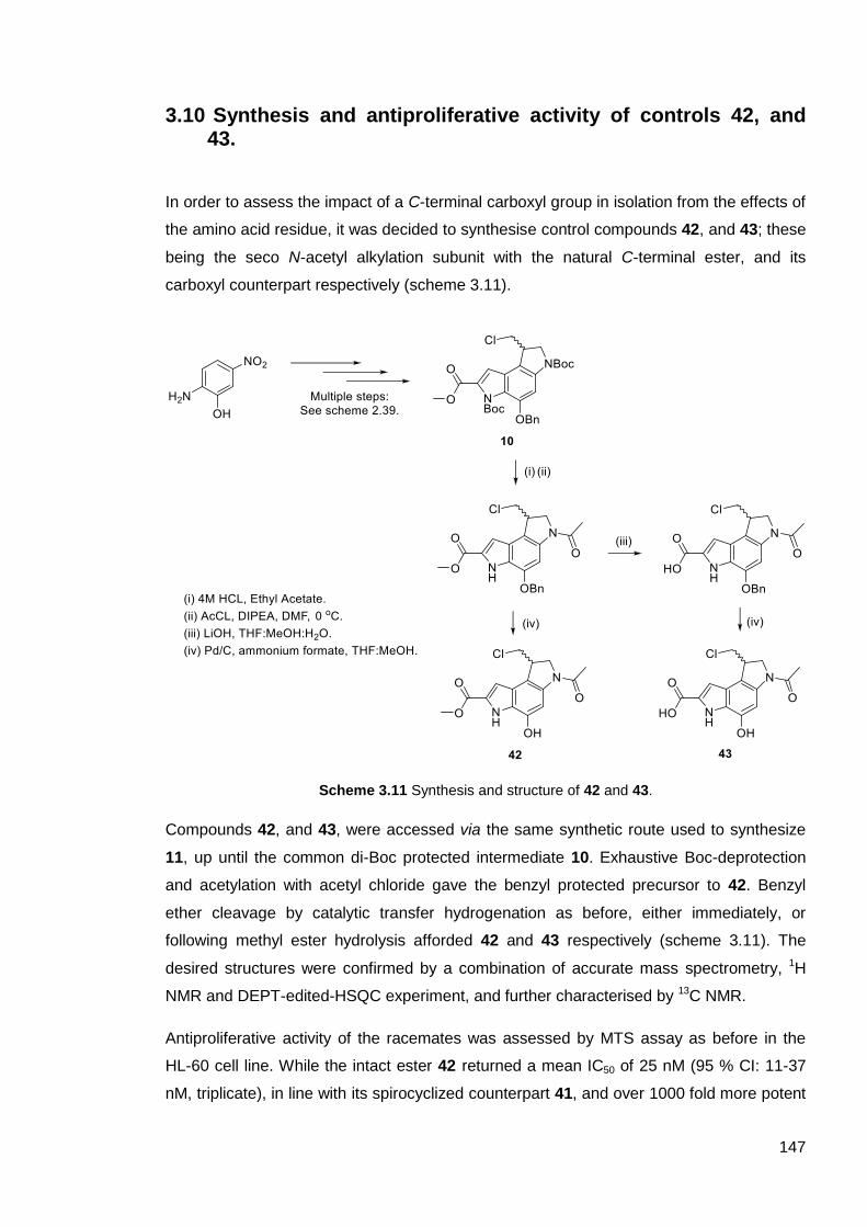


![Ultrashort self-assembling Fmoc-peptide gelators for anti ... · phase Fmoc peptide synthesis protocols using methods previously demonstrated by our group [4]. Peptides were cleaved](https://static.documents.pub/doc/80x56/5f0b640e7e708231d430495b/ultrashort-self-assembling-fmoc-peptide-gelators-for-anti-phase-fmoc-peptide.jpg)


