THE VALUE OF DYNAMIC CONTRAST ENHANCED BREAST MRI IN MAMMOGRAPHICALLY DETECTED BI-RADS 5 MICROCALCIFICATIONS
Dana Houserkovaa*, Sachin Prasad Na, Ivan Svachc, Ladislava Kucerovab, Milada Duskovab, Jiri Bucila, Ivan Sisolaa, Nora Zlamalovac, Hana Svebisovad
a Department of Radiology, Faculty of Medicine and Dentistry, Palacky University, 775 15 Olomouc, Czech Republicb Department of Pathology, Faculty of Medicine and Dentistry, Palacky University, 775 15 Olomoucc Department of Surgery, Faculty of Medicine and Dentistry, Palacky University, 775 15 Olomouc d Department of Oncology, Faculty of Medicine and Dentistry, Palacky University, 775 15 Olomouce-mail: [email protected]
Received: November 11, 2007; Accepted (with revisions): February 8, 2008
Key words: Microcalcifi cation/MRI – mammography/Ductal carcinoma in situ/Multifocality
Aims: To evaluate the role of dynamic contrast-enhanced magnetic resonance imaging (MRI) in the diagnosis of BI-RADS-5 type of microcalcifi cations of the breast, to compare the size of the microcalcifi cation lesions using mammography (MG) and MRI, and to determine the value of MRI in surgery for microcalcifi cations. The study also determines the morphology of microcalcifi cation lesions, assesses kinetic curves and compare MRI features of ductal carcinoma in situ (DCIS) for diff erent histopathological grades.
Methods: Our group consisted of 32 patients with mammographically detected BI-RADS 5 microcalcifi cations. The MRI was done in this group of women which was later followed by stereotactic vaccum-assisted biopsy (SVAB). Surgery was performed on all patients with a biopsy that resulted in a diagnosis of breast cancer or atypical ductal hyperplasia (ADH).
Results: Of our group of 32 patients, there were 35 mammograhically detected microcalcifi cation lesions, 32 DCIS, one ADH and two benign fi ndings according to the fi nal histology.
The microcalcifi cation lesions were larger using MRI than in MG in 10 women. We diagnosed DCIS multifocality in 6 women and bilateral carcinoma in one woman. As with kinetic curve assessment, we found in 67 % of DCIS a rapid rise, 27 % a moderate and in 6 % a slow initial rise. With the pattern of enhancement in the delayed phase, we found in 30 % of DCIS a washout pattern, 67 % a plateau and in 3 % a persistent pattern. Noted diff erence between high and low grade DCIS was confi rmed.
Conclusions: MRI sensitivity in the detection of DCIS was 94 % in our group of patients and was the sole evidence for detection of multifocality and bilateral incidence of carcinoma. In 26 % of women the outcome of MRI was the most important for converting breast conserving surgery to mastectomy.
INTRODUCTION
Magnetic resonance imaging (MRI) is a method also used in breast imaging. It has changed its role recent-ly with the development of modern scanners and new sequences. Breast MRI is an imaging method with big importance not only concerning the morphology of the lesion, but also the evaluation of its functional parameters. The evaluation of the enhancement from the quantitative and qualitative points of view, is in fact the assessment of vascularization of the lesion. The attribute of neoangio-genesis is used in malignant lesions which are often too small to be proved by another imaging method.
The value of breast MRI for ductal carcinoma in situ (DCIS) detection is being discussed. The most common presentations of DCIS are the malignant microcalcifi -cations in mammography (MG). In the case of DCIS manifesting by microcalcifi cation in mammography, the real advantage of breast MRI is the ability to evaluate ac-curately the microcalcifi cation lesion and even the mam-
mographically occult area without microcalcifi cations by detecting the DCIS with microinvasion. The great benefi t of preoperative breast MRI is in the evaluation of multifo-cality and bilateral incidence of carcinoma.
The aim of our study was the following:1) to evaluace the sensitivity, accuracy, positive predictive
value (PPV) and negative predictive value (NPV) of dynamic contrast-enhanced breast MRI for evaluation of malignancy in BIRADS-5 (Breast Imaging Report-ing and Data System) microcalcifi cations.
2) to compare the size of the microcalcifi cation lesions in MG and MRI.
3) to determine the value of MRI for evaluating the multi-focality and bilateral incidence of carcinoma.
4) to determine the MRI morphologic features and to fi nd out the enhancement parameters in microcalcifi -cation lesions.
5) to compare MRI enhancement parameters in DCIS of diff erent histopathological grades.
108 D. Houserkova, S. Prasad N, I. Svach, L. Kucerova, M. Duskova, J. Bucil, I. Sisola, N. Zlamalova, H. Svebisova
6) to assess the benefi t of breast MRI for following surgi-cal therapy.
MATERIALS AND METHODS
Patient populationConsecutive patients with mammographically detected
BI-RADS 5 microcalcifi cations and mammographically dense type of breast (type 4 and 5 according to Tabár1, 2) were recruited from January 2004 to December 2006. Three women with contraindications to MRI (with claus-trophobia or pacemaker) were excluded. These entrance criterias fulfi lled 32 women, aged 34–72 years (50.5 years of age in an average). In all women, after mammography we performed an ultrasound examination and contrast dynamic-enhanced MRI of the breast prior to stereotactic vaccum-assisted biopsy (SVAB).
Mammography protocolBilateral mammography was performed (Diamond,
Instrumentarium Imaging, Tuusula, Finland) and includ-ed craniocaudal and mediolateral oblique view of the breasts and spot – magnifi cation views over the area of microcalcifi cations. The mammograms were independ-ently double-read using BI-RADS assesment categories by two or four radiologists with 5–15 years of experience in mammography. Radiologists also scored breast density ac-cording to Tabár’s classifi cation (1-5). Microcalcifi cations were classifi ed according to BI-RADS for mammographic features including calcifi cation morphology and distribu-tion.
Breast ultrasound protocolIn all women, bilateral breast ultrasound (US) was
perfomed with the knowledge of mammographic fi ndings of BI-RADS 5 microcalcifi catons. US using a linear-ar-ray transducer with a center frequency of 8–11 MHz was performed by the same group of radiologists as mammog-raphy in breast US (Logiq 500 MD, General Electric, Solingen, Germany). We were able to detect the area of microcalcifi cations in breast in 12 women, but we found no mass. In the remaining 23 women, the microcalcifi ca-tios were not seen in ultrasound.
Breast MRI protocol In premenopausal women, we performed breast MRI
within the second week of the menstrual cycle to avoid contrast uptake due to hormonal dependent changes of the breast. We examined these women in prone position with their breasts fi xed in a double breast coil. The scan-ner was a 1.5T system (Siemens Symphony, Erlangen, Germany, Siemens). Using double breast coil enabled comparison of both breast in one image. Image protocol included a localizer followed by transverse turbo-spin echo T2–weighted sequence (TR/TE, 4430/97 ms, matrix 512 × 512) and by coronal STIR (Short Tau Inversion Recovery) sequence (TR/TE, 9370/70 ms). Other parameters were section thickness 3 mm and interslice gap 0.8 mm. These
sequences were followed by a dynamic study consisting of serial imaging of three-dimensional transverse fast low an-gle shot T1-weighted sequence (TR/TE, 4.42/1.67; matrix, 512x512; section thickness 1.2 mm) with fat supression. For dynamic study we acquired one pre- and six post-con-trast sequences. Contrast media Prohance (Gadoteridol, 0.5 M) was administered immediately after the end of fi rst (precontrast) sequence as a bolus intravenous injection at a dose of 0.1 mmol/kg body weight and was followed by a 20 ml saline solution. The total time of the dynamic study was 6.26 minutes. The necessary part of the examination was postprocessing by creating subtraction images of each postcontrast sequence and by creating maximum intensity projection (MIP) images.
For dynamic contrast-enhanced breast MRI, a malig-nant lesion was considered to be diagnosed successfully (true positive) if it showed contrast uptake in the area of microcalcifi cation. The absence of contrast uptake in the area of microcalcifi cation that was assessed to be malig-nant in SVAB or surgery was classifi ed as false-negative MRI fi ndings. Sensitivity, specifi city, accuracy, positive predictive value and negative predictive value were calcu-lated on the basis of the absence or presence of contrast uptake in the area of BI-RADS 5 microcalcifi cation.
MRI morphology of enhancement and kinetic curves were evaluated for all the 31 enhanced microcalcifi ca-tion lesions. We evaluated two main morphological MRI patterns of enhanced microcalcifi cation lesions. By the quantitative evaluation, we assessed the increase in signal intensity on the second post-contrast sequence compared to the pre-contrast sequence. We determined three levels of intensity of enhancement, slow (1st level, 0–50 %), medium (2nd level, 50–100 %) and the rapid (3rd level, above 100 %). For qualitative evaluation we used time to intensity curves (TIC) for this evaluation. We diff erenti-ated three types of enhancement patterns (three types of TIC). Curve type 1 – “persistent pattern“, with a con-tinuous increase in signal intensity on each successive contrast-enhanced image. Type 2 – “plateau pattern“, in which an initial increase in signal intensity was followed by a fl attening and fl uctuation of the enhancement curve (±10 %). Type 3 – “washout pattern“, with an initial in-crease and subsequent decrease in signal intensity. We evaluated the initial rise of enhancement and type of enhancement pattern with regard to the fi nal histology in microcalcifi cation lesions. We used chi-square test to compare the diff erences in enhancement parameters in DCIS of various histopathological grades.
Each examination was evaluated by radiologist with 4 years experience in breast MRI.
SVAB protocolSVAB using 11 or 8-gauge probes (Mammotome,
Ethicon Endo-Surgery, Cincinnati, USA) was performed. A radiologist with 5 years experience in SVAB performed the biopsy. The average number of samples taken during the biopsy was 15. Specimen radiography was performed routinely on all samples. Complete or partial removal of the microcalcifi cations was assessed in all cases on two
109The value of dynamic contrast enhanced breast MRI in mammographically detected BI-RADS 5 microcalcifi cations
view mammograms immediately. If the microcalcifi cations were removed completely, clips were placed through the probe to identify the SVAB site for subsequent surgical excision. The histopathological result was correlated with the mammographic fi ndings by radiologist.
Histological diagnosisHistological diagnosis was determined by two patholo-
gists with 10–20 years of experience in breast histology. The histological fi ndings were classifi ed into three groups – malignant, high risk and benign. Malignant lesions in-cluded invasive carcinoma and DCIS. The grade of DCIS was scored as low, intermediate or high. Atypical ductal hyperplasia (ADH) was considered to be high-risk lesion for which, the associated presence of carcinoma can be underestimated with SVAB. Lesions that were not clas-sifi ed as histologically malignant or high risk were cat-egorized as benign. All women with malignant or high risk microcalcifi cation lesions in SVAB were operated. If malignancy was found in SVAB, the surgeon performed a therapeutic operation including axillary surgery if indi-cated. In case of ADH the surgeon performed surgical excision. If a benign microcalcifi cation lesion was found in SVAB, the woman was followed by mammography of the ipsilateral breast at 6 months interval. If there was no calcifi cation progression in post-SVAB mammographic follow-up, mammography was recommended in usual screening interval.
RESULTS
The histopathological report of our group of 32 pa-tients (aged between 34–72 years) revealed 31 lesions with DCIS diagnosed in SVAB, two with ADH and two with benign fi ndings. 33 lesions with malignant or suspicious fi nding from previous biopsy were operated. SVAB, histo-logic fi ndings and the subsequent surgical excision were compared.
Final histology after surgery confi rmed 32 DCIS le-sions. Among them, 22 pure DCIS were found and in 10 DCIS with microinvasion was proved. One ADH was upgraded to DCIS after surgery and in one ADH was cofi rmed by open biopsy. Regarding the histopathological grade of pure DCIS, high-grade DCIS was confi rmed in 7 women, intermediate-grade DCIS in 9 women and low-grade DCIS in 6 women. Two women with benign micro-calcifi cation lesions in SVAB were followed at 6 months interval and no false-negative diagnosis occured, there was no microcalcifi cation progression at mammographic fol-low-up.
In two benign microcalcifi cation lesions, there was no contrast enhancement in breast MRI. In one case of ADH we found contrast uptake in the microcalcifi cation lesion. Dynamic contrast-enhanced MRI showed uptake of contrast in the area of microcalcifi cations in 31 (94 %) with DCIS. In 2 (6 %) women with histologically proved low grade DCIS, we could not assess the contrast en-hancement in breast MRI and the result of breast MRI
did not prove the existence of DCIS. The sensitivity of contrast-enhanced breast MRI in assessment of BI-RADS 5 microcalcifi cation lesion was 94 %, the accuracy 94 %, PPV 100 % and NPV 50 %.
Regarding the size of microcalcifi cation lesion in breast MRI, we proved the same size of lesion in comparison to MG in 21 (68 %) patients, and in 10 (32 %) patients the size of enhancement in MR imaging was larger – up to 10 mm in 2 patients, up to 15 mm in 3 patients and up to 20 mm in 5 women.
Multifocality of DCIS was found by MRI in 6 (19 %) women (Fig. 1). Bilateral carcinoma was found in one of these patients.
In 31 contrast-enhanced microcalcifi cation lesions, we assessed the morphology pattern of enhancement. Two main patterns of contrast enhancement were evaluated; clumped or stippled and heterogeneous in 21 (68 %) wom-en (pattern 1) and ductal, branching, and heterogeneous in 10 (32 %) women (pattern 2).
As with the level of enhancement we found slow ini-tial peak in 2 (6 %) enhanced DCIS, medium peak in 8 (27 %) and rapid peak in 20 (67 %) of them. In only 1 (3 %) patient with low-grade DCIS we evaluated persist-ent enhancement pattern. In 20 (67 %) of the DCIS, we assessed the plateau enhancement pattern (Fig. 1, 3) and in 9 (30 %) DCIS we found the washout enhancement pattern (Fig. 2) (Table 1).
In one case of ADH we found the medium initial rise of enhancement and the plateau enhancement pattern (Table 1). Microcalcifi cation lesions with rapid peak and type 2 or 3 of enhancement pattern were considered suspi-cious of malignancy.
As with the enhancement features of DCIS of various histopathological grades, signifi cant diff erence was found between the group of low grade and high grade DCIS (p = 0.021) and between the group of low grade DCIS and DCIS with microinvasion (p = 0.006) (Table 2).
Altogether in 11 (35 %) patients, we proved the chang-es (larger size, multifocality) of microcalcifi cation lesions in breast MRI in comparison to MG. In 8 (26 %) patients breast MRI gave the most important information to con-vert the breast conserving surgery to mastectomy.
DISCUSSION
Magnetic resonance imaging (MRI) is also used in breast imaging. The evaluation of breast MRI should be done by radiologists who are experienced with other imag-ing methods used for the breast as well. The assessment of MRI together with other imaging methods mostly mam-mography, increases the sensitivity and specifi city of this method greatly3.
Breast MRI has defi nite indications, and its use should be carefully considered by the radiologist. This examina-tion should not substitute mammography or ultrasound examination even in clear indications for making diag-nostic interventions. The only one indication for native breast MRI is evaluating of the integrity of the silicone
110 D. Houserkova, S. Prasad N, I. Svach, L. Kucerova, M. Duskova, J. Bucil, I. Sisola, N. Zlamalova, H. Svebisova
Fig. 1. Woman 43 years of age, screening mammography of the left breast. (a) Microcalcifi cation in the upper outer quadrant (black arrow) and small nodules in the lower inner quadrant (white arrow) of the left breast. (b) Spot compression magnifi cation mammogram demonstrate microcalcifi cations classifi ed as BI-RADS category 5 (black arrow). (c) Transverse native T2 TSE sequence reveals the small round hypointense lesions in the left breast (black arrows). The area of microcalcifi cation lesion is not visible. (d) Transverse contrast-enhanced T1 FLASH 3D MRI, subtraction image. Small hyperintense round lesions (left and middle images, white arrows) and the clumped and heterogeneous enhancement of microcalcifi cation lesion (right image, white arrow) in the left breast. (e) Time to intensity curve. The rapid initial rise and plateau pattern (type 2) of enhancement (the line curve, the small dotted curve). Final histology was multifocal high – grade DCIS with microinvasion.
a b
c
d
e
111The value of dynamic contrast enhanced breast MRI in mammographically detected BI-RADS 5 microcalcifi cations
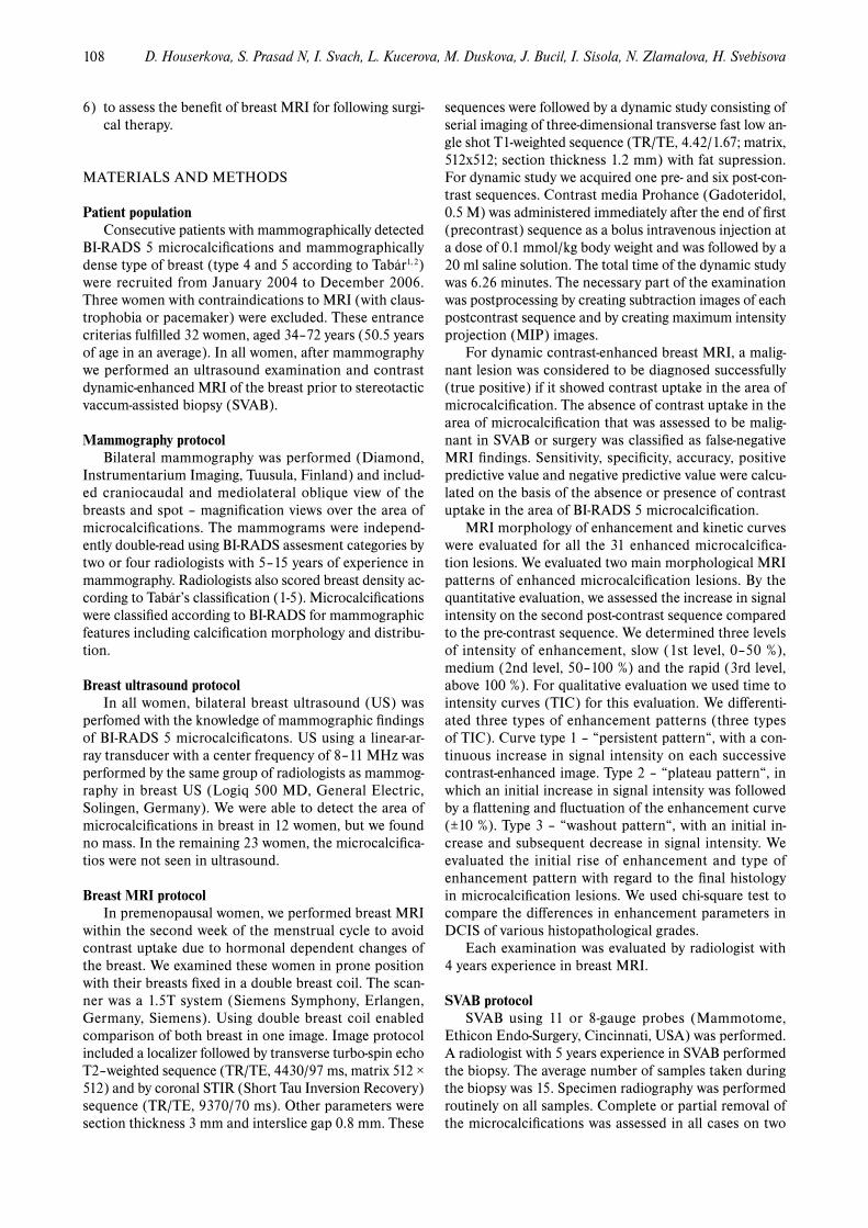
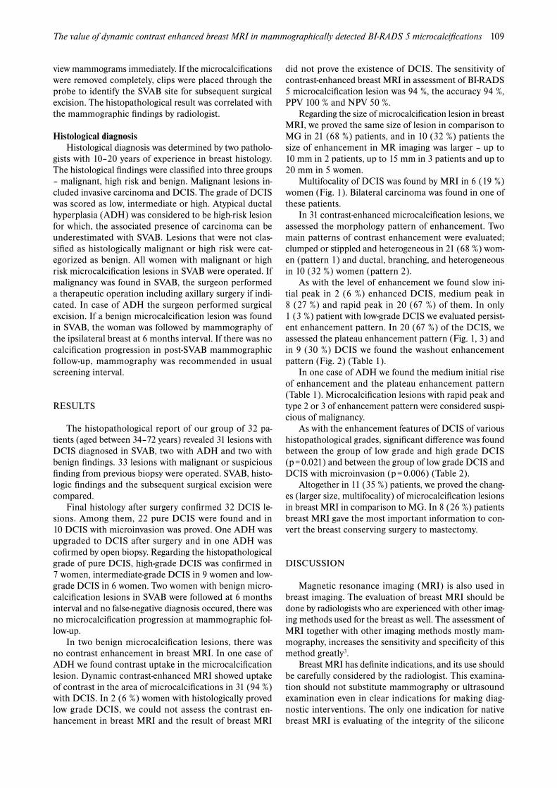
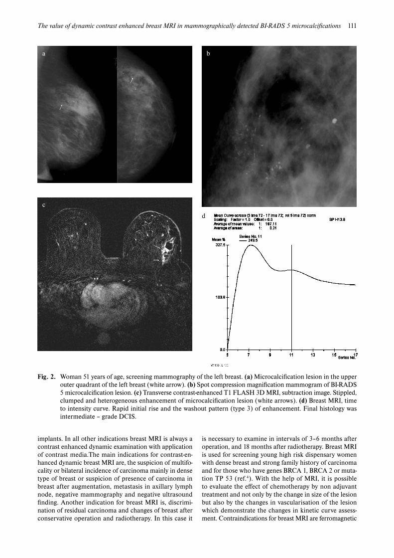
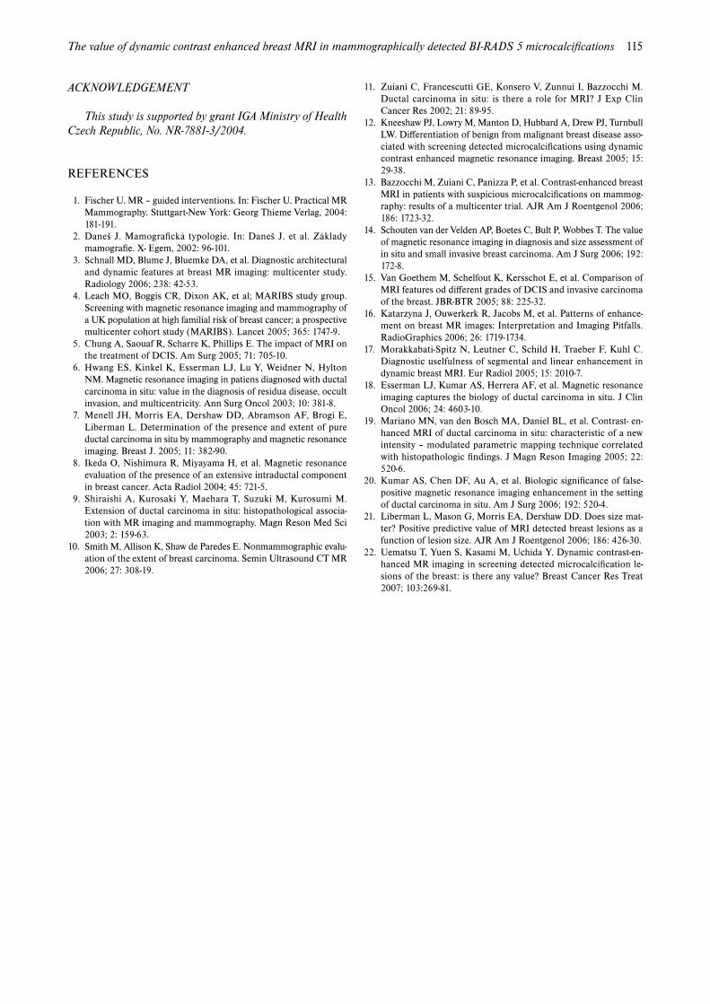
Fig. 2. Woman 51 years of age, screening mammography of the left breast. (a) Microcalcifi cation lesion in the upper outer quadrant of the left breast (white arrow). (b) Spot compression magnifi cation mammogram of BI-RADS 5 microcalcifi cation lesion. (c) Transverse contrast-enhanced T1 FLASH 3D MRI, subtraction image. Stippled, clumped and heterogeneous enhancement of microcalcifi cation lesion (white arrows). (d) Breast MRI, time to intensity curve. Rapid initial rise and the washout pattern (type 3) of enhancement. Final histology was intermediate – grade DCIS.
a b
d
c
implants. In all other indications breast MRI is always a contrast enhanced dynamic examination with application of contrast media.The main indications for contrast-en-hanced dynamic breast MRI are, the suspicion of multifo-cality or bilateral incidence of carcinoma mainly in dense type of breast or suspicion of presence of carcinoma in breast after augmentation, metastasis in axillary lymph node, negative mammography and negative ultrasound fi nding. Another indication for breast MRI is, discrimi-nation of residual carcinoma and changes of breast after conservative operation and radiotherapy. In this case it
is necessary to examine in intervals of 3–6 months after operation, and 18 months after radiotherapy. Breast MRI is used for screening young high risk dispensary women with dense breast and strong family history of carcinoma and for those who have genes BRCA 1, BRCA 2 or muta-tion TP 53 (ref.4). With the help of MRI, it is possible to evaluate the eff ect of chemotherapy by non adjuvant treatment and not only by the change in size of the lesion but also by the changes in vascularisation of the lesion which demonstrate the changes in kinetic curve assess-ment. Contraindications for breast MRI are ferromagnetic
112 D. Houserkova, S. Prasad N, I. Svach, L. Kucerova, M. Duskova, J. Bucil, I. Sisola, N. Zlamalova, H. Svebisova
a c
b
Fig. 3. Woman 38 years of age. Suspicious microcalcifi cations in the screening mammography of the right breast. (a) Spot compression magnifi cation mammogram of BI-RADS 4 microcalcifi cation in the right breast (white arrow). (b) Transverse native T2 TSE sequence reveals small hypointense irregular area of microcalcifi cation lesion (left image, black arrow). Clumped and heterogeneous enhancement of microcalcifi cation lesion (white arrow, right image) in transverse contrast-enhanced T1 FLASH 3D MRI, subtraction image. (c) Breast MRI, time to intensity curve. The rapid initial rise and the washout pattern of enhancement. Final histology was intermediate – grade DCIS.
material in patients body, claustrophobia, pregnancy and lactating women.
Previously MRI had big changes which were related with the progressing of new examination MR technique.The optimal power of the magnet for examination of the breast is from 1.5 Tesla. It is better to use special double breast coil. In this way examination of both the breasts are done and it is possible to compare them. During the examination, the breasts are fi xed in the hole of double breast coil. Dynamic echo sequences with application of
contrast media in an amount of 0.1 mmol/kg are pro-vided in transverse and coronal planes and eventually completed by sagittal plane scanning. Axial plane is used for good imaging of retroareolar and prepectoral areas, in coronal plane there is better visualisation of axillary and parasternal areas. Dynamic contrast study usually consists of native sequence and another 6 (eventually 12) post-contrast sequences. A part of the examination is post-processing by creating subtraction images and maximum intensity projection (MIP) images. Breast MRI is an im-
113The value of dynamic contrast enhanced breast MRI in mammographically detected BI-RADS 5 microcalcifi cations
Table 1. Detailed study of the parameters of enhancement according to the histology of microcalcifi cation lesions.
Final histology35 patients
Enhancement
MRI enhancementInitial rise (%)
MRI enhancementType of pattern (%)
Slow Medium Rapid Type 1 Type 2 Type 3
Benign 2 No enhancement – – – – – –
ADH 1 Enhancement – 1 – – 1 –
DC
IS
Low grade 6No enhancement 2 – – – – – –
Enhancement 4 1 3 – 1 3 –
Intermediate 9 Enhancement 1 3 5 – 7 2
High grade 7 Enhancement – 1 6 – 4 3
DCIS with microinvasion
10 Enhancement – 1 9 – 6 4
Table 2. Enhancement parameters of DCIS of diff erent histopathological grades.
Histological grade of DCIS
MRI enhancementInitial rise (%)
MRI enhancementType of pattern (%)
Slow Medium Rapid Type 1 Type 2 Type 3
Low grade DCIS 4 25 75 – 25 75 –
Intermediate grade DCIS 9 11 33 56 – 78 22
High grade DCIS 7 – 14 86 – 57 43
High grade DCIS with microinvasion
10 – 10 90 – 60 40
aging method which diff ers from other methods used for breast imaging, because it insists not only on the imaging of morphology of lesions, but also on the functional evalu-ation. Evaluation of enhancement from quantitative and qualitative point of view is infact the evaluation of vascu-larisation of the lesion. The principle of dynamic contrast enhanced breast MRI is based on the feature of neoangio-genesis of malignant lesion which is sometimes too small to be detected by another imaging method (MG, USG). Enhancement of normal breast during menstrual cycle, dramatically fl uctuate by the infl uence of hormone levels. In premenopausal women, performing breast MRI within the second week of the menstrual cycle may improve the sensitivity and specifi city of dynamic contrast – enhanced breast MRI because diff use enhancement such as uniden-tifi ed breast objects (UBOś) are reduced.
Breast MR is an imaging method, which has high sen-sitivity (in invasive carcinoma 95–100 %) and relatively
low specifi city (40–80 %). The problem with the signifi -cance of dynamic contrast enhanced MRI in patients with suspicious microcalcifi cations of the breast is still being discussed. Of course MRI is unable to image microcal-cifi cations of the breast. Its great importance is in the imaging of a mammographically occult microcalcifi cation lesion of breast. Clustered malignant microcalcifi cations are the most common presentation of DCIS in mammo-graphy. The great benefi t of preoperative breast MRI is in imaging the surrounding DCIS with microinvasion and ductal invasive carcinomas that were already diagnosed by mammography. The other benefi t of preoperative MRI is assessing multifocality and bilateral incidence of the carcinoma5-7.
The sensitivity, specifi city and accuracy of breast MR imaging in detecting DCIS with microinvasion were found high in some studies, 71 %, 85 % and 76 %, respectively8. Particularly in dense type of breast during MRI, the extent
114 D. Houserkova, S. Prasad N, I. Svach, L. Kucerova, M. Duskova, J. Bucil, I. Sisola, N. Zlamalova, H. Svebisova
of microcalcifi cation lesion can be bigger in comparison with that of MG and it is possible to detect another mam-mographically and ultrasonographically occult foci of DCIS and observe more accurately the tumour size5, 9–11.
Microcalcifi cations as other lesions of the breast are categorized according to BI-RADS. Microcalcifi cations classifi ed as BI-RADS 4 and 5 are recommended to under-go a biopsy. It is quite diffi cult to determine the BI-RADS 3 microcalcifi cations. The short–term mammographic follow-up which is recommended for probable benign le-sions is questionable in this case. Recent studies suggest that, histological evaluation of BI-RADS 3 microcalcifi ca-tion is necessary until a more reliable system for the de-scription and classifi cation of these microcalcifi cations is available. The last study of Japanese authors recommends BI-RADS 3 microcalcifi cations to be evaluated by MRI prior to SVAB especially in those women who does not want to undergo biopsy22. This is due to the high sensitiv-ity of breast MRI for the assessment of malignant and benign microcalcifi cations12.
In literatures, high percentage of agreement regarding the size of malignant lesion detected in breast MRI with defi nitive histology is mentioned11. MRI is not an abso-lutely reliable method and overestimation and underesti-mation of the size of the lesion is found13, 14. It was found that quite a high percentage of re-operations in cases of DCIS was diagnosed only by MG. MRI is understood to be an invaluable adjunct in the evaluation of DCIS or in-vasive carcinoma especially in those women who consider breast conserving therapy5, 6, 9, 10.
Very typical morphological feature for high but also for low DCIS is ductal pattern of enhancement7, 15, 16.Usually it is clumped, and the heterogeneous or homogeneous enhancement sometimes can be of branching appearance and follows the course of the duct8. The enhancement can also have a non regular stippled, spotty and micronodular appearance. Segmental and linear character of this en-hancement is described and this morphological fi nding was assessed to have high PPV for DCIS lesions16, 17. It was assessed that MRI morphologic features of DCIS can refl ect diff erences in biology and pathology of these tumours18. The region of interest (ROI) which is necessary to gain relevant quantitative and qualitative evaluation of enhancement must be very small in this ductal pattern of enhancement to obtain the most accurate measurement. The ROI must cover only part of the lesion, and should be placed in the region of strongest enhancement on the fi rst contrast – enhanced image. When the ROI is randomly placed in the mass, the enhancement curve may be vari-able and yield lower specifi city16.
However, regarding the detection of ductal carcino-ma in situ (DCIS), the sensitivity of MRI varies between 70 % and 100 % according to the latest studies9, 11-13, 15, 19. According to Menell’s study, breast MRI was signifi cantly more sensitive than mammography in DCIS detection7. It was shown that, MRI had relatively high sensitivity for high grade DCIS which often showed rapid initial rise and 2 or 3 of enhancement pattern and thus resembles invasive carcinoma by its characteristics. Low grade DCIS
had often quite nontypical form of enhancement pattern and initial rise was usually lower and is of course diffi -cult to diff erentiate it from benign proliferative processes that often enhance on MRI (ref.20, 21). To Mariano et al. study contrast-enhanced MRI using parametric mapping technique was useful in identifying all intermediate and high-grade DCIS lesions10. Regarding the kinetic curve assessment of DCIS, the rapid initial peak of enhance-ment was found in almost 77 % and washout pattern of enhancement in nearly 54 % of women13.
If the ductal pattern of enhancement (during nega-tive MG and USG fi nding) and kinetic curve assessment suspicious of malignancy are found in MRI, most of the authors recommend biopsy under MRI control3, 15. Some authors recommend further work-up in case only if the segmental or ductal pattern of enhancement is identifi ed on breast MRI. They recommend either direct MR-guided vaccum-assisted biopsy or short-term follow-up breast MRI within 3 months15. Libermann et al. found that the PPV of biopsy for lesions identifi ed in breast MRI signifi cantly increased with increasing size of the lesion and biopsy is rarely necessary for lesions smaller than 5 mm because of their low likelihood of cancer19. If the diagnostic centre for breast is interested in MRI examination seriously, it should be well equipped for doing biopsy and localization under MRI control. It is highly sensitive and indicated in the hands of the experts in breast imaging and upon the correlation with MG and USG.
CONCLUSION
1. Contrast dynamic – enhanced MRI had sensitivity of 94 %, PPV of 100 %, NPV of 50 % and accuracy of 100 % for detection of DCIS in our group of BI-RADS 5 microcalcifi cation lesions.
2. Larger size of microcalcifi cation lesion in breast MRI in comparison with MG was proved in 32 % of wom-en. In other 68 %, the size of microcalcifi cation lesion in breast MRI and MG was the same.
3. In 19 % of DCIS, we found multifocality of malig-nant lesion, and bilateral carcinoma was proved in one patient among these women.
4. In 94 % of women with malignancy or ADH, we proved enhancement of microcalcification lesions in breast MRI. Two main patterns of contrast enhancement were evaluated that were typical for DCIS microcalcifi -caton lesions. In DCIS rapid initial rise of enhancement (in 67 %), and the plateau (in 67 %) or washout (in 30 %) enhancement patterns were found most frequently.
5. As with MRI enhancement features, noted diff erenc-es between the low grade and high grade DCIS (p = 0.021) and low grade and high grade DCIS with microinvasion, (p = 0.006) were found.
6. In 26 % of women, the result of breast MRI was the most important to convert the breast conserving opera-tion to mastectomy.
115The value of dynamic contrast enhanced breast MRI in mammographically detected BI-RADS 5 microcalcifi cations
ACKNOWLEDGEMENT
This study is supported by grant IGA Ministry of Health Czech Republic, No. NR-7881-3/2004.
REFERENCES
1. Fischer U. MR – guided interventions. In: Fischer U. Practical MR Mammography. Stuttgart-New York: Georg Thieme Verlag, 2004: 181-191.
2. Daneš J. Mamografi cká typologie. In: Daneš J. et al. Základy mamografi e. X- Egem, 2002: 96-101.
3. Schnall MD, Blume J, Bluemke DA, et al. Diagnostic architectural and dynamic features at breast MR imaging: multicenter study. Radiology 2006; 238: 42-53.
4. Leach MO, Boggis CR, Dixon AK, et al; MARIBS study group. Screening with magnetic resonance imaging and mammography of a UK population at high familial risk of breast cancer; a prospective multicenter cohort study (MARIBS). Lancet 2005; 365: 1747-9.
5. Chung A, Saouaf R, Scharre K, Phillips E. The impact of MRI on the treatment of DCIS. Am Surg 2005; 71: 705-10.
6. Hwang ES, Kinkel K, Esserman LJ, Lu Y, Weidner N, Hylton NM. Magnetic resonance imaging in patiens diagnosed with ductal carcinoma in situ: value in the diagnosis of residua disease, occult invasion, and multicentricity. Ann Surg Oncol 2003; 10: 381-8.
7. Menell JH, Morris EA, Dershaw DD, Abramson AF, Brogi E, Liberman L. Determination of the presence and extent of pure ductal carcinoma in situ by mammography and magnetic resonance imaging. Breast J. 2005; 11: 382-90.
8. Ikeda O, Nishimura R, Miyayama H, et al. Magnetic resonance evaluation of the presence of an extensive intraductal component in breast cancer. Acta Radiol 2004; 45: 721-5.
9. Shiraishi A, Kurosaki Y, Maehara T, Suzuki M, Kurosumi M. Extension of ductal carcinoma in situ: histopathological associa-tion with MR imaging and mammography. Magn Reson Med Sci 2003; 2: 159-63.
10. Smith M, Allison K, Shaw de Paredes E. Nonmammographic evalu-ation of the extent of breast carcinoma. Semin Ultrasound CT MR 2006; 27: 308-19.
11. Zuiani C, Francescutti GE, Konsero V, Zunnui I, Bazzocchi M. Ductal carcinoma in situ: is there a role for MRI? J Exp Clin Cancer Res 2002; 21: 89-95.
12. Kneeshaw PJ, Lowry M, Manton D, Hubbard A, Drew PJ, Turnbull LW. Diff erentiation of benign from malignant breast disease asso-ciated with screening detected microcalcifi cations using dynamic contrast enhanced magnetic resonance imaging. Breast 2005; 15: 29-38.
13. Bazzocchi M, Zuiani C, Panizza P, et al. Contrast-enhanced breast MRI in patients with suspicious microcalcifi cations on mammog-raphy: results of a multicenter trial. AJR Am J Roentgenol 2006; 186: 1723-32.
14. Schouten van der Velden AP, Boetes C, Bult P, Wobbes T. The value of magnetic resonance imaging in diagnosis and size assessment of in situ and small invasive breast carcinoma. Am J Surg 2006; 192: 172-8.
15. Van Goethem M, Schelfout K, Kersschot E, et al. Comparison of MRI features od diff erent grades of DCIS and invasive carcinoma of the breast. JBR-BTR 2005; 88: 225-32.
16. Katarzyna J, Ouwerkerk R, Jacobs M, et al. Patterns of enhance-ment on breast MR images: Interpretation and Imaging Pitfalls. RadioGraphics 2006; 26: 1719-1734.
17. Morakkabati-Spitz N, Leutner C, Schild H, Traeber F, Kuhl C. Diagnostic uselfulness of segmental and linear enhancement in dynamic breast MRI. Eur Radiol 2005; 15: 2010-7.
18. Esserman LJ, Kumar AS, Herrera AF, et al. Magnetic resonance imaging captures the biology of ductal carcinoma in situ. J Clin Oncol 2006; 24: 4603-10.
19. Mariano MN, van den Bosch MA, Daniel BL, et al. Contrast- en-hanced MRI of ductal carcinoma in situ: characteristic of a new intensity – modulated parametric mapping technique correlated with histopathologic fi ndings. J Magn Reson Imaging 2005; 22: 520-6.
20. Kumar AS, Chen DF, Au A, et al. Biologic signifi cance of false-positive magnetic resonance imaging enhancement in the setting of ductal carcinoma in situ. Am J Surg 2006; 192: 520-4.
21. Liberman L, Mason G, Morris EA, Dershaw DD. Does size mat-ter? Positive predictive value of MRI detected breast lesions as a function of lesion size. AJR Am J Roentgenol 2006; 186: 426-30.
22. Uematsu T, Yuen S, Kasami M, Uchida Y. Dynamic contrast-en-hanced MR imaging in screening detected microcalcifi cation le-sions of the breast: is there any value? Breast Cancer Res Treat 2007; 103:269-81.