Modica et al. BMC Genomics (2015) 16:441 DOI 10.1186/s12864-015-1648-4
RESEARCH ARTICLE Open Access
The venomous cocktail of the vampire snailColubraria reticulata (Mollusca, Gastropoda)
Maria Vittoria Modica1, Fabrizio Lombardo2, Paolo Franchini3 and Marco Oliverio1*
Abstract
Background: Hematophagy arose independently multiple times during metazoan evolution, with several lineagesof vampire animals particularly diversified in invertebrates. However, the biochemistry of hematophagy has beenstudied in a few species of direct medical interest and is still underdeveloped in most invertebrates, as in general isthe study of venom toxins. In cone snails, leeches, arthropods and snakes, the strong target specificity of venomtoxins uniquely aligns them to industrial and academic pursuits (pharmacological applications, pest control etc.)and provides a biochemical tool for studying biological activities including cell signalling and immunologicalresponse. Neogastropod snails (cones, oyster drills etc.) are carnivorous and include active predators, scavengers,grazers on sessile invertebrates and hematophagous parasites; most of them use venoms to efficiently feed. It hasbeen hypothesized that trophic innovations were the main drivers of rapid radiation of Neogastropoda in the lateCretaceous.We present here the first molecular characterization of the alimentary secretion of a non-conoidean neogastropod,Colubraria reticulata. Colubrariids successfully feed on the blood of fishes, throughout the secretion into the host ofa complex mixture of anaesthetics and anticoagulants. We used a NGS RNA-Seq approach, integrated with differentialexpression analyses and custom searches for putative secreted feeding-related proteins, to describe in detail the salivaryand mid-oesophageal transcriptomes of this Mediterranean vampire snail, with functional and evolutionary insights onmajor families of bioactive molecules.
Results: A remarkably low level of overlap was observed between the gene expression in the two target tissues,which also contained a high percentage of putatively secreted proteins when compared to the whole body.At least 12 families of feeding-related proteins were identified, including: 1) anaesthetics, such as ShK Toxin-containingproteins and turripeptides (ion-channel blockers), Cysteine-rich secretory proteins (CRISPs), Adenosine Deaminase(ADA); 2) inhibitors of primary haemostasis, such as novel vWFA domain-containing proteins, the Ectonucleotidepyrophosphatase/phosphodiesterase family member 5 (ENPP5) and the wasp Antigen-5; 3) anticoagulants, such asTFPI-like multiple Kunitz-type protease inhibitors, Peptidases S1 (PS1), CAP/ShKT domain-containing proteins, Astacinmetalloproteases and Astacin/ShKT domain-containing proteins; 4) additional proteins, such the Angiotensin-Converting Enzyme (ACE: vasopressive) and the cytolytic Porins.
Conclusions: Colubraria feeding physiology seems to involve inhibitors of both primary and secondary haemostasis,anaesthetics, a vasoconstrictive enzyme to reduce feeding time and tissue-degrading proteins such as Porins andAstacins. The complexity of Colubraria venomous cocktail and the divergence from the arsenal of the fewneogastropods studied to date (mostly conoideans) suggest that biochemical diversification of neogastropodsmight be largely underestimated and worth of extensive investigation.
* Correspondence: [email protected] of Biology and Biotechnologies “C. Darwin”, Sapienza University,I-00185 Rome, ItalyFull list of author information is available at the end of the article
rticle distributed under the terms of the Creative Commons Attribution Licensewhich permits unrestricted use, distribution, and reproduction in anyly credited. The Creative Commons Public Domain Dedication waiverro/1.0/) applies to the data made available in this article, unless otherwise stated.
Modica et al. BMC Genomics (2015) 16:441 Page 2 of 21
BackgroundHematophagy arose independently multiple times duringmetazoan evolution, leading to the appearance of severallineages of vampire animals that exploit hosts’ blood, a re-newable and nutrient-rich source. Beside vertebrates (batsand lampreys), in invertebrates hematophagous parasitesare found amongst platyhelminths (flukes), nematodes(hookworms), annelids (leeches), molluscs and in arthro-pods with more than 14,000 hematophagous species, ran-ging from acarines (ticks) to insects (mosquitoes, sandflies,bugs, fleas, horseflies, midges, moths) and crustaceans (sealice, fish lice) [1, 2].Adaptations to blood-feeding life style include behav-
ioral traits, fundamental for host location, anatomicalfeatures, often including specialized mouthparts and bio-chemical specialization, with the production of complexsecretions (generally including molecules acting as anti-hemostatic, anesthetic and anti-inflammatory) in special-ized glands [2, 3].The biochemical bases of blood feeding have been in-
vestigated to date only in a reduced number of medicallyimportant species, either for their therapeutic use, as isthe case of leeches [4, 5], or for their role as vectors ofdeadly diseases such as ticks and insects [6–8] that areundoubtedly the most explored group. These investiga-tions revealed that hematophagous animals have conver-gently recruited a series of molecules acting on differentphysiological stages of host hemostasis, including vasocon-striction, formation of both platelet plugs and fibrinclots and fibrinolysis [3, 5, 7, 9]. Platelet aggregationis the first stage of hemostasis, and is activated by colla-gen, thrombin, ADP and thromboxane A [9]. Secondaryhemostasis, or blood coagulation cascade, consists of aseries of enzymatic reaction where coagulation factors (in-active proenzymes) are converted in their active forms,generally proteases, that in turn activate the next pro-enzyme in the cascade, converging on a final commonpathway concluded by thrombin that converts fibrinogenin fibrin clot. Generally speaking, hematophagous animalstarget the hemostatic system of the host at the level ofboth primary and secondary hemostasis, producing anumber of proteins of different molecular masses, that actinteracting with several targets and exert a variety of in-hibitory mechanisms [3, 5, 7, 9].Among the less studied hematophagous groups, vampire
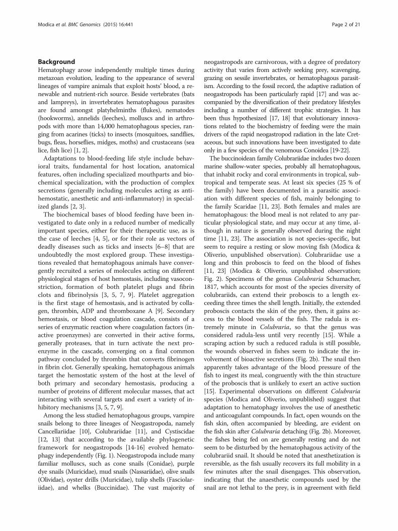
snails belong to three lineages of Neogastropoda, namelyCancellariidae [10], Colubrariidae [11], and Cystiscidae[12, 13] that according to the available phylogeneticframework for neogastropods [14-16] evolved hemato-phagy independently (Fig. 1). Neogastropoda include manyfamiliar molluscs, such as cone snails (Conidae), purpledye snails (Muricidae), mud snails (Nassariidae), olive snails(Olividae), oyster drills (Muricidae), tulip shells (Fasciolar-iidae), and whelks (Buccinidae). The vast majority of
neogastropods are carnivorous, with a degree of predatoryactivity that varies from actively seeking prey, scavenging,grazing on sessile invertebrates, or hematophagous parasit-ism. According to the fossil record, the adaptive radiation ofneogastropods has been particularly rapid [17] and was ac-companied by the diversification of their predatory lifestylesincluding a number of different trophic strategies. It hasbeen thus hypothesized [17, 18] that evolutionary innova-tions related to the biochemistry of feeding were the maindrivers of the rapid neogastropod radiation in the late Cret-aceous, but such innovations have been investigated to dateonly in a few species of the venomous Conoidea [19-22].The buccinoidean family Colubrariidae includes two dozen
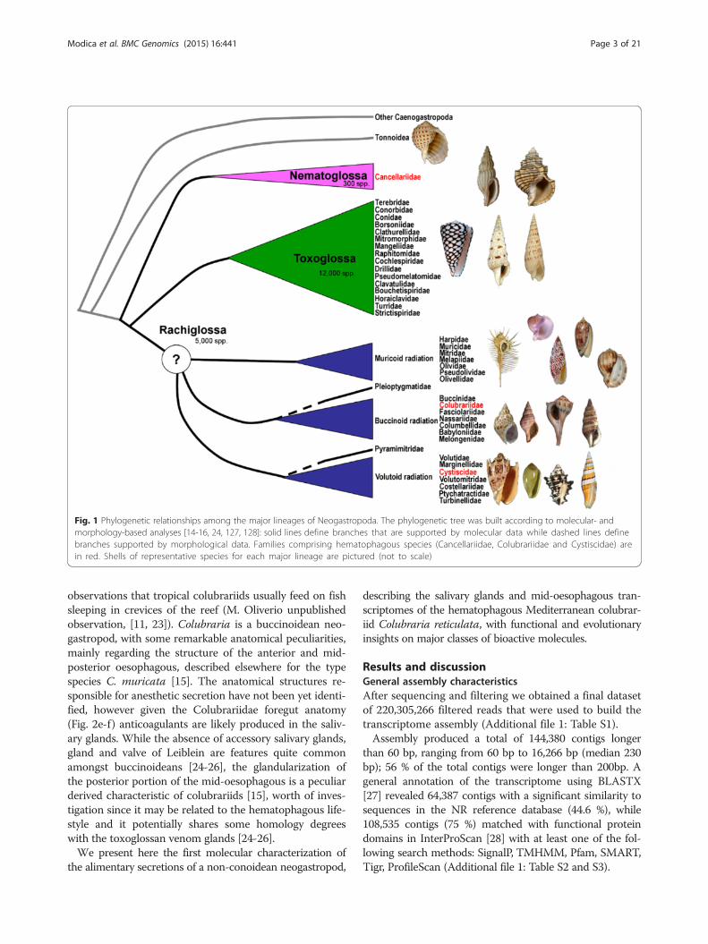
marine shallow-water species, probably all hematophagous,that inhabit rocky and coral environments in tropical, sub-tropical and temperate seas. At least six species (25 % ofthe family) have been documented in a parasitic associ-ation with different species of fish, mainly belonging tothe family Scaridae [11, 23]. Both females and males arehematophagous: the blood meal is not related to any par-ticular physiological state, and may occur at any time, al-though in nature is generally observed during the nighttime [11, 23]. The association is not species-specific, butseem to require a resting or slow moving fish (Modica &Oliverio, unpublished observation). Colubrariidae use along and thin proboscis to feed on the blood of fishes[11, 23] (Modica & Oliverio, unpublished observation;Fig. 2). Specimens of the genus Colubraria Schumacher,1817, which accounts for most of the species diversity ofcolubrariids, can extend their proboscis to a length ex-ceeding three times the shell length. Initially, the extendedproboscis contacts the skin of the prey, then, it gains ac-cess to the blood vessels of the fish. The radula is ex-tremely minute in Colubraria, so that the genus wasconsidered radula-less until very recently [15]. While ascraping action by such a reduced radula is still possible,the wounds observed in fishes seem to indicate the in-volvement of bioactive secretions (Fig. 2b). The snail thenapparently takes advantage of the blood pressure of thefish to ingest its meal, congruently with the thin structureof the proboscis that is unlikely to exert an active suction[15]. Experimental observations on different Colubrariaspecies (Modica and Oliverio, unpublished) suggest thatadaptation to hematophagy involves the use of anestheticand anticoagulant compounds. In fact, open wounds on thefish skin, often accompanied by bleeding, are evident onthe fish skin after Colubraria detaching (Fig. 2b). Moreover,the fishes being fed on are generally resting and do notseem to be disturbed by the hematophagous activity of thecolubrariid snail. It should be noted that anesthetization isreversible, as the fish usually recovers its full mobility in afew minutes after the snail disengages. This observation,indicating that the anaesthetic compounds used by thesnail are not lethal to the prey, is in agreement with field
Fig. 1 Phylogenetic relationships among the major lineages of Neogastropoda. The phylogenetic tree was built according to molecular- andmorphology-based analyses [14-16, 24, 127, 128]: solid lines define branches that are supported by molecular data while dashed lines definebranches supported by morphological data. Families comprising hematophagous species (Cancellariidae, Colubrariidae and Cystiscidae) arein red. Shells of representative species for each major lineage are pictured (not to scale)
Modica et al. BMC Genomics (2015) 16:441 Page 3 of 21
observations that tropical colubrariids usually feed on fishsleeping in crevices of the reef (M. Oliverio unpublishedobservation, [11, 23]). Colubraria is a buccinoidean neo-gastropod, with some remarkable anatomical peculiarities,mainly regarding the structure of the anterior and mid-posterior oesophagous, described elsewhere for the typespecies C. muricata [15]. The anatomical structures re-sponsible for anesthetic secretion have not been yet identi-fied, however given the Colubrariidae foregut anatomy(Fig. 2e-f) anticoagulants are likely produced in the saliv-ary glands. While the absence of accessory salivary glands,gland and valve of Leiblein are features quite commonamongst buccinoideans [24-26], the glandularization ofthe posterior portion of the mid-oesophagous is a peculiarderived characteristic of colubrariids [15], worth of inves-tigation since it may be related to the hematophagous life-style and it potentially shares some homology degreeswith the toxoglossan venom glands [24-26].We present here the first molecular characterization of
the alimentary secretions of a non-conoidean neogastropod,
describing the salivary glands and mid-oesophagous tran-scriptomes of the hematophagous Mediterranean colubrar-iid Colubraria reticulata, with functional and evolutionaryinsights on major classes of bioactive molecules.
Results and discussionGeneral assembly characteristicsAfter sequencing and filtering we obtained a final datasetof 220,305,266 filtered reads that were used to build thetranscriptome assembly (Additional file 1: Table S1).Assembly produced a total of 144,380 contigs longer
than 60 bp, ranging from 60 bp to 16,266 bp (median 230bp); 56 % of the total contigs were longer than 200bp. Ageneral annotation of the transcriptome using BLASTX[27] revealed 64,387 contigs with a significant similarity tosequences in the NR reference database (44.6 %), while108,535 contigs (75 %) matched with functional proteindomains in InterProScan [28] with at least one of the fol-lowing search methods: SignalP, TMHMM, Pfam, SMART,Tigr, ProfileScan (Additional file 1: Table S2 and S3).
Fig. 2 Anatomy and feeding activity of Colubraria reticulata. a: a specimen of C. reticulata crawling towards a Gobius bucchichi (Perciformes:Gobiidae), with the proboscis extended. The fish appear to be semi-anesthetized. b: circular bleeding wound found after C. reticulata detachmentbehind the pelvic fin of Uranoscopus scaber (Perciformes: Uranoscopidae). c: C. reticulata probing the sub-opercular area of G. bucchichi; d: C. reticulatafeeding on the caudal fin of G. bucchichi; e: Dissected foregut of C. reticulata, from the proboscis tip to the mid-posterior oesophagous f: Bodyof C. reticulata. Mantle and body wall are medially dissected to show mantle organs and organization of the foregut. Abbreviations: an,anus; aoe, anterior oesophagus; bm, buccal mass; ct, ctenidium; dg, digestive gland; fo, foot; go, gonad; he, head; hyg, hypobranchialgland; lsg, left salivary gland; mo, mouth; mpoe, mid-posterior oesophagus; mtl, mantle; nf, nerve fibre; nr, nerve ring; os, osphradium; pa,proboscideal artery; p, penis; pr, proboscis; prr, proboscis retractor muscle; psh, proboscis sheath; re, rectum; rsg, right salivary gland; ry,rhynchodaeum; sd, salivary gland duct; si, siphon; st, stomach
Modica et al. BMC Genomics (2015) 16:441 Page 4 of 21
Modica et al. BMC Genomics (2015) 16:441 Page 5 of 21
The total number of transcripts retrieved for C. reticu-lata transcriptome is congruent with previously reporteddatasets from other non-model species of invertebrates,including gastropods [29-31].A total of 1437 transcripts possibly derive from transpos-
able elements, possessing RT PFAM domains indicative ofreverse transcriptase. The presence of these transcriptsmay indicate transposition activity taking place in the gen-ome of C. reticulata.Protein families (PFAM) that are enriched in the whole-
body with respect to both target tissues (Additional file 1:Table S4; Additional file 2: Figure S1) include large multi-functional families such as Thrombospondin-, Kelch-,ARM-, and WD40-containing proteins, putative oxygencarriers containing globin and haemocyanin domains, proteinwith kinase and tyrosinase domains, HMG-box-containingproteins involved in DNA-regulation processes, immunitycomponents such as complement and immunoglobulins,and sugar transporters. The iron-storage Ferritin proteinsthat in mollusks have been related with immunity, devel-opment and shell formation processes [32], and LEA (LateEmbryogenesis Abundant) proteins that are thought to beinvolved in the response to drying and osmotic stresses inplants and nematodes [33] were also enriched in thewhole body. Enrichment in the whole body involves alsoproteins containing a ShK toxin domain, whose expres-sion is enhanced in the salivary subset as well (see below).
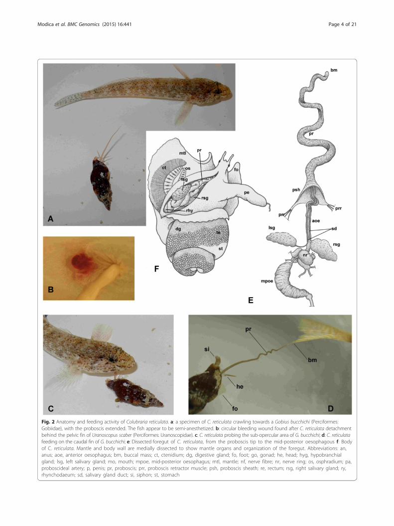
Gene-expression profiling of glandular tissuesOur NGS-based transcriptome analysis revealed that,with a conservative approach (logFC > =3, FDR < 0.05),935 contigs showed enhanced expression in the salivary
Fig. 3 Tissue-specific gene expression. Volcano plots displaying the relativesalivary glands (right) versus the whole body. The x-axis represent the log2(tissue specific logCPM: whole body logCPM, where CPM stands for Countscorrected for the false discovery rate. Red points represent transcripts withlogFC values indicate transcripts enhanced in the tissue-specific subset, wh
glands when compared to whole body while 184 contigsshowed enriched expression in the mid-esophagus (Fig. 3;Additional file 1: Table S5, Table S6).Among these, 378 and 93 contigs respectively did not
display a significant similarity with known proteins in theNR database by BLAST search, corresponding to 40.3 %for salivary glands and 50.5 % for mid-oesophagus. Thishigh rate of transcripts that were unidentifiable with aBLAST search is congruent with corresponding rates ob-served in the transcriptomic analyses of venom-producingglands of other taxa such as arthropods [34-36], wheremore than 30 % to more than 50 % of transcript re-sulted unknown. However, when we examined singu-larly the most overexpressed contigs (SI > 65 %, where SIis the normalized tissue-specific logFC value for each con-tig) with no BLAST, we were able to recover similaritiesfor about half of them.The expression of only 32 contigs, 13 of which had no
BLAST hits, was enhanced in both tissues. Limiting theanalysis to contigs with a SI > =50 %, the overlap be-tween the two tissue-specific subsets decreases to 9 con-tigs, two of which have no BLAST hit (Fig. 4; Additionalfile 1: Table S7).The most represented contigs of the tissue-specific
subsets encode for proteins/domains that belong to dif-ferent PFAM families (Fig. 5). Specifically, Kunitz-BPTI,FMRFamide-related peptides (FARPs), WD40 proteinsand whey acidic proteins (WAP) are the protein domainsthat dominate the oesophageal subset, which is generallyscarcely diversified, with only 7 families represented bymore than 2 contigs (Additional file 1: Table S8). Salivarysubset includes 50 families represented by more than 2
expression levels of transcripts in the mid-oesophagous (left) andof the expression ratio for each transcript of Colubraria transcriptomePer Million reads); the y-axis represents the log10 of the p-valuelogFDR < 0.05 a greater than 3-folds difference in logCPM). Positiveile negative values indicate transcripts enhanced in the whole body
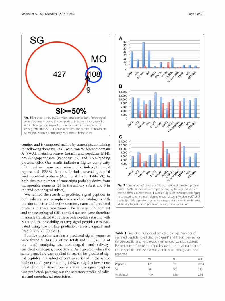
Fig. 4 Enriched transcripts pairwise tissue comparison. ProportionalVenn diagrams showing the comparison between salivary-specificand mid-oesophagous-specific transcripts, with a tissue-specificityindex greater than 50 %. Overlap represents the number of transcriptswhose expression is significantly enhanced in both tissues
Fig. 5 Comparison of tissue-specific expression of targeted proteinclasses. a Abundance of transcripts belonging to targeted venomprotein classes in each tissue; b Median logFC of transcripts belongingto targeted venom protein classes in each tissue; c Median logCPM oftranscripts belonging to targeted venom protein classes in each tissue.Mid-oesophageal transcripts in red, salivary transcripts in red
Table 1 Predicted number of secreted contigs. Number ofsecreted peptides predicted by SignalP and PrediSi servers fortissue-specific and whole-body enhanced contigs subsets.Percentages of secreted peptides over the total number oftissue-specific and whole-body enhanced contigs are alsoreported
MO SG WB
Peptides 178 929 1048
SP 80 305 235
% SP/total 44.9 32.8 22.4
Modica et al. BMC Genomics (2015) 16:441 Page 6 of 21
contigs, and is composed mainly by transcripts containingthe following domains: ShK Toxin, von Willebrand domainA (vWA), metalloproteases (astacin and peptidase M14),prolyl-oligopeptidases (Peptidase S9) and RNA-bindingproteins (KH). Our results indicate a higher complexityof the salivary gene expression profile: indeed, the mostrepresented PFAM families include several potentialfeeding-related proteins (Additional file 1: Table S9). Inboth tissues a number of transcripts probably derive fromtransposable elements (24 in the salivary subset and 3 inthe mid-oesophageal subset).We refined the search of predicted signal peptides in
both salivary- and oesophageal-enriched catalogues withthe aim to better define the secretory nature of predictedproteins in these repertoires. The salivary (935 contigs)and the oesophageal (184 contigs) subsets were thereforemanually translated (to retrieve only peptides starting withMet) and the probability to carry signal peptides was eval-uated using two on-line prediction servers, SignalP andPrediSi [37, 38] (Table 1).Putative proteins carrying a predicted signal sequence
were found 80 (43.5 % of the total) and 305 (32.6 % ofthe total) analysing the oesophageal- and salivary-enriched catalogues, respectively. As expected, when thesame procedure was applied to search for predicted sig-nal peptides in a subset of contigs enriched in the wholebody (a catalogue containing 1,048 contigs), a lower rate(22.4 %) of putative proteins carrying a signal peptidewas predicted, pointing out the secretory profile of saliv-ary and oesophageal repertoires.
Modica et al. BMC Genomics (2015) 16:441 Page 7 of 21
The rate of predicted signal peptides in the C. reticu-lata secretory organs is consistent with previous obser-vations in other hematophagous arthropods such as, forinstance, the malaria mosquito Anopheles gambiae, theyellow fever mosquito Aedes aegypti, and the tick Ixodesscapularis, where respectively the 18 %, 22.1 % and 29 %of the salivary transcripts were predicted as secreted[39–41].Custom search for putative feeding-related proteins in
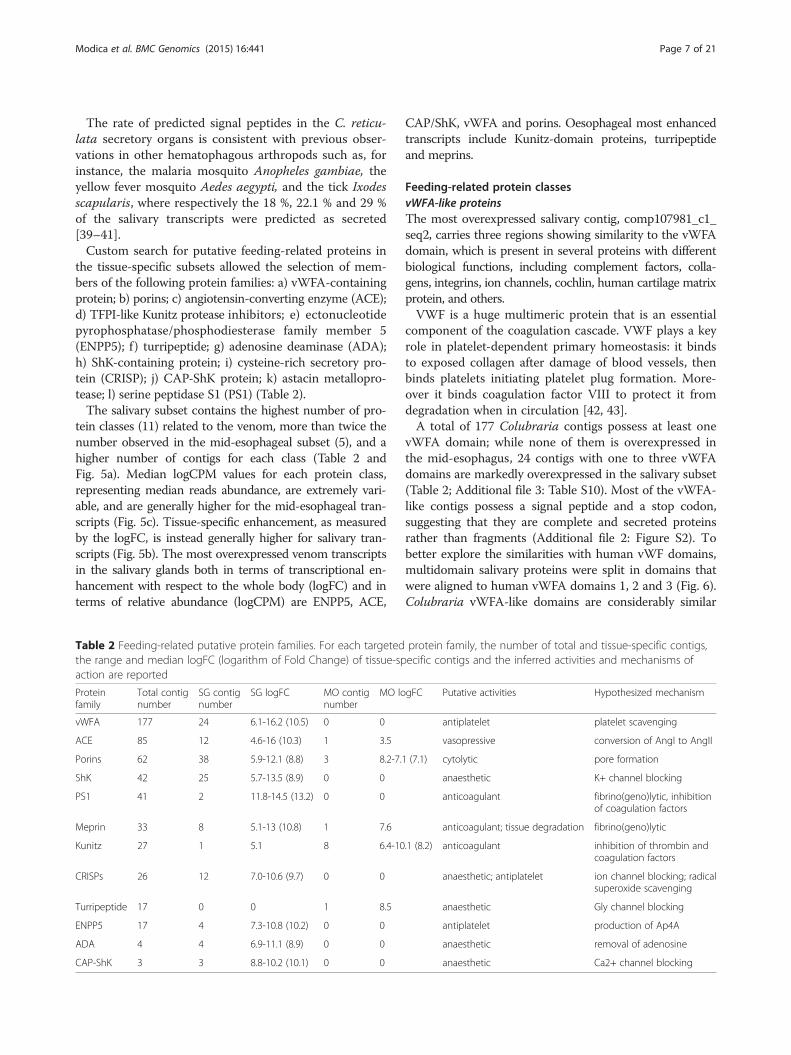
the tissue-specific subsets allowed the selection of mem-bers of the following protein families: a) vWFA-containingprotein; b) porins; c) angiotensin-converting enzyme (ACE);d) TFPI-like Kunitz protease inhibitors; e) ectonucleotidepyrophosphatase/phosphodiesterase family member 5(ENPP5); f ) turripeptide; g) adenosine deaminase (ADA);h) ShK-containing protein; i) cysteine-rich secretory pro-tein (CRISP); j) CAP-ShK protein; k) astacin metallopro-tease; l) serine peptidase S1 (PS1) (Table 2).The salivary subset contains the highest number of pro-
tein classes (11) related to the venom, more than twice thenumber observed in the mid-esophageal subset (5), and ahigher number of contigs for each class (Table 2 andFig. 5a). Median logCPM values for each protein class,representing median reads abundance, are extremely vari-able, and are generally higher for the mid-esophageal tran-scripts (Fig. 5c). Tissue-specific enhancement, as measuredby the logFC, is instead generally higher for salivary tran-scripts (Fig. 5b). The most overexpressed venom transcriptsin the salivary glands both in terms of transcriptional en-hancement with respect to the whole body (logFC) and interms of relative abundance (logCPM) are ENPP5, ACE,
Table 2 Feeding-related putative protein families. For each targetedthe range and median logFC (logarithm of Fold Change) of tissue-spaction are reported
Proteinfamily
Total contignumber
SG contignumber
SG logFC MO contignumber
MO lo
vWFA 177 24 6.1-16.2 (10.5) 0 0
ACE 85 12 4.6-16 (10.3) 1 3.5
Porins 62 38 5.9-12.1 (8.8) 3 8.2-7.1
ShK 42 25 5.7-13.5 (8.9) 0 0
PS1 41 2 11.8-14.5 (13.2) 0 0
Meprin 33 8 5.1-13 (10.8) 1 7.6
Kunitz 27 1 5.1 8 6.4-10
CRISPs 26 12 7.0-10.6 (9.7) 0 0
Turripeptide 17 0 0 1 8.5
ENPP5 17 4 7.3-10.8 (10.2) 0 0
ADA 4 4 6.9-11.1 (8.9) 0 0
CAP-ShK 3 3 8.8-10.2 (10.1) 0 0
CAP/ShK, vWFA and porins. Oesophageal most enhancedtranscripts include Kunitz-domain proteins, turripeptideand meprins.
Feeding-related protein classesvWFA-like proteinsThe most overexpressed salivary contig, comp107981_c1_seq2, carries three regions showing similarity to the vWFAdomain, which is present in several proteins with differentbiological functions, including complement factors, colla-gens, integrins, ion channels, cochlin, human cartilage matrixprotein, and others.VWF is a huge multimeric protein that is an essential
component of the coagulation cascade. VWF plays a keyrole in platelet-dependent primary homeostasis: it bindsto exposed collagen after damage of blood vessels, thenbinds platelets initiating platelet plug formation. More-over it binds coagulation factor VIII to protect it fromdegradation when in circulation [42, 43].A total of 177 Colubraria contigs possess at least one
vWFA domain; while none of them is overexpressed inthe mid-esophagus, 24 contigs with one to three vWFAdomains are markedly overexpressed in the salivary subset(Table 2; Additional file 3: Table S10). Most of the vWFA-like contigs possess a signal peptide and a stop codon,suggesting that they are complete and secreted proteinsrather than fragments (Additional file 2: Figure S2). Tobetter explore the similarities with human vWF domains,multidomain salivary proteins were split in domains thatwere aligned to human vWFA domains 1, 2 and 3 (Fig. 6).Colubraria vWFA-like domains are considerably similar
protein family, the number of total and tissue-specific contigs,ecific contigs and the inferred activities and mechanisms of
.1 (8.2) anticoagulant inhibition of thrombin andcoagulation factors
anaesthetic; antiplatelet ion channel blocking; radicalsuperoxide scavenging
anaesthetic Gly channel blocking
antiplatelet production of Ap4A
anaesthetic removal of adenosine
anaesthetic Ca2+ channel blocking
Fig. 6 Alignment of the colubrarian vWFA domains with the human vWFA1. Cysteines in yellow, key residues for the interaction with plateletGpIb in bold, gain-of-function mutations in red. Identical and conserved residues with respect to human vWFA1 are highlighted respectively ingrey and light grey and indicated respectively by asterisk and colon. Multiple identical copies of domains were excluded from the alignment
Modica et al. BMC Genomics (2015) 16:441 Page 8 of 21
(pairwise identity 17-23 %; pairwise similarity 35-35 %) todomain A1 of the human vWF, which is responsible forbinding platelet GpIb [43, 44]. Therefore, by competingwith the vWF of their prey, they may bind thrombocytespreventing their interaction with collagen and thus inhibitingthrombocyte aggregation, with a resulting anti-hemostaticaction. The alignment of Colubraria vWFA-like domainswith human vWFA1 domain highlights the conservationof most residues responsible for platelet binding (Fig. 6),according to molecular studies of several von Willebranddisease phenotypes (reviewed in [44]) and to molecularmodeling of the interaction with platelets [45, 46]. More-over, several residues in vWFA1 domain were found mu-tated in Type IIB von Willebrand disease, resulting in anincreased affinity for platelets [44]; interestingly, two ofsuch gain-of-function mutations were observed in someof the Colubraria contigs (R543W or R543Q: [45, 47];I546V: [48]). This observation suggests that the affinity forfish thrombocytes may be higher in Colubraria vWFA-like proteins than in native fish vWFA1.
TFPI-like Kunitz-domain protease inhibitorsA total of 27 contigs possessing one or more Kunitz-domains were identified (Table 2). Only one of them,comp101225_c0_seq1, is enriched in both subsets (Table 2;Additional file 3: Table S11 and S12), while seven contigs
(six of which are retrieved as isoforms of the same contigcomp105558_c0_seq1-seq6), are enriched in the oesophagealsubset (Table 2; Additional file 3: Table S12).The most complete contig, the 136-residues long
comp105558_c0_seq3, highly overexpressed in MO, pos-sesses two Kunitz domains separated by a short linker anddisplays high similarity values with hard ticks thrombin in-hibitors, particularly with haemalin (pairwise identity 45 %,pairwise similarity 60 %), a recently described thrombininhibitor from the bush tick Haemaphysalis longicornis [49](Fig. 7). Signal P prediction identified a putative cleavagesite between residues 15 and 16. The cysteine pattern isfully conserved between sequences and similar to whatobserved in TFPI, with 3 disulfide bonds predicted for eachdomain (Fig. 7).A second contig, comp101225_c0_seq1, encoding for a
putative protein of 167 residues with 4 Kunitz domainsand a predicted signal peptide of 19 residues, is overex-pressed in both tissues, especially in the mid-esophagealsubset (Additional file 3: Table S11 and S12). This contigshows similarities with the Tetralaris, a 4 Kunitz-domainspeptide isolated from the salivary gland of the Zebra tickRhipicephalus pulchellus [50] (pairwise identity 33 %, pair-wise similarity 39 %) and a high conservation level of keyresidues with human TFPI (although the latter possessesonly three Kunitz domains) (Fig. 8).
Fig. 7 Alignment of two-Kunitz domains protease inhibitors. Predicted signal peptides in blue, Kunitz domains in light orange, conserved cysteinesin yellow, residues binding Thrombin exosite in green, protease binding residues in purple, residues binding Thrombin active cleft in red. Identical andconserved residues are indicated respectively by asterisk and colon. Roman numbering indicate disulfide bonds pattern
Modica et al. BMC Genomics (2015) 16:441 Page 9 of 21
A third contig, comp108520_c0_seq4 is remarkably long(1654 residues) and includes 6 Kunitz domains and 3 WAPdomains. The WAP domain contains 8 characteristicallyspaced cysteine residues forming disulfide bonds. This mul-tidomain contig is enriched in the MO subset (Additionalfile 3: Table S11).Kunitz-like protease inhibitors have been found in the
venom of a variety of venomous organisms, includingsnakes, cnidarians, wasps, spiders and scorpions [51], andare major components of the salivary secretions of blood-feeding arthropods [52]. In gastropods, they have beenfound in the venom gland secretion of Conus snails, wherethey were called ConKunitzins [53]. However, ColubrariaKunitz-like peptides are only distantly related with con-Kunitzin, being instead more similar to Kunitz peptidesfrom hard ticks, especially the contig comp101225_c0_seq1(the only Kunitz-like predicted peptide overexpressed inthe salivary glands) that displays a high similarity withTetralaris isolated in hard ticks. Intriguingly, multidomainKunitz proteins are widely present in hard ticks (including
Fig. 8 Alignment of the colubrarian four-Kunitz domain transcript with tickdomains in light orange, active sites in red. Identical and conserved residueindicate disulfide bonds pattern
the genera Ixodes, Amblyomma, and Rhipicephalus) butuntil now have not been detected in other species [52, 54].A number of them exhibit a potent anticoagulant activity,often involving non-canonical bindings to inhibit specificcoagulation factors, despite their overall similarity withTFPI [55].Oesophagus produces different kinds of Kunitz-type
TFPI-like protease inhibitor. Beside comp101225_c0_seq1,already described for the salivary subset, we detectedthe overexpression of a multidomain Kunitz/WAP(comp108520_c0_seq4) and a two Kunitz domain proteaseinhibitor (comp105558_c0_seq3). The latter is very similarto members of a hard-tick family of thrombin inhibitorsthat consist of pairs of Kunitz modules and include haema-lin, amblin and boophilin [49, 55-57]. Although thesemolecules are only preliminarily characterized so far, arecent research highlighted their divergence from the anti-hemostatic factors identified in soft ticks (e.g. savigninin,monobin, ornithodorin) and evidenced their similarity tocanonical Kunitz-type molecules such as BPTI [55].
Tetralaris and human TFPI. Predicted signal peptides in blue, Kunitzs are indicated respectively by asterisk and colon. Roman numbering
Modica et al. BMC Genomics (2015) 16:441 Page 10 of 21
The activity of haemalin, that is similar to contigcomp105558_c0_seq3, was tested in vitro and resulted inthe inhibition, even at low concentration, of plasma andfibrinogen clot, formation and platelet aggregation, indicat-ing that haemalin is an anticoagulant for the commonpathway of coagulation [49]. Boophilin, isolated from thewhole body of Rhipicephalus microplus, is highly similar tohaemalin, but it directly inhibits thrombin with a non-canonical mechanism, where a few N-terminal residuesbind across the thrombin active-site cleft while C-terminalmodules interact with the basic exosite I [57]. Amblinisolated from the haemolymph of Amblyomma hebraeumis quite different from other natural proteinase inhibitors,and its thrombin inhibitor activity still waits for anexperimental confirmation [57, 58]. Although its activity re-mains to be tested in vitro, we can hypothesize thatcomp105558_c0_seq3 could act as a thrombin inhibitor inpreventing blood clot formation to allow efficient feedingand digestion while passing through the oesophagus, assuggested for H. longicornis [49].
Adenosine deaminase (ADA)We found 21 putative contigs showing similarity withadenosine deaminase (ADA), an enzyme that catalyzes theconversion of adenosine and 2’-deoxyadenosine to inosineand 2’-deoxyinosine [59] two of which are enriched in thesalivary glands subset (Table 2; Additional file 3: Table S13).These putative proteins display high levels of similarity
with ADAs of other mollusk species (e.g. Aplysia califor-nica) and hematophagous arthropods (including Lutzomiyalongipalpis, Phlebotomus dubosqui, Aedes albopictus, Culexquinquefasciatus and Anopheles gambiae). The alignment(Additional file 2: Figure S3) depicts the high levels ofsequence similarity (up to 40 % pairwise identity and 58 %pairwise similarity) and the conservation of key residues forthe enzymatic activity.ADAs have been particularly studied in the salivary
secretion of blood-feeding insects, namely L. longipalpis[60], C. quinquefasciatus and Ae. aegypti [61], Phlebotomusduboscqui [62], Chtenocephalides felii [63] and Glossinamorsitans [64]. In these species, the proposed activity forthese molecules is the hydrolysis of adenosine, a moleculeinvolved in pain perception [60, 65], thus reducing theperception of the parasite wound by the host. Addition-ally, the removal of adenosine produces inosine, a potentinhibitor of the production of inflammatory cytokines invertebrates [61, 62]. Paradoxically, adenosine is also animmunosuppressive, vasodilatory and platelet aggrega-tion inhibitor, and the presence of an enzyme thatremoves such a potentially useful molecule was explainedin insects with the production of other more effectivecompounds counteracting these actions [60]. In C. reticu-lata, vasoconstriction apparently is not an issue, beinginstead advantageous to the parasite, while platelet
aggregation can be counteracted by the action of ENPP5,Antigen-5 and vWFA-containing proteins.
Angiotensin-converting enzyme (ACE – M2)We found a total of 85 contigs displaying similarity withsingle-domain angiotensin converting enzyme (ACE2) frommouse, and most interestingly with the only two knownsequences identified in hematophagous animals (the duckleech Theromyzon tessulatus and the buffalo fly Haematobiairritans) (Table 2). ACE is a dipeptidyl carboxypeptidase be-longing to the M2-metalloprotease family. Transcription oftwelve contigs is enriched in the salivary glands (Additionalfile 3: Table S14) while a single putative contig enriched inmid oesophagous was retrieved (Additional file 3: TableS15). Partial contigs with a reduced length (less than 100residues) and redundant sequences were excluded from thealignment that includes two Colubraria contigs over-expressed in the salivary subset, with a similarity to T. tessu-latus and H. irritans ACEs up to 62 %, and a high level ofconservation of residues in the active sites (Additional file 2:Figure S4). Both Colubraria contigs appear to be incom-plete, with comp1091011_c0_seq4 having a predicted signalpeptide but missing the C-terminal tail and comp109011_c1_seq2 aligning with the C-terminal end of the alignment.In vertebrates, this zinc-dependant metalloprotease takes
part in the renin-angiotensin system, converting the inactivedecapeptide Ang I (angiotensin I) into the vasopressor octa-peptide Ang II (angiotensin II). ACE also inactivates brady-kinin, a vasodilator peptide, and thus contributes to bloodpressure increase in mammals. In invertebrates, ACE-related genes have been cloned in a few insects [66-68], inthe duck leech Theromyzon tessulatus [69], and recentlyACE-encoding transcript was reported in the venom ducttranscriptome of Conus victoriae [70], while the peptidewas found in the proteomes of C. purpuranscens and C.ermineus [71]. For the cone snails it was suggested that thevasoconstrictory activity of ACE enzymes might play a rolein envenomation, perhaps increasing the local concentra-tion and effectiveness of the venom, or, more generally,interfering with cardiovascular homeostatic mechanisms asseen in other venomous animals. For a hematophagousfeeder like Colubraria, ACE may play a role in the trophicphysiology of the snail. In fact the proboscis in Colubrariais extremely thin and scarcely muscularized suggesting thatit is not able to exert a strong suction [15]. Most likely, thepassage of blood through the proboscis is due to the bloodpressure of the fish, as confirmed by feeding observation(Oliverio & Modica, unpublished observations). In thiscontext, increasing the blood pressure of the fish wouldmaximize the blood income for Colubraria.
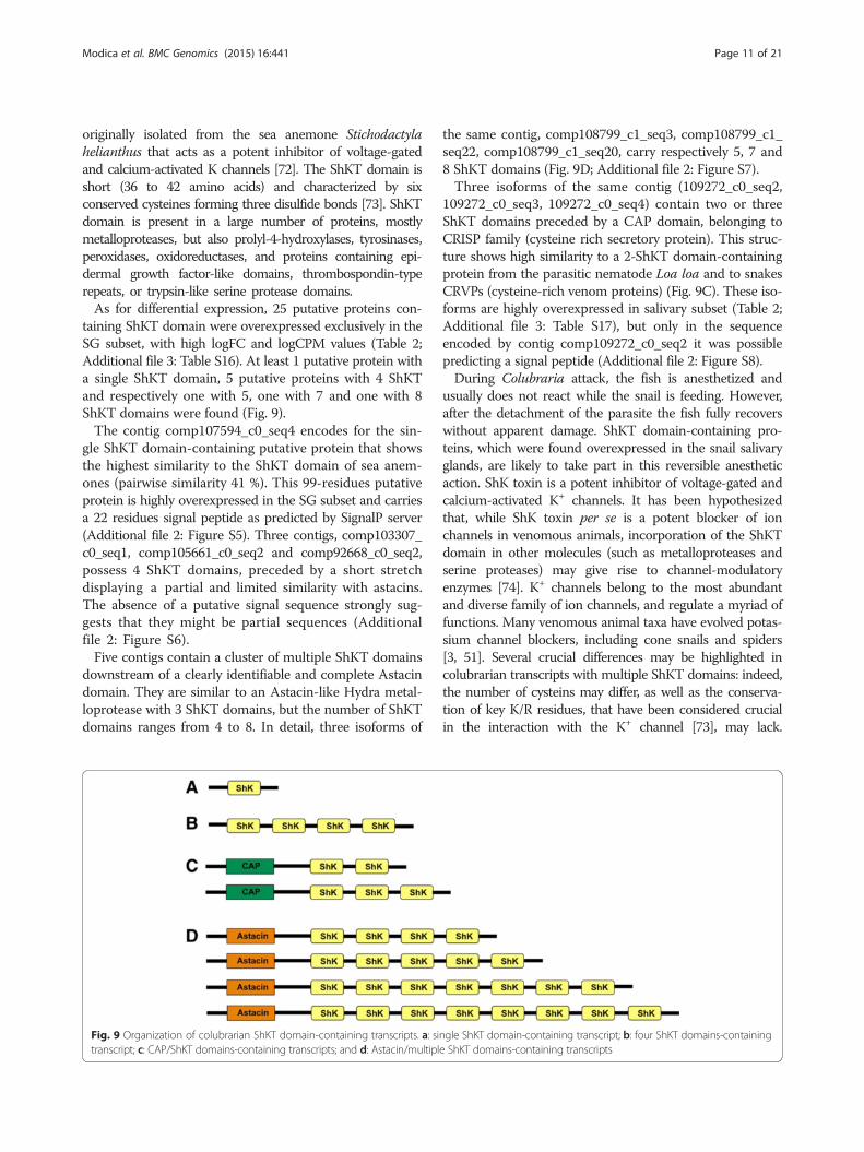
ShKT domain-containing proteinsColubraria transcriptome contains 61 contigs with a ShKTdomain (Table 2). ShK is a 35-residue peptide toxin
Modica et al. BMC Genomics (2015) 16:441 Page 11 of 21
originally isolated from the sea anemone Stichodactylahelianthus that acts as a potent inhibitor of voltage-gatedand calcium-activated K channels [72]. The ShKT domain isshort (36 to 42 amino acids) and characterized by sixconserved cysteines forming three disulfide bonds [73]. ShKTdomain is present in a large number of proteins, mostlymetalloproteases, but also prolyl-4-hydroxylases, tyrosinases,peroxidases, oxidoreductases, and proteins containing epi-dermal growth factor-like domains, thrombospondin-typerepeats, or trypsin-like serine protease domains.As for differential expression, 25 putative proteins con-
taining ShKT domain were overexpressed exclusively in theSG subset, with high logFC and logCPM values (Table 2;Additional file 3: Table S16). At least 1 putative protein witha single ShKT domain, 5 putative proteins with 4 ShKTand respectively one with 5, one with 7 and one with 8ShKT domains were found (Fig. 9).The contig comp107594_c0_seq4 encodes for the sin-
gle ShKT domain-containing putative protein that showsthe highest similarity to the ShKT domain of sea anem-ones (pairwise similarity 41 %). This 99-residues putativeprotein is highly overexpressed in the SG subset and carriesa 22 residues signal peptide as predicted by SignalP server(Additional file 2: Figure S5). Three contigs, comp103307_c0_seq1, comp105661_c0_seq2 and comp92668_c0_seq2,possess 4 ShKT domains, preceded by a short stretchdisplaying a partial and limited similarity with astacins.The absence of a putative signal sequence strongly sug-gests that they might be partial sequences (Additionalfile 2: Figure S6).Five contigs contain a cluster of multiple ShKT domains
downstream of a clearly identifiable and complete Astacindomain. They are similar to an Astacin-like Hydra metal-loprotease with 3 ShKT domains, but the number of ShKTdomains ranges from 4 to 8. In detail, three isoforms of
Fig. 9 Organization of colubrarian ShKT domain-containing transcripts. a: sitranscript; c: CAP/ShKT domains-containing transcripts; and d: Astacin/multipl
the same contig, comp108799_c1_seq3, comp108799_c1_seq22, comp108799_c1_seq20, carry respectively 5, 7 and8 ShKT domains (Fig. 9D; Additional file 2: Figure S7).Three isoforms of the same contig (109272_c0_seq2,
109272_c0_seq3, 109272_c0_seq4) contain two or threeShKT domains preceded by a CAP domain, belonging toCRISP family (cysteine rich secretory protein). This struc-ture shows high similarity to a 2-ShKT domain-containingprotein from the parasitic nematode Loa loa and to snakesCRVPs (cysteine-rich venom proteins) (Fig. 9C). These iso-forms are highly overexpressed in salivary subset (Table 2;Additional file 3: Table S17), but only in the sequenceencoded by contig comp109272_c0_seq2 it was possiblepredicting a signal peptide (Additional file 2: Figure S8).During Colubraria attack, the fish is anesthetized and
usually does not react while the snail is feeding. However,after the detachment of the parasite the fish fully recoverswithout apparent damage. ShKT domain-containing pro-teins, which were found overexpressed in the snail salivaryglands, are likely to take part in this reversible anestheticaction. ShK toxin is a potent inhibitor of voltage-gated andcalcium-activated K+ channels. It has been hypothesizedthat, while ShK toxin per se is a potent blocker of ionchannels in venomous animals, incorporation of the ShKTdomain in other molecules (such as metalloproteases andserine proteases) may give rise to channel-modulatoryenzymes [74]. K+ channels belong to the most abundantand diverse family of ion channels, and regulate a myriad offunctions. Many venomous animal taxa have evolved potas-sium channel blockers, including cone snails and spiders[3, 51]. Several crucial differences may be highlighted incolubrarian transcripts with multiple ShKT domains: indeed,the number of cysteins may differ, as well as the conserva-tion of key K/R residues, that have been considered crucialin the interaction with the K+ channel [73], may lack.
Modica et al. BMC Genomics (2015) 16:441 Page 12 of 21
However, the absence of these key residues in functionallyactive ShKT proteins was already described in snakes [75].Interestingly, similar multidomain ShKT proteins have
been recently identified in the venom of several species ofthe polychaete Glycera [75].CAP proteins have been identified in the venom of a
wide range of taxa, from Cnidaria to mammals [3, 51], andare particularly prominent in the venom of toxic reptiles[76]. The biological activity of these cysteine-rich proteins(CRISPs) has been subject of debate. As for snake CAPs,nevertheless, inhibition of smooth muscles contraction andof cyclic nucleotide-gated ion channels was reported [77].Colubraria CAP-ShKT proteins appear related to snakeCRVPs (cysteine-rich venom proteins) that may targetvoltage gated Ca2+ channels on smooth muscles [78].Astacin metalloproteases play essential roles in diverse
physiological mechanisms including digestion, early embry-onic development, processing of the extracellular matrixand egg hatching. Astacins might also be good candidatesto assist in the anticoagulant action, thanks to theirfibrino(geno)lytic activity reported in spider venoms [79].Astacin/ShKT-containing proteins of Colubraria couldtherefore both inhibit ion channels and contribute to im-pair hemostasis. Moreover, by degrading extracellularmatrix molecules and basal lamina proteins, astacins mightfacilitate the spreading of the toxins in the body of the prey[79, 80] and inactivate several endogenous vasoactivepeptides [81].
Meprin-like proteinsBeside the Astacin-ShKT containing proteins describedabove, in Colubraria transcriptome we found a total of 33Meprin-like metalloproteases, comprising a contig en-coding for an astacin domain followed by a MAMdomain, overexpressed both in the salivary an in theoesophageal subset (Table 2). Among them, 8 contigs (allputative isoforms of the same putative protein encoded bycomp108388_c4) are highly overexpressed in the salivarysubset while a single contig (comp109603_c4_seq1) isoverexpressed in the mid-oesophagus (Table 2; Additionalfile 3: Table S18, S19). These contigs show a pairwise simi-larity up to 50 % to Meprin A subunit B isolated from thebivalve Crassostrea gigas and a good level of conservationof active residues (Additional file 2: Figure S9).Generally speaking, meprins are multimeric proteins,
whose subunits are characterized by a unique combinationof domains, namely Astacin, MAM (Meprin, A5 protein,and protein tyrosine phosphatase Mu), TRAF (tumor ne-crosis factor (TNF) receptor-associated factor), EGF (epi-dermal growth factor)-like domain, a transmembrane anda cytosolic domain [80]. Astacin is the catalytic domain re-sponsible of the proteolytic activity, while MAM domainis involved in the homo-oligomerizations [82]. Colubrariacontigs are devoid of most of the domains generally
included in meprins architecture, possessing exclusivelyan astacin and a MAM domain. Despite the missing do-mains, the sequences obtained appear to be complete,with a signal peptide and a stop codon, suggesting thatthey may act as oligomeric astacins.The presence of a MAM domain was detected to date
in a number of other astacin-like proteins such as squidmyosinases, HMP2 from Hydra and an enzyme fromNematostella vectensis [83-85]. Astacins as component ofanimal venom were described only in the brown spiderLoxosceles venoms, where, beside a digestive role, it hasbeen suggested that they could fulfill a fibrinogenolytic ac-tivity promoting anticoagulation of prey blood. It has alsobeen hypothesized that astacins might contribute tovenom spreading and inactivate prey vasoactive peptides[79-81]. Together astacins and meprins belong to the M12family of metalloproteases that includes important com-ponents of snake venoms [86]. In Viperidae, metallopro-teases M12 are the most abundant venom toxins [87, 88],and are involved in a variety of physiological activitiesincluding hemorrhagic and fibrinolytic effects [89].
Cysteine-Rich Secretory Proteins (CRISPs)Beside the CAP-ShKT group, 25 other contigs with hom-ology to CRISPs (Table 2) were found in Colubraria tran-scriptome, 12 of which are overexpressed in the salivarysubset (Table 2; Additional file 3: Table S20), a numberthat elevates to 15 if we include the CAP-ShKTcontigs de-scribed above (Additional file 3: Table S17). Among themost enriched in the salivary subset (logFC: 10.6; logCPM:8.1; SI: 65 %), contig comp107140_c0_seq1 possesses apredicted signal peptide of 26 residues and aligns well withvenom allergen 5, a major constituent of vespid venom. Italso displays a certain degree of similarity with homologsisolated from the hematophagous arthropods Dipetaloga-ster maximus and Triatoma infestans [90, 91] (pairwiseidentity 21 % and 19 %, pairwise similarity 30 %), includingthe conservation of crucial Histidine residues (Fig. 10).Contig comp107140_c0_seq1 is shorter than its arthro-pods counterparts, suggesting that the sequence mightbe incomplete. Antigen-5 family comprises a consider-able number of members, whose biological functionsare mostly unknown. Some of them have been recruitedto accomplish roles in blood feeding during the evolutionof hematophagous taxa, and have been identified in mostof the hematophagous arthropods studied to date wherethey can fulfill different tasks. Inhibition of collagen-induced platelet aggregation has been recently reportedfor antigen-5 of the hematophagous Triatomine bugsDipetalogaster maximus and Triatoma infestans, the vec-tors of Chagas disease [90, 91]. A recent study revealedthat these proteins are Cu2+ antioxidant enzymes that areable to inhibit collagen-induced platelet aggregation, ATPsecretion and thromboxane-A production by a unique
Fig. 10 Alignment of the colubrarian putative Ant5 with Dolichovespula maculata Ant5. Predicted signal peptide in blue, SCP-domain in lightorange, Histidine residues in red, Cysteine residues in yellow. Identical and conserved residues are indicated respectively by asterisk and colon
Modica et al. BMC Genomics (2015) 16:441 Page 13 of 21
mechanism involving the removal of radical superoxide[92]. Moreover, scavenging of radical superoxide down-regulates a myriad of pro-inflammatory reaction, as itlimits the production of inflammatory extracellular ROSby neutrophils, platelets and other cell types [92]. It canbe hypothesized that antigen 5 protein could have thesame activity in Colubraria, to reinforce anti-platelet-aggregation capabilities of the snail.The other contigs encoding for CRISPs appear to be
largely incomplete, but despite being quite different inlength, they are generally similar to Mr30 and Tex31proteins isolated in two Conus species, C. marmoreus andC. textile [93, 94] (Additional file 2: Figure S10). Althoughthe biological function of these proteins still needs to beclarified, an inhibitory activity on ion channels was hypoth-esized for them [93, 94].
Peptidases S1A total number of 41 contigs similar to Peptidase S1were found (Table 2), only 2 of which are differentiallyexpressed and enhanced in the salivary subset (Table 2;Additional file 3: Table S21). These putative proteins arequite different: comp108333_c0_seq1 appears to be achymotrypsin, while comp98826_c0_seq1 is more similarto trypsin-like peptidases isolated in cephalopods [95]and the fibrinolytic enzyme of the Echiurid worm Urechisunicinctus. The trypsin-like comp98826_c0_seq1 does notpossess a predicted signal peptide, thus might be incom-plete, and displays only a partial level of conservation ofkey residues at both the C-terminal active site and thesubstrate binding site (Additional file 2: Figure S11).The other contig encoding for a putative peptidase S1,
comp108333_c0_seq1, aligns with chymotrypsin of theant Harpegnatos saltator, the fly Glossina morsitans andwith a secreted salivary protein of the tick Ixodes scapu-laris. However, the residues corresponding to the activesites of the catalytic triad are not conserved, raising
doubts on its effective putative activity (Additional file 2:Figure S12).The peptidase S1 (PS1) family of serine proteases is a
large peptidase family, including chimotrypsin, trypsinand elastase activities. Biological roles of the PS1 are ex-tremely diverse, ranging from digestion to immune re-sponses. The venom of several taxa of both vertebratesand invertebrates contains PS1 that are the dominantvenom component in several species of cephalopods, inmale platypuses, and in Remipedia [51, 96]. Activities ofPS1 in the venom are diverse and are involved in pain,inflammation, smooth muscle contraction, vasodilationand prevention of blood clotting. In Colubraria, mem-bers of PS1 family constitute only a reduced fraction ofthe salivary secretion, but include two well-differentiatedputative proteins that may participate to the overall en-venomation process, possibly exploiting an anticoagulantaction. These proteins are the trypsin-like protein similarto cephalopods peptidases S1 and to echiurid fibrinolyticenzyme, and the chymotrypsin-like peptidase that is de-void of the conventional catalytic triad. Recent researchidentified in snake venoms a number of PS1 with uncon-ventional catalytic triads that show activity towards awide array of proteins involved in hemostasis, being ableto degrade fibrinogen, fibrin, prothrombin, factor X andplasminogen [97].
Ectonucleotide pyrophosphatase/phosphodiesterase family5 (ENPP5)A total number of 17 contigs encode for members of theENPP5 family, which hydrolyze 5’-phosphodiester bondsof nucleotides and their derivatives, resulting in the re-lease of 5’-nucleotide monophosphates (Table 2). Fourslightly different isoforms of a same contig are overex-pressed in the salivary glands (Table 2; Additional file 3:Table S22) and encode for putative proteins of 441 resi-dues long (except for comp109445_c0_seq3, that carries agap of 82 residues), with a signal peptide of 26 residues,
Modica et al. BMC Genomics (2015) 16:441 Page 14 of 21
and highly similar (up to 44.3 % identity) to ENPP5 en-zyme from Crassostrea gigas. Molluscan sequences lackthe transmembrane region that is instead present inhuman ENPP5 (Additional file 2: Figure S13).ENPP5 family is a versatile group of enzymes with a
broad substrate specificity [98, 99]. It was shown thatmembers of the ENPP5 family are capable of hydrolyzingdinucleoside polyphosphates, a group of nucleotides thatrecently has attracted considerable interest because itsmembers act as extracellular signaling molecules in abroad variety of tissues [99]. It was also shown that theyare involved in platelet aggregation, with a potent inhibi-tory effect reported for diadenosine 5’,5”’-P1,P4-tetrapho-sphate (Ap4A) [100]. Ap4A can be produced by ENPP5hydrolysing Ap5A, suggesting an indirect anti-aggregantactivity for Colubraria ENPP5, as indicated by the over-expression in the salivary glands.
PorinsA total of 62 contigs were described in the transcriptome,showing sequence similarity to echotoxins and conoporins,pore-forming lectins with lethal and haemolytic effects de-scribed respectively in the caenogastropod Monoplex echoand in the Conoidean Conus consors [101, 102] (Table 2).These proteins belong to the sea-anemones cytolysine fam-ily of Actinoporins [103, 104], whose members are able toform pores in the cell membrane.Porin contigs are mostly represented in the salivary
subset that contains 38 overexpressed contigs (Table 2),one of which is shared with the MO subset (but witha higher enhancement in the SG subset; Additionalfile 3: Table S23). Two contigs, comp89661_c0_seq1 andcomp93847_c1_seq1, are specific of the MO subset (Table 2;Additional file 3: Table S24).Alignment of the 8 most complete contigs (length be-
tween 192 and 268 residues and signal peptide pre-dicted) with echotoxins and conoporins is displayed insupplementary Additional file 2: Figure S14; noticeably,residues important for the interaction with the membraneare not completely conserved [105]. Overall, Colubrariaporins display the highest similarity to conoporin and toechotoxin A (pairwise identity values ranging respectivelyfrom 27 % to 41 % and from 25 % to 33 %; pairwise simi-larity from 40 % to 53 % and from 40 % to 51 %).Noteworthy, the mid-esophageal contig comp89661_
c0_seq1 displays the highest similarity to conoporin, afinding that might be related to the common ontogen-etic origin of the mid-oesophagous of C. reticulata andthe venom duct of Conoidea.Porins are able to perforate cellular membranes in a
multistep process. Such process involves i) recognitionof membrane sphingomyelin using aromatic rich regionand adjacent phosphocholine (POC) binding site, ii) firmbinding to the membrane (mainly driven by hydrophobic
interactions) accompanied by the transfer of the N-terminalregion to the lipid-water interface and iii) eventually, poreformation after oligomerization of several monomers [106].Echotoxins, the first protein toxins isolated from marinegastropods, differ from actinoporins in having affinity forgangliosides instead of sphingomyelin [101, 105]. For someof the gastropod echotoxins identified so far, a cardiacstimulation activity has been reported. The high expressionlevels of porins in the salivary glands of C. reticulata can berelated to the need to access blood vessels.
TurripeptideA single contig, comp109534_c9_seq2, enhanced in theMO subset (Table 2), displays a high level of similarity(pairwise identity 55.7 %, pairwise similarity 69.5 %) withknown turripeptides, from Lophiotoma olangoensis, Iotyrriscingulifera, and Gemmula speciosa (Additional file 3:Table S25; Additional file 2: Figure S15) [107]. These areconotoxin-like peptides discovered in the venom gland ofthe Conoidean family Turridae, characterized by threedisulfide bonds arranged according to the IX cysteineframework. This pattern is typical of the poorly known Psuperfamily of conotoxins, whose sequences are howeverquite divergent when compared to turripeptides [108].Contig comp109534_c9_seq2 encodes for a 72 residues
peptide, with a 20 aminoacids predicted signal peptide,and possibly a 5 residues propeptide region, accordingto similarity with known turripeptides (Additional file 2:Figure S15).Turritoxins are reported to act as neurotoxins by inhi-
biting ion channels, and possibly also as serine proteaseinhibitors, given that they posses the Kazal serine proteaseinhibitor signature. Their 6-Cys pattern was first charac-terized in the peptide BeTXIIa from the venom of the ver-mivorous species Conus betulinus [109], and subsequentlyfound in a few “spasmodic peptides”, that define the P-superfamily of conotoxins and the Cys framework IX[110]. Framework-IX venom peptides have additionallybeen found in members of families Turridae and Terebridae[111, 112], however the sequence similarity to peptides ofConidae P-superfamily is generally low [108].An analysis of feeding habits of conideans reveals that
framework IX peptides are not produced by fish-huntingcone snails but they are expressed in the venom duct ofmolluscivorous cone snails and vermivorous conoideans(including also Terebridae and Turridae beside somespecies of cone snails) [108]. The molecular target of anyframework IX-conopeptide has not been identified thusfar. However, experimental evidences suggest, at least forsome members of the P-subfamily, an action on glycinereceptors [110], ligand-gated ion channels belonging tothe nicotinic acetylcholine receptor family, which are im-portant targets for neuroactive drugs [113]. Gly receptorsare in fact widely distributed inhibitory receptors, that are
Modica et al. BMC Genomics (2015) 16:441 Page 15 of 21
involved in the regulation of the motor circuits of thespinal cord and have inhibitory synapses in afferentsensory neurons, including pain fibers [113, 114], beingideal targets for peripheral anesthetic compounds.The oesophageal overexpression of turritoxin-like contigcomp109534_c9_seq2 is in agreement with the structuralhomology of Conoidean venom duct with the colubrarianmid-oesophagus [26]. The expression of other families ofconopeptides in salivary glands was reported for Conuspulicarius [115], but no conopeptides were found in arecent transcriptome of salivary glands of C. geographus[20]. Expression of turritoxins does not appear to berestricted to gastropods, as it was also recently detected inthe venom of the polychaete Glycera [75].
ConclusionWe present here the first molecular characterization ofthe alimentary secretion of a non-conoidean neogastro-pod, by deciphering secretory repertoires of salivary glandsand mid-oesophagus of a hematophagous snail.
Physiology of feedingThe biochemical characteristics of the feeding-relatedclasses of molecules identified in the Colubraria tran-scriptome allow to hypothesize their functional role andthe physiology of the glandular tissues in the light ofColubraria feeding habits. The transcriptomic profiles ofthe two secretory tissues studied are different, with a re-duced overlap in the classes of molecules dominating theirsecretions, and indicate a large degree of specialization,with a significantly higher complexity in the salivary glands.Anticoagulant activities in the saliva, which is secretedboth into the prey and along the extremely long anterioroesophagus, can be ascribed to the synergic action ofseveral molecules, with a crucial role possibly played bythe vWFA domain-containing proteins, encoded bynumerous contigs overexpressed at the highest levels inthe salivary subset. The mid-oesophageal transcriptome isapparently less complex than the salivary one and is domi-nated by transcripts encoding for a turritoxin-like proteinand for a two Kunitz-domains protease inhibitorThese expression patterns reflect the different physio-
logical roles of the two tissues. We hypothesize that themid-oesophageal turritoxin is produced when the snailapproaches the fish and, as in the case of the venomouscocktail of Conus geographus, it may be involved in theinduction of a “hypnotic” state in the fish. C. geographus,as other so called net-hunter cone snails, engulfs fish (oreven a small school of fishes) with its highly extensiblerostrum before stinging. Apparently, when the fish isapproached by a cone snail, it remains in an almost hyp-notic state until the snail is close enough to engulf it [22]displaying a behavior extremely similar to Colubrariapredatory activity. The biochemistry of the induction of
“hypnosis” has not been investigated to date in Conus. InColubraria the use of an oesophageal secretion in the ex-ternal environment could be possible due to the loss ofthe valve of Leiblein, a structure that in neogastropod sep-arates the mid from the anterior oesophagus and thusfrom the environment [15, 26].Afterwards, when the blood meal is ingested, the main
task of the oesophagus would be avoiding blood clottingduring its passage through the stomach, where it willbe digested. Such a physiological role may be sustainedby the haemalin-like two-Kunitz domain protease inhibi-tor. Haemalin is a thrombin inhibitor, produced in theoesophagus of the bush tick Haemaphysalis longicornis,able to avoid clotting of the blood meal until digestion iscompleted [49].The salivary glands, that discharge their secretion at
the tip of the proboscis, very close to the external environ-ment, produce a more complex venom probably in re-sponse to their more diversified tasks. First, when theproboscis contacts the fish, the snail has to gain access tothe blood. The massive secretion of porins, maybe assistedby the metalloproteases, probably elicit a potent cyto-lytic activity [79, 106], that associated with the scrap-ing action of the radula allows the snail to open largecircular wounds on the fish skin (Fig. 2B). Unquestionably,an anesthetic activity is crucial to efficiently accomplishblood-feeding in any hematophagous taxa. In Colubrariathis anesthetic activity is likely due to the concerted activityof ShKT-containing proteins, especially the CAP-ShKTthat act as K channel modulators [78], and ADA, that mayreduce local pain perception by removing adenosine [60].The anticoagulant action at the site of the wound and inthe anterior digestive tract is mediated by the salivary pro-duction of a complex mixture of anticoagulant com-pounds. The vWFA-containing proteins, novel proteinshere described for the first time, may act as platelet anti-aggregants and target primary hemostasis, throughout amechanism still to be defined but probably including dir-ect scavenging of thrombocytes. Anti-platelet activity canbe assisted by the Antigen-5 protein, via a radical super-oxide removal mechanism [92]. ENPP5 can participate todisarrange primary hemostasis, producing Ap4A thatreinforce the inhibition of thrombocyte aggregation [100].Secondary hemostasis may be targeted by the 4 Kunitz do-main protein, similar to the tick Rhipicephalus pulchellusTetralaris, a potent thrombin and TF/FVIIa complex in-hibitor [52, 54], and to TFPI, that modulates factor Xa.Astacins and peptidases S1 also likely contribute to impairhemostasis, as described e.g. in snakes and spiders, due tothe fibrinogenolytic activity of astacins [79], and to thehemorrhagic–anticoagulant activity of PS1 that involvesthe degradation of several hemostatic factors [97]. Finally,salivary glands produce ACE that owing to its vasopressiveactivity can increase cardiac frequency in the fish and
Modica et al. BMC Genomics (2015) 16:441 Page 16 of 21
maximize blood income through the snail foregut, consid-erably reducing Colubraria feeding time. Vasoconstrictionis a defense mechanism that is part of the hemostatic sys-tem, and is generally counteracted by hematophagous ani-mals; interestingly, in a “passive feeder” such as Colubrariait becomes advantageous to the parasite, explaining the re-cruitment of ACE enzyme in the venomous cocktail.
Evolutionary considerationsMost hematophagous animals produce a complex mix-ture of anti hemostatic molecules that act synergisticallyto impair hemostasis in the host, the natural response tovascular injury aimed to arrest bleeding and based onthe triad of platelet aggregation, blood clotting and vaso-constriction. While Colubraria secretion does not blockvasoconstriction, which is advantageous to this parasite,it counteracts both primary and secondary hemostasis.Primary hemostasis is targeted by several molecules pro-duced in different hematophagous species, that act at vari-ous stages, including salivary apyrases, epinephrine andserotonine binding molecules and nitrophorins in severalarthropods; lipocalins in Rhodnius prolixus, triatominesand ticks; collagen binding proteins as antiplatelets inmosquitos and leeches; integrin antagonists in ticks andhookworms [6]. Inhibitors of secondary hemostasis are ex-tremely variable as well, in their molecular masses, targetsand inhibitory mechanisms, with thrombin and FXa beingthe most common targets [5, 7].Apparently, while inhibition of secondary hemostasis
in Colubraria involves mechanisms already described inother animal groups, this snail evolved a unique moleculararsenal to impair primary hemostasis, as a key role wouldbe played by vWFA-like proteins, assisted by ENPP5 andAntigen-5. The evolution of such arsenal could have beentriggered by the high efficiency of the coagulation systemin fishes that need to rapidly repair vascular damage, espe-cially in the gills, where blood supply is maximum andvery close to the external environment. In fact, althoughhemostasis in fish involves the same mechanisms andfactors described in mammals (except for the presencein fish of nucleated thrombocytes instead of anucleatedplatelets), coagulation time is generally shorter, due to thehigh levels of coagulation factors contributing to high ac-tivity of the extrinsic and intrinsic pathway [116]. Furthermolecular and biochemical studies on the anticoagulantsecretions of other hematophagous parasites of fishes,such as fish lice and lampreys, could elucidate mecha-nisms of evolutionary “arms race” between fishes and theirparasites.Surprisingly, we detected a very low level of similarity
between Colubraria and Conus biochemical predatoryarsenals, restricted to a single molecule, the turripeptide.Additionally, such molecules are not exclusive of gastro-pods as demonstrated by their recent discovery in the
venom gland of the polychaete Glycera [75]. On the otherhand, the presence in Colubraria of multiple Kunitz-domains proteins that were found to date only in hardticks (Ixodidae) [52] indicate some degree of physio-logical convergence between colubrariids and ixodid ticks.It should be remarked, for example, that both hard ticksand Colubraria share a feeding behavior that involves areversible attachment to the host that can last severalhours [117].Given the close phylogenetic relationship of Mollusca
and Annelida [118], we expected to find strong affinitiesbetween the biochemical arsenal of C. reticulata and thatof leeches and of the recently investigated polychaete Gly-cera. Again, very limited similarities were instead detected,namely the presence in Glycera of porins, turripeptide-like, CAP/ShKT and Astacin/ShKT [75] and the similarityof leech ACE to its Colubraria counterpart. This indicatesa different evolutionary pathway for the onset of hema-tophagy arsenal in the two lineages, and a dominantinfluence of convergence between distantly related butfunctionally similar taxa, such as Colubraria and hardticks. Moreover, the presence of anesthetic multidomainShKT proteins is shared with lower metazoans such asCnidaria, suggesting that they might represent a basalanimal toxin arsenal.More tailored investigations on the main feeding-related
protein classes here identified are needed to shed light ontheir activities, and also to define proteins involved in otherbiological roles such as the modulation of the inflamma-tory response of the host. However, our results undoubt-edly indicate that marine predatory gastropods could bea valuable study target to identify novel bioactive mole-cules. Conoidea investigated to date constitute only afraction of the overall Neogastropoda trophic diversity,suggesting that this animal group as a whole is a plen-tiful reservoir of bioactive peptides worth of detailedinvestigations.
MethodsSamples and tissues collectionA total of 30 specimens of Colubraria reticulata werecollected alive at Sidi Jmour (Djerba Is., Tunisia: 7-12.vi.2013; 33°49.5’N, 010°44.5’E), in shallow waters (0.3-2.5 m depth), under rocks and boulders. Specimens werekept alive in aquarium until sample preparation. Twenty-four specimens were dissected on ice, and target tissues(mid-oesophagous and salivary glands; hereafter MO andSG) were collected and used to prepare three tissue-specific biological replicates constituted of 8 samples each(SG 1-3 and MO 1-3). Additionally, 6 animals were usedto prepare three biological replicates of whole-body refer-ence, made up of 2 samples each (WB 1-3). During prep-aration of the whole-body samples, where we encounteredengorged snails, the stomach full of blood was
Modica et al. BMC Genomics (2015) 16:441 Page 17 of 21
excluded from the sample, to avoid contamination withfish transcripts.Samples were preserved in RNA later at -80 °C until
RNA extraction.
RNA extraction, library preparation and next generationsequencingTotal RNA from each sample was isolated using a Qiagen-RNeasy Mini Kit according to manufacturer’s instructions.FastPrep-24 homogenizer (MP Biomedicals) was usedto process approximately 30 μg of each sample (30sec at 4.0 m/s). RNA quality and quantity was assessedusing a Bioanalyzer 2100 (Agilent Technologies, Palo Alto,USA) and a Qubit v2.0 fluorometer (Life Technologies,Darmstadt, Germany), respectively.For each sample, 250 ng of high-quality RNA (RIN
value > 8) was used to construct nine barcoded sequen-cing libraries with the Illumina TruSeq RNA samplepreparation kit (Low-Throughput protocol) according tothe manufacturer’s instructions (Illumina, San Diego, USA).To increase the average library insert size, input RNA waschemical fragmented at 94 °C for 1 min.The nine barcoded samples were equimolar-pooled
and clustered template cDNA was sequenced in anIllumina HiSeq2000 platform with 209 cycles (101 cyclesfor each paired-read and seven cycles for the barcodesequences).
Bioinformatics analyses and differential expressionAfter sequencing we obtained 329,576,088 raw reads (from33,213,610 to 38,909,488 reads per sample), each 101 bp inlength that were quality-controlled (quality score higherthan Q36 where Q40 is the maximum value) before assem-bly, read mapping and downstream analyses. First, SeqPrep(https://github.com/jstjohn/SeqPrep) was used to removethe remaining adapters and to merge the overlapping paired-reads. The reads were then trimmed by quality in CLCGenomics Workbench v6.5 (CLC bio, Aarhus, Denmark).Low-quality reads (CLC parameter ‘limit’ set to 0.02) andreads shorter than 50 nucleotides were excluded. Finally, acustom database constituted by bacteria, fungi, virusand protozoa (source: NCBI Reference Sequence, RefSeq,March 2014) was used as reference to align the reads ofeach sample. Reads that aligned to these potential sourceof contamination were removed. A final dataset of220,305,266 filtered reads (140,615,306 paired reads, meanlength 99 bp, and 79,689,960 single merged reads, meanlength 154 bp) ranging from 17,993,689 to 27,248,309reads per sample was used to build the transcriptomeassembly.The filtered reads were de novo assembled using
Trinity v20140717 [119]. Trinity was run on the two setsof paired-end and single-end (merged) sequences withthe fixed default k-mer size of 25 and a minimum contig
length of 60 bp. To reduce the number of isoforms, thePasa assembly algorithm implemented in the Butterflymodule of Trinity was used. The produced assembly wassubjected to similarity search against three differentdatabases: a) NCBI’s non-redundant (NR) database; b)EMBL’s UniProtKB/Swiss-Prot; c) a custom nucleotidedatabase including the transcriptomes of three molluscspecies Ilyanassa obsoleta (PRJNA79721), Nucella lapil-lus (PRJNA217409), Conus geographus (venom ducttranscriptome; PRJNA167726), plus the salivary glandsEST library of the leech Macrobdella decora (LIB-EST_028114). BLASTx (NR and Swiss-Prot databases)and BLASTn (mollusc databases) algorithms [120] with acut-off e-value of 10−6 were used for similarity searches.The assembly was further scanned for the presence offunctional protein domains using InterProScan [28]with the following search methods: SignalP, TMHMM,Pfam, SMART, Tigr, ProfileScan. Transcripts that alignedto proteins or transcripts contained in the databases and/or had InterPro match were retained for downstreamanalyses.The obtained final set of sequences (144,380 transcripts)
was used as reference for read mapping and differentialexpression (DE) analyses. Read mapping for each samplewas performed using Bowtie2.2.3 [121] and gene expres-sion level for each transcript was estimated by RSEMv1.2.15 [122]. The read-count table was exported and DEanalysis was carried out using the Bioconductor edgeRv2.14 [123] R v3.1.0 package.Three different differential expression (DE) analyses were
carried out comparing the three different tissues (WB, MOand SG) in a pairwise manner. False discovery rate (FDR)was applied to correct p-values generated by edgeR’s exacttest (transcripts showing FDR < 0.05 were consideredDE). Following a conservative approach, we consideredenriched those DE contigs having logFC values (the log2fold change between tissue-specific vs. whole body relativeabundances) of 3 or higher.Functional annotation of the DE transcripts was carried
out by Blast2GO [124], setting the NCBI’s NR as referencedatabase (E-value < e−6, annotation cut-off > 55, GOweight > 5). Enrichment analyses, using the Fisher’s exacttest implemented in Blast2GO, were applied to identifysignificantly over-represented gene ontology (GO) termsin the DE transcripts (test sets) when compared to thewhole assembly (baseline set).The open reading frame (ORF) for each transcript was
detected using two methods: a) ORF were predicted usingthe TransDecoder module implemented in the Trinitypackage, and the longest ORF among those matchingthe same protein was selected; b) ORF was detected,for each transcript, using the output coordinates of theBLASTx similarity search against NR or Swiss-Prot (whenno BLAST hit to NR was recorded).
Modica et al. BMC Genomics (2015) 16:441 Page 18 of 21
Further inspection, to identify peptides potentiallyinvolved in feeding was carried out with user-definedscripts in R applying two strategies.First, the whole transcriptome was screened out for
contigs with NR hits to known proteins with potentialneurotoxic and hemotoxic activity, according to previouslypublished reference data [3, 51]. Similarity with the follow-ing protein classes was investigated: a) conopeptides; b)protease inhibitors; c) peptidases S1; d) phospholipases A2;e) lectins; f) metalloproteases; g) cysteine-rich secretoryproteins (CRISPs); h) apyrases.Second, particular attention was given to the contigs
(even with no NR hits) that are enriched over a specificitythreshold in each tissue. We defined a specificity index (SI)for each contig in the two tissue-specific subsets, normaliz-ing the logFC of each contig to the logFC of the most over-expressed contig in each tissue. Contigs with a SI > 65 %were singularly screened to identify potential feeding-related activity in both tissues, and manually annotated.Alignments for the main protein classes were built
using ClustalW [125].Salivary (935 contigs), oesophageal (184 contigs) and
whole body (1048 contigs) catalogues were also manuallytranslated using Virtual Ribosome - version 1.1 server[126] and only peptides starting with Met were consid-ered. The probability to encode for a signal peptide wasevaluated using two on-line prediction servers (SignalPand PrediSi) [37, 38].
Ethics statementAll experiments were conducted in accordance withinItalian laws, and thus required no ethics approval for theanimals used in the study. Field permits are not requiredfor this species.
Availability of supporting dataThe short read DNA sequences and the de novotranscriptome assembly have been deposited in theEuropean Nucleotide Archive (ENA) under the accessioncode PRJEB9058.
Additional files
Additional file 1: Tables S1-S9. Tables describing general transcriptomefeatures and tissue-specific differential expression, including enriched PFAMprotein families.
Additional file 2: Figures S1-S15. Tissue-specific histogram ofenriched PFAM families; alignments of the putatively feeding-relatedcolubrarian sequences.
Additional file 3: Tables S10-S25. Tables describing tissue-specificexpression values for each feeding-related protein class.
Competing interestsThe authors declare that they have no competing interests.
Authors’ contributionsMVM conceived, designed and coordinated the study, dissected secretorytissues, carried out custom searches for bioactive peptides and sequencealignments, prepared most of the figures and wrote the manuscript. PFisolated RNA and made cDNA libraries, processed the raw data, assembledand annotated the transcriptomes, and run differential expression analyses.FL run signal peptide searches, assisted in sequence alignments and inmanuscript writing. MO conceived the study and assisted in its coordination,collected the specimens and participated to figures and manuscriptpreparation. All authors read and approved the final manuscript.
AcknowledgmentsThis research was supported by Grant C26A13H2H7 of Sapienza University ofRome to MO (FEEGEX - FEEding-related Gene EXpression in non-modelinvertebrates). Philippe Bouchet (Muséum National d’Histoire Naturelle,Paris) provided logistic support for the sampling expedition to Djerba(Tunisia). Thanks are due to Bruno Arcà, Maria Carmela Bonaccorsi, MarenWatkins, and two anonymous reviewers for careful reading and helpfulcomments on the manuscript. Maurizio Mei is acknowledged for helpwith anatomical drawings and Paolo Colangelo for statistics advice.
Author details1Department of Biology and Biotechnologies “C. Darwin”, Sapienza University,I-00185 Rome, Italy. 2Department of Public Health and Infectious Diseases,Sapienza University, I-00185 Rome, Italy. 3Department of Biology, Universityof Konstanz, D-78745 Konstanz, Germany.
Received: 20 January 2015 Accepted: 20 May 2015
References1. Ribeiro JM. Blood-feeding arthropods: live syringes or invertebrate
pharmacologists? Infect Agents Dis. 1995;4:143–52.2. Lehane MJ. The Biology of Blood-Sucking in Insects. Cambridge, UK: Cambridge
University Press; 2005.3. Casewell NR, Wuster W, Vonk FJ, Harrison RA, Fry BG. Complex cocktails:
the evolutionary novelty of venoms. Trends Ecol Evol. 2013;28:219–29.4. Kvist S, Min G-S, Siddall ME. Diversity and selective pressures of anticoagulants
in three medicinal leeches (Hirudinida: Hirudinidae, Macrobdellidae). Ecol Evol.2013;3:919–33.
5. Salzet M. Anticoagulants and inhibitors of platelet aggregation derived fromleeches. FEBS Lett. 2001;429:187–92.
6. Francischetti IM. Platelet aggregation inhibitors from hematophagousanimals. Toxicon. 2010;56:1130–44.
7. Ribeiro JM, Francischetti IM. Role of arthropod saliva in blood feeding:sialome and post-sialome perspectives. Annu Rev Entomol. 2003;48:73–88.
8. Kazimirova M, Stibraniova I. Tick salivary compounds: their role inmodulation of host defences and pathogen transmission. Front Cell InfectMicrobiol. 2013;3:43.
9. Hiller E. Basic Principles of Hemostasis. In: Munker R, Hiller E, Glass J,Paquette R, editors. Modern Hematology. Totowa, New Jersey: HumanaPress. 2007, pp. 327-345.
10. O’Sullivan JB, McConnaughey RR, Huber ME. A blood-sucking snail: theCooper’s nutmeg Cancellaria cooperi Gabb, parasitizes the california electricray, Torpedo californica Ayres. Biol Bull. 1987;172:362–6.
11. Bouchet P, Perrine D. More gastropods feeding at night on parrotfishes.Bull Mar Sci. 1996;59:224–8.
12. Bouchet P. A marginellid gastropod parasitize sleeping fishes. Bull Mar Sci.1989;45:76–84.
13. Kosuge S. Description of a new species of ecto-parasitic snail on fish.Bull Inst Malacol. 1986;2:77–8.
14. Cunha RL, Grande C, Zardoya R. Neogastropod phylogenetic relationshipsbased on entire mitochondrial genomes. BMC Evol Biol. 2009;9:210.
15. Oliverio M, Modica MV. Relationships of the haematophagous marinesnailColubraria(Rachiglossa: Colubrariidae), within the neogastropodphylogenetic framework. Zool J Linnean Soc. 2010;158:779–800.
16. Zou S, Li Q, Kong L. Additional gene data and increased sampling give newinsights into the phylogenetic relationships of Neogastropoda, within thecaenogastropod phylogenetic framework. Mol Phylogenet Evol.2011;61:425–35.
Modica et al. BMC Genomics (2015) 16:441 Page 19 of 21
17. Taylor JD, Morris NJ, Taylor CN. Food specialization and the evolution ofpredatory prosobranch gastropods. Palaeontology. 1980;23:375–409.
18. Modica MV, Holford M. The Neogastropoda: Evolutionary Innovations ofPredatory Marine Snails with Remarkable Pharmacological Potential. 2010.p. 249–70.
19. Castelin M, Puillandre N, Kantor YI, Modica MV, Terryn Y, et al.Macroevolution of venom apparatus innovations in auger snails(Gastropoda; Conoidea; Terebridae). Mol Phylogenet Evol. 2012;64:21–44.
20. Dutertre S, Jin AH, Vetter I, Hamilton B, Sunagar K, et al. Evolution ofseparate predation- and defence-evoked venoms in carnivorous cone snails.Nat Commun. 2014;5:3521.
21. Imperial JS, Kantor Y, Watkins M, Heralde 3rd FM, Stevenson B, et al.Venomous auger snail Hastula (Impages) hectica (Linnaeus, 1758): molecularphylogeny, foregut anatomy and comparative toxinology. J Exp Zool B MolDev Evol. 2007;308:744–56.
22. Olivera BM. CONUS VENOM PEPTIDES: Reflections from the Biology ofClades and Species. Annu Rev Ecol Syst. 2002;33:25–47.
23. Johnson S, Johnson J, Jazwinski S. Parasitism of sleeping fish by gastropodmollusks in the Colubrariidae and Marginellidae at Kwajalein, MarshallIslands. Festivus. 1995;27:121–6.
24. Kantor Y. Phylogeny and relationships of Neogastropoda. In: Taylor JD,editor. Origin and evolutionary radiation of the Mollusca. Oxford: OxfordUniversity Press; 1996. p. 221–30.
25. Kantor Y. Morphological prerequisite for understanding neogastropodphylogeny. Bollettino Malacologico Supplement. 2002;4:161–74.
26. Ponder WF. The origin and evolution of the Neogastropoda. Malacologia.1973;12:295–338.
27. Altschul SF, Madden TL, Schaffer AA, Zhang J, Zhang Z, Miller W, Lipman DJ.Gapped BLAST and PSI-BLAST: a new generation of protein database searchprograms. Nucleic Acids Res. 1997;25:3389–3402.
28. Quevillon E, Silventoinen V, Pillai S, Harte N, Mulder N, et al. InterProScan:protein domains identifier. Nucleic Acids Res. 2005;33:W116–120.
29. Feldmeyer B, Wheat CW, Krezdorn N, Rotter B, Pfenninger M. Short readIllumina data for the de novo assembly of a non-model snail speciestranscriptome (Radix balthica, Basommatophora, Pulmonata), and acomparison of assembler performance. BMC Genomics. 2011;12:317.
30. Riesgo A, Andrade SC, Sharma PP, Novo M, Perez-Porro AR, et al. Comparativedescription of ten transcriptomes of newly sequenced invertebrates andefficiency estimation of genomic sampling in non-model taxa. FrontZool. 2012;9:33.
31. Sun J, Wang M, Wang H, Zhang H, Zhang X, et al. De novo assembly of thetranscriptome of an invasive snail and its multiple ecological applications.Mol Ecol Resour. 2012;12:1133–44.
32. Huan P, Liu G, Wang H, Liu B. Multiple ferritin subunit genes of the Pacificoyster Crassostrea gigas and their distinct expression patterns during earlydevelopment. Gene. 2014;546:80–8.
34. Liu ZC, Zhang R, Zhao F, Chen ZM, Liu HW, et al. Venomic andtranscriptomic analysis of centipede Scolopendra subspinipes dehaani.J Proteome Res. 2012;11:6197–212.
35. Morgenstern D, Rohde BH, King GF, Tal T, Sher D, et al. The tale of a restinggland: transcriptome of a replete venom gland from the scorpionHottentotta judaicus. Toxicon. 2011;57:695–703.
36. Vincent B, Kaeslin M, Roth T, Heller M, Poulain J, et al. The venomcomposition of the parasitic wasp Chelonus inanitus resolved by combinedexpressed sequence tags analysis and proteomic approach. BMC Genomics.2010;11:693.
37. Hiller K, Grote A, Scheer M, Munch R, Jahn D. PrediSi: prediction ofsignal peptides and their cleavage positions. Nucleic Acids Res.2004;32:W375–379.
38. Petersen TN, Brunak S, von Heijne G, Nielsen H. SignalP 4.0: discriminatingsignal peptides from transmembrane regions. Nat Methods. 2011;8:785–6.
39. Arca B, Lombardo F, Valenzuela JG, Francischetti IM, Marinotti O, et al.An updated catalogue of salivary gland transcripts in the adult femalemosquito, Anopheles gambiae. J Exp Biol. 2005;208:3971–86.
40. Ribeiro JM, Arca B, Lombardo F, Calvo E, Phan VM, et al. An annotatedcatalogue of salivary gland transcripts in the adult female mosquito,Aedes aegypti. BMC Genomics. 2007;8:6.
41. Ribeiro JM, Alarcon-Chaidez F, Francischetti IM, Mans BJ, Mather TN, et al.An annotated catalog of salivary gland transcripts from Ixodes scapularisticks. Insect Biochem Mol Biol. 2006;36:111–29.
42. Schneppenheim R, Budde U. von Willebrand factor: the complex moleculargenetics of a multidomain and multifunctional protein. J Thromb Haemost.2011;9 Suppl 1:209–15.
43. Terraube V, O’Donnell JS, Jenkins PV. Factor VIII and von Willebrand factorinteraction: biological, clinical and therapeutic importance. Haemophilia.2010;16:3–13.
44. Keeney S, Cumming AM. The molecular biology of von Willebrand disease.Clin Lab Haematol. 2001;23:209–30.
45. Huizinga EG, Tsuji S, Romijn RA, Schiphorst ME, de Groot PG, et al.Structures of glycoprotein Ibalpha and its complex with von Willebrandfactor A1 domain. Science. 2002;297:1176–9.
46. Vasudevan S, Roberts JR, McClintock RA, Dent JA, Celikel R, et al. Modelingand functional analysis of the interaction between von Willebrand factor A1domain and glycoprotein Ibalpha. J Biol Chem. 2000;275:12763–8.
47. Cooney KA, Nichols WC, Bruck ME, Bahou WF, Shapiro AD, et al. Themolecular defect in type IIB von Willebrand disease. Identification of fourpotential missense mutations within the putative GpIb binding domain.J Clin Investig. 1991;87:1227–33.
48. Federici AB, Mannucci PM, Stabile F, Canciani MT, Di Rocco N, et al.A type 2b von Willebrand disease mutation (Ile546→Val) associatedwith an unusual phenotype. Thromb Haemost. 1997;78:1132–7.
49. Liao M, Zhou J, Gong H, Boldbaatar D, Shirafuji R, et al. Hemalin, a thrombininhibitor isolated from a midgut cDNA library from the hard tickHaemaphysalis longicornis. J Insect Physiol. 2009;55:164–73.
50. Garcia GR, Gardinassi LG, Ribeiro JM, Anatriello E, Ferreira BR, et al.The sialotranscriptome of Amblyomma triste, Amblyomma parvum andAmblyomma cajennense ticks, uncovered by 454-based RNA-seq.Parasit Vectors. 2014;7:430.
51. Fry BG, Roelants K, Champagne DE, Scheib H, Tyndall JD, et al. Thetoxicogenomic multiverse: convergent recruitment of proteins into animalvenoms. Annu Rev Genomics Hum Genet. 2009;10:483–511.
52. Dai SX, Zhang AD, Huang JF. Evolution, expansion and expression of theKunitz/BPTI gene family associated with long-term blood feeding in IxodesScapularis. BMC Evol Biol. 2012;12:4.
53. Bayrhuber M, Vijayan V, Ferber M, Graf R, Korukottu J, et al. Conkunitzin-S1 isthe first member of a new Kunitz-type neurotoxin family. Structural andfunctional characterization. J Biol Chem. 2005;280:23766–70.
54. Louw E, van der Merwe NA, Neitz AW, Maritz-Olivier C. Evolution of thetissue factor pathway inhibitor-like Kunitz domain-containing protein familyin Rhipicephalus microplus. Int J Parasitol. 2013;43:81–94.
56. Iwanaga S, Okada M, Isawa H, Morita A, Yuda M, et al. Identification andcharacterization of novel salivary thrombin inhibitors from the ixodidae tick,Haemaphysalis longicornis. Eur J Biochem. 2003;270:1926–34.
57. Macedo-Ribeiro S, Almeida C, Calisto BM, Friedrich T, Mentele R, et al.Isolation, cloning and structural characterisation of boophilin, amultifunctional Kunitz-type proteinase inhibitor from the cattle tick.PLoS One. 2008;3:e1624.
59. Cristalli G, Costanzi S, Lambertucci C, Lupidi G, Vittori S, et al. Adenosinedeaminase: functional implications and different classes of inhibitors.Med Res Rev. 2001;21:105–28.
60. Charlab R, Rowton ED, Ribeiro JM. The salivary adenosine deaminase fromthe sand fly Lutzomyia longipalpis. Exp Parasitol. 2000;95:45–53.
61. Ribeiro JM, Charlab R, Valenzuela JG. The salivary adenosine deaminaseactivity of the mosquitoes Culex quinquefasciatus and Aedes aegypti.J Exp Biol. 2001;204:2001–10.
62. Kato H, Jochim RC, Lawyer PG, Valenzuela JG. Identification andcharacterization of a salivary adenosine deaminase from the sand flyPhlebotomus duboscqi, the vector of Leishmania major in sub-SaharanAfrica. J Exp Biol. 2007;210:733–40.
63. Ribeiro JM, Assumpcao TC, Ma D, Alvarenga PH, Pham VM, et al. An insightinto the sialotranscriptome of the cat flea, Ctenocephalides felis. PLoS One.2012;7:e44612.
Modica et al. BMC Genomics (2015) 16:441 Page 20 of 21
64. Alves-Silva J, Ribeiro JM, Van Den Abbeele J, Attardo G, Hao Z, et al.An insight into the sialome of Glossina morsitans morsitans. BMC Genomics.2010;11:213.
65. Burnstock G, Wood JN. Purinergic receptors: their role in nociception andprimary afferent neurotransmission. Curr Opin Neurobiol. 1996;6:526–32.
66. Tatei K, Cai H, Ip YT, Levine M. Race: a Drosophila homologue of theangiotensin converting enzyme. Mech Dev. 1995;51:157–68.
67. Taylor CA, Coates D, Shirras AD. The Acer gene of Drosophila codesfor an angiotensin-converting enzyme homologue. Gene.1996;181:191–7.
68. Wijffels G, Fitzgerald C, Gough J, Riding G, Elvin C, et al. Cloning andcharacterisation of angiotensin-converting enzyme from the dipteranspecies, Haematobia irritans exigua, and its expression in the maturingmale reproductive system. Eur J Biochem. 1996;237:414–23.
69. Riviere G, Michaud A, Deloffre L, Vandenbulcke F, Levoye A, et al.Characterization of the first non-insect invertebrate functionalangiotensin-converting enzyme (ACE): leech TtACE resembles theN-domain of mammalian ACE. Biochem J. 2004;382:565–73.
70. Robinson SD, Safavi-Hemami H, McIntosh LD, Purcell AW, Norton RS, et al.Diversity of conotoxin gene superfamilies in the venomous snail,Conus victoriae. PLoS One. 2014;9:e87648.
71. Safavi-Hemami H, Moller C, Mari F, Purcell AW. High molecular weightcomponents of the injected venom of fish-hunting cone snails target thevascular system. J Proteomics. 2013;91:97–105.
72. Castaneda O, Sotolongo V, Amor AM, Stocklin R, Anderson AJ, et al.Characterization of a potassium channel toxin from the Caribbean Seaanemone Stichodactyla helianthus. Toxicon. 1995;33:603–13.
73. Dauplais M, Lecoq A, Song J, Cotton J, Jamin N, et al. On the convergentevolution of animal toxins. Conservation of a diad of functional residues inpotassium channel-blocking toxins with unrelated structures. J Biol Chem.1997;272:4302–9.
74. Rangaraju S, Khoo KK, Feng ZP, Crossley G, Nugent D, et al. Potassiumchannel modulation by a toxin domain in matrix metalloprotease 23.J Biol Chem. 2010;285:9124–36.
75. von Reumont BM, Campbell LI, Richter S, Hering L, Sykes D, et al. A Polychaete’spowerful punch: venom gland transcriptomics of Glycera reveals a complexcocktail of toxin homologs. Genome Biol Evol. 2014;6:2406–23.
76. Sunagar K, Johnson WE, O’Brien SJ, Vasconcelos V, Antunes A. Evolution ofCRISPs associated with toxicoferan-reptilian venom and mammalianreproduction. Mol Biol Evol. 2012;29:1807–22.
77. Yamazaki Y, Morita T. Structure and function of snake venom cysteine-richsecretory proteins. Toxicon. 2004;44:227–31.
78. Fry BG, Vidal N, Norman JA, Vonk FJ, Scheib H, et al. Early evolution of thevenom system in lizards and snakes. Nature. 2006;439:584–8.
79. Trevisan-Silva D, Gremski LH, Chaim OM, da Silveira RB, Meissner GO, et al.Astacin-like metalloproteases are a gene family of toxins present in thevenom of different species of the brown spider (genus Loxosceles).Biochimie. 2010;92:21–32.
80. Sterchi EE, Stocker W, Bond JS. Meprins, membrane-bound and secretedastacin metalloproteinases. Mol Aspects Med. 2008;29:309–28.
81. Chaim OM, Trevisan-Silva D, Chaves-Moreira D, Wille ACM, Pereira Ferrer V,et al. Brown Spider (Loxosceles genus) Venom Toxins: Tools for BiologicalPurposes. Toxins. 2011;3:309–44.
82. Chen SL, Li ZS, Fang WH. Theoretical investigation of astacin proteolysis.J Inorg Biochem. 2012;111:70–9.
83. Putnam NH, Srivastava M, Hellsten U, Dirks B, Chapman J, et al. Seaanemone genome reveals ancestral eumetazoan gene repertoire andgenomic organization. Science. 2007;317:86–94.
84. Yan L, Fei K, Zhang J, Dexter S, Sarras Jr MP. Identification and characterizationof hydra metalloproteinase 2 (HMP2): a meprin-like astacin metalloproteinasethat functions in foot morphogenesis. Development. 2000;127:129–41.
85. Yokozawa Y, Tamai H, Tatewaki S, Tajima T, Tsuchiya T, et al. Cloning andbiochemical characterization of astacin-like squid metalloprotease. J Biochem.2002;132:751–8.
86. Brust A, Sunagar K, Undheim EA, Vetter I, Yang DC, et al. Differentialevolution and neofunctionalization of snake venom metalloproteasedomains. Mol Cell Proteomics. 2013;12:651–63.
87. Casewell NR, Harrison RA, Wuster W, Wagstaff SC. Comparative venomgland transcriptome surveys of the saw-scaled vipers (Viperidae: Echis)reveal substantial intra-family gene diversity and novel venom transcripts.BMC Genomics. 2009;10:564.
88. Rokyta DR, Wray KP, Lemmon AR, Lemmon EM, Caudle SB. A high-throughput venom-gland transcriptome for the Eastern DiamondbackRattlesnake (Crotalus adamanteus) and evidence for pervasive positiveselection across toxin classes. Toxicon. 2011;57:657–71.
90. Assumpcao TC, Charneau S, Santiago PB, Francischetti IM, Meng Z, et al.Insight into the salivary transcriptome and proteome of Dipetalogastermaxima. J Proteome Res. 2011;10:669–79.
91. Assumpcao TC, Francischetti IM, Andersen JF, Schwarz A, Santana JM, et al.An insight into the sialome of the blood-sucking bug Triatoma infestans,a vector of Chagas’ disease. Insect Biochem Mol Biol. 2008;38:213–32.
92. Assumpcao TC, Ma D, Schwarz A, Reiter K, Santana JM, et al. Salivaryantigen-5/CAP family members are Cu2 + -dependent antioxidant enzymesthat scavenge O(2)(-). and inhibit collagen-induced platelet aggregation andneutrophil oxidative burst. J Biol Chem. 2013;288:14341–61.
93. Milne TJ, Abbenante G, Tyndall JD, Halliday J, Lewis RJ. Isolation andcharacterization of a cone snail protease with homology to CRISP proteinsof the pathogenesis-related protein superfamily. J Biol Chem.2003;278:31105–10.
94. Qian J, Z-y G, C-w C. Cloning and isolation of a conus cysteine-rich proteinhomologous to Tex31 but without proteolytic activity. Acta Biochim BiophysSin. 2008;40:174–81.
95. Fry BG, Roelants K, Norman JA. Tentacles of venom: toxic proteinconvergence in the Kingdom Animalia. J Mol Evol. 2009;68:311–21.
96. von Reumont BM, Blanke A, Richter S, Alvarez F, Bleidorn C, et al.The first venomous crustacean revealed by transcriptomics and functionalmorphology: remipede venom glands express a unique toxin cocktaildominated by enzymes and a neurotoxin. Mol Biol Evol. 2014;31:48–58.
97. Kurtovic T, Brgles M, Leonardi A, Lang Balija M, Sajevic T, et al.VaSP1, catalytically active serine proteinase from Vipera ammodytesammodytes venom with unconventional active site triad. Toxicon.2014;77:93–104.
98. Goding JW, Grobben B, Slegers H. Physiological and pathophysiologicalfunctions of the ecto-nucleotide pyrophosphatase/phosphodiesterasefamily. Biochim Biophys Acta. 2003;1638:1–19.
100. Zamecnik PC, Kim B, Gao MJ, Taylor G, Blackburn GM. Analogues ofdiadenosine 5′,5‴-P1, P4-tetraphosphate (Ap4A) as potential anti-platelet-aggregation agents. Proc Natl Acad Sci U S A. 1992;89:2370–3.
101. Shiomi K, Kawashima Y, Mizukami M, Nagashima Y. Properties ofproteinaceous toxins in the salivary gland of the marine gastropod(Monoplex echo). Toxicon. 2002;40:563–71.
102. Terrat Y, Biass D, Dutertre S, Favreau P, Remm M, et al. High-resolutionpicture of a venom gland transcriptome: case study with the marine snailConus consors. Toxicon. 2012;59:34–46.
103. Anderluh G, Macek P. Dissecting the actinoporin pore-forming mechanism.Structure. 2003;11:1312–3.
104. Garcia-Ortega L, Alegre-Cebollada J, Garcia-Linares S, Bruix M, Martinez-Del-Pozo A, et al. The behavior of sea anemone actinoporins at thewater-membrane interface. Biochim Biophys Acta. 2011;1808:2275–88.
105. Kawashima Y, Nagai H, Ishida M, Nagashima Y, Shiomi K. Primary structureof echotoxin 2, an actinoporin-like hemolytic toxin from the salivary glandof the marine gastropod Monoplex echo. Toxicon. 2003;42:491–7.
106. Kristan KC, Viero G, Dalla Serra M, Macek P, Anderluh G. Molecularmechanism of pore formation by actinoporins. Toxicon. 2009;54:1125–34.
107. Olivera BM, Watkins M, Bandyopadhyay P, Imperial JS, de la Cotera EP, et al.Adaptive radiation of venomous marine snail lineages and the acceleratedevolution of venom peptide genes. Ann N Y Acad Sci. 2012;1267:61–70.
108. Aguilar MB, de la Rosa RA, Falcon A, Olivera BM, Heimer de la Cotera EP.Peptide pal9a from the venom of the turrid snail Polystira albida from theGulf of Mexico: purification, characterization, and comparison withP-conotoxin-like (framework IX) conoidean peptides. Peptides. 2009;30:467–76.
109. Chen JS, Fan CX, Hu KP, Wei KH, Zhong MN. Studies on conotoxins ofConus betulinus. J Nat Toxins. 1999;8:341–9.
110. Lirazan MB, Hooper D, Corpuz GP, Ramilo CA, Bandyopadhyay P, et al.The spasmodic peptide defines a new conotoxin superfamily. Biochemistry.2000;39:1583–8.
Modica et al. BMC Genomics (2015) 16:441 Page 21 of 21
111. Imperial JS, Watkins M, Chen P, Hillyard DR, Cruz LJ, et al. The augertoxins:biochemical characterization of venom components from the toxoglossategastropod Terebra subulata. Toxicon. 2003;42:391–8.
112. Watkins M, Hillyard DR, Olivera BM. Genes expressed in a turrid venom duct:divergence and similarity to conotoxins. J Mol Evol. 2006;62:247–56.
113. Lynch JW. Molecular structure and function of the glycine receptor chloridechannel. Physiol Rev. 2004;84:1051–95.
114. Dutertre S, Drwal M, Laube B, Betz H. Probing the pharmacologicalproperties of distinct subunit interfaces within heteromeric glycinereceptors reveals a functional betabeta agonist-binding site. J Neurochem.2012;122:38–47.
116. Tavares-Dias M, Oliveira SR. A review of the blood coagulation system offish. Revista Brasileira de Biociencias. 2009;7:205–24.
117. Chmelar J, Calvo E, Pedra JH, Francischetti IM, Kotsyfakis M. Tick salivarysecretion as a source of antihemostatics. J Proteomics. 2012;75:3842–54.
118. Edgecombe GD, Giribet G, Dunn CW, Hejnol A, Kristensen RM, et al.Higher-level metazoan relationships: recent progress and remainingquestions. Organisms Divers Evol. 2011;11:151–72.
119. Haas BJ, Papanicolaou A, Yassour M, Grabherr M, Blood PD, et al. De novotranscript sequence reconstruction from RNA-seq using the Trinity platformfor reference generation and analysis. Nat Protoc. 2013;8:1494–512.
120. Altschul SF, Gish W, Miller W, Myers EW, Lipman DJ. Basic local alignmentsearch tool. J Mol Biol. 1990;215:403–10.
121. Langmead B, Salzberg SL. Fast gapped-read alignment with Bowtie 2.Nat Methods. 2012;9:357–9.
122. Li B, Dewey CN. RSEM: accurate transcript quantification from RNA-Seq datawith or without a reference genome. BMC Bioinformatics. 2011;12:323.
123. Robinson MD, McCarthy DJ, Smyth GK. edgeR: a Bioconductor package fordifferential expression analysis of digital gene expression data.Bioinformatics. 2010;26:139–40.
124. Conesa A, Gotz S, Garcia-Gomez JM, Terol J, Talon M, et al. Blast2GO:a universal tool for annotation, visualization and analysis in functionalgenomics research. Bioinformatics. 2005;21:3674–6.
125. Larkin MA, Blackshields G, Brown NP, Chenna R, McGettigan PA, et al.Clustal W and Clustal X version 2.0. Bioinformatics. 2007;23:2947–8.
126. Wernersson R. Virtual Ribosome–a comprehensive DNA translation tool withsupport for integration of sequence feature annotation. Nucleic Acids Res.2006;34:W385–388.
127. Kantor Y, Lozouet P, Puillandre N, Bouchet P. Lost and found: the Eocenefamily Pyramimitridae (Neogastropoda) discovered in the recent fauna ofthe Indo-Pacific. Zootaxa. 2014;3754:239–76.
128. Puillandre N, Kantor YI, Sysoev A, Couloux A, Meyer C, et al. The DragonTamed? A Molecular Phylogeny of the Conoidea (Gastropoda). J MolluscanStud. 2011;77:259–72.
Submit your next manuscript to BioMed Centraland take full advantage of:
• Convenient online submission
• Thorough peer review
• No space constraints or color figure charges
• Immediate publication on acceptance
• Inclusion in PubMed, CAS, Scopus and Google Scholar
• Research which is freely available for redistribution
Submit your manuscript at www.biomedcentral.com/submit