The veins mostly responsible for the venous drainage of the heart in mammals are the great cardiac vein, the middle cardiac vein, the right cardiac veins, and the minute cardiac veins 1-4 . The great and middle cardiac veins terminate in the coronary sinus 1,5-7 . Yet, the minute cardiac veins draining the interventricular septum in cattle and sheep open either into the coronary sinus or join Kafkas Univ Vet Fak Derg 15 (2): 279-286, 2009 DOI:10.9775/kvfd.2009.015-A RESEARCH ARTICLE The Venous Drainage of the Heart in the Tuj Sheep Gürsoy AKSOY * Erdal ÖZMEN ** İbrahim KÜRTÜL * Sami ÖZCAN * Hüseyin KARADAĞ *** Yayn Kodu (Article Code): 2009/015-A Department of Anatomy, Faculty of Veterinary Medicine, University of Kafkas, Kars - TURKEY Department of Anatomy, Faculty of Veterinary Medicine, University of Mustafa Kemal, Hatay - TURKEY Department of Anatomy, Faculty of Veterinary Medicine, University of Yüzüncü Yıl, Van - TURKEY İletişim (Correspondence) ℡ +90 474 242 68 00/1327 [email protected]Summary This research aimed at observing the veins of the hearts of 10 Tuj sheep by latex injection. The results documented that the veins draining the heart of Tuj sheep were the great and middle cardiac veins, the right cardiac veins, and the minute cardiac veins. The coronary sinus was determined to be the continuation of the left azygos vein, receiving the great and middle cardiac veins. The left marginal ventricular vein in one heart and the veins draining the left atrium in five hearts were observed to terminate in the coronary sinus. In one cadaver, venous blood of the left atrium was shown to be emptied both into the left azygos vein and the caudal vena cava. Moreover, the left distal ventricular vein was displayed to discharge the venous blood into the coronary sinus in five hearts and into the great cardiac vein in two hearts, yet was not present in three hearts. Venous blood of the left atrium near the aortic arch was displayed to empty into the cranial vena cava in two cadavers. There was another vein ending within the angle where the paraconal interventricular vein became the left circumflex vein, which reflected the angular vein present in the cat and horse. The right semicircumflex vein was seen to be constituted by the right marginal ventricular vein and the right proximal ventricular vein and the right conal vein. The right distal ventricular vein was determined to drain directly into the right atrium. There were also eminent anastomoses between the branches of the cardiac veins observed. Keywords: Cardiac veins, Heart, Sheep Tuj Koyununda Kalbin Venöz Drenajı Özet Bu araştrma, 10 adet Tuj koyununda kalbin drenajn gerçekleştiren venalarn lateks enjeksiyonu ile ortaya çkarlmasn amaçlad. Araştrma sonucunda kalbin drenajnn; v. cordis magna, v. cordis media, vv. cordis dextrae ve vv. cordis minimae tarafndan sağlandğ tespit edildi. Sinus coronarius’un v. azygos sinistra’nn devam olduğu ve v. cordis magna ile v. cordis media’nn sinus coronarius’a açldğ belirlendi. Ayn zamanda, bir kalpte v. marginis ventricularis sinistri’nin ve beş kalpte atrium sinistrum’u drene eden damarlarn da sinus coronarius’a döküldüğü saptand. Bir materyalde atrium sinistrum’un venöz kannn hem v. azygos sinistra’ya hem de v. cava caudalis’e boşaldğ tespit edildi. V. distalis ventriculi sinistri’nin beş kalpte sinus coronarius’un oluşumuna katldğ, iki kalpte v. cordis magna’ya açldğ ve üç kalpte de bulunmadğ görüldü. Atrium sinistrum’un arcus aorta’ya yakn bölümünün venöz kannn iki kadavrada v. cava cranialis’e döküldüğü saptand. V. interventricularis paraconalis’in v. circumflexa sinistra haline geldiği yerden ayrlan ve kedi ile attaki v. angularis’i anmsatan bir vena tespit edildi. V. marginis ventricularis dextri, v. proximalis ventriculi dextri ve v. coni arteriosi olarak isimlendirilen damarlarn birleşerek v. semicircumflexa dextri’yi meydana getirdikleri görüldü. V. distalis ventriculi dextri’nin ise, doğrudan atrium dextrum’a drene olduğu belirlendi. Koroner venlerin dallar arasnda belirgin derecede anastomozlarn şekillendiği gözlendi. Anahtar sözcükler: Koroner venler, Kalp, Koyun INTRODUCTION * ** ***
Transcript
The veins mostly responsible for the venousdrainage of the heart in mammals are the greatcardiac vein, the middle cardiac vein, the rightcardiac veins, and the minute cardiac veins 1-4. The
great and middle cardiac veins terminate in thecoronary sinus 1,5-7. Yet, the minute cardiac veinsdraining the interventricular septum in cattle andsheep open either into the coronary sinus or join
Kafkas Univ Vet Fak Derg15 (2): 279-286, 2009DOI:10.9775/kvfd.2009.015-A
RESEARCH ARTICLE
TThhee VVeennoouuss DDrraaiinnaaggee ooff tthhee HHeeaarrtt iinn tthhee TTuujj SShheeeeppGürsoy AKSOY *�� Erdal ÖZMEN ** İbrahim KÜRTÜL * Sami ÖZCAN * Hüseyin KARADAĞ ***
YayFn Kodu (Article Code): 2009/015-A
Department of Anatomy, Faculty of Veterinary Medicine, University of Kafkas, Kars - TURKEYDepartment of Anatomy, Faculty of Veterinary Medicine, University of Mustafa Kemal, Hatay - TURKEYDepartment of Anatomy, Faculty of Veterinary Medicine, University of Yüzüncü Yıl, Van - TURKEY
This research aimed at observing the veins of the hearts of 10 Tuj sheep by latex injection. The results documentedthat the veins draining the heart of Tuj sheep were the great and middle cardiac veins, the right cardiac veins, and theminute cardiac veins. The coronary sinus was determined to be the continuation of the left azygos vein, receiving thegreat and middle cardiac veins. The left marginal ventricular vein in one heart and the veins draining the left atrium infive hearts were observed to terminate in the coronary sinus. In one cadaver, venous blood of the left atrium was shownto be emptied both into the left azygos vein and the caudal vena cava. Moreover, the left distal ventricular vein wasdisplayed to discharge the venous blood into the coronary sinus in five hearts and into the great cardiac vein in twohearts, yet was not present in three hearts. Venous blood of the left atrium near the aortic arch was displayed to emptyinto the cranial vena cava in two cadavers. There was another vein ending within the angle where the paraconalinterventricular vein became the left circumflex vein, which reflected the angular vein present in the cat and horse. Theright semicircumflex vein was seen to be constituted by the right marginal ventricular vein and the right proximalventricular vein and the right conal vein. The right distal ventricular vein was determined to drain directly into the rightatrium. There were also eminent anastomoses between the branches of the cardiac veins observed.
Bu araştSrma, 10 adet Tuj koyununda kalbin drenajSnS gerçekleştiren venalarSn lateks enjeksiyonu ile ortaya çSkarSlmasSnSamaçladS. AraştSrma sonucunda kalbin drenajSnSn; v. cordis magna, v. cordis media, vv. cordis dextrae ve vv. cordisminimae tarafSndan sağlandSğS tespit edildi. Sinus coronarius’un v. azygos sinistra’nSn devamS olduğu ve v. cordis magna ilev. cordis media’nSn sinus coronarius’a açSldSğS belirlendi. AynS zamanda, bir kalpte v. marginis ventricularis sinistri’nin vebeş kalpte atrium sinistrum’u drene eden damarlarSn da sinus coronarius’a döküldüğü saptandS. Bir materyalde atriumsinistrum’un venöz kanSnSn hem v. azygos sinistra’ya hem de v. cava caudalis’e boşaldSğS tespit edildi. V. distalis ventriculisinistri’nin beş kalpte sinus coronarius’un oluşumuna katSldSğS, iki kalpte v. cordis magna’ya açSldSğS ve üç kalpte debulunmadSğS görüldü. Atrium sinistrum’un arcus aorta’ya yakSn bölümünün venöz kanSnSn iki kadavrada v. cava cranialis’edöküldüğü saptandS. V. interventricularis paraconalis’in v. circumflexa sinistra haline geldiği yerden ayrSlan ve kedi ile attakiv. angularis’i anSmsatan bir vena tespit edildi. V. marginis ventricularis dextri, v. proximalis ventriculi dextri ve v. coniarteriosi olarak isimlendirilen damarlarSn birleşerek v. semicircumflexa dextri’yi meydana getirdikleri görüldü. V. distalisventriculi dextri’nin ise, doğrudan atrium dextrum’a drene olduğu belirlendi. Koroner venlerin dallarS arasSnda belirginderecede anastomozlarSn şekillendiği gözlendi.
Anahtar sözcükler: Koroner venler, Kalp, Koyun
INTRODUCTION
***
***
the anterior cardiac veins 8. Likewise, the leftmarginal ventricular vein in ruminants enters thelast part of the coronary sinus 2. Finally, Yadm andGad 4 and Hegazi 9 have indicated that the leftmarginal ventricular vein in goats is a main branchof the great cardiac vein. Moreover, Hegazi 9 andMay 10 have reported that the middle cardiac veinin sheep drains directly into the right atrium.
The coronary sinus in general is locatedimmediately ventral to the opening of the caudalvena cava, discharging the venous blood to theright atrium 1,11. The average length of the coronarysinus is 2-2.5 cm in goat 4 while it is wider butshorter in sheep 6.
The great cardiac vein originates from the apexof the heart, and advances in the paraconal inter-ventricular groove through the basis of the heart 1,8.As a veiled structure with lipid tissue, it courses inthe coronary groove, as being wrapped with theleft auricle, and terminates in the coronary sinus 11.During this course, it drains subbranches from theleft atrium and the right and left ventricles 9.
Collecting the atrial surface, the middle cardiacvein also arises from the apex of the heart, advancesin the subsinuosal interventricular groove throughthe basis of the heart, and subsequently opens intothe coronary sinus 1,2,7. It drains subbranches fromboth atrial and auricular surfaces of the heart 6.
A number of 4-5 right cardiac veins 4 draining theright ventricles, open directly to the right atrium1,4,6,9,10 Contrarily, Dursun 7 has reported that thesevessels terminate generally in the coronary sinus.
Very small and thin minute cardiac veins returndirectly from the myocardial substance withoutjoining the venous flow, terminating in nearbycardiac cavity 1,6,10,11. Besoluk and Tipirdamaz 1
have documented that these vessels lack in the leftatrium and ventricle of the sheep while are presentin those of the goats. On the other hand, Nickel etal.2 have indicated the presence of these vessels inboth the left atrium and ventricle.
Tuj sheep is mainly raised in northeast Anatolia.Its body is covered with white wool, and whilemale has horn female does not. The sheep is alsocalled Tuchin in south of Russia, and Caucasia. Ithas a short and fatty tail 12.
Sheep is mainly regarded as an appropriate
model for cardiovascular surgery because of itsease of handling, size, and vascular anatomy whichpossesses very close resemblance to that of humanbeing 13-15. A study 16 on the intrarenal arterial patternof the Tuj sheep has found significant variation ascompared to those of other sheep species. Therehas been no literature report so far on revealingthe cardiac venous current on this animal. Withthose in mind, this study has therefore beenperformed to observe the venous drainage of theheart macroanatomically in the Tuj sheep.
MATERIAL and METHODS
The study examined macroanatomically thehearts of ten mature Tuj sheep, weighing 45-60kg, regardless of the sex, since the preliminaryresults showed no sex-related variation. The animalswere exsanguinated through the abdominal aortaunder deep anesthesia with a combination ofxylazine HCl (Rompun, 0.2 mg/kg/IV, Bayer;Istanbul, Turkey) and ketamine HCl (Ketalar, 2.2mg/kg/IV, Parke-Davis; Istanbul, Turkey). Thethoracic cavities were firstly opened, and heparin(5.000 IU/ml) was administered through thecaudal vena cava to prevent blood coagulation.Vessels were then washed with 0.9% saline viathe abdominal aorta, hearts were removed, andthe related vessels were ligated. The colored-latex(ZPK-582-G, Educational & Scientific Products Ltd.;West Sussex, UK) was injected through the leftazygos vein, as suggested by the literature 17. Thevessels were kept in 10% formaldehyde fixativesolution at room temperature for 48 hours forpolymerization. Finally, dissection was performedand the undermined vessels were grosslyobserved. Nomina Anatomica Veterinaria 18 wasused for the anatomical nomenclature.
RESULTS
The veins draining the heart of the Tuj sheepwere the great cardiac vein (Figs 1-3, 5, 6C, 4,9B), the middle cardiac vein (Figs 1I, 2, 3, 5H, 6E,7C), the right cardiac veins (Figs 7G, 7H, 8B, 8C,8D, 8E) and the minute cardiac veins. Moresubepicardial fat tissue was located in theintersectional areas between the coronary grooveand paraconal and subsinuosal interventriculargrooves. The main veins coursed subepicardiallywhile subbranches were situated intramyocardially.
The Venous Drainage of the...280
However, the left marginal ventricular vein waslocated subepicardially. In two hearts, the sub-sinuosal interventricular vein lied subepicardiallyduring the proximal 2/3 of its course in the groovewhile the rest was intramyocardial. The veinsdraining the interventricular septum and joiningthe paraconal interventricular vein were larger andmuch more in number than those opening into thesubsinuosal interventricular vein.
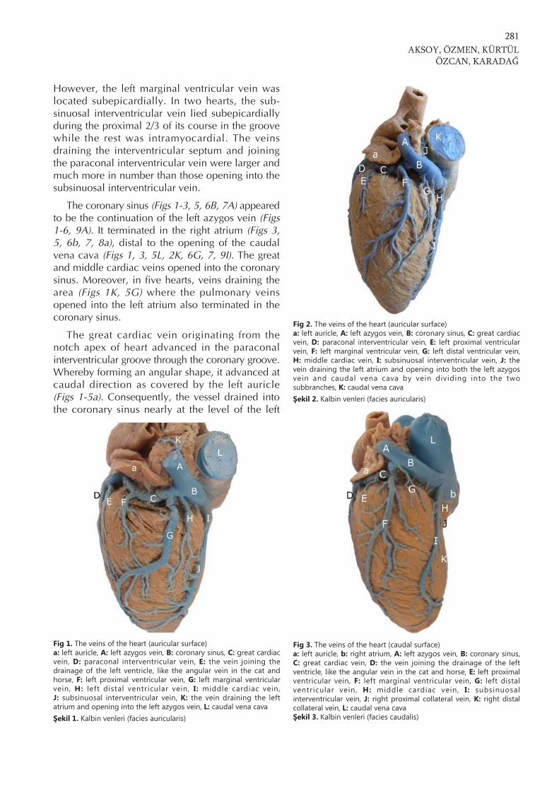
The coronary sinus (Figs 1-3, 5, 6B, 7A) appearedto be the continuation of the left azygos vein (Figs1-6, 9A). It terminated in the right atrium (Figs 3,5, 6b, 7, 8a), distal to the opening of the caudalvena cava (Figs 1, 3, 5L, 2K, 6G, 7, 9I). The greatand middle cardiac veins opened into the coronarysinus. Moreover, in five hearts, veins draining thearea (Figs 1K, 5G) where the pulmonary veinsopened into the left atrium also terminated in thecoronary sinus.
The great cardiac vein originating from thenotch apex of heart advanced in the paraconalinterventricular groove through the coronary groove.Whereby forming an angular shape, it advanced atcaudal direction as covered by the left auricle(Figs 1-5a). Consequently, the vessel drained intothe coronary sinus nearly at the level of the left
AKSOY, ÖZMEN, KÜRTÜLÖZCAN, KARADAĞ
281
Fig 1. The veins of the heart (auricular surface)a: left auricle, A: left azygos vein, B: coronary sinus, C: great cardiacvein, D: paraconal interventricular vein, E: the vein joining thedrainage of the left ventricle, like the angular vein in the cat andhorse, F: left proximal ventricular vein, G: left marginal ventricularvein, H: left distal ventricular vein, I: middle cardiac vein,J: subsinuosal interventricular vein, K: the vein draining the leftatrium and opening into the left azygos vein, L: caudal vena cavaŞekil 1. Kalbin venleri (facies auricularis)
Fig 3. The veins of the heart (caudal surface)a: left auricle, b: right atrium, A: left azygos vein, B: coronary sinus,C: great cardiac vein, D: the vein joining the drainage of the leftventricle, like the angular vein in the cat and horse, E: left proximalventricular vein, F: left marginal ventricular vein, G: left distalventricular vein, H: middle cardiac vein, I: subsinuosalinterventricular vein, J: right proximal collateral vein, K: right distalcollateral vein, L: caudal vena cavaŞekil 3. Kalbin venleri (facies caudalis)
Fig 2. The veins of the heart (auricular surface)a: left auricle, A: left azygos vein, B: coronary sinus, C: great cardiacvein, D: paraconal interventricular vein, E: left proximal ventricularvein, F: left marginal ventricular vein, G: left distal ventricular vein,H: middle cardiac vein, I: subsinuosal interventricular vein, J: thevein draining the left atrium and opening into both the left azygosvein and caudal vena cava by vein dividing into the twosubbranches, K: caudal vena cavaŞekil 2. Kalbin venleri (facies auricularis)
ventricular border. During its course, the greatgardiac vein was named paraconal interventricularvein (Figs 1, 2, 4D) in the paraconal interventriculargroove, and left circumflex vein in the coronarygroove. Right after its origin from the notch of theapex of the heart, the vessel anastomosed with theinitial branches of the middle cardiac vein on theauricular surface. The great cardiac vein drainedboth the left and right ventricles. It also receivedsmaller veins from the interventricular septum.
Branches draining the left ventricle were thefollowings:
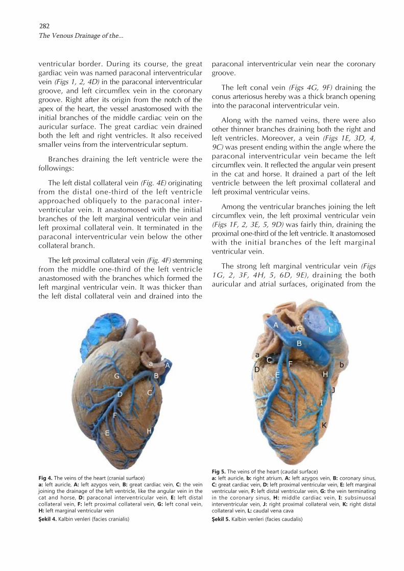
The left distal collateral vein (Fig. 4E) originatingfrom the distal one-third of the left ventricleapproached obliquely to the paraconal inter-ventricular vein. It anastomosed with the initialbranches of the left marginal ventricular vein andleft proximal collateral vein. It terminated in theparaconal interventricular vein below the othercollateral branch.
The left proximal collateral vein (Fig. 4F) stemmingfrom the middle one-third of the left ventricleanastomosed with the branches which formed theleft marginal ventricular vein. It was thicker thanthe left distal collateral vein and drained into the
paraconal interventricular vein near the coronarygroove.
The left conal vein (Figs 4G, 9F) draining theconus arteriosus hereby was a thick branch openinginto the paraconal interventricular vein.
Along with the named veins, there were alsoother thinner branches draining both the right andleft ventricles. Moreover, a vein (Figs 1E, 3D, 4,9C) was present ending within the angle where theparaconal interventricular vein became the leftcircumflex vein. It reflected the angular vein presentin the cat and horse. It drained a part of the leftventricle between the left proximal collateral andleft proximal ventricular veins.
Among the ventricular branches joining the leftcircumflex vein, the left proximal ventricular vein(Figs 1F, 2, 3E, 5, 9D) was fairly thin, draining theproximal one-third of the left ventricle. It anastomosedwith the initial branches of the left marginalventricular vein.
The strong left marginal ventricular vein (Figs1G, 2, 3F, 4H, 5, 6D, 9E), draining the bothauricular and atrial surfaces, originated from the
The Venous Drainage of the...282
Fig 4. The veins of the heart (cranial surface)a: left auricle, A: left azygos vein, B: great cardiac vein, C: the veinjoining the drainage of the left ventricle, like the angular vein in thecat and horse, D: paraconal interventricular vein, E: left distalcollateral vein, F: left proximal collateral vein, G: left conal vein,H: left marginal ventricular veinŞekil 4. Kalbin venleri (facies cranialis)
Fig 5. The veins of the heart (caudal surface)a: left auricle, b: right atrium, A: left azygos vein, B: coronary sinus,C: great cardiac vein, D: left proximal ventricular vein, E: left marginalventricular vein, F: left distal ventricular vein, G: the vein terminatingin the coronary sinus, H: middle cardiac vein, I: subsinuosalinterventricular vein, J: right proximal collateral vein, K: right distalcollateral vein, L: caudal vena cavaŞekil 5. Kalbin venleri (facies caudalis)
AKSOY, ÖZMEN, KÜRTÜLÖZCAN, KARADAĞ
283
Fig 6. The veins of the heart (caudal surface)a: left atrium, b: right atrium, A: left azygos vein, B: coronary sinus,C: great cardiac vein, D: left marginal ventricular vein, E: middlecardiac vein, F: subsinuosal interventricular vein, G: caudal vena cavaŞekil 6. Kalbin venleri (facies caudalis)
Fig 7. The veins of the heart (atrial surface)a: right atrium, A: coronary sinus, B: left distal ventricular vein,C: middle cardiac vein, D: subsinuosal interventricular vein, E: rightproximal collateral vein, F: right distal collateral vein, G: right distalventricular vein, H: right marginal ventricular vein, I: caudal venacavaŞekil 7. Kalbin venleri (facies atrialis)
Fig 8. The right cardiac veins (atrial surface)a: right atrium, A: right semicircumflex vein, B: right marginalventricular vein, C: right proximal ventricular vein, D: right conalvein, E: right distal ventricular vein, F: a trunk, G: paraconalinterventricular veinŞekil 8. Vv. cordis dextrae (facies atrialis)
Fig 9. The veins of the heart (auricular surface)a: left atrium, b: aorta, A: left azygos vein, B: great cardiac vein,C: the vein joining the drainage of the left ventricle, like the angularvein in the cat and horse, D: left proximal ventricular vein, E: leftmarginal ventricular vein, F: left conal vein, G: the vein joining thedrainage of the left atrium near the aortic arch, H: cranial vena cava,I: caudal vena cavaŞekil 9. Kalbin venleri (facies auricularis)
apex of the heart, lied on the caudal border, andterminated in the left circumflex vein. Itscommencing branches united with the leftproximal and left distal collateral veins, the leftdistal ventricular vein, and the subsinuosal inter-ventricular vein. In one cadaver, it (Fig. 6D)opened directly into the coronary sinus.
The left distal ventricular vein originating fromthe middle one-third of the left ventricleanastomosed with the branches joining theformation of the subsinuosal interventricular vein.It drained the area between the left marginalventricular vein and the subsinousal inter-ventricular vein. Yet, it was determined to openinto the coronary sinus in five cadavers (Figs 2,3G, 7B), and into the great cardiac vein in twocadavers (Figs 1H, 5F). The left distal ventricularvein was not formed in three hearts that wereexamined and the left marginal ventricular veindrained the area that was supposed to be drainedby this vein.
Several very thin veins draining the left atrium,including the proximal, intermedius, and distalveins of the left atrium, joined the left circumflexvein. In one cadaver, venous blood of the leftatrium (Fig. 2J) was determined to drain into theboth left azygos vein and caudal vena cava by avein dividing into the two subbranches. Besides,venous blood of the left atrium (Fig. 9G) near theaortic arch was displayed to empty into the cranialvena cava in two cadavers.
The middle cardiac vein, which is called thesubsinuosal interventricular vein (Figs 1J, 2, 3, 5I,6F, 7D) during the course in the subsinuosalinterventricular groove, was formed by thebranches stemming from the auricular surface ofthe apex of the heart. Originally, there were twostrong branches. It also took the branches from theinterventricular septum. Branches from originatingboth the right and left ventricles also joined thisvein; of those the right proximal collateral vein(Figs 3, 5J, 7E) was highly thick, while the rightdistal collateral vein (Figs 3, 5K, 7F) was thinner.
The veins comprising the right cardiac veinswere observed to be from different origins. Theveins draining the right atrium were shown to bevery thin. Those draining the right ventricle werestronger. The right marginal ventricular (Figs 7H,8B), the right proximal ventricular (Fig. 8C) veins
and the right conal vein (Fig. 8D), all draining theright ventricle, united to form the right semi-circumflex vein (Fig. 8A). This vein, in turn, liedcaudally in the coronary groove, and opened intothe right atrium at the level of the right ventricularborder. The right proximal ventricular vein formeda trunk (Fig. 8F) with the right conal vein beforeopening into the right semicircumflex vein. Theright distal ventricular vein (Figs 7G, 8E), originatingfrom the distal one-third of the right ventricle,advanced caudodorsally, anastomosed with theright marginal ventricular vein, and terminateddirectly in the right atrium.
The number of the minute cardiac veinsobserved grossly was higher in the right atriumand ventricle than in the left atrium and ventricle.
DISCUSSION
Reports have indicated that the venous cardiacveins including the great and middle cardiac veinsterminate in the coronary sinus 1,5-7. On the otherhand, Hegazi 9 and May 10 have reported thesevessels in sheep to drain directly into the rightatrium. Our study has also found that these veinsin the Tuj sheep drain into the coronary sinus.Besoluk and Tipirdamaz 1 have observed that theleft marginal ventricular vein in sheep terminatesin the coronary sinus. Yadm and Gad 4 and Hegazi 9
have reported this vein in goat to be a branch ofthe great cardiac vein. Even though, Nickel et al.2
have also said that the left marginal ventricularvein is a very thin vessel which opens into the lastpart of the coronary sinus, in our study weobserved this vein to be a very thick vessel joiningthe great cardiac vein. However, the vessel in justone cadaver was determined to open directly intothe coronary sinus.
In five cadavers, the veins collecting the venousblood of the area nearby the opening of thepulmonary veins into the left atrium were observedto terminate in the coronary sinus. Likewise, theleft distal ventricular vein was also opened intothe coronary sinus in five cadavers. So far, no suchdata have been reported on the sheep in theliterature. Although, Besoluk and Tipirdamaz 1 havesuggested the coronary sinus to be the continuationof the left azygos vein, Yadm and Gad 4 andGhoshal et al.6 have proposed it to be the cranialcontinuation of the great cardiac vein. Our
The Venous Drainage of the...284
findings in this study have led us to think inparallel with Besoluk and Tipirdamaz 1.
McKibben and Christensen 8 have reported thatthe great cardiac vein in sheep reaches as far asthe proximal two-thirds of the paraconal inter-ventricular groove. However, the great cardiacvein observed in our study advanced as far as thedistal one-third of the paraconal interventriculargroove. This report has also documented that thevenous blood of the cranial one-third of theinterventricular septum is usually drained eitherdirectly into the coronary sinus or into an anteriorcardiac vein. Contrarily, our study revealed thatthe venous blood of the interventricular septumwas emptied into either the paraconal orsubsinuosal interventricular veins.
It is very interesting to mention hereby that thevein observed in our study, supposedly called v.angularis, is present in the cat and horse 2, and asfar as to our knowledge, no literature report hasmentioned the presence of such a structure insmall ruminants.
TSpSrdamaz et al.3 have indicated that the distalone-third of the middle cardiac vein in sheep liesintramyocardially, while the proximal half issubepicardial. Our study revealed that theproximal two-thirds of the middle cardiac vein intwo hearts coursed subepicardially, while distalone-third distended intramyocardially.
In our study, the venous blood of the left atriumwas determined to drain both into the left azygosvein and caudal vena cava in one heart, and intothe cranial vena cava in two hearts. No suchfindings have been reported so far in the literature.
The right cardiac veins with different numbershave been documented to drain either into theright atrium 1,3,4,6,9 or into the coronary sinus 7. Wedisplayed that this vein opened directly into theright atrium. There was no such vein observed inthis study opening directly into the coronary sinusas indicated by Dursun 7. Nickel et al.2 haveinformed that the right semicircumflex vein isformed by the right proximal ventricular vein, theright conal vein and the right proximal atrial vein,and the right marginal ventricular vein drainsdirectly into the right atrium. On the other hand,we documented in our study that the rightsemicircumflex vein was formed by the right
marginal ventricular vein, the right proximalventricular vein and the right conal vein, and thisvein drained into right atrium. Yet, the right distalventricular vein opened directly into the rightatrium. This was similar to that reported byTSpSrdamaz et al.3 in sheep.
Studies 1,2 have reported different and contradictorycases on the presence of the minute cardiac veins.Yet, our study observed grossly that it was higherin number in the right atrium and ventricle ascompared to the left atrium and ventricle.
Consequently, the coronary sinus was determinedto be the continuation of the right azygos vein.Broad variation was observed among the veinsdraining the heart. The results obtained herebywill surely contribute to the anatomy of sheep, aswell as to the clinical researches using the heart asa model and conducted on animals and humanbeings.
REFERENCES
1. Besoluk K, Tipirdamaz S: Comparative macroanatomicinvestigations of the venous drainage of the heart inAkkaraman sheep and Angora goats. Anat Histol Embryol, 30,249-252, 2001.
2. Nickel R, Schummer A, Seiferle E: The Anatomy of theDomestic Animals. The Circulatory System, the Skin and theCutaneous Organs of the Domestic Mammals. Verlag PaulParey, Berlin, 1981.
7. Dursun N: Veteriner Anatomi II. 1. baskS. MedisanYaySnevi, Ankara, 1994.
8. McKibben JS, Christensen GC: The venous return from theinterventricular septum of the heart: a comparative study. AmJ Vet Res, 25, 512-517, 1964.
9. Hegazi H: Die Blutgefassversorgung des Herzens von Rind,Schaf und Ziege. Inaugural Dissertation, Giessen, 1958.
10. May NDS: The Anatomy of the Sheep. 2nd ed. Universityof Queensland Press, Brisbane, 1964.
11. Getty R: General heart and blood vessels. In, Getty R (Ed):Sisson and Grossman’s the Anatomy of the DomesticAnimals. 5th ed. Vol 1. 164-175, W.B. Saunders Company,
AKSOY, ÖZMEN, KÜRTÜLÖZCAN, KARADAĞ
285
Philadelphia, 1975.
12. AkçapFnar H: Koyun Yetiştiriciliği. 2. baskS. İsmatMatbaacSlSk, Ankara, 2000.
13. Shofti R, Zaretzki A, Cohen E, Engel A, Bar-el Y: Thesheep as a model for coronary artery bypass surgery. LabAnim, 38, 149-157, 2004.
14. Solem JO, Boumzebra D, Al-Buraiki J, Nakee S, Rafeh W,Al-Halees Z: Evaluation of a new device for quick suturelesscoronary artery anastomosis in surviving sheep. EuropeanJournal of Cardiothoracic Surgery, 17, 313-318, 2000.
15. Timek TA, Dagum P, Lai DT, Tibayan F, Liang D, DaughtersGT, Hayase M, Ingels NB Jr, Miller DC: Will a partial posterior
18. International Committee on Veterinary Gross AnatomicalNomenclature: General Assembly of the World Association ofVeterinary Anatomists. Nomina Anatomica Veterinaria. 5thed. Gent, 2005.


![Love -f] TUJ - Sarah TitusLove -f] TUJ . Created Date: 10/11/2017 8:02:16 PM](https://static.documents.pub/doc/80x56/5f50c5db472da84b43136754/-love-f-tuj-sarah-titus-love-f-tuj-created-date-10112017-80216-pm.jpg)


