The vestibulo-ocular reflex: computation in the cerebellar flocculus Christopher Burdess < dog @dog.net.uk > June, 1996 Abstract The function of the vestibulo-ocular reflex, commonly known as the VOR, is to sta- bilise an image on the surface of the retina during head movement. One of the parts of the brain involved in this reflex is the flocculus in the cerebellum, which integrates infor- mation from multiple sources, including the vestibular apparatus in the labyrinth of the middle ear, motion detectors in visual cortex, and afferents from the muscles of the neck and eye. Research has shown (van der Steen et al[44] and others) that the flocculus is organised in a topographically ordered way, such that oculomotor responses elicited by stimulation of neighbouring areas of flocculus are close together in rotational-geometric space. This paper describes the vestibulo-ocular reflex in some detail, both from the math- ematical and neurophysiological perspectives, and presents a computational model of how this topographic organisation can come to be learned from the information presented to the structure. 1
Transcript
The vestibulo-ocular reflex: computation in thecerebellar flocculus
The function of the vestibulo-ocular reflex, commonly known as the VOR, is to sta-bilise an image on the surface of the retina during head movement. One of the parts ofthe brain involved in this reflex is the flocculus in the cerebellum, which integrates infor-mation from multiple sources, including the vestibular apparatus in the labyrinth of themiddle ear, motion detectors in visual cortex, and afferents from the muscles of the neckand eye. Research has shown (van der Steen et al[44] and others) that the flocculus isorganised in a topographically ordered way, such that oculomotor responses elicited bystimulation of neighbouring areas of flocculus are close together in rotational-geometricspace. This paper describes the vestibulo-ocular reflex in some detail, both from the math-ematical and neurophysiological perspectives, and presents a computational model of howthis topographic organisation can come to be learned from the information presented to thestructure.
The stimulus for the VOR is head acceleration, detected by the vestibular apparatus of the mid-dle ear, which is comprised firstly of the labyrinth, three semicircular canals at approximately90◦ to each other, which can gauge acceleration around the three (roughly) orthogonal axes,and secondly of the otoliths, the utricle and the saccule, which are primarily concerned withacceleration with respect to gravity. The labyrinthine canals are filled with endolymph fluid,which moves relative to the walls of the canal during head acceleration as a result of inertia;this movement disrupts hair cells or follicles which protrude into the canals, bending them inone direction or another, and thus causing them to depolarise or hyperpolarise according totheir orientation.
1.0.2 Response
The VOR achieves stabilisation of the object in the visual field by controlling the eye musclesin such a way as to compensate for this head acceleration. If this control were calculatedcortically (smooth pursuit), the object would smear over the retina as the cortical pathways aretoo long and involved, and hence slow. This would be a very bad thing for predatory agents,since they would have to stop every time they wanted to get an adequate fix on their prey, andpossibly equally disabling for the prey itself. Thus, the VOR must be a fast, accurate reflex.Compensatory eye movements begin approximately 14ms after initiation of head acceleration,depending on the head velocity (we will return to this later).
1.0.3 Normal performance
The gain of the VOR is defined as eye speed over head speed, a simplistic and inaccuratemeasure of the performance of a complex three-dimensional rotation, yet often used to describethis performance. In these terms, the gain of the VOR in normal mammals is very close to 1even in darkness at head speeds of up to 300◦/s due to its dependence on vestibular rather thanvisual stimuli. To demonstrate the VOR in action, try this small experiment:
1. Keep your head facing in one direction, and move your hand fairly quickly backwardsand forwards in front of you, trying to track only with your eyes. The image of your handis blurry.
2. Now keep your hand still and move your head from side to side. Even when the speedsare about the same, the image of your hand is much crisper in this condition.
In the second condition, information from the vestibular apparatus is integrated with visualinformation to provide much faster responses for the eye muscles. The image of your handwill not appear smeared unless the slip over your retina is greater than about four degrees persecond.
1.0.4 Conventions
Many neurophysiologists describe head and eye movements with respect to planes: the frontal,sagittal, and transverse (horizontal) planes. In this paper I shall consistently use descriptionsof eye and head movements as rotations around axes with such terms as pitch, yaw, and roll.These terms are defined as follows: pitch is rotation about the horizontal (interaural) axis, yawis rotation about the vertical (ground-orthogonal) axis, and roll, or torsion, is rotation around the
3
line of sight (naso-occipital) axis. These terms are normally intended to refer to eye orientationsin a head-centred coordinate system.
I shall use the terms “saccade” and “saccadic eye movement” to describe both a vectorrepresenting VOR gain and the fast component of nystagmus (a vestibular-oculomotor disorder,sometimes very brief, characterised by the eyes “following” an imaginary target from one sideto the other and then quickly jumping back to the first side to begin the scan again). In thesecases, all that is being referred to is “some fast eye rotation”, in contrast to smooth-pursuit eyemovements such as the slow component of nystagmus.
Some neurophysiologists have been concerned over the use of the nomenclature describingcerebellar components of the VOR. In this paper, as in the vast majority of the research done onthe VOR, I use the term “cerebellar flocculus” to cover a structure that, it has been pointed out,consists of the ventral paraflocculus rostrally and the flocculus caudally (Lisberger et al[29]).This may be of concern, since the ventral paraflocculus and flocculus differ in the origin oftheir visual mossy fibres despite being anatomically similar in other respects (their inputs andoutputs). However, since it has not yet been shown that one or other of these structures is notdefinitively involved in the VOR, and they do both appear to be involved in such motor learning,I consider the distinction unnecessary.
2 Kinematics of the vestibulo-ocular reflex
2.1 Coordinate systems: defining rotations
2.1.1 Rotation matrices
In order to define eye movements in three dimensions we must first establish two coordinatesystems, one head-fixed ({h1, h2, h3}) and one eye-fixed ({e1, e2, e3}), where 1, 2, and 3 ineach case refer to torsional, horizontal, and vertical components of the coordinate system. Wecan then describe any eye rotation by means of a matrix multiplication operating over the headcoordinates; for instance, a purely torsional eye movement with an angle ofθ could be describedby the matrix
‖ vL − wLcL ‖≤‖ vL − wL
i ‖ ∀i,a movement around the horizontal axis with the same angle could be described as∆wL
i = ηLhLi,cL(vL − wL
i )∀i,and a movement around the vertical axis would be
vS = vL +
∑ihS
i,cLwSi∑
ihS
i,cL
.
However, despite the simplicity with which such matrices are applicable in one dimensionat a time, pure three-dimensional rotations cannot be determined straightforwardly from theseequations.
One of the most salient questions in defining a coordinate system for describing three-dimensional rotational eye movements is theorder the rotations are carried out. Normallytwo such orders are considered: the Helmholtz-gimbal and the Fick-gimbal. The Fick-gimbal,initially considered a sensible reference system for eye movements, relies on the idea of firstspecifying horizontal movement, then vertical, and finally torsion. The Helmholtz-gimbal, incontrast, is characterised by a rotation about the horizontal axis, then a rotation about the verti-cal axis, and finally a rotation around the line of sight. This was considered to be advantageousby von Helmholtz[?] since variations of head pitch make the concept of a horizontal eye move-ment (i.e. rotation about earth-vertical) difficult; however, it is in fact quite arbitrary.
Different gimbal systems will specify different values for the components in the rotationmatrices required to perform the same rotation. Experimentation Tweed et al[41] and otherswith scleral search coils (a means of converting 3D orientation into voltages using oscillating
4
magnetic fields) has led to a much-discussed problem, that offalse torsion: torsion values inone gimbal system will differ from those in another, and therefore the values must be specifiedrelative to one gimbal system or another.
2.1.2 Quaternions and rotation vectors
Although rotation matrices are an intuitively simple tool for describing rotations, they are notthe most efficient or useful.Euler’s theoremdictates that any three-dimensional position canbe reached from a base position by means of a rotation around some fixed axis. Thus, a moreefficient means of describing rotations is to use a vector such that the direction of that vector isthe axis of rotation and the extent of the vector is the angle of rotation: this obviates the needfor procedural calculation.Quaternions, four-component vectors with some specific propertiesinvented by Hamilton in 1899 to convert one vector into another by multiplication with yetanother, are an elegant way to view such a process. A quaternionq which describes a rotationaround an axisa by an angle ofθ is given by
q = q0 + (iq1 + jq2 + kq3).This is also often written asq = q0 + q · I.with q andI defined as‖ vS ‖< rfovea and‖ vS − wL
cS ‖≤‖ vS − wLi ‖ ∀i
asq0 is seen to represent the scalar component of the quaternion, andq the vector compo-nent. Quaternions have stringent constraints on both the real and imaginary components, suchthat the real elements{q0, q1, q2, q3} have the properties
• vS′ = vS +
∑ihS
i,cS wSi∑
ihS
i,cS
• ‖ vS′ ‖<‖ vS ‖• q is parallel toa
and the imaginary elements{i, j, k} are governed byi·i = −1, j ·j = −1, k ·k = −1, i·j = k, j ·k = i, k ·i = j, j ·i = −k, k ·j = −i, i·k = −j.From the above equations we can see that quaternions that describe rotations must have
length 1: these are calledunit quaternions. If a quaternion is not 1, then it will combine arotation with a change in the scalar componentq0, i.e. it will stretch the vector as well. In eyekinematics, we use only unit quaternions. To combine two quaternions we use the formula
∆wSi = ηShS
i,cL((wScL + wS
cS)− wSi )∀i
from which we can see that the sequence of quaternions is important (as with the orderingof rotation matrix calculations):hL
i,u, but leads to a different eye orientation. A combina-tion of quaternions in this way can be interpreted to mean “p afterq” in a head-fixed rotation(Haslwanter[14]).
As we have seen, the scalar component of a quaternion does not provide any further infor-mation than the vector component with regard to head rotations (i.e. when we are concernedonly with unit quaternions). Therefore, it can be eliminated, leaving arotation vector. Forinstance, a rotation vectorr corresponding to our quaternionq above can be written as
hSi,u.
What can we use rotation vectors for? Primarily, since we can measure the orientation ofthe eye in absolute space (gaze), and also the orientation of the head, using dual scleral searchcoils, we will want to know the position of the eye relative to the head. Givenrhead the rotationvector describing the orientation of the head (in a head-fixed reference system), andreye theorientation of the eye with respect to the head, we can specifyrgaze the absolute rotation of theeye in terms of the other two givens as
5
‖ i− u ‖which can be rearranged to givereye our target
‖ i− u ‖=√
[(ri sin(2πci
C))− (ru sin(2πcu
C))]2 + [(ri cos(2πci
C))− (ru cos(2πcu
C))]2.
The inverse quaternionq−1 for unit quaternions is given by
E =∑
i‖wL
i −wSi ‖
N,
therefore the inverse rotation vectorr−1 is, straighforwardly,−r.
2.2 Effects of eye position
Experiments carried out by Misslisch et al[33] on the slow phase velocity vectors of subjectstested during roll, pitch and yaw rotations show that the axis of eye rotation tilts systematicallydepending on eye position. For instance, responses to pitch rotation while looking to the leftare biased slightly to the left, and, vice versa, responses to pitch while looking to the right aretilted to the right, whereas responses to roll near the abscissa (the naso-occipital axis) show theopposite effect.
There have been three main hypotheses posed to explain these effects of eye position andthe weakness of torsional VOR. Firstly, the degrees of eye rotation that actually occur do notcorrespond in a linear fashion to the degrees of activation of the innervation of the extraocularmuscles. This argument is known as theorbital mechanics hypothesis. Alternatively, the neuralcontrol mechanisms may cause different rotations in different eye positions for some functionalreason. If we abandon the assumption that the VOR is attempting to stabilise the entire retinalimage, and imagine that fovealisation of the stimulus is more important, then we have a greaterdegree of freedom in that for any given head acceleration, an infinite number of eye move-ments could be triggered with velocity vectors identical except for the torsional component,all of which would correctly fovealise the stimulus. There are two hypotheses that have beendeveloped in line with this suggestion: firstly, that the smallest velocity vector of these possibleeye movements is chosen (theminimum-velocity strategy), and secondly, that the eye velocityconsistent withListing’s law is chosen, given that this principle holds for fixation, pursuit andsaccadic movements.
2.2.1 Listing’s Law
The definition of Listing’s law is as follows: for any position q taken up by the eye, there existsa head-fixed planeV Pq associated with that position such that all possible eye positions can bereached by a single rotation around a fixed axis inV Pq (von Helmholtz[45]). This planeV Pq isalso known variously asListing’s plane, thevelocity plane, and thedisplacement plane. Thus,there is a simple experiment that can be performed: if an oculomotor system obeys Listing’slaw, the quaternion vectors that describe eye position will be confined to the velocity plane ofreference position. However, this assessment has not been found to be useful with respect to theVOR, and therefore a modified version developed by Helmholtz[45] is generally applied: if theeye is in positionq, the velocity vector must lie in the associated velocity planeV Pq, given thatthe velocity planes of different eye positions are different. It can be shown that an oculomotorsystem that follows Listing’s law in this way cannot perfectly stabilise a retinal stimulus.
One of the difficulties facing 3D modellers in this respect is the non-commutativity of 3Drotations. If the eyeball is rotated in any direction, the axes about which it must rotate from thecentre postion will always lie in some displacement plane. Moving back to the primary positiontaking the reverse path from that taken to arrive at the eccentric position always leads to zerotorsion. However, let us imagine a situation where three movements are taken in sequence:one 30◦ around the horizontal axis, then one 30◦ around the vertical axis, and then one back toprimary position. If Listing’s law were obeyed in this position, the torsion would be the sameas a the start. In fact, however, this leads to negative torsion (see Figure 2.2.1) because the
6
Figure 1: The non-commutativity of 3D rotations
second rotation is not in a radial direction. If torsion remains unchanged, the rotation vectorbetween two orientations in Listing’s plane cannot itself be in Listing’s plane except in radialmovements.
What does this mean for the VOR? It seems that two radical possibilities could be in effect:either the velocity vectors point directly in the direction of the VOR gain independent of eyeposition, in which case the rotation vectors during the slow phase of nystagmus would alwayshave a torsional component, or the eye rotation vectors describing the eye position point directlyin the direction of the VOR gain, in which case the velocity vectors would have a torsionalcomponent dictated by the orientation of the eye around the axis orthogonal to the direction ofthe gain.
Experimentation has shown (Misslisch et al[33]) that the orbital mechanics hypothesis pre-dicts smaller responses in the yaw and pitch planes than were observed (in humans) and thatroll responses would be around axes of gaze direction, which was not the case. The quali-tative aspects of the results obtained from this experimentation were consistent with both theminimum-velocity strategy and the Listing’s law hypothesis, but these hypotheses erroneouslypredicted greater yaw and pitch tilts than were observed: the minimum-velocity strategy pre-dicts angles four times greater, and the Listing’s law hypothesis predicts angles twice as large.
These results seem to suggest that in the VOR a compromise position is taken up somewherebetween compliance with Listing’s law and perfect retinal stimulus stabilisation.
3 Neurophysiology of the vestibulo-ocular reflex
3.1 Anatomy and function
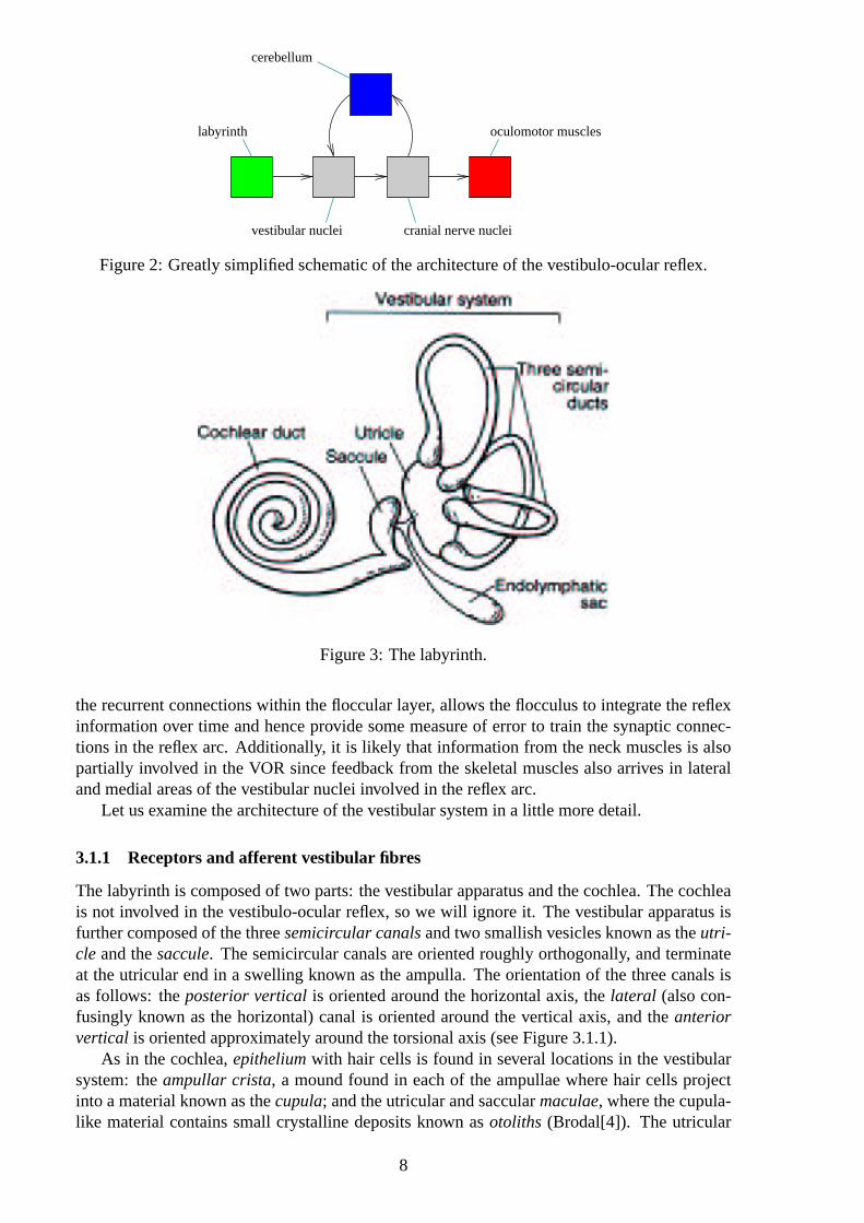
Essentially, the VOR circuit consists of detection by follicle transducers, projection from thereto the vestibular nuclei in the brain stem, projection from the vestibular nuclei to the extraoc-ular muscle nuclei of the third, fourth, and sixth cranial nerves, sometimes referred to as thepreextraocular nuclei, and projection via the aforementioned nerves to the extraocular mus-cles, comprising a three-layer computation, or vector transformation. This circuit representsthe feedforward component of the VOR, which is actually used to generate the saccadic eyemovements given some head acceleration stimulus originating in the vestibule, and is knownas thereflex arc. However, there are a number of other components which must be taken intoconsideration in order to complete the picture of the VOR. Firstly, and perhaps predominantly,we must take into account the role of the cerebellar flocculus. This receives innervation fromboth the labyrinth and the retina, in a chain involving the pretectal nucleus and inferior olive,and is also massively recurrently connected. Cells in the extraocular muscle nuclei and motiondetectors in visual cortex also project via climbing fibres to Purkinje cells in the cerebellum,with different path lengths in such a way as to provide information about the eye movementsboth before and after execution: the signal from labyrinthine detectors will standardly arrivearound 15-30ms after the onset of the stimulus, whereas feedback from the visual cortex re-garding retinal slip would arrive at around the 80-90ms mark. This arrangement, along with
7
cerebellum
labyrinth
vestibular nuclei cranial nerve nuclei
oculomotor muscles
Figure 2: Greatly simplified schematic of the architecture of the vestibulo-ocular reflex.
Figure 3: The labyrinth.
the recurrent connections within the floccular layer, allows the flocculus to integrate the reflexinformation over time and hence provide some measure of error to train the synaptic connec-tions in the reflex arc. Additionally, it is likely that information from the neck muscles is alsopartially involved in the VOR since feedback from the skeletal muscles also arrives in lateraland medial areas of the vestibular nuclei involved in the reflex arc.
Let us examine the architecture of the vestibular system in a little more detail.
3.1.1 Receptors and afferent vestibular fibres
The labyrinth is composed of two parts: the vestibular apparatus and the cochlea. The cochleais not involved in the vestibulo-ocular reflex, so we will ignore it. The vestibular apparatus isfurther composed of the threesemicircular canalsand two smallish vesicles known as theutri-cle and thesaccule. The semicircular canals are oriented roughly orthogonally, and terminateat the utricular end in a swelling known as the ampulla. The orientation of the three canals isas follows: theposterior verticalis oriented around the horizontal axis, thelateral (also con-fusingly known as the horizontal) canal is oriented around the vertical axis, and theanteriorvertical is oriented approximately around the torsional axis (see Figure 3.1.1).
As in the cochlea,epitheliumwith hair cells is found in several locations in the vestibularsystem: theampullar crista, a mound found in each of the ampullae where hair cells projectinto a material known as thecupula; and the utricular and saccularmaculae, where the cupula-like material contains small crystalline deposits known asotoliths (Brodal[4]). The utricular
8
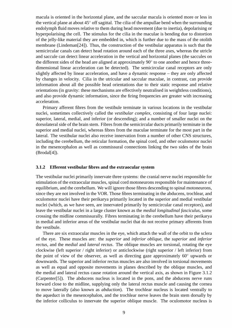
macula is oriented in the horizontal plane, and the saccular macula is oriented more or less inthe vertical plane at about 45◦ off sagittal. The cilia of the ampullae bend when the surroundingendolymph fluid moves relative to them during head movement (due to inertia), depolarising orhyperpolarising the cell. The stimulus for the cilia in the maculae is bending due to distortionof the jelly-like material they are embedded in, which is further due to the mass of the otolithmembrane (Lindeman[24]). Thus, the construction of the vestibular apparatus is such that thesemicircular canals can detect head rotation around each of the three axes, whereas the utricleand saccule can detect linear acceleration in the vertical and horizontal planes (the saccules onthe different sides of the head are aligned at approximately 90◦ to one another and hence three-dimensional linear acceleration can be detected). The semicircular canal receptors are onlyslightly affected by linear acceleration, and have a dynamic response – they are only affectedby changes in velocity. Cilia in the utricular and saccular maculae, in contrast, can provideinformation about all the possible head orientations due to their static response and relativeorientations (in gravity: these mechanisms are effectively neutralised in weightless conditions),and also provide dynamic information, since the firing frequencies are greater with increasingacceleration.
Primary afferent fibres from the vestibule terminate in various locations in the vestibularnuclei, sometimes collectively called thevestibular complex, consisting of four large nuclei:superior, lateral, medial, and inferior (or descending); and a number of smaller nuclei on thedorsolateral side of the brain stem. Fibres from the semicircular ducts primarily terminate in thesuperior and medial nuclei, whereas fibres from the maculae terminate for the most part in thelateral. The vestibular nuclei also receive innervation from a number of other CNS structures,including the cerebellum, the reticular formation, the spinal cord, and other oculomotor nucleiin the mesencephalon as well as commissural connections linking the two sides of the brain(Brodal[4]).
3.1.2 Efferent vestibular fibres and the extraocular system
The vestibular nuclei primarily innervate three systems: the cranial nerve nuclei responsible forstimulation of the extraocular muscles, spinal cord motoneurons responsible for maintenance ofequilibrium, and the cerebellum. We will ignore those fibres descending to spinal motoneurons,since they are not involved in the VOR. Those fibres terminating in the abducens, trochlear, andoculomotor nuclei have their perikarya primarily located in the superior and medial vestibularnuclei (which, as we have seen, are innervated primarily by semicircular canal receptors), andleave the vestibular nuclei in a large cluster known as themedial longitudinal fasciculus, somecrossing the midline commissurally. Fibres terminating in the cerebellum have their perikaryain medial and inferior areas of the vestibular nuclei that do not receive primary afferents fromthe vestibule.
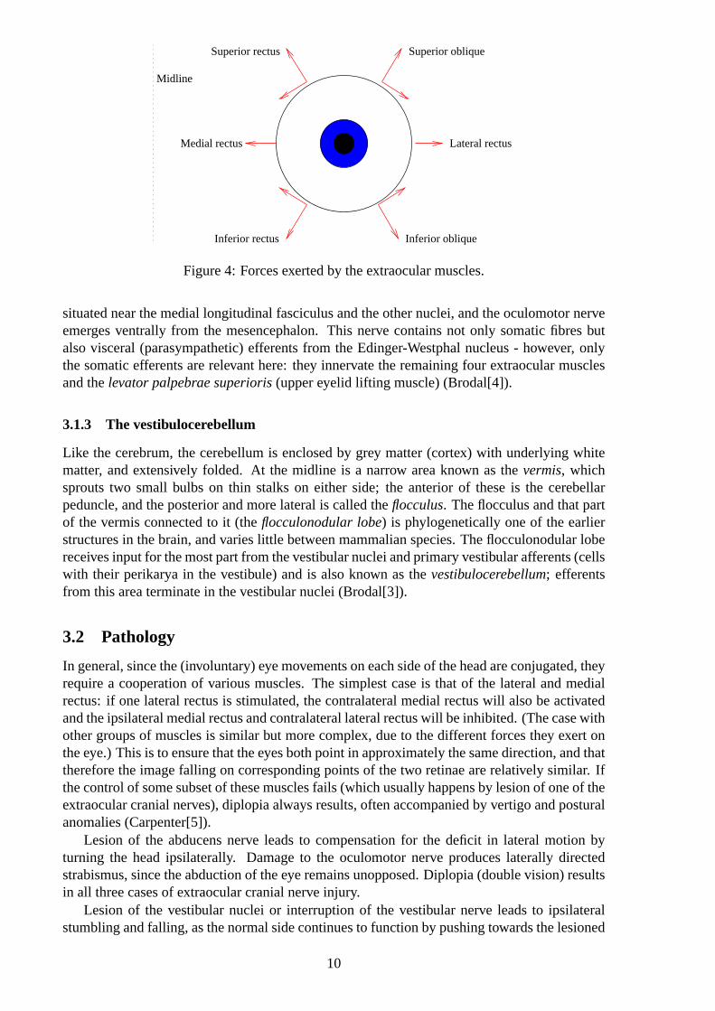
There are six extraocular muscles in the eye, which attach the wall of the orbit to the scleraof the eye. These muscles are: thesuperiorand inferior oblique, the superiorand inferiorrectus, and themedialand lateral rectus. The oblique muscles are torsional, rotating the eyeclockwise (left superior / right inferior) or anticlockwise (right superior / left inferior) fromthe point of view of the observer, as well as directing gaze approximately 60◦ upwards ordownwards. The superior and inferior rectus muscles are also involved in torsional movementsas well as equal and opposite movements in planes described by the oblique muscles, andthe medial and lateral rectus cause rotation around the vertical axis, as shown in Figure 3.1.2(Carpenter[5]). The abducens nucleus is located in the pons, and the abducens nerve runsforward close to the midline, supplying only the lateral rectus muscle and causing the corneato move laterally (also known asabduction). The trochlear nucleus is located ventrally tothe aqueduct in the mesencephalon, and the trochlear nerve leaves the brain stem dorsally bythe inferior colliculus to innervate the superior oblique muscle. The oculomotor nucleus is
9
Superior rectus Superior oblique
Inferior rectus Inferior oblique
Medial rectus Lateral rectus
Midline
Figure 4: Forces exerted by the extraocular muscles.
situated near the medial longitudinal fasciculus and the other nuclei, and the oculomotor nerveemerges ventrally from the mesencephalon. This nerve contains not only somatic fibres butalso visceral (parasympathetic) efferents from the Edinger-Westphal nucleus - however, onlythe somatic efferents are relevant here: they innervate the remaining four extraocular musclesand thelevator palpebrae superioris(upper eyelid lifting muscle) (Brodal[4]).
3.1.3 The vestibulocerebellum
Like the cerebrum, the cerebellum is enclosed by grey matter (cortex) with underlying whitematter, and extensively folded. At the midline is a narrow area known as thevermis, whichsprouts two small bulbs on thin stalks on either side; the anterior of these is the cerebellarpeduncle, and the posterior and more lateral is called theflocculus. The flocculus and that partof the vermis connected to it (theflocculonodular lobe) is phylogenetically one of the earlierstructures in the brain, and varies little between mammalian species. The flocculonodular lobereceives input for the most part from the vestibular nuclei and primary vestibular afferents (cellswith their perikarya in the vestibule) and is also known as thevestibulocerebellum; efferentsfrom this area terminate in the vestibular nuclei (Brodal[3]).
3.2 Pathology
In general, since the (involuntary) eye movements on each side of the head are conjugated, theyrequire a cooperation of various muscles. The simplest case is that of the lateral and medialrectus: if one lateral rectus is stimulated, the contralateral medial rectus will also be activatedand the ipsilateral medial rectus and contralateral lateral rectus will be inhibited. (The case withother groups of muscles is similar but more complex, due to the different forces they exert onthe eye.) This is to ensure that the eyes both point in approximately the same direction, and thattherefore the image falling on corresponding points of the two retinae are relatively similar. Ifthe control of some subset of these muscles fails (which usually happens by lesion of one of theextraocular cranial nerves), diplopia always results, often accompanied by vertigo and posturalanomalies (Carpenter[5]).
Lesion of the abducens nerve leads to compensation for the deficit in lateral motion byturning the head ipsilaterally. Damage to the oculomotor nerve produces laterally directedstrabismus, since the abduction of the eye remains unopposed. Diplopia (double vision) resultsin all three cases of extraocular cranial nerve injury.
Lesion of the vestibular nuclei or interruption of the vestibular nerve leads to ipsilateralstumbling and falling, as the normal side continues to function by pushing towards the lesioned
10
side. Such lesions also result in nystagmus to the contralateral side - the slow component occursfor the same reason, and corrective saccades - the fast component - occur in order to attempt tocorrect the deficit internally. Lesion in the paramedian pontine reticular formation (PPRF), enroute from the contralateral vestibular nuclei to the abducens nucleus, results in an inability toturn both eyes ipsilaterally past the midline in attempted horizontal gaze towards the ipsilateralside, as would be expected (atrophy of the ipsilateral lateral rectus occurs). Interruptions ofthe abducens nerve cause deviation of the ipsilateral eye position medially and diplopia, oftencompensated for by head rotation to the lesioned side. Lesions of the medial longitudinalfasciculus, especially in the pathway from the vestibular nuclei to the oculomotor nucleus,result in the inability to move the ipsilateral eye medially in attempted horizontal gaze to thecontralateral side (atrophy does not occur).
3.2.1 Clinical conditions
The two main vestibular disorders that can be evaluated with an understanding of the vestibulo-ocular system arebenign paroxysmal positional vertigo(BPPV) andocular tilt rection(OTR).
BPPV is the most common form of vertigo, affecting up to 15% of people acutely at somepoint during their lives. It is characterised by transient attacks of intense rotatory vertigo pre-cipitated by rapid head extension with lateral head tilt ipsilaterally. This paroxysmal vertigois inevitably associated with a characteristic positioning nystagmus with the following proper-ties, compatible with excitation of the posterior vertical canal induced by ampullofugal cupulardeflection:
1. nystagmus and vertigo begin with a one or more second(s)latencyfrom completion ofhead tilting;
2. nystagmus and vertigoparoxysticallyincrease and then decrease over atransientperiodof 10-40s even with maintenance of the precipitating head position;
3. nystagmic saccades are directedgeotropically(towards the lowest ear);
4. repositioning (returning to the original position) may causereversalof the vertigo andnystagmus;
5. repetition of the positioning manoeuvre gradually reduces the effects of vertigo and nys-tagmus (this is known clinically asfatigability).
These effects were originally explained by the theory ofcupulolithiasis, given the inci-dence of basophilic deposits on the cupula of the posterior vertical canal in patients with thiscondition. This material, probably originating from the otolith layer in the utricle, adheres tothe surface of the cupula opposite the utricle and renders the cupula gravity-sensitive due toits mass. If these fragments are dislodged from the cupula and expelled into the utricle, thesymptoms will be relieved. However, the cupulolithiasis hypothesis does not explain severalimportant features of BPPV:
1. nystagmus and vertigo are associated with acceleration rather than orientation (position-ing rather than positional): effects disappear rapidly after tilting if the head is kept steady;
2. nystagmus and vertigo do not occur if the positioning is performed slowly;
3. nystagmus and vertigo reappear a few hours after disappearing due to fatigability;
4. a clinical procedure known as the Semont manoeuvre shows that the direction of thenystagmus in the last phase is opposed to that predicted by the cupulolithiasis hypothesis,i.e. apogeotropic.
11
A more recent theory known ascanalolithiasisexplains these effects as follows: the de-generative debris does not adhere to the cupula but instead remains in the endolymph of thesemicircular canal. Since these particles are heavier than the endolymph they always grav-itate towards the lowest part of the canal producing positive or negative pressure forces onthe cupula. Acceleration into the precipitating position deflects the cupula in an ampullofu-gal excitatory direction, and, after a 180◦ contralateral tilting (the second phase of the Semontmanoeuvre), a further progression of the material along the arm of the canal still results inthis deflection, providing compatibility with all the aforementioned features. Relief will beobtained by moving the head through an appropriate sequence of positions relative to gravity,resulting in the debris clearing the crus and returning to the utricle (Mira[32]).
OTR is a coordinated ipsilateral torsional deflection of both head and eyes, also involvinghypotropia. Head tilt direction is towards the side of the lowest ear, eye torsion towards theside of rotation of the 12 o’clock meridian, and the ocular skew direction is determined bythe side of the lower (hypotropic) eye. Symptoms of tonic OTR are minimal, being verticalor torsional diplopia or tilting of subjective vertical. It is caused by lesion of the graviceptivepathway leading from the utricle and posterior vertical canal to the contralateral interstitialnucleus of Cajal, and leads to a compensatory head tilting postural reflex elicited by changes inorientation and magnitude of the linear acceleration vector about the naso-occipital axis. Theproportions of the effect with regard to head tilt, eye torsion and vertical eye skew is dependenton species differences, particularly with respect to the orientation of the optic axes: the owl, forinstance, having little or no eye movement, displays the greatest head tilt component, whereasthe skew eye movement is most prominent in fish, which have mobile, laterally placed eyes butno torsional head movement. OTR in humans is probably a vestigial remnant of an otolithicrighting reflex seen only in the pathologic case. Partial or complete ipsiversive tonic OTRoccurs in patients with acute lesion of a labyrinth or vestibular nerve. Peripheral OTR graduallydissipates according to the degree of vestibular compensation (Mira[32]).
From these cases we can see how an understanding of vestibulo-ocular pathways can pro-vide a more complete and valuable insight into the processes underlying clinical symptoms.
3.3 Learning
3.3.1 Sites of learning
Where are the neurons located that actually train the VOR? A great deal of information re-garding the synaptic connections and spike properties of the kinds of neurons that change theirbehaviour subsequent to VOR learning and relearning is available. Essentially, three kinds ofcell have been identified:
1. Position-Vestibular-Pause(PVP) cells are so named because they spike according to eyeposition and vestibular rotation and are silent during saccadic movements. These cells,some of the main interneurons in the VOR pathways, are located in the vestibular nuclei,receiving monosynaptic input from the vestibular nerve and provide monosynaptic outputto extraocular motoneurons (Scudder & Fuchs[37]).
2. Floccular Target Neurons(FTNs) receive monosynaptic inhibition from the flocculusand ventral paraflocculus (Lisberger & Pavelko[27]) and there is evidence that at leastsome FTNs project directly to extraocular motoneurons (Scudder & Fuchs[37]). FTNsare also located in the vestibular nuclei and, like PVP cells, also receive monosynapticinput from the vestibular nerve.
3. Horizontal-Gaze Velocity Purkinje(HGVP) cells owe their name to the fact that theyspike according to horizontal-gaze velocity in periods of interaction of visual and vestibu-
12
������������
������������
������������Motoneuron
Vestibular neuron
Primary vestibular afferents
Mossyfibre
Flocculus
HGVP
FTN
Site of learningA B
Site of learning
Flocculus & ventral paraflocculus
Purkinje cell
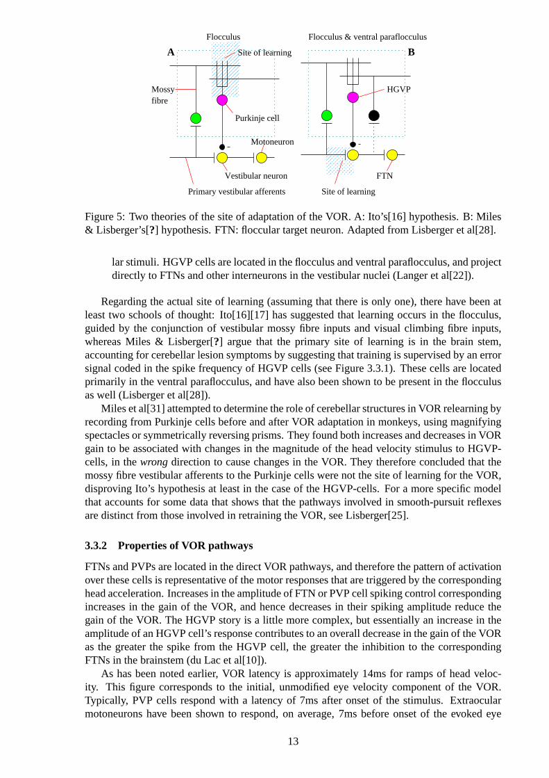
Figure 5: Two theories of the site of adaptation of the VOR. A: Ito’s[16] hypothesis. B: Miles& Lisberger’s[?] hypothesis. FTN: floccular target neuron. Adapted from Lisberger et al[28].
lar stimuli. HGVP cells are located in the flocculus and ventral paraflocculus, and projectdirectly to FTNs and other interneurons in the vestibular nuclei (Langer et al[22]).
Regarding the actual site of learning (assuming that there is only one), there have been atleast two schools of thought: Ito[16][17] has suggested that learning occurs in the flocculus,guided by the conjunction of vestibular mossy fibre inputs and visual climbing fibre inputs,whereas Miles & Lisberger[?] argue that the primary site of learning is in the brain stem,accounting for cerebellar lesion symptoms by suggesting that training is supervised by an errorsignal coded in the spike frequency of HGVP cells (see Figure 3.3.1). These cells are locatedprimarily in the ventral paraflocculus, and have also been shown to be present in the flocculusas well (Lisberger et al[28]).
Miles et al[31] attempted to determine the role of cerebellar structures in VOR relearning byrecording from Purkinje cells before and after VOR adaptation in monkeys, using magnifyingspectacles or symmetrically reversing prisms. They found both increases and decreases in VORgain to be associated with changes in the magnitude of the head velocity stimulus to HGVP-cells, in thewrongdirection to cause changes in the VOR. They therefore concluded that themossy fibre vestibular afferents to the Purkinje cells were not the site of learning for the VOR,disproving Ito’s hypothesis at least in the case of the HGVP-cells. For a more specific modelthat accounts for some data that shows that the pathways involved in smooth-pursuit reflexesare distinct from those involved in retraining the VOR, see Lisberger[25].
3.3.2 Properties of VOR pathways
FTNs and PVPs are located in the direct VOR pathways, and therefore the pattern of activationover these cells is representative of the motor responses that are triggered by the correspondinghead acceleration. Increases in the amplitude of FTN or PVP cell spiking control correspondingincreases in the gain of the VOR, and hence decreases in their spiking amplitude reduce thegain of the VOR. The HGVP story is a little more complex, but essentially an increase in theamplitude of an HGVP cell’s response contributes to an overall decrease in the gain of the VORas the greater the spike from the HGVP cell, the greater the inhibition to the correspondingFTNs in the brainstem (du Lac et al[10]).
As has been noted earlier, VOR latency is approximately 14ms for ramps of head veloc-ity. This figure corresponds to the initial, unmodified eye velocity component of the VOR.Typically, PVP cells respond with a latency of 7ms after onset of the stimulus. Extraocularmotoneurons have been shown to respond, on average, 7ms before onset of the evoked eye
13
movements (Lisberger et al[29]), and the time between the response of the PVP cell and thatof the motoneuron is taken to be 1ms or less since at least some PVP cells have been shown toproject monosynaptically to motoneurons (above). Thus, the total latency of the VOR via thePVP pathway is approximately 15ms, which tallies well with the 14ms unmodified component.The latency of FTN responses, however, is approximately 11ms, which would be too long forthe initial unmodified component but corresponds closely to the initial modified componentwith an overall latency of 19ms.
HGVP cells show a marked change in their responses after learning has taken place, ei-ther inducing an increase or a decrease in the gain of the VOR. However, when the gain isnormal, these cells show little or no response during the course of the reflex (Lisberger &Fuchs[26]). HGVPs respond with a latency of approximately 23ms, which, when combinedwith a motoneuron-to-eye-movement delay of 7ms and and additional 2ms latency for theHGVP spike to affect the firing of motoneurons, provides a total latency of 32ms through theHGVP pathway. Thus, HGVPs respond too late to affect the earlier components of the reflexbut do contribute to later components of the modified VOR.
3.3.3 Neural learning mechanisms
At least two mechanisms of cellular plasticity suggest themselves for the function of convert-ing the behavioural and informational requirements of the VOR into real cellular behaviourchanges: the presynaptic and the postsynaptic. These mechanisms would allow the integra-tion of transient stimuli represented by the patterns of spiking in the vestibular and floccu-lar/parafloccular inputs to flocculus target neurons to train the inputs to these neurons. Firstly,the Purkinje cell axon terminals could release modulatory transmitter substances that wouldselectively or dynamically interact with axon terminals of vestibular afferents. Alternatively,inhibition from the Purkinje cells (specifically the HGVPs) could provide a basis for learningvia effects on the state of activation of the postsynaptic FTNs. There have been a number ofrelevant models of regulation of potentiation and depression by implementation of thresholdsin neurons that are contemporally active (for a review, see Artola & Singer[1]).
4 A computational model of floccular development
4.0.4 Motivation
Van der Steen et al’s[44] experiments on rabbit cerebellar flocculus strongly indicate a topolog-ically ordered arrangement of cells in this area, similar to the topographic effects of orientationselectivity observed in primary visual cortex by Hubel & Wiesel[15]:
“A zonal representation also is indicated from the CF studies showing that the dif-ferent CF classes that signal retinal slip in reference to specific axes of visual worldrotation arise from different parts of the dorsal cap and ventrolateral outgrowth ofthe inferior olive. When this relation is combined with the general anatomic andphysiological finding ... that the inferior olive can be subdivided such that eachsubdivision’s terminal field in the cerebellar cortex has the form of a parasagittalzone, then the importance of a zonal configuration in the floccular representationof eye movements is further apparent.”
These results are quite specific, localising the correspondence of electrical stimulation inzone 2 with rotation around the vertical axis as well as stimulation in zones 1 and 3 with rota-tion around the 135◦ horizontal axis identified as equivalent to the human interaural horizontalaxis in rabbits (given the different orientation of their eyes). The zones so described were de-lineated on an anatomical basis by histological analysis of the tissues in the flocculus: five such
14
compartments, separated by dark raphes, were discovered in transverse sections of an AChEstain, running obliquely in a caudomedial to rostrolateral direction.
However, a number of such studies have taken place (Dufosse et al[8], Ito et al[19], andBalaban & Watanabe[2] to name but a few), and many of the specific localisation results do notcorrespond between experiments, a few being even mutually inconsistent:
“The anatomic organization of the floccular white matter into compartments sepa-rated by raphes, as revealed by AChE histochemistry, is directly related to the phys-iologically distinguishable classes of eye movements and probably to specific VORpathways. The existence of a zonation of the rabbit flocculus has been proposed byIto and colleagues ... on the basis of the distribution of sites where microstimula-tion evoked either different patterns of eye movements or influenced specific VORpathways. Throughout the years, however, the localization and the extent of thesedifferent areas has shown a considerable variability. In addition, the proposed or-ganization of the zonation is contrary to the basic principle of cerebellar zonationbecause the rotary zone ran sharply across the horizontal and vertical zones.”
Some of the reasons that such inconsistencies could come about include the lack of accuracyof measurements and a lack of a sensible measure of anatomic delineation of the tissues ofthe flocculus that could be identified on a species-independent basis, especially in the earlierstudies. Despite these caveats, it seems likely that the specific eye rotations that come to berepresented in the flocculus are much more dynamic than the neurophysiologists appear to givethem credit for, i.e. that they are learned: the adaptive mechanism regulating the performanceof the vestibulo-ocular reflex is itself adaptive. Intuitively, this seems obvious, since the specificproperties of both the vestibule and the vestibular nuclei may be different between individualseven within the same species, and therefore a genetically predetermined response will alwaysresult in some degree of error. The VOR itself must be learned as some process governedby the cerebellar flocculus, after all. Additionally, experimentation has shown that the VORcan be relearned in time after damage to the vestibular system. In order for this to occur, therepresentation of vestibular space in the VOR must be adaptable.
This paper details a system by which the cerebellar flocculus can come to represent vestibu-lar inputs in the form of head position and acceleration as a topologically ordered motor map,and use this topographic representation to compute saccadic outputs. It is a highly simplifiedversion of that part of the vestibulo-ocular reflex reponsible for the analysis of head move-ments and prediction of the corresponding eye rotations that must be executed in order to trainthe reflex, and without which the VOR will not be learned or recalibrated after damage to theoculomotor system. Such avestibulotopic mapmust be formed in order to adequately repre-sent the space of possible head movements before a degree of error between the eye movementactually executed and the correct eye movement can be calculated.
The method by which this map is formed is in accordance with the principles of competi-tive unsupervised learning. Given that the flocculus has access to feedback information fromthe visual system regarding whether or not a particular saccade actually succeeded, a linear,Hebbian learning rule can then train a second set of output connections to predict the correctmotor vectors required for a stimulus at these vestibular coordinates. These vectors can thenbe fed back into the vestibular nuclei as error, allowing the vestibular system to compare itsactions with the correct motor output in a Hebbian manner.
4.0.5 Method
The architecture of the model is as follows: there are three nodes representing the stimulus inthe form of a three-dimensional rotation from a static head position. This is a simplification of amore detailed model in which this rotational vector (information from the semicircular canals)
15
would be specified in addition to a linear (Cartesian) acceleration vector as an offset from astatic head position vector supplied by the utricle and saccule. These units feed into a three-dimensional lattice of Gaussian feature detectors. This means that there is a number of units forwhich the situational geometry is defined in three dimensions (they exist at relative distancesfrom one another as distinct points in cortical space), and that for each unit, the receptivefield is of the centre-surround kind common in cortical representations of perceptual stimuli.The lattice architecture is in the form of a cube with 10 nodes on each side, leading to 1000units in all in this layer; the dimensionality of the lattice is suggested by the dimensionalityof the input space. These lattice units further feed into three nodes representing the necessaryrotations around orthogonal axes that specify the ultimate eye movements required given thestimuli. Therefore there are two sets of synaptic connection weights: those connecting thestimulus with the lattice units, which will be referred to as thelattice weights(wL), and thoseconnecting the lattice units with the outputs, which will be called thesaccade weights(wS).
This model is not intended to represent successive stimuli occurring in time, and thereforeno attempt to limit the values of the three dimensions of input space has been implemented;these are chosen with equal probability. All stimuli are intended to represent the extent of headrotations around three orthogonal axes.
The algorithm for the development of this model of the flocculus follows this schedule:
1. SetwL andwS to small random values
2. Present a vestibular stimulus vectorvL over the input units
3. Find the unit in the lattice representing the centre of excitationc according to
‖ vL − wLc ‖≤‖ vL − wL
i ‖ ∀i4. Update the lattice weights to form the vestibulotopic map over the lattice units according
to
∆wLi = ηLhL
i,c(vL − wL
i )∀i5. Execute a normalised saccadic eye movement centred onc such that the response vector
vS occurs according to
vS = vL −∑
ihS
i,cwSi∑
jhS
j,c
6. If the horizontal and vertical components of the saccadevS are equal and opposite to thehorizontal and vertical components of the stimulusvL, perform the learning step for thesaccade weights according to
∆wSi = ηShS
i,c(vS − wS
i )∀i7. Go to step (1)
The termshLi,j andhS
i,j are Gaussian functions of the magnitude of the distance‖ i − j ‖,contingent onσL andσS, respectively, which in turn decrease over time, like the learning ratesηL andηS, according to a standard exponential decay. These parameters were chosen as
• ηL(t) = 0.3 exp(−0.0002t)
• σL(t) = 5.5 exp(−0.0003t)
• ηS(t) = 0.2 exp(−0.0001t)
• σS(t) = 4.0 exp(−0.0003t)
16
wheret indexes the presentation of the stimulus (trial). The choice of parameters in themodel is relatively arbitrary: what is required is that the learning rate for the saccade weightsstarts lower than that for the lattice weights but is still relatively strong in the later trials, sincethe receptive fields of the lattice units must be learned before any significantly useful informa-tion can be gleaned from the saccade weights.
4.0.6 Results
The above algorithm was implemented over 20,000 trials. Due to the multidimensional natureof the stimulus and saccade vectors, I have not attempted to provide a graphical representationof the results. However, I determined two measures which appear to be adequate yardsticks ofthe network’s performance during the trials. The first is a global error measureEG, which isdefined as
EG =∑
i‖wL
i +wSi ‖
N
with N representing the number of lattice units.EG thus measures the distance by whichthe saccade weight vector differs from the idealised saccade, which is equal and opposite tothe stimulus, represented as the lattice weight vector. Note that the network is not guaranteedto learn the torsional components ofwL andwS according to the algorithm, and thereforeEG
is not expected to reach 0 in the limit, only approach it closely. The second measureER is ofhow well the lattice comes to represent the input space. Note that all that is required is for unitsin the lattice to represent stimuli closer in lattice weight space to their situational neighboursthan to those units in the lattice further away. Therefore, for each lattice uniti we can define ameasureER
i which is dictated by
ERi =
∑j
fi,j(‖g(wLj )−g(wL
i )‖)f ′i,j(‖g(wL
j )−g(wLi )‖)
such that the functionfi,j is a function involving the normalised Manhattan-distance be-tweeni andj of the magnitude of the distance between the weight vectors ofi andj, and thefunctionf ′i,j is the inverse of that function. The functiong is simply a modification to take intoaccount the fact that high values of the components ofWL are closer to the low values than theintermediary values (due to the rotational geometry). Thus, the measureER
i is smaller whenthe units closer toi have weight vectors more similar toi than the units further away, and viceversa. This allows us to define the whole-lattice measureER as the sum of theER
i for all i
ER =∑
iER
i
N.
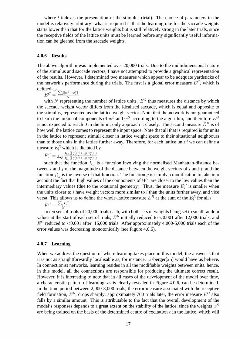
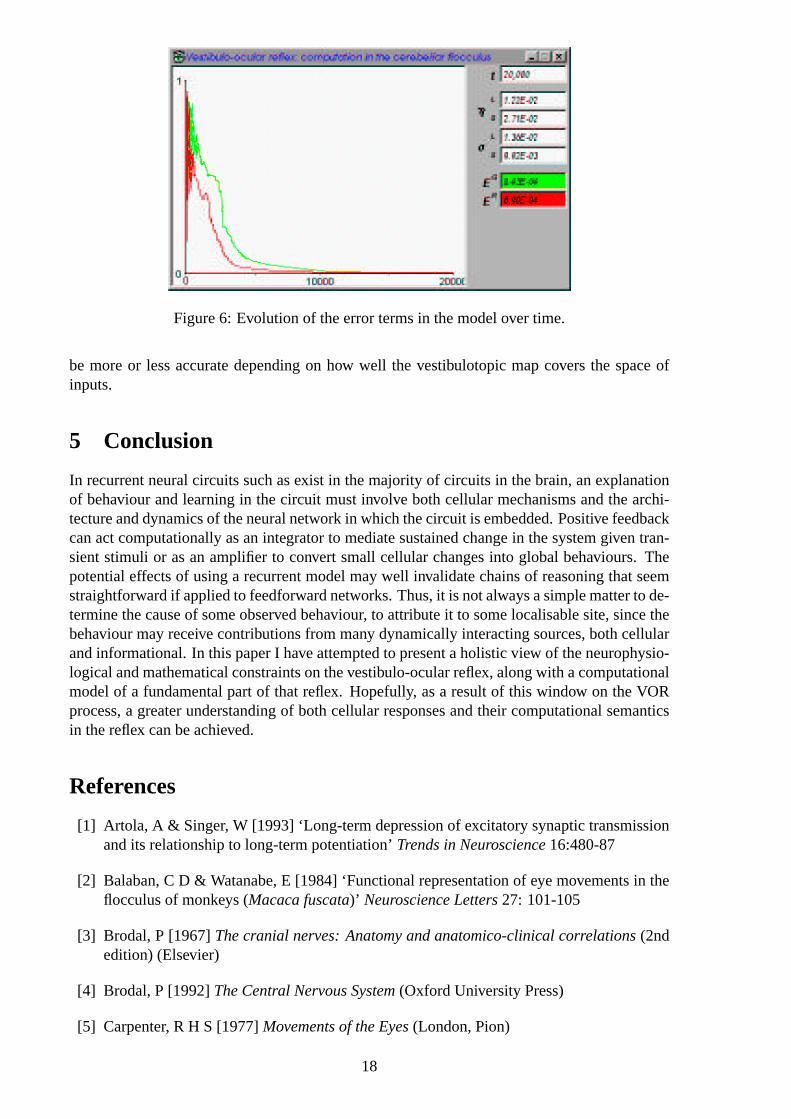
In ten sets of trials of 20,000 trials each, with both sets of weights being set to small randomvalues at the start of each set of trials,ER initially reduced to<0.001 after 12,000 trials, andEG reduced to<0.001 after 16,000 trials. After approximately 4,000-5,000 trials each of theerror values was decreasing monotonically (see Figure 4.0.6).
4.0.7 Learning
When we address the question of where learning takes place in this model, the answer is thatit is not as straightforwardly localisable as, for instance, Lisberger[25] would have us believe.In connectionist networks, learning resides in all the modifiable weights between units, hence,in this model, all the connections are responsible for producing the ultimate correct result.However, it is interesting to note that in all cases of the development of the model over time,a characteristic pattern of learning, as is clearly revealed in Figure 4.0.6, can be determined.In the time period between 2,000-3,000 trials, the error measure associated with the receptivefield formation,ER, drops sharply; approximately 700 trials later, the error measureEG alsofalls by a similar amount. This is attributable to the fact that the overall development of themodel’s responses depends to a great extent on the stability of the lattice, since the weightswS
are being trained on the basis of the determined centre of excitationc in the lattice, which will
17
Figure 6: Evolution of the error terms in the model over time.
be more or less accurate depending on how well the vestibulotopic map covers the space ofinputs.
5 Conclusion
In recurrent neural circuits such as exist in the majority of circuits in the brain, an explanationof behaviour and learning in the circuit must involve both cellular mechanisms and the archi-tecture and dynamics of the neural network in which the circuit is embedded. Positive feedbackcan act computationally as an integrator to mediate sustained change in the system given tran-sient stimuli or as an amplifier to convert small cellular changes into global behaviours. Thepotential effects of using a recurrent model may well invalidate chains of reasoning that seemstraightforward if applied to feedforward networks. Thus, it is not always a simple matter to de-termine the cause of some observed behaviour, to attribute it to some localisable site, since thebehaviour may receive contributions from many dynamically interacting sources, both cellularand informational. In this paper I have attempted to present a holistic view of the neurophysio-logical and mathematical constraints on the vestibulo-ocular reflex, along with a computationalmodel of a fundamental part of that reflex. Hopefully, as a result of this window on the VORprocess, a greater understanding of both cellular responses and their computational semanticsin the reflex can be achieved.
References
[1] Artola, A & Singer, W [1993] ‘Long-term depression of excitatory synaptic transmissionand its relationship to long-term potentiation’Trends in Neuroscience16:480-87
[2] Balaban, C D & Watanabe, E [1984] ‘Functional representation of eye movements in theflocculus of monkeys (Macaca fuscata)’ Neuroscience Letters27: 101-105
[3] Brodal, P [1967]The cranial nerves: Anatomy and anatomico-clinical correlations(2ndedition) (Elsevier)
[4] Brodal, P [1992]The Central Nervous System(Oxford University Press)
[5] Carpenter, R H S [1977]Movements of the Eyes(London, Pion)
18
[6] Churchland, P S [1989]Neurophilosophy - Toward a Unified Science of the Mind/Brain(MIT Press)
[7] Churchland, P S & Sejnowski, T [1990]The Computational Brain(MIT Press)
[8] Dufosse, M, Ito, M & Miiyashita, Y [1977] ‘Functional localization in the rabbit’s cere-bellar flocculus determined in relationship with eye movements’Neuroscience Letters5:273-277
[9] Demer, J L [1992] ‘Mechanisms of Human Vertical Visual-Vestibular Interaction’Journalof Neurophysiology68, 6: 2128-2146
[10] du Lac, S, Raymond, J L, Sejnowski, T J & Lisberger, S G [1995] ‘Learning and Memoryin the Vestibulo-Ocular Reflex’Annual Review of Neuroscience18: 409-441
[11] Escudero, M, Dewaele, C, Vibert, N, Berthoz, A & Vidal, P P [1993] ‘Saccadic Eye-Movements and the Horizontal Vestibuloocular and Vestibulo-collic Reflexes in the IntactGuinea-Pig’Experimental Brain Research97, 2: 254-262
[12] Gilbert, C D [1983] ‘Microcircuitry of the visual cortex’Annual Review of Neuroscience6: 217-47
[13] Guitton, D, Munoz, D P & Galiana, H L [1990] ‘Gaze Control in the Cat: Studies andModeling of the Coupling between Orienting Eye and Head Movements in Different Be-havioral Tasks’Journal of Neurophysiology64(2): 509-531
[14] Haslwanter, T [1995] ‘Mathematics of Three-dimensional Eye Rotations’Vision Research35, 12: 1727-1739
[15] Hubel, D H & Wiesel, T N [1974] ‘Sequence Regularity and Geometry of OrientationColumns in the Monkey Striate Cortex’Journal of Comparative Neurology158: 267-294
[16] Ito, M [1972] ‘Neural design of the cerebellar motor control system’Brain Research40:80-84
[17] Ito, M [1982] ‘Cerebellar control of the vestibulo-ocular reflex - around the flocculushypothesis’Annual Review of Neuroscience5:275-298
[18] Ito, M [1984] The cerebellum and neural control(Raven Press)
[19] Ito, M, Orlov, I & Yamamoto, M [1982] ‘Topographical representation of vestibulo-ocularreflexes in rabbit cerebellar flocculus’Neuroscience7: 1657-1664
[20] Kaas, J H, Merzenich, M M & Killackey, H P [1983] ‘The Reorganization of Somatosen-sory Cortex Following Peripheral Nerve Damage in Adult and Developing Mammals’Annual Review of Neuroscience6: 325-356
[21] Knox, P C & Donaldson, I M L [1995] ‘The effect of afferent signals from extraocularmuscles on visual responses of cells in the optic tectum of the pigeon’Proceedings of theRoyal Society, LondonB 259: 285-291
[22] Langer, T, Fuchs, A F, Chubb, M C, Scudder, C A & Lisberger, S G [1985] ‘Floccular ef-ferents in the rhesus macaque as revealed by autoradiography and horseradish peroxidase’Journal of Comparative Neurology235: 26-37
[23] Leigh, R J & Zee, D S [1991]The Neurology of Eye Movements(Philadelphia) (2ndedition)
19
[24] Lindeman, H H [1992] ‘Anatomy of the Otolith organs’Advances in Oto-Laryngology20: 405-433
[25] Lisberger, S G [1994] ‘Neural Basis for Motor Learning in the Vestibuloocular Reflex ofPrimates 3. Computational and Behavioral-Analysis of the Sites of Learning’Journal ofNeurophysiology72, 2: 974-998
[26] Lisberger, S G & Fuchs, A F [1974] ‘Response of flocculus Purkinje cells to adequatevestibular stimulation in the alert monkey: fixation vs. compensatory eye movements’Brain Research69: 347-353
[27] Lisberger, S G & Pavelko, T A [1988] ‘Brainstem neurons in modified pathways for motorlearning in the primate vestibulo-ocular reflex’Science242: 771-773
[28] Lisberger, S G, Pavelko, T A, Brontestewart, H M & Stone, L S [1994a] ‘Neural Basis forMotor Learning in the Vestibuloocular Reflex of Primates 2. Changes in the Responses ofHorizontal Gaze Velocity Purkinje-Cells in the Cerebellar Flocculus and Ventral Parafloc-culus’Journal of Neurophysiology72, 2: 954-973
[29] Lisberger, S G, Pavelko, T A & Broussard, D M [1994b] ‘Neural Basis for Motor Learningin the Vestibuloocular Reflex of Primates 1. Changes in the Responses of Brain-StemNeurons’Journal of Neurophysiology72, 2: 928-953
[30] Maxwell, J S & Schor, C M [1996] ‘Adaptation of Vertical Eye Alignment in Relation toHead Tilt’ Vision Research36, 8: 1195-1205
[31] Miles, F A, Braitman, D J & Dow, B M [1980] ‘Long-term adaptive changes in primatevestibuloocular reflex IV. Electrophysiological observations in flocculus of adapted mon-keys’Journal of Neurophysiology43: 1477-1493
[32] Mira, E [1995] ‘General View of Vestibular Disorders’Acta Oto-LarygologicaS519: 13-16
[33] Misslisch, H, Tweed, D, Fetter, M, Sievering, D & Koenig, E [1994] ‘Rotational Kinemat-ics of the Human Vestibuloocular Reflex III. Listing’s Law’Journal of Neurophysiology72, 5: 2490-2502
[34] Rang, H P, Dale, M M, & Ritter, J M [1995]Pharmacology(Churchill Livingstone) (3rdedition)
[35] Robinson, D A [1964] ‘The mechanics of human saccadic eye movement’Journal ofPhysiology, London174:245-264
[36] Robinson, D A [1989] ‘Integrating with Neurons’Annual Review of Neuroscience12:33-45
[37] Scudder, C A & Fuchs, A F [1992] ‘Physiological and behavioral identification of vestibu-lar nucleus neurons mediating the horizontal vestibuloocular reflex in trained rhesus mon-keys’Journal of Neurophysiology68: 244-64
[38] Seidman, S H, Leigh, R J, Tomsak, R L, Grant, M P & del’Osso, L F [1995] ‘DynamicProperties of the Human Vestibulo-ocular Reflex During Head Rotations in Roll’VisionResearch35, 5: 679-689
[39] Shelhamer, M, Tiliket, C, Roberts, D, Kramer, P D & Zee, D S [1994] ‘Short-TermVestibuloocular Reflex Adaptation in Humans II. Error Signals’Experimental Brain Re-search100, 2: 328-336
20
[40] Tiliket, C, Shelhamer, M, Roberts, D & Zee, D S [1994] ‘Short-Term Vestibule-OcularReflex Adaptation in Humans I. Effect on the Ocular Motor Velocity-to-Position NeuralIntegrator’Experimental Brain Research100, 2: 316-327
[41] Tweed, D, Cadera, W & Vilis, T [1990] ‘Geometric relation of eye position and velocityvectors during saccades’Vision Research30: 111-127
[42] Tweed, D, Fetter, M, Sievering, D, Misslisch, H & Koenig, E [1994a] ‘Rotational Kine-matics of the Human Vestibuloocular Reflex II. Velocity Steps’Journal of Neurophysiol-ogy72, 5: 2480-2489
[43] Tweed, D, Sievering, D, Misslisch, H, Fetter, M, Zee, D & Koenig, E [1994b] ‘RotationalKinematics of the Human Vestibuloocular Reflex I. Gain Matrices’Journal of Neurophys-iology72, 5: 2467-2479
[44] van der Steen, J, Simpson, J I & Tan, J [1994] ‘Functional and Anatomic Organizationof Three-Dimensional Eye Movements in Rabbit Cerebellar Flocculus’Journal of Neu-rophysiology72, 1: 31-46
[45] von Helmholtz, H [1867]Handbuch der Physiologischen Optik(Hamburg: Voss)
[46] Wiesel, T N & Hubel, D H [1974] ‘Ordered Arrangement of Orientation Columns inMonkeys Lacking Visual Experience’Journal of Comparative Neurology158: 307-318