The Hog1 Stress-activated Protein Kinase TargetsNucleoporins to Control mRNA Export upon Stress*
Received for publication, December 11, 2012, and in revised form, April 27, 2013 Published, JBC Papers in Press, May 3, 2013, DOI 10.1074/jbc.M112.444042
Sergi Regot‡1, Eulàlia de Nadal‡1,2,3,4, Susana Rodríguez-Navarro§5, Alberto González-Novo‡,Jorge Pérez-Fernandez¶6, Olivier Gadal¶6, Gerhard Seisenbacher‡, Gustav Ammerer�, and Francesc Posas‡2,3,7
From the ‡Cell Signaling Unit, Departament de Ciències Experimentals i de la Salut, Universitat Pompeu Fabra, E-08003 Barcelona,Spain, §Centro de Investigación Principe Felipe, Primo Yúfera, E-46012 Valencia, Spain, ¶Laboratoire de Biologie MoleculaireEucaryote du CNRS, Université de Toulouse, 118 route de Narbonne, F-31000 Toulouse, France , and the �Department forBiochemistry, Max F. Perutz Laboratories, University of Vienna, Dr. Bohrgasse 9, A-1030 Vienna, Austria
Background: The Hog1 stress-activated protein kinase (SAPK) regulates mRNA biogenesis in response to osmostress.Results: The nuclear pore complex is regulated upon osmostress by the Hog1 SAPK.Conclusion:mRNA export is controlled by the HOG signaling pathway.Significance: Efficient mRNA biogenesis of stress-responsive genes requires of the coordination between synthesis andmRNAexport.
The control ofmRNAbiogenesis is exerted at several steps. Inresponse to extracellular stimuli, stress-activated proteinkinases (SAPK) modulate gene expression to maximize cell sur-vival. In yeast, the Hog1 SAPK plays a key role in reprogram-ming the gene expression pattern required for cell survival uponosmostress by acting during transcriptional initiation and elon-gation. Here, we genetically show that an intact nuclear porecomplex is important for cell survival and maximal expressionof stress-responsive genes. The Hog1 SAPK associates withnuclear pore complex components and directly phosphorylatesthe Nup1, Nup2, and Nup60 components of the inner nuclearbasket.Mutationof those factors resulted in adeficient export ofstress-responsive genes upon stress. Association ofNup1,Nup2,and Nup60 to stress-responsive promoters occurs upon stressdepending onHog1 activity. Accordingly, STL1 gene territory ismaintained at the nuclear periphery uponosmostress in aHog1-dependent manner. Cells containing non-phosphorylatablemutants in Nup1 or Nup2 display reduced expression of stress-responsive genes. Together, proper mRNA biogenesis of stress-responsive genes requires of the coordinate action of synthesisand export machineries by the Hog1 SAPK.
In response to stress, cells regulate gene expression to maxi-mize cell survival. Cellular signaling cascades regulate specific
transcriptional activities that convert extracellular informationinto modulation of gene expression (1, 2). Activation of theyeast HOG8 (high osmolarity glycerol) signaling pathway inresponse to osmostress results in a transient and acute regula-tion of the transcriptional program of the cell (3). The centralcore of the HOG pathway consists of a tier of kinases highlyconserved among eukaryotic cells that results in the phosphor-ylation and nuclear accumulation of the Hog1 stress-activatedprotein kinase (SAPK) when extracellular stimuli is sensed(4–6). Transcription profiling studies together with bindinganalyses of Hog1-dependent transcription factors have shownthat Hog1 is a major regulator of the osmostress-regulatedtranscription that involves a complex and highly specific net-work of genes (7–15).The Hog1 SAPK regulates gene expression at multiple levels
(3). The SAPK directly phosphorylates several unrelated tran-scription factors such as Sko1, Hot1, Smp1, or Rtg1/Rtg3 (16–21). Moreover, in response to stress, the kinase associates withseveral target loci through physical interaction with specifictranscription factors (22, 23). Once stably associatedwith chro-matin, Hog1 has diverse functions in the stimulation of geneexpression. Recruitment of Hog1 to promoters is essential tocoordinate chromatin remodeling and assembly of the tran-scriptional machinery upon stress (19, 23–26). Moreover, theSAPK physically interacts with elongating RNA polymerase IIand cross-links to osmostress-transcribed regions allowing forefficient histone eviction during transcriptional elongation(27–29). In addition, Hog1 has also been involved in the controlof mRNA stability as well as in the translation of stress-respon-sive genes (13, 14, 30, 31).The nuclear pore complex (NPC) mediates nucleocytoplas-
mic transport of macromolecules in all eukaryotes (for review,see Refs. 32–34). NPCs have largely symmetrical, doughnut-shape structures that lie within a pore connecting the inner and
* This work was supported by MINECO (Spanish government) GrantBFU2012-33503, the Consolider Ingenio 2010 program (Grant CSD2007-0015), the Fundación Marcelino Botín (to F. P.), and Grant BFU2011-26722(to E. d. N.).Author’s Choice—Final version full access.
1 Both authors contributed equally to this work.2 Both are co-senior authors.3 Recipients of an ICREA Acadèmia (Generalitat de Catalunya).4 To whom correspondence may be addressed. Tel.: 34-93-316-0849; Fax:
34-93-316-0901; E-mail: [email protected] Supported by the MINECO Grant BFU2011-23418.6 Supported by Agence Nationale de la Recherche (Nucleopol and
ODynRib-Jeune chercheur program) and Jeune équipe from Fondationpour la Recherche Médicale.
7 To whom correspondence may be addressed. Tel.: 34-93-316-0849; Fax:34-93-316-0901; E-mail: [email protected].
8 The abbreviations used are: HOG, high osmolarity glycerol; NPC, nu-clear pore complex; YPD, yeast extract/peptone/dextrose; qV,quadruple-Venus; TAP, tandem affinity purification.
outer layers of the nuclear envelope. In the yeast Saccharomycescerevisiae the NPC is 50 MDa in size and consists of multiplecopies of �30 different proteins called nucleoporins (Nups)positioned in a flexible structure (34–37). Not all nucleoporinsare equally accessible, and they have completely different rolesdepending on their location at the scaffold, the nuclear basket,or the cytoplasmic filaments (for review, see Refs. 34 and 38).Besides trafficking of macromolecules, the nuclear pore has
been proposed to exert a direct role in gene expression throughphysical interactions of Nups with the nuclear genome; indeed,recent work in yeast has revealed that some genes can undergodynamic recruitment to the periphery upon transcriptionalactivation (for review, see Refs. 39–42). Localization of individ-ual geneswithin the nucleus can be dynamically controlled, andmany inducible yeast genes such as theGAL genes rapidly relo-calize from the nucleoplasm to the nuclear periphery after acti-vation by their association with Nups (43–47). Other examplesof nuclear pore components interacting with active genes dur-ing the initial steps of transcription are described for INO1 (48–50), RAP1/GCR1/GCR2 genes (51), or �-factor-induced genes(52). Although many studies pointed out that recruitment tothe nuclear periphery appears to have a functional role in pro-moting transcriptional activation, gene anchoring may not be ageneral requirement for gene expression (53, 54). For instance,it was reported that repressive environment at the nuclearperiphery establishes a negative feedback loop that enables theGAL locus to respond rapidly to changes in environmental con-ditions (55).In this study we show that nuclear pore integrity is essential
formaximal gene expression and adaptation to osmostress. TheHog1 SAPK interacts with and phosphorylates specific compo-nents of the NPC to facilitate mRNA export upon osmostress.Therefore, the Hog1 SAPK regulates several steps in mRNAbiogenesis from initiation tomRNAexport, whichmight definea dedicated path for optimal expression of osmostress-respon-sive genes.
EXPERIMENTAL PROCEDURES
Yeast Strains—S. cerevisiae strain BY4741 (MATa his3-�1leu2-�0 met15-�0 ura3-�0) and its derivatives (nup42,nup100, nup53, nup157, seh1, nup188, nup170, nup133,nup120, nup84, nup2, nup60, pom34, pom152, nup59, nup1,and gle2) as well as TAP-tagged (Nup42, Nup100, Nup53,Nup157, Seh1, Nup188, Nup170, Nup133, Nup120, Nup84,Nup2, Nup60, Pom34, Pom152, Nup59, Nup1, Gle2, Nic96,Nup116, Nup49, Nup159, Nup57, Nup85, Nup192, Nup145,Gle1, and Nup82) and GFP-tagged (Nup1, Nup2, and Nup60)strains were obtained from the EROSCARF collection. Forphospho-mass spectrometry, NUP2-TAP::HIS3 HOG1::kanMX4was obtained. For localization studies, strains were HOT1-GFP::HIS3 with NUP2::kanMX4, NUP60 ::kanMX4, andNUP188::kanMX4 (YAG200, YAG201, and YAG202), MSN2-GFP::HIS3 with NUP2::kanMX4, NUP60::kanMX4, andNUP188::kanMX4 (YAG203, YAG204, and YAG205) andSKO1-GFP::HIS3 with NUP2::kanMX4, NUP60 ::kanMX4,and NUP188::kanMX4 (YAG206, YAG207, and YAG208). Tag-ging strains obtained in this study were YSR43 (NUP1-6HA::HIS3), YSR44 (NUP1-6HA::HIS3HOG1::kanMX4), YSR218
(NUP60-6HA::HIS3), YSR221 (NUP60-HA::HIS3 HOG1::kanMX4), YSR38 (NUP2-6HA::HIS3), YSR47 (NUP2-6HA::HIS3 HOG1 ::kanMX4), and YAG209 (CSE2-6HA::KanMX4). Tagging of genomic ORFs with HA epitope wasdone with a PCR-based strategy. Strains used for FACS cytom-etry analysis were YSR237 (NUP2::kanMX4 STL1-qVenus::LEU2 pRS413), YSR238 (NUP2::kanMX4 STL1-qVenus::LEU2 pRS413-Nup2), YSR239 (NUP2::kanMX4 STL1-qVenus::LEU2 pRS413-Nup2T361A), YSR325 (NUP1::kanMX4STL1-qVenus::LEU2 pRS413), YSR240 (NUP1::kanMX4STL1-qVenus::LEU2 pRS413-Nup1), and YSR273 (NUP1::kanMX4 STL1-qVenus::LEU2 pRS413-Nup1S11A/T159A/S161A).Plasmids—The plasmids used in this study were pGEX4T-
Hog1 and pGEX4T-Pbs2EE (PBS2with S514E andT518Emuta-tions), which are described in Bilsland-Marchesan et al. (56).NUP1 (PSR19), NUP1S11AT159AS161A (PSR45), NUP2 (PSR3),NUP2T361A (PSR44), and NUP60 (PSR21) were cloned intopGEX4T. RPN2 was cloned into pGEX6P.1 (pAG36). NUP1(pSR60), NUP1S11A/T159A/S161A (pSR77), NUP2 (pSR57), andNUP2T361A (pSR66) were cloned into pRS413. STL1-qVenusconstruct is described in Pelet et al. (57). pRS416 HOG-GFP isdescribed in Ferrigno et al. (6).Northern Blot Analysis—Yeast strains were grown tomid-log
phase in rich medium at an absorbance at 660 nm of 0.6–0.9and then subjected to osmostress (0.4 or 1 M NaCl) for thelength of time indicated in Figs. 2 and 3. Total RNA and expres-sion of specific genes were probed using radiolabeled PCR frag-ments containing the entire ORF of STL1 (1.7 kbp), CTT1 (1.7kbp), GRE2 (1.0 kbp), and the non-coding exon of RDN18 (1.8kbp). Signals were quantified with phosphorimaging (FujifilmBAS-5000), and autoradiographs were obtained on Kodak Bio-max XAR film (Sigma).GST Pulldown Experiments—Cells expression expressing
GST or GST-Hog1 and specific TAP-tagged proteins grown inmid-log phasewere treatedwith 0.4 MNaCl for 10min and thencollected by brief centrifugation at 4 °C. Pellets were harvestedwith glass beads in the FastPrep-24 (Qbiogene) in lysis buffer A(50 mM Tris-HCl, pH 7.5, 150 mM NaCl, 15 mM EDTA, 15 mM
EGTA, 2 mM DTT, 0.1% Triton X-100, 1 mM PMSF, 1 mM
benzamidine, 2 mg/ml leupeptin, 2 mg/ml pepstatin, 25mM �-glycerophosphate, 1 mM sodium pyrophosphate, 10 mM
sodium fluoride, 100 mM sodium orthovanadate), and lysateswere clarified by centrifugation and quantified by the Bradfordassay (Bio-Rad). As a control, 25 mg of whole-cell extract wasblotted with anti-GST antibody to check the expression levelsof the tagged proteins. Alternatively, 2 mg of cleared superna-tant was incubated with 50 �l of glutathione-Sepharose beads(GE Healthcare) overnight at 4 °C. Beads were washed exten-sively with buffer A, resuspended in loading buffer, andresolved by sodium dodecyl sulfate-polyacrylamide gel electro-phoresis. PAP antibody (Sigma) was used to detect the TAP-tagged proteins.Kinase Assay—The GST fusion proteins encoding NUPs,
Hog1, and Pbs2EE were expressed in Escherichia coliDH5� andpurified using glutathione-Sepharose beads (GE Healthcare) inSTET buffer (10 mM Tris-HCl, pH 8.0, 100 mM NaCl, 1 mM
g/ml pepstatin). A 1-�g sample of Hog1 was activated with 0.5�g of Pbs2EE in the presence of kinase buffer (50 mM Tris-HClpH 7.5, 10 mM MgCl2, 2 mM DTT) and 50 mM ATP. After 20min at 30 °C, eluted NUP protein was added to the previousmixture together with [�-32P]ATP (0.1mCi/ml) and incubatedfor 15 min at 30 °C. The reaction was terminated by the addi-tion of SDS-loading buffer. Labeled proteins were resolved bySDS/PAGE, stained, dried, and detected by autoradiography.TAP Purification—27 TAP tagged strains were grown to
mid-log exponential phase in 250 ml of YPD. Protein extractswere extracted in buffer A as above and incubated with ProtA-Sepharose beads (Sigma) for 2 h at 4 °C. Beads were washedfour times with Buffer A and four times with kinase buffer.Kinase assay was performed with Pbs2EE-activated GST-Hog1 as above. The reaction was stopped by the addition ofSDS-loading buffer. Labeled proteins were resolved by SDS/PAGE, transferred for Western blotting, and detected byautoradiography.Phospho-mass Spectrometry—NUP2-TAP-tagged wild type
and hog1� strains were grown in 1000 ml of YPD to A660 2.Unstressed and stressed (0.4 M NaCl, 5 min) cultures were har-vested, and pellets were frozen on dry ice. The pellet was resus-pended in 8 ml of lysis buffer supplied with protease and phos-phatase inhibitors, and standard glass bead lysiswas performed.Lysates were cleared by spinning 45 min at 4750 rpm at 4 °C.0.5-ml IgG beads were added, and the sample was incubated for120min at 4 °C. IgG beadswere subsequently washed 3 times inlysis buffer supplied with 0.5 M NaCl followed by 3 washes with1� Tris/DTT. Purified TAP-tagged NUP2 was eluted with 3.5M MgCl2 (1st elution 0.5 ml, 2nd elution 1 ml, and 3rd elution0.5 ml). The eluate was immediately diluted by adding 3 mlTris/DTT and concentrated in a Centricon column to0.5ml, rediluted with 3.5ml Tris/DTT, and concentrated to 0.5ml. The concentrated eluate was precipitated by adding 2 ml ofice-cold acetone, incubated overnight at �20 °C, and pelletedby 14.000 rpm centrifugation for 10 min. Pellet was air-driedand stored at �80 °C. Phosphopeptide enrichment was per-formed according to Zhou et al. (58). No fractionation step wasincluded. 50–100 �g of purified protein was trypsinized andsubjected to phosphopeptide enrichment. Phospho-enrichedsamples were analyzed in a LTQ-Orbitrap, and results wereprocessed with MaxQuant (59). In the case of double-phos-phorylated peptides, only intensities of the respective singlephosphorylation events that could be unambiguously mappedwere analyzed in this study.In Situ Hybridization of Yeast Cells—Localization of STL1
transcripts was performed following the detailed protocol fromthe Singer laboratory (64). 5�Cy3-end-labeled 60-bp oligonu-cleotides were designed covering the ORF region of STL1 gene.Wild type, mex67-5, nup2�, and nup60� cells were grown toA660 0.5. To inactivate Mex67, the thermosensitive mutantmex67-5 was shifted to 37 °C for 20 min. NaCl was added to afinal concentration of 0.4 M and incubated for 20min. After salttreatment, the cultures were fixedwith formaldehyde, and eachstrain was used to detect STL1 RNA specifically. Pictures weretaken using a Leica DM6000B fluorescence microscope with a63� PL APO objective.
Chromatin Immunoprecipitation—Chromatin immunopre-cipitation was performed as described previously (25). Yeastcultures were grown to early log phase (A660 0.6–1.0) beforealiquots of the culture were exposed to osmostress (0.4 or 1.2 M
NaCl) for the length of time specified in legends. For cross-linking, yeast cells were treated with 1% formaldehyde for 20min at room temperature. Antibody used in this study wasmonoclonal anti-HA 12CA5. Quantitative PCR analysis ofstress genes and constitutively expressed genes used the follow-ing primers with locations indicated by the distance from therespective ATG initiation codon: STL1 (�372/�112 and�1000/�1280),CTT1 (�432/�302 and�736/�836), andTEL(telomeric region on the right arm of chromosome VI). Exper-iments were done on three independent chromatin prepara-tions, and quantitative PCR analysis was done in real time usingan Applied Biosystems 7700 sequence detector. Immunopre-cipitation efficiency was calculated in triplicate by dividing theamount of PCR product in the immunoprecipitated sample bythat in the TEL sequence control. The binding data are pre-sented as -fold induction with respect to the non-treatedcondition.
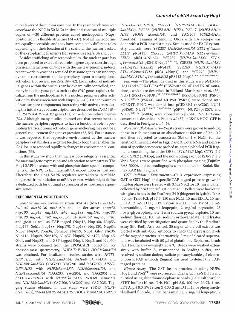
FIGURE 1. NPC is essential for growth upon osmostress. Mutations on spe-cific NPC components render cells osmosensitive. Wild type (wt) and the indi-cated mutant strains were grown to mid-log exponential phase and spotted,making serial dilutions in YPD plates without or with 1.2 M NaCl or 2 M sorbitol.Growth at 30 °C was scored after 3 days.
Control of mRNA Export by Hog1
17386 JOURNAL OF BIOLOGICAL CHEMISTRY VOLUME 288 • NUMBER 24 • JUNE 14, 2013
StatisticalMapping of STL1Gene Territory—In vivo localiza-tion of STL1 locus was performed as previously described (60).Briefly, a mCherry-Nop1 Nup49GFP TetRGFP strain was usedto introduce an array of TetRDNAbinding sites downstreamofthe STL1 locus by homologous recombination. Agar patches ofrich media with or without 0.4 M NaCl were used to image cellsin cover slides sealed with VaLaP (1/3 Vaseline�, 1/3 lanoline,1/3 paraffin). Confocal microscopy was limited to 20 min aftermounting and using a confocal microscope with an incubatorfor live cell experiments (The Cube and The Box; Life ImagingService) at 30 °C and performedwith anAndor RevolutionNip-kow-disk confocal system installed on a Olympus IX-81 featur-ing aYokogawaCSU22 confocal spinning disk unit and a cooledAndor EMCCD camera (DU 888). The system was controlledusing the mode “Revolution FAST” of Andor Revolution IQsoftware (Andor Technology). Images were acquired using anOlympus 100� objective (Plan APO, 1.4 NA, oil immersion).Single laser lines used for excitation were diode-pumped solid
state lasers (DPSSL) exciting GFP fluorescence at 488 nm (50milliwatts, Coherent) and mCherry fluorescence at 561 nm (50milliwatts, Cobolt JiveTM); a Semrock bi-bandpass emission fil-ter (Em01-R488/568–15) allowed collection of green and redfluorescence. Pixel size was 65 nm. For three-dimensional anal-ysis, Z-stacks of 41 images with a 250-nm Z-step were used.Exposure time was 200 ms. Image analysis was done withMATLABby aligning nuclei using the axis betweennucleus andnucleolus three-dimensional centroids. Between 2000 and 3000cells were analyzed (wild type control, n � 3047 cells; wild typeNaCl, n� 2762 cells; hog1� control, n� 2882 cell; hog1�NaCl,n � 2195). The statistical analyses (p value) presented in thefigure correspond to a Kolmogorov-Smirnov test testing forequal distribution.Flow Cytometry—Saturated overnight cultures grown in syn-
theticmediumweredilutedandgrown for24hat logphase (belowA660 0.4). Cells were stressed bymixing 200�l of culture with 100�l of stress solution (SDmedium�NaCl). Protein translationwas
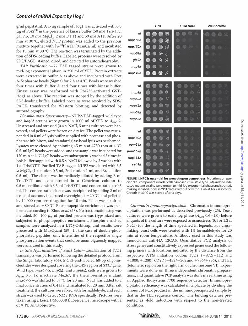
FIGURE 2. NPC is essential for gene expression upon osmostress. WT and the indicated viable NPC mutant strains were grown to mid-log phase in richmedium and subjected to 0.4 M NaCl or 1.2 M NaCl osmostress for the indicated lengths of time. Total RNA was extracted and blotted against STL1 and CTT1 andRDN18 as the loading control.
Control of mRNA Export by Hog1
JUNE 14, 2013 • VOLUME 288 • NUMBER 24 JOURNAL OF BIOLOGICAL CHEMISTRY 17387
FIGURE 3. NPC is essential for gene expression upon osmostress. WT and the indicated strains were grown to mid-log phase in rich medium and thensubjected to osmostress (1 M NaCl) for the indicated length of time. Total RNA was extracted and assayed by Northern blot for transcript levels of STL1, CTT1, andGRE2 and RDN18 as the loading control.
FIGURE 4. Localization of Hog1 or transcription factors Hot1, Msn2, and Sko1 is not altered in NPC mutant strains. A, Hog1 localization was not affectedin NPC mutant strains. WT and the indicated NPC mutant strains expressing Hog1-GFP were grown in SD medium to mid-log phase and subjected toosmostress (0.4 M NaCl, 5 min). DAPI staining to reveal nuclei and epifluorescence pictures were taken without (basal conditions) and with stress. Thepercentage of cells with Hog1 nuclear accumulation upon stress is shown in the lower panel; data are the mean and S.D. of three independent experiments. B,Hot1, Msn2, and Sko1 localization is not affected in NPC mutant strains. WT and the indicated NPC mutant strains expressing Hot1-GFP, Mns2-GFP, andSko1-GFP were grown in SD medium to mid-log phase and subjected to osmostress (0.4 M NaCl, 5 min). Nomarsky and epifluorescence pictures were takenwithout (�) and with stress (�). DAPI staining to reveal nuclei in the wild type strain upon stress is shown in the lower panel.
Control of mRNA Export by Hog1
17388 JOURNAL OF BIOLOGICAL CHEMISTRY VOLUME 288 • NUMBER 24 • JUNE 14, 2013
stoppedafter45minby theadditionof cycloheximide (0.1mg/ml).Cells were briefly sonicated, and fluorescence was measured byflow cytometry (FACSCalibur, BD Biosciences).
RESULTSNuclear Pore Integrity Is Essential for Growth and Gene
Expression upon Osmostress—We performed an exhaustivegenome-wide genetic screening searching for mutations thatrender cells sensitive at high osmolarity to systematically iden-tify novel activities required for full expression of the gene pro-gram required for cell survival upon osmostress. This screenidentified several transcriptional-related complexes such as theRpd3 histone deacetylase complex, SAGA,Mediator, the Ubp3ubiquitin protease, and components of the TEC as well as theRSC complex that are important for gene expression in
response to osmostress (24–26, 61). In addition, we also foundthat mutations in three genes encoding components of the NPCyielded cells osmosensitive (NUP120, NUP170, and NUP188).Then, we systematically extended our screen and monitored cellgrowth in the presence of high osmolarity in all viable nullmutantyeast strains deficient in components of theNPC (17 in total).Weassessed the sensitivity of all those strains in 1.2 M NaCl and 2 M
sorbitol and found that severalmutants incomponentsof theNPCcomplex (nup188�, nup170�, nup84�, gle2�, nup1�, andnup120�) were sensitive to osmostress (Fig. 1). The mutationsincluded nucleoporins acting as adaptors, coat, cytoplasmic fila-ments, and components of the nuclear basket.We then also assessed systematically whether mutations in
components of the NPC resulted in reduced osmostress gene
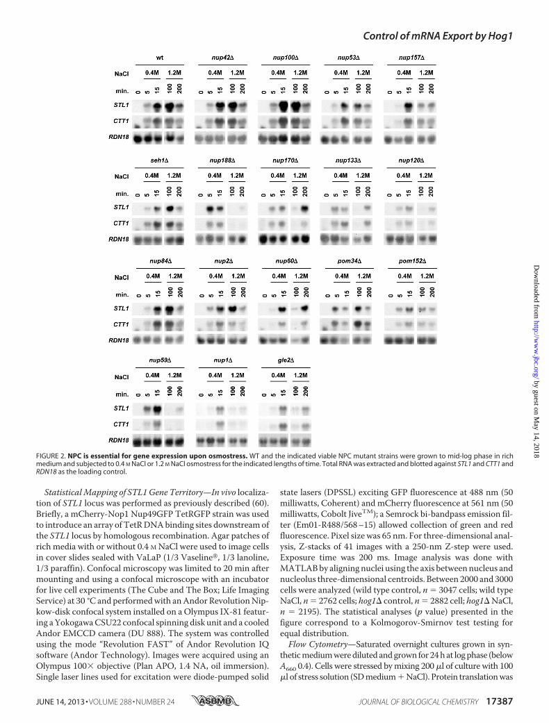
FIGURE 5. Hog1 co-precipitates with the NPC and phosphorylates some of its components. A, in vivo binding of Hog1 and the NPC is shown. TAP-taggedNup188, Nup84, Nup82, Nup60, or Nup2 strains expressing GST or GST-Hog1 were grown to mid-log phase and subjected to brief osmotic shock (0.4 M NaCl,10 min). GST proteins were pulled down by glutathione-Sepharose 4B, and the presence of TAP-tagged proteins was probed by immunoblotting usinganti-TAP (PAP; Sigma) (upper panel). Total extract represents �20% of total input protein (middle panel). Amounts of precipitated (prec.) GST proteins weredetected using anti-GST (lower panel). B, GST-Hog1 and Nup2-HA or Nup60-HA reciprocal immunoprecipitations are shown. Nup2-HA and Nup60-HA co-immunoprecipitates with GST-Hog1 in a GST pulldown assay (left panels). Cse2-HA was added as negative control. Correspondingly, Hog1 co-immunoprecipi-tates with Nup2-HA or Nup60-HA in a HA pulldown assay (right panels). C, in vitro phosphorylation of Nup2, Nup60, and Nup1 by Hog1 is shown. RecombinantGST-fused proteins from E. coli were phosphorylated in vitro by Hog1. A non-relevant protein (GST-tagged Rpn2) was added as a control to exclude possiblenonspecific phosphorylation of Nups by Hog1. Phosphorylated proteins were resolved by SDS-PAGE and detected by autoradiography (upper panel). Totalprotein levels were detected by staining with Coomassie Brilliant Blue (lower panel).
Control of mRNA Export by Hog1
JUNE 14, 2013 • VOLUME 288 • NUMBER 24 JOURNAL OF BIOLOGICAL CHEMISTRY 17389
expression.We found that expression of osmoresponsive genessuch as STL1, CTT1, and GRE2 was significantly affected ordelayed under severe osmostress in nup188�, nup170�, gle2�,nup120�, nup133�, nup59�, nup157�, nup1�, and nup60�mutant strains (Figs. 2 and 3). Of note, localization of Hog1upon stress was not altered in any of the mutant strains tested(Fig. 4A). Again, all those mutations included componentspresent in diverse domains of theNPC.The expression of STL1,CTT1, andGRE2 is driven by different transcription factors (i.e.Hot1, Msn2/Msn4, and Sko1, respectively) and thus indicate ageneral defect on stress-responsive mRNA accumulationrather than a defect associatedwith a given transcription factor.Of note, localization of Sko1, Msn2, or Hot1 (fused to GFP) inall nucleoporin mutants analyzed was similar to wild type (Fig.4B). Taken together, our data suggest that a subset of compo-nents of theNPCcomplex, which includes elements in differentregions of the NPC, is important for maximal gene expressionand cell growth in response to osmostress.The Hog1 SAPK Associates with the NPC Complex—Hog1
interacts with a number of substrates to control gene expres-sion. The possibility thatHog1 and theNPC complex physicallyinteract was addressed by performing GST pulldown experi-ments in extracts from osmotically stressed cells expressingGST-Hog1 and a selected group of TAP-tagged nucleoporins(Nup2, Nup188, Nup84, Nup82, and Nup60). In all cases, GST-Hog1 but not the GST control, co-precipitated the TAP-taggedNPC components (Fig. 5A). Similar results were obtainedwhenassociation of GST-Hog1 to HA-tagged Nup2 or Nup60 wasassessed in reciprocal co-precipitations (Fig. 5B). Thus, thepulldown experiments indicated that Hog1 physically associ-ates with the NPC complex, which provides biochemical evi-dence for the relationship between NPC and the SAPK.Components in the Nuclear Basket of the NPC Are Phosphor-
ylated by Hog1—Then we used an in vitro phosphorylationassay to assess whether Hog1 directly phosphorylates any com-ponent of the NPC. Initially, we purified 27 TAP-tag nucleo-porins from the yeast TAP-tag collection (Open Biosystems)and subjected them to an in vitro kinase assay using E. coli-purified Hog1 that was activated in the presence of a constitu-tive MAPKK allele (Pbs2EE) (18). 3 of 27 of the purified TAP-nucleoporins from yeast, Nup1, Nup2, and Nup60 werephosphorylated by theHog1 SAPK. To validate the results fromthe TAP assay, we fused Nup1, Nup2, and Nup60 to GSTexpressed and purified from E. coli. All three proteins, but notthe Rpn2 negative control, were phosphorylated when theywere incubated with active Hog1, indicating that these Nupsare direct substrates for the Hog1 SAPK (Fig. 5C). Of note, allthree target nucleoporins are components of the nuclearbasket.Export of Stress-responsive mRNAs Is Altered upon Osmo-
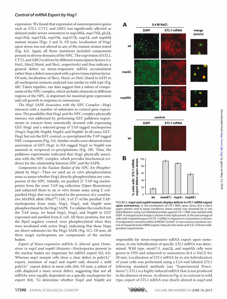
stress in nup2 and nup60 Mutants—Nucleoporins present inthe nuclear basket are important for mRNA export (62, 63).Whereas nup1 mutant cells show a clear defect in poly(A)�export, mutation of nup2 and nup60 only showed a mildpoly(A)� export defect in most cells (64). Of note, a subset ofcells displayed a more severe defect, suggesting that not allmRNAs were equally dependent on a specific nucleoporin forexport (64). To determine whether Nup2 and Nup60 are
responsible for stress-responsive mRNA export upon osmo-stress, in situ hybridization of specific STL1mRNA was deter-mined. Wild type, mex67-5, nup2�, and nup60� cells weregrown in YPD and subjected to osmostress (0.4 M NaCl) for20 min. Localization of STL1mRNA by in situ hybridizationof yeast cells was performed using a Cy3-end-labeled STL1following standard methods (see “Experimental Proce-dures”). STL1 is a highly inducedmRNA that is not producedin the absence of stress. As shown in Fig. 6, in contrast to wildtype, export of STL1 mRNA was clearly altered in nup2 and
FIGURE 6. nup2 and nup60 mutants display defects in STL1 mRNA exportupon osmostress. A, the localization of STL1 RNA upon stress (0.4 M NaCl;upper panels) and in basal conditions (lower panel) was assessed by in situhybridization using Cy3-labbeled probes against STL1. DNA was stained withDAPI. A merged zoom image is shown in the right panels. B, the percentage ofcells with impaired export of STL1 mRNA in response to osmostress is shown.A temperature-sensitive allele of MEX67 (mex67-5) was used as a positive con-trol of impaired total mRNA export. Data are the mean and S.D. of three inde-pendent experiments.
Control of mRNA Export by Hog1
17390 JOURNAL OF BIOLOGICAL CHEMISTRY VOLUME 288 • NUMBER 24 • JUNE 14, 2013
nup60 mutant strains in response to osmostress. A temper-ature-sensitive allele ofMEX67 (mex67-5) was used as a con-trol of impaired total mRNA export. Thus, export of theSTL1 stress-responsive mRNA is altered in nup2 and nup60mutants.Nuclear Basket Proteins Nup1, Nup2, and Nup60 Associate
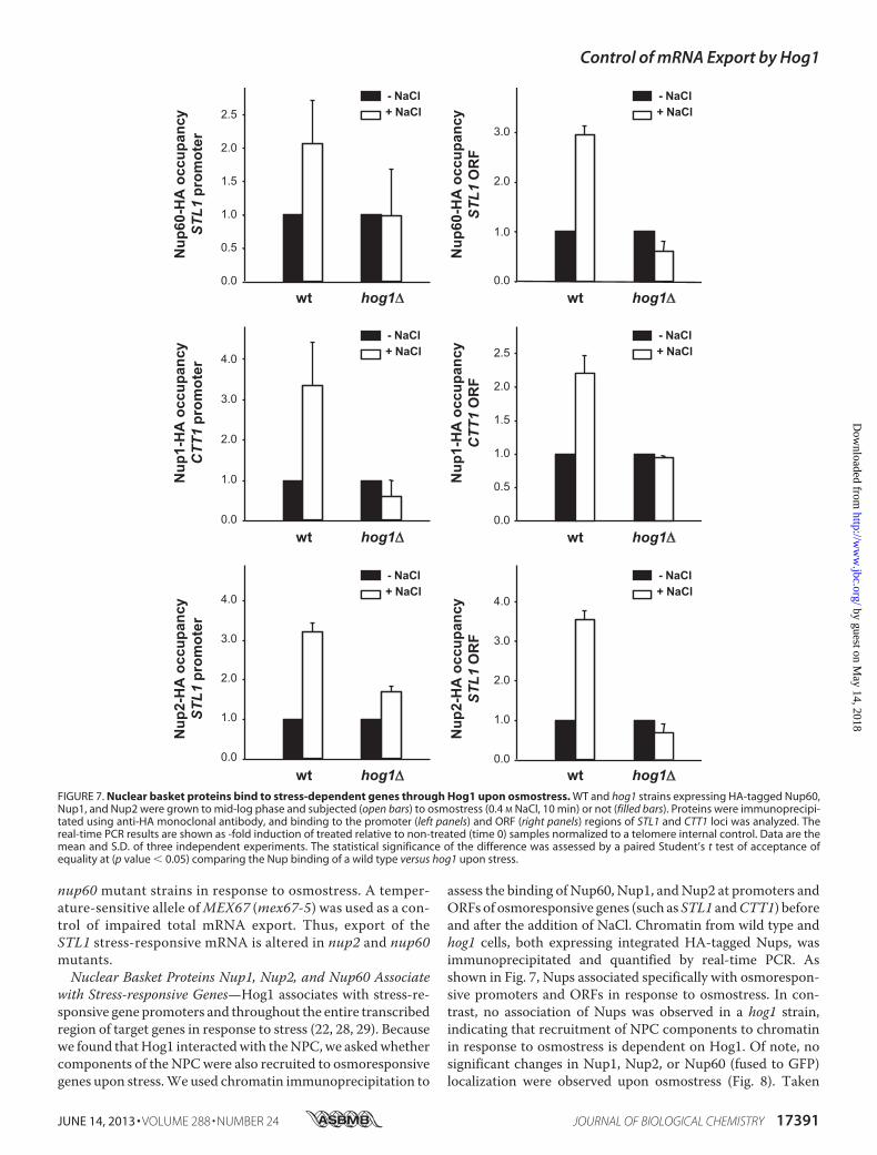
with Stress-responsive Genes—Hog1 associates with stress-re-sponsive gene promoters and throughout the entire transcribedregion of target genes in response to stress (22, 28, 29). Becausewe found thatHog1 interactedwith theNPC,we askedwhethercomponents of the NPCwere also recruited to osmoresponsivegenes upon stress.We used chromatin immunoprecipitation to
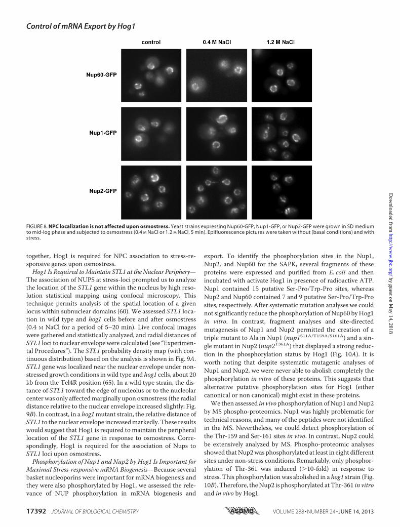
assess the binding of Nup60, Nup1, andNup2 at promoters andORFs of osmoresponsive genes (such as STL1 andCTT1) beforeand after the addition of NaCl. Chromatin from wild type andhog1 cells, both expressing integrated HA-tagged Nups, wasimmunoprecipitated and quantified by real-time PCR. Asshown in Fig. 7, Nups associated specifically with osmorespon-sive promoters and ORFs in response to osmostress. In con-trast, no association of Nups was observed in a hog1 strain,indicating that recruitment of NPC components to chromatinin response to osmostress is dependent on Hog1. Of note, nosignificant changes in Nup1, Nup2, or Nup60 (fused to GFP)localization were observed upon osmostress (Fig. 8). Taken
FIGURE 7. Nuclear basket proteins bind to stress-dependent genes through Hog1 upon osmostress. WT and hog1 strains expressing HA-tagged Nup60,Nup1, and Nup2 were grown to mid-log phase and subjected (open bars) to osmostress (0.4 M NaCl, 10 min) or not (filled bars). Proteins were immunoprecipi-tated using anti-HA monoclonal antibody, and binding to the promoter (left panels) and ORF (right panels) regions of STL1 and CTT1 loci was analyzed. Thereal-time PCR results are shown as -fold induction of treated relative to non-treated (time 0) samples normalized to a telomere internal control. Data are themean and S.D. of three independent experiments. The statistical significance of the difference was assessed by a paired Student’s t test of acceptance ofequality at (p value � 0.05) comparing the Nup binding of a wild type versus hog1 upon stress.
Control of mRNA Export by Hog1
JUNE 14, 2013 • VOLUME 288 • NUMBER 24 JOURNAL OF BIOLOGICAL CHEMISTRY 17391
together, Hog1 is required for NPC association to stress-re-sponsive genes upon osmostress.Hog1 Is Required toMaintain STL1 at theNuclear Periphery—
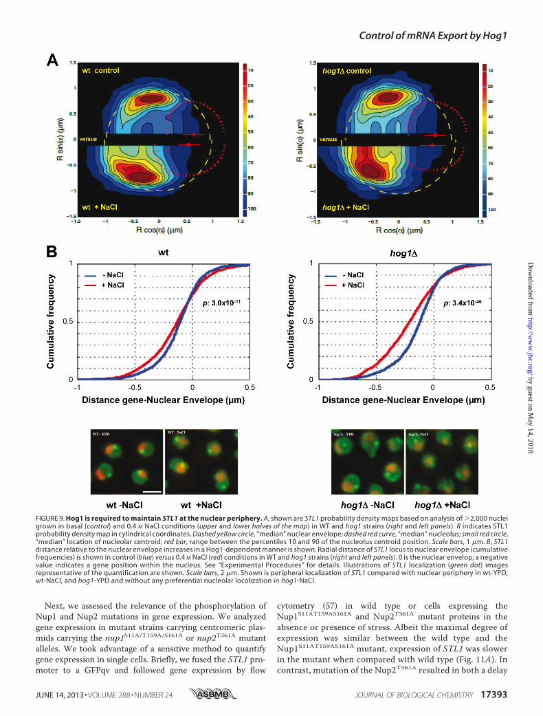
The association of NUPS at stress-loci prompted us to analyzethe location of the STL1 gene within the nucleus by high reso-lution statistical mapping using confocal microscopy. Thistechnique permits analysis of the spatial location of a givenlocus within subnuclear domains (60). We assessed STL1 loca-tion in wild type and hog1 cells before and after osmostress(0.4 M NaCl for a period of 5–20 min). Live confocal imageswere gathered and statistically analyzed, and radial distances ofSTL1 loci to nuclear envelopewere calculated (see “Experimen-tal Procedures”). The STL1 probability density map (with con-tinuous distribution) based on the analysis is shown in Fig. 9A.STL1 gene was localized near the nuclear envelope under non-stressed growth conditions in wild type and hog1 cells, about 20kb from the Tel4R position (65). In a wild type strain, the dis-tance of STL1 toward the edge of nucleolus or to the nucleolarcenter was only affectedmarginally upon osmostress (the radialdistance relative to the nuclear envelope increased slightly; Fig.9B). In contrast, in a hog1mutant strain, the relative distance ofSTL1 to the nuclear envelope increasedmarkedly. These resultswould suggest that Hog1 is required to maintain the peripherallocation of the STL1 gene in response to osmostress. Corre-spondingly, Hog1 is required for the association of Nups toSTL1 loci upon osmostress.Phosphorylation of Nup1 and Nup2 by Hog1 Is Important for
Maximal Stress-responsive mRNA Biogenesis—Because severalbasket nucleoporins were important for mRNA biogenesis andthey were also phosphorylated by Hog1, we assessed the rele-vance of NUP phosphorylation in mRNA biogenesis and
export. To identify the phosphorylation sites in the Nup1,Nup2, and Nup60 for the SAPK, several fragments of theseproteins were expressed and purified from E. coli and thenincubated with activate Hog1 in presence of radioactive ATP.Nup1 contained 15 putative Ser-Pro/Trp-Pro sites, whereasNup2 and Nup60 contained 7 and 9 putative Ser-Pro/Trp-Prosites, respectively. After systematic mutation analyses we couldnot significantly reduce the phosphorylation of Nup60 byHog1in vitro. In contrast, fragment analyses and site-directedmutagenesis of Nup1 and Nup2 permitted the creation of atriple mutant to Ala in Nup1 (nup1S11A/T159A/S161A) and a sin-gle mutant in Nup2 (nup2T361A) that displayed a strong reduc-tion in the phosphorylation status by Hog1 (Fig. 10A). It isworth noting that despite systematic mutagenic analyses ofNup1 and Nup2, we were never able to abolish completely thephosphorylation in vitro of these proteins. This suggests thatalternative putative phosphorylation sites for Hog1 (eithercanonical or non canonical) might exist in these proteins.We then assessed in vivo phosphorylation of Nup1 andNup2
by MS phospho-proteomics. Nup1 was highly problematic fortechnical reasons, andmany of the peptides were not identifiedin the MS. Nevertheless, we could detect phosphorylation ofthe Thr-159 and Ser-161 sites in vivo. In contrast, Nup2 couldbe extensively analyzed by MS. Phospho-proteomic analysesshowed thatNup2was phosphorylated at least in eight differentsites under non-stress conditions. Remarkably, only phosphor-ylation of Thr-361 was induced (10-fold) in response tostress. This phosphorylationwas abolished in a hog1 strain (Fig.10B). Therefore, theNup2 is phosphorylated atThr-361 in vitroand in vivo by Hog1.
FIGURE 8. NPC localization is not affected upon osmostress. Yeast strains expressing Nup60-GFP, Nup1-GFP, or Nup2-GFP were grown in SD mediumto mid-log phase and subjected to osmostress (0.4 M NaCl or 1.2 M NaCl, 5 min). Epifluorescence pictures were taken without (basal conditions) and withstress.
Control of mRNA Export by Hog1
17392 JOURNAL OF BIOLOGICAL CHEMISTRY VOLUME 288 • NUMBER 24 • JUNE 14, 2013
Next, we assessed the relevance of the phosphorylation ofNup1 and Nup2 mutations in gene expression. We analyzedgene expression in mutant strains carrying centromeric plas-mids carrying the nup1S11A/T159A/S161A or nup2T361A mutantalleles. We took advantage of a sensitive method to quantifygene expression in single cells. Briefly, we fused the STL1 pro-moter to a GFPqv and followed gene expression by flow
cytometry (57) in wild type or cells expressing theNup1S11AT159AS161A and Nup2T361A mutant proteins in theabsence or presence of stress. Albeit the maximal degree ofexpression was similar between the wild type and theNup1S11AT159AS161A mutant, expression of STL1 was slowerin the mutant when compared with wild type (Fig. 11A). Incontrast, mutation of the Nup2T361A resulted in both a delay
FIGURE 9. Hog1 is required to maintain STL1 at the nuclear periphery. A, shown are STL1 probability density maps based on analysis of 2,000 nucleigrown in basal (control) and 0.4 M NaCl conditions (upper and lower halves of the map) in WT and hog1 strains (right and left panels). R indicates STL1probability density map in cylindrical coordinates. Dashed yellow circle, “median” nuclear envelope; dashed red curve, “median” nucleolus; small red circle,“median” location of nucleolar centroid; red bar, range between the percentiles 10 and 90 of the nucleolus centroid position. Scale bars, 1 �m. B, STL1distance relative to the nuclear envelope increases in a Hog1-dependent manner is shown. Radial distance of STL1 locus to nuclear envelope (cumulativefrequencies) is shown in control (blue) versus 0.4 M NaCl (red) conditions in WT and hog1 strains (right and left panels). 0 is the nuclear envelop; a negativevalue indicates a gene position within the nucleus. See “Experimental Procedures” for details. Illustrations of STL1 localization (green dot) imagesrepresentative of the quantification are shown. Scale bars, 2 �m. Shown is peripheral localization of STL1 compared with nuclear periphery in wt-YPD,wt-NaCl, and hog1-YPD and without any preferential nucleolar localization in hog1-NaCl.
Control of mRNA Export by Hog1
JUNE 14, 2013 • VOLUME 288 • NUMBER 24 JOURNAL OF BIOLOGICAL CHEMISTRY 17393
and a reduction in mRNA accumulation that was compara-ble with the null nup2 mutant (Fig. 11B). Therefore, muta-tion of the phosphorylation sites by the SAPK in Nup1 andNup2 resulted in altered mRNA accumulation uponosmostress.Because the transcriptional defect was more exacerbated in
the Nup2T361A mutant, we assessed STL1 mRNA export byin situ hybridization in nup2-deficient cells carrying a wildtype allele of NUP2 or the nup2T361A mutant. Whereas thewild type strain only displayed 10% of cells with mRNAspecks, 60% of the cells carrying the nup2T361A mutationpresented defects in mRNA export (Fig. 11C). Overall, ourresults suggest that the phosphorylation of nuclear basketnucleoporins by Hog1 is important for proper mRNA exportin response to osmostress.
DISCUSSION
Yeast cells respond to increases in external osmolarity byactivating the stress-activatedHog1 SAPK.Amajor outcome ofthe activation of Hog1 is the regulation of gene expression, andit has been shown that Hog1 controls gene expression by actingin several steps of mRNA biogenesis (2, 3). Here, we provideevidence that mRNA export, an additional step in mRNA bio-genesis, is also controlled by the SAPK.The identification by genetic means of mutants in the NPC
that were osmosensitive and displayed reduced expression ofrepresentative stress-responsive mRNAs prompted us to sys-tematically assess which components of the NPC were impor-tant for stress adaptation andwhether theywere targeted by theSAPK.We found that specific mutations in nucleoporins local-
FIGURE 10. A, Nup1 and Nup2 are phosphorylated in vitro by Hog1. Recombinant GST-fused wild type and mutant proteins were purified from E. coli, andin vitro kinase assays were performed as in Fig. 2B. Phosphorylated proteins were resolved by SDS-PAGE and detected by autoradiography (upper panel).GST-tagged Nup proteins were detected by staining with Coomassie Brilliant Blue (lower panel). B, shown is -fold change in phosphorylation upon0.4 M NaCl treatment (5 min) at the indicated residues in Nup2 in wild type (blue) and hog1 mutant (red) strains. Ratios have been calculated fromabsolute phosphorylation intensities of unstressed and osmostressed wild type and hog1 mutant samples. The table shows the Nup2 residues that havebeen found to be phosphorylated in this study (first column), the position of the phosphorylated residue (red) in the MS-detected phosphopeptide(second column), and ratios of wild type stressed/wild type unstressed and hog1 mutant stressed/hog1 mutant unstressed (third and fourth column,respectively).
Control of mRNA Export by Hog1
17394 JOURNAL OF BIOLOGICAL CHEMISTRY VOLUME 288 • NUMBER 24 • JUNE 14, 2013
ized in the adaptor, coat, cytoplasmic filaments, and nuclearbasket resulted in defects in adaptation and mRNA accumula-tion upon stress. Mutations in NUPs did not lead to alterationon transcription factor localization. This suggests that integrityof the NPC is important for proper mRNA accumulation uponosmostress. Remarkably, only nuclear basket nucleoporinswere directly phosphorylated by the Hog1 SAPK. Thus, thismight suggest that several components of the NPC are impor-tant for proper stress-responsive mRNA accumulation/export,but only the components that remain at the nuclear side can bedirectly regulated by the SAPK. Correspondingly, active Hog1accumulates into the nucleus in response to stress (6).Nucleoporins have been involved in macromolecular traf-
ficking, proteins, and mRNAs as well as in regulatory functions(33, 66). The phosphorylation of nucleoporins by cyclin-depen-dent kinases andMAPKs has been reported in yeast and mam-mals (67, 68). Here, we have shown that the recruitment of
nucleoporins occurs at stress loci in response to Hog1 activa-tion. In contrast, Hog1 protein transport is not altered by thedeletion of those nucleoporins. Thus, the role of nucleoporinregulation upon osmostress seems to be related to mRNAexport. Correspondingly, STL1 mRNA export is altered uponstress in cells deficient in nuclear basket nup2 and nup60mutants. Furthermore, a point mutation in Nup2 (Nup2T361A)that reduces Hog1-dependent phosphorylation also results indefective STL1 export. Taken together, our data support thatmRNA export is controlled by the phosphorylation of NUPcomponents by the SAPK. The fact that null mutations of theNUPs showed a more dramatic effect on gene expression thanthe non-phosphorylatable mutants could be due to additionalphosphorylation sites targeted by the SAPK. Alternatively, it ispossible that phosphorylation of the NUPs serves to modulatethe efficiency of the transport that would be completelyimpaired in the null mutants.
FIGURE 11. Phosphorylation of Nup1 and Nup2 by Hog1 mediates proper stress-responsive mRNA biogenesis. nup1 (A) and nup2 (B) mutant strainsharboring the quadruple-Venus (qV) fluorescence reporter driven by the STL1 promoter (pSTL1) and the indicated low copy number (pRS413) constructs weremeasured by flow cytometry upon osmostress (1 M NaCl, 70 min). C, shown is analysis of nuclear STL1 mRNA export in the nup2 mutant strain carrying the NUP2wild type or the nup2T361A allele upon osmostress. The localization of STL1 RNA (left panel) was assessed by in situ hybridization using Cy3-labeled probesagainst STL1 in response to 0.4 M NaCl. DNA was stained with DAPI (middle panel). Merged images are shown (right panel). Scale bars, 1 �m (white line). Mergedzoom images are shown with the percentage of impaired export of STL1 mRNA in response to osmostress.
Control of mRNA Export by Hog1
JUNE 14, 2013 • VOLUME 288 • NUMBER 24 JOURNAL OF BIOLOGICAL CHEMISTRY 17395
Tethering genomic loci to the nuclear envelope has beenpos-tulated as amechanism to optimize gene expression (62, 63, 69).However, the molecular mechanism and functional signifi-cance of peripheral localization of transcription-induced genesis poorly understood andwhether it is the cause or consequenceof transcription activation is still unclear (48, 55, 70). However,several studies have shown a direct link between NPC and genelocalization (43, 45–47, 49, 51). Moreover, peripheral localiza-tion of certain loci has been linked toNPCphosphorylation. Forinstance, it has been reported that phosphorylation of Nup1 byCdk1 alters INO1 andGAL1 nuclear localization (68). Here, wehave shown that STL1 is located near the nuclear envelopeunder normal conditions possibly due to its close proximity totheTer4R location.However, it is only in response to stress thatNUPs associate to the STL1 loci, indicating the direct tetheringof the loci to NPC upon stress. In a hog1 strain, NUPs do notassociate to STL1, and the STL1 locus is not retained at thenuclear periphery. Taken together, our data suggest that asso-ciation of NPC to stress-responsive loci might serve to opti-mally couple mRNA production and export. Alternatively, theassociation of NUPs to promotersmight be relevant to stabilizetranscriptional complexes at the loci of transcribed genes.Actually, the mutation of the phosphorylation sites for Hog1 inNup1 andNup2 seem not only to diminish gene expression butalso delays mRNA production. A similar scenario is observedwhen chromatin remodeling at stress-loci is altered yielding abimodal response similar to the observed here (57). We cannotformally exclude the presence of nucleoplasmic pool of phos-phorylated Nups. InDrosophila, the presence of nucleoplasmicNups away from the NPC, in the nucleoplasm, has beenreported (71). Although we cannot directly address this pointwith STL1 due to its telomer proximity, it is worth mentioningthat upon induction of a telomer proximal gene, HXK1, thedistance to periphery decreased upon NPC association (46).In response to stress, general transcription seems to be
down-regulated in contrast to an increase of stress-responsivemRNAs (13–15). Thus, the data provided here also pointtoward the existence of a specialized pathway that wouldinclude the control of several steps in mRNA biogenesis. Thiswould favor the production of stress-responsive mRNAs,whereas there is amajor down-regulation ofmRNAsynthesis ofgeneral and housekeeping genes. The Hog1 SAPK has beeninvolved inmRNA synthesis, the control ofmRNA stability (13,30), and translation (31). The data provided here suggest thatthe SAPK also controls the export of stress-responsive mRNAsvia the phosphorylation of NUPS at the nuclear basket. Indeed,there are many examples reporting the importance of addi-tional levels in the regulation of gene expression after its syn-thesis in the nucleus (72). All together, the data support amodelby which a single signaling molecule is able to exert the controlof several steps in mRNA biogenesis.
Acknowledgments—Weare grateful to S. Obejas andA. Fernandez fortechnical assistance and L. Subirana for experimental support.
REFERENCES1. de Nadal, E., Ammerer, G., and Posas, F. (2011) Controlling gene expres-
sion in response to stress. Nat. Rev. Genet. 12, 833– 845
2. Weake, V. M., and Workman, J. L. (2010) Inducible gene expression. Di-verse regulatory mechanisms. Nat. Rev. Genet. 11, 426 – 437
3. de Nadal, E., and Posas, F. (2010) Multilayered control of gene expressionby stress-activated protein kinases. EMBO J. 29, 4 –13
4. Macia, J., Regot, S., Peeters, T., Conde, N., Solé, R., and Posas, F. (2009)Dynamic signaling in the Hog1 MAPK pathway relies on high basal signaltransduction. Sci. Signal. 2, ra13
5. Saito, H., and Posas, F. (2012) Response to hyperosmotic stress. Genetics192, 289 –318
6. Ferrigno, P., Posas, F., Koepp, D., Saito, H., and Silver, P. A. (1998) Regu-lated nucleo/cytoplasmic exchange of HOG1 MAPK requires the impor-tin � homologs NMD5 and XPO1. EMBO J. 17, 5606 –5614
7. Gasch, A. P., Spellman, P. T., Kao, C. M., Carmel-Harel, O., Eisen, M. B.,Storz, G., Botstein, D., and Brown, P. O. (2000) Genomic expression pro-grams in the response of yeast cells to environmental changes. Mol. Biol.Cell 11, 4241– 4257
8. Posas, F., Chambers, J. R., Heyman, J. A., Hoeffler, J. P., de Nadal, E., andAriño, J. (2000) The transcriptional response of yeast to saline stress.J. Biol. Chem. 275, 17249 –17255
9. Rep, M., Krantz, M., Thevelein, J. M., and Hohmann, S. (2000) The tran-scriptional response of Saccharomyces cerevisiae to osmotic shock. Hot1pand Msn2p/Msn4p are required for the induction of subsets of high os-molarity glycerol pathway-dependent genes. J. Biol. Chem. 275,8290 – 8300
10. Yale, J., and Bohnert, H. J. (2001) Transcript expression in Saccharomycescerevisiae at high salinity. J. Biol. Chem. 276, 15996 –16007
11. Ni, L., Bruce, C., Hart, C., Leigh-Bell, J., Gelperin, D., Umansky, L., Ger-stein, M. B., and Snyder, M. (2009) Dynamic and complex transcriptionfactor binding during an inducible response in yeast. Genes Dev. 23,1351–1363
12. Capaldi, A. P., Kaplan, T., Liu, Y., Habib, N., Regev, A., Friedman, N., andO’Shea, E. K. (2008) Structure and function of a transcriptional networkactivated by the MAPK Hog1. Nat. Genet. 40, 1300 –1306
13. Miller, C., Schwalb, B., Maier, K., Schulz, D., Dümcke, S., Zacher, B.,Mayer, A., Sydow, J., Marcinowski, L., Dölken, L., Martin, D. E., Tresch, A.,and Cramer, P. (2011) Dynamic transcriptome analysis measures rates ofmRNA synthesis and decay in yeast. Mol. Syst. Biol. 7, 458
14. Nadal-Ribelles, M., Conde, N., Flores, O., González-Vallinas, J., Eyras, E.,Orozco, M., de Nadal E., and Posas, F. (2012) Hog1 bypasses stress-medi-ated down-regulation of transcription by RNA polymerase II redistribu-tion and chromatin remodeling. Genome Biol. 13, R106
15. Cook, K. E., and O’Shea, E. K. (2012) Hog1 controls global reallocation ofRNA Pol II upon osmotic shock in Saccharomyces cerevisiae. G3 2,1129 –1136
16. Proft, M., and Serrano, R. (1999) Repressors and upstream repressingsequences of the stress-regulated ENA1 gene in Saccharomyces cerevisiae.bZIP protein Sko1p confers HOG-dependent osmotic regulation. Mol.Cell. Biol. 19, 537–546
17. Rep, M., Reiser, V., Gartner, U., Thevelein, J. M., Hohmann, S., Ammerer,G., and Ruis, H. (1999) Osmotic stress-induced gene expression in Sac-charomyces cerevisiae requires Msn1p and the novel nuclear factor Hot1p.Mol. Cell. Biol. 19, 5474 –5485
18. Proft, M., Pascual-Ahuir, A., de Nadal E., Ariño, J., Serrano, R., and Posas,F. (2001) Regulation of the Sko1 transcriptional repressor by the Hog1MAP kinase in response to osmotic stress. EMBO J. 20, 1123–1133
19. Alepuz, P. M., de Nadal, E., Zapater, M., Ammerer, G., and Posas, F. (2003)Osmostress-induced transcription by Hot1 depends on a Hog1-mediatedrecruitment of the RNA Pol II. EMBO J. 22, 2433–2442
20. de Nadal, E., Casadomé, L., and Posas, F. (2003) Targeting the MEF2-liketranscription factor Smp1 by the stress-activated Hog1 mitogen-activatedprotein kinase. Mol. Cell. Biol. 23, 229 –237
21. Ruiz-Roig, C., Noriega, N., Duch, A., Posas, F., and de Nadal E. (2012) TheHog1 SAPK controls the Rtg1/Rtg3 transcriptional complex activity bymultiple regulatory mechanisms. Mol. Biol. Cell 23, 4286 – 4296
22. Alepuz, P. M., Jovanovic, A., Reiser, V., and Ammerer, G. (2001) Stress-induced map kinase Hog1 is part of transcription activation complexes.Mol. Cell 7, 767–777
23. Proft, M., and Struhl, K. (2002) Hog1 kinase converts the Sko1-Cyc8-Tup1
Control of mRNA Export by Hog1
17396 JOURNAL OF BIOLOGICAL CHEMISTRY VOLUME 288 • NUMBER 24 • JUNE 14, 2013
repressor complex into an activator that recruits SAGA and SWI/SNF inresponse to osmotic stress. Mol. Cell 9, 1307–1317
24. De Nadal, E., Zapater, M., Alepuz, P. M., Sumoy, L., Mas, G., and Posas, F.(2004) The MAPK Hog1 recruits Rpd3 histone deacetylase to activateosmoresponsive genes. Nature 427, 370 –374
25. Zapater, M., Sohrmann, M., Peter, M., Posas, F., and de Nadal E. (2007)Selective requirement for SAGA in Hog1-mediated gene expression de-pending on the severity of the external osmostress conditions. Mol. Cell.Biol. 27, 3900 –3910
26. Solé, C., Nadal-Ribelles, M., Kraft, C., Peter, M., Posas, F., and de Nadal, E.(2011) Control of Ubp3 ubiquitin protease activity by the Hog1 SAPKmodulates transcription upon osmostress. EMBO J. 30, 3274 –3284
27. Pascual-Ahuir, A., Struhl, K., and Proft, M. (2006) Genome-wide locationanalysis of the stress-activated MAP kinase Hog1 in yeast. Methods 40,272–278
28. Pokholok, D. K., Zeitlinger, J., Hannett, N. M., Reynolds, D. B., and Young,R. A. (2006) Activated signal transduction kinases frequently occupy tar-get genes. Science 313, 533–536
29. Proft, M., Mas, G., de Nadal E., Vendrell, A., Noriega, N., Struhl, K., andPosas, F. (2006) The stress-activated Hog1 kinase is a selective transcrip-tional elongation factor for genes responding to osmotic stress. Mol. Cell23, 241–250
30. Romero-Santacreu, L., Moreno, J., Pérez-Ortín, J. E., and Alepuz, P. (2009)Specific and global regulation of mRNA stability during osmotic stress inSaccharomyces cerevisiae. RNA 15, 1110 –1120
31. Warringer, J., Hult, M., Regot, S., Posas, F., and Sunnerhagen, P. (2010)The HOG pathway dictates the short-term translational response afterhyperosmotic shock. Mol. Biol. Cell 21, 3080 –3092
32. Tran, E. J., and Wente, S. R. (2006) Dynamic nuclear pore complexes. Lifeon the edge. Cell 125, 1041–1053
33. Lim, R. Y., Ullman, K. S., and Fahrenkrog, B. (2008) Biology and biophysicsof the nuclear pore complex and its components. Int. Rev. Cell Mol. Biol.267, 299 –342
34. Hoelz, A., Debler, E. W., and Blobel, G. (2011) The structure of the nuclearpore complex. Annu. Rev. Biochem. 80, 613– 643
35. Rout, M. P., Aitchison, J. D., Suprapto, A., Hjertaas, K., Zhao, Y., and Chait,B. T. (2000) The yeast nuclear pore complex. composition, architecture,and transport mechanism. J. Cell Biol. 148, 635– 651
36. Kampmann, M., and Blobel, G. (2009) Three-dimensional structure andflexibility of a membrane-coating module of the nuclear pore complex.Nat. Struct. Mol. Biol. 16, 782–788
37. Aitchison, J. D., and Rout, M. P. (2012) The yeast nuclear pore complexand transport through it. Genetics 190, 855– 883
38. D’Angelo, M. A., and Hetzer, M. W. (2008) Structure, dynamics, and func-tion of nuclear pore complexes. Trends Cell Biol. 18, 456 – 466
39. Brown, C. R., and Silver, P. A. (2007) Transcriptional regulation at thenuclear pore complex. Curr. Opin. Genet. Dev. 17, 100 –106
40. Taddei, A. (2007) Active genes at the nuclear pore complex. Curr. Opin.Cell Biol. 19, 305–310
41. Ahmed, S., and Brickner, J. H. (2007) Regulation and epigenetic control oftranscription at the nuclear periphery. Trends Genet. 23, 396 – 402
42. Akhtar, A., and Gasser, S. M. (2007) The nuclear envelope and transcrip-tional control. Nat. Rev. Genet. 8, 507–517
43. Casolari, J. M., Brown, C. R., Komili, S., West, J., Hieronymus, H., andSilver, P. A. (2004) Genome-wide localization of the nuclear transportmachinery couples transcriptional status and nuclear organization. Cell117, 427– 439
44. Cabal, G. G., Genovesio, A., Rodriguez-Navarro, S., Zimmer, C., Gadal, O.,Lesne, A., Buc, H., Feuerbach-Fournier, F., Olivo-Marin, J. C., Hurt, E. C.,and Nehrbass, U. (2006) SAGA interacting factors confine sub-diffusionof transcribed genes to the nuclear envelope. Nature 441, 770 –773
45. Schmid, M., Arib, G., Laemmli, C., Nishikawa, J., Durussel, T., and Laem-mli, U. K. (2006) Nup-PI. The nucleopore-promoter interaction of genesin yeast. Mol. Cell 21, 379 –391
46. Taddei, A., Van Houwe, G., Hediger, F., Kalck, V., Cubizolles, F., Schober,H., and Gasser, S. M. (2006) Nuclear pore association confers optimalexpression levels for an inducible yeast gene. Nature 441, 774 –778
47. Luthra, R., Kerr, S. C., Harreman, M. T., Apponi, L. H., Fasken, M. B.,
Ramineni, S., Chaurasia, S., Valentini, S. R., and Corbett, A. H. (2007)Actively transcribed GAL genes can be physically linked to the nuclearpore by the SAGA chromatin modifying complex. J. Biol. Chem. 282,3042–3049
48. Brickner, D. G., Cajigas, I., Fondufe-Mittendorf, Y., Ahmed, S., Lee, P. C.,Widom, J., and Brickner, J. H. (2007) H2A.Z-mediated localization ofgenes at the nuclear periphery confers epigenetic memory of previoustranscriptional state. PLoS Biol. 5, e81
49. Brickner, J. H., and Walter, P. (2004) Gene recruitment of the activatedINO1 locus to the nuclear membrane. PLoS. Biol. 2, e342
50. Ahmed, S., Brickner, D. G., Light, W. H., Cajigas, I., McDonough, M.,Froyshteter, A. B., Volpe, T., and Brickner, J. H. (2010) DNA zip codescontrol an ancient mechanism for gene targeting to the nuclear periphery.Nat. Cell Biol. 12, 111–118
51. Menon, B. B., Sarma, N. J., Pasula, S., Deminoff, S. J., Willis, K. A.,Barbara, K. E., Andrews, B., and Santangelo, G. M. (2005) Reverserecruitment. The Nup84 nuclear pore subcomplex mediates Rap1/Gcr1/Gcr2 transcriptional activation. Proc. Natl. Acad. Sci. U.S.A. 102,5749 –5754
52. Casolari, J. M., Brown, C. R., Drubin, D. A., Rando, O. J., and Silver, P. A.(2005) Developmentally induced changes in transcriptional programalter spatial organization across chromosomes. Genes Dev. 19,1188 –1198
53. Dieppois, G., Iglesias, N., and Stutz, F. (2006) Cotranscriptional recruit-ment to the mRNA export receptor Mex67p contributes to nuclearpore anchoring of activated genes. Mol. Cell. Biol. 26, 7858 –7870
54. Abruzzi, K. C., Belostotsky, D. A., Chekanova, J. A., Dower, K., and Ros-bash, M. (2006) 3�-End formation signals modulate the association ofgenes with the nuclear periphery as well as mRNP dot formation. EMBO J.25, 4253– 4262
55. Green, E. M., Jiang, Y., Joyner, R., and Weis, K. (2012) A negative feedbackloop at the nuclear periphery regulates GAL gene expression. Mol. Biol.Cell 23, 1367–1375
56. Bilsland-Marchesan, E., Ariño, J., Saito, H., Sunnerhagen, P., and Posas, F.(2000) Rck2 kinase is a substrate for the osmotic stress-activated mitogen-activated protein kinase Hog1. Mol. Cell. Biol. 20, 3887–3895
57. Pelet, S., Rudolf, F., Nadal-Ribelles, M., de Nadal E., Posas, F., and Peter, M.(2011) Transient activation of the HOG MAPK pathway regulates bi-modal gene expression. Science 332, 732–735
58. Zhou, H., Ye, M., Dong, J., Corradini, E., Cristobal, A., Heck, A. J., Zou, H.,and Mohammed, S. (2013) Robust phosphoproteome enrichment usingmonodisperse microsphere-based immobilized titanium (IV) ion affinitychromatography. Nat. Protoc. 8, 461– 480
59. Cox, J., and Mann, M. (2008) MaxQuant enables high peptide identifica-tion rates, individualized p.p.b.-range mass accuracies and proteome-wideprotein quantification. Nat. Biotechnol. 26, 1367–1372
60. Berger, A. B., Cabal, G. G., Fabre, E., Duong, T., Buc, H., Nehrbass, U.,Olivo-Marin, J. C., Gadal, O., and Zimmer, C. (2008) High-resolutionstatistical mapping reveals gene territories in live yeast. Nat. Methods 5,1031–1037
61. Mas, G., de Nadal E., Dechant, R., Rodríguez de la Concepción ML, Logie,C., Jimeno-González, S., Chávez, S., Ammerer, G., and Posas, F. (2009)Recruitment of a chromatin remodelling complex by the Hog1 MAP ki-nase to stress genes. EMBO J. 28, 326 –336
62. Liang, Y., and Hetzer, M. W. (2011) Functional interactions betweennucleoporins and chromatin. Curr. Opin. Cell Biol. 23, 65–70
63. Mekhail, K., and Moazed, D. (2010) The nuclear envelope in genomeorganization, expression, and stability. Nat. Rev. Mol. Cell Biol. 11,317–328
64. Powrie, E. A., Zenklusen, D., and Singer, R. H. (2011) A nucleoporin,Nup60p, affects the nuclear and cytoplasmic localization of ASH1 mRNAin S. cerevisiae. RNA 17, 134 –144
65. Therizols, P., Duong, T., Dujon, B., Zimmer, C., and Fabre, E. (2010) Chro-mosome arm length and nuclear constraints determine the dynamic rela-tionship of yeast subtelomeres. Proc. Natl. Acad. Sci. U.S.A. 107,2025–2030
66. Köhler, A., and Hurt, E. (2010) Gene regulation by nucleoporins and linksto cancer. Mol. Cell 38, 6 –15
Control of mRNA Export by Hog1
JUNE 14, 2013 • VOLUME 288 • NUMBER 24 JOURNAL OF BIOLOGICAL CHEMISTRY 17397
67. Kosako, H., Yamaguchi, N., Aranami, C., Ushiyama, M., Kose, S., Imamoto,N., Taniguchi, H., Nishida, E., and Hattori, S. (2009) Phosphoproteomics re-veals new ERK MAP kinase targets and links ERK to nucleoporin-mediatednuclear transport. Nat. Struct. Mol. Biol. 16, 1026–1035
68. Brickner, D. G., and Brickner, J. H. (2010) Cdk phosphorylation of anucleoporin controls localization of active genes through the cell cycle.Mol. Biol. Cell 21, 3421–3432
69. Capelson, M., and Hetzer, M. W. (2009) The role of nuclear pores in generegulation, development and disease. EMBO Rep. 10, 697–705
70. Light, W. H., Brickner, D. G., Brand, V. R., and Brickner, J. H. (2010)Interaction of a DNA zip code with the nuclear pore complex promotesH2A.Z incorporation and INO1 transcriptional memory. Mol. Cell 40,112–125
71. Kalverda, B., Pickersgill, H., Shloma, V. V., and Fornerod, M. (2010)Nucleoporins directly stimulate expression of developmental and cell-cycle genes inside the nucleoplasm. Cell 140, 360 –371
72. Choder, M. (2011) mRNA imprinting. Additional level in the regulation ofgene expression. Cell Logist. 1, 37– 40
Control of mRNA Export by Hog1
17398 JOURNAL OF BIOLOGICAL CHEMISTRY VOLUME 288 • NUMBER 24 • JUNE 14, 2013
Francesc PosasJorge Pérez-Fernandez, Olivier Gadal, Gerhard Seisenbacher, Gustav Ammerer and Sergi Regot, Eulàlia de Nadal, Susana Rodríguez-Navarro, Alberto González-Novo,
Export upon StressThe Hog1 Stress-activated Protein Kinase Targets Nucleoporins to Control mRNA
doi: 10.1074/jbc.M112.444042 originally published online May 3, 20132013, 288:17384-17398.J. Biol. Chem.
10.1074/jbc.M112.444042Access the most updated version of this article at doi:
Alerts:
When a correction for this article is posted•
When this article is cited•
to choose from all of JBC's e-mail alertsClick here
The Hog1 stress-activated protein kinase targetsnucleoporins to control mRNA export upon stress.Sergi Regot, Eulalia de Nadal, Susana Rodríguez-Navarro, Alberto Gonzalez-Novo,Jorge Perez-Fernandez, Olivier Gadal, Gerhard Seisenbacher, Gustav Ammerer,and Francesc Posas
We regretfully failed to acknowledge Piero Giansanti, Maarten Altelaar,and Albert Heck from Utrecht University for the generation and analysisof the mass spectrometry-based phosphoproteomics data, which wereperformed within the framework of the PRIME-XS Project Grant262067, funded by the European Union 7th Framework Program.
JANUARY 23, 2015 • VOLUME 290 • NUMBER 4 JOURNAL OF BIOLOGICAL CHEMISTRY 2301
ADDITIONS AND CORRECTIONS
Authors are urged to introduce these corrections into any reprints they distribute. Secondary (abstract) services are urged to carry notice ofthese corrections as prominently as they carried the original abstracts.