52
TECA TECHNOLOGIES and PRACTICES for SMALL AGRICULTURAL PRODUCERS THEMATIC CATALOGUE for SMALLHOLDER FARMERS to PROMOTE INNOVATION Main bee diseases: Good beekeeping practices
PB
TECATECHNOLOGIES and PRACTICES
for SMALL AGRICULTURAL
PRODUCERS
THEMATIC CATALOGUE for SMALLHOLDER FARMERS to PROMOTE INNOVATION
Main bee diseases:
Good beekeeping practices

THEMATIC CATALOGUE for SMALLHOLDER FARMERS to PROMOTE INNOVATION
Main bee diseases:
Good beekeeping practices
Food and Agriculture Organization of the United Nations Rome, 2018

The designations employed and the presentation of material in this information product do not imply the expression of any opinion whatsoever on the part of the Food and Agriculture Organization of the United Nations (FAO) concerning the legal or development status of any country, territory, city or area or of its authorities, or concerning the delimitation of its frontiers or boundaries. The mention of specific companies or products of manufacturers, whether or not these have been patented, does not imply that these have been endorsed or recommended by FAO in preference to others of a similar nature that are not mentioned.
The views expressed in this information product are those of the author(s) and do not necessarily reflect the views or policies of FAO.
ISBN 978-92-5-130544-7© FAO, 2018
FAO encourages the use, reproduction and dissemination of material in this information product. Except where otherwise indicated, material may be copied, downloaded and printed for private study, research and teaching purposes, or for use in non-commercial products or services, provided that appropriate acknowledgement of FAO as the source and copyright holder is given and that FAO’s endorsement of users’ views, products or services is not implied in any way.
All requests for translation and adaptation rights, and for resale and other commercial use rights should be made via www.fao.org/contact-us/licence-request or addressed to [email protected].
FAO information products are available on the FAO website (www.fao.org/publications) and can be purchased through [email protected].
iii
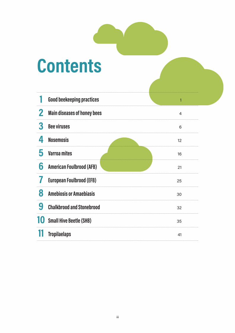
ContentsGood beekeeping practices 1
Main diseases of honey bees 4
Bee viruses 6
Nosemosis 12
Varroa mites 16
American Foulbrood (AFB) 21
European Foulbrood (EFB) 25
Amebiosis or Amaebiasis 30
Chalkbrood and Stonebrood 32
Small Hive Beetle (SHB) 35
Tropilaelaps 41
o
1234567891011
iv
ContributorsThe good beekeeping practices contained in this catalogue have been developed in collaboration with the International Federation of Beekeepers’ Associations (APIMONDIA) (A. Menegotto and R. Jannoni-Sebastianini), Istituto Zooprofilattico Sperimentale del Lazio e della Toscana (IZSLT) “M. Aleandri” (G. Formato and J. Rivera-Gomis), and the BPractices project, with the support of the Food and Agriculture Organization of the United Nations’ Animal Health Service and Research and Extension Unit.
Organizations interested in developing and sharing their technologies and practices on TECA can contact the TECA Team.
Good beekeeping practices
Summary
Apiculture (beekeeping) is the practice of honeybee management in hives for pollination, production of honey and other products such as wax, royal jelly, propolis and pollen. In addition, an important aspect of beekeeping is the production of bees, queens, package bees, etc. The best beekeeping practices involve proper management of the apiary that can prevent bee diseases and, at the same time, allow obtain high quality products respecting the consumer’s health.
Description
Good beekeeping practices that normally should be adopted in the apiary involve:
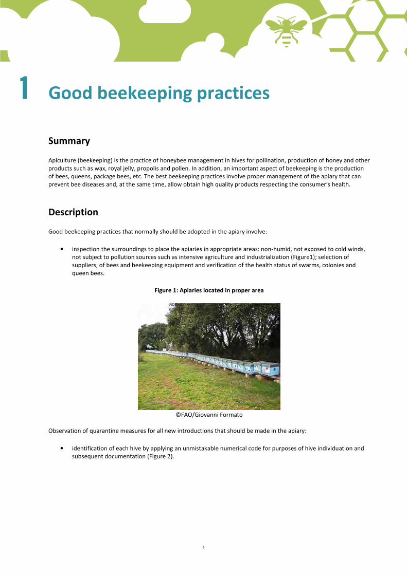
• inspection the surroundings to place the apiaries in appropriate areas: non-humid, not exposed to cold winds, not subject to pollution sources such as intensive agriculture and industrialization (Figure1); selection of suppliers, of bees and beekeeping equipment and verification of the health status of swarms, colonies and queen bees.
Figure 1: Apiaries located in proper area
©FAO/Giovanni Formato
Observation of quarantine measures for all new introductions that should be made in the apiary:
• identification of each hive by applying an unmistakable numerical code for purposes of hive individuation and subsequent documentation (Figure 2).
iv 1
1
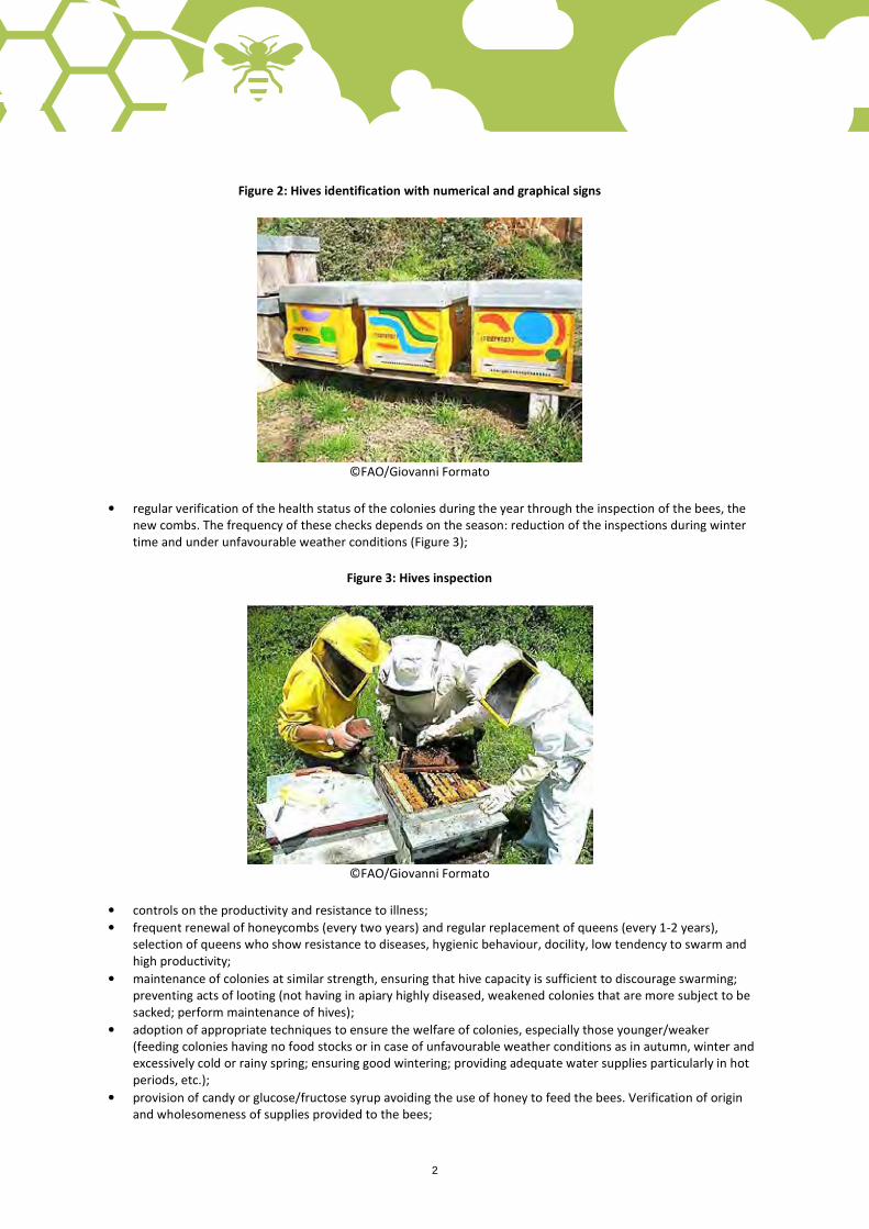
Figure 2: Hives identification with numerical and graphical signs
©FAO/Giovanni Formato
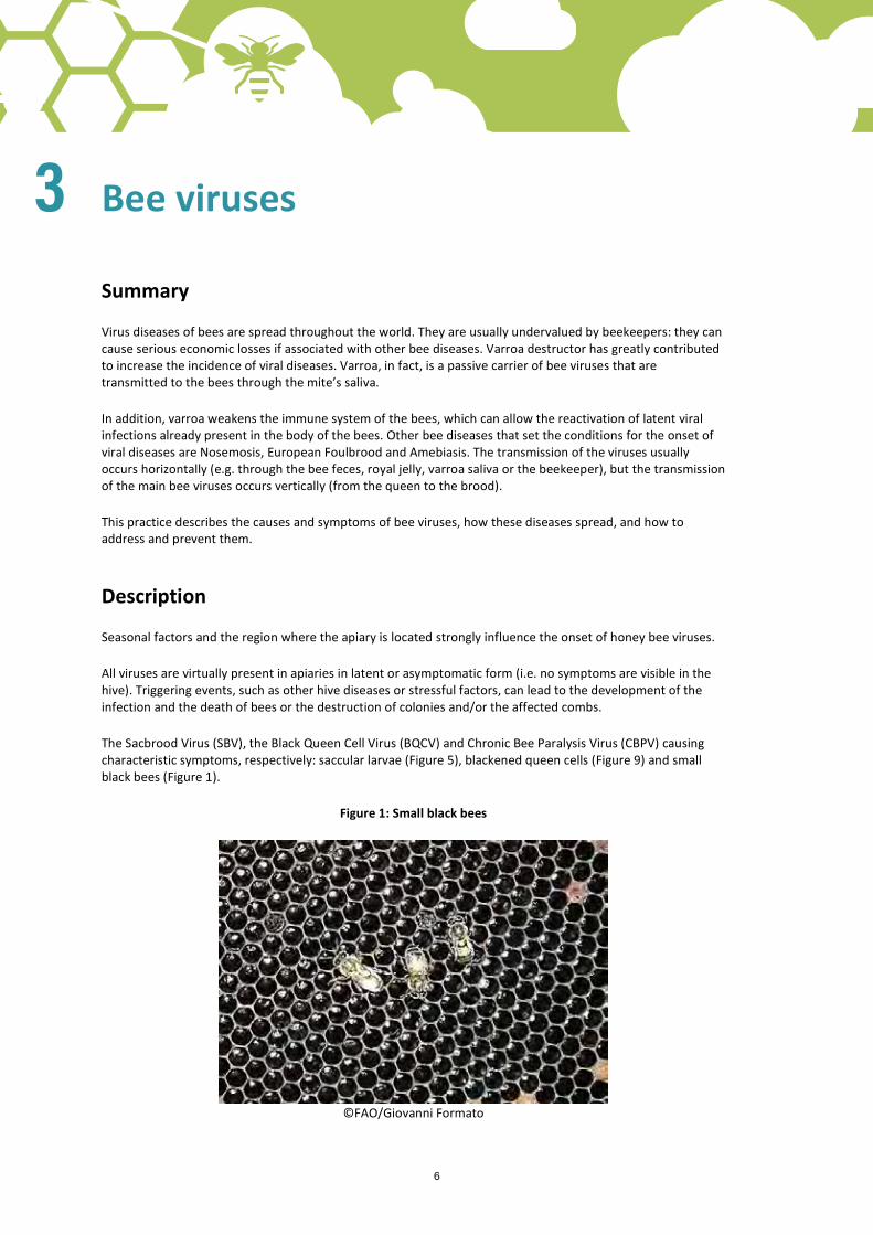
• regular verification of the health status of the colonies during the year through the inspection of the bees, the new combs. The frequency of these checks depends on the season: reduction of the inspections during winter time and under unfavourable weather conditions (Figure 3);
Figure 3: Hives inspection
©FAO/Giovanni Formato
• controls on the productivity and resistance to illness; • frequent renewal of honeycombs (every two years) and regular replacement of queens (every 1-2 years),
selection of queens who show resistance to diseases, hygienic behaviour, docility, low tendency to swarm and high productivity;
• maintenance of colonies at similar strength, ensuring that hive capacity is sufficient to discourage swarming; preventing acts of looting (not having in apiary highly diseased, weakened colonies that are more subject to be sacked; perform maintenance of hives);
• adoption of appropriate techniques to ensure the welfare of colonies, especially those younger/weaker (feeding colonies having no food stocks or in case of unfavourable weather conditions as in autumn, winter and excessively cold or rainy spring; ensuring good wintering; providing adequate water supplies particularly in hot periods, etc.);
• provision of candy or glucose/fructose syrup avoiding the use of honey to feed the bees. Verification of origin and wholesomeness of supplies provided to the bees;
2

• appropriate use of the bee smoker (respecting the bees’ welfare and avoiding using toxic material that cancontaminate the honey);
• elimination of the use of toxic substances or paints for hives (e.g. disinfectants, chemical treatments for wood,etc.);
• elimination of the transfer of honeycombs from one colony to another if the health status of the colonies isunknown. Separation of the sick from the healthy hives; destroying, if necessary, infected colonies;
• exclusive application of drugs registered for use in bees respecting instructions and guidelines and recordingdrug use in the logbook. Improper and untimely use of chemicals during honey production may lead to itscontamination;
• periodic mowing of grass in front of the hives;• maintenance of the apiary and the beekeeping equipment in good order and clean; ensuring the required
maintenance and, when necessary, renewing the materials;• referral to expert assistance in case of anomalies, whenever necessary.
Please note that applying good beekeeping practices in the apiary does not mean the bees will not get sick any more, but the incidence of diseases will decrease.
Further reading
Maine State Beekeepers Association, Inc. 2007. Best Management Practices for Beekeeping, (http://mainebeekeepers.org/beekeeping-resources/best-management-practices-for-beekeeping/).
Dr Somerville, D. 2007. The Australian Honey Bee Industry Council, 2007 National Best Management Practice for Beekeeping in the Australian Environment (http://honeybee.org.au/pdf/NBPFBIAE.pdf).
Ritter, W. Veterinary Institute Freiburg (CVUA-Freiburg) 2013. Good Beekeeping practice - knowledge in a Nutshell, Bees for Development Journal 107 (http://www.beesfordevelopment.org/phocadownload/userupload/bfd%20j107%20good%20beekeeping%20practice.pdf).
Heintz, C., Ribotto, M., Ellis, M., Delaplane, K.S. 2011. Best Management Practices (BMPs) for Beekeepers Pollinating California’s Agricultural Crops, jointly published in the American Bee Journal and in Bee Culture (http://www.beeccdcap.uga.edu/documents/bmpcalagr.html).
Formato, G., Vari G. 2007. Le buone prassi di allevamento in apiario. In “Aspetti igienico-sanitari in apicoltura” published by IZSLT (8-10).
Formato, G. (IZSLT), Smulders F.J.M. (Department of Production Animal Medicine and Veterinary Public Health, University of Veterinary Medicine, Vienna, Austria) 2011. Risk management in primary apicultural production (Part 1: bee health and disease prevention and associated best practices).
Source
APIMONDIA
IZSLT
2 3

Main diseases of honey bees
Summary
Honeybees are susceptible to various diseases, some of which are very contagious and diffusive. It is very important that the beekeeper is able to recognize the first signs of disease or infestation in hives and knows how to proceed. This practice outlines the factors that play an important role in the outbreak of a disease and describes the classification of bee diseases.
Description
The occurrence of diseases in honeybees depends on three factors:
1. Bees (genetic): the hygienic behavior and resistance to various diseases varied from colony to colony and it is based on the genetic heritage of the queen bees. 2. Pathogens (presence, infectious load and virulence): the disease needs the presence of the responsible agent to manifest itself (virus, bacteria, fungus and protozoa), but the quantity and ability to spread of the pathogen is also very important. 3. Environment (temperature, relative humidity, presence of nectar plants): environmental conditions and seasonal factors strongly influence the onset of diseases, in many cases they are real key triggers.
CLASSIFICATION OF BEE DISEASES
The diseases of honeybees can be classified depending on:
• the nature of the agent responsible for the disease: parasitic, fungal, bacterial or viral infection (Table 1). This type of classification is more accurate;
• the function of the individuals who are affected in the hive: brood diseases (Table 2) and diseases of adult bees (Table 3).
Table 1: Main diseases of honeybees depending on the nature of the causative agent
Disease Causative agent Type Acariasis Acarapis woodi Parasitic Varroatosis Varroa destructor Parasitic Aethinosis Aethina Tumida (Small hive beetle) Parasitic Tropilaelapsosis Tropilaelaps spp. Parasitic American foulbrood Paenibacillus larvae Bacterial European foulbrood Melissococcus pluton Bacterial Chalkbrood Ascosphera apis Fungal Stonebrood Aspergillus flavus Fungal Nosemosis Nosema apis – Nosema ceranae Fungal Amebiasis Malpighamoeba mellificae Protozoal Sacbrood Virus (SBV) Virus Picorna-like Viral Chronic Bee Paralysis Virus (CBPV) Cripaviridae Viral Acute Bee Paralysis Virus (ABPV) Dicistroviridae Viral Deformed Wing Virus (DWV) Iflaviridae Viral Black Queen Cell Virus (BQCV) Dicistroviridae Viral Israeli Acute Paralisysis Virus (IAPV) Dicistroviridae Viral Kashmir Bee Virus (KBV) Dicistroviridae Viral Kakugo Virus Iflaviridae Viral Invertrebrate Iridescent Virus type 6 Iridoviridae Viral Tobacco ringspot virus Secoviridae Viral 4
2

Main diseases of honey bees
Summary
Honeybees are susceptible to various diseases, some of which are very contagious and diffusive. It is very important that the beekeeper is able to recognize the first signs of disease or infestation in hives and knows how to proceed. This practice outlines the factors that play an important role in the outbreak of a disease and describes the classification of bee diseases.
Description
The occurrence of diseases in honeybees depends on three factors:
1. Bees (genetic): the hygienic behavior and resistance to various diseases varied from colony to colony and it is based on the genetic heritage of the queen bees. 2. Pathogens (presence, infectious load and virulence): the disease needs the presence of the responsible agent to manifest itself (virus, bacteria, fungus and protozoa), but the quantity and ability to spread of the pathogen is also very important. 3. Environment (temperature, relative humidity, presence of nectar plants): environmental conditions and seasonal factors strongly influence the onset of diseases, in many cases they are real key triggers.
CLASSIFICATION OF BEE DISEASES
The diseases of honeybees can be classified depending on:
• the nature of the agent responsible for the disease: parasitic, fungal, bacterial or viral infection (Table 1). This type of classification is more accurate;
• the function of the individuals who are affected in the hive: brood diseases (Table 2) and diseases of adult bees (Table 3).
Table 1: Main diseases of honeybees depending on the nature of the causative agent
Disease Causative agent Type Acariasis Acarapis woodi Parasitic Varroatosis Varroa destructor Parasitic Aethinosis Aethina Tumida (Small hive beetle) Parasitic Tropilaelapsosis Tropilaelaps spp. Parasitic American foulbrood Paenibacillus larvae Bacterial European foulbrood Melissococcus pluton Bacterial Chalkbrood Ascosphera apis Fungal Stonebrood Aspergillus flavus Fungal Nosemosis Nosema apis – Nosema ceranae Fungal Amebiasis Malpighamoeba mellificae Protozoal Sacbrood Virus (SBV) Virus Picorna-like Viral Chronic Bee Paralysis Virus (CBPV) Cripaviridae Viral Acute Bee Paralysis Virus (ABPV) Dicistroviridae Viral Deformed Wing Virus (DWV) Iflaviridae Viral Black Queen Cell Virus (BQCV) Dicistroviridae Viral Israeli Acute Paralisysis Virus (IAPV) Dicistroviridae Viral Kashmir Bee Virus (KBV) Dicistroviridae Viral Kakugo Virus Iflaviridae Viral Invertrebrate Iridescent Virus type 6 Iridoviridae Viral Tobacco ringspot virus Secoviridae Viral
Table 2: Main brood diseases Varroatosis
Small hive beetle
Tropilaelapsosis
American foulbrood
European foulbrood
Chalkbrood
Stonebrood
Black queen cell virus (BQCV)
Sacbrood virus (SBV)
Other Virosis
Table 3: Main adult bee diseases Varroatosis
Nosemosis
Virosis
Further reading
FAO, 2006. Honey bee diseases and pests: a practical guide (http://teca.fao.org/sites/default/files/resources/Honey%20bee%20diseases%20and%20pests%20-%20a%20practical%20guide.pdf). Fries I., Camazine S. 2001. Implications of horizontal and vertical pathogen transmission for honey bee epidemiology, INRA/DIB-AGIB/EDP Sciences (http://www.apidologie.org/index.php?option=com_article&access=standard&Itemid=129&url=/articles/apido/pdf/2001/03/fries.pdf).
Formato G., Vari G. 2007. Le buone prassi di allevamento in apiario, in “Aspetti igienico-sanitari in apicoltura” (8-10), published by IZSLT.
Source
APIMONDIA
IZSLT
4 5
6
Bee viruses
Summary
Virus diseases of bees are spread throughout the world. They are usually undervalued by beekeepers: they can cause serious economic losses if associated with other bee diseases. Varroa destructor has greatly contributed to increase the incidence of viral diseases. Varroa, in fact, is a passive carrier of bee viruses that are transmitted to the bees through the mite’s saliva.
In addition, varroa weakens the immune system of the bees, which can allow the reactivation of latent viral infections already present in the body of the bees. Other bee diseases that set the conditions for the onset of viral diseases are Nosemosis, European Foulbrood and Amebiasis. The transmission of the viruses usually occurs horizontally (e.g. through the bee feces, royal jelly, varroa saliva or the beekeeper), but the transmission of the main bee viruses occurs vertically (from the queen to the brood).
This practice describes the causes and symptoms of bee viruses, how these diseases spread, and how to address and prevent them.
Description
Seasonal factors and the region where the apiary is located strongly influence the onset of honey bee viruses.
All viruses are virtually present in apiaries in latent or asymptomatic form (i.e. no symptoms are visible in the hive). Triggering events, such as other hive diseases or stressful factors, can lead to the development of the infection and the death of bees or the destruction of colonies and/or the affected combs.
The Sacbrood Virus (SBV), the Black Queen Cell Virus (BQCV) and Chronic Bee Paralysis Virus (CBPV) causing characteristic symptoms, respectively: saccular larvae (Figure 5), blackened queen cells (Figure 9) and small black bees (Figure 1).
Figure 1: Small black bees
©FAO/Giovanni Formato
3
6 7
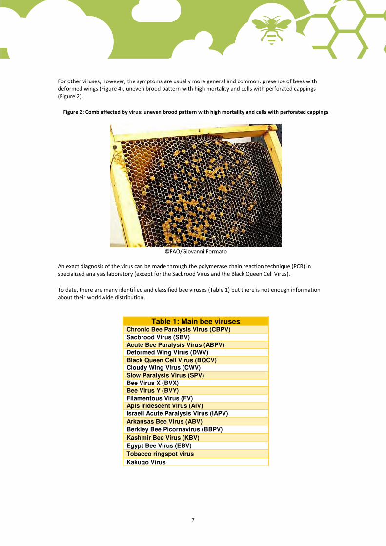
For other viruses, however, the symptoms are usually more general and common: presence of bees with deformed wings (Figure 4), uneven brood pattern with high mortality and cells with perforated cappings (Figure 2).
Figure 2: Comb affected by virus: uneven brood pattern with high mortality and cells with perforated cappings
©FAO/Giovanni Formato
An exact diagnosis of the virus can be made through the polymerase chain reaction technique (PCR) in specialized analysis laboratory (except for the Sacbrood Virus and the Black Queen Cell Virus).
To date, there are many identified and classified bee viruses (Table 1) but there is not enough information about their worldwide distribution.
Table 1: Main bee viruses Chronic Bee Paralysis Virus (CBPV)
Sacbrood Virus (SBV)
Acute Bee Paralysis Virus (ABPV)
Deformed Wing Virus (DWV)
Black Queen Cell Virus (BQCV)
Cloudy Wing Virus (CWV)
Slow Paralysis Virus (SPV)
Bee Virus X (BVX)
Bee Virus Y (BVY)
Filamentous Virus (FV)
Apis Iridescent Virus (AIV)
Israeli Acute Paralysis Virus (IAPV)
Arkansas Bee Virus (ABV)
Berkley Bee Picornavirus (BBPV)
Kashmir Bee Virus (KBV)
Egypt Bee Virus (EBV)
Tobacco ringspot virus
Kakugo Virus

8
CHRONIC BEE PARALYSIS VIRUS (CBPV or CPV)
This is an infectious and contagious disease of adult honeybees caused by a virus (CBPV). The infection has no seasonal pattern, often remains latent and is present in many countries. The CBPV is more frequently found in colonies infested with varroa.
The CBPV is the only common viral disease of adult bees that has well-described symptoms, for this it has been given a variety of names, such as “hairless black syndrome” and “little blacks“.
Affected bees become almost hairless, dark in appearance and suffer nibbling attacks from healthy bees of their colony. They become wobbly and flightless in the upper part of the honeycomb, crawling on the ground and on the stems of grass, where they die (Figure 3).
Figure 3: Wobbly and flightless bees in the upper part of the honeycomb
©FAO/Giovanni Formato
Some bees present enlarged abdomen due to accumulation of liquid in the honey sac and wings spread out in “K“ form. Sick individuals die within a few days of the onset of symptoms.
Thousands of paralyzed bees from each colony die throughout the year and severely affected colonies can suddenly collapse.
The cells are hollow inside and the larvae become yellowish-brownish. Their internal organs become fluid while the integument remains intact, assuming the typical "saccular" larvae aspect when they die (Figure 5).
Figure 5: Saccular larva typical of Sacbrood virus
©FAO/Giovanni Formato

8 9
Subsequently, the infected larvae dry taking the form of blackish mummified flakes (Figure 6). Normally no smell is present even if, sometimes, the brood may have a mild sour smell.
Figure 6: The infected larvae dry taking the form of blackish mummified flakes
©FAO/Giovanni Formato
The infection has a typical seasonal pattern with higher incidence in spring and early summer, and normally disappearing by the autumn.
However, associated to varroa, these viruses can cause devastating effects and take epidemic form. The virus is transmitted to the larvae by the royal jelly and to the bees when they go to clean up the infected cells from dead larvae. Recently it has also been demonstrated that the virus is passed on from the queen to the brood (i.e. vertical transmission).
ACUTE BEE PARALYSIS VIRUS (ABPV)
This virus can normally be found in the fatty tissue of the bee and does not cause symptoms. Combined with varroa, the infection becomes particularly serious, causing mortality both in brood and adult bees. This virus is usually combined with the CBPV, however in case of massive varroa infestation ABPV prevails on CBPV because of its rapid replication activity.
DEFORMED WING VIRUS (DWV)
This virus is relatively widespread in apiaries, although often present in subclinical form if not associated with varroa (no symptoms are visible). However, in combination with varroa this virus can cause the death of the brood and of adult bees. This virus affects immature bees during their development in the cells. Unlike the ABPV, it is characterised by a very slow replication cycle, generally allowing the bees to fly despite the serious wings deformations, the reduced body size and very short life expectancy (Figure 7).

10
Figure 7: Bee with normal wings (left) and another with deformed ones (right)
©FAO/Giovanni Formato
SACBROOD VIRUS (SBV)
This is a Picorna-like virus not very resistant to chemical, physical and environmental agents (it dies in 10 minutes at 55-65 °C and resists six days to direct sunlight; in honey, it loses its virulence after 5-6 weeks). SBV infects the young bee larvae orally. The symptoms are evident in larvae in capped cells (Figure 8), while the adults are asymptomatic. The capped cells are hollow inside and the larvae first become yellowish-brownish, then the internal organs become fluid while the integument remains intact assuming the typical "saccular" aspect (Figure 5).
Figure 8: Sacbrood Virus (SBV)
©FAO/FERA
BLACK QUEEN CELL VIRUS (BQCV)
This virus only affects the cells of queen bees and is one of the most frequent causes of mortality among the queen larvae. The name of the virus comes from the blackish colour of larval forms and the cell walls (Figure 9). Although worker bee and drone brood can be infected by BQCV, generally these do not develop any kind of symptoms. The infection is more common when colonies are affected by nosemosis as the lesions of the small intestine facilitate the passage of the virus in the hemolymph.

10 11
Figure 9: Black Queen Cell Virus (BQCV)
©FAO/Giovanni Formato
VIRUSES PREVENTION AND CONTROL
There are not still specific and effective therapeutic remedies for the viral diseases of bees. In the case of particularly severe symptoms, the only remedy is the destruction of affected colonies. In other cases where the symptoms are less severe, you can try to replace the queen and the infected honeycombs, which will be destroyed.
The infected hives must be properly cleaned and disinfected before being used (disinfection can be carried out with bleach and then passing a blue flame on the hives).
Because of the demonstrated transovarian transmissibility for some viruses (an infected queen bee can produce infected eggs and brood), when introducing new queens in the apiary it is recommended to observe a quarantine period and monitoring of the health of the brood.
Good beekeeping practices are essential to prevent diseases and the stress factors should be kept to a minimum level, in fact they may serve as predisposing factors for the viruses, such as: chemical (e.g. drug treatments), physical (e.g. frequent visits in winter), metabolic and infectious (it is fundamental to keep varroa and nosema infestation under control).
Further reading
Formato, G., Cardeti, G. 2007. Le virosi delle api. In “Aspetti igienico-sanitari in apicoltura” (34-39) published by IZSLT
Source
APIMONDIA
IZSLT
12
Nosemosis
Summary
Nosemosis is a disease of adult bees caused by unicellular fungi belonging to the class: Microsporidia, family: Nosimatidi, gender: Nosema.
There are two different sub-species of Nosema that affect Apis mellifera with different prevalence depending on the area: Nosema apis (N. apsi) and Nosema ceranae (N. ceranae), responsible for two different forms of the disease. Both N. apis and N. ceranae have a dormant stage, a long-lived spore.
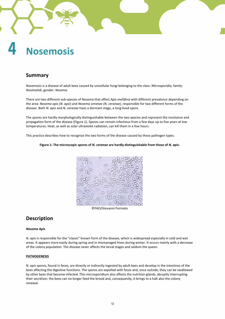
The spores are hardly morphologically distinguishable between the two species and represent the resistance and propagation form of the disease (Figure 1). Spores can remain infectious from a few days up to five years at low temperatures. Heat, as well as solar ultraviolet radiation, can kill them in a few hours.
This practice describes how to recognize the two forms of the disease caused by these pathogen types.
Figure 1: The microscopic spores of N. ceranae are hardly distinguishable from those of N. apis.
©FAO/Giovanni Formato
Description
Nosema Apis
N. apis is responsible for the "classic" known form of the disease, which is widespread especially in cold and wet areas. It appears more easily during spring and in mismanaged hives during winter. It occurs mainly with a decrease of the colony population. The disease never affects the larval stages and seldom the queen.
PATHOGENESIS
N. apis spores, found in feces, are directly or indirectly ingested by adult bees and develop in the intestines of the bees affecting the digestive functions. The spores are expelled with feces and, once outside, they can be swallowed by other bees that become infected. This microsporidium also affects the nutrition glands, abruptly interrupting their secretion: the bees can no longer feed the brood and, consequently, it brings to a halt also the colony renewal.
4
12 13
SYMPTOMS
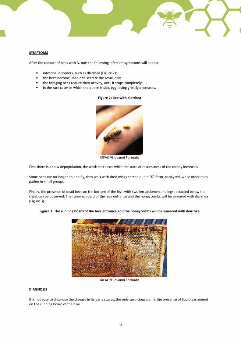
After the contact of bees with N. apis the following infection symptoms will appear:
• intestinal disorders, such as diarrhea (Figure 2); • the bees become unable to secrete the royal jelly; • the foraging bees reduce their activity, until it stops completely; • in the rare cases in which the queen is sick, egg-laying greatly decreases.
Figure 2: Bee with diarrhea
©FAO/Giovanni Formato
First there is a slow depopulation, the work decreases while the state of restlessness of the colony increases.
Some bees are no longer able to fly, they walk with their wings spread out in "K" form, paralyzed, while other bees gather in small groups.
Finally, the presence of dead bees on the bottom of the hive with swollen abdomen and legs retracted below the chest can be observed. The running board of the hive entrance and the honeycombs will be smeared with diarrhea (Figure 3).
Figure 3: The running board of the hive entrance and the honeycombs will be smeared with diarrhea
©FAO/Giovanni Formato
DIAGNOSIS
It is not easy to diagnose the disease in its early stages; the only suspicious sign is the presence of liquid excrement on the running board of the hive.

14
A field test consists of examining the colour of the terminal portion of the digestive system of some bees: in healthy bees it has a reddish colour, while in sick bees it is milky white. This sign, however, is seen only when the disease has already reached a certain severity. Only a laboratory test can make an early diagnosis by searching with the microscope the spores at intestinal level or directly on feces.
TRANSMISSION
Transmission occurs primarily by fecal-oral route. N. apis can easily spread through the droppings of sick bees, especially within the hive.
The spread from hive to hive and apiary to apiary may occur through:
• drifting of infected workers; • drone displacement; • looting of infected colonies; • interchanging infected honeycombs from a hive to another; • feeding of bees with contaminated honey; • use of infected materials or equipment.
This disease is influenced by many climatic factors:
• bad weather increases the chances of infection among the bees of the same hive because it forces the bees indoors;
• seasonal pattern can also affect the spread of infection. During long, cold winters, and cold, rainy springs the bees may not find nectar and pollen;
• frequent hive inspections with adverse weather conditions (e.g. winter season, windy or rainy weather) can trigger the onset of the disease as well as its propagation due to the induced stress;
• the presence of other diseases (such as amebiasis or viruses) exacerbates the symptoms of N. apis.
CONTROL
• prevent the infection by adopting good management practices and by taking special care when selecting the apiary location (non-humid, not exposed to cold winds) and the correct orientation of the hives (prefer sunny and slightly ventilated areas);
• adopt correct wintering measures (removing honeycombs not populated by bees, providing good quality food if necessary and applying appropriate treatments against varroa);
• place pollen plants near the hives that can provide protein food to the colony in the late summer and autumn;
• in cold climates, keep the hives warm during wintering until late spring; • use an adequate number of honeycombs in relation to the colony population; • disturb the bees as little as possible during winter.
Unfortunately, when N. apis occurs, the prognosis is frequently serious because its onset is almost always unnoticed and symptoms occur only at an advanced stage. Generally, the affected colonies do not heal spontaneously, therefore the beekeeper’s intervention becomes necessary. If the disease is well developed, particularly in weak families its destruction is definitely suggested.
It is possible to retrieve the materials after killing the bees, sterilizing the hives (with boiling water, soda six percent and blue flame) and destroying the combs. Infected honey and pollen should absolutely not be used to feed other bees to avoid their infection. In case the affected family is very strong, move it in an area exposed to the sun (not windy and cold), with clean hive and combs thus decreasing the possibility of re-infection from diarrhea and provide proper feeding (e.g. molasses, herbs or medicated feed).

14 15
The infected combs should be destroyed and the hive should be sterilized as mentioned above or destroyed. The honey can be used for human consumption. To destroy a hive and avoid further contamination, a hole deep at least 50 cm should be dug in the ground, the hive and combs should be burnt and the hole should be duly covered.
Nosema Ceranae
N. ceranae is a new species of microsporidium isolated for the first time in 1996 by Fries on Apis cerana, a bee species widespread in Southeast Asia.
In 2006, it was isolated for the first time by Higes in Apis mellifera. N. ceranae has spread in vast areas of Europe replacing the indigenous form of N. apis on Apis mellifera, resulting in quite different clinical signs from the classical nosemosis. Typical of this disease are the severe injuries and the absence of gastro-enteritis (diarrhea) as a typical symptom and the appearance of the disease in different periods from those of N. apis. It was listed among the possible causes of depopulation of the hives, even though its pathogenic effect on the honeybees is still unclear.
PATHOGENESIS
The ingestion of microsporidium by the bees occurs directly or indirectly (e.g. through honey contaminated by the spores). N. apis develops and attacks the intestines of the bees inducing malabsorption. The spores of N. ceranae are very resistant in the environment (they can withstand very cold or very hot temperatures), facilitating the re-infection of the colonies and the recurrence of the disease after a long time.
SYMPTOMS
The disease can occur throughout the year. Typical is the absence of diarrhea in foraging bees. It seems that they go to die away from the hive, causing a progressive depopulation of the colonies (without noticing the presence of dead bees) until the total loss of the family.
DIAGNOSIS
The microscopic spores of N. ceranae are hardly morphologically distinguishable from those of N. apis. It is possible to make a diagnosis only through the Polymerase chain reaction (PCR), a biology molecular technique, which allows the sequencing of a very specific and characteristic part of N. ceranae genome on the spores. Cost and availability of this exam depends on each country and laboratory.
Further reading
Palazzetti, M. 2007. La nosemosi, in “Aspetti igienico-sanitari in apicoltura” (22-24) published by IZSLT.
Source
APIMONDIA
IZSLT
16
Varroa mites (Varroatosis or Varroosis)
Summary
Varroa destructor (Figure 1) is the mite responsible of Varroatosis (or Varroosis), an external parasitic disease that attacks honeybee colonies (adult bees and especially the brood). Varroa destructor causes the major economic losses to the beekeeping sector because it is widespread and it has a strong adaptability to the treatments.
This mite affects both the brood and the adult bees. It weakens the adult bees by sucking their hemolymph. The weakened bees are more susceptible to other diseases, especially viral pathologies. The first to suffer are the stronger colonies with more brood because of the higher possibility of the mite to replicate at the brood level.
Description
MORPHOLOGY
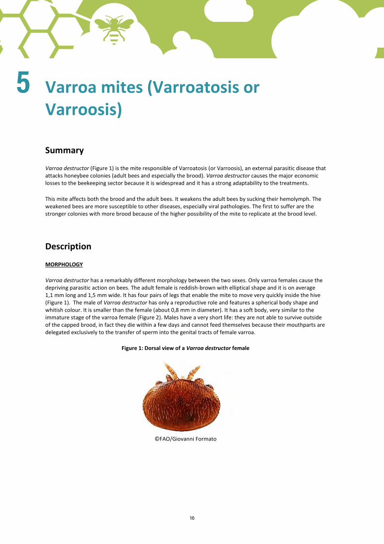
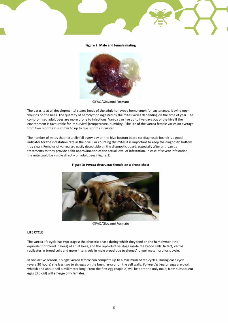
Varroa destructor has a remarkably different morphology between the two sexes. Only varroa females cause the depriving parasitic action on bees. The adult female is reddish-brown with elliptical shape and it is on average 1,1 mm long and 1,5 mm wide. It has four pairs of legs that enable the mite to move very quickly inside the hive (Figure 1). The male of Varroa destructor has only a reproductive role and features a spherical body shape and whitish colour. It is smaller than the female (about 0,8 mm in diameter). It has a soft body, very similar to the immature stage of the varroa female (Figure 2). Males have a very short life: they are not able to survive outside of the capped brood, in fact they die within a few days and cannot feed themselves because their mouthparts are delegated exclusively to the transfer of sperm into the genital tracts of female varroa.
Figure 1: Dorsal view of a Varroa destructor female
©FAO/Giovanni Formato
5
16 17
Figure 2: Male and female mating
©FAO/Giovanni Formato
The parasite at all developmental stages feeds of the adult honeybee hemolymph for sustenance, leaving open wounds on the bees. The quantity of hemolymph ingested by the mites varies depending on the time of year. The compromised adult bees are more prone to infections. Varroa can live up to five days out of the hive if the environment is favourable for its survival (temperature, humidity). The life of the varroa female varies on average from two months in summer to up to five months in winter.
The number of mites that naturally fall every day on the hive bottom board (or diagnostic board) is a good indicator for the infestation rate in the hive. For counting the mites it is important to keep the diagnostic bottom tray clean. Females of varroa are easily detectable on the diagnostic board, especially after anti-varroa treatments as they provide a fair approximation of the actual level of infestation. In case of severe infestation, the mite could be visible directly on adult bees (Figure 3).
Figure 3: Varroa destructor female on a drone chest
©FAO/Giovanni Formato
LIFE CYCLE
The varroa life cycle has two stages: the phoretic phase during which they feed on the hemolymph (the equivalent of blood in bees) of adult bees, and the reproductive stage inside the brood cells. In fact, varroa replicates in brood cells and more intensively in male brood due to drones’ longer metamorphosis cycle.
In one active season, a single varroa female can complete up to a maximum of ten cycles. During each cycle (every 30 hours) she lays two to six eggs on the bee's larva or on the cell walls. Varroa destructor eggs are oval, whitish and about half a millimeter long. From the first egg (haploid) will be born the only male; from subsequent eggs (diploid) will emerge only females.

18
The parasites colonize the brood when the bee larvae have six days of life. Within 24 hours a hexapod larva develops; after another 24 hours became a protonymph and emerges from the egg (Figure 4) The newly born varroa parasites start immediately feeding on the developing bee.
Figure 4: Protonymph (left), deutonymph (center), adult (right) of Varroa destructor female
©FAO/G. San Martin
The varroa reproduction period is related to the duration of the metamorphosis of the pupae in the brood cells: 12 days for the worker bees and 15 days for the drones. If the varroa male dies before mating, females remain irreversibly sterile and unable to procreate because of an involution of their genitalia.
The varroa feeding site is always located in the abdomen of the pupa in order not to compromise its survival. At the time of the bee‘s emergence from the cocoon, the varroa offspring is inside the cell. The varroa adult daughters that emerge from the cell as the young bee is leaving the same cell will try to jump on adult bees where they will spend their phoretic stage (stage during which they feed on adult bee hemolymph) before entering the brood cell where they will reproduce.
SYMPTOMS
In bees’ colonies contaminated by varroa it is possible to observe:
• the parasites on the body of adult bees; • scattered brood (index of high mortality of larvae) (Figure 5); • a typical stench of dead brood; • smaller bees (Figure 6); • bees with deformed wings (Figure 7); • clusters of bees restless and unable to fly; • weakening of the colony as it becomes less populated and due to the reduced capacity of the bees in the
collection and storage of supplies; • abnormal swarming (especially at the end of the season) and replacement of the queen.
Figure 5: Comb with scattered brood
©FAO/Giovanni Formato

18 19
Figure 6: Smaller bee on the left; healthy bee on the right
©FAO/Charlotte Lietaer
Figure 7: Healthy honey bee with normal wings (left) and parasitized honey bee with deformed wings (right)
©FAO/Charlotte Lietaer
The average lifespan of adult bees in heavily parasitized colonies decreases ranging from 25 to 50 percent. Varroa not only sucks the hemolymph of the larvae and adult bees, it also causes little wounds on the body of the bees and makes them more vulnerable to other pathogens such as viruses, fungi and bacteria. This effect is augmented also by the possibility of viral multiplication in the salivary glands of varroa.
TRANSMISSION
This parasitic disease is transmitted very easily by direct contact from infested to healthy bees (e.g. during the visit of a flower, by drones who can freely enter different hives, during robbing of infested hives, as effect of drifting of infested worker bees among adjacent hives, etc.). But the transmission may also occur by the direct action of the beekeeper for example by transferring parasitized brood combs from one colony to another or by the migratory beekeeping practice.

20
Moreover, another factor of varroa transmission is linked to migratory beekeeping due to the transfer of heavily infested colonies or due to the delayed application of treatments. In fact this practice increases exponentially the physical contact between healthy and infested colonies.
Hence the importance of simultaneous and coordinated anti-varroa treatments both within the same apiary and between closely located apiaries.
MONITORING
Since the evolution of the disease is not very evident, the monitoring of the number of parasites in each hive through periodic inspections is very important. The diagnosis of infestation can be carried out by:
• checking the number of parasites that fall on the hive bottom; • checking the number of varroa mites affecting the male brood (which is the most affected); • checking if the parasites are visible to the naked eye on adult bees, meaning that there are high levels of
infestation; • applying the World Organization for Animal Health (OIE)-endorsed method by which adult bees are
dipped in alcohol and stirred in order to separate the varroa mites from the bees; • applying the powder sugar empirical method, which entails sprinkling powder sugar on bees collected in
a jar and shaking it to cause the varroa to fall through a mesh as this allows to count the mites easily.
It is interesting to mention that there are some bee subspecies which have the so-called hygienic behaviour by which they groom themselves and are able to contain the varroa infestation level (e.g. Apis mellifera capensis, Apis mellifera intermissa).
WARNING: Please consult the relevant authorities or expert beekeepers in your country for the most efficient treatments and the authorized products in your region/country. It is important to use the treatments properly and according to the prescriptions to avoid creating resistance of varroa mites to the products. Some products can only be used after the harvesting of honey as they can leave residues in the honey.
Further reading
Comini, A., Pietropaoli, M., Giacomelli, A., Formato, G. 2007. Varroa destructor: morfologia e ciclo biologico, published by IZSLT.
Formato, G., Vari, G. 2007. La varroatosi in “Aspetti igienico-sanitari in apicoltura” (11-14) published by IZSLT.
Source
APIMONDIA
IZSLT
20 21
American Foulbrood (AFB)
Summary
American Foulbrood (AFB) is a bacterial disease of honeybees that affects the brood. It is considered the most widespread and destructive bee disease and it can cause serious economic losses to beekeeping. The term "american" does not refer to the fact that the disease has been imported from the United States of America, rather because it has been studied and identified in this country.
This practice describes the causes and symptoms of AFB, how it spreads, and how to address and prevent this disease.
Description
CAUSES
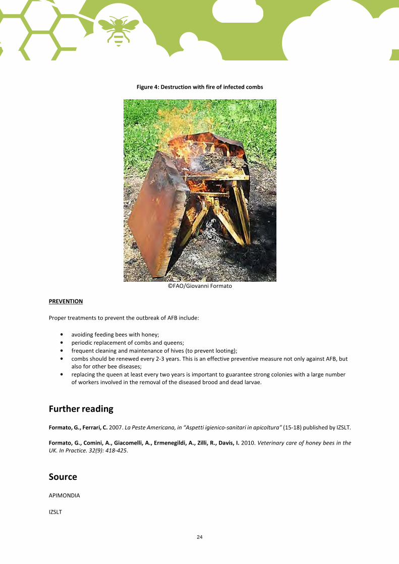
The causative agent of AFB is a spore-forming bacterium, the Paenibacillus larvae (P. larvae). The spores are able to withstand several minutes a temperature of 100 °C and in a suitable environment (e.g. in the intestine of the larvae) a single spore is able to produce 250 million new bacilli after only 24 hours. The spores can remain viable for more than 30 years in an infected hive, being able to contaminate new colonies. This explains why, in severe forms, the only remedy consists in the destruction with fire of both the colonies and the infected combs (Figure 4).
Bee larvae are the main target of P. larvae in their first 24 hours of life. The spores become active in the digestive tract of young larvae. After seven days of infection, the infected larvae die and the P. larvae turns back into the spore form not finding the suitable conditions for development.
SYMPTOMS
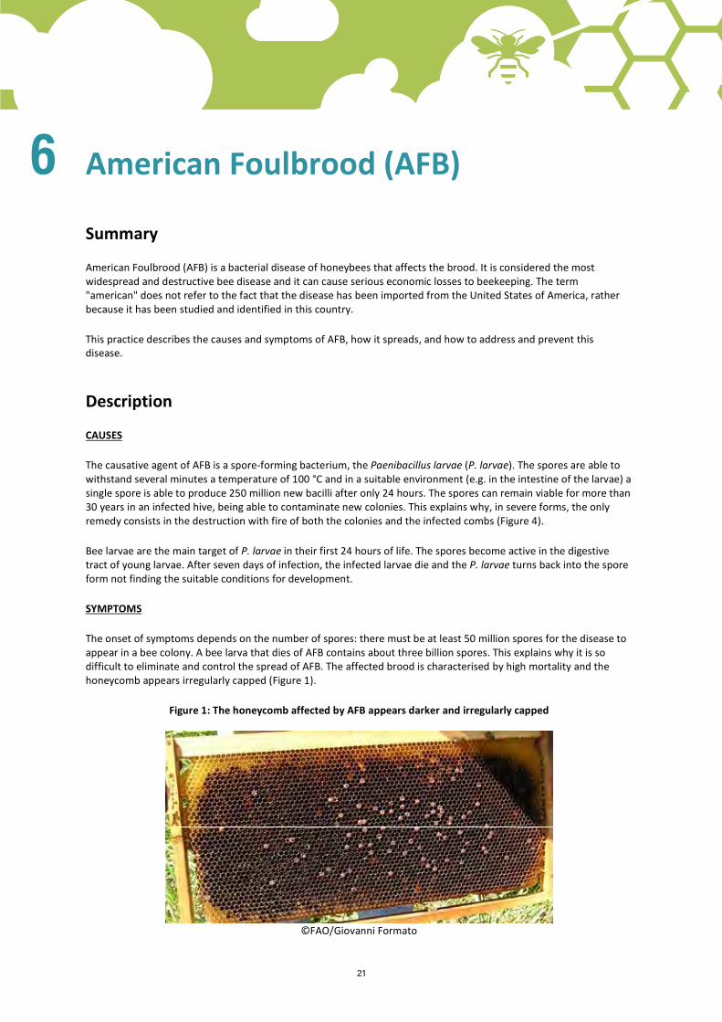
The onset of symptoms depends on the number of spores: there must be at least 50 million spores for the disease to appear in a bee colony. A bee larva that dies of AFB contains about three billion spores. This explains why it is so difficult to eliminate and control the spread of AFB. The affected brood is characterised by high mortality and the honeycomb appears irregularly capped (Figure 1).
Figure 1: The honeycomb affected by AFB appears darker and irregularly capped
©FAO/Giovanni Formato
6
22
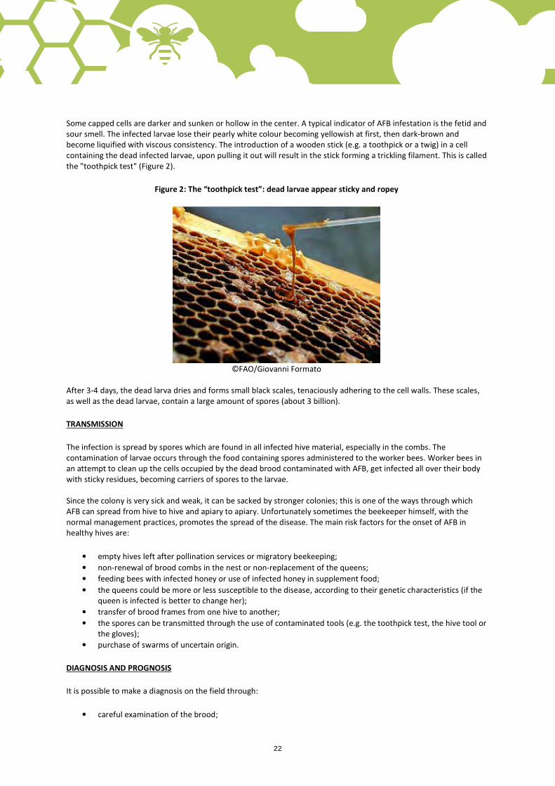
Some capped cells are darker and sunken or hollow in the center. A typical indicator of AFB infestation is the fetid and sour smell. The infected larvae lose their pearly white colour becoming yellowish at first, then dark-brown and become liquified with viscous consistency. The introduction of a wooden stick (e.g. a toothpick or a twig) in a cell containing the dead infected larvae, upon pulling it out will result in the stick forming a trickling filament. This is called the "toothpick test" (Figure 2).
Figure 2: The “toothpick test”: dead larvae appear sticky and ropey
©FAO/Giovanni Formato
After 3-4 days, the dead larva dries and forms small black scales, tenaciously adhering to the cell walls. These scales, as well as the dead larvae, contain a large amount of spores (about 3 billion).
TRANSMISSION
The infection is spread by spores which are found in all infected hive material, especially in the combs. The contamination of larvae occurs through the food containing spores administered to the worker bees. Worker bees in an attempt to clean up the cells occupied by the dead brood contaminated with AFB, get infected all over their body with sticky residues, becoming carriers of spores to the larvae. Since the colony is very sick and weak, it can be sacked by stronger colonies; this is one of the ways through which AFB can spread from hive to hive and apiary to apiary. Unfortunately sometimes the beekeeper himself, with the normal management practices, promotes the spread of the disease. The main risk factors for the onset of AFB in healthy hives are:
• empty hives left after pollination services or migratory beekeeping; • non-renewal of brood combs in the nest or non-replacement of the queens; • feeding bees with infected honey or use of infected honey in supplement food; • the queens could be more or less susceptible to the disease, according to their genetic characteristics (if the
queen is infected is better to change her); • transfer of brood frames from one hive to another; • the spores can be transmitted through the use of contaminated tools (e.g. the toothpick test, the hive tool or
the gloves); • purchase of swarms of uncertain origin.
DIAGNOSIS AND PROGNOSIS
It is possible to make a diagnosis on the field through:
• careful examination of the brood;

22 23
• presence of characteristic symptoms as described above; • positivity of the toothpick test.
Symptoms are usually quite clear. However, in case of doubt, an AFB kit for field diagnosis can be used (Figure 3) or a sample of the suspected brood comb can be sent to a laboratory for microbiologic analysis. Honey samples can be analysed for the presence of AFB spores as a tool for monitoring the presence of AFB in the territory. The prognosis of this disease is serious: when in a hive dead larvae are found in the liquified stage, usually the colony will die in one or a few seasons with high risk to endanger the health of other hives because of the spread of the infections to other colonies.
Figure 3: AFB kit for field diagnosis (positive above, negative below)
©FAO/Giovanni Formato
CONTROL
For all the reasons mentioned above, it is really important to take appropriate actions as soon as possible, such as:
• destruction by incineration of the infected colonies (honeycombs and honey bees; the hives, if in good state, could be disinfected). This action is suggested in case of advanced stage of the disease, weak colonies or low prevalence of the disease in the apiary (Figure 4);
• accurate disinfection of all objects used for the manipulation of infected hives, including equipment used for operations by the beekeeper (e.g. the hive tool, the gloves, the suit, the honey extractor, etc.);
• treatment of the infected colonies. In some countries the treatments against AFB are on-trade (antibiotics). It should be considered that medicines against AFB able to guarantee a total disinfection of the hive from the P. larvae do not exist and the inappropriate use of antibiotics encourages the development of drug resistance and the risk of the presence of residues in the hive products. This action is suggested in case of early stages of the disease, in case of strong colonies and in case of high prevalence of the disease in the apiary;
• shook swarm method, consisting in shaking the hives from the infected combs into a clean hive with new foundation.
24
Figure 4: Destruction with fire of infected combs
©FAO/Giovanni Formato
PREVENTION
Proper treatments to prevent the outbreak of AFB include:
• avoiding feeding bees with honey; • periodic replacement of combs and queens; • frequent cleaning and maintenance of hives (to prevent looting); • combs should be renewed every 2-3 years. This is an effective preventive measure not only against AFB, but
also for other bee diseases; • replacing the queen at least every two years is important to guarantee strong colonies with a large number
of workers involved in the removal of the diseased brood and dead larvae.
Further reading
Formato, G., Ferrari, C. 2007. La Peste Americana, in “Aspetti igienico-sanitari in apicoltura” (15-18) published by IZSLT. Formato, G., Comini, A., Giacomelli, A., Ermenegildi, A., Zilli, R., Davis, I. 2010. Veterinary care of honey bees in the UK. In Practice. 32(9): 418-425.
Source
APIMONDIA
IZSLT

24 25
7
European Foulbrood (EFB)
Summary
European Foulbrood (EFB) is a bacterial disease that affects the honey bee brood. The genetic resistance of some species of bees to this disease may allow, especially in favourable environmental conditions, to overcome the infection without suffering serious damage. However, it should be noted that, even if characterised by a better prognosis than the American Foulbrood, in some areas the EFB has a more malignant manifestation, seriously damaging even very strong bee colonies.
This practice describes the causes and symptoms of EFB, how the disease spreads, and how to address and prevent the disease.
Description
CAUSES
EFB is caused by the streptococcus bacterium Melissococcus pluton (M. pluton), often associated with other bacterial agents, including: Bacillus alvei, Streptococcus faecalis, Achromobacter eurydice, Paenibacillus alvei and Bacillus laterosporus. Depending on the type of bacteria associated with the bacterium M. pluton, the EFB can occur with different symptoms (e.g. the presence/absence of an unpleasant acid smell).
M. pluton is a germ that is quite resistant to adverse environmental conditions (e.g. it remains viable for several months in pollen).
TRANSMISSION
The bacterium develops inside the hive at the brood level. This disease spreads orally inside the hive by the nurse bees that, in an attempt to clean up the cells by the dead larvae, get contaminated with the spores and they transmit them to the brood when they go to feed it.
The disease can be spread from hive to hive or apiary to apiary by the bees (especially when bees rob a diseased hive) and by the beekeeper (with the use of infected honey to feed healthy colonies, moving diseased colonies during migratory beekeeping, trade of infected tools, use of contaminated equipment, moving of combs from one hive to another, etc.).
The disease, while being able to occur throughout the year, is more common in spring when there is more brood. The bacterium can spread through honey with infected combs (through pollen, honey, brood, etc.).
The development of EFB could be favoured by an imbalance between the number of larvae and that of the nurse bees. In addition, the EFB would seem to be more common in cold and rainy springs, where there may be food shortages, particularly of protein for the brood (lack of pollen). It has also been observed that the quality and quantity of the sources of nectar and pollen are able to influence the course of the disease.
The health status of the colony is very important for the development of the disease inside the colony: weak colonies or colonies that are stressed for any reason (food shortage, migratory beekeeping, pesticides, etc.), as well as genetically more sensitive colonies, are especially prone to this disease.
Healthy and strong colonies will be able to recover from the disease by themselves if the season guarantees adequate food sources (pollen, nectars and flowers).

26
SYMPTOMS
The transmission of EFB from the adult bee to the larva takes place orally. After the infection, the larvae die in a few days (regardless of whether the larvae are working bees, drones or queens). Unlike the American Foulbrood, M. pluton kills the larvae before the cells’ capping.
The death of the larvae occurs with open cells and this is one of the features that allows to differentiate the EFB from the AFB (Figure 1). Only in the case of serious infection with EFB, the larvae can die in capped cells.
Figure 1: Larval death with open cells
©FAO/Giovanni Formato
Another important feature useful to recognize this disease is that the affected larvae instead of being horizontally positioned on one side in a C-shape, adhering to the back of the cells, they often change position.
The infected larvae initially lose their pearly white colour to become first opaque, then yellowish and finally yellowish-brown (Figure 2). After death, the larva becomes darker and decomposes, turning into a soft brown mass which is neither viscous nor stringy, unlike the larvae infested with AFB (Figure 3).
Figure 2: The infected larvae initially lose their pearly white colour to become first opaque, then yellowish and finally yellowish-brown
©FAO/Massimo Palazzetti
26 27
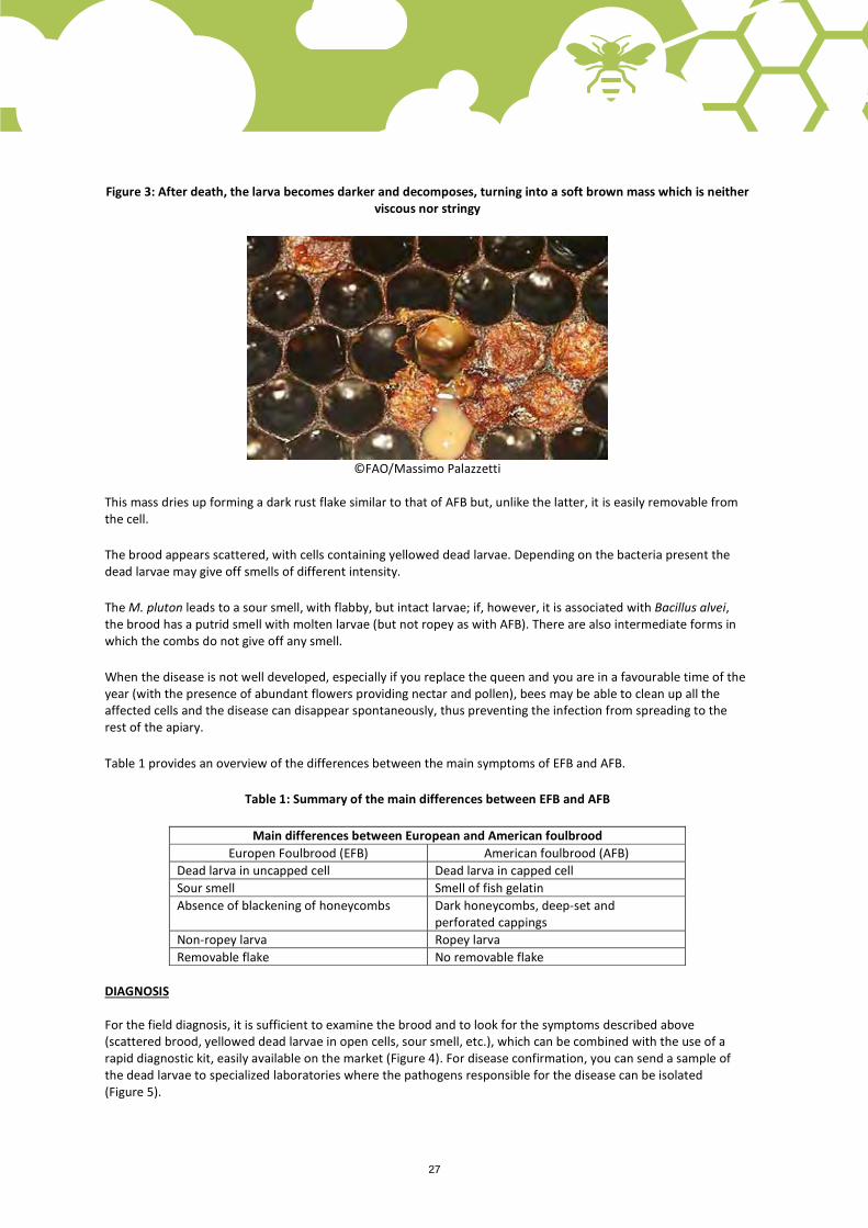
Figure 3: After death, the larva becomes darker and decomposes, turning into a soft brown mass which is neither viscous nor stringy
©FAO/Massimo Palazzetti
This mass dries up forming a dark rust flake similar to that of AFB but, unlike the latter, it is easily removable from the cell.
The brood appears scattered, with cells containing yellowed dead larvae. Depending on the bacteria present the dead larvae may give off smells of different intensity.
The M. pluton leads to a sour smell, with flabby, but intact larvae; if, however, it is associated with Bacillus alvei, the brood has a putrid smell with molten larvae (but not ropey as with AFB). There are also intermediate forms in which the combs do not give off any smell.
When the disease is not well developed, especially if you replace the queen and you are in a favourable time of the year (with the presence of abundant flowers providing nectar and pollen), bees may be able to clean up all the affected cells and the disease can disappear spontaneously, thus preventing the infection from spreading to the rest of the apiary.
Table 1 provides an overview of the differences between the main symptoms of EFB and AFB.
Table 1: Summary of the main differences between EFB and AFB
Main differences between European and American foulbrood Europen Foulbrood (EFB) American foulbrood (AFB)
Dead larva in uncapped cell Dead larva in capped cell Sour smell Smell of fish gelatin Absence of blackening of honeycombs Dark honeycombs, deep-set and
perforated cappings Non-ropey larva Ropey larva Removable flake No removable flake
DIAGNOSIS For the field diagnosis, it is sufficient to examine the brood and to look for the symptoms described above (scattered brood, yellowed dead larvae in open cells, sour smell, etc.), which can be combined with the use of a rapid diagnostic kit, easily available on the market (Figure 4). For disease confirmation, you can send a sample of the dead larvae to specialized laboratories where the pathogens responsible for the disease can be isolated (Figure 5).

28
Figure 4: Rapid diagnostic kit (positive result above, negative below)
©FAO/Giovanni Formato
Figure 5: Sample of dead larvae taken to be sent to specialized laboratories for the diagnosis
©FAO/Giovanni Formato
CONTROL
Take the appropriate actions as soon as possible to control the infection, such as:
• destruction by incineration of the infected colonies (honeycombs and honey bees; the hives, if in good state, could be disinfected). This action is suggested in case of advanced stage of the disease, weak colonies or low prevalence of the disease in the apiary;
• accurate disinfection of all objects used for the manipulation of infected hives, including equipment used for operations by the beekeeper (e.g. the hive tool, the gloves, the suit, the honey extractor, etc.);

28 29
• shook swarm method, consisting in shaking the hives from the infected combs into a clean hive with new foundation.
PREVENTION
• ensure that colonies have always available stocks of food (pollen and honey); • do not use honeys suspected of being infected to feed the bees; • do not move combs from a hive to another without checking their healthy conditions; • renew the combs every 2-3 years (about 30 percent of the combs per year); • remove the queen from the infected colonies.
Further reading
Ferrari, C. 2007. La Peste Europea, in “Aspetti igienico-sanitari in apicoltura” (19-21), published by IZSLT.
Source
APIMONDIA
IZSLT

30
8
Amebiosis or Amaebiasis
Summary
Malpighamoeba mellificae is a protozoan responsible for the amoebiosis disease which affects adult honey bees (Apis mellifera). Amebiosis has similar symptoms to nosemosis. In fact, amebiosis and nosemosis are frequently observed together in a mixed infection. This pathogen is present in the temperate regions of both hemispheres. It affects adult bees causing swollen abdomen and diarrhoea. The beekeeper can observe at the hive entrance feces and bees that are unable to fly and with quivering, trembling wings.
This information sheet describes the causes and symptoms of Amebiosis. It also provides some indications on how to identify the disease and how to address and prevent this disease.
Description
CAUSE
Amebiosis is caused by the protozon Malpighamoeba mellificae. The infection has generally a benign course and usually regresses spontaneously. Bees become infected by ingestion of honey or pollen contaminated with the faeces of infected bees. The disease is transmitted within colonies by bees returning to the wrong hive (drifting), robbing and watering stations.
The entire cycle of infection of the parasite lasts 18-28 days. Amebiosis is characterized by an inflammation of the intestines of the adult bees that progressively become unable to eliminate their excretions. The disease occurs mainly in spring and then disappears after a few months. In severe cases it kills adult bees. As a consequence, there are not enough bees in the hive to take care of the brood that dies.
This pathogen is present in the temperate regions of both hemispheres, but it seems absent in tropical and sub-tropical zones. Nevertheless, this disease affects a very low proportion of colonies and is rarely identified. Beekeepers should be aware of the symptoms and control measures in order to be able to act against amebiosis if necessary.
SYMPTOMS AND DIAGNOSIS
Symptoms are similar to those described in nosemosis (caused by Nosema apis): swollen abdomen, inability to fly, quivering wings, diarrhea that smears honeycombs and the entrance of the hive (Figure 1). Diagnosis is confirmed by laboratory identification of microscopic cysts in the tubules and faeces of the bees.

30 31
Figure 1. The hive entrance and the honeycombs smeared with diarrhea
©FAO/Giovanni Formato
CONTROL
Control measures are similar to the ones for nosemosis:
• clean and disinfect regularly beekeeping equipment and hive tools (e.g. using bleach), ideally after each use;
• ensure that the hives are located in good place: sunny and dry places, avoid humidity and wind; • strengthen and stimulate the colonies in autumn and spring with the administration of feeding
fortified with vegetal substances or vitamin supplements specific for bees; • control of other pathogens (mainly Varroa), to ensure a good health status of the colony; • remove combs from colonies with signs of disease, melt their wax; • administer supplements to infected colonies; • do not feed bees with honey or pollen taken from unhealthy colonies; • do not exchange any combs between diseased and healthy colonies.
Further reading
IZSLT, 2007. Aspetti igienico-sanitari in apicoltura.
articles.extension.org/pages/27064/nosema-ceranae-the-inside-story
Costa, C., 2014. Malattie da protozoi, in: Carpana E., Lodesani M. (eds) Patologia e avversità dell’alveare, Springer, Milano (https://doi.org/10.1007/978-88-470-5650-3_7 - http://www.nationalbeeunit.com/index.cfm?pageid=193
Source
APIMONDIA
IZSLT

32
9
Chalkbrood and Stonebrood
Summary
Chalkbrood and Stonebrood are fungal diseases of honeybees (Apis mellifera) that occur worldwide. Chalkbrood is caused by Ascosphaera apis and affects the brood. Stonebrood is caused by Aspergillus flavus and Aspergillus fumigatus and affects both the brood and adult bees.
This practice describes the causes and symptoms of Chalkbrood and Stonebrood, how they spread, how to address and prevent these diseases.
Description
Chalkbrood
CAUSE
Bee larvae become infected by ingesting spores of Ascosphera apis with food. The spores germinate in the intestines leading to the death of the larvae. Each dead larva of chalkbrood produces billions of spores and, if not removed by the worker bees, they can remain infectious for several years within the hive.
Ascosphera apis grows better in larvae situated more externally in the brood because it is colder. This phenomenon may occur especially during the colony spring growth, when the number of adult bees is not enough to allow an adequate nest temperature control to cover the whole brood area. Less populated and weaker colonies are more susceptible as the bees are not able to keep all brood warm. Drone larvae are usually the most affected because of their location on the margins of the brood chamber.
SYMPTOMS AND DIAGNOSIS
The larvae may be affected in different life stages, more frequently on the third or fourth day of larval life. They then die in the first two days after capping, so bees must uncap the cells to remove the dead larvae. Chalkbrood produces a mummification and/or calcification of the larvae (Figure 1). Firstly, larvae appear soft, assuming the hexagonal shape of the cell, then they dry out and become hard. The larger part of affected larvae appears white, but some become grey or black (Figure 2); they may not present any symptoms if the infection is less than 12 percent. The presence of little stones (chalkbrood) on the bottom or at the entrance of the hive is typical.
Figure 1: Calcification of the larvae
©FAO/Massimo Palazzetti

32 33
Figure 2: Some larvae become grey or black
©FAO/Massimo Palazzetti
PREDISPOSING CAUSES
The disease is influenced by the genetics of the queen, like hygenic behaviour that is able to prevent or contain the disease by removal of affected larvae. Moreover, low temperatures, high humidity in the apiary and poor ventilation of the hives all contribute to the disease being more severe. Avoiding any practice that causes heat loss in the colonies can help prevent chalkbrood disease. Some practices include: too many/too long hive inspections during winter time or during cold days, colony splits for artificial swarming and nest enlargement with interposition of wax combs between brood combs (especially during unfavourable periods for wax comb construction such as early spring, autumn or winter, when the bees do not find enough food resources).
Chalkbrood and Stonebrood may even appear in the hives after antibiotic treatment due to a lack of microbial competition.
CONTROL
Many drugs have been tested, but the persistence of spores makes the disease eradication impossible. The best solution seems to be the administration of sucrose syrup (1:1) acidified with lemon juice or vinegar or ascorbic acid powder until pH4, and prevention with the application of good management practices in the apiary, such as the selection of appropriate locations, selection of resistant queens and ensuring enough food reserves in the hive (also feeding artificially when necessary). This disease, as well as Stonebrood, frequently causes constant spring losses but the evolution of the disease is usually benign: affected colonies can recover by themselves especially if they increase their population (e.g. in favourable environmental conditions as in sunny days of spring-early summer with presence of abundant nutritional resources).
Stonebrood
CAUSE
Stonebrood is a disease present worldwide and caused by the fungus Aspergillus flavus or, less frequently, Aspergillus fumigatus, both commonly disseminated in the soil. The temperature limits for its development are between 7 °C and 40 °C; with an ideal range around 33 °C and 37 °C.
Stonebrood can affect larvae as well as adult bees. The infection is oral by feeding (from one bee to another by passing nectar) or cleaning the hive, but the fungus are also able to develop at the surface of the bees‘ body, causing the damage from the outside.

34
It affects capped and un capped brood. At the beginning the larvae appear white and fluffy, then they become yellow (A. flavus) or greenish brown (A. fumigatus), mummified and with a hard consistency. The appearance is very similar to the chalkbrood affected larvae (Figures 1 and 2). Behavioural changes are observed in adult bees: agitation, weakness, paralysis, inability to fly away from the hive and morphological alterations with distended abdomen and subsequent mummification.
CONTROL
There are no treatments to eradicate the disease that is transitional and disappears spontaneously, but the correct apiary location (avoidance of wetlands and good exposure), proper management (preventing water infiltration inside the hives) and the regular disinfection of beekeeping equipment (eg. sterilising by torch flames) are effective prevention measures. Although death of entire colonies affected by the fungus may occur, the disease is usually transitional and subsides spontaneously, especially if the previous measures are applied.
Further reading
IZSLT, 2007. Aspetti igienico-sanitari in apicoltura (20-21).
Source
APIMONDIA
IZSLT

34 35
10
Small Hive Beetle (SHB) Summary
The Small Hive Beetle (SHB), or Aethina tumida MURRAY, is a pest native to Southern Africa that affects the honey bee colonies and other and other pollinating insects of the Apoidea family such as the bumble bees (genus Bombus).
Actually, the SHB is present in North America, in Central America and the Caribbean, and SHB was reported in South America (Brazil, 2016). It was detected in North Africa (Egypt, 2000) but a later survey did not confirm it. In Australia the SHB is present since 2002, and has been recorded in Asia (Philippines, 2015). In Europe, the SBH is present in Italy since 2014.
This information sheet describes the morphology and the life cycle of SHB, how it spreads and how to prevent or control it.
Description
MORPHOLOGY OF THE PARASITE
The eggs of the SHB are white-pearly with a shape quite similar to those of bees but smaller (about 1/3). They are 1.4 mm long and 0.26 mm wide and are laid by the fecundated females of the SHB in the hive interstices and in the small gaps (difficult for bees to access and remove the eggs) or inside the capped brood cell (after perforating the cap). The incubation period of the eggs varies from one to three days.
The larvae of the SHB are responsible for the greater damage inside the hive. They are cream-coloured and about 11 mm long at the end of their development stage. The larvae can be recognized by four rows of dorsal spikes along the back, three pairs of legs and two rear spines (Figure 1). These are three very clear characteristics that can allow the beekeeper to distinguish larvae of the SHB from larvae of the wax moth (Galleria melonella) (Figure 2).
Adults of the SHB are oval-shaped. With increasing age, adults are first yellow-reddish, then become gradually brown, dark brown and eventually black when they reach sexual maturity (Figure 3). They can survive inside the hive up to six months. The body is rather flattened, 0.5 - 0.7 cm long and 0.3 - 0.45 cm wide (about 1/3 of the adult bee size). The antennas are club-shaped and the rather long legs enable the SHB to move easily and quickly inside the hives. Its natural armour on the back and the characteristic "turtle position" (retracting head and legs under the body). They assume when attacked, protect them by the honey bee bites and stings.

36
Figure 1: Larva of the SHB, Aethina tumida Murray, dorsal (right) and ventral (left) view. The SHB larva can be distinguished from the wax moth larva by its four rows of dorsal spikes along the back, three pairs of
legs and two rear spines (in red circles).
©FAO/Josephine Ratikan
Figure 2: Galleria mellonella (wax moth) larva on bottom tray of the hive
©FAO/Daniele Olivotti
Figure 3: Adult of Aethina tumida
©FAO/James D. Ellis
36 37
THE LIFE CYCLE OF THE SHB
Adult beetles can penetrate in hives through the entrance or cracks; they are excellent fliers, even though accurate estimates of speed and maximum possible distance are lacking so far. Beetles are attracted by the smell of live bees and combs containing pollen and/or larvae. Adult beetles spend the winter season inside the hives feeding on pollen, honey and bee brood.
Once inside the hives, females begin to lay hundreds of eggs, preferably on the brood combs by drilling the cap of the brood or in the hive cracks. In in their four to six months life cycle they may lay more than a thousand eggs. Many species of bees are able to identify and remove the more accessible beetle eggs from the hive. However, when eggs are lain in cracks in the hive where bees cannot reach, the bees cannot remove the eggs and the larvae can freely develop.
Larvae cause enormous damage to the hives, digging tunnels among the cells of the honeycomb (Figure 5) to feed on pollen, honey and bee brood. They defecate on honey and on the combs. The combs becomes slimy and acquire a characteristic smell of rotten oranges. The fermented smell is a typical sign of infestation by the beetle. A high number of SHB larvae in the hives can totally destroy the combs. They may also cause the colony to swarm.
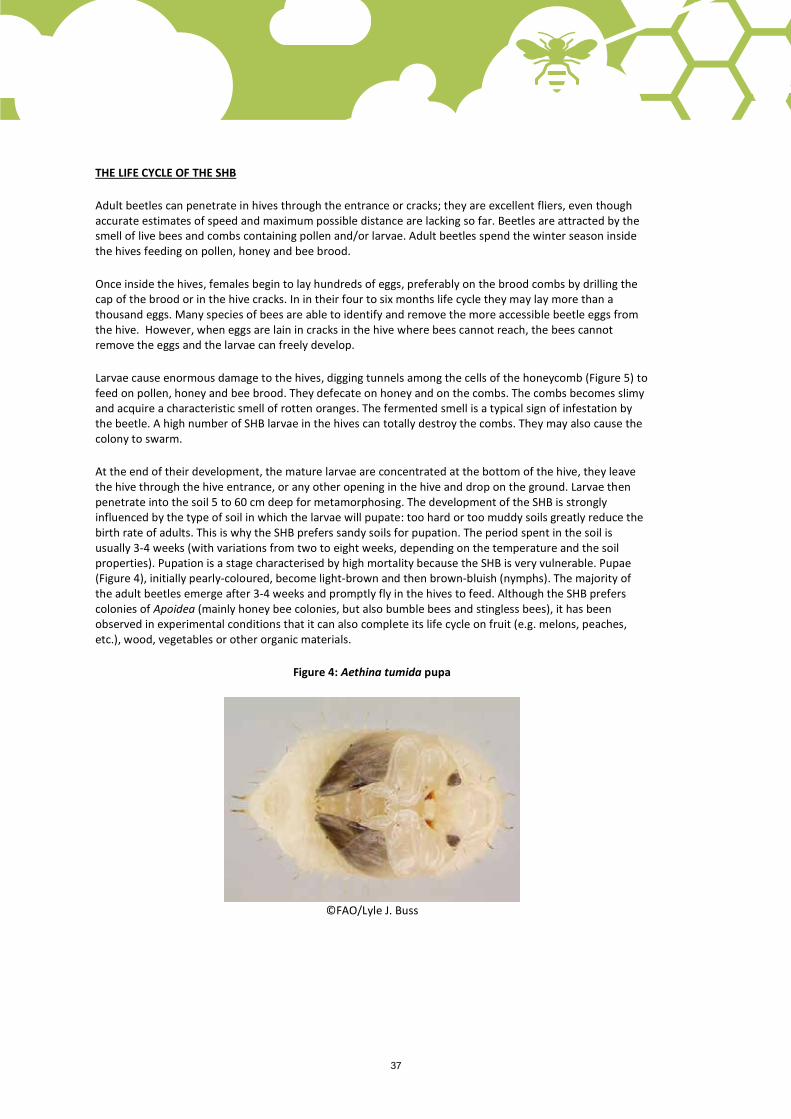
At the end of their development, the mature larvae are concentrated at the bottom of the hive, they leave the hive through the hive entrance, or any other opening in the hive and drop on the ground. Larvae then penetrate into the soil 5 to 60 cm deep for metamorphosing. The development of the SHB is strongly influenced by the type of soil in which the larvae will pupate: too hard or too muddy soils greatly reduce the birth rate of adults. This is why the SHB prefers sandy soils for pupation. The period spent in the soil is usually 3-4 weeks (with variations from two to eight weeks, depending on the temperature and the soil properties). Pupation is a stage characterised by high mortality because the SHB is very vulnerable. Pupae (Figure 4), initially pearly-coloured, become light-brown and then brown-bluish (nymphs). The majority of the adult beetles emerge after 3-4 weeks and promptly fly in the hives to feed. Although the SHB prefers colonies of Apoidea (mainly honey bee colonies, but also bumble bees and stingless bees), it has been observed in experimental conditions that it can also complete its life cycle on fruit (e.g. melons, peaches, etc.), wood, vegetables or other organic materials.
Figure 4: Aethina tumida pupa
©FAO/Lyle J. Buss

38
Figure 5: Damage caused by Aethina tumida (SHB) larvae in honey combs (up) and in the brood combs (center). Damage caused by Galleria mellonella (waxmoth) in a comb (down).
©FAO/Mark Dykes
©FAO/Jeffrey W. Lotz
©FAO/Elsa Demoulin
38 39
SHB’S BEHAVIOUR IN THE HIVE
The SHB can adopt attitudes of attack, defence ("turtle position") or it can fool the bees by antennal contacts asking for transfer of food by trophallaxis (bees behaviour consisting on sharing collected nectar among worker bees by mouth-to-mouth). This means that the bees will actually feed honey to the SHB. Bees can ignore the parasites, can try to move them away from the hive or try to confine them in small spaces into the nest closed with propolis (as a kind of prison). In case of massive presence of the parasite, the flight activity of bee colonies is reduced with a subsequent impact on the productivity. Weakest colonies are at greater risk of massive infestation, while stronger ones are able to ward off the larval forms of SHB by removing the beetle larvae and eggs from the hive, or containing the adults.
Factors conditioning the damage of the parasite to the beehive:
• environmental factors, especially temperature (development of any stage of SHB stops below 10 °C and temperatures over 35 °C cause high mortality of all SHB life stages) and rainfall (the soil moisture should be above 5 percent for pupation of SHB);
• genetics and behaviour of bees (the species and race of the bees) affects the number of beetle cycles. SHB is not a serious threat to the African bees (Apis mellifera capensis and Apis mellifera scutellata) because they can defend themselves very well from the parasite by adopting different behavioural strategies, such as being more efficient fighting and trapping the beetles. Unfortunately, European bees do not show the same aggressiveness against SHB.
SHB EFFECTS
The SHB can spread very rapidly flying from apiary to apiary but also through the trade of bee packages, artificial swarms, queen bees, raw wax and beekeeping materials. Typically SHB infestation leads to death of weak colonies already affected by other diseases (such as Varroa). The presence of SHB in the hive can also cause swarming. In addition, as the SHB larvae are feacating on the honey, they compromise the quality of the honey. The SHB can also cause considerable damage on stored unextracted honey combs in warehouses and honeyhouses.
Adult SHB can survive several days without food so it can be easily introduced, even accidentally, in a SHB-free country through international trade. The SHB represents a strong threat to the environment and to the economy of beekeeping.
The damages to the bee colonies and the stored honey are caused by the adult beetles but especially by the larval forms.
Super storage is a stage during which honeycombs and honey are particularly exposed to SHB because it finds the ideal conditions of development (temperature, humidity and availability of food) and there are no adult bees that may prevent the infestation.
At this level the excrements of larvae can cause honey alterations (such as unpleasant odours and flavours) and fermentation, until honey becomes no longer suitable for human consumption.
CONTROL OF THE SHB
Control methods that can be applied against SHB:
• keep only strong colonies in the apiary; • adopt traps to control and monitor the parasite inside the hives; • achieve good hygiene practices during extraction and at the apiary level as follows:
use of queen excluder: this will prevent the presence of brood in the frames of the supers and will reduce the attraction of SHB;
40
hygienic storage of honeycombs and supers during winter in clean warehouses, preventing the access and reproduction of SHB;
honey supers must be extracted as soon as possible after their collection from the hives in order to prevent SHB colonisation and damage;
the stored honeycombs must be regularly checked in order to reveal any signs of infestation; freeze the honeycombs reaching at least –1 °C for one hour in every part of the comb or apply
sulphur dioxide on empty combs to kill the parasite; it is important to observe good hygiene rules in the honey extraction rooms in order to not
leave organic material available to the parasite (such as comb pieces, wax or honey); do not leave combs or wax (cappings) around the apiary because they may contain SHB eggs; ensure that the relative humidity is < 50 percent (optimum of relative humidity is < 34 percent
to allow 100 percent of egg mortality) in the areas where the honey is stored: this will prevent the egg-hatching of the parasite and avoid damages such as fermentation caused by the SHB larvae to honey. This result may be achieved by performing an appropriate re-circulation of air using a fan and a dehumidifier. Finally, place supers in stacks on pallets and not directly on the ground;
keep temperature inside the honey house or in the storage areas below 15 °C to kill the eggs (48-72h) and 10 °C to kill all the parasite’s life stages;
fluorescent light sources positioned on the floor of the extraction room overnight, attract the SHB larvae. In this way they may be collected and destroyed by putting them in alcohol or detergent solution.
The combination of different control strategies seems the best solution to apply. The first strategy should be to install mechanical traps or biological control methods and only subsequently chemical control methods (i.e. when the population of beetles threats the survival of the colony).
Regulations restricting the importation of bees are the main defences against the introduction of SHB in SHB-free countries. It is essential that all beekeepers adhere to this rule to ensure the effective prevention of the SHB introduction in SHB-free areas.
Further reading
Annand, N., 2011. Investigations of small hive beetle biology to develop better control options (MSc thesis, University of Western Sydney, Australia).
IZSLT, 2007. “Aspetti igienico-sanitari in apicoltura” (22-26).
EFSA, 2015. Panel on Animal Health and Welfare (AHAW), Survival, spread and establishment of thesmall hive beetle (Aethina tumida). EFSA Journal 2015;13(12):4328. doi:10.2903/j.efsa.2015.4328
https://www.ars.usda.gov/northeast-area/beltsville-md/beltsville-agricultural-research-center/bee-research-laboratory/docs/small-hive-beetle/
Neumann, P., Pettis, J. S., & Schäfer, M. O., 2016. Quo vadis Aethina tumida? Biology and control of small hive beetles. Apidologie, (1-40).
Source
APIMONDIA
IZSLT

40 41
11
Tropilaelaps Summary
Tropilaelaps is caused by mites of the genus Tropilaelaps. They are parasites that feed on bee larvae and pupae, Tropilaelaps spp. causes malformation, death of bees and subsequent swarming or colony decline.
This information sheet describes shortly the biology, the life cycle of Tropilaelaps, how it spreads and how to control it.
Description
BIOLOGY
Tropilaelaps mites are reddish-brown in colour, about 1 mm long and 0.6 mm wide (Figure 1). They move freely and quickly among the honeycombs and feed on brood. The buccal apparatus is not able to penetrate the cuticle of adult bees, consequently they are not able to survive in colonies without brood (e.g. swarms, natural brood interruption during winter or artificial brood interruption by caging the queen bee).
Figure 1: Tropilaelaps spp. adults (seen from above on the left, seen from below on the right)
©FAO/Giovanni Formato
The Tropilaelaps mite could be confused by the beekeeper with the varroa mite because they look somewhat similar. The body of the female Varroa mite is wider than it is long (measuring 1.1 to 1.2 mm in length and 1.5 to 1.6 mm in width) and it moves quite slowly, whereas the body of Tropilaelaps is elongated and females measure about 1 mm in length and 0.6 mm in width (males are a little smaller) and moves much faster. (Figure 2 and Figure 3).

42
Figure 2. Adults of Varroa destructor (left) and Tropilaelaps spp. mite (right)
©FAO/Giovanni Formato
Fig. 3: Varroa destructor (left) and Tropilaelaps spp. (right) seen from below
©FAO/Giovanni Formato
LIFE CYCLE
Tropilelaps mite has some similarities with the Varroa mite. It feeds and reproduces in the bee brood, but unlike Varroa, it cannot feed on adults because its buccal apparatus cannot penetrate the cuticula of honey bees and suck their haemolymph. The Tropilelaps mite has a shorter reproductive cycle and a higher reproductive rate but it is not able to survive brood interruption periods of the honey bee (natural absence of brood during the winter or artificially induced by queen caging) due to its inability to feed on adult honey bee.
The adult mites enter the cells containing the bee larvae (both of worker bees and drones) to breed. Eggs hatch after about twelve hours and the larvae feed on the bee hemolymph. The mites cause damage on the larvae which results in high mortality rate of bee brood (up to 50 percent).
The Tropilaelaps mite can spread from hive to hive through honey bee drift, looting and swarming. The spread of these parasites however may occur also through common beekeeping practices, such as: moving affected brood combs from one hive to another, migratory beekeeping and buying and selling of parasitized colonies/nucs.

42 43
SYMPTOMS
In colonies affected by a large number of mites, Tropilaelaps is able to cause damage very similar to varroa with high mortality of brood, weak adults with deformed wings and legs and abdomens smaller than normal. Bees can also be found paralysed at the entrance of the hive. Other symptoms are: irregular brood pattern, hollow cappings for the attempt of worker bees to clean up sick or dead larvae. In cases of severe infestation, up to 50 percent of the brood may die; in these cases, a bad smell of dead brood associated with the Tropilaelaps infestation can be observed. At these levels of infestation bees frequently swarm, contributing to the spread of the mite.
DIAGNOSIS AND BIOLOGICAL CONTROL
The diagnosis of this parasitic disease is visual and is carried out through the observation of infected bee brood or by the observation of Tropilaelaps fallen on the bottom of the hive after an acaricide treatments.
a) Adult honey bee examination
Take a sample of about 300 adult bees (more or less equal to one glass full of bees) from suspected infested colonies and put them into a jar (1 kg jar) with three tea spoons of powder sugar (about 30 g), putting a screen on the top of the jar. Then shake the jar onto a white piece of paper to allow the mites to fall through the screened top. You will be able to count the number of mites fallen onto the paper. This method is very similar to the powder sugar method for diagnosing and quantifying varroa infestation (see: http://teca.fao.org/read/8663).
Otherwise, the same number of adult bees could be put into a container (e.g. 1 kg jar) with 70 percent ethyl alcohol or with soapy water. After closing the container, shake them, pour the bees into a sieve onto a white paper and the fallen mites can be identified and counted on a white paper.
b) Brood examination
When monitoring honey bee colonies for the presence of Tropilaelaps, an examination of both drone and worker brood may provide an early indication of infestation. Mites can be observed inside capped bee brood by using a honey scratcher to pull up capped pupae. The mites are clearly visible.
c) Sticky board examination
A precise diagnosis can be made using a sticky board on the bottom of the hive, covered with a mesh with size of 2 mm that prevents the bees from removing the dislodged mites. The mesh of 2 mm is large enough for mites to pass through. Make a sticky board with poster board, cardboard or other white, stiff paper coated with Vaseline or other sticky substance, or use a sheet of sticky shelf paper. Cut the paper to fit the bottom board of a hive. Leave the bottom board in the colony, under the hive for up to three days, collecting and examining the debris for mites.
For faster mite diagnosis, smoke each colony adding 25 g (1 oz) pipe tobacco in the smoker. Puff the bees six to ten times, close up the hive for 10/20 minutes. Pull out the sticky board after at least ten minutes and count the mites.
A mite control can be carried out by the queen caging method or through artificial swarming: these techniques allow to reduce the number of Tropilaelaps due to its inability to feed on adult bees or to survive for more than two days without the brood.
Even though Tropilaelaps spp. has not spread all around the world as other bee pathogens (such as varroa), it represents a potential pathogen for the Western honeybee Apis mellifera spp.
4444
Further reading
IZSLT, 2007. Aspetti igienico-sanitari in apicoltura, (27-28).
Source
APIMONDIA
IZSLT
4444
List of related technologiesReaders of this catalogue could also be interested in related technologies and practices available in the TECA Knowledge Base or in specific moderated discussions hosted by TECA. The technologies/practices and/or moderated discussion listed below can be consulted online (http://teca.fao.org).
Language Title of the practice/technology in the languages available
Practice/technology by Partner
Sustainable Development Goals
Practice/TechnologyField method to determine level of varroa infestation
English IRACHFrench Méthode pour déterminer le taux de
varroas sur le terrainSpanish Método para determinar niveles de
varroa en terrenoPortuguese
How to collect raw propolis from the hive
English How to collect raw propolis from the hive
FAO
French Comment récolter la propolis brute d’une ruche
Spanish
Portuguese
How to build a steam wax extractor
English SwisscontactFrench Fabrication d’un cérificateur à vapeur
artisanalSpanish Fundidor de cera de abejas a vapor
artesanalPortuguese
Use of stingless bees for pollination
English Use of Stingless Bees for Pollination The University of the Philippines Los Banos (UPLB)
French
Spanish Uso de Abejas sin Aguijón para la Polinización
PortugueseHow to build a smoker
English How to build a smoker Swisscontact/The Beekeeping Network North-South (BNNS)
French Fabrication d’un enfumoir métallique à soufflet
Spanish Confección de ahumador para apicultor
PortugueseModerated discussions
English Use of veterinary products in beekeeping
IZSLT
English Field diagnosis of main bee diseases
IZSLT
French Comment fabriquer des amorces de cire pour la conduite de ruches à barrettes
BNNS
FAO and the SDGs Co-branding Guidelines 12
GRAPHIC ELEMENTSSDG ICONS
FAO and the SDGs Co-branding Guidelines 12
GRAPHIC ELEMENTSSDG ICONS
FAO and the SDGs Co-branding Guidelines 12
GRAPHIC ELEMENTSSDG ICONS
FAO and the SDGs Co-branding Guidelines 12
GRAPHIC ELEMENTSSDG ICONS
FAO and the SDGs Co-branding Guidelines 12
GRAPHIC ELEMENTSSDG ICONS
FAO and the SDGs Co-branding Guidelines 12
GRAPHIC ELEMENTSSDG ICONS
FAO and the SDGs Co-branding Guidelines 12
GRAPHIC ELEMENTSSDG ICONS
FAO and the SDGs Co-branding Guidelines 12
GRAPHIC ELEMENTSSDG ICONS
FAO and the SDGs Co-branding Guidelines 12
GRAPHIC ELEMENTSSDG ICONS
FAO and the SDGs Co-branding Guidelines 12
GRAPHIC ELEMENTSSDG ICONS
FAO and the SDGs Co-branding Guidelines 12
GRAPHIC ELEMENTSSDG ICONS
FAO and the SDGs Co-branding Guidelines 12
GRAPHIC ELEMENTSSDG ICONS
FAO and the SDGs Co-branding Guidelines 12
GRAPHIC ELEMENTSSDG ICONS
FAO and the SDGs Co-branding Guidelines 12
GRAPHIC ELEMENTSSDG ICONS
FAO and the SDGs Co-branding Guidelines 12
GRAPHIC ELEMENTSSDG ICONS
FAO and the SDGs Co-branding Guidelines 12
GRAPHIC ELEMENTSSDG ICONS
FAO and the SDGs Co-branding Guidelines 12
GRAPHIC ELEMENTSSDG ICONS

e-mail: [email protected] website: http://teca.fao.orgFOOD AND AGRICULTURE ORGANIZATION OF THE UNITED NATIONSViale delle Terme di Caracalla Rome, Italy
I9466EN/1/05.18
ISBN 978-92-5-130544-7
9 7 8 9 2 5 1 3 0 5 4 4 7
The TECA PlatformTechnologies and practices for small agricultural producers (TECA) (http://teca.fao.org), is an FAO online platform for the exchange and sharing of knowledge on agricultural technologies and practices for smallholder farmers and producers. These support innovation needs in the field, contribute to further enhance knowledge access for a better life and increase capacities to innovate and contribute to achieving the Sustainable Development Goals (SDGs). The TECA platform is hosted by the Food and Agriculture Organization of the United Nations’ Research and Extension Unit.
The TECA Knowledge Base currently contains around 900 technologies and practices. They include practices or techniques, tools, equipment, know-how, and skills, or combinations of the aforementioned elements that: (i) have been tested and/or adopted by small producers under actual field conditions; (ii) are easy to replicate; (iii) are presented in an easy to understand, non-academic language illustrated with drawings, video and pictures to facilitate the application of the technologies in the field; and (iv) are validated by technical experts.
TECA also provides online forums (Exchange Groups) where stakeholders interested in a certain topic can interact and discuss sustainable and innovative solutions for challenges that smallholders face. TECA currently hosts three thematic Exchange Groups, including the TECA Beekeeping group which runs in three languages (English, French and Spanish).
About this catalogueIn an effort to support beekeepers in the sustainable production of good quality beehive products, TECA together with its partners, have developed a set of information sheets on good beekeeping practices for the main bee diseases. They are designed following the TECA standards, to help beekeepers, and those providing advisory services to the beekeepers, correctly identify honey bee diseases, apply the correct treatment and to prevent them. The information sheets available in this catalogue can also be consulted online through the TECA Knowledge Base: http://teca.fao.org/technologies/browse
Objective of this catalogueTo facilitate the early and correct identification of a honey bee disease for the efficient management/treatment of the disease.
FAO and the SDGs Co-branding Guidelines 12
GRAPHIC ELEMENTSSDG ICONS
FAO and the SDGs Co-branding Guidelines 12
GRAPHIC ELEMENTSSDG ICONS
FAO and the SDGs Co-branding Guidelines 12
GRAPHIC ELEMENTSSDG ICONS


![PRIX N.202 - H135 MT[Own.:Allevamento Equini Di Brotto Sante Giorgio] Fabio Brotto ITA 10016424 0/0 26,09 110,00 7° 253 KLEONS COCARIO 105IH12 - 8 cast. bay os carrico/(chambertin)](https://static.documents.pub/doc/80x56/5f52f93f2cc62b02452aabf7/prix-n202-h135-mt-ownallevamento-equini-di-brotto-sante-giorgio-fabio-brotto.jpg)

![Arena JUMPING 5 Y.O 17/07/2020 12,00 h €. 500 · 106WD27 - 5 cast. bay hol adagio de talm/united iii(con air) [Own.:Allevamento Guldenboom & G&G Snc] Gianluca Gennusa ITA 10098653](https://static.documents.pub/doc/80x56/5f581e23246e1278147aacc9/arena-jumping-5-yo-17072020-1200-h-a-500-106wd27-5-cast-bay-hol-adagio.jpg)


