Nanomedicine: Nanotechnology, Biology, and Medicinexx (2014) xxx–xxx
nanomedjournal.com
Theranostic tumor homing nanocarriers for the treatment of lung cancerApurva R. Patel, PhDa, Mahavir B. Chougule, PhDb, Ed Lim, PhDc, Kevin P. Francis, PhDc,
Stephen Safe, PhDd, Mandip Singh, PhDa,⁎aCollege of Pharmacy and Pharmaceutical Sciences, Florida A&M University, Tallahassee, FL, USA
bDepartment of Pharmaceutical Sciences, College of Pharmacy, University of Hawai'i at Hilo, Hilo, HI, USAcCalipers-Life Sciences & Technology, A Perkin Elmer Company, Alameda, CA, USAdInstitute of Biosciences and Technology, Texas A&M University, Houston, TX, USA
Lung cancer is the leading cause of cancer death (28% of allcancer deaths) in both men and women in the United States.1
Despite recent advances in lung cancer management includingchemotherapy, the survival rates in lung cancer patients areunsatisfactorywith five-year survival rate of 16.3%,which is lowerthan other types of cancer such as colon, breast and prostate.1,2
Currently, newer approaches in the treatment of lung cancer withnovel antiangiogenic drugs have generated clinical interest.3 Thevascular endothelial growth factor (VEGF) over-expression (61%to 92% of NSCLC) is associated with poor survival among lung
Conflict of Interest and Disclosure: The authors report no financialinterest that might pose a potential, perceived, or real conflict of interest.
U.S. Patent filed “Modified Nanodelivery System and Method forEnhanced In vivo Medical and Preclinical Imaging.” (U.S. 13/851610).
All sources of support for research: This workwas supported byNationalInstitute of Health MBRS-SC1 Program (Grant # SC1 GM092779-01).
⁎Corresponding author at: College of Pharmacy and PharmaceuticalSciences, Florida A&M University, Tallahassee, FL, USA.
Please cite this article as: Patel A.R., et al., Theranostic tumor homing nanocarhttp://dx.doi.org/10.1016/j.nano.2013.12.002
cancer patients.4,5 Among newer approaches, the use ofantiangiogenic agents in combination with other anticancer drugshas generated clinical interest that selectively inhibits the tumorblood supply thus controlling cancer cell survival, proliferationand/or metastasis.6 Previous studies demonstrated that DIM-C-pPhC6H5 (DIM-P), a c-substituted diindolylmethanes has prom-ising anticancer activity against lung cancer and in combinationwith Docetaxel showed additive to synergistic action by activatinggrowth inhibitory and apoptotic pathways in lung tumors.7 Studiesconducted in our laboratory strongly suggest that DIM-P exhibitsantiangiogenic activity which is evident from down regulation ofVEGF and CD31 expression and, decrease in the microvesseldensity.8 This warrants further investigations into the antiangio-genic role of DIM-P in the management of lung cancer; however,the pharmacokinetic (PK) studies conducted in our laboratoryshowed that DIM-P has poor oral bioavailability9 as well as shortplasma half-life following intravenous administration. To over-come this, we used nanoparticle system to deliver DIM-P. Earlyclinical results have suggested that nanoparticles can enhanceefficacy and reduce side effects of therapeutic agents compared to
riers for the treatment of lung cancer. Nanomedicine: NBM 2014;xx:1-11,
2 A.R. Patel et al / Nanomedicine: Nanotechnology, Biology, and Medicine xx (2014) xxx–xxx
conventional delivery systems.10,11 However, the outcomes ofthese passively targeted nanoparticles are hampered due toinefficient tumor cell internalization and toxicity to the normalcells.12,13 Solid lipid nanoparticles (SLN) have several advantagesas compared to other nanoparticles in terms of enhanced stabilityand alteration of biodistribution.14–16 However, the use of SLN islimited due to a) low drug loading, and b) drug expulsion therebydecreasing stability. The nanostructured lipid carriers (NCs) havebeen developed as the second generation lipid nanoparticles17,18 toovercome barriers of SLN. The higher drug loading capacity andminimal expulsion during storage could be achieved by thedevelopment ofNCs due to higher solubility of drugs in oils than insolid lipids.19 The NCs have been utilized to deliver variouschemotherapeutic agents20 and the outer lipid core of the NCs isflexible which allows surface modification with specific groupslike PEG-DSPE or DOGS-NTA-Ni which can be utilized fordesign of multi-targeted delivery systems.
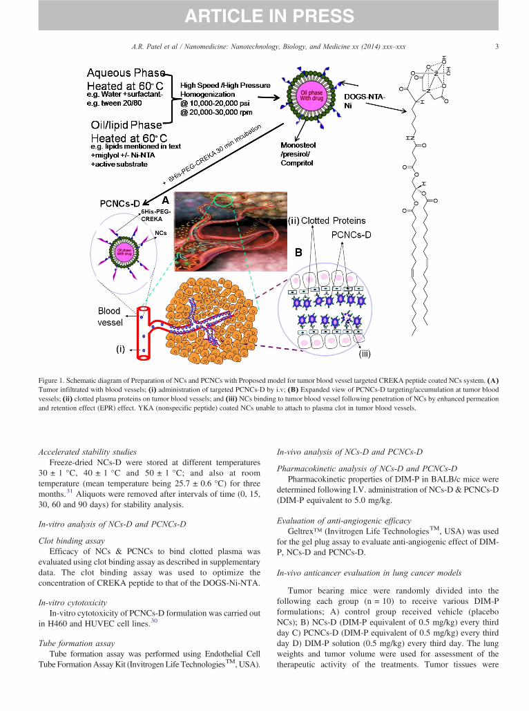
Multifunctional nanoparticles have tremendous potential toimprove the clinical outcome of cancer therapeutics as evidentfrom published reports by several researchers.21–23 One of thetumor homing five amino acid peptides, CREKA (Cys-Arg-Glu-Lys-Ala) was identified using in vivo phage display in MMTV-PyMT transgenic mice.24 CREKA binds to clotted plasmaproteins and homes to the interstitial tissue of tumors and vesselwalls containing clotted plasma proteins, whereas normal bloodvessels lacks these proteins.24,25 The selective homing ofCREKA peptide to tumor blood vessels and stroma is a novelway to enhance targeting efficacy to tumor blood vasculature.CREKA peptide coupled on the surface of super-paramagneticamino dextran-coated iron oxide nanoparticles showed effectiveanticancer effect in vitro as well as in vivo conditions in breastcancer.26 Further, CREKA peptide conjugated to Abraxane, aclinically approved paclitaxel-albumin nanoparticle, also showedpromising accumulation of CREKA-abraxane in breast tumorblood vessels.27
The conjugation of polythethylene glycol (PEG) to drugdelivery systems has been well reported to increase the half-lifeand overcome the clearance by the reticuloendothelialsystem.28,29 DIM-P showed a short plasma half-life andpromising anti-angiogenic activity in lung cancer. Therefore, adelivery approach using PEGylated NCs coupled with targetingto clotted proteins on tumor vasculature using a well studiedpeptide, CREKA, will overcome limitations associated withdelivery of DIM-P and will help to explore its anti-angiogenicpotential in the treatment of lung cancer. The proposedmechanistic function of targeted NCs is graphically elaboratedin Figure 1. Thus we hypothesize that “Tumor homingPEGylated nanolipidcarriers of DIM-P will target tumor bloodvasculature and increase its plasma half life thereby inhibitingtumor growth by exerting antiangiogenic activity against lungtumors”. The specific objectives of this study are: 1) to formulatetumor homing PEGylated CREKA peptide conjugated nanopar-ticles of DIM-P (PCNCs-D), in conjunction with versions thatcontain XenolightDiR (PCNCs-Di), 2) to investigate antitumoractivity and antiangiogenic potential of PCNCs-D in orthotopicand metastatic lung tumor models, and 3) to evaluate the in-vivoimaging of tumor progression/vasculature and tracking of thenanoparticle using PCNCs-Di. This is the first study to
demonstrate delivery of a novel anticancer agent, DIM-P in apegylated nanocarrier system using CREKA peptide in lungcancer models (orthotopic and metastatic) and demonstrates theapplication of theranostic nanocarriers in lung tumors. Theresults from these studies will give researchers critical andimportant formation for benefits of peptide targeted theranosticnanoparticles in imaging and treatment of lung tumors.
Materials and methods
Materials and animals
Further information are given in the Supplementary Data. Allanimal experimental procedures were done according to theanimal experimental ethics committee from our university.
Preparation and optimization of NCs
NCs were prepared by modified hot melt homogenizationtechnique30 using triglycerides and optimized process variables.The three formulation variables such as lipid (3, 6, 9%w/w),peptide/DOGS-NTA-Ni ratio and PEG molecular weight werevaried at three concentration levels to achieve desired efficiency.
Freeze drying of NCs
Formulations were lyophilized (SMART Freeze Drying, FTSSystems, SP Scientific, USA) using a universal stepwise freezedrying cycle. Formulations were lyophilized using 5% w/vtrehalose (cryoprotectant) and the viscosity of the NCsformulations was adjusted to 2.5-3 cP by re-suspending thelyophilized formulation in distilled water prior to use.
Preparation and optimization of PCNCs
For surface modification, 200 μl of NCs was mixed with50 μl of 6-Histidine -tagged PEGylated (PEG-2000) CREKApeptide aqueous solution (5 mg/ml) and incubated for 2 h atroom temperature with constant stirring.
Characterization of NCs & PCNCs
Particle size, zeta potential, entrapment efficiency and drugloading measurement
The particle size and zeta potential of NCs or PCNCs weremeasured in distilled water using Nicomp 380 ZLS (ParticleSizing Systems, Port Richey, FL) as described previously.30
Entrapment efficiency was determined as reported earlier usingvivaspin columns, molecular weight cut-off (MWCO)10,000 Da.30 In-vitro drug release studies: In-vitro drug releasestudies were conducted with a cellulose membrane using a USPType-I dissolution apparatus (Vankel, NC) for 72 h with the helpof 200 ml of phosphate buffer saline (PBS) pH 7.4 containing0.5% w/v Volpo-20 & 0.5% TPGS as dissolution medium. Thein-vitro drug release of DIM-P solution, NCs-D and PCNCs-Dwas carried out as described in supplementary data.
Differential scanning calorimetryThe interaction of DIM-P with lipids and association of DIM-
P in NCs formulations were determined using a DSCQ100 (TAinstrument, DE).
Figure 1. Schematic diagram of Preparation of NCs and PCNCs with Proposed model for tumor blood vessel targeted CREKA peptide coated NCs system. (A)Tumor infiltrated with blood vessels; (i) administration of targeted PCNCs-D by i.v; (B) Expanded view of PCNCs-D targeting/accumulation at tumor bloodvessels; (ii) clotted plasma proteins on tumor blood vessels; and (iii) NCs binding to tumor blood vessel following penetration of NCs by enhanced permeationand retention effect (EPR) effect. YKA (nonspecific peptide) coated NCs unable to attach to plasma clot in tumor blood vessels.
3A.R. Patel et al / Nanomedicine: Nanotechnology, Biology, and Medicine xx (2014) xxx–xxx
Accelerated stability studiesFreeze-dried NCs-D were stored at different temperatures
30 ± 1 °C, 40 ± 1 °C and 50 ± 1 °C; and also at roomtemperature (mean temperature being 25.7 ± 0.6 °C) for threemonths.31 Aliquots were removed after intervals of time (0, 15,30, 60 and 90 days) for stability analysis.
In-vitro analysis of NCs-D and PCNCs-D
Clot binding assayEfficacy of NCs & PCNCs to bind clotted plasma was
evaluated using clot binding assay as described in supplementarydata. The clot binding assay was used to optimize theconcentration of CREKA peptide to that of the DOGS-Ni-NTA.
In-vitro cytotoxicityIn-vitro cytotoxicity of PCNCs-D formulation was carried out
in H460 and HUVEC cell lines.30
Tube formation assayTube formation assay was performed using Endothelial Cell
Tube FormationAssayKit (Invitrogen Life TechnologiesTM,USA).
In-vivo analysis of NCs-D and PCNCs-D
Pharmacokinetic analysis of NCs-D and PCNCs-DPharmacokinetic properties of DIM-P in BALB/c mice were
determined following I.V. administration of NCs-D & PCNCs-D(DIM-P equivalent to 5.0 mg/kg.
Evaluation of anti-angiogenic efficacyGeltrex™ (Invitrogen Life TechnologiesTM, USA) was used
for the gel plug assay to evaluate anti-angiogenic effect of DIM-P, NCs-D and PCNCs-D.
In-vivo anticancer evaluation in lung cancer models
Tumor bearing mice were randomly divided into thefollowing each group (n = 10) to receive various DIM-Pformulations; A) control group received vehicle (placeboNCs); B) NCs-D (DIM-P equivalent of 0.5 mg/kg) every thirdday C) PCNCs-D (DIM-P equivalent of 0.5 mg/kg) every thirdday D) DIM-P solution (0.5 mg/kg) every third day. The lungweights and tumor volume were used for assessment of thetherapeutic activity of the treatments. Tumor tissues were
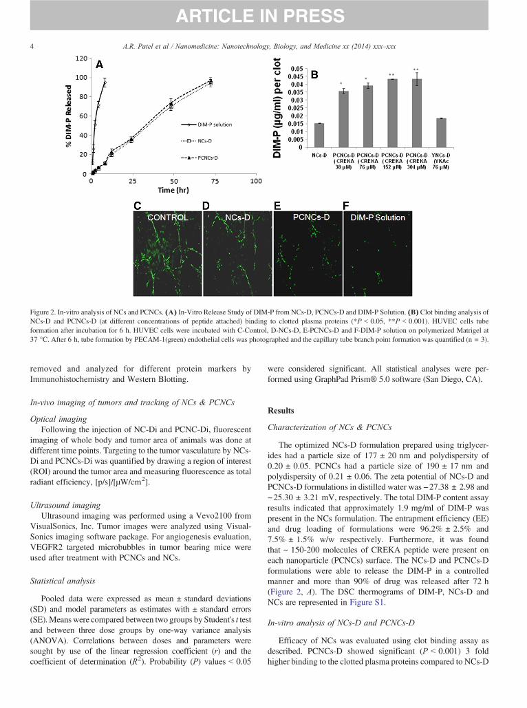
Figure 2. In-vitro analysis of NCs and PCNCs. (A) In-Vitro Release Study of DIM-P from NCs-D, PCNCs-D and DIM-P Solution. (B) Clot binding analysis ofNCs-D and PCNCs-D (at different concentrations of peptide attached) binding to clotted plasma proteins (*P b 0.05, **P b 0.001). HUVEC cells tubeformation after incubation for 6 h. HUVEC cells were incubated with C-Control, D-NCs-D, E-PCNCs-D and F-DIM-P solution on polymerized Matrigel at37 °C. After 6 h, tube formation by PECAM-1(green) endothelial cells was photographed and the capillary tube branch point formation was quantified (n = 3).
4 A.R. Patel et al / Nanomedicine: Nanotechnology, Biology, and Medicine xx (2014) xxx–xxx
removed and analyzed for different protein markers byImmunohistochemistry and Western Blotting.
In-vivo imaging of tumors and tracking of NCs & PCNCs
Optical imagingFollowing the injection of NC-Di and PCNC-Di, fluorescent
imaging of whole body and tumor area of animals was done atdifferent time points. Targeting to the tumor vasculature by NCs-Di and PCNCs-Di was quantified by drawing a region of interest(ROI) around the tumor area and measuring fluorescence as totalradiant efficiency, [p/s]/[μW/cm2].
Ultrasound imagingUltrasound imaging was performed using a Vevo2100 from
VisualSonics, Inc. Tumor images were analyzed using Visual-Sonics imaging software package. For angiogenesis evaluation,VEGFR2 targeted microbubbles in tumor bearing mice wereused after treatment with PCNCs and NCs.
Statistical analysis
Pooled data were expressed as mean ± standard deviations(SD) and model parameters as estimates with ± standard errors(SE).Meanswere compared between two groups by Student's t testand between three dose groups by one-way variance analysis(ANOVA). Correlations between doses and parameters weresought by use of the linear regression coefficient (r) and thecoefficient of determination (R2). Probability (P) values b 0.05
were considered significant. All statistical analyses were per-formed using GraphPad Prism® 5.0 software (San Diego, CA).
Results
Characterization of NCs & PCNCs
The optimized NCs-D formulation prepared using triglycer-ides had a particle size of 177 ± 20 nm and polydispersity of0.20 ± 0.05. PCNCs had a particle size of 190 ± 17 nm andpolydispersity of 0.21 ± 0.06. The zeta potential of NCs-D andPCNCs-D formulations in distilled water was −27.38 ± 2.98 and−25.30 ± 3.21 mV, respectively. The total DIM-P content assayresults indicated that approximately 1.9 mg/ml of DIM-P waspresent in the NCs formulation. The entrapment efficiency (EE)and drug loading of formulations were 96.2% ± 2.5% and7.5% ± 1.5% w/w respectively. Furthermore, it was foundthat ~ 150-200 molecules of CREKA peptide were present oneach nanoparticle (PCNCs) surface. The NCs-D and PCNCs-Dformulations were able to release the DIM-P in a controlledmanner and more than 90% of drug was released after 72 h(Figure 2, A). The DSC thermograms of DIM-P, NCs-D andNCs are represented in Figure S1.
In-vitro analysis of NCs-D and PCNCs-D
Efficacy of NCs was evaluated using clot binding assay asdescribed. PCNCs-D showed significant (P b 0.001) 3 foldhigher binding to the clotted plasma proteins compared to NCs-D
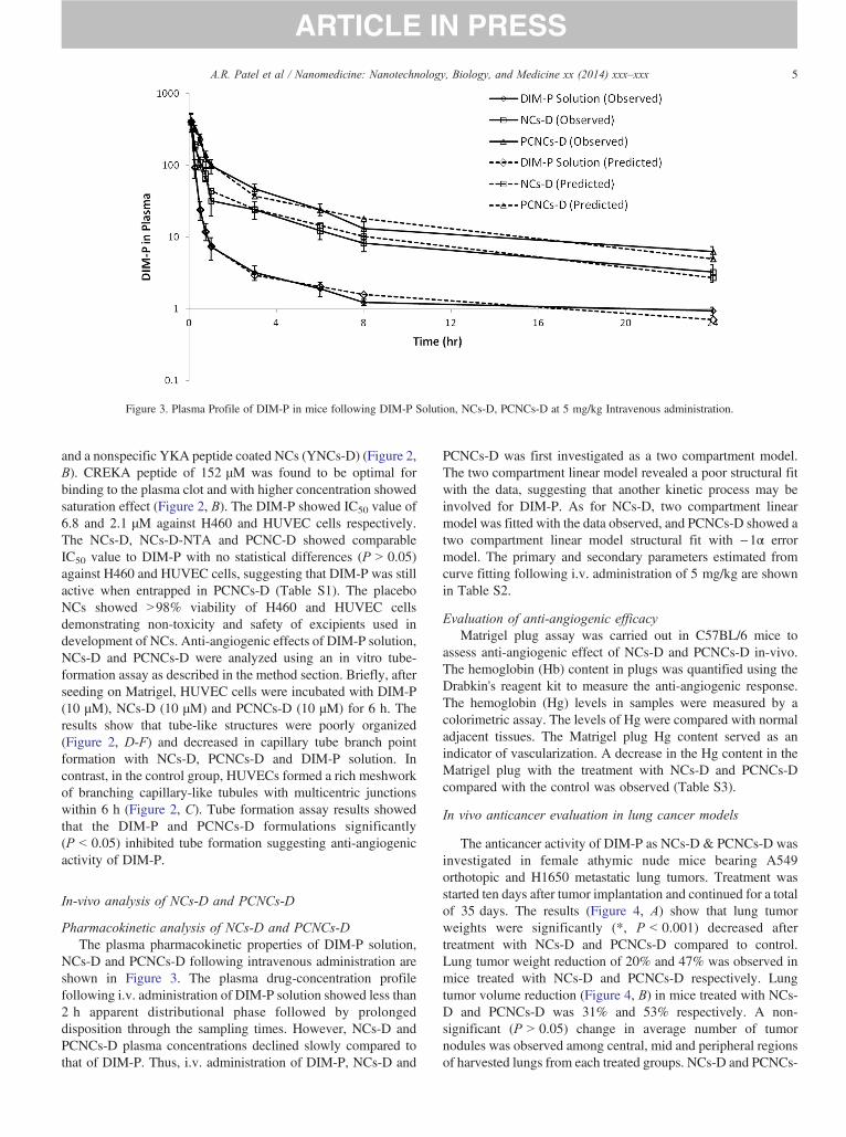
Figure 3. Plasma Profile of DIM-P in mice following DIM-P Solution, NCs-D, PCNCs-D at 5 mg/kg Intravenous administration.
5A.R. Patel et al / Nanomedicine: Nanotechnology, Biology, and Medicine xx (2014) xxx–xxx
and a nonspecific YKA peptide coated NCs (YNCs-D) (Figure 2,B). CREKA peptide of 152 μM was found to be optimal forbinding to the plasma clot and with higher concentration showedsaturation effect (Figure 2, B). The DIM-P showed IC50 value of6.8 and 2.1 μM against H460 and HUVEC cells respectively.The NCs-D, NCs-D-NTA and PCNC-D showed comparableIC50 value to DIM-P with no statistical differences (P N 0.05)against H460 and HUVEC cells, suggesting that DIM-P was stillactive when entrapped in PCNCs-D (Table S1). The placeboNCs showed N98% viability of H460 and HUVEC cellsdemonstrating non-toxicity and safety of excipients used indevelopment of NCs. Anti-angiogenic effects of DIM-P solution,NCs-D and PCNCs-D were analyzed using an in vitro tube-formation assay as described in the method section. Briefly, afterseeding on Matrigel, HUVEC cells were incubated with DIM-P(10 μM), NCs-D (10 μM) and PCNCs-D (10 μM) for 6 h. Theresults show that tube-like structures were poorly organized(Figure 2, D-F) and decreased in capillary tube branch pointformation with NCs-D, PCNCs-D and DIM-P solution. Incontrast, in the control group, HUVECs formed a rich meshworkof branching capillary-like tubules with multicentric junctionswithin 6 h (Figure 2, C). Tube formation assay results showedthat the DIM-P and PCNCs-D formulations significantly(P b 0.05) inhibited tube formation suggesting anti-angiogenicactivity of DIM-P.
In-vivo analysis of NCs-D and PCNCs-D
Pharmacokinetic analysis of NCs-D and PCNCs-DThe plasma pharmacokinetic properties of DIM-P solution,
NCs-D and PCNCs-D following intravenous administration areshown in Figure 3. The plasma drug-concentration profilefollowing i.v. administration of DIM-P solution showed less than2 h apparent distributional phase followed by prolongeddisposition through the sampling times. However, NCs-D andPCNCs-D plasma concentrations declined slowly compared tothat of DIM-P. Thus, i.v. administration of DIM-P, NCs-D and
PCNCs-D was first investigated as a two compartment model.The two compartment linear model revealed a poor structural fitwith the data, suggesting that another kinetic process may beinvolved for DIM-P. As for NCs-D, two compartment linearmodel was fitted with the data observed, and PCNCs-D showed atwo compartment linear model structural fit with −1α errormodel. The primary and secondary parameters estimated fromcurve fitting following i.v. administration of 5 mg/kg are shownin Table S2.
Evaluation of anti-angiogenic efficacyMatrigel plug assay was carried out in C57BL/6 mice to
assess anti-angiogenic effect of NCs-D and PCNCs-D in-vivo.The hemoglobin (Hb) content in plugs was quantified using theDrabkin's reagent kit to measure the anti-angiogenic response.The hemoglobin (Hg) levels in samples were measured by acolorimetric assay. The levels of Hg were compared with normaladjacent tissues. The Matrigel plug Hg content served as anindicator of vascularization. A decrease in the Hg content in theMatrigel plug with the treatment with NCs-D and PCNCs-Dcompared with the control was observed (Table S3).
In vivo anticancer evaluation in lung cancer models
The anticancer activity of DIM-P as NCs-D & PCNCs-D wasinvestigated in female athymic nude mice bearing A549orthotopic and H1650 metastatic lung tumors. Treatment wasstarted ten days after tumor implantation and continued for a totalof 35 days. The results (Figure 4, A) show that lung tumorweights were significantly (*, P b 0.001) decreased aftertreatment with NCs-D and PCNCs-D compared to control.Lung tumor weight reduction of 20% and 47% was observed inmice treated with NCs-D and PCNCs-D respectively. Lungtumor volume reduction (Figure 4, B) in mice treated with NCs-D and PCNCs-D was 31% and 53% respectively. A non-significant (P N 0.05) change in average number of tumornodules was observed among central, mid and peripheral regionsof harvested lungs from each treated groups. NCs-D and PCNCs-
Figure 4. Effects of NCs-D and PCNCs-D on orthotopic A549 lung tumor weight (A); tumor volume (B); mice body weight (C) as well as on metastatic H1650lung tumor weight (D); tumor volume (E); mice body weight (F). Lung weights and tumor volumes were determined for measurement of therapeutic activity ofthe treatments. One-way ANOVA followed by post Tukey test was used for statistical analysis. P b 0.05 (*, significantly different from untreated controls; **,significantly different from NCs-D treatments). Data presented are means ± SD (n = 8).
6 A.R. Patel et al / Nanomedicine: Nanotechnology, Biology, and Medicine xx (2014) xxx–xxx
D treatment showed a significant (*P b 0.001) decrease inaverage number of tumor nodules in central, mid and peripheralregions compared to control groups. We did not observe anyweight loss or other signs of toxicity in mice treated with NCs-Dor PCNCs-D (Figure 4, C). Also, the anticancer activity of DIM-P as NCs-D & PCNCs-D in female athymic nude mice bearingH1650 metastatic lung tumors showed similar results (Figure 4,D-F).
Immunohistochemistry (VEGF & CD31 expression) andWestern blot analysis
To examine if the NCs-D & PCNCs-D inhibited lung tumorgrowth through inhibition of angiogenesis and to confirm ourpreliminary result of antiangiogenic activity of DIM-P, wedetermined VEGF expression in tumor tissue sections by IHC. Asignificantly (*P b 0.05) decreased expression of VEGF(Figure 5, A) was observed in tumors treated with the NCs-D& PCNCs-D treatment compared to the untreated group. CD31(+) endothelial cells were also identified, as illustrated inFigure 5, B. The staining of microvessels in NCs-D & PCNCs-Dtreated groups was significantly (*P b 0.05) decreased comparedto control group. The average number of microvessels per fieldin groups treated with NCs-D & PCNCs-D was found to be 99 ±6.6 (*P b 0.05), 52 ± 10.5 (**P b 0.001) respectively com-pared to 179.0 ± 28.4 in the control group. The analysis ofproliferation marker Ki-67 (Figure S2) indicates the inhibition(*P b 0.05) of lung tumors progression in NCs-D and PCNCs-Dtreated groups of animals. The average number of proliferativeKi-67 positive cells per field in groups treated with NCs-D &PCNCs-D was found to be 86 ± 9 (*P b 0.05), 41 ± 11(**P b 0.001) respectively compared to 158.0 ± 22.0 in thecontrol group. We compared expression of several proteins in
normal lung tissue lysates, tumor lysates from control and treatedmice by Western blot analysis using β-actin as loading control(Figure 5, C). NCs-D & PCNCs-D treatment significantly(*P b 0.05) decreased MMP-9 expression to 0.26 and 0.54-foldin regressed tumor samples compared to controls groupsrespectively. In regressed tumors, the PCNCs-D (*P b 0.001)and NCs-D (*P b 0.01) significantly decreased HIF-1α expres-sion to 0.48, and 0.15-fold, respectively of the controls (Figure 5,C). PCNCs-D treatment showed increased Erk2 proteinexpression (**P b 0.05) to 0.67-fold compared to 0.28-foldNCs-D (*P b 0.01) respectively of the controls in regressedtumors (Figure 5, C). The NCs-D & PCNCs-D decreased Sp1expression significantly (*P b 0.001) compared to tumorsharvested from the control group (Figure 5, C). PCNCs-D &NCs-D treatment significantly (*P b 0.05) decreased Sp3expression to 0.29 and 0.09-fold respectively in regressedtumor samples compared to the control groups (Figure 5, C).The expression of HSP-27 protein was significantly decreased by0.13 fold (*P b 0.01) and 0.44 fold (*P b 0.05) with PCNCs-D& NCs-D treatment compared to control group respectively(Figure 5, C). Results illustrated in Figure 5, C show that thePCNCs-D treatment significantly decreased expression of VEGFto 0.6-fold (*P b 0.001) compared to control.
In-vivo imaging of tumors and tracking of NCs & PCNCs
In-vivo imaging following exposure of PCNCs-Di demon-strated their targeting to the tumor vasculature (Figure 6), wherethe PCNCs-Di were found to migrate more in newly formedblood vessels with total radiant efficiency [p/s]/[μW/cm2] of2.1*1012 ± 0.5*1012 over the period of 0.5 h to 3 h. NCs-Dididn't show any specific migration to tumors confirming thespecific targeting of PCNCs-Di with total radiant efficiency [p/
Figure 5. Immunohistochemical staining of lung tumor tissues for VEGF (A) and CD31 (B). Percentages of VEGF and CD31 positive cells were quantitated bycounting 100 cells from 6 random microscopic fields. Data were expressed as mean + SD (N = 6). One-way ANOVA followed by post Tukey test was used forstatistical analysis to compare control and treated groups. Original magnification × 40 (Micron bar = 100 μm). (C) Expression of VEGF, SP1, SP3, HIF-α,HSP-27, MMP-9, and Erk2 proteins in tumor lysates by western blotting. β-actin protein acts as a loading control. Similar results were observed in triplicateexperiments. Protein expression levels (relative to β-actin) were determined.
7A.R. Patel et al / Nanomedicine: Nanotechnology, Biology, and Medicine xx (2014) xxx–xxx
s]/[μW/cm2] of 0.6*1011 ± 0.18*1011. Also, nanoparticle dis-tribution analysis showed that NCs-Di were not uniformlydistributed throughout the body and migrated to organs such asliver, lung etc. On the contrary, PCNCs-Di showed uniformdistribution of nanoparticles and did show more migration totumor region compared to other organs in the body (Figure S3).Furthermore, targeting efficiency and delivery of entrappedmaterial by NCs-D and PCNCs-D were confirmed usingluciferase reporter (bioluminescence in-vivo imaging) system(Figure S4).
The particles conjugated to ligands targeting VEGFR2 wereutilized and the Vevo system was able to quantify angiogenesisrelative to the tumor. There was a significant (P b 0.05) decreasein contrast enhancement in mice treated with PCNCs-D andNCs-D compared with the control-treated group (Figure 6).Relative contrast mean intensity values [linear arbitrary units(a.u.)] for animals treated with PCNCs (3.9 ± 1.5) weresignificantly (P b 0.01) decreased than in control-treatedanimals (19.8 ± 1.1) and NCs-D treated animals (12.5 ± 1.4)(P b 0.05). Furthermore, we analyzed the vascular blood flow/perfusion kinetics of tumor and surrounding tissue (Figure S5).Similar to in-vivo angiogenesis assay, we found decrease intumor blood flow with treatment of PCNCs-Di compared toNCs-Di and control. The relative contrast mean intensity values[linear arbitrary units (a.u.)] for bolus perfusion/blood flowkinetics in animals treated with PCNCs (8.93 ± 1.90) weresignificantly (P b 0.01) decreased than in control-treated
animals (57.83 ± 16.89) and NCs-D treated animals (40.85 ±1.69) in orthotopic tumors, whereas non-tumor bearing miceshowed intensity values of 2.6 ± 1.65.
Discussion
In this study, we have developed a novel theranostic approachfor treatment and imaging of lung cancer using targetedmultifunctional PCNCs. The triglycerides such as compritol,monosterol, precirol and miglyol were used to prepare the NCsformulations. The NCs are flexible, which allows surfacemodification with specific groups like PEG-DSPE or DOGS-NTA-Ni, and useful for design of multi-targeted deliverysystems. Thus, we have utilized a rational approach in designingstable and multifunctional NCs and evaluated its efficacy againstlung cancer.
The approach of using DOGS-NTA-Ni spacer for linking tohistidine tagged CREKA peptide exploits the interactionbetween chelated divalent metal ions such as nickel and a shortsequence of histidine residues, referred to as histidine-tags (His-tag). Which provides a simple, efficient and reproducibletechnique to conjugate the His tag PEGylated CREKA peptideonto the NCs surface using metal chelating lipids as spacer. Inthe present study, we used six histidine groups at the N-terminalend of the CREKA peptide for conjugation on to the NCssurface. We have evaluated DOGS-NTA-Ni toxicity under in
Figure 6. In-Vivo Imaging. (A) A549 and H460 lung cancer cell tumor bearing mouse in in-vivo imaging system and Spectrally Unmixed Image of Vasculaturewith, (B) PCNCs-Di targeting vasculature and (C) NCs-Di. Micro-ultrasound is a real-time modality, molecular imaging and quantification of angiogenesisusing the microbubbles conjugated to ligands targeting VEGFR2 Control tumor bearing mice (D)No treatment & (E) PCNCs-D. Nanoparticels conjugated with6His-PEG2000-CREKA through DOGS-Ni-NTA for targeting tumor vasculature (PCNCs-D) by (i) intravenous administration binds to the (ii) clotted plasmaproteins on tumor blood vessels and (iii) release the payload. In-Vivo Imaging of (A) tumor bearing mouse with (B) PCNCs-Di targeting reveals tumorvasculature and 30-60 fold enhanced delivery of payload carried by nanoparticle compared to non-targeted nanoparticle (C) NCs-Di.
8 A.R. Patel et al / Nanomedicine: Nanotechnology, Biology, and Medicine xx (2014) xxx–xxx
vitro conditions due to presence of Ni by exposing normal smallairway epithelial cells. Our results showed that DOGS-NTA-Ni(0.1%-27% w/v) showed N 97% viability for normal smallairway epithelial cells suggesting safety profile of DOGS-NTA-Ni (Figure S6). To prepare PCNCs-D, we have used DOGS-NTA concentration of 0.1%-0.3% w/v which is 66 fold lowerthan the evaluated dose by Chikh et al in vivo32 and 90 foldlower than the highest concentration of our in vitro safety dataagainst normal airway epithelial cells.
Anti-angiogenic approaches have been found to be effectivein limiting tumor growth and also have advantages compared toconventional therapies such as (i) overcoming the physiologicalbarrier, (ii) decreasing the tumor growth and metastasis; and (iii)diminishing secondarily acquired drug resistance.33–35 VEGFreceptors, αvβ3 integrins, and matrix metalloproteinase receptorsare main targets which have been explored using nanoparticlesystems against different cancers.13 Current treatment with FDA
approved bevacizumab, an anti-VEGF antibody, is associatedwith severe cardiovascular side effects.36 Therefore, newertherapeutic options need to be investigated to improve theclinical outcome of lung cancer. The CREKA peptide binds toclotted plasma proteins and homes to the interstitial tissue oftumors and vessel walls containing clotted plasma proteins,whereas normal blood vessels lack these proteins.24,25 Ruoslahtiet al26,27 showed CREKA coated iron oxide nanoparticles andCREKA coated abraxane nanoparticles delivered to tumors bindto the walls of tumor vessels and cause clotting in them. CREKApeptide coated DIM-P nanoparticles also showed an accumula-tion in tumor vessels as evident from co-localization of thenanoparticles evaluated using in-vivo imaging studies (Figure 6).Our in vivo results showed an effective tumor therapy and animaging strategy that are based on synergistic and self-amplifying accumulation of homing peptide-coated nano-lipidcarriers in lung tumor blood vessels.
9A.R. Patel et al / Nanomedicine: Nanotechnology, Biology, and Medicine xx (2014) xxx–xxx
The NCs showed a desirable size of b 200 nm for targetinglung tumors. Our in-vitro studies confirm the encapsulation andcontrolled release of DIM-P from NCs-D and PCNCs-D. Therelease pattern of DIM-P from NCs-D was found to be zero orderkinetics. Cytotoxicity analysis showed N98% viability of H460and HUVEC cells with placebo NCs demonstrating non-toxicityand safety of excipients used in the preparation of NCsformulations, while NCs-D treatment confirmed anticancer andantiangiogenic activity of DIM-P. The NCs-D and PCNCs-Dformulations were found to be effective in inhibition of NSCLCand HUVEC cells in vitro. Furthermore, the targeted PCNCs-Dshowed more inhibition of tumors compared to NCs-D.
The efficiency of the PCNCs-D may be correlated with thedegree of tumor vessel blockade achieved with treatment. In-vivo angiogenic assay using Geltrex showed that NCs-Dtreatment induced 27% (P b 0.05) reduction in Hb contentcompared to the 72% (P b 0.01) reduction in Hb content byPCNCs-D therapy. The reduction in Hb content was incorrelation with increased amount of PCNCs-D at the site ofaction. Similarly, Ruoslahti et al24 showed that the efficiency ofthe CREKA nanoworms was strongly correlated with the degreeof tumor vessel blockade achieved with the various treatments.Significantly, our results were obtained using only the inherentproperties of the nanoparticles, which also allowed imaging ofthe tumors and further enhanced the utility of this theranosticnanosystem.
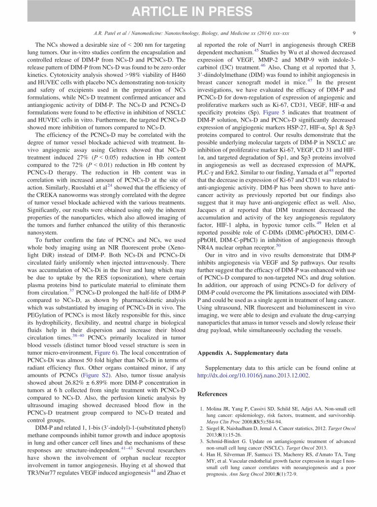
To further confirm the fate of PCNCs and NCs, we usedwhole body imaging using an NIR fluorescent probe (Xeno-light DiR) instead of DIM-P. Both NCs-Di and PCNCs-Dicirculated fairly uniformly when injected intravenously. Therewas accumulation of NCs-Di in the liver and lung which maybe due to uptake by the RES (opsonization), where certainplasma proteins bind to particulate material to eliminate themfrom circulation.37 PCNCs-D prolonged the half-life of DIM-Pcompared to NCs-D, as shown by pharmacokinetic analysiswhich was substantiated by imaging of PCNCs-Di in vivo. ThePEGylation of PCNCs is most likely responsible for this, sinceits hydrophilicity, flexibility, and neutral charge in biologicalfluids help in their dispersion and increase their bloodcirculation times.38–40 PCNCs primarily localized in tumorblood vessels (distinct tumor blood vessel structure is seen intumor micro-environment, Figure 6). The local concentration ofPCNCs-Di was almost 50 fold higher than NCs-Di in terms ofradiant efficiency flux. Other organs contained minor, if anyamounts of PCNCs (Figure S2). Also, tumor tissue analysisshowed about 26.82% ± 6.89% more DIM-P concentration intumors at 6 h collected from single treatment with PCNCs-Dcompared to NCs-D. Also, the perfusion kinetic analysis byultrasound imaging showed decreased blood flow in thePCNCs-D treatment group compared to NCs-D treated andcontrol groups.
DIM-P and related 1, 1-bis (3′-indolyl)-1-(substituted phenyl)methane compounds inhibit tumor growth and induce apoptosisin lung and other cancer cell lines and the mechanisms of theseresponses are structure-independent.41–43 Several researchershave shown the involvement of orphan nuclear receptorinvolvement in tumor angiogenesis. Huying et al showed thatTR3/Nur77 regulates VEGF induced angiogenesis44 and Zhao et
al reported the role of Nurr1 in angiogenesis through CREBdependent mechanism.45 Studies by Wu et al showed decreasedexpression of VEGF, MMP-2 and MMP-9 with indole-3-carbinol (I3C) treatment.46 Also, Chang et al reported that 3,3′-diindolylmethane (DIM) was found to inhibit angiogenesis inbreast cancer xenograft model in mice.47 In the presentinvestigations, we have evaluated the efficacy of DIM-P andPCNCs-D for down-regulation of expression of angiogenic andproliferative markers such as Ki-67, CD31, VEGF, HIF-α andspecificity proteins (Sp). Figure 5 indicates that treatment ofDIM-P solution, NCs-D and PCNCs-D significantly decreasedexpression of angigiogenic markers HSP-27, HIF-α, Sp1 & Sp3proteins compared to control. Our results demonstrate that thepossible underlying molecular targets of DIM-P in NSCLC areinhibition of proliferative marker Ki-67, VEGF, CD 31 and HIF-1α, and targeted degradation of Sp1, and Sp3 proteins involvedin angiogenesis as well as decreased expression of MAPK,PLC-γ and Erk2. Similar to our finding, Yamada et al48 reportedthat the decrease in expression of Ki-67 and CD31 was related toanti-angiogenic activity. DIM-P has been shown to have anti-cancer activity as previously reported but our findings alsosuggest that it may have anti-angiogenic effect as well. Also,Jacques et al reported that DIM treatment decreased theaccumulation and activity of the key angiogenesis regulatoryfactor, HIF-1 alpha, in hypoxic tumor cells.49 Helen et alreported possible role of C-DIMs (DIMC-pPhOCH3, DIM-C-pPhOH, DIM-C-pPhCl) in inhibition of angiogenesis throughNR4A nuclear orphan receptor.50
Our in vitro and in vivo results demonstrate that DIM-Pinhibits angiogenesis via VEGF and Sp pathways. Our resultsfurther suggest that the efficacy of DIM-P was enhanced with useof PCNCs-D compared to non-targeted NCs and drug solution.In addition, our approach of using PCNCs-D for delivery ofDIM-P could overcome the PK limitations associated with DIM-P and could be used as a single agent in treatment of lung cancer.Using ultrasound, NIR fluorescent and bioluminescent in vivoimaging, we were able to design and evaluate the drug-carryingnanoparticles that amass in tumor vessels and slowly release theirdrug payload, while simultaneously occluding the vessels.
Appendix A. Supplementary data
Supplementary data to this article can be found online athttp://dx.doi.org/10.1016/j.nano.2013.12.002.
References
1. Molina JR, Yang P, Cassivi SD, Schild SE, Adjei AA. Non-small celllung cancer: epidemiology, risk factors, treatment, and survivorship.Mayo Clin Proc 2008;83(5):584-94.
2. Siegel R, Naishadham D, Jemal A. Cancer statistics, 2012. Target Oncol2013;8(1):15-26.
3. Schmid-Bindert G. Update on antiangiogenic treatment of advancednon-small cell lung cancer (NSCLC). Target Oncol 2013.
4. Han H, Silverman JF, Santucci TS, Macherey RS, d'Amato TA, TungMY, et al. Vascular endothelial growth factor expression in stage I non-small cell lung cancer correlates with neoangiogenesis and a poorprognosis. Ann Surg Oncol 2001;8(1):72-9.
10 A.R. Patel et al / Nanomedicine: Nanotechnology, Biology, and Medicine xx (2014) xxx–xxx
5. Fontanini G, Faviana P, Lucchi M, Boldrini L, Mussi A, Camacci T, et al.A high vascular count and overexpression of vascular endothelial growthfactor are associated with unfavourable prognosis in operated small celllung carcinoma. Br J Cancer 2002;86(4):558-63.
6. Gasparini G, Longo R, Fanelli M, Teicher BA. Combination ofantiangiogenic therapy with other anticancer therapies: results, chal-lenges, and open questions. J Clin Oncol 2005;23(6):1295-311.
7. Ichite N, Chougule MB, Jackson T, Fulzele SV, Safe S, Singh M.Enhancement of docetaxel anticancer activity by a novel diindolyl-methane compound in human non-small cell lung cancer. Clin CancerRes 2009;15(2):543-52.
8. Ichite N, Chougule M, Patel AR, Jackson T, Safe S, Singh M. Inhalationdelivery of a novel diindolylmethane derivative for the treatment of lungcancer. Mol Cancer Ther 2010;9(11):3003-14.
9. Patel AR, Spencer SD, Chougule MB, Safe S, SinghM. Pharmacokineticevaluation and in vitro-in vivo correlation (IVIVC) of novel methylene-substituted 3,3′ diindolylmethane (DIM). Eur J Pharm Sci 2012;46(1–2):8-16.
10. Farokhzad OC, Langer R. Impact of nanotechnology on drug delivery.ACS Nano 2009;3(1):16-20.
11. Cho K, Wang X, Nie S, Chen ZG, Shin DM. Therapeutic nanoparticlesfor drug delivery in cancer. Clin Cancer Res 2008;14(5):1310-6.
12. Sinha R, Kim GJ, Nie S, Shin DM. Nanotechnology in cancertherapeutics: bioconjugated nanoparticles for drug delivery.Mol CancerTher 2006;5(8):1909-17.
13. Byrne JD, Betancourt T, Brannon-Peppas L. Active targeting schemesfor nanoparticle systems in cancer therapeutics. Adv Drug Deliv Rev2008;60(15):1615-26.
14. Kirpotin DB, Drummond DC, Shao Y, Shalaby MR, Hong K, NielsenUB, et al. Antibody targeting of long-circulating lipidic nanoparticlesdoes not increase tumor localization but does increase internalization inanimal models. Cancer Res 2006;66(13):6732-40.
15. Iinuma H, Maruyama K, Okinaga K, Sasaki K, Sekine T, Ishida O, et al.Intracellular targeting therapy of cisplatin-encapsulated transferrin-polyethylene glycol liposome on peritoneal dissemination of gastriccancer. Int J Cancer 2002;99(1):130-7.
16. Wong HL, Bendayan R, Rauth AM, Li Y, Wu XY. Chemotherapy withanticancer drugs encapsulated in solid lipid nanoparticles. Adv DrugDeliv Rev 2007;59(6):491-504.
17. Souto EB, Wissing SA, Barbosa CM, Muller RH. Development of acontrolled release formulation based on SLN and NLC for topicalclotrimazole delivery. Int J Pharm 2004;278(1):71-7.
18. Pardeike J, Hommoss A, Muller RH. Lipid nanoparticles (SLN, NLC) incosmetic and pharmaceutical dermal products. Int J Pharm 2009;366(1–2):170-84.
19. Muller RH, Radtke M, Wissing SA. Solid lipid nanoparticles (SLN) andnanostructured lipid carriers (NLC) in cosmetic and dermatologicalpreparations. Adv Drug Deliv Rev 2002;54(Suppl 1):S131-55.
20. Selvamuthukumar S, Velmurugan R. Nanostructured lipid carriers: apotential drug carrier for cancer chemotherapy. Lipids Health Dis2012;11:159.
21. van Vlerken LE, Amiji MM. Multi-functional polymeric nanoparticlesfor tumour-targeted drug delivery. Expert Opin Drug Deliv 2006;3(2):205-16.
22. Muthu MS, Kulkarni SA, Raju A, Feng SS. Theranostic liposomes ofTPGS coating for targeted co-delivery of docetaxel and quantum dots.Biomaterials 2012;33(12):3494-501.
23. WuW, Shen J, Gai Z, Hong K, Banerjee P, Zhou S. Multi-functional core-shell hybrid nanogels for pH-dependent magnetic manipulation, fluores-cent pH-sensing, and drug delivery. Biomaterials 2011;32(36):9876-87.
24. Simberg D, Duza T, Park JH, Essler M, Pilch J, Zhang L, et al.Biomimetic amplification of nanoparticle homing to tumors. Proc NatlAcad Sci U S A 2007;104(3):932-6.
25. Dvorak HF, Senger DR, Dvorak AM, Harvey VS, McDonagh J.Regulation of extravascular coagulation by microvascular permeability.Science 1985;227(4690):1059-61.
26. Agemy L, Sugahara KN, Kotamraju VR, Gujraty K, Girard OM, KonoY, et al. Nanoparticle-induced vascular blockade in human prostatecancer. Blood 2010;116(15):2847-56.
27. Karmali PP, Kotamraju VR, Kastantin M, Black M, Missirlis D, TirrellM, et al. Targeting of albumin-embedded paclitaxel nanoparticles totumors. Nanomedicine 2009;5(1):73-82.
29. Stefanick JF, Ashley JD, Kiziltepe T, Bilgicer B. A systematic analysisof peptide linker length and liposomal polyethylene glycol coating oncellular uptake of peptide-targeted liposomes. ACS Nano 2013;7(4):2935-47.
30. Patel AR, Chougule MB, I T, Patlolla R, Wang G, Singh M. Efficacy ofaerosolized celecoxib encapsulated nanostructured lipid carrier in non-small cell lung cancer in combination with docetaxel. Pharm Res2013;30(5):1435-46.
31. Singh A, Ahmad I, Akhter S, Jain GK, Iqbal Z, Talegaonkar S, et al.Nanocarrier based formulation of thymoquinone improves oral delivery:stability assessment, in vitro and in vivo studies. Colloids Surf BBiointerfaces 2013;102:822-32.
32. Chikh GG, Li WM, Schutze-Redelmeier MP, Meunier JC, Bally MB.Attaching histidine-tagged peptides and proteins to lipid-based carriersthrough use of metal-ion-chelating lipids. Biochim Biophys Acta2002;1567(1–2):204-12.
33. Folkman J. Fighting cancer by attacking its blood supply. Sci Am1996;275(3):150-4.
34. Giaccone G. The potential of antiangiogenic therapy in non-small celllung cancer. Clin Cancer Res 2007;13(7):1961-70.
35. Kumar S, Li C. Targeting of vasculature in cancer and other angiogenicdiseases. Trends Immunol 2001;22(3):129.
36. Ferrara N, Hillan KJ, Novotny W. Bevacizumab (Avastin), a humanizedanti-VEGF monoclonal antibody for cancer therapy. Biochem BiophysRes Commun 2005;333(2):328-35.
37. Moghimi SM, Hunter AC, Murray JC. Long-circulating and target-specific nanoparticles: theory to practice. Pharmacol Rev2001;53(2):283-318.
38. Zhu S, Hong M, Tang G, Qian L, Lin J, Jiang Y, et al. Partly PEGylatedpolyamidoamine dendrimer for tumor-selective targeting of doxorubicin:the effects of PEGylation degree and drug conjugation style. Biomater-ials 2010;31(6):1360-71.
39. Lim SM, Kim TH, Jiang HH, Park CW, Lee S, Chen X, et al. Improvedbiological half-life and anti-tumor activity of TNF-related apoptosis-inducing ligand (TRAIL) using PEG-exposed nanoparticles. Biomater-ials 2011;32(13):3538-46.
40. Fang YP, Wu PC, Huang YB, Tzeng CC, Chen YL, Hung YH, et al.Modification of polyethylene glycol onto solid lipid nanoparticlesencapsulating a novel chemotherapeutic agent (PK-L4) to enhancesolubility for injection delivery. Int J Nanomedicine 2012;7:4995-5005.
41. Chintharlapalli S, Papineni S, Safe S. 1,1-bis(3′-indolyl)-1-(p-substitu-tedphenyl)methanes inhibit growth, induce apoptosis, and decrease theandrogen receptor in LNCaP prostate cancer cells through peroxisomeproliferator-activated receptor gamma-independent pathways. MolPharmacol 2007;71(2):558-69.
42. Chintharlapalli S, Papineni S, Baek SJ, Liu S, Safe S. 1,1-Bis(3′-indolyl)-1-(p-substitutedphenyl)methanes are peroxisome proliferator-activated receptor gamma agonists but decrease HCT-116 colon cancercell survival through receptor-independent activation of early growthresponse-1 and nonsteroidal anti-inflammatory drug-activated gene-1.Mol Pharmacol 2005;68(6):1782-92.
43. Inamoto T, Papineni S, Chintharlapalli S, Cho SD, Safe S, Kamat AM.1,1-Bis(3′-indolyl)-1-(p-chlorophenyl)methane activates the orphannuclear receptor Nurr1 and inhibits bladder cancer growth. Mol CancerTher 2008;7(12):3825-33.
44. Zeng H, Qin L, Zhao D, Tan X, Manseau EJ, Van Hoang M, et al. Orphannuclear receptor TR3/Nur77 regulates VEGF-A-induced angiogenesisthrough its transcriptional activity. J Exp Med 2006;203(3):719-29.
11A.R. Patel et al / Nanomedicine: Nanotechnology, Biology, and Medicine xx (2014) xxx–xxx
45. Zhao D, Desai S, Zeng H. VEGF stimulates PKD-mediated CREB-dependent orphan nuclear receptor Nurr1 expression: role in VEGF-induced, angiogenesis. Int J Cancer 2011;128(11):2602-12.
46. Wu HT, Lin SH, Chen YH. Inhibition of cell proliferation and in vitromarkers of angiogenesis by indole-3-carbinol, a major indole metabolitepresent in cruciferous vegetables. J Agric FoodChem 2005;53(13):5164-9.
47. Chang X, Tou JC, Hong C, Kim HA, Riby JE, Firestone GL, et al. 3,3′-Diindolylmethane inhibits angiogenesis and the growth of transplantablehuman breast carcinoma in athymic mice. Carcinogenesis 2005;26(4):771-8.
48. Yamada S, Bu X-Y, Khankaldyyan V, Gonzales-Gomez I, McComb JG,Laug WE. Effect of the angiogenesis inhibitor cilengitide (Emd 121974)on glioblastoma growth in nude mice. Neurosurgery 2006;59(6):1304-12.
49. Riby JE, Firestone GL, Bjeldanes LF. 3,3′-diindolylmethane reduceslevels of HIF-1alpha and HIF-1 activity in hypoxic cultured humancancer cells. Biochem Pharmacol 2008;75(9):1858-67.
50. Mohan HM, Aherne CM, Rogers AC, Baird AW, Winter DC, MurphyEP. Molecular pathways: the role of NR4A orphan nuclear receptors incancer. Clin Cancer Res 2012;18(12):3223-8.