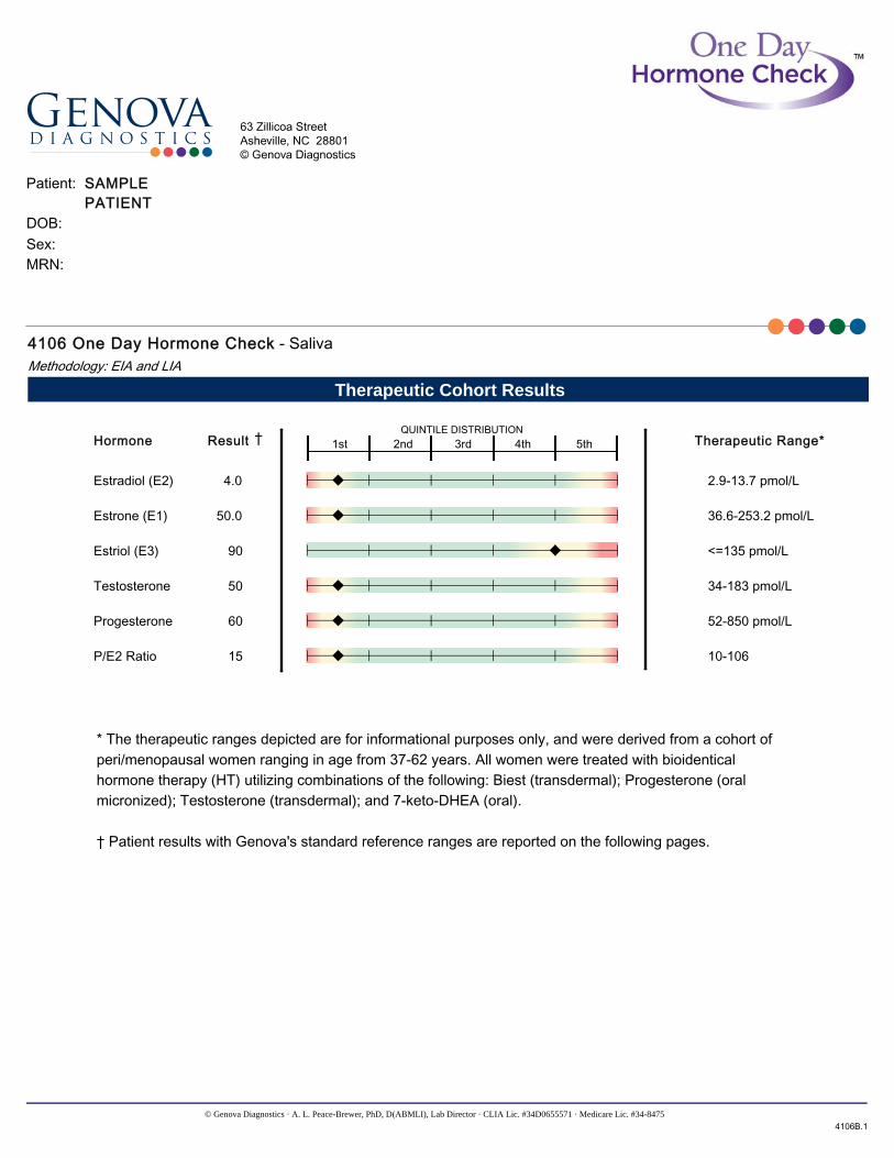
* The therapeutic ranges depicted are for informational purposes only, and were derived from a cohort ofperi/menopausal women ranging in age from 37-62 years. All women were treated with bioidenticalhormone therapy (HT) utilizing combinations of the following: Biest (transdermal); Progesterone (oralmicronized); Testosterone (transdermal); and 7-keto-DHEA (oral).
† Patient results with Genova's standard reference ranges are reported on the following pages.
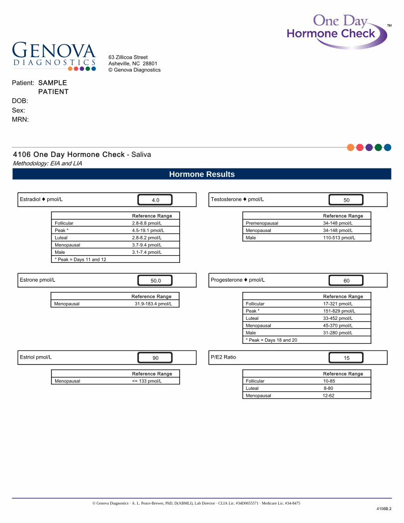
4106 One Day Hormone Check - SalivaMethodology: EIA and LIA
The performance characteristics of all assays have been verified by Genova Diagnostics, Inc. Unless otherwise noted with , the assay has not been cleared by the U.S. Food and Drug Administration.
Commentary is provided to the practitioner for educational purposes, and should not be interpreted as diagnostic or as treatment recommendations. Diagnosis and treatment decisions are the practitioner's responsibility.
Estrogens play a critical role in female sexual development, menstrual function, protein synthesis, cardiovascular function, bone formation and remodeling, cognitive function, emotional balance and other important health factors. The estrogenic potency of estradiol is 12 times that of estrone and 80 times that of estriol. Estradiol is the primary estrogen in premenopausal women. Estrone is the second most potent estrogen compared to estradiol. After menopause, estrone becomes the primary estrogen as the ovary loses its ability to manufacture estradiol, and it is synthesized in the adrenal glands and fat cells. Estriol is considered to be the mildest and briefest-acting of the three estrogens. Estrogen metabolism and synthesis in men appear to remain relatively stable across the life course. • In women, lower levels of estrogens have been associated with a variety of clinical symptoms: peri/menopausalsymptoms (vasomotor symptoms; mood and memory alterations; atrophic vaginitis, a condition associated with decreased vaginal lubrication and thinner vaginal epithelial; lining diminished skin tone); altered lipid metabolism; accelerated rate of bone loss. Excessive estrogen levels have been associated with increased risk of some hormone-dependent cancers. • In men, low levels of estrogen may be associated with decreased bone density, cognitive decline andcardiovascular disease. Excessive estradiol levels have been associated with greater risk of stroke and cardiovascular disease, as well as BPH, gynecomastia, decreased sexual function and weight gain. A source of elevated estrogen in men may be associated with men who have a higher body fat percentage, as increased aromatization of testosterone to estradiol can occur in adipose tissue. • In a large, population based study of salivary sex hormone levels in older adults researchers found: Older men andwomen had similar estradiol concentrations. Estradiol concentrations have been associated with cognition, mood, and memory in women and, in combination with testosterone and other factors, preservation of memory and cognitive function in men.
Progesterone is important for normal reproductive and menstrual function, and influences the health of bone, blood vessels, heart, brain, skin, and many other tissues and organs. As a precursor, progesterone is used by the body to make other steroid hormones, including DHEA, cortisol, estrogen and testosterone. In addition, progesterone plays an important role in mood, blood sugar balance, libido, and thyroid function, as well as adrenal gland health. Progesterone is primarily produced in the ovaries in premenopausal women and in the adrenal cortex in postmenopausal women. Although progesterone is found in both women and men, the physiologic role in men is poorly understood. • In women, lower levels of progesterone have been associated with dysfunctional uterine bleeding, and may play arole in osteoporosis and impaired neurological function. Excessive amounts can result in problems such as dysglycemia, alopecia, acne and breast tenderness. • The clinical significance of elevated or low levels in men is poorly understood. Low progesterone levels may beinvolved in male infertility. Increased levels of progesterone have been found in states of stress and anxiety in men and women: this may relate to its sedative or stress countering effects.
Testosterone is an androgenic sex steroid/hormone that helps maintain libido, influences muscle mass and weight loss, and plays a role in the production of several other hormones. During the aging process, testosterone levels gradually decline in both sexes, which can lead to loss of bone density. Testosterone concentrations tend to be higher in men versus women. • In women, imbalances of testosterone have been associated with various forms of coronary heart disease and
cardiovascular events, including myocardial infarction in postmenopausal women. Low salivary testosterone levels have also been shown in women with breast cancer compared to age-matched controls. Obese women exhibit higher levels of free salivary testosterone. Excessive amounts are associated with PCOS, acne, oily skin and hirsutism. • In men, lower levels of testosterone are associated with aortic, peripheral vascular, and cardiovascular disease inmiddle-aged and older men. In some but not all studies, lower levels of testosterone predict increased incidence of cardiovascular events and mortality. Additionally, elevated testosterone can be associated with CVD risk. Men with excessive testosterone may exhibit aggressive behavior or increased irritability, and hair loss (scalp). • In men and women, low levels of testosterone have been associated with lower coital frequency and loss of sexualdesire in men and women. Low levels are also associated with reduced stamina and lean muscle mass, anxiety, depression and cognitive decline in both men and women.
The P/E2 ratio describes the relationship between progesterone and estradiol levels, and is used clinically to ascertain dominance of one hormone compared to the other. • An elevated ratio may indicate progesterone dominance, and symptoms may be consistent with progesterone excess. • A low ratio may indicate estrogen dominance, and symptoms may be consistent with estrogen excess.
The performance characteristics of all assays have been verified by Genova Diagnostics, Inc. Unless otherwise noted with , the assay has not been cleared by the U.S. Food and Drug Administration.
Commentary is provided to the practitioner for educational purposes, and should not be interpreted as diagnostic or as treatment recommendations. Diagnosis and treatment decisions are the practitioner's responsibility.
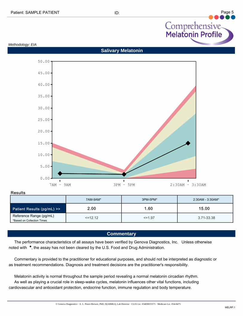
Melatonin activity is normal throughout the sample period revealing a normal melatonin circadian rhythm. As well as playing a crucial role in sleep-wake cycles, melatonin influences other vital functions, including cardiovascular and antioxidant protection, endocrine function, immune regulation and body temperature.
Commentary is provided to the practitioner for educational purposes, and should not be interpreted as diagnostic or as treatment recommendations. Diagnosis and treatment decisions are the practitioner's responsibility.
The performance characteristics of all assays have been verified by Genova Diagnostics, Inc. All assay have been cleared by the U.S. Food and Drug Administration, unless otherwise noted with ♦.
The Reference Range is a statistical interval representing 95% or 2 Standard Deviations (2 S.D.) of the reference population.
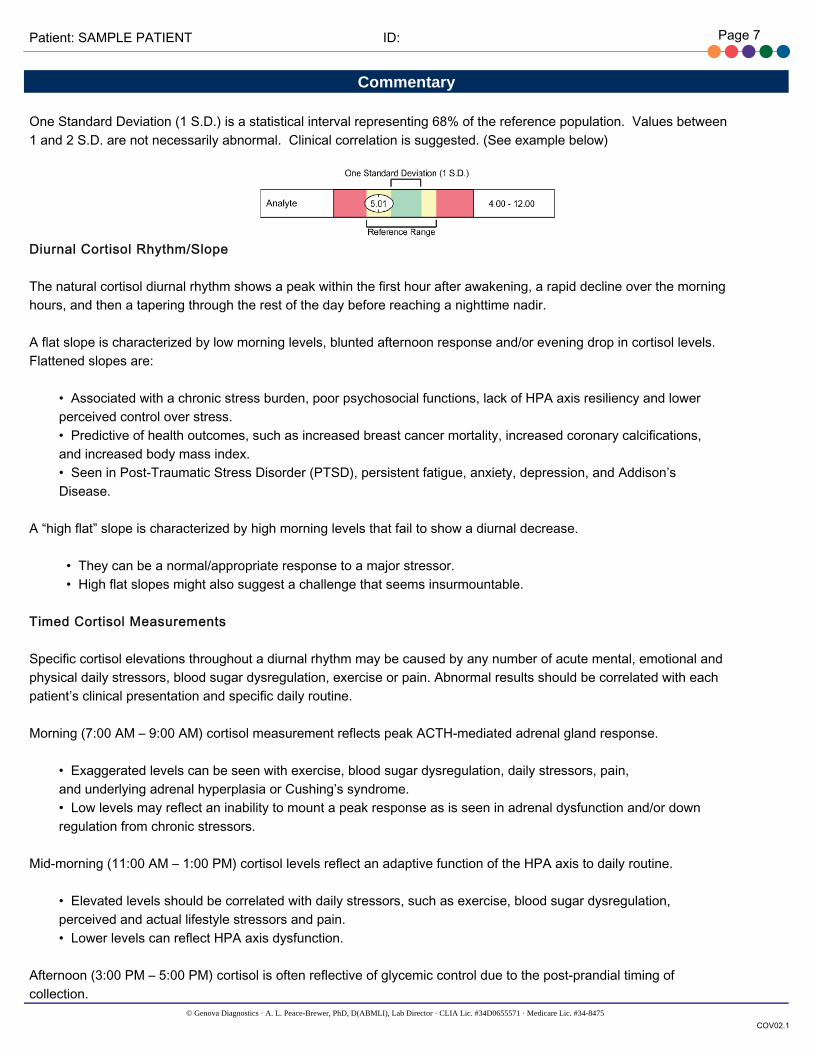
One Standard Deviation (1 S.D.) is a statistical interval representing 68% of the reference population. Values between 1 and 2 S.D. are not necessarily abnormal. Clinical correlation is suggested. (See example below)
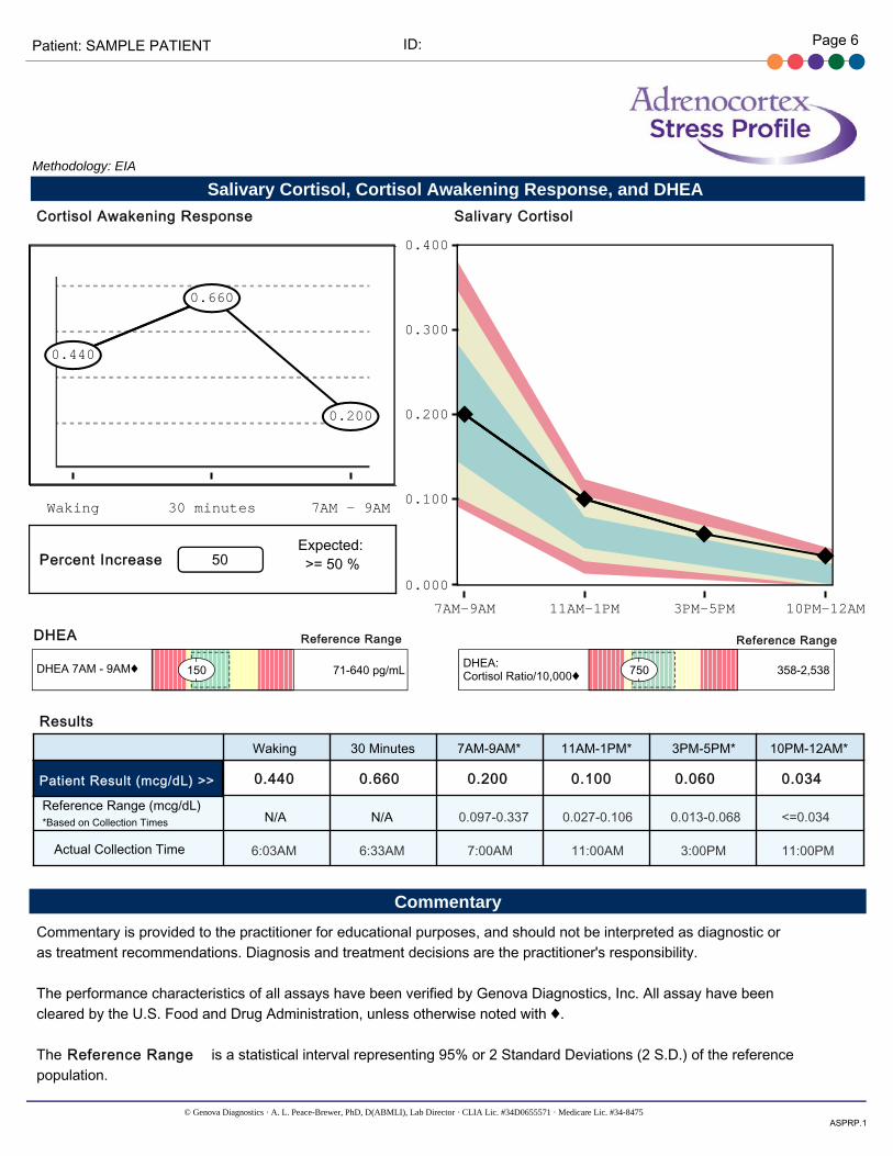
Diurnal Cortisol Rhythm/Slope
The natural cortisol diurnal rhythm shows a peak within the first hour after awakening, a rapid decline over the morning hours, and then a tapering through the rest of the day before reaching a nighttime nadir.
A flat slope is characterized by low morning levels, blunted afternoon response and/or evening drop in cortisol levels. Flattened slopes are:
• Associated with a chronic stress burden, poor psychosocial functions, lack of HPA axis resiliency and lower perceived control over stress. • Predictive of health outcomes, such as increased breast cancer mortality, increased coronary calcifications, and increased body mass index. • Seen in Post-Traumatic Stress Disorder (PTSD), persistent fatigue, anxiety, depression, and Addison’s Disease.
A “high flat” slope is characterized by high morning levels that fail to show a diurnal decrease.
• They can be a normal/appropriate response to a major stressor. • High flat slopes might also suggest a challenge that seems insurmountable.
Timed Cortisol Measurements
Specific cortisol elevations throughout a diurnal rhythm may be caused by any number of acute mental, emotional and physical daily stressors, blood sugar dysregulation, exercise or pain. Abnormal results should be correlated with each patient’s clinical presentation and specific daily routine.
• Exaggerated levels can be seen with exercise, blood sugar dysregulation, daily stressors, pain, and underlying adrenal hyperplasia or Cushing’s syndrome. • Low levels may reflect an inability to mount a peak response as is seen in adrenal dysfunction and/or down regulation from chronic stressors.
Mid-morning (11:00 AM – 1:00 PM) cortisol levels reflect an adaptive function of the HPA axis to daily routine.
• Elevated levels should be correlated with daily stressors, such as exercise, blood sugar dysregulation, perceived and actual lifestyle stressors and pain. • Lower levels can reflect HPA axis dysfunction.
Afternoon (3:00 PM – 5:00 PM) cortisol is often reflective of glycemic control due to the post-prandial timing of collection.
• Elevated levels can reflect any number of daily stressors as previously outlined. • Low levels can reflect underlying HPA axis dysfunction.
Evening (10:00 PM – 12:00 AM) cortisol levels are a good indication of baseline HPA axis function since they represent the lowest level during the circadian rhythm.
• Elevated levels may be due to stress, exercise, alcohol, and specific lifestyle stressors. • Elevated evening salivary cortisol is linked to insomnia • High evening cortisol levels are also associated with various diseases such as diabetes, cardiovascular disease, hormonally driven cancers, and osteoporosis.
Treatment of elevated cortisol should be directed at the root cause of the stressor. Lifestyle modification with relaxation methods, dietary changes, pain management, and overall HPA axis support with nutrition and/or adaptogens can be helpful. Glandulars may be added if additional support is necessary.
Cortisol Awakening Response (CAR)
CAR is calculated by a direct percent increase: difference between 30 minutes and wake, divided by wake, then multiplied by 100. In literature, there are several ways to calculate CAR. Expected increases may differ depending on which calculation is used. Most literature demonstrates an expected increase of greater than 50% as a reflection of HPA axis resiliency.¹
CAR represents the momentum of rising cortisol levels that begins several hours prior to awakening and an additional transient increase. The initial cortisol rise begins due to ACTH-mediated normal HPA axis activities with the additional CAR increase caused by supra-chiasmic nucleus (SCN) light activation.
CAR reflects a person’s ability to cope with anticipated challenges and the perceptions of control around chronic stress. CAR is calculated based on the percent cortisol rise from awakening to 30 minutes. A value of approximately 50% is expected.
Approximately 25% of healthy adults do not mount a CAR, and are termed non-responders. Response is defined as an increase of at least 2.5 nmol/l (0.09 mcg/dL) above individual baseline. Any patient with a result less than this is considered a “non-responder” if sampling was performed correctly and the rest of the diurnal curve shows adequate cortisol response.
• Blunted CAR is seen in clinical burnout, self-reported health problems, early loss experiences, material hardship, depression, PTSD, and amnesia. • Elevated CAR can be adaptive as a reflection of anticipation for daily stress. It may play a literal role in “preparing for action” by stimulating motor function, immunity responses, and alertness. • If CAR is abnormal, and the rest of the diurnal pattern is not, then this would imply that a CAR-specific mechanism (SCN-related signaling) is implicated instead of a CRH or ACTH-mediated mechanism. Any abnormality of the hippocampus may blunt the CAR response and not affect the diurnal slope.
• If both the CAR and the diurnal rhythm are abnormal, this may represent a more general HPA dysfunction. It may also be useful to look at DHEA for a complete assessment of the HPA axis.
CAR treatment involves HPA axis and adrenal support using lifestyle modification, nutrition and adaptogens. However,
Commentaryinsight into blunted or elevated CAR may help direct additional modalities such as behavioral modification and psychological therapies.
Evening (10:00 PM – 12:00 AM) cortisol levels are a good indication of baseline HPA axis function since they represent the lowest level during the circadian rhythm.
• Elevated levels may be due to stress, exercise, alcohol, and specific lifestyle stressors. • Elevated evening salivary cortisol is linked to insomnia. • High evening cortisol levels are also associated with various diseases such as diabetes, cardiovascular disease, hormonally driven cancers, and osteoporosis.
DHEA
DHEA levels peak at around age 25, then decline steadily through the following decades. DHEA can be converted downstream in the steroidogenic pathway to create androgens and estrogens. It has antioxidant and anti-inflammatory properties and can be protective against corticosterone’s neurotoxic effects.
• Lower levels of DHEA are seen with advancing age and have been associated with immune dysregulation, cardiovascular disease, arthritis, osteoporosis, insomnia, declining cognition, depression, fatigue, and decreased libido. • Elevated levels of DHEA may reflect endogenous exposure and supplementation. Other considerations include Polycystic Ovarian Syndrome (PCOS,) adrenal hyperplasia and adrenal tumors.
General recommendations include overall control of the cortisol response, HPA axis support using nutrition, adaptogens, and behavioral modification.
DHEA:Cortisol Ratio
This calculation represents anabolic and catabolic balance. Since DHEA acts not only as an anabolic hormone, but appears to down-regulate the cellular effects of cortisol, this measurement can theoretically enhance the predictive value of HPA axis dysfunction.
• An elevated ratio reflects elevated DHEA levels as compared to cortisol, which favors anabolic activity. Causes of DHEA and cortisol abnormalities should be evaluated. • A decreased ratio generally reflects a more catabolic state. It is associated with cortisol elevations and HPA-axis imbalances. Causes of DHEA and cortisol abnormalities should be addressed. • An optimal ratio indicates proper HPA axis homeostasis.
References:
1. Clow A, Thorn L, Evans P. Hucklebridge F. The awakening cortisol response: methodological issues and significance. Stress. 2004;7(1):29-37. 2. Stalder T, Kirschbaum C, Kudielka BM, et al. Assessment of the cortisol awakening response: Expert consensus guidelines. Psychoneuroendocrino. 2016;63:414-432. 3. Wust S, Wolf J, Hellhammer DH, Federenko I, Schommer N, Kirschbaum C. The cortisol awakening response-normal values and confounds. Noise health. 2000;2(7):79. 4. Fries E, Dettenborn L, Kirschbaum C. The cortisol awakening response (CAR): facts and future directions. IntJPsychophysiol. 2009;72(1):67-73.
Commentary5. Saxbe DE. A field (researcher's) guide to cortisol: tracking HPA axis functioning in everyday life. Health Psychol Rev. 2008;2(2):163-190.