j ourna l homepage: www.e lsev ie r .com/ locate / f i to te
Thermal stability of ginkgolic acids from Ginkgo biloba and theeffects of ginkgol C17:1 on the apoptosis and migration ofSMMC7721 cells
Xiao-Ming Yang a,⁎, Yun-Fei Wang a, Yue-Ying Li b, Hai-Le Ma a
a School of Food and Biological Engineering, Jiangsu University, Zhenjiang 212013, Chinab School of Medical Science and Laboratory Medicine, Jiangsu University, Zhenjiang 212013, China
Article history:Received 4 May 2014Accepted in revised form 26 June 2014Accepted 4 July 2014Available online 10 July 2014
Ginkgolic acids are alkylsalicylic acid derivatives with a thermolabile carboxylic group fromGinkgo biloba L., and they exhibit anticancer activity. Their anticancer effects are closelyassociated with their thermal stability. In this study, the thermal decomposition of ginkgolicacids was analyzed at temperatures of 30, 50, 70 and 250 °C. The results clearly showed thatan obvious slow decarboxylation of the ginkgolic acids was detected at 70 °C. When thetemperature increased to 250 °C, the decarboxylation reaction was rapidly completed. Theginkgolic acids were decarboxylated to yield ginkgols. The ginkgols C13:0, C15:1 and C17:1were separated and definitively identified by IR, NMR and GC-MS. The cytotoxic effects ofginkgols C13:0, C15:1 and C17:1 were tested and compared with those of the correspondingginkgolic acids. An MTT assay showed that ginkgol C17:1 (48-h IC50 = 8.5 μg · ml-1) has thestrongest inhibition on SMMC-7721 cells in a dose- and time-dependent manner. Theanticancer action may occur via the induction of apoptosis by the activation of caspases-3, theupregulation of Bax expression, and the inhibition migration of SMMC7721 cells. The resultsindicated that ginkgol C17:1 might be useful for the further development of a hepatocellularcarcinoma preventive agent.
Ginkgo biloba L. has been therapeutically used forthousands of years in Chinese traditional medicine. Theextract and infusion made from the ginkgo leaf have beenutilized to treat lung ailments such as asthma and bronchitisand as a remedy for cardiovascular diseases in China [1]. Overthe past several decades, the extract of the ginkgo leaf hasentered the herbal spotlight mainly because of its provenbenefits for treating Alzheimer’s disease [2,3] and its promiseas a therapeutic agent for many other chronic and acuteforms of diseases. Now, the extract of the ginkgo leaf isbecoming one of the most popular and scientifically explored
pharmaceutical- and nutraceutical-grade raw materials [4].The standardized extract of the ginkgo leaf (EGb 761) isone of the best-selling herbal supplements in Europe, andginkgo products are ranked among the top 10 bestsellingover-the-counter dietary supplements in the USA [5,6]. Theginkgo nut is consumed as a food and medicine throughoutthe world [1]. The ginkgo nut has been known to treatpulmonary disorders, alcohol abuse and bladder inflammation,and Chinese and Japanese cuisines often involve ginkgo nutconsumption as a part of their diet [7]. Compared with the leafand nut however, the ginkgo sarcotesta is almost completelyneglected in commercial terms. The ginkgo sarcotesta is animportant agricultural byproduct obtained during the process-ing of the ginkgo nut. The potential annual availability of driedginkgo sarcotesta is 26 million pounds in China [8]. However,no industrial application has been developed until now.
Themain active ingredients of the ginkgo sarcotesta includeflavonoid glycosides, terpene lactones, polysaccharides andginkgolic acids. In addition to the sarcotesta, ginkgolic acids arealso present in the leaf and nut of Ginkgo biloba [8]. Thepotential annual availability of this class of components, whichaccounts for approximately 5% of the sarcotesta dry weight [9],is enormous. Because this class of compounds possessescontact allergens [10], it has been identified as a hazardousconstituent in the crude ginkgo extract [11]. However, severaltherapeutically desired effects have been reported for ginkgolicacids, such as antioxidant activity [12], antitumor activity [11],inhibition of phospholipase Cγ1 [13] and antifungal activi-ty [14]. Our previous studies showed that ginkgolic acidsexhibit molluscicidal activity against Oncomelania hupensis[15]. These prominent bioactivities lead to interest in ginkgolicacid research with regard to its potential applications as atherapeutic agent in the treatment of cancer and other diseasesor as a botanical molluscicide.
Ginkgolic acids are 2-hydroxy-6-alkylbenzoic acids. The alkylside chain varies from 13 to 17 carbons in length with zero totwo double bonds [16]. Because of the thermolability of thecarboxylic group, ginkgolic acids can be converted into ginkgols(3-alkylphenols) by heating. This decarboxylation reaction isgenerally accomplished by immersing ginkgolic acids in a hot oilbath preheated to 200 °C [17], but the thermal stability ofginkgolic acids below 200 °C is unclear. The bioactive propertiesof ginkgolic acids are closely associated with their thermalstability. To reduce the loss of ginkgolic acids caused bypharmaceutical processing, it is important to understand thethermal stability of ginkgolic acids below 200 °C. Furthermore,the effects of ginkgols on cancer cells still require furtherinvestigation. Ginkgols have shown antiproliferation effects onvarious cancer cells [13]; however, the effect is unknown fornecrosis or apoptosis. To promote ginkgols toward a chemopre-vention agent, ginkgols’ underlying mechanism of action oncancer cells must be examined.
In our present work, the thermal stabilities of ginkgolicacids at 30, 50, 70 and 250 °C were investigated. Theirthermal decomposition products, ginkgol monomers, wereseparated and identified. The antiproliferation effects on thevarious cancer cell lines of ginkgolic acid and ginkgolmonomers were compared. Ginkgol C17:1 showed excellentanticancer activity, particularly on hepatocellular carcinomaSMMC7721 cells. We investigated whether a ginkgol C17:1treatment will cause necrosis and/or apoptosis in SMMC7721cells and migration inhibition in SMMC7721 cells.
2. Materials and methods
2.1. Materials and chemicals
Sunlight-dried Ginkgo biloba L. sarcotestas, collected in theJiangsu province, China in October, 2010, were identified by thePharmacognosy Laboratory, School of Pharmaceuticals. Voucherspecimens were deposited in the Pharmacognosy Laboratory,School of Pharmaceutical, Jiangsu University.
The ginkgolic acids C13:0, C15:1 and C17:1 were isolatedand prepared in our laboratory. The isolation procedure ofthe ginkgolic acids from the Ginkgo sarcotesta was depictedin the reference [15]. The purities were determined as N98%by HPLC according to Yang et al. [15,18]. Methanol (LC grade)
was purchased from China National Medicines Co., Ltd.Wahaha purified water (Hangzhou Wahaha Group Co., Ltd.)was used in the HPLC experiments. The other chemicals usedin this study were all analytical grade unless specified.
The DMEM and fetal bovine serum were purchased fromGibco. 3-(4, 5-dimethylthiazol-2-yl)-2, 5-diphenyltetrazoliumbromide (MTT), Hoechst 33342 and propidium iodide (PI)fluorescent stain were purchased from Sigma. Mouseanti-Bcl-2, mouse anti-Bax, and rabbit anti β-actin werepurchased from Beijing Zhongshan Golden Bridge Biotech(Beijing, China). A caspase-3 apoptosis-detection kit waspurchased from the Beyotime biotechnology company.
2.2. General Analysis
UV spectra were obtained on a UV-2450 spectrometer(Shimadzu, Japan) using methanol as the solvent. FT-IRspectra were recorded on a Nicolet Nexus 470 FT-IRspectrometer (Thermo Electron Corp., USA) using dichloro-methane as the diluting agent. NMR spectra were collectedon a Bruker DRX500 NMR spectrometer in CDCl3 operating at500 MHz for 1H analysis and at 125 MHz for 13C analysis,using tetramethylsilane (TMS) as an internal standard.GC-MS analysis was performed using a 5973 Mass SelectiveDetector coupled to an HP 6890 gas chromatograph (Agilent,US) fitted with a DB-1701 capillary column. Helium was usedas the carrier gas with a linear pressure of 0.38 MPa. Samplealiquots of 1 μl were injected. The oven temperature programwas as follows: initial temperature at 100 °C for 2 min andthen ramped by 20 °C · min-1 to 240 °C, ending in 20 min at240 °C. The injector temperature was maintained at 250 °C.EI spectra were acquired between 10 and 800 Da.
2.3. Analytical and semi-preparative HPLC
An Agilent 1200 HPLC system consisting of a G1311A pumpand a G1314b VWD detector (Agilent Corp., USA) was used toperform all experiments. Agilent Chemstation software B.02was used for data collection and processing. The analysis wasperformed on a ZORBAX Eclipse XDB-C18 column(150 mm × 4.6 mm, 5 μm). The mobile phase used wasmethanol and water containing 1% acetic acid (90:10, v/v) ata flow of 1.0 ml · min-1. The ginkgolic acids in the eluent weredetected at 310 nm.
A ZORBAX SB-C18 (250 mm × 9.4 mm, 5 μm) semi-preparative columnwas employed for the isolation of individualcompounds in the decarboxylated ginkgolic acids.
2.4. Thermal stability study of the ginkgolic acids
2.4.1. TG-DTA analysisTG-DTA were performed simultaneously using a STA 449 C
instrument (NETZSCH, Germany). Approximately 10 mg ofginkgolic acids from the 4 °C storing condition were weighedon an alumina crucible and then heated to 600 °C in a static airatmosphere. The heating rate was 10 °C · min-1. Calcinedkaolinite was used as the reference material.
2.4.2. Thermal-stability testsThe thermal stability of ginkgolic acids was investigated
at temperatures of 30, 50 and 70 °C. The samples of the
68 X.-M. Yang et al. / Fitoterapia 98 (2014) 66–76
ginkgolic acids (purity of 98%, 50 mg) in equal portions werekept in open containers in a dark, thermocontrolled oven.Another equal portion of ginkgolic acids was weighed on analumina crucible and heated to 250 °C for 30 min in the STA449 C instrument. The heating rate was 50 °C · min-1.
The heat decomposition was evaluated by the UV spectralanalysis and by measuring the contents of the ginkgolic acidsin the samples with HPLC.
2.4.3. LC-MS analysis of the thermal-decomposition productsLC-MS analysis was performed with a Surveyor HPLC
system connected with a Surveyor LCQ class ion trap massdetector using electrospray ionization (ESI) in negative-ionization mode (Thermo Finnigan, USA). An Agilent TC-C18column (150 mm × 4.6 mm, 5 μm) was operated at 35 °C at aflow rate of 1.0 ml · min-1. The mobile phases were methanoland water containing 1% acetic acid (90:10, v/v). The decom-position products and ginkgolic acids in the eluting phase weredetected with a diode-array detector set at 275 and 310 nm.
2.5. Isolation and identification of the ginkgol monomers
Decarboxylation of the ginkgolic acids was conducted asdescribed by references [17,19]. The ginkgolic acids were mixedwith calcium hydroxide at 140 °C for 2 h. The mixture wasextracted with petroleum ether (60-90 °C) and concentrated togive brown oil. The oil was purified to yield ginkgols. Theginkgols was separated by semi-preparative HPLC elution withmethanol and water (90:10, v/v) at a flow rate of3.0 ml · min-1. The ginkgols C13:0, C15:1 and C17:1 in theeluate were detected at 275 nm, collected and subsequentlyfreeze-dried. The structures of ginkgols C13:0, C15:1 and C17:1were definitively identified by IR, NMR and GC-MS.
2.6. Cancer cell lines and culture
The cancer cell lines of A549 (lung cancer), SMMC7721(hepatocellular carcinoma) and U251 (human glioma) wereobtained from the Institute of Biochemistry and Cell Biology.The cells were adapted in a DMEM medium containing 10%fetal bovine serum, 100 unit · ml-1 penicillin and 125 mg · L-1
streptomycin at 37 °C in a humidified atmosphere of 5% CO2.
2.7. MTT assay
The cells were pooled, diluted to the cell density105 cells · ml-1 and dispensed into a 96-well plate. The testcompounds were dissolved in DMSO, sonicated and thendiluted 10-fold with the culture medium and added to thecells to a final volume of 100 μl per well. After incubation forvarious times, 20 μl of the MTT solution (5 mg · ml-1) wasadded to each well and incubated for another 4 h. Thegrowth medium was removed, and 100 μl DMSO was addedand mixed thoroughly. The absorbance was measured at492 nm using a microplate reader (Bio-Rad, USA). Thepositive and negative controls were included in all assays.
2.8. Cellular-morphology analysis
The exponential phase SMMC-7721 cells were seededonto a 24-well plate (sterile glass coverslips early in the
24-well plate) and incubated overnight. Then, cells weretreated with various concentrations of ginkgol C17:1 for 12and 24 h, and the cells were triple washed with PBS andstained with fluorescence staining fluid containing Ho.33342(10 μg · ml-1) and PI (50 μg · ml-1) for 20 min in a 37 °Chumidified box. After staining, the glass coverslips weretriple washed with PBS, and then the cover slips weremounted onto microscope slides, and the nuclear morphol-ogy was observed under a fluorescence microscope (200×).
2.9. Caspase-3-activity assay
An in vitro caspase-3-activity assay was performed aspreviously described [20]. Briefly, SMMC-7721 cells wereincubated with ginkgol C17:1 (0, 10, 20 μg · ml-1) for 48 hand lysed on ice for 15 min. The assays were performed on96-well microtiter plates by incubating 10 μl protein of the celllysate per sample in 80 μl reaction buffer (1% NP-40, 20 mMTris-HCl (pH 7.5), 137 mM Nad, and 10% glycerol) containing10 μL caspase-3 substrate (Ac-DEVD-pNA) (2 mM). The lysateswere incubated at 37 °C for 2 h. The absorbance of the sampleswas measured at 405 nm, and the caspase-3 activities wereexpressed as the percentage of enzyme activity comparedwiththat of the control. All of the experiments were performed intriplicate.
2.10. Western blot analysis
Western blotting was used to evaluate the expression ofBax and Bcl-2 proteins. SMMC-7721 cells were seeded intosix-well plates at a density of 1 × 10⁶ cells · mL-1 andcultured 12 h for cell attachment. Then, the cells were treatedwith various concentrations of ginkgol C17:1 for 24, 48, and72 h. The cultures were harvested and washed twice with PBSand then suspended in cell lysis buffer for incubating for10 min at 100 °C. Finally, it is necessary to expose the cells toultrasonic treatment for 30 seconds to ensure thoroughcracking. The proteins of each group were extracted using theProtein Extraction Reagent Kit (Pierce, USA) according to themanufacturer’s instructions. Equal amounts of protein wereseparated by electrophoresis with 10% SDS-PAGE and weretransferred to PVDF membranes. Antibodies against Bax(1:500), Bcl-2 (1:200) and β-actin (1:10000) were diluted inTBST. Afterward, 5% nonfatmilk was used for 1 h, and the blotswere washed three times. Antibodies against Bcl-2 and β-actinwere incubated with the membranes for 2 h with gentleagitation, and then the membranes were washed three timeswith PBS and incubated with a rabbit secondary antibody foran additional 1 h. After washing three times with PBS, theWestern blot chemiluminescence regent was added, andchemiluminescent signals were detected using a TyphoonMolecular imaging system (GE Company).
2.11. Migration assay
A cell-migration assay was performed using TranswellBoyden chambers (Corning, Acton, MA). Briefly, 500 μl DMEMcontaining 10% FBS was added to the bottom chamber.The exponential phase SMMC-7721 cells were collected, and5 × 103 cells were transferred into 300 μl of serum-freemedium containing various concentrations of ginkgol C17:1 to
69X.-M. Yang et al. / Fitoterapia 98 (2014) 66–76
the top chamber. The cells were allowed to migrate throughthe polyethylene terephthalate membrane between thebottom and the top chamber. After incubation at 37 °C in5% CO2, the cells that had not migrated were removed fromthe upper surface of the membranes using cotton swabs, andthose that migrated to the lower surface of the membraneswere fixed in 4% paraformaldehyde and stained with Giemsastaining. The migration was determined by counting thecell number with a microscope at 200×. Five visual fieldswere chosen randomly for each assay. The mean numberof the migrating cells in the five fields was taken as thecell-migration number of the group. All experiments wereperformed in triplicate.
2.12. Statistical analysis
The data were presented as the means ± SD. A statisticalanalysis was evaluated with Student’s t test by the SPSS 16.0software.
3. Results
3.1. Thermal stability of the ginkgolic acids
TG-DTA analysis was used to observe the thermal decom-position of the ginkgolic acids. The TG-DTA curves (Fig. 1) of theginkgolic acids showed two endothermic peaks at temperaturesranging from 4 to 250 °C. The first endothermic peak appearedat temperatures between 4 and 50 °C with no mass loss,corresponding to the heat absorption of the sample fromthe 4 °C storage condition. The second endothermic peak at200-250 °C with a mass loss of approximately 13.95% wasconsidered to be the result of the decarboxylation reaction of theginkgolic acids. Tyman et al. [17] reported that anacardic acids innatural cashew nut shell liquid (CNSL) were decarboxylated byimmersion in a hot oil bath preheated to 200 °C.
Additionally, the initial decarboxylation temperature ofthe ginkgolic acids from the TG-DTA curves was 200 °C.According to Fig. 1 only, we were still not sure that theginkgolic acids were stable under 200 °C. The stability of the
50 100 150 200 250 300 350 400 450 500 550
0
20
40
60
80
100
Temperature / OC
Mas
s/%
-0.4
-0.3
-0.2
-0.1
0.0
0.1
DTA
/(uV/m
g)
Fig. 1. TG-DTA diagram of the ginkgolic acids.
carboxyl group of the ginkgolic acids below 200°C requiresfurther investigation.
The ginkgolic acid samples were heated to 30, 50 and70 °C for 10, 20 and 30 days, respectively, and at 250 °C for0.5 h. The UV absorption spectra of the original sample andthe thermally treated samples obtained at 30, 50, 70 and250 °C are shown in Fig. 2. After heating at 30 and 50 °C for30 days, the profiles of the UV spectra of the samples wereunchanged, comparing to that of the original sample. Whenthe temperature reached 70 °C, significant breakdowns at243 and 310 nm were observed with prolonged treatmenttime, suggesting that the carboxyl chromophores wereremoved in the thermally decomposed derivatives. Whenthe samples were heated at 250 °C for 0.5 h, UV detectionindicated a clear build-up at 275 nm for the thermally treatedproducts with similar UV absorption at λmax to that of theginkgols.
The contents of the ginkgolic acids in the thermallytreated samples were analyzed by HPLC. The contents of theginkgolic acids in the sample heated at 30 and 50 °C for30 days were 99.8% and 99.6%, respectively, and the thermaldecomposition rate was negligible. For ginkgolic acids heatedat 70 °C for 30 days, the content of ginkgolic acid was of75.3%. The lowest ginkgolic acid content of 4.2% was observedfor the sample heated at 250 °C for 0.5 h. The ginkgolic acidswere rapidly decomposed at 250 °C.
HPLC-ESI-MS analyses were performed, and the total ionchromatograms (TICs) and HPLC-UV chromatograms of thesample heated at 250 °C for 0.5 h are shown in Fig. 3. TheHPLC chromatograms obtained at 275 nm exhibited similarprofiles to those of the HPLC chromatograms of the ginkgolicacids detected at 310 nm. LC-ESI mass spectrometricstudies revealed that, as expected, the thermally decomposedproducts of peaks 1, 2 and 3 exhibit ions at m/z 275[M-44-H]-, 301 [M-44-H]- and 329 [M-44-H]-, which corre-spond to the deprotonated and singly decarboxylatedmolecular ions of the ginkgolic acids C13:0, C15:1 andC17:1, respectively. The UV spectra of peaks 1, 2 and 3exhibited similar profiles to those of the ginkgols. This resultis consistent with the well-known fact that ginkgolic acidtends to become converted into ginkgol because of thethermolability of its carboxyl group [17].
200 225 250 275 300 325 350 375 4000.0
0.5
1.0
1.5
2.0
2.5
3.0
Abs
Wavelength/nm
heating at 250oC for 0.5h
heating at 70oC for 30d
heating at 70oC for 10d
ginkgolic acids(original and heating at 30, 50oC)
ginkgols
Fig. 2. The UV spectra of the ginkgolic acids and their thermal decompositionderivatives.
RT: 0.00 - 19.99 SM: 5G
0 2 4 6 8 10 12 14 16 18
Time (min)
0
10000
20000
30000
uAU
0
100000
200000
300000
uAU
20
40
60
80
100
Rel
ativ
e A
bund
ance
RT: 0.05
RT: 12.06RT: 18.36
RT: 16.70
RT: 10.96
10.00
9.19
15.4611.50 14.32
1.73 17.562.681.18 12.994.36 7.813.91 18.784.98
11.45
17.56
10.49
12.981.73 16.211.181.91
NL:5.87E5
TIC F: MS Genesis 03
NL:3.80E5
Channel A UV 03
NL:3.28E4
Channel B UV 03
A
B
CC13:0
C15:1
C17:2 C15:0
C17:1
Peak 1
Peak 2
Peak 3
1 #1106 RT: 10.53 AV: 1 NL: 7.62E4T: - c ESI Full ms [ 250.00-600.00]
Fig. 3. LC-DAD-ESI-MS analysis of the ginkgolic acids at 250 °C for 0.5 h. (A) HPLC-MS chromatogram; (B) HPLC-UV chromatogram detected at 275 nm; (C) HPLC-UVchromatogram detected at 310 nm; (D) MS spectra and UV spectra of peak 1;(E) MS spectra and UV spectra of peak 2; and (F) MS spectra and UV spectra of peak 3.
70 X.-M. Yang et al. / Fitoterapia 98 (2014) 66–76
71X.-M. Yang et al. / Fitoterapia 98 (2014) 66–76
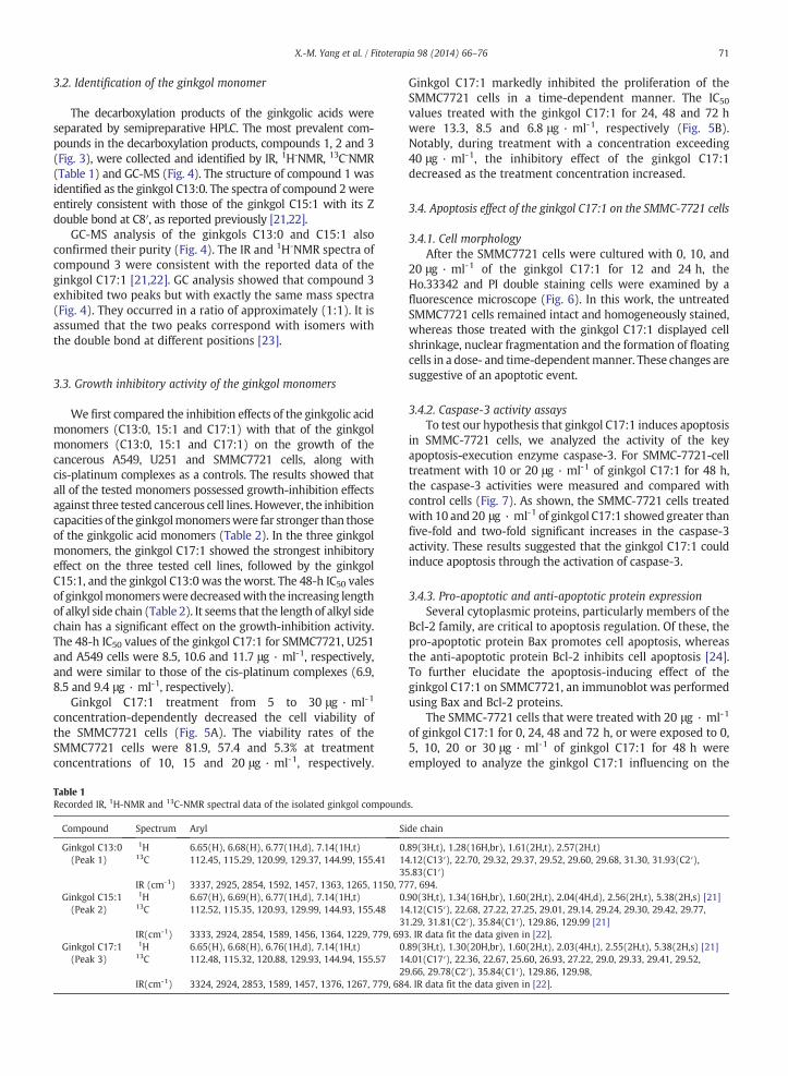
3.2. Identification of the ginkgol monomer
The decarboxylation products of the ginkgolic acids wereseparated by semipreparative HPLC. The most prevalent com-pounds in the decarboxylation products, compounds 1, 2 and 3(Fig. 3), were collected and identified by IR, 1H-NMR, 13C-NMR(Table 1) and GC-MS (Fig. 4). The structure of compound 1 wasidentified as the ginkgol C13:0. The spectra of compound 2wereentirely consistent with those of the ginkgol C15:1 with its Zdouble bond at C8′, as reported previously [21,22].
GC-MS analysis of the ginkgols C13:0 and C15:1 alsoconfirmed their purity (Fig. 4). The IR and 1H-NMR spectra ofcompound 3 were consistent with the reported data of theginkgol C17:1 [21,22]. GC analysis showed that compound 3exhibited two peaks but with exactly the same mass spectra(Fig. 4). They occurred in a ratio of approximately (1:1). It isassumed that the two peaks correspond with isomers withthe double bond at different positions [23].
3.3. Growth inhibitory activity of the ginkgol monomers
We first compared the inhibition effects of the ginkgolic acidmonomers (C13:0, 15:1 and C17:1) with that of the ginkgolmonomers (C13:0, 15:1 and C17:1) on the growth of thecancerous A549, U251 and SMMC7721 cells, along withcis-platinum complexes as a controls. The results showed thatall of the tested monomers possessed growth-inhibition effectsagainst three tested cancerous cell lines. However, the inhibitioncapacities of the ginkgolmonomerswere far stronger than thoseof the ginkgolic acid monomers (Table 2). In the three ginkgolmonomers, the ginkgol C17:1 showed the strongest inhibitoryeffect on the three tested cell lines, followed by the ginkgolC15:1, and the ginkgol C13:0 was theworst. The 48-h IC50 valesof ginkgolmonomerswere decreasedwith the increasing lengthof alkyl side chain (Table 2). It seems that the length of alkyl sidechain has a significant effect on the growth-inhibition activity.The 48-h IC50 values of the ginkgol C17:1 for SMMC7721, U251and A549 cells were 8.5, 10.6 and 11.7 μg · ml-1, respectively,and were similar to those of the cis-platinum complexes (6.9,8.5 and 9.4 μg · ml-1, respectively).
Ginkgol C17:1 treatment from 5 to 30 μg · ml-1
concentration-dependently decreased the cell viability ofthe SMMC7721 cells (Fig. 5A). The viability rates of theSMMC7721 cells were 81.9, 57.4 and 5.3% at treatmentconcentrations of 10, 15 and 20 μg · ml-1, respectively.
Table 1Recorded IR, 1H-NMR and 13C-NMR spectral data of the isolated ginkgol compound
Ginkgol C17:1 markedly inhibited the proliferation of theSMMC7721 cells in a time-dependent manner. The IC50values treated with the ginkgol C17:1 for 24, 48 and 72 hwere 13.3, 8.5 and 6.8 μg · ml-1, respectively (Fig. 5B).Notably, during treatment with a concentration exceeding40 μg · ml-1, the inhibitory effect of the ginkgol C17:1decreased as the treatment concentration increased.
3.4. Apoptosis effect of the ginkgol C17:1 on the SMMC-7721 cells
3.4.1. Cell morphologyAfter the SMMC7721 cells were cultured with 0, 10, and
20 μg · ml-1 of the ginkgol C17:1 for 12 and 24 h, theHo.33342 and PI double staining cells were examined by afluorescence microscope (Fig. 6). In this work, the untreatedSMMC7721 cells remained intact and homogeneously stained,whereas those treated with the ginkgol C17:1 displayed cellshrinkage, nuclear fragmentation and the formation of floatingcells in a dose- and time-dependentmanner. These changes aresuggestive of an apoptotic event.
3.4.2. Caspase-3 activity assaysTo test our hypothesis that ginkgol C17:1 induces apoptosis
in SMMC-7721 cells, we analyzed the activity of the keyapoptosis-execution enzyme caspase-3. For SMMC-7721-celltreatment with 10 or 20 μg · ml-1 of ginkgol C17:1 for 48 h,the caspase-3 activities were measured and compared withcontrol cells (Fig. 7). As shown, the SMMC-7721 cells treatedwith 10 and 20 μg · ml-1 of ginkgol C17:1 showed greater thanfive-fold and two-fold significant increases in the caspase-3activity. These results suggested that the ginkgol C17:1 couldinduce apoptosis through the activation of caspase-3.
3.4.3. Pro-apoptotic and anti-apoptotic protein expressionSeveral cytoplasmic proteins, particularly members of the
Bcl-2 family, are critical to apoptosis regulation. Of these, thepro-apoptotic protein Bax promotes cell apoptosis, whereasthe anti-apoptotic protein Bcl-2 inhibits cell apoptosis [24].To further elucidate the apoptosis-inducing effect of theginkgol C17:1 on SMMC7721, an immunoblot was performedusing Bax and Bcl-2 proteins.
The SMMC-7721 cells that were treated with 20 μg · ml-1
of ginkgol C17:1 for 0, 24, 48 and 72 h, or were exposed to 0,5, 10, 20 or 30 μg · ml-1 of ginkgol C17:1 for 48 h wereemployed to analyze the ginkgol C17:1 influencing on the
s.
de chain
.89(3H,t), 1.28(16H,br), 1.61(2H,t), 2.57(2H,t)4.12(C13′), 22.70, 29.32, 29.37, 29.52, 29.60, 29.68, 31.30, 31.93(C2′),5.83(C1′)7, 694..90(3H,t), 1.34(16H,br), 1.60(2H,t), 2.04(4H,d), 2.56(2H,t), 5.38(2H,s) [21]4.12(C15′), 22.68, 27.22, 27.25, 29.01, 29.14, 29.24, 29.30, 29.42, 29.77,1.29, 31.81(C2′), 35.84(C1′), 129.86, 129.99 [21]. IR data fit the data given in [22]..89(3H,t), 1.30(20H,br), 1.60(2H,t), 2.03(4H,t), 2.55(2H,t), 5.38(2H,s) [21]4.01(C17′), 22.36, 22.67, 25.60, 26.93, 27.22, 29.0, 29.33, 29.41, 29.52,9.66, 29.78(C2′), 35.84(C1′), 129.86, 129.98,. IR data fit the data given in [22].
expression levels of the Bax and Bcl-2 proteins. Western boltanalysis in this study showed that the expression of Bcl-2achieved slight growth, whereas Bax increased markedly withprolonged treatment times (Fig. 8A). As seen in Fig. 8B, the 48-hginkgol C17:1 treatment showed the concentration-dependencyand significantly upregulated protein levels of Bax. Additionally,the anti-apoptotic Bcl-2 was upregulated slightly by ginkgolC17:1. These results suggested that ginkgol C17:1 regulatesthe Bax/Bcl-2 level in SMMC7721 cells and demonstrated thatthe Bax/Bcl-2 ratios increased significantly when the ginkgolC17:1 treatment times and concentrations increased (Fig. 8),correlating stronglywith the ginkgol C17:1-induced apoptosis inSMMC7721 cells.
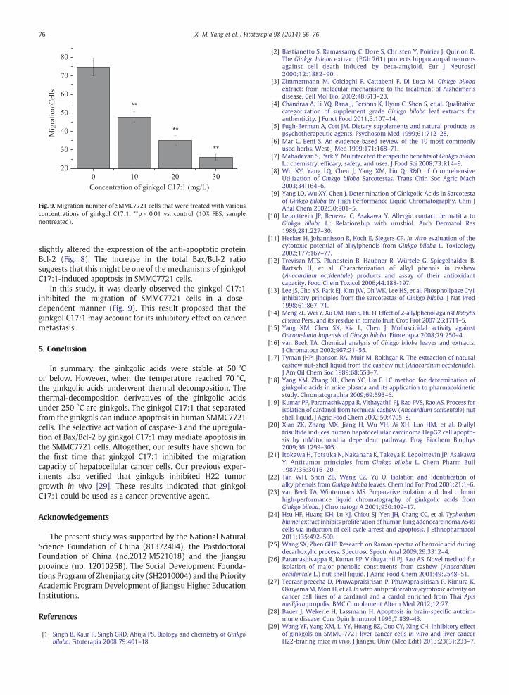
3.5. Effect of ginkgol on SMMC-7721-cell migration
The effect of ginkgol C17:1 on the migration ofSMMC7721, as evaluated using a modified Boyden chamber,
Table 2Inhibition effect of the alkylphenolic compounds on the A549, U251 and SMMC772
was as shown in Fig. 9. The cellular migration was inducedusing 10% FBS that acted as a chemotactic factor andincreased the basal migration of SMMC7721 (74.8±4.6cells) in the control group, whereas the treatment of theSMMC7721 cells with 10, 20 or 30 μg · ml-1 of ginkgol C17:1for 12 h resulted in a concentration-dependent inhibition ofcell migration by 47.8±2.94, 35.2±2.59 and 26.2±1.79 cells,respectively (P b 0.01; Fig. 9).
4. Discussion
G. biloba have been planted widely in many provincesof China as ornamental and fruit trees. The sarcotesta ofG. biloba is a rich source of ginkgolic acids and has not beeneffectively utilized until now [8]. Our study had determinedthat ginkgolic acids exhibit outstanding molluscicidal activityagainst O. hupensis, the intermediate host of Schistosomajaponicum [15], and could further develop into an effective
Fig. 5. Time- and concentration-dependent effect of ginkgol C17:1 on SMMC7721 cells growth. (A) The viability under treatment with various concentrations ofginkgol C17:1 for 24 h. (B) IC50 values under various treatment durations.
74 X.-M. Yang et al. / Fitoterapia 98 (2014) 66–76
plant molluscicide in an O. hupensis control. So, it is importantto know the thermal stability of ginkgolic acids to avoidtheir thermal decomposition during molluscicide processing.The thermal decomposition of anacardic acids, analoguesof ginkgolic acids, has been reported. Anacardic acids arecommonly decarboxylated at 200 °C in industrial practice [17].Our TG-DTA analysis (Fig. 1) revealed that the ginkgolic acidswere decarboxylated at temperatures ranging from 200 to250 °C. However, based only on TG-DTA analysis, we cannot besure whether ginkgolic acids are stable at temperature below200 °C.
Therefore, the thermal stability of ginkgolic acids attemperatures of 30, 50 and 70 °C was examined first. Theresults showed that the ginkgolic acids decomposed slowlyby heating at 70 °C but exhibited almost no change whenheated at 50 °C for 30 days. The results were similar to those
a b
d e
Fig. 6.Morphology changes of the SMMC-7721 cells upon treatment with ginkgol C1was observed. SMMC-7721 cells were treated with ginkgol C17:1 at concentrations oat 200× using a fluorescent microscope.
reported by Wang and Zheng [25] for the decarboxylation ofbenzoic acid. They found that the characteristic Raman peakof benzoic acid did not change below 50 °C but becameweaker between 50 and 100 °C and disappeared when thetemperature reached 170 °C. Our results showed that theginkgolic acids can be stored and processed below 50 °C.They cannot be separated by fractional distillation [26]. Theresults can provide useful suggestions for a suitable way tolimit the loss of ginkgolic acids in pharmaceutical processing.
The ginkgols C13:0, C15:1 and C17:1 were separated fromthe thermally decomposed products of the ginkgolic acids bysemipreparative HPLC. We confirmed that the ginkgol C17:1actually consisted of two isomers in a ratio of 1:1, differingonly in the position of their Z double bond (Fig. 4). These twocompounds are considered to be inseparable by HPLC and canonly be separated by capillary GC [23]. Ginkgol C15:1 has a
c
f
7:1. Ho.33342 and PI double staining of the nuclei from the SMMC-7721 cellsf 0, 10 and 20 μg · ml-1 for 12 h (a-c) and 24 h (d-f). Images were visualized
-5 0 5 10 15 20 250
100
200
300
400
500ca
spas
e-3
activ
ity(%
con
trol)
Concentration(mg/ml)
Fig. 7. The effects of ginkgol C17:1 on caspase-3 activation in SMMC-7721cells.
75X.-M. Yang et al. / Fitoterapia 98 (2014) 66–76
double bond at the C8′ position, and ginkgol C13:0 is a purecompound (Table 1).
Comparing the growth-inhibition effects of ginkgolic acidwith those of ginkgol, the ginkgols exhibited higher inhibi-tory effects than the corresponding ginkgolic acids (Table 2).Support for this can be found in the study of Lee et al. [13],who demonstrated that ginkgol had a higher toxicity towardthe A549, MCF-7 and HT-1197 cell lines than ginkgolicacid. Itokawa et al. [21] obtained a similar result on Sarcoma180 ascites in mice. This results indicate that the carboxylgroup of ginkgolic acid has a negative effect on cell-growthinhibition.
C
D
Fig. 8. Effect of ginkgol C17:1 on the expression of apoptosis-related proteins in SMand 72 h (from 1 to 4); (B) treatment with 0, 5, 10, 20 and 30 μg · ml-1 of ginkgol(D) the Bax/Bcl-2 ratios at various concentrations.
Ginkgol C17:1 showed the greatest anticancer capacityamong the three ginkgol monomers, especially for SMMC7721. In the present study, the SMMC7721 cells treated with0-30 μg · ml-1 ginkgol C17:1 underwent obvious viabilitychanges and showed time- and dose-dependent behavior(Fig. 5). The 48-h IC50 value of ginkgol C17:1 on theSMMC-7721 cells was 8.5 μg · ml-1 and was far lower thanthat of cardanol from Apis mellifera propolis on the BT474,Chaco, SW620 and Hep-G2 cell lines (the IC50 values rangedfrom 10.8 to 29.3 μg · ml-1) [27]. However, Dungpom et al.[27] did not affirm if cardanol affected the SW620 cancer cellsby necrosis or by apoptosis.
In our study, highly condensed chromatin in a fragmentednucleus, which is the typical characteristic of apoptotic cells,was clearly confirmed by Ho.33342 and PI double staining(Fig. 6). This experiment indicated that the ginkgol C17:1induces cytotoxicity in the SMMC7721 cells through a mecha-nism involving apoptosis. Inducing apoptosis in malignant cellsis a very important mechanism of action for certain chemopre-ventive agents [28].
To understand the mechanism of how the ginkgol C17:1produces cytotoxic effects in the SMMC7721 cells, we utilizedvarious molecular techniques to label and measure selectedmarkers. We observed that ginkgol C17:1 was able to activatecaspase-3. Therefore, ginkgol C17:1-induced apoptosis in theSMMC7721 cells may be occurring through the activation ofcaspase-3 (Fig. 7).
To support this view, we also found that the ginkgol C17:1increased the Bax/Bcl-2 ratio by significantly increasing theexpression of the pro-apoptotic protein Bax, although it only
Ginkgol C17:1 (µg·ml -1)0 5 10 20 30
MC7721 cells: (A) treatment with 20 μg · ml-1 of gingkol C17:1 for 0, 24, 48C17:1 for 48 h (from 1 to 5); (C) the Bax/Bcl-2 ratios at various times; and
0 10 20 3020
30
40
50
60
70
80
**
**Mig
ratio
n C
ells
Concentration of ginkgol C17:1 (mg/L)
**
Fig. 9. Migration number of SMMC7721 cells that were treated with variousconcentrations of ginkgol C17:1. **p b 0.01 vs. control (10% FBS, samplenontreated).
76 X.-M. Yang et al. / Fitoterapia 98 (2014) 66–76
slightly altered the expression of the anti-apoptotic proteinBcl-2 (Fig. 8). The increase in the total Bax/Bcl-2 ratiosuggests that this might be one of the mechanisms of ginkgolC17:1-induced apoptosis in SMMC7721 cells.
In this study, it was clearly observed the ginkgol C17:1inhibited the migration of SMMC7721 cells in a dose-dependent manner (Fig. 9). This result proposed that theginkgol C17:1 may account for its inhibitory effect on cancermetastasis.
5. Conclusion
In summary, the ginkgolic acids were stable at 50 °Cor below. However, when the temperature reached 70 °C,the ginkgolic acids underwent thermal decomposition. Thethermal-decomposition derivatives of the ginkgolic acidsunder 250 °C are ginkgols. The ginkgol C17:1 that separatedfrom the ginkgols can induce apoptosis in human SMMC7721cells. The selective activation of caspase-3 and the upregula-tion of Bax/Bcl-2 by ginkgol C17:1 may mediate apoptosis inthe SMMC7721 cells. Altogether, our results have shown forthe first time that ginkgol C17:1 inhibited the migrationcapacity of hepatocellular cancer cells. Our previous exper-iments also verified that ginkgols inhibited H22 tumorgrowth in vivo [29]. These results indicated that ginkgolC17:1 could be used as a cancer preventive agent.
Acknowledgements
The present study was supported by the National NaturalScience Foundation of China (81372404), the PostdoctoralFoundation of China (no.2012 M521018) and the Jiangsuprovince (no. 1201025B). The Social Development Founda-tions Program of Zhenjiang city (SH2010004) and the PriorityAcademic Program Development of Jiangsu Higher EducationInstitutions.
References
[1] Singh B, Kaur P, Singh GRD, Ahuja PS. Biology and chemistry of Ginkgobiloba. Fitoterapia 2008;79:401–18.
[2] Bastianetto S, Ramassamy C, Dore S, Christen Y, Poirier J, Quirion R.The Ginkgo biloba extract (EGb 761) protects hippocampal neuronsagainst cell death induced by beta-amyloid. Eur J Neurosci2000;12:1882–90.
[3] Zimmermann M, Colciaghi F, Cattabeni F, Di Luca M. Ginkgo bilobaextract: from molecular mechanisms to the treatment of Alzheimer’sdisease. Cell Mol Biol 2002;48:613–23.
[4] Chandraa A, Li YQ, Rana J, Persons K, Hyun C, Shen S, et al. Qualitativecategorization of supplement grade Ginkgo biloba leaf extracts forauthenticity. J Funct Food 2011;3:107–14.
[5] Fugh-Berman A, Cott JM. Dietary supplements and natural products aspsychotherapeutic agents. Psychosom Med 1999;61:712–28.
[6] Mar C, Bent S. An evidence-based review of the 10 most commonlyused herbs. West J Med 1999;171:168–71.
[7] Mahadevan S, Park Y. Multifaceted therapeutic benefits of Ginkgo bilobaL.: chemistry, efficacy, safety, and uses. J Food Sci 2008;73:R14–9.
[8] Wu XY, Yang LQ, Chen J, Yang XM, Liu Q. R&D of ComprehensiveUtilization of Ginkgo biloba Sarcotestas. Trans Chin Soc Agric Mach2003;34:164–6.
[9] Yang LQ, Wu XY, Chen J. Determination of Ginkgolic Acids in Sarcotestaof Ginkgo Biloba by High Performance Liquid Chromatography. Chin JAnal Chem 2002;30:901–5.
[10] Lepoittevin JP, Benezra C, Asakawa Y. Allergic contact dermatitia toGinkgo biloba L.: Relationship with urushiol. Arch Dermatol Res1989;281:227–30.
[11] Hecker H, Johannisson R, Koch E, Siegers CP. In vitro evaluation of thecytotoxic potential of alkylphenols from Ginkgo biloba L. Toxicology2002;177:167–77.
[12] Trevisan MTS, Pfundstein B, Haubner R, Würtele G, Spiegelhalder B,Bartsch H, et al. Characterization of alkyl phenols in cashew(Anacardium occidentale) products and assay of their antioxidantcapacity. Food Chem Toxicol 2006;44:188-197.
[13] Lee JS, Cho YS, Park EJ, Kim JW, OhWK, Lee HS, et al. Phospholipase Cγ1inhibitory principles from the sarcotestas of Ginkgo biloba. J Nat Prod1998;61:867–71.
[14] Meng ZL, Wei Y, Xu DM, Hao S, Hu H. Effect of 2-allylphenol against Botrytiscinerea Pers., and its residue in tomato fruit. Crop Prot 2007;26:1711–5.
[15] Yang XM, Chen SX, Xia L, Chen J. Molluscicidal activity againstOncomelania hupensis of Ginkgo biloba. Fitoterapia 2008;79:250–4.
[16] van Beek TA. Chemical analysis of Ginkgo biloba leaves and extracts.J Chromatogr 2002;967:21–55.
[17] Tyman JHP, Jhonson RA, Muir M, Rokhgar R. The extraction of naturalcashew nut-shell liquid from the cashew nut (Anacardium occidentale).J Am Oil Chem Soc 1989;68:553–7.
[18] Yang XM, Zhang XL, Chen YC, Liu F. LC method for determination ofginkgolic acids in mice plasma and its application to pharmacokineticstudy. Chromatographia 2009;69:593–6.
[19] Kumar PP, Paramashivappa R, Vithayathil PJ, Rao PVS, Rao AS. Process forisolation of cardanol from technical cashew (Anacardium occidentale) nutshell liquid. J Agric Food Chem 2002;50:4705–8.
[20] Xiao ZK, Zhang MX, Jiang H, Wu YH, Ai XH, Luo HM, et al. Diallyltrisulfide induces human hepatocellular carcinoma HepG2 cell apopto-sis by mMitochondria dependent pathway. Prog Biochem Biophys2009;36:1299–305.
[21] Itokawa H, Totsuka N, Nakahara K, Takeya K, Lepoittevin JP, AsakawaY. Antitumor principles from Ginkgo biloba L. Chem Pharm Bull1987;35:3016–20.
[22] Tan WH, Shen ZB, Wang CZ, Yu Q. Isolation and identification ofalkylphenols from Ginkgo biloba leaves. Chem Ind For Prod 2001;21:1–6.
[23] van Beek TA, Wintermans MS. Preparative isolation and dual columnhigh-performance liquid chromatography of ginkgolic acids fromGinkgo biloba. J Chromatogr A 2001;930:109–17.
[24] Hsu HF, Huang KH, Lu KJ, Chiou SJ, Yen JH, Chang CC, et al. Typhoniumblumei extract inhibits proliferation of human lung adenocarcinoma A549cells via induction of cell cycle arrest and apoptosis. J Ethnopharmacol2011;135:492–500.
[25] Wang SX, Zhen GHF. Research on Raman spectra of benzoic acid duringdecarboxylic process. Spectrosc Spectr Anal 2009;29:3312–4.
[26] Paramashivappa R, Kumar PP, Vithayathil PJ, Rao AS. Novel method forisolation of major phenolic constituents from cashew (Anacardiumoccidentale L.) nut shell liquid. J Agric Food Chem 2001;49:2548–51.
[27] Teerasripreecha D, Phuwapraisirisan P, Phuwapraisirisan P, Kimura K,Okuyama M, Mori H, et al. In vitro antiproliferative/cytotoxic activity oncancer cell lines of a cardanol and a cardol enriched from Thai Apismellifera propolis. BMC Complement Altern Med 2012;12:27.
[28] Bauer J, Wekerle H, Lassmann H. Apoptosis in brain-specific autoim-mune disease. Curr Opin Immunol 1995;7:839–43.
[29] Wang YF, Yang XM, Li YY, Huang BZ, Guo CY, Xing CH. Inhibitory effectof ginkgols on SMMC-7721 liver cancer cells in vitro and liver cancerH22-braring mice in vivo. J Jiangsu Univ (Med Edit) 2013;23(3):233–7.