Thermo-induced ElectromagneticCoupling in Gold/Polymer HybridPlasmonic Structures Probed by Surface-Enhanced Raman ScatteringHelene Gehan,† Laure Fillaud,† Mohamed M. Chehimi,† Jean Aubard,† Andreas Hohenau,‡ Nordin Felidj,†
and Claire Mangeney†,*†ITODYS, Universite Paris Diderot-Paris 7 (UMR CNRS 7086), 15 rue Jean de Baıf, 75013 Paris, France, and ‡Institute of Physics, Karl Franzens University, Universitatsplatz5, A-8010 Graz, Austria
Since its discovery four decades ago,surface-enhanced Raman scattering(SERS) has been considered as one of
the most powerful and sensitive tools for
the chemical analysis of molecules ad-
sorbed onto metallic nanoparticles (NPs),
especially gold and silver.1 This technique
leads to a giant increase of the Raman scat-
tering cross section for molecules adsorbed
onto metallic nanostructured surfaces and
thus has a large potential in analytical
chemistry and biological and optical
applications.2,3 Many aspects of the mecha-
nisms involved in SERS have been described
and resolved to a reasonable degree over
the last two decades.4 It is now well estab-
lished that the overall mechanism can be di-
vided into electromagnetic and chemical
contributions: (i) the electromagnetic con-
tribution arises from enhanced optical fields
close to the metallic surface, due to the ex-
citation of localized surface plasmon (LSP)
with a typical Raman enhancement factor
(EF) of |E|4 ca. 106�107 (where E is the local
electric field);5�9 (ii) a further chemical en-
hancement can be observed for molecules
adsorbed onto specific metallic sites when a
resonant charge transfer occurs.10,11
One challenging aspect in SERS concen-
trates on the demonstration that a single
molecule or very few molecules could be
detected under specific SERS conditions.12�17
It requires very huge electric field enhance-
ments occurring within the gap between
two NPs or between NPs and a gold film (so-
called a hot-spot regime).18�21 Substrates
made of gold nanoparticles linked by rigid
alkyl layers of self-assembled molecules
(SAMs) to a gold flat film were recently in-
vestigated for the detection of very low
concentrations (zeptomolar) of molecular
probes. These systems were shown to be ul-
trasensitive for surface-enhanced Raman
scattering.22 Indeed, when the NPs are
within a distance typically between 1 and
2�50 nm from the gold surface, a light con-
centration is induced between the NPs and
the film, whose intensity decreases rapidly
with the distance. However, distances
around 20 nm still lead to efficient Raman
gains (typically 103�104), as recently calcu-
lated by R. Hill et al.23 This light concentra-
tion results from the interaction between
the localized surface plasmon of the NPs
and the delocalized surface plasmon polari-
ton of the metal film.21
Of particular interest is the possibility tocontrol this interaction in order to optimize
the Raman enhancement factor, for example, through
the use of polyelectrolyte layers of different
thicknesses.23,24 Recently, sophisticated systems made
of 3D core�shell colloidal NPs coated with PNIPAM,
silver-loaded agarose gels or LBL films were proposed
for SERS applications, allowing the authors to dynami-
cally control the distance between the gold or silver
particles and the molecular probes.25�28 These experi-
ments nicely showed a strong variation of the SERS sig-
nal depending on the distance particle/molecules. Oth-
ers studied the plasmon band variation of gold or silver-
NPs coated on top of stimuli-responsive brushes
(polystyrene, poly2-vinylpyridine),29�36 following an ex-
ternal stimulus (such as pH or solvent modification).
However, to the best of our knowledge, optimization
of the Raman signals through the use of a stimuli-
responsive linker between the gold NPs and a gold
film has never been reported so far.
In this work, we propose to address this issue by de-
signing a stimulable device made of gold colloidal
nanoparticles connected to a gold flat film through an
active thermosensitive polymer brush layer, capable to
externally modulate the distance between the NPs and
the substrate. As a linker, we used poly(N-
isopropylacrylamide) (PNIPAM), which is interesting in
this regard due to its well-known phase behavior in
aqueous solutions. Indeed, it undergoes a reversible, in-
verse phase transition at a lower critical solution tem-
perature (LCST) of about 32 °C in pure water.37 Below
the LCST, PNIPAM is hydrated and the chains are in an
extended conformational state. Above the LCST,
PNIPAM is in a hydrophobically collapsed conforma-
tional state. These conformational changes of the
PNIPAM linker between the gold nanoparticle assem-
blies and the gold surface are expected to induce dra-
matic modifications of the optical properties of the sub-
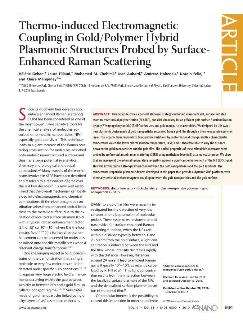
strate. Below the LCST, the gold nanoparticles and the
gold surface are far from each other (for instance, dis-
tance � 120 nm) and there should be no coupling be-
tween these two components: SERS spectra originating
from noncoupled NPs should be observed. In contrast,
above the LCST, the proximity of the colloidal particles
to the gold film (distance � 20 nm) should lead to a
strong interaction regime with higher SERS spectra.
For designing the hybrid plasmonic device, we de-
veloped an original multistep functionalization strat-
egy, (Figure 1), relying on three major steps: (i) the well-
known electroreduction of diazonium salts38,39 for the
covalent grafting of atom-transfer radical polymeriza-
tion (ATRP) initiators40�45 to the gold surface; (ii) the
grafting of PNIPAM chains from the surface via surface-
initiated ATRP (SI-ATRP)46,47 and their further modifica-
tion via click chemistry leading to amine chain-ends;48
(iii) the immobilization of gold nanoparticles exclusively
at the very end PNIPAM chains.
RESULTS AND DISCUSSIONPreparation of PNIPAM-Modified Gold Surfaces. The
initiator-modified gold surfaces were prepared by atwo-step procedure that consists of the following: (i)first, the electrochemical reduction of aryl diazoniumions (HO(CH2)2BD) to produce �OH terminated arylmoieties, covalently anchored to the surface, and then(ii) esterification of the anchored OH groups with2-bromopropionyl bromide to produce ethyl2-bromopropionate (EBrP)-bearing aryl grafted groups.
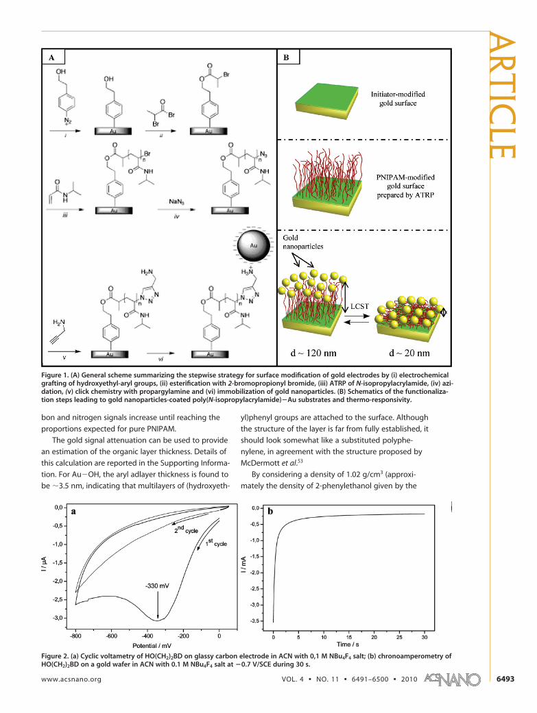
Figure 2a shows the cyclic voltammogram ofHO(CH2)2BD on a glassy carbon electrode in acetoni-trile (ACN) with 0.1 M NBu4BF4 salt. One can observe abroad, irreversible, monoelectronic wave at Epc �
�0.33 V/saturated calomel electrode (SCE), which corre-sponds to the reduction of the diazonium salt.
The electron transfer is concerted with the cleavageof dinitrogen, giving an aryl radical which binds to thesurface according to reaction (i) in Figure 1. Upon re-petitive scanning, this wave decreases to negligible val-ues, as usually observed with diazonium salts; this de-crease corresponds to the formation of the organiclayer on top of the electrode surface. On gold plates,grafting was achieved by chronoamperometry (see Fig-ure 2b) maintaining for 30 s a 300 mV negative poten-tial relative to the peak potential measured on carbon.The very steep decrease of the current with time isagain characteristic of the formation of the organiclayer which blocks the electron transfer from the elec-trode. The gold electrodes were then thoroughly rinsedwith ethanol before being immersed in a2-bromopropionyl bromide solution during 5 min, in or-der to achieve the esterification of the hydroxyl groups,leading to bromo-terminated gold surfaces (Au�Br).PNIPAM brushes were prepared by ATRP, after expos-ing the initiator-modified surfaces for various times (10,30, and 120 min) at room temperature to a polymeriz-ing solution of N-isopropylacrylamide in water at a lowmethanol concentration (2.6%). The growing polymerbrush adopts an extended conformation49,50 underthese reaction conditions, and brush growth is fa-vored.51 In ATRP, a highly reactive and, in our case,surface-tethered, organic radical is generated alongwith a stable Cu(II) species that can be regarded as apersistent metalloradical, which is not able to initiateradical polymerization in the polymerizing solution.52
This means that polymerization is strictly confined tothe surface-attached, growing polymer chains.
XPS Analysis. XPS analysis was performed in order tomonitor the progressive build-up of the final hybrid sys-tem along all the functionalization steps depicted inFigure 1. Details of the spectra are presented in the Sup-porting Information. Table 1 gathers the atomic sur-face composition obtained by the integration of thecore level peaks. One observes a progressive attenua-tion of the gold signal as the electrode is covered by theinitiator layers and the polymer coatings while the car-
ART
ICLE
VOL. 4 ▪ NO. 11 ▪ GEHAN ET AL. www.acsnano.org6492
bon and nitrogen signals increase until reaching the
proportions expected for pure PNIPAM.
The gold signal attenuation can be used to provide
an estimation of the organic layer thickness. Details of
this calculation are reported in the Supporting Informa-
tion. For Au�OH, the aryl adlayer thickness is found to
be �3.5 nm, indicating that multilayers of (hydroxyeth-
yl)phenyl groups are attached to the surface. Although
the structure of the layer is far from fully established, it
should look somewhat like a substituted polyphe-
nylene, in agreement with the structure proposed by
McDermott et al.53
By considering a density of 1.02 g/cm3 (approxi-
mately the density of 2-phenylethanol given by the
Figure 1. (A) General scheme summarizing the stepwise strategy for surface modification of gold electrodes by (i) electrochemicalgrafting of hydroxyethyl-aryl groups, (ii) esterification with 2-bromopropionyl bromide, (iii) ATRP of N-isopropylacrylamide, (iv) azi-dation, (v) click chemistry with propargylamine and (vi) immobilization of gold nanoparticles. (B) Schematics of the functionaliza-tion steps leading to gold nanoparticles-coated poly(N-isopropylacrylamide)�Au substrates and thermo-responsivity.
Figure 2. (a) Cyclic voltametry of HO(CH2)2BD on glassy carbon electrode in ACN with 0,1 M NBu4F4 salt; (b) chronoamperometry ofHO(CH2)2BD on a gold wafer in ACN with 0.1 M NBu4F4 salt at �0.7 V/SCE during 30 s.
supplier) for the grafted aryl adlayer and the thicknessof 3.5 nm estimated by XPS, it follows that the electro-chemical grafting yields a surface coverage � of 3.0 �
10�9 mol · cm�2. It is noteworthy that this value isaround 2�3 times higher than the surface concentra-tion of a close-packed monolayer �CPML of phenyl (or4-substituted phenyl) groups estimated from molecu-lar models: �CPML � 1.35 � 10�9 mol · cm�2.38
Concerning the surface reaction yield of the esterifi-cation reaction of the OH-terminated aryl groups with2-bromopropionyl bromide, a rough estimation can bemade by comparing the experimental Br/C and Br/Oatomic ratios (0.04 and 0.2, respectively) to the oneswhich would be expected in the case of a 100% reac-tion yield (0.1 and 0.5, respectively). These results indi-cate that the esterification reaction has indeed occurredbut with a reaction yield close to around 50%. Thiswould correspond to a surface coverage (�Br) of EBrPderived aryl groups of �Br � 1.5 � 10�9 mol · cm�2, stillsuperior to �CPML.
As the conformation of tethered chains and thefilm thickness in polymer-brush layers are directly af-fected by the surface coverage of the initiating groupson the flat surface, the value of �Br is a crucial param-eter. For instance, it was shown by Fukuda et al.54 thatincreasing the surface density of initiators grafted on asilicon substrate leads to the formation of “high-density” PMMA brushes, in which high-order interac-tions among graft chains are important. Nevertheless,
the polymer chain density cannot exceed a certainvalue due to steric effects in the surface-graft polymer-ization, and if the surface coverage of initiators isaround 4 molecules/nm2 (experimental value for amonolayer), the polymer graft efficiency only reaches0.2. By comparison, the �Br value obtained in thepresent paper (�9 molecules/nm2) appears highenough to allow the growing of dense polymer layersin a brush regime.
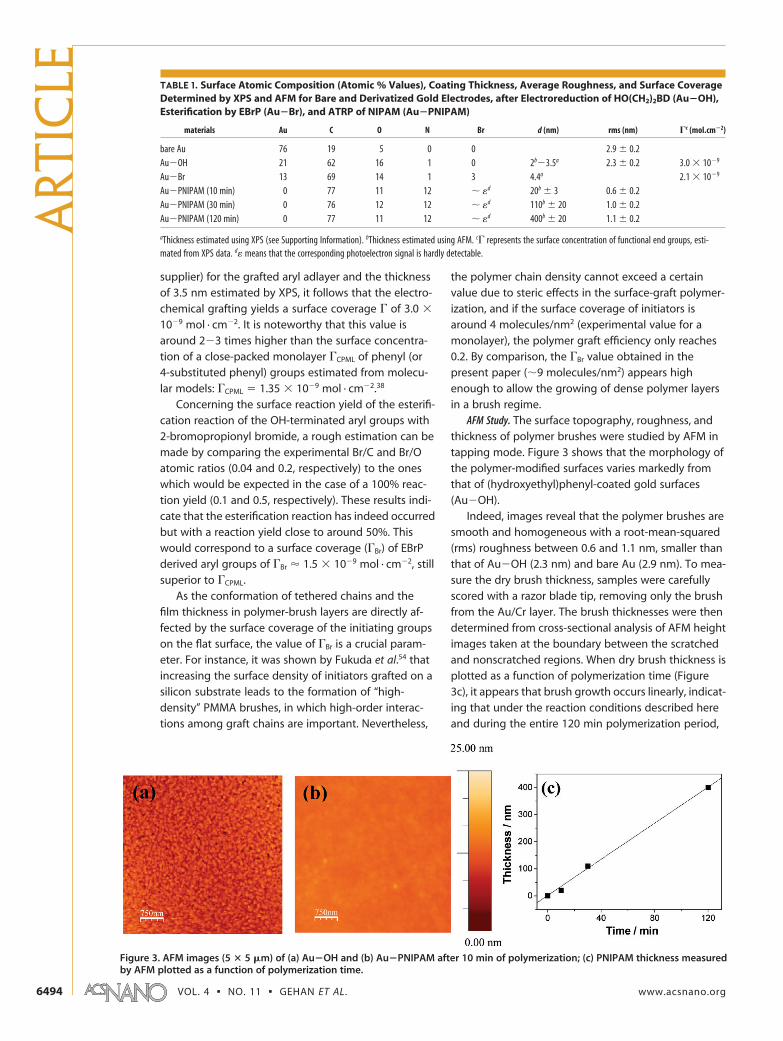
AFM Study. The surface topography, roughness, andthickness of polymer brushes were studied by AFM intapping mode. Figure 3 shows that the morphology ofthe polymer-modified surfaces varies markedly fromthat of (hydroxyethyl)phenyl-coated gold surfaces(Au�OH).
Indeed, images reveal that the polymer brushes aresmooth and homogeneous with a root-mean-squared(rms) roughness between 0.6 and 1.1 nm, smaller thanthat of Au�OH (2.3 nm) and bare Au (2.9 nm). To mea-sure the dry brush thickness, samples were carefullyscored with a razor blade tip, removing only the brushfrom the Au/Cr layer. The brush thicknesses were thendetermined from cross-sectional analysis of AFM heightimages taken at the boundary between the scratchedand nonscratched regions. When dry brush thickness isplotted as a function of polymerization time (Figure3c), it appears that brush growth occurs linearly, indicat-ing that under the reaction conditions described hereand during the entire 120 min polymerization period,
TABLE 1. Surface Atomic Composition (Atomic % Values), Coating Thickness, Average Roughness, and Surface CoverageDetermined by XPS and AFM for Bare and Derivatized Gold Electrodes, after Electroreduction of HO(CH2)2BD (Au�OH),Esterification by EBrP (Au�Br), and ATRP of NIPAM (Au�PNIPAM)
materials Au C O N Br d (nm) rms (nm) �c (mol.cm�2)
aThickness estimated using XPS (see Supporting Information). bThickness estimated using AFM. c� represents the surface concentration of functional end groups, esti-mated from XPS data. d� means that the corresponding photoelectron signal is hardly detectable.
Figure 3. AFM images (5 � 5 �m) of (a) Au�OH and (b) Au�PNIPAM after 10 min of polymerization; (c) PNIPAM thickness measuredby AFM plotted as a function of polymerization time.
ART
ICLE
VOL. 4 ▪ NO. 11 ▪ GEHAN ET AL. www.acsnano.org6494
good control of the polymerization process is achieved.
The thermoresponsive system gold electrode/PNIPAM
was shown to be highly stable with time: indeed, it was
able to undergo several cycles of temperature changes
in water on both sides of the LCST without altering its
integrity (see experiments on gold nanoarrays reported
in the Supporting Information).
Click Chemistry and Au Nanoparticles Self-Assembly. Post-
functionalization of PNIPAM chain-ends was performed
via Cu(I)-catalyzed Huisgen 1,3-dipolar cycloadditions
(“click chemistry approach”). It consists of first convert-
ing the bromo-termination to azido-terminated func-
tionality and then coupling it with alkyne-terminated
molecules, here propargylamine. The reaction product
is the stable heterocyclic linker, 1,4-disubstituted 1,2,3-
triazole (see Figure 1).
The PNIPAM-coated gold substrates were modified
with azides by immersion in a DMF solution contain-
ing 0.1 M of sodium azide. The azide-modified PNIPAM
brushes were then treated with a solution of 10 mM
propargylamine in 1:1 EtOH/H2O containing Cu(I) (cata-
lyst generated in situ from Cu(II) sulfate and sodium
ascorbate as reducing agent) to form an amino-
modified surface (Au�PNIPAM�NH2).55
The Au�PNIPAM�NH2 substrates were then incu-
bated in a gold colloidal suspension. As the pH of the
solution is around 5, the free amine end groups of
grafted PNIPAM chains are protonated and positively
charged. The electrostatic attraction between these
groups and the citrate counterions coated on Au NPs
resulted in the self-assembly of Au nanoparticles at the
chain-end of polymer brushes.
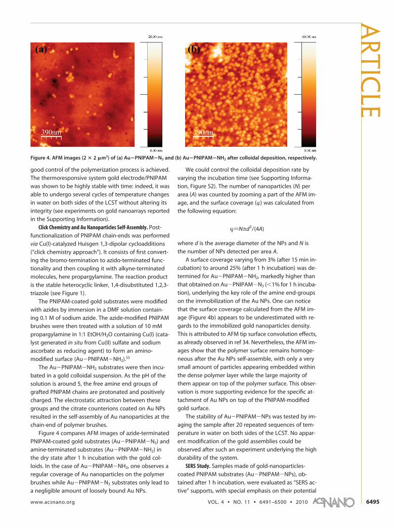
Figure 4 compares AFM images of azide-terminated
PNIPAM-coated gold substrates (Au�PNIPAM�N3) and
amine-terminated substrates (Au�PNIPAM�NH2) in
the dry state after 1 h incubation with the gold col-
loids. In the case of Au�PNIPAM�NH2, one observes a
regular coverage of Au nanoparticles on the polymer
brushes while Au�PNIPAM�N3 substrates only lead to
a negligible amount of loosely bound Au NPs.
We could control the colloidal deposition rate by
varying the incubation time (see Supporting Informa-
tion, Figure S2). The number of nanoparticles (N) per
area (A) was counted by zooming a part of the AFM im-
age, and the surface coverage () was calculated from
the following equation:
�)Nπd2/(4A)
where d is the average diameter of the NPs and N is
the number of NPs detected per area A.
A surface coverage varying from 3% (after 15 min in-
cubation) to around 25% (after 1 h incubation) was de-
termined for Au�PNIPAM�NH2, markedly higher than
that obtained on Au�PNIPAM�N3 (1% for 1 h incuba-
tion), underlying the key role of the amine end-groups
on the immobilization of the Au NPs. One can notice
that the surface coverage calculated from the AFM im-
age (Figure 4b) appears to be underestimated with re-
gards to the immobilized gold nanoparticles density.
This is attributed to AFM tip surface convolution effects,
as already observed in ref 34. Nevertheless, the AFM im-
ages show that the polymer surface remains homoge-
neous after the Au NPs self-assemble, with only a very
small amount of particles appearing embedded within
the dense polymer layer while the large majority of
them appear on top of the polymer surface. This obser-
vation is more supporting evidence for the specific at-
tachment of Au NPs on top of the PNIPAM-modified
gold surface.
The stability of Au�PNIPAM�NPs was tested by im-
aging the sample after 20 repeated sequences of tem-
perature in water on both sides of the LCST. No appar-
ent modification of the gold assemblies could be
observed after such an experiment underlying the high
durability of the system.
SERS Study. Samples made of gold-nanoparticles-
coated PNIPAM substrates (Au�PNIPAM�NPs), ob-
tained after 1 h incubation, were evaluated as “SERS ac-
tive” supports, with special emphasis on their potential
Figure 4. AFM images (2 � 2 �m2) of (a) Au�PNIPAM�N3 and (b) Au�PNIPAM�NH2 after colloidal deposition, respectively.
thermoresponsive behavior. The molecular probe usedfor the SERS experiments was the methylene blue (MB),a weakly fluorescent molecule absorbing and emittingat 630 and 670 nm, respectively. We used a laser excita-tion wavelength at 633 nm (He�Ne laser) for the Ra-man experiments since we expect that LSP excitationof the gold NPs, in interaction with the metallic film,might be between 600 and 700 nm, if we refer to re-cent light scattering experiments investigated on simi-lar systems made of gold NPs coupled to a gold filmthrough an electrolyte.23,24 Furthermore, the matchingbetween the absorption band and the laser excitationwavelength leads to a resonance with an additional en-hancement factor.56 Two kinds of samples were stud-ied, with PNIPAM thicknesses varying from ca. 20 nm(sample A) to 200 nm (sample B) in the dried state, re-spectively. As the swelling ratio57 of polymer brushes isaround 6 in our experimental conditions, it provides, inaqueous solvent and below the LCST, a swollen poly-mer brush thickness of around 120 nm for sample A andmore than 1 �m for sample B. This polymer thickness,which assesses the distance between the gold NPs andthe gold film, is expected to play a crucial role in the in-teraction between these two components. Indeed,when the distance between the NPs and the gold filmis too high (superior to 50 nm), there should be no in-teraction between them. On the contrary, a distance in-ferior to 50 nm should lead to an interaction betweenthe LSP and the surface plasmon polariton (SPP) on thegold film inducing a huge electric field that becomesstronger as this distance becomes smaller.21 Figure 5shows the SERS spectra of MB, recorded at various tem-peratures (below and above the LCST of PNIPAM) afterdipping the Au�PNIPAM�NPs substrates into a 10�4 MMB solution (followed by an intense rinsing procedurewith pure ethanol and drying under an argon flow).
The MB molecules are expected to be located at dif-ferent levels on the substrate, that is, adsorbed on the
NPs, on the gold film, and trapped within the PNIPAMbrush layer. All spectra display the characteristic Ramanbands of MB, with the C�N�C skeletal deformationmode at 443 cm�1, the C�C ring stretch �CC at 1502cm�1, and the C�N stretch �CN at 1618 cm�1. One cannotice that SERS signals are accompanied by a widebackground, attributed to surface enhanced fluores-cence (SEF).56,58 At 24 °C, the SERS intensities observedin samples A and B are the same, indicating an overallNPs density which is similar for both samples.
For sample A (Figure 5a), the SERS intensities in-crease with temperature. This could be explained byan enhancement of the intensity of the local field (|E|2)when the gap between the NPs and the gold film is de-creasing.23 Therefore, as the SERS signal is proportionalto |E|4, it strongly depends on the distance between theNPs and the gold film, which varies with the PNIPAMconformation when changing the temperature. Thetemperature dependence of the integrated Raman in-tensities plotted in Figure 5b (red curve) shows that itfollows a steep increase within the temperature rangeof the LCST, before reaching a plateau value above theLCST, with a significant enhancement factor increase ofat least 5 times. This result demonstrates that our ther-moresponsive system allows us to dynamically tuneand increase the interaction between the NPs and thegold film.
In contrast, for sample B (200 nm thickness in thecollapsed state and 1.2 �m in the swollen state), theSERS intensities of MB are independent of temperatureand remain constant from 23 to 45 °C (see Figure 5b,black curve).
As the temperature increases above the LCST, thephase transition turns the PNIPAM layer from hydro-philic to hydrophobic, breaking the interactions be-tween the chains and water molecules. Thepolymer�polymer interactions dominate resulting in acollapse of the chains59,60 bringing the NPs closer to the
Figure 5. (a) SERS spectra recorded at various temperatures (below and above the LSCT) of MB molecules adsorbed onAu�PNIPAM�NPs (sample A); (b) Integrated SERS intensity of MB molecules versus temperature, for the two Au�PNIPAM�NPs sub-strates: sample A with 20 nm PNIPAM dry thickness (red curve) and sample B with 200 nm PNIPAM dry thickness (black curve). Ex-perimental conditions: laser excitation (632.8 nm, 10 �W power) in backscattering configuration. Focused laser spot area onto thesubstrate is ca. 1 �m2. All the Raman spectra were recorded with a 1 s integration time and within the 300�2800 cm�1 spectral range.
ART
ICLE
VOL. 4 ▪ NO. 11 ▪ GEHAN ET AL. www.acsnano.org6496
flat gold film. The distance between the gold NPs andthe gold film then scales with the collapsed polymerbrush thickness which is around 20 nm for sample Aand 200 nm for sample B. In these conditions, uniquelyin the case of sample A, a near-field coupling canemerge in the gap between the NPs and the flat film. In-deed, for sample B, the NPs are too far from the goldsubstrate, whatever the external temperature. These re-sults evidence the importance of the underlying goldfilm for enhancing the Raman signature of the MB.
Furthermore, it is remarkable that the variations ofthe SERS intensities observed for sample A are fully re-versible (see Figure 6) when switching the temperaturebelow and above the LCST, evidencing a strong and re-versible response of the Au�PNIPAM�NPs system tothe external temperature.
This temperature dependence of the coupling re-gime in these new hybrid gold/polymer plasmonicstructures, opens exciting outlooks for the elaborationof smart optical devices. Future work will concern theinfluence of the PNIPAM length upon the Raman en-hancement factor.
CONCLUSIONA new stepwise strategy has been developed for
the surface modification of gold substrates by PNIPAM-
grafted gold NPs for SERS applications. This strategy is
based on a combination of diazonium salt electrograft-
ing, surface-initiated atom transfer radical polymeriza-
tion (SI-ATRP), and click chemistry. The gold/PNIPAM/
NPs hybrids were fully characterized using XPS and
AFM. A regular coverage of gold nanoparticles (25%
surface coverage) was achieved with robust bonding
of gold NPs to the PNIPAM brushes through triazole-
functionalized end groups. When the polymer organic
linker layer was sufficiently thin, around 20 nm in the
dried state, it was shown to undergo a phase transition
on both sides of the PNIPAM LCST, modifying signifi-
cantly the SERS intensities of MB molecules adsorbed
on the substrates, depending on the external tempera-
ture. Importantly, these variations appearing on both
sides of the LCST are fully reversible, making our sys-
tem a thermal switch for near-field coupling regime.
The results presented in this paper stress the enor-
mous potentialities of polymer brush-supported NPs
as new advanced platforms for SERS applications. These
novel stimuli-responsive systems offer the following ad-
vantages: (i) a reversible near-field coupling between
the gold NPs and the gold film, depending on the exter-
nal temperature, and leading to a control over the SERS
intensities of molecular probes adsorbed on the sub-
strate; (ii) an easy means for identifying a wide variety
of molecular species (drugs, complex biological com-
pounds.. .).61 One should remember that such a remark-
able control over the optical properties of these ‘smart’
plasmonic substrates rests on the fine tailoring of the
surface and interface chemistry options offered by the
combination of diazonium salts, polymer brushes and
click chemistry.
MATERIAL AND METHODSMaterials. Reagent grade solvents were purchased from VWR-
Prolabo and Alfa Aesar. 2-Bromopropionyl bromide (BPB) (97%,Aldrich), triethylamine (TEA) (99%, Merck), Cu(I)Br (98%, Sigma-Aldrich), N,N,N=,N==,N==-pentamethyldiethyltriamine (PMDETA)(99%, Acros Organics), sodium azide (NaN3) (99%, Prolabo), pro-pargylamine (99%, Acros Organics), Cu(II) sulfate pentahydrate(99%, Prolabo), L-ascorbic acid sodium salt (99%, Alfa Aesar) wereused as received. N-Isoproylacrylamide (NIPAM) (99%, Acros Or-ganics) was purified by recrystallization in (40/60 v/v) toluene/
hexane solution. Gold NP suspensions, prepared following themethod described by Frens,62 and synthesis of4-hydroxyethylbenzene diazonium tetrafluoroborate salt (notedHO(CH2)2BD) are reported in the Supporting Information. Gold-coated silicon wafers ( 111� oriented, 1000 Å coating, titaniumadhesion layer, 4 in. � 500 �m) were purchased from Aldrich.
Gold-Surface Functionalization by PNIPAM Brushes. Gold-coated sili-con wafers were first cleaned using a UV-ozone light during 10min. Substrates were then rinsed with ethanol and dried underan argon flow. All reaction steps were carried out at room tem-
Figure 6. SERS spectra of MB molecules adsorbed on Au�PNIPAM�NPs substrates (sample A) and recorded at various temperatures:from 23 to 33 °C and back to 24 °C (�exc � 632.8 nm, 10 �W power).
perature and followed by rinsing with the reaction solvent andethanol, and subsequently dried under a flush of argon.
Initiator-Modified Gold Surfaces. The atom transfer radical polym-erization initiator was grafted in 2 steps. (i) Electrochemical graft-ing of HO(CH2)2BD was achieved on cleaned gold-coated siliconwafer by chronoamperometry for 30 s in acetonitrile at �0.7V/SCE. The �OH-terminated gold surfaces are abbreviated asAu�OH. (ii) Then, the terminal hydroxyl groups were treatedwith 2-bromopropionyl bromide (0.1 M, dichloromethane) in thepresence of TEA (0.12 M) for 5 min to produce bromo-terminatedester groups. The �Br terminated gold surfaces are abbreviatedas Au�Br.
Atomic Transfer Radical Polymerization (ATRP) of NIPAM. (Step iii inFigure 1) Solutions were prepared and kept at room tempera-ture during degassing by passing a continuous stream of argonthrough the solution while being stirred. The polymerization so-lution was prepared by adding a solution of an organometalliccatalyst to a solution of NIPAM monomer. The extremely oxygen-sensitive organometallic catalyst was prepared by adding a 5mL solution of PMDETA in MeOH (100 �L, 0.48 mmol) to 12.5mg of Cu(I)Br (0.085 mmol). A 1 ml portion of the resulting greensolution (which could possibly turn blue due to presence of Cu(I-I)Br and provide unsuccessful ATRP) was added to a solution ofNIPAM monomer (8.4 g, 74 mmol) in 38 mL of deionized waterunder a continuous stream of argon. The polymerization solutionwas allowed to stir during degassing for 5 min and then trans-ferred into a flask containing the initiator-modified gold surface.The resulting solution was allowed to stir at room temperatureunder argon for 10, 30, or 120 min depending on the polymerthickness targeted. Substrates were then removed from the flaskand rinsed thoroughly with ethanol and water and subsequentlydried under a flush of argon. Resulting gold-modified substrateswere stored under argon. The PNIPAM-terminated gold surfacesare abbreviated as Au�PNIPAM.
PNIPAM brushes Modification by Gold NPs Using Click Chemistry. (Stepiv in Figure 1) Substitution of the bromo-end groups (of PNIPAMbrushes) to azide functions was carried out by reacting theAu�PNIPAM substrates in NaN3 solution (0.1 M, DMF) for 24 hat room temperature in order to obtain click active substrates,abbreviated as Au�PNIPAM�N3. Then, (step v in Figure 1) theazide-terminated substrates were immersed in a solution con-taining propargylamine (10 mM, ethanol/water 1:1), Cu(II) sulfatepentahydrate (0.1 mM) and L-ascorbic acid sodium salt (2.5 mM)following a typical click-chemistry procedure (azide�alkyne cy-cloaddition).55 Solutions were degassed by argon bubbling whilebeing stirred during 24 h, and the resulting amino-terminatedgold surfaces were abbreviated as Au�PNIPAM�NH2. Finally,last step (step vi in Figure 1) consisted in the immobilization ofgold nanoparticules (NPs) onto the amino-terminated substrateswhich were immersed in the colloidal solution for 1 h and thenthoroughly rinsed with water. NPs-terminated gold surfaceswere stored under argon atmosphere and were abbreviated asAu�PNIPAM�NPs.
Instrumentation. X-ray photoelectron spectra were recordedusing a Thermo VG Scientific ESCALAB 250 system fitted with amicrofocused, monochromatic Al K� X-ray source (h� � 1486.6eV; spot size � 650 �m; power � 15 kV � 200 W). The pass en-ergy was set at 150 and 40 eV for the survey and the narrow re-gions, respectively. Spectral calibration was determined by set-ting the main C1s component at 285 eV. The surfacecomposition was determined using the integrated peak areasand the corresponding Scofield sensitivity factors corrected forthe analyzer transmission function. AFM studies were performedwith Nanoscope III digital instrument microscope in tappingmode to map the film morphology at ambient conditions. Ra-man spectra were recorded using a Jobin-Yvon LABRAM HR 800microspectrometer, using a He�Ne laser excitation (632.8 nm,100 �W power) in backscattering mode. In our experiments, themicroscope was equipped with an �100 immersion objective(NA no. 1), which leads to a focused laser spot area onto the sub-strate of ca. 1 �m2 and ensures a very efficient collection of theRaman light. All spectra, recorded in distilled water, were takenwith a 1 s integration time and recorded within the 300�2800cm�1 spectral range.
Acknowledgment. The authors gratefully acknowledge the fi-nancial support from Ministere de la Defense, Direction Gen-erale de l’Armement DGA (Project DGA-REI 07 34024 and Ph.D.support 2007�2010).
Supporting Information Available: Detailed methods for nano-particles and hydroxyethylbenzene diazonium tetrafluoroboratesalt synthesis, XPS and AFM measurements, stability studies ofthe samples. This material is available free of charge via the Inter-net at http://pubs.acs.org.
REFERENCES AND NOTES1. Moskovitz, M. Surface-Enhanced Spectroscopy. Rev. Mod.
Phys. 1985, 57, 783–826.2. Anker, J. N.; Hall, W. P.; Lyandres, O.; Shah, N. C.; Zhao, J.;
Van Duyne, R. P. Biosensing with Plasmonic Nanosensors.Nat. Mater. 2008, 7, 442–453.
3. Penn, S.; He, L.; Natan, M. Nanoparticles for Bioanalysis.Curr. Opin. Chem. Biol. 2003, 7, 609–615.
4. Stiles, P.; Dieringer, J.; Shah, N. C.; Van Duyne, R. P. Surface-Enhanced Raman Spectroscopy. Ann. Rev. Anal. Chem.2008, 1, 601–626.
5. Kerker, M.; Wang, D. S.; Chew, H. Surface Enhanced RamanScattering (SERS) by Molecules Adsorbed at SphericalParticle. Appl. Opt. 1980, 19, 4159–4174.
6. Aravind, P. K.; Nitzan, A.; Metiu, H. The Interaction betweenElectromagnetic Resonances and Its Role in SpectroscopicStudies of Molecules Adsorbed on Colloidal Particles orMetal Spheres. Surf. Sci. 1981, 110, 189–204.
7. McCall, S. L.; Platzman, P. M.; Wolf, P. A. Surface EnhancedRaman Scattering. Phys. Lett. A 1980, 77, 381–383.
8. Gersten, J.; Nitzan, A. Electromagnetic Theory of EnhancedRaman Scattering by Molecules Adsorbed on RoughSurfaces. J. Chem. Phys. 1980, 73, 3023–3037.
9. Ruppin, R. Electric Field Enhancement Near a SurfaceBump. Solid State Commun. 1981, 39, 903–906.
10. Otto, A. Surface-Enhanced Raman Scattering ofAdsorbates. J. Raman Spectrosc. 1991, 22, 743–752.
11. Aussenegg, F. R.; Lippitsch, M. E. On Raman Scattering inMolecular Complexes Involving Charge Transfer. Chem.Phys. Lett. 1978, 59, 214–216.
12. Le Ru, E. C.; Etchegoin, P. G. In Principles of Surface-Enhanced Raman Spectroscopy and Related PlasmonicEffects; Elsevier: Amsterdam, The Netherlands, 2009.
13. Etchegoin, P. G.; Le Ru, E. C. A Perspective on SingleMolecule SERS: Current Status and Future Challenges.Phys. Chem. Chem. Phys. 2008, 10, 6079–6089.
14. Kneipp, K.; Wang, Y.; Kneipp, H.; Perelman, L.; Itzkan, I.;Dasari, R.; Feld, M. Single Molecule Detection UsingSurface-Enhanced Raman Scattering. Phys. Rev. Lett. 1997,78, 1667–1670.
15. Le Ru, E. C.; Etchegoin, P. G. Comment on ChemicalContribution to Surface-Enhanced Raman Scattering. Phys.Rev. Lett. 2006, 97, 199701.
16. Dadosh, T.; Sperling, J.; Bryant, G. W.; Breslow, R.; Shegai,T.; Dyshel, M.; Haran, G.; Bar-Joseph, I. Plasmonic Controlof the Shape of the Raman Spectrum of a Single Moleculein a Silver Nanoparticle Dimer. ACS Nano 2009, 3,1988–1994.
17. Acimovic, S. S.; Kreuzer, M. P.; Gonzalez, M. U.; Quidant, R.ACS Nano 2009, 3, 1231–1237.
18. Camden, J. P.; Dieringer, J. A.; Wang, Y.; Masiello, D. J.;Marks, L. D.; Schatz, G. C.; Van Duyne, R. P. Probing theStructure of Single-Molecule Surface-Enhanced RamanScattering Hot Spots. J. Am. Chem. Soc. 2008, 130, 12616–12617.
20. Wells, M. S.; Retterer, S. D.; Oran, J. M.; Sepaniak, M. J.Controllable Nanofabrication of Aggregate-likeNanoparticle Substrates and Evaluation for Surface-Enhanced Raman Spectroscopy. ACS Nano 2009, 3, 3845–3853.
ART
ICLE
VOL. 4 ▪ NO. 11 ▪ GEHAN ET AL. www.acsnano.org6498
21. Yoon, I.; Kang, T.; Choi, W.; Kim, J.; Yongdong, Y.; Yoo, S. J.;Park, Q.; Ihee, H.; Kim, B. Single Nanowire on a Film as anEfficient SERS-Active Platform. J. Am. Chem. Soc. 2009, 131,758–762.
22. Lorenzo, L.; Alvarez-Puebla, R.; Pastoriza-Santos, I.;Mazzucco, S.; Stephan, O.; Kociak, M.; Liz-Marzan, L. M.;Garcia de Abajo, J. Zeptomol Detection ThroughControlled Ultrasensitive Surface-Enhanced RamanScattering. J. Am. Chem. Soc. 2009, 131, 4616–4618.
23. Hill, R. T.; Mock, J. J.; Urzhumov, Y.; Sebba, D. S.;Oldenburg, S. J.; Chen, S.-Y.; Lazarides, A. A.; Chilkoti, A.;Smith, D. R. Leveraging Nanoscale Plasmonic Modes toAchieve Reproducible Enhancement of Light. Nano Lett.2010, 10, 4150–4154.
24. Mock, J. J.; Hill, R. T.; Degiron, A.; Zauscher, S.; Chikoti, A.;Smith, D. R. Distance-Dependent Plasmon ResonantCoupling between a Gold Nanoparticle and Gold Film.Nano Lett. 2008, 8, 2245–2252.
25. Alvarez-Puebla, R.; Contreras-Caceras, R.; Pastoriza-Santos,I.; Perez-Juste, J.; Liz-Marzan, L. M. Au@pNIPAM Colloidsas Molecular Traps for Surface-Enhanced, Spectroscopic,Ultrasensitive Analysis. Angew. Chem., Int. Ed. 2009, 48,138–143.
26. Qian, X.; Li, J.; Nie, S. Stimuli-Responsive SERSNanoparticles: Conformational Control of PlasmonicCoupling and Surface Raman Enhancement. J. Am. Chem.Soc. 2009, 131, 7540–7541.
27. Aldeanueva-Potel, P.; Faoucher, E.; Alvarez-Puebla, R. A.;Liz-Marzan, L. M.; Brust, M. Recyclable Molecular Trappingand SERS Detection in Silver-Loaded Agarose Gels withDynamic Hot Spots. Anal. Chem. 2009, 81, 9233–9238.
28. Abalde-Cela, S.; Ho, S.; Rodriguez-Gonzalez, B.; Correa-Duarte, M. A.; Alvarez-Puebla, R. A.; Liz-Marzan, L. M.;Kotov, N. A. Loading of Exponentially Grown LBL Filmswith Silver Nanoparticles and Their Application toGeneralized SERS Detection. Angew. Chem., Int. Ed. 2009,48, 5326–5329.
30. Sanchez-Iglesias, A.; Grzelczak, M.; Rodriguez-Gonzalez, B.;Guardia-Giros, P.; Pastoriza-Santos, O.; Perez-Juste, J.;Prato, M.; Liz-Marzan, L. M. Synthesis of MultifunctionalComposite Microgels via in Situ Ni Growth on pNIPAM-Coated Au Nanoparticles. ACS Nano 2009, 3, 3184–3190.
31. Volden, S.; Kjoniksen, A.-L.; Zhu, K.; Genzer, J.; Nystrom, B.;Glomm, W. R. Temperature-Dependent Optical Propertiesof Gold Nanoparticles Coated with a Charged DiblockCopolymer and an Uncharged Triblock Copolymer. ACSNano 2010, 4, 1187–1201.
32. Mistark, P. A.; Park, S.; Yalcin, S. E.; Lee, D. H.; Yavuzcetin,O.; Tuominen, M. T.; Russell, T. P.; Achermann, M. Block-Copolymer-Based Plasmonic Nanostructures. ACS Nano2009, 3, 3987–3992.
33. Gupta, S.; Uhlmann, P.; Agrawal, M.; Chapuis, S.; Oertel, U.;Stamm, M. Immobilization of Silver Nanoparticles onResponsive Polymer Brushes. Macromolecules 2008, 41,2874–2879.
34. Gupta, S.; Agrawal, M.; Uhlmann, P.; Simon, F.; Oertel, U.;Stamm, M. Gold Nanoparticles Immobilized on StimuliResponsive Polymer Brushes as Nanosensors.Macromolecules 2008, 41, 8152–8158.
35. Oren, R.; Liang, Z.; Barnard, J. S.; Warren, S. C.; Wiesner, U.;Huck, W. T. S. Organization of Nanoparticles in PolymerBrushes. J. Am. Chem. Soc. 2009, 131, 1670–1671.
36. Tokareva, I.; Minko, S.; Fendler, J. H.; Hutter, E. NanosensorsBased on Responsive Polymer Brushes and GoldNanoparticle Enhanced Transmission Surface PlasmonResonance Spectroscopy. J. Am. Chem. Soc. 2004, 126,15950–15951.
37. Malham, I. B.; Bureau, L. Density Effects on Collapse,Compression, and Adhesion of Thermoresponsive PolymerBrushes. Langmuir 2010, 26, 4762–4768.
38. Pinson, J.; Podvorica, F. Attachment of Organic Layers toConductive or Semiconductive Surfaces by Reduction ofDiazonium Salts. Chem. Soc. Rev. 2005, 34, 429–439.
39. Bernard, M. C.; Chausse, A.; Cabet-Deliry, E.; Chehimi,M. M.; Pinson, J.; Podvorica, F.; Vautrin-Ul, C. OrganicLayers Bonded to Industrial, Coinage, and Noble Metalsthrough Electrochemical Reduction of Aryldiazonium Salts.Chem. Mater. 2003, 15, 3450–3462.
40. Gehan, H.; Fillaud, L.; Felidj, N.; Aubard, J.; Lang, P.;Chehimi, M. M.; Mangeney, C. A General ApproachCombining Diazonium Salts and Click Chemistries for GoldSurface Functionalization by Nanoparticle Assemblies.Langmuir 2010, 26, 3975–3980.
41. Dahoumane, S. A.; Nguyen, M. N.; Thorel, A.; Boudou, J. P.;Chehimi, M. M.; Mangeney, C. Protein-Functionalized HairyDiamond Nanoparticles. Langmuir 2009, 25, 9633–9638.
42. Gam-Derouich, S.; Nguyen, M. N.; Madani, A.; Maouche, N.;Lang, P.; Perruchot, C.; Chehimi, M. M. Aryl diazonium saltsurface chemistry and ATRP for the preparation ofmolecularly imprinted polymer grafts on gold substrates.Surf. Interface Anal. 2010, 42, 1050–1056.
43. Mahouche, S.; Abbas, N.; Matrab, T.; Turmine, M.; BonNguyen, E.; Losno, R.; Pinson, J.; Chehimi, M. M. Uptake ofCopper Ions by Carbon Fiber/Polymer Hybrids Preparedby Tandem Diazonium Salt Chemistry and in Situ AtomTransfer Radical Polymerization. Carbon 2010, 48,2106–2111.
44. Mahouche, S.; Mekni, N.; Abbassi, L.; Lang, P.; Perruchot, C.;Jouini, M.; Mammeri, F.; Turmine, M.; Romdhane, H. B.;Chehimi, M. M. Tandem Diazonium Salt Electroreductionand Click Chemistry as a Novel, Efficient Route for GraftingMacromolecules to Gold Surface. Surf. Sci. 2009, 603,3205–3211.
45. Matrab, T.; Save, M.; Charleux, B.; Pinson, J.; Cabet-Deliry,E.; Adenier, A.; Chehimi, M. M.; Delamar, M. GraftingDensely-Packed Poly(N-butyl methacrylate) Chains froman Iron Substrate by Aryl Diazonium Surface-InitiatedATRP: XPS Monitoring. Surf. Sci. 2007, 601, 2357–2366.
46. Tsarevsky, N. V.; Sumerlin, B. S.; Matyjaszewski, K. Step-Growth “Click” Coupling of Telechelic Polymers Preparedby Atom Transfer Radical Polymerization. Macromolecules2005, 38, 3558–3561.
48. Kolb, H. C.; Finn, M. G.; Sharpless, K. B. Click Chemistry:Diverse Chemical Function from a Few Good Reactions.Angew. Chem., Int. Ed. 2001, 40, 2004–2021.
49. Schild, H. G.; Muthukumar, M.; Tirrell, D. A. Cononsolvencyin Mixed Aqueous Solutions of Poly(N-isopropylacrylamide). Macromolecules 1991, 24, 948–952.
50. Winnink, F. M.; Ringsdorf, H.; Venzmer, J. Methanol�Wateras a Co-nonsolvent System for Poly(N-isopropylacrylamide). Macromolecules 1990, 23,2415–2416.
51. Kaholek, M.; Lee, W.-K.; Ahn, S.-J.; Ma, H.; Caster, K. C.;LaMattina, B.; Zauscher, S. Stimulus-Responsive Poly(N-isopropylacrylamide) Brushes and Nanopatterns Preparedby Surface-Initiated Polymerization. Chem. Mater. 2004,16, 3688–3696.
52. Matyjaszewski, K.; Xia, J. H. Atom Transfer RadicalPolymerization. Chem. Rev. 2001, 101, 2921–2990.
53. Kariuki, J. K.; McDermott, M. T. Nucleation and Growth ofFunctionalized Aryl Films on Graphite Electrodes.Langmuir 1999, 15, 6534–6540.
54. Yamamoto, S.; Ejaz, M.; Tsujii, Y.; Fukuda, T. SurfaceInteraction Forces of Well-Defined, High-Density PolymerBrushes Studied by Atomic Force Microscopy. 2. Effect ofGraft Density. Macromolecules 2000, 33, 5608–5612.
55. Ciampi, S.; Bocking, T.; Kilian, K. A.; James, M.; Harper, J. B.;Gooding, J. J. Functionalization of Acetylene-TerminatedMonolayers on Si(100) Surfaces: A Click ChemistryApproach. Langmuir 2007, 23, 9320–9329.
56. Le Ru, E. C.; Etchegoin, P.; Grand, J.; Felidj, N.; Aubard, J.;Levi, G. Mechanisms of Spectral Profile Modification inSurface-Enhanced Fluorescence. J. Phys. Chem. C 2007,111, 16076–16079.
57. The swelling ratio, i.e., the ratio of the swollen to thecollapsed thickness, is defined as � � h0/hdry, where h0 isthe brush thickness in water and hdry is the dry thickness.On the basis of the swelling ratio data obtained recentlyby Bureau et al.37 using the surface forces apparatus, wecan expect in our experimental conditions a swelling ratioof around 6 below the LCST leading to a polymer brushthickness of ca. 120 nm and a swelling ratio close to unityabove the LCST, leading to a 20 nm-brush thickness.
58. Le Ru, E. C.; Grand, J.; Felidj, N.; Aubard, J.; Levi, G.;Hoheneau, A.; Krenn, J. R.; Blackie, E.; Etchegoin, P. G. J.Phys. Chem. C 2008, 112, 8117–8121.
59. Ahan, S.; Kaholek, M.; Lee, W.; LaMattina, B.; LaBean, T.;Zauscher, S. Experimental Verification of the SERSElectromagnetic Model beyond the |E|4 Approximation:Polarization Effects. Adv. Mater. 2004, 16, 2141–2145.