95
© 2015 Thompson Educational Publishing, Inc. 1 THE STRUCTURE OF THE CARDIOVASCULAR SYSTEM

© 2015 Thompson Educational Publishing, Inc. 1
THE STRUCTURE OF THE
CARDIOVASCULAR SYSTEM


• 250-350g• Left of the
midline• Size of a
closed fist• Two sides are
separated by the septum

The pericardium is the fluid filled sac that surrounds and protects the heart and its great vessels.

Pericardium-protective sac around the heart


The Heart Acts as a “Double Pump”
© 2015 Thompson Educational Publishing, Inc. 8
Myocardium
•Specialized muscle tissue (cardiac muscle) that forms the
heart. The heart is considered a “double pump” that is
divided into right and left sides.
Pulmonary circulation
•The main function of the right side of the heart is to
pump deoxygenated blood, which has just returned to
the body, to the lungs.
Systemic circulation
•The role of the left side of the heart is to pump
oxygenated blood, which has just returned from the
lungs, to the rest of the body.

Arteries and Veins
© 2015 Thompson Educational Publishing, Inc. 9
Arteries are blood vessels that carry blood away from the
heart.
• In the systemic circulation, arteries carry oxygenated blood
from the left side of the heart towards body tissues. In the
pulmonary circulation, arteries carry deoxygenated blood
from the right side of the heart towards the lungs.
Veins are blood vessels that carry blood towards the
heart.
• In the systemic circulation, veins carry deoxygenated blood
towards the right side of the heart from body tissues. In the
pulmonary circulation, veins carry oxygenated blood
towards the left side of the heart.

Other Vascular Structures
© 2015 Thompson Educational Publishing, Inc. 1
0
Arterioles
• Arterioles are vessels in the
blood circulation system that
branch out from arteries and
lead to capillaries, where gas
exchange eventually occurs.
Capillaries
• The smallest of the blood vessels,
capillaries help to enable the
exchange of water, oxygen, carbon
dioxide, and other nutrients and
waste substances between blood
and the tissues of the body.


Overview of the Human Vascular System
© 2015 Thompson Educational Publishing, Inc. 1
2

Atria and Ventricles
Atria and Ventricles
The heart is made up of four chambers (two sides). The
upper chambers are called atria (singular: “atrium”), and the
lower chambers are called ventricles. Blood is received into
the atria and pushed out from the ventricles.
© 2015 Thompson Educational Publishing, Inc. 1
3

Major Structures of the Heart
© 2015 Thompson Educational Publishing, Inc. 1
4

The Flow of Blood through the Heart
© 2015 Thompson Educational Publishing, Inc. 1
5
Blood is delivered to the right atrium from the
superior and inferior vena cava. It passes through
the tricuspid valve and enters the right ventricle.
From there, the blood is pumped through the
pulmonary semilunar valve and out through the
pulmonary arteries to the lungs.
The blood returns from the lungs through the
pulmonary veins to the left atrium. It then passes
through the bicuspid valve and enters into the left
ventricle. The blood is then pumped out through the
aortic semilunar valve into the aorta and throughout
the systemic circulation.
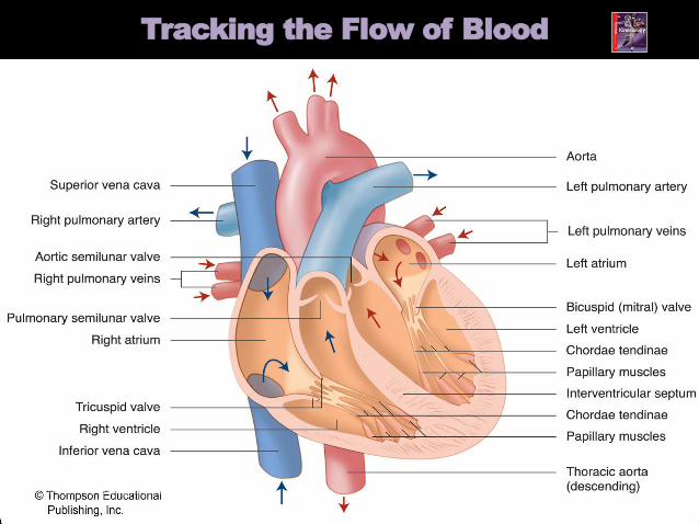
Tracking the Flow of Blood
© 2015 Thompson Educational Publishing, Inc. 10


The Skeletal Muscle Pump
•The low pressure within
the veins causes a
problem for the
cardiovascular system.
•The skeletal muscle pump
aids in the return of blood
back to the heart through
the veins.
•With each contraction of
the skeletal muscle, blood
is pushed back to the
heart.
© 2015 Thompson Educational Publishing, Inc. 20


Pressure in veins (in the chest) decrease while pressure in veins (in the abdominal cavity) increase upon intake of breath
Difference in pressure pushes blood from veins in the abdominal cavity into veins in the thoracic cavity

Nervous system sends a signal to the veins to
constrict
Veins constrict allowing more blood back to the heart


Two main components:
Plasma
Fluid component of blood (mostly water)
Formed Elements
Red blood cells (erythrocytes)
Made in bone marrow
Transport O2 and CO2 in the blood
Transport nutrients and waste
Contain hemoglobin
White blood cells (leukocytes)
Destroy foreign elements
Critical in the function of the immune system
Platelets
Regulate blood clotting
Plasma 55%90% water
7% plasma proteins
3% other (acids,
salts)
Formed elements
45%>99% red blood cells
<1% white blood cells
and platelets
Buffy Layer


The Cardiac Cycle
© 2015 Thompson Educational Publishing, Inc. 27
• The cardiac cycle is the series of
events that occurs through one heart
beat. During this cycle there is both a
phase of relaxation (diastole), in
which the ventricle is filling with
blood, and a phase of contraction
(systole), in which the heart
contracts and ejects the blood.
• Blood pressure is the force exerted
by the blood against the walls of the
arteries and other vascular vessels.
Blood pressure in each of the two
phases—diastole and systole—is
measured in millimetres of mercury
(abbreviated as mmHg).

Systolic and Diastolic Blood Pessure
© 2015 Thompson Educational Publishing, Inc. 28
When blood pressure is reported or
measured, it is often stated as being the
systolic pressure over the diastolic
pressure (e.g., 120/80 mmHg).
Systolic blood pressure
• refers to the maximum pressure
observed in the arteries during the
contraction phase of the ventricle (e.g.,
120 mmHg).
Diastolic blood pressure
• is the minimum pressure observed
in the arteries during the
relaxation phase of the ventricle
(e.g., 80 mmHg).

Normal Blood Pressure
120/80 mmHg
High Blood Pressure: (hypertension)
140/90 mmHg or higher
Low Blood Pressure: (hypotension)
90/60 mmHg

The Heart’s Electrical Conduction System
© 2015 Thompson Educational Publishing, Inc. 30
The cardiac muscle cells are excitable, meaning that with electrical
stimulation they will all contract (this is known as a “syncytium”).
Within the heart there are areas of specialized tissue that are
important in the regulation and coordination of this electrical
activity.
These specialized tissues are:
• the sinoatrial node (SA node)
• the atrioventrical node (AV node)
The contraction of the heart leads to the pumping of blood.
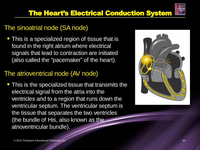
The Heart’s Electrical Conduction System
© 2015 Thompson Educational Publishing, Inc. 20
The sinoatrial node (SA node)
• This is a specialized region of tissue that is
found in the right atrium where electrical
signals that lead to contraction are initiated
(also called the “pacemaker” of the heart).
The atrioventrical node (AV node)
• This is the specialized tissue that transmits the
electrical signal from the atria into the
ventricles and to a region that runs down the
ventricular septum. The ventricular septum is
the tissue that separates the two ventricles
(the bundle of His, also known as the
atrioventricular bundle).

© 2015 Thompson Educational Publishing, Inc. 32


Cardiac conduction

An Electrocardiogram
© 2015 Thompson Educational Publishing, Inc. 35

Measured using an electrocardiogram (ECG) or EKG
Graphical representation of electrical sequence of events occurring with each contraction of the heart
Current on body surface derived almost entirely form heard
12 tracings or leads

Normal sinus rhythm: 60-100 bpm

Bradycardia: < 60 bpm, conditioned athlete, drugs, hyopothermia, sleeping

Tachycardia: > 100 bpm, exercise, smoking, fever, hypoxia (lack of O2 to brain)

ST segment elevation -represents tissue injury due to acute, prolonged reduction in BF. May show up min or hours after a heart attack (due to clot or vascular coronary spasm)

Inverted T wave: represents ischemia (temporary and reversible reduction in blood flow; may show up hours or days after ischemic attack)
Abnormal Q wave: irreversible death of heart tissue

Myocardial infarction: heart attack
-occurs when an artery that feeds the heart becomes blocked

Angina: chest pains
Ischemia: insufficient blood flow to provide adequate oxygenation. (ischemia leads to hypoxia)
Hypoxia: reduction of oxygen supply to tissues
Stroke: lack of oxygen to the brain

Heart Rate (HR): is the
number of times the
heart contracts in a
minute (bpm)
Avg. HR @ rest
=72bpm
Highly trained
athlete=40bpm
During intense exercise
HR may increase to up
to 200 bpm
Maximum HR=220-age

The lowest resting heart beat on
record is 28 bpm and belongs to
the cyclist Miguel Indura in
(Spain) who was tested at the
University of Navarra,
Pamplona, Spain, in 1995.
Lance Armstrong-his heart is 30
percent larger than average;
however, an enlarged heart is a
common trait for many other
athletes. He has a resting heart
rate of 32-34 beats per minute
(bpm) with a maximum heart
rate of 201 bpm.


Activity Heart RateSpeed skater
Downhill skier
Cyclist
Distance runner
Sprint athlete
Tennis player
Shooter
1-Highest
7-Lowest
2
3
4
5
6

Bradycardia and Tachycardia
© 2015 Thompson Educational Publishing, Inc. 49
Regular aerobic exercise results in
improvements in the efficiency of
the cardiovascular system at rest
and during exercise.
• Bradycardia is one of the most easily
observed adaptations that occurs with
training. Bradycardia is characterized
by a heart rate of 60 beats per minute
or less at rest, while tachycardia is a
heart rate of more than 100 beats per
minute at rest.
•Generally, a lower heart rate is
regarded as an indication of an
athletic or strong heart.

The Effects of Exercise
© 2015 Thompson Educational Publishing, Inc. 50
During exercise, dramatic changes ocur in the cardiovascular
system—changes known as cardiovascular dynamics.
The heart and the vessels constantly adapt to accommodate the
ever-changing requirements of the body during exercise.
Some of the factors that are considered when discussing
cardiovascular dynamics are:
• Cardiac output (Q),
• Blood pressure (BP),
• Distribution of blood flow, and
• Oxygen consumption (VO2).

-Regular aerobic exercise leads to
alterations of the cardiovascular
system
-These are functional & structural
With exercise the size and mass
of heart increase
Ventricular walls become thicker
and volume of ventricle increases
(due to increase in venous return)
This leads to a more forceful
contraction= increase in stroke
volume= increase in cardiac
output

Increase in number of capillaries that deliver blood to the heart tissue
Possible increase in the diameter of the coronary arteriesincrease in O2 to the heart in order to work harder
Increase in blood volume up to 15% within 2 days
This causes increase in venous return= increase in stroke volume (volume of blood ejected by left
ventricle per beat) = increase in cardiac output (the volume of blood pumped out of left
ventricle in 1 min)
Capillaries around the soleus muscle.

Cardiovascular system adapts to meet the demands that are placed on it
Heart adjusts amount of blood pumped by altering:
Heart rate (HR) (beats/min)
duration of each cardiac cycle
Stroke volume (SV) (mL)
volume of blood ejected by left ventricle per beat
Avg=70 mL per beat
Cardiac output (Q) (L/min)
HR SV = Q
The volume of blood pumped out of left ventricle in 1 min
Frank-Starling Law:
Ability of the heart to stretch and increase the force of contraction

Q (L/min)=SV (mL) x HR (bpm)
Q = SV x HR
Eg. An avg. HR at rest would be 72 bpm and an avg. SV at
rest would be 71 mL.
Therefore using the equation for Q = SV x HR Q= 71mL x 72 bpm
Q= 5040 mL/min or 5.04 L/min.
During exercise Q can increase to 15-25 L/min
depending on the intensity of exercise
Athletes tend to have slower HR and large SV creating a
more efficient circulatory system.
A slower HR with an increased SV requires less oxygen.

Cardiac Output during Exercise
© 2015 Thompson Educational Publishing, Inc. 55

The Cardiovascular Effects of Training
© 2015 Thompson Educational Publishing, Inc. 56

Heart Disease
© 2015 Thompson Educational Publishing, Inc. 57
The system of vessels that supply essential materials
via blood to the heart muscle itself is called the
coronary circulation.
Serious health repercussions and even death can
occur if a narrowing or blockage of blood vessels
restricts the flow of blood to the heart muscle. For
example, a heart attack (myocardial infarction) can
result when blood flow to a section of the heart
muscle becomes blocked due to plaque buildup or
some other reason.

The Coronary Vessels (Anterior View)
© 2015 Thompson Educational Publishing, Inc. 58

Atherosclerosis
• Coronary artery disease (also known as atherosclerosis) involves a
gradual narrowing of the coronary arteries resulting from the
accumulation of hard deposits of cholesterol (plaque), on the lining
of the blood vessels.
© 2015 Thompson Educational Publishing, Inc. 59

The Causes of Coronary Artery Disease
© 2015 Thompson Educational Publishing, Inc. 30
• Poor Diet,
• Smoking,
• Elevated blood lipids,
• Hypertension,
• Family history, and
• Physical inactivity.
Each factor individually increases the risk of development of coronary artery
disease. When the factors are combined, the risk of coronary artery disease is
magnified.


angioplasty

stent

The Functions of the Respiratory System
The three main functions
of the respiratory system
are to:
•Supply O2 to the
blood,
•Remove CO2 from the
blood, and
•Regulate blood pH (acid-
base balance).
© 2015 Thompson Educational Publishing, Inc. 65

Respiration
© 2015 Thompson Educational Publishing, Inc. 66
External Respiration
• External respiration refers to the processes that occur within the
lungs involving the exchange of O2 and CO2.
Internal Respiration
• Internal respiration refers to the exchange of gases at the tissue
level, where O2 is delivered and CO2 is removed.
Cellular Respiration
• Finally, cellular respiration is the process in which the cells use O2 to
generate energy in the mitochondria of cells.

The Structure of the Respiratory System
67
The respiratory system can be
divided into two main zones — the
“conductive zone” and the
“respiratory zone.”
• The conductive zone
transports filtered air to the
lungs. This zone consists of the
mouth and nose; pharynx;
larynx; trachea; primary and
secondary bronchi; and tertiary
bronchioles and terminal
bronchioles.
• The respiratory zone is where gas
exchange occurs. Bronchioles,
alveolar ducts, and the alveolar sacs
are all structures of the respiratory
zone that are involved with the
exchange of gases between inspired
air and the blood.

The Mechanics of Breathing
© 2015 Thompson Educational Publishing, Inc. 68
The combination of inspiration and
expiration together is known as
“ventilation.”
• Inspiration is an active process,
requiring the contraction of various
respiratory muscles and therefore
the expenditure of significant
amounts of energy.
• Expiration, on the other hand, may be
passive, as in quiet breathing (which
may not require much energy) or
active (as in forced breathing).


Inspiration
Air flows into
the lungs due to
increased lung
volume following
the contraction
of the diaphragm
and intercostal
muscles.
© 2015 Thompson Educational Publishing, Inc. 40

Expiration
Air is expelled from
the lungs due
to relaxation of the
diaphragm and the
intercostal
muscles.
© 2015 Thompson Educational Publishing, Inc. 72

• Ventilation (VE) is the volume
of air moved by the lungs in 1
min.
• Influenced by two factors:
• Tidal volume (VT)
• Volume of air in each breath
• Respiratory frequency (f)
• Number of breaths taken per
minute

Ve is influenced by:
VT (tidal volume): vol. of air in each breath
f (respiratory frequency): # of breaths taken per min
@ rest VT = 0.5 L/breath
@ Exercise VT= 4 L/breath
@ rest f = 12 breaths/min
@ Exercise f = 40 breaths/min
Therefore, Ve = VT x f
note: during strenuous xcise, Ve = 200 L of air per
min

Gas Exchange
© 2015 Thompson Educational Publishing, Inc. 75
The average person’s
lungs have about 300
million alveolar sacs (that
is about a tennis court’s
worth), each of which is
surrounded by a web of
capillaries.
•The walls of each
capillary are one
cell thick, which
provides a very
short distance for
gases to diffuse.

Diffusion
© 2015 Thompson Educational Publishing, Inc. 76
The primary factor that mediates gas
exchange both at the lung (where
blood becomes oxygenated and CO2
is removed) and at the tissue (where
O2 is delivered for metabolism and
CO2 is removed) is diffusion.
• Diffusion is the movement of a
gas, liquid, or solid from a region
of high concentration to a region
of low concentration through
random movement.
• Diffusion can only occur if a
difference in concentration
exists, and such a difference is
called a concentration
gradient.

O2 Transport
© 2015 Thompson Educational Publishing, Inc. 77
O2 Transport
• The process by which O2 is absorbed in the lungs and carried to the
peripheral tissues.
CO2 Transport
• The process by which CO2 in blood is moved into the alveoli and
then exhaled from the body.
a-vO2 diff
• The difference between the amount of O2 in the artery and vein
reflects the amount of O2 that was delivered to the muscle.

Monitoring Cardiorespiratory Functioning
© 2015 Thompson Educational Publishing, Inc. 78

The Rest-to-Exercise Transition
© 2015 Thompson Educational Publishing, Inc. 79
The delivery of O2 to the working skeletal muscle is
achieved through a combination of physiological
mechanisms. However, this is not instantaneous. During
this “lag,” a phenomenon called oxygen deficit (O2 deficit)
occurs.
Oxygen deficit is the difference between the oxygen
required to perform a task and the oxygen actually
consumed prior to reaching a new steady state.
•The trained individual will reach this steady-state
plateau faster than an untrained individual.

Oxygen Deficit: difference between total oxygen
consumed during exercise and amount that would
have been used at steady-rate of aerobic metabolism.

--Trained person reaches steady-rate quicker, has smaller oxygen deficit.

Light aerobic exercise
rapidly attains steady-rate
with small oxygen deficit.
Moderate to heavy
aerobic takes longer to
reach steady-rate and
oxygen deficit
considerably larger.
Maximal exercise
(aerobic-anaerobic) VO2
plateaus without matching
energy requirement.


Excess post-exercise oxygen consumption: The extra oxygen required to replenish oxygen to the various systems that were taxed during the exercise.
Eg: refilling phosphocreatine reserves, replenishing O2 in blood and tissues, lowering breathing rate, lowering body temp. and increasing blood lactate removal.
Active recovery can aid in the removal of blood lactate.

O2 Deficit
Ventilatory threshold
• A state in which
ventilation
increases much
more rapidly than
workload
Lactate threshold
• The point where
blood lactate
concentrations
begin to increase
© 2015 Thompson Educational Publishing, Inc. 85

o Ve increases more rapidly than workload
o 65-85% VO2max
o increase Ve due to increase in lactic acid, and
therefore there is a decrease in blood pH
(increase in H+ ions in blood)
o increase in Ve, increases expelling of CO2 and
increases H+ to combine with bicarbonate to
form CACO2 + H2O, therefore decrease in H+
ions increases pH to normal levels,
therefore, increase in Ve aids to return blood Ph to
normal

Onset of Blood Lactate Accumulation
When lactate levels begin to accumulate rapidly in the blood, this is referred to as the
onset of blood lactate accumulation (OBLA).
• With training, the curve for lactate threshold and OBLA can shift to the right.
© 2015 Thompson Educational Publishing, Inc. 87

VO2max
Maximal rate of
oxygen
consumption
(VO2max)
VO2max is the maximum volume(V) of oxygen (O2) inmillilitres that the humanbody can use in oneminute, per kilogram of body weight, while breathing air at sea level.
© 2015 Thompson Educational Publishing, Inc. 88

VO2max Scores for Athletes/Non-Athletes
© 2015 Thompson Educational Publishing, Inc. 89

Physiological Adaptations to Training
© 2015 Thompson Educational Publishing, Inc. 90

Physiological Adaptations to Training
© 2015 Thompson Educational Publishing, Inc. 91

Respiratory Diseases
© 2015 Thompson Educational Publishing, Inc. 60
Asthma is a disease that is characterized
by spasm of the smooth muscles that line
the respiratory system, an oversecretion
of mucous, and swelling of the cells lining
the respiratory tract.
• Many factors can lead to an asthma
attack, including exercise, allergic
reaction, contaminates, and stress.
Fortunately, most cases of asthma
can be controlled through the use
of different medications.
• Some Olympic-level athletes have
been diagnosed with asthma and
yet are able to compete
internationally.

Asthma Continued
In people with asthma, the airways are chronically inflamed.
Certain triggers can make the inflammation worse and cause a
narrowing of the airways. At the same time, the body may
produce extra mucus that clogs the airways. These changes
work together to restrict the flow of air to the lungs. As too little
air gets through, wheezing and breathlessness occur.

Respiratory Diseases
© 2015 Thompson Educational Publishing, Inc. 94
Chronic obstructive pulmonary
disease (COPD) is a general term
that describes a family of diseases
that lead to a dramatic reduction in
airflow through the respiratory
system.
• Individuals with COPD cannot
perform normal everyday
activities without experiencing
dyspnea (shortness of breath).
• Treatment of COPD conditions
includes not only medications but
also supplemental oxygen therapy
for severe cases, as well as
respiratory muscle training.

© 2015 Thompson Educational Publishing, Inc. 95
A Diseased Lung


