1 Thiamine Biochemistry in Ethanol Consumption Thiamine Biochemistry in Ethanol Consumption ................................................................1 1. Biology of Substance ....................................................................................................6 1.1. Forms of thiamine .................................................................................................7 1.1.1. Different Supplemental Forms of Thiamine ..................................................8 1.2. Requirement for Thiamine ..................................................................................10 1.2.1. Therapeutic Dose of Thiamine.....................................................................11 1.2.2. Toxic Doasage of Thiamine ..........................................................................11 1.3. Thiamine Antagonists..........................................................................................12 1.3.1. Sulfites .........................................................................................................13 1.3.1.1. Sulfites cause thiamine deficiency .......................................................16 1.3.1.2. Sulfites and sulfur dioxide toxicity linked to heart failure ...................16 1.3.1.3. Sulfites in food and drinks contribute to cardiac disfunction ..............17 1.3.1.4. The high sensitivity of thiamine to sulfites ..........................................17 1.3.1.5. Thiamine deficiencies caused by sulfite preservatives animal study ..17 1.3.1.6. Sulfites destroy thiamine in preventing browning of parboiled rice ...21 1.3.1.7. Sulfites in Alcoholic Beverages .............................................................21 1.3.1.8. The Molybdenum Biology of Sulfite Oxidase .......................................22 1.3.1.8.1. Sulfite Oxidase: Molybdenum enzyme converts Sulfite to Sulfate 26 1.3.2. Caffeic Acid and Polyphenols.......................................................................27 1.3.3. Losses in Food Preparation ..........................................................................29 1.4. Experimental Models of Thiamine Deficit .........................................................31 1.5. Thiamine in Enzymic Reactions .........................................................................33 2. Deficiency...................................................................................................................37 2.1. Diagnosis and Identification Problems ..............................................................39 2.2. Thiamine Deficiency in Alcoholic Population ....................................................42 2.2.1. Liver Storage of Thiamine ............................................................................42 2.3. Comorbidity of vitamin deficits .........................................................................43 2.4. Beri Beri .............................................................................................................43 2.4.1. Clinical Vignette for Wernicke's thiamine deficiency ..................................44 2.5. Subclinical Thiamine Defciencies .......................................................................45 2.6. Elderly Predisposition to Thiamine Deficiency ..................................................47 2.7. Variations in causes of thiamine deficiency ......................................................49 2.7.1. Thiamine Transporter Proteins and Biotin ..................................................49 2.8. Gastrointestinal Problems .................................................................................50 3. Carbohydrate Metabolism.........................................................................................50 3.1. Carbohydrate loading produces similar polyneuropathys to Wernicke's/Korsakoff's psychosis ..................................................................................51 3.2. Thiamine in Diabetic Advanced Glycation Endproducts ...................................51 3.3. Thiamine and dextrose in management of the comatose patient ....................52 3.4. Emergency Treatment and Coma ......................................................................52 4. Alcohol Consumption ................................................................................................53
Transcript
1
Thiamine Biochemistry in Ethanol Consumption
Thiamine Biochemistry in Ethanol Consumption ................................................................11. Biology of Substance ....................................................................................................6
1.1. Forms of thiamine .................................................................................................71.1.1. Different Supplemental Forms of Thiamine ..................................................8
1.2. Requirement for Thiamine..................................................................................101.2.1. Therapeutic Dose of Thiamine.....................................................................111.2.2. Toxic Doasage of Thiamine ..........................................................................11
1.3.1.1. Sulfites cause thiamine deficiency .......................................................161.3.1.2. Sulfites and sulfur dioxide toxicity linked to heart failure ...................161.3.1.3. Sulfites in food and drinks contribute to cardiac disfunction ..............171.3.1.4. The high sensitivity of thiamine to sulfites ..........................................171.3.1.5. Thiamine deficiencies caused by sulfite preservatives animal study ..171.3.1.6. Sulfites destroy thiamine in preventing browning of parboiled rice ...211.3.1.7. Sulfites in Alcoholic Beverages .............................................................211.3.1.8. The Molybdenum Biology of Sulfite Oxidase .......................................22
1.3.1.8.1. Sulfite Oxidase: Molybdenum enzyme converts Sulfite to Sulfate 261.3.2. Caffeic Acid and Polyphenols.......................................................................271.3.3. Losses in Food Preparation..........................................................................29
1.4. Experimental Models of Thiamine Deficit .........................................................311.5. Thiamine in Enzymic Reactions .........................................................................33
2. Deficiency...................................................................................................................372.1. Diagnosis and Identification Problems ..............................................................392.2. Thiamine Deficiency in Alcoholic Population ....................................................42
2.2.1. Liver Storage of Thiamine ............................................................................422.3. Comorbidity of vitamin deficits .........................................................................432.4. Beri Beri .............................................................................................................43
2.4.1. Clinical Vignette for Wernicke's thiamine deficiency ..................................442.5. Subclinical Thiamine Defciencies .......................................................................452.6. Elderly Predisposition to Thiamine Deficiency ..................................................472.7. Variations in causes of thiamine deficiency ......................................................49
2.7.1. Thiamine Transporter Proteins and Biotin ..................................................492.8. Gastrointestinal Problems .................................................................................50
3. Carbohydrate Metabolism.........................................................................................503.1. Carbohydrate loading produces similar polyneuropathys to Wernicke's/Korsakoff's psychosis..................................................................................513.2. Thiamine in Diabetic Advanced Glycation Endproducts ...................................513.3. Thiamine and dextrose in management of the comatose patient....................523.4. Emergency Treatment and Coma ......................................................................52
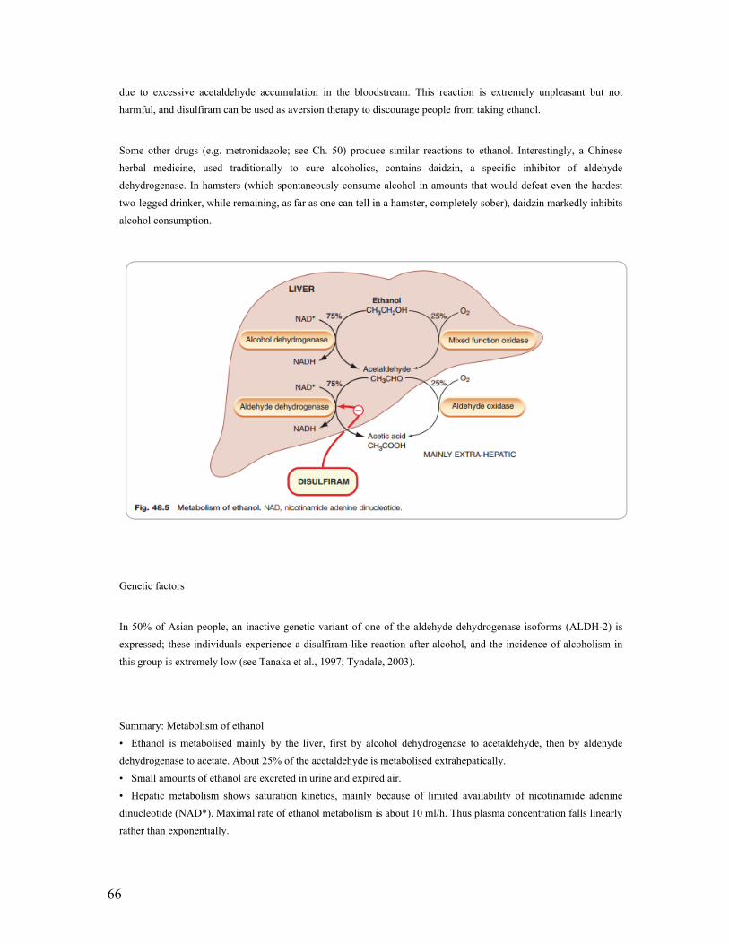
4.1. Pharmacology of Ethanol...................................................................................584.2. Thiamine Deficiency as a predisopostion to and consequence of alcohol consumption ..................................................................................................................684.3. Alcohol Intoxication ...........................................................................................694.4. Thiamine Deficiency as a result of Alcoholism leading to Wernicke-Korsakoff Disease ...........................................................................................................................724.5. Memory Dysfunction in Alcohol Intoxication ....................................................734.6. Alcoholic Cerebellar Degeneration ....................................................................74
4.6.1. Cerebellar Sensitivity to Lesions in Thiamine Deficit...................................764.7. Alcohol Induced Neuropathies ..........................................................................76
4.7.1. Thiamine and vitamin related neuropathies associated with ethanol and tabacco 77
4.8. Management of Alcohol Withdrawal ................................................................774.8.1. Thiamine treatment in alcoholism ..............................................................80
4.9. Fetal Alcohol Syndrome .....................................................................................814.9.1. Increased natal requirement for thiamine ..................................................814.9.2. The effect of ethanol on fetal development ...............................................82
5. Neural and Psychiatric Symptoms .............................................................................835.1. Depression like symptoms in thiamine deficiency ............................................835.2. Complex patterns of dementia in thiamine and vitamin deficiency .................845.3. Thiamine and Alzheimer's Disease ....................................................................845.4. Psychiatric disorder resulting from Thiamine Deficit ........................................865.5. Wernickes Encephalopathy ...............................................................................87
5.5.1. Memory Dysfunction in Wernicke's Encephalopathy .................................905.5.2. Wernicke's physiology similar to Alzheimer's .............................................905.5.3. Glutamate neurotoxicity (or excitotoxicity) is the primary cause of the Thiamine Deficiency Encephalopathy .......................................................................915.5.4. Age-related differences in the areas of Broca and Wernicke .....................915.5.5. Damage to Wernicke's area linked to thiamine deficit and memory damage
925.5.6. Clinical Features of Wernicke's leading to Korsakoff's ................................925.5.7. Glutamate Excitotoxcity Suggested as Causing Wernicke's Lesions ...........93
5.6. Korsakoff Syndrome ..........................................................................................945.6.1. Memory Dysfunction in Alcoholic Korsakoff Syndrome..............................945.6.2. Anterograde amnesia in Korsakoff's syndrome ..........................................955.6.3. Memory, Amnesia and Thiamine Deficiency ...............................................95
5.7. Wernicke-Korsakoff's is the third most common dementia .............................996. Neurotransmission ..................................................................................................100
6.1. Thiamine deficit impairs cholinergic functioning ............................................1006.1.1. Acetyl Choline linked with Alzheimer's Disease ........................................1006.1.2. Thiamine deficiency and the loss of cholinergic cells................................1016.1.3. Cholinergic neurons co-release other neurotransmitters such as GABA and glutamate.................................................................................................................1016.1.4. Thiamine and Acetyl Choline Functioning .................................................102
3
6.2. Demyelination in Thiamine Deficiency ............................................................1026.3. Thiamine functioning in the nervous system ..................................................1036.4. Thiamine in Neurotransmitter Metabolism ....................................................103
7. Cardiovascular Disease ............................................................................................1037.1. Thiamine deficiency is defined by heart disease.............................................1047.2. Thiamine deficiency common in heart failure patients ..................................1047.3. Thiamine supplementation improves forms of heart failure ..........................1057.4. Thiamine triphosphate postulated as important for heart contraction .........1057.5. Pathologically High Cardiac Output in thiamine deficiency ............................1057.6. Systemic Vascular Resistance, Heart Failure and Thiamine ............................1077.7. Alcohol induced heart problems .....................................................................108
4
Based on the research which I have reviewed and brought together it makes me think that key strategies involve:
Remove all sources of sulfites and sulfur dioxide from diet and environment
Protection of supplemental thiamine from destruction
Clinical thiamine supplementation
Thiamine is a vitamin we have to derive from the diet we eat. It plays various roles in the body including the creation and breaking down of sugars. It also plays a critical role in heart function, with thiamine triphosphate having been suggested as vital for heart contraction.
Historically thiamine deficiency was known and identified by its manifesting in heart failure. Currently, in the industrialised world we live in, sulfites and sulfur dioxide have been included as additives in our food and drink chain.
Sulfites cause thiamine deficiency by cleaving the vitamin into two pieces neither of which have biological activity. In relation to taking thiamine supplementation it is essential to remove anything which is destroying the vitamin which has therapeutic effects.
The strategy to remove the toxin and restock the tissues which need thiamine for their functioning, like the heart. Simply removing sulfites and sulfur dioxide alone will improve the health of the heart.
There is considerable evidence that restoring the bodies own ability to breakdown sugars (glycolysis) and create sugars (gluconeogenesis) is an important strategy in relieving certain cravings for seeking alcohol and sugar through dietary means.
Ethanol is a partially metabolised sugar and thus is used as a source of energy and production of various complex molecules. The alcoholic body has a disrupted ability to function normally and as a result it seems rational that the mammalian body will crave what its physiology requires to function - in this case sugars and their intermediates (ethanol, starch, and sugars).
As well as this means for generating craving and addiction, the condensation of acetaldehyde (a breakdown product of ethanol) with dopamine generates tetrahydroisoquinones which interact with the opiate receptors. Significantly, it has been hypothesised that glutamate excitotoxicity is an underlying mechanism of lesion production in Wernicke's linking this pathophysiology into a large body of research on addiction mechanisms.
5
In summary, there potentially exists a triad of mechanisms reinforcing the behaviour of ethanol consumption: (a) the need to provide in the diet sugar intermediates for normal function, (b) the stimulation of the opiate system, and (c) the stimulation of excitatory glutamate.
The widespread use of sulfites in the dietary intake is one of the most significant damages to thiamine functioning. These preservatives and common food additives destroy the vitamin/enzyme in the food stuff and in the body as well as rendering supplemental forms ineffective.
The metabolic significance of consuming sulfites is several fold. Not only do they destroy enzymes for gluconeogenesis and glycolysis etc, but they put a heavy demand on sulfite oxidase. This is significant because the constituents necessary for the enzymic transformation of detrimental sulfites to inert sulfates (molybdenum enzymes) are also required for the metabolism of aldehydes, to which an ethanol load greatly contributes.
In summary, the consumption of sulfites alone is not as toxicologically significant as the consumption of sulfites and aldehydes due to the requirement of a common enzymatic cofactor.
There is considerable evidence that thiamine compounds are required for the normal functioning of the heart and that they contribute vital roles to neurotransmission. This is suggestive that cardiac disease is significantly correlated with sulfite intake and other factors which create a burden on thiamine enzymes.
Lastly, carbohydrate loaded diets (ethanol, sugars and starches) proportionally increase the requirement for thiamine and in its absence represent sources of toxicity - and potentially predispositions to craving/addiction. There is significant evidence that carbohydrate loads trigger thiamine deficits.
If this proves to be correct then the industrial food chain is a major shit show due to the culturally engrained nature of the food stuffs, the use of alcohol to self medicate in contexts of sociocultural trauma and the endemic use of sulfites throughout the industrial-food complex.
What follows are direct verbatim excerpts from textbooks and peer reviewed papers with their sources embedded in a distributed bibliography throughout the preliminary document.
The choice for presenting this through such a method is aimed to eliminate a level of doubt about the interpretation of the original source and promote to the reader the accessing of the original source of text and verify the context and provenance.
This version (12.03.2019) is preliminary and represents a small part of the final study but is written to show prospective avenues being fleshed out. Significant sections yet to come include detailed enzymology, molybdenum biochemistry, oncology, opiate metabolism, detailed neuropsychiatry, and other sections.
This document is a work in progress and not for public distribution. Please get in touch if you have any questions, comments, helpful challenges or suggestions.
In mammalian cells, ThDP is the coenzyme of fve enzymes or enzyme complexes, among which PDHC and OGDHC,
localized at strategically important crossroads of cell metabolism. PDHC controls the oxidative metabolism of sugars
and the production of acetyl-CoA required for fatty acid, cholesterol, and acetylcholine synthesis. OGDHC is the rate-
limiting enzyme of the Krebs cycle in the brain and links sugar and amino acid metabolism.
An important point is the role of thiamine in the nervous system, which is particularly sensitive to thiamine defciency. This is not unexpected, as the functioning of the brain is heavily dependent on oxidative metabolism and the synthesis of neurotransmitters requires the activity of ThDP-dependent enzymes: PDHC is required for the synthesis of acetyl-CoA, the precursor of acetylcholine, and OGDHC is required for the synthesis of glutamate and GABA.
Among possible non-coenzyme roles, a positive effect of (unphosphorylated) thiamine on acetylcholine release has been consistently reported
7
1.1. Forms of thiamine
The analysis of the chemical properties of thiamine and its naturally occurring phosphate derivatives shows a
complexity at three levels.
First, there is an intrinsic complexity of the thiamine molecule with, in particular, the unique properties of the thiazole heterocycle, critical for its catalytic properties.
Second, the existence of three sequential phosphate derivatives is reminiscent of nucleotides and makes thiamine the only nonnucleotide molecule known with such a feature.
8
Third, the recent discovery of adenylated thiamine derivatives adds an additional level of complexity, closing the gap with other structurally related vitamin-derived “dimeric” molecules such as flavine or pyrimidine nucleotides (FAD, NAD+).
The existence of energy-rich di- or triphosphorylated as well as adenylated thiamine derivatives strongly suggests that the biological role of vitamin B1 is not limited to catalysis by ThDP.
Handbook of vitamins. (2014). Boca Raton: CRC Press.Page 274
Thiamin phosphate derivatives are found in all organisms. In most cases the cofactor ThDP is the major thiamin compound. In most animal cells, ThDP represents 70 to 90% of total thiamin, and in the brain most of it is bound to apoenzymes.
Unlike other B-vitamins, oral administration of thiamin does not lead to significantly increased plasma vitamin
levels (Davis and Icke, 1983), probably because intestinal thiamin absorption is a relatively slow process,
especially in humans.
This probably contributes to the fact that marginal thiamin deficiency in humans is more common than initially thought. In order to overcome this problem, several thiamin precursors with higher bioavailability were developed (Figure 17.3 ).
In the 1950s, M. Fujiwara and colleagues in Tokyo discovered that a thiamin derivative with high bioavailability was formed in crushed garlic (Allium sativum) bulbs through the action of a plant enzyme on thiamin and allicin (diallyl thiosulphinate) (Fujiwara et al., 1954 ). They named this compound allithiamin, which they later identified as thiamin allyl disulfide.
Other synthetic thiamin disulfides such as sulbutiamine (O - isobutyrylthiamin disulfide) and fursultiamine (thiamin tetrahydrofurfuryl disulfide) were developed. All have a higher bioavailability than thiamin, probably because their hydrophobic character means that they easily cross intestinal membranes and no transporter is required.
In the bloodstream, these disulfide compounds are easily reduced to thiamin in the presence of cysteine or glutathione. Intraperitoneal administration of sulbutiamine in rats leads to a significant increase in the levels of
9
thiamin phosphate esters in the brain and it has a documented effect on the central nervous system as a psychotropic drug (Bizot et al., 2005 ).
Fursultiamine improves energy metabolism and physical performance during physical fatigue loading in rats (Nozaki et al., 2009 ). Central nervous effects of fursultiamine are less well documented but it was suggested to have beneficial effects on speech, behavior and sleep in autistic children (Lonsdale et al., 2002 ). Benfotiamine (S - benzoylthiamin O - monophosphate), another synthetic thiamin precursor, is being extensively studied.
In contrast to the lipophilic thiamin disulfides, benfotiamine, a thioester, because of its hydrophilic phosphoryl group is not lipid - soluble. In contrast, it dissolves in aqueous solvents at slightly alkaline pH. To be absorbed, benfotiamine must be dephosphorylated to S - benzoylthiamin by ecto - alkaline phosphatases present in the intestinal mucosa (Volvert et al., 2008 ).
The more lipophilic S - benzoylthiamin may then cross the brush - border membrane. It can be hydrolyzed to thiamin by thioesterases present in the liver. The different modes of transport and degradation of these three compounds probably explain their different effects, though all raise blood thiamin levels well above those obtained by administration of an equivalent dose of thiamin.
Benfotiamine mainly acts on peripheral tissues through increase in transketolase activity and thus is effective in preventing diabetic complications such as retinopathy (Hammes et al., 2003 ).
However, benfotiamine is unable to significantly raise thiamin phosphate levels in the rodent brain, which probably explains why, until recently, it had no documented central nervous system effects. However, very recently, benfotiamine was shown to improve cognitive functions and to dramatically decrease amyloid plaques and neurofibrillary tangles in a mouse model of Alzheimer ’ s disease (Pan et al., 2010 ).
It is generally considered that the recommended dietary allowance (RDA) is 1.2 mg/day for adult males and 1.1
mg/day for adult females (Table 7.1). The RDA is increased to 1.4 mg/day during pregnancy and even 1.5 mg/day
for lactating women. In children, the RDA increases with age and is 0.5, 0.6, and 0.9 mg/day, respectively, for 1–3,
4–8, and 9–13 years of age. Such values should however be taken with some caution as they depend on lifestyle:
factors such as alcohol or sugar intake must be considered. Therefore, thiamine supplementation is advisable for
alcohol abusers and might be useful for elderly people, as both might have decreased intestinal thiamine
absorption. Some rare conditions require large thiamine intake (100 mg/day orally or intravenously). These include
Wernicke’s encephalopathy and some rare genetic diseases such as thiamine-responsive megaloblastic anemia
(TRMA) or thiamine-responsive maple syrup urine disease.
Handbook of vitamins. (2014). Boca Raton: CRC Press.
11
Page 274
1.2.1. Therapeutic Dose of Thiamine
Because of the slow rates of thiamine
transport (especially in humans), derivatives with higher bioavailability than thiamine may be ofadvantage (benfotiamine, sulbutiamine, or fursultiamine; see Section 7.10.7).
Therapeutic doses may vary from 10 to 200 mg/day. For instance, 100 mg thiamine intravenousfor several days are generally recommended for the treatment of cardiovascular (“wet”) beriberi. Itis advised that intravenous thiamine administration precedes intravenous glucose administration asthe latter may worsen thiamine defciency (Hack and Hoffman 1998).
Indeed, it has been observedthat high carbon uptake increases the requirement for thiamine, possibly as a result of an instability of enzyme-bound ThDP during the catalytic reactions (McCourt et al. 2006): increased glucoseintake would result in increased flux through ThDP-dependent enzymes, hence precipitating breakdown of ThDP and worsening the thiamine status.
Handbook of vitamins. (2014). Boca Raton: CRC Press.Page 275
1.2.2. Toxic Doasage of Thiamine
Thiamine has a very low toxicity and is without adverse effects after oral intake of even large doses. However,
intravenous administration of large doses (125 mg/kg in mice) can lead, in humans, to allergic reactions
(anaphylactic shock), respiratory depression, and neuromuscular blockade. In dogs, blood thiamine levels of 10
mg/100 ml (0.3 mM) are invariably fatal (Davis and Icke 1983), probably through a curare-like action of thiamine
(Ngai et al. 1961; Smith et al. 1948).
Handbook of vitamins. (2014). Boca Raton: CRC Press.Page 275
Oral intake of large doses generally has no adverse effects. However, intravenous administration of large doses (125 mg/kg in mice) can lead to respiratory depression and neuromuscular blockade. In dogs, blood thiamin levels of 10μg/100 ml (300μmol/L) are invariably fatal (Davis and Icke, 1983 ). In humans, allergic reactions (anaphylactic shock) are a rare complication of intravenous thiamin administration.
Many organisms contain thiamine-degrading enzymes (thiaminases). Thiaminase I (EC 2.5.1.2), present in
microorganisms and some higher multicellular eukaryotes such as fern, shellfsh, and fsh, is a pyrimidine
transferase.
Handbook of vitamins. (2014). Boca Raton: CRC Press.Page 275
Several thiamine antimetabolites are potent competitive inhibitors or substrates of TPK. Thus, pyrithiamine (Ki = 1 μM with respect to thiamine) is a substrate of TPK, though not very effcient (Liu et al. 2006). On the other hand, oxythiamine is less effective as an inhibitor, but a better substrate than pyrithiamine (Rindi et al. 1963). Oxythiamine diphosphate is a potent inhibitor of transketolase (Datta and Racker 1961) as well as of PDHC (Rogers 1970).
Handbook of vitamins. (2014). Boca Raton: CRC Press.Page 277
Several synthetic thiamine antagonists have been developed. The most potent is pyrithiamine, a competitive inhibitor of thiamine transport and TPK. Other analogs such as oxythiamine, amprolium, and the chemically unrelated diuretic amiloride also inhibit thiamine transport (Bettendorff and Wins 1994). While pyrithiamine has an affnity comparable to thiamine (Ki <1 μM) for TPK, oxythiamine has a 1000 times lower affnity (Peterson et al. 1975).
Both compounds may be pyrophosphorylated by TPK. Oxythiamine diphosphate is a potent inhibitor of thiamine-dependent enzymes, while pyrithiamine diphosphate is not. Amprolium cannot be phosphorylated. Pyrithiamine easily crosses the blood–brain barrier, in contrast to oxythiamine, which does not enter the brain (Rindi et al. 1963). This property was used in the development of animal models of Wernicke–Korsakoff syndrome.
In contrast, oxythiamine-treated animals have no neurological symptoms, but suffer weight loss, anorexia, and cardiac enlargement, probably as a result of inhibition of ThDP-dependent enzymes by oxythiamine diphosphate. There are several possible mechanisms to explain the poisonous effects of pyrithiamine:
13
(1) it is a potent competitive inhibitor of the high-affnity thiamine transporter (Bettendorff and Wins 1994; Casirola et al. 1988; Rindi and Laforenza 1997),
(2) it competitively inhibits thiamine pyrophosphorylation by TPK (Peterson et al. 1975),
(3) it is a substrate of TPK (Liu et al. 2006) and pyrithiamine diphosphate may be an inhibitor of ThDP-dependent enzymes (though probably not a very potent one), and fnally
(4) a direct toxic effect of pyrithiamine cannot be excluded (Rindi and Sciorelli 1970). Sulfites, added as preservatives to food, cleave the vitamin at the level of the methylene bridge, yielding separate pyrimidine and thiazole moieties (Figure 7.2).
Cases of thiamine deficiency in dogs as a result of feeding on sulfite-preserved meat have been described (Singh et al. 2005).
Some foodstuff may contain thiaminases (thiamine-destroying) enzymes. Some fish (e.g., carp, eel, Baltic herring, or catfish) and shellfish may contain thermolabile thiaminase I, a pyrimidine transferase (EC 2.5.1.2). This is also the case of some ferns (Pteris aquilina) that, when consumed by grazing cattle or horses, may result in severe thiamine deficiency. Thiaminase I is destroyed by cooking, but consumption of raw food containing this enzyme may cause beriberi in humans.
Another thiaminase found in microorganisms, thiaminase II (EC 3.5.99.2), catalyzes the hydrolysis of thiamine in separate pyrimidine and thiazole moieties. Recent results suggest that this enzyme may be involved in a thiamine salvage rather than a thiamine degradation pathway (Jenkins et al. 2007). Indeed, thiamine degradation in the soil leads to the formation of aminopyrimidine, and thiaminase II catalyzes the conversion of aminopyrimidine to hydroxypyrimidine, a building block for the biosynthesis of thiamine by some bacteria.
Sulfites in various forms have been added to food materials as preservative agents and for other purposes for
centuries. concern over possible hazard also goes back a considerable length of time, to an article published by
Kionka in 1896 on the possible toxicity of sulfites in food Various forms of sulfites have been used to prevent
browning during processing of such light-colored fruits and vegetables as dried apples and instant potatoes.
14
They are also used in wine-making as selective antibacterial agents which do not inhibit the desired development of yeast. Sulfites serve a special function in the wet-milling of corn, where they have the effect of softening the hard kernel to permit removal of cornstarch. Sulfite levels in processed foods, regardless of their specific chemical source, are conventionally expressed as SO2-equivalent, and range from zero to about 3,000 ppm on a dry-weight basis.
Dehydrated, light-colored fruits such as apples, apricots, bleached raisins, pears and peaches contain the greatest amounts in this range. Dehydrated vegetables and prepared soup mixes range from a few hundred to about 2,000 ppm; instant potatoes, for example, average about 400 ppm.
A worldwide average for wines would be about 100-400 ppm, with beers about 2-8 ppm. Many wines produced in the United States have less than 100 ppm, although the maximum legally permitted by the Food and Drug Administration is 350 ppm. Sulfite is naturally produced from sulfate during the fermentation process itself, and may account for 16-125 ppm of SO2 even when no sulfites are deliberately added (Wurdig and Schlotter, 1967)
However, the most commonly used figure for per-capita daily intake from solid foods and non-alcoholic beverages in the US. Is approximately 2 mg of SO2. The U . S . wine and beer consumption figures for 1971 correspond to an additional daily sulfite intake of approximately 5 mg of SO2 per capita, assuming that these beverages are consumed by 75% of the population. The wide variations in preferences, however, make this “average” figure almost meaningless. For example, an individual drinking several 12-02 cans of beer daily would be consuming 5 to 15 mg of SO2 per day, whereas a pint of wine contributes 100 mg or more.
Thus it is probable that the bulk of the U.S. population consumes no more than 10-15 mg of SO2 per day per capita, although some individuals may consume as much as 120 mg or more per day. Essentially the same levels have been estimated for other developed countries, for example Belgium (Bigwood, 1968; 1970)
Extensive data on dietary intake of sulfites in the US. are contained in a survey prepared by the National Academy of Sciences under contract with the FDA and submitted to that agency in October 1972. These data indicate that consumption of SO2 per capita may reach approximately 600 mg per day. However, the NAS report indicates that the intake data are overstated in most cases, often by considerable margins, because of the basic assumptions involved in their collection. Thus, the NAS is currently re-evaluating these data. The Acceptable Daily Intake (ADI) of sulfites for adults, as established by the United Nations’ FAO/WHO, is 0.70 mg of S02- equivalent per kg of body weight, equivalent to 50 mg of SO2 per day for a 70-kg (155-lb) person.
It should be noted that in addition to dietary sulfites, the human body is exposed to airborne sulfur dioxides from a variety of sources, both natural and manmade. Extensive data on dietary intake of sulfites in the US. are contained in a survey prepared by the National Academy of Sciences under contract with the FDA and submitted to that agency in October 1972. These data indicate that consumption of SO2 per capita may reach approximately 600 mg per day. However, the NAS report indicates that the intake data are overstated in most cases, often by considerable margins, because of the basic assumptions involved in their collection. Thus, the NAS is currently re-evaluating these data.
15
The Acceptable Daily Intake (ADI) of sulfites for adults, as established by the United Nations’ FAO/WHO, is 0.70 mg of S02- equivalent per kg of body weight, equivalent to 50 mg of SO2 per day for a 70-kg (155-lb) person. It should be noted that in addition to dietary sulfites, the human body is exposed to airborne sulfur dioxides from a variety of sources, both natural and manmade. The normal metabolic processes of the body convert the excess sulfur in these amino acids first to sulfites and then -with the aid of an enzyme, sulfite oxidase - to sulfates, which are excreted in theurine. Adult human beings “in balance” with respect to the intake and excretion of sulfur normally excrete on the average about 25 millimoles (2,400 mg) of sulfate in the urine per day.
Animal studies have indicated that sulfite oxidase is present to some extent in most body tissues. However, the liver, heart, and kidney appear to possess the greatest capacity to oxidize sulfite (MacLeod et al., 1961), As countries become more affluent and more urbanized the consumption of sulfited foods tend to rise, and it is possible that their intake may become relatively high in a few cases destruction of thiamine (vitamin 61)by sulfites might lead to a deficiency of this vitamin. Also, the recent discovery of the action of sulfites on nucleic acid components has raised questions as to possible genetic effects. Mutations have in fact been produced in Escherichia coli and in phage lambda by exposing them to strong (1 to 3-molar) solutions of sodium bisulfite (Mukai et at., 1970)
Furthermore, investigators working prior to 1935 were unaware of the fact that sulfites can destroy thiamine, and their test results may have been complicated t v deficiency ofthis vitamin. Applying the usual 100-fold safety factor to this figure yields the 0.70 mg/kg/day figure mentioned earlier as the FAONHO Acceptable Daily Intake. In these studies, the diets were fed to rats for three generations, to quail for four generations, and to pigs for one year. The diets were mixed fresh every two weeks and stored at -18°C (0°F) until used, and were fortified with 50 mg of thiamine per kg ofbody weight to prevent any deficiency of this vitamin.
These studies indicate that dietary sulfites are not highly toxic, provided that the diets are not stored for long periods after mixing and that adequate thiamine is provided. Hotzel et at. (1966) found that rats maintained on diets providing adequate thiamine suffered no ill effects attributable to consumption of sulfites in doses of up to 300 mg/kg/day. Thiamine-deficient animals in the same tests, however, showed toxic effects at doses as low as 50 mg/kg/day. Fitzhugh et at. (1946) reported that diets containing sulfites equivalent to 615 ppm of SO2 or less had no significant effect on the growth of rats. Higher levels, however, were toxic, and the toxicity was only partly counteracted by administration of additional thiamine to the animals.
Bhagat and Lockett (1964) observed that diets containing 0.6% sodium metabisulfite (4,044 ppm of SOZ) produced two types of toxic effects in growing rats: Growth was reduced in those rats fed on diets stored for 7 weeks; this was shown to be attributable to lack of thiamine. However, diets stored for 3-4 months produced toxic effects that were not reversed or prevented by thiamine; this may have been due to changes in the fats contained in the diet during storage. Ti1 et al. (1972) found that when rat diets consisting of corn meal, casein, vitamins, soybean oil, cellulose, and minerals were stored in the presence of 1% sodium metabisulfite for 3 months at room temperature, the mixtures became toxic; this is probably because of interaction between sulfite and unsaturated fats
16
SPECIAL REPORTSULFITES AS FOOD ADDITIVESA Scientific Status Summary by the Institute of Food Technologists’ Expert Panel on Food Safety & Nutrition and the Committee on Public Information, NUTRITION REVIEWSIVOL. 34, NO. PIFEBRUARY 1976 pp 58-62
1.3.1.1. Sulfites cause thiamine deficiency
Various physical and chemical conditions can cause the breakdown of thiamine. Conditions of high pH,
elevated temperatures, and the presence of sulfites (Figure 2), which are used as preservatives of meat
products, are common causes of thiamine loss. Sulfiting agents used for food preservation include bisulfites,
sulfites, metabisulfites, and sulfur dioxide.
These preservatives serve to prevent the oxidation of oxymyoglobin to metmyoglobin in meat, which causes its discoloration from red to brown upon exposure to air. Aside from their use as anti-oxidants, they also have anti-microbial properties, delaying the onset and rate of growth of bacteria. In the United States, the use of sulfiting agents as meat preservatives, as well as in other foods recognized as a source of thiamine, is prohibited. Aside from causing allergic reactions in sensitive people, sulfites cleave thiamine at its methylene bridge (Figure 2), causing its destruction.
Foods and beverages that contain high concentrations of polyphenolic compounds can also cause thiamine deficiency. Polyphenolic compounds are plant extracts including tannins and catechins (Figure 3), that are commonly found in coffee and tea.
[Causes of Thiamine Deficiency, Department of Natural Resources, Cornell University, 2017, Drawn from internet 4.10.2018 http://thiamine.dnr.cornell.edu/Thiamine_causes.html
1.3.1.2. Sulfites and sulfur dioxide toxicity linked to heart failure
Sulfur dioxide and sulfites are oxidized in the body to sulfate, which is harmless, and excreted in the urine. It
has generally been believed that this detoxification mechanism is adequate to handle the quantities that are
likely to be ingested
It has long been known that the aged and patients with bronchial asthma, chronic bronchitis and degrees of heart failure may suffer fatal consequences during periods of severe smog when the concentration of atmospheric sulfur dioxide is high. But even normal persons suffer bronchospasm at 5 ppm SO2
[Sulfite Sensitivity — Unrecognized Threat : — Is Molybdenum Deficiency the Cause ? Rhoda Papaioannou, Carl PfeifferPublished 2007 https://www.semanticscholar.org/paper/Sulfite-Sensitivity-%E2%80%94-Unrecognized-Threat-%3A-%E2%80%94-Is-%3F-Papaioannou-Pfeiffer/acafec6185f86e7c38c0d91aaf7acf1b6cd494e4
1.3.1.3. Sulfites in food and drinks contribute to cardiac disfunction
This evidence, though restricted to an in vitro cell model, should raise a fundamental concern about sulfite
preservatives used in wine industry or food and the consequent role of grapes-derived flavonoids in human
health. Actually, one of the main sources of sulfites in the human body comes from the addition of sulfites
and sulfur dioxide to many food products. The pejorative effect of sulfite preservatives may dampen the
beneficial action of wine polyphenols.
[Front Cardiovasc Med. 2016; 3: 15. Commentary: Sulfur Dioxide Contributes to the Cardiac and Mitochondrial Dysfunction in Rats Salvatore Chirumbolo1,* and Geir Bjørklund]
1.3.1.4. The high sensitivity of thiamine to sulfites
Thiamine is highly sensitive to sulftes, which cleave the vitamin between the two heterocycles, yielding (6-
amino-2-methylpyrimid-5-yl)methanesulfonic acid (VI) and 5-β-hydroxyethyl-4- methylthiazole (VII) (Leichter
and Joslyn 1969). As sulftes are widely used as food preservatives, this reaction can be responsible for
thiamine cleavage in food during storage, even at low temperatures.
Handbook of vitamins. (2014). Boca Raton: CRC Press.Page 272
1.3.1.5. Thiamine deficiencies caused by sulfite preservatives animal study
Sulfites, added as preservatives to food, cleave the vitamin at the level of the methylene bridge yielding
separate pyrimidine and thiazole moieties, and cases of thiamin deficiency in dogs due to feeding on sulfite -
Causes of thiamine deficiency in small animals include the ingestion of fish high in thiaminase,7, 8 inactivation of thiamine by cooking or processing5 and the addition of sulphur dioxide or sulphite preservatives to meat.4, 5 These include preservatives 220, 221, 223, 224, 225 and 228.
Sulphating agents delay spoilage by inhibiting the oxidation of myoglobin into metmyoglobin, decreasing odour and preserving the red colour of meat.9 These agents also increase the shelf life and palatability of cooked meat. Thiamine is cleaved by sulphites into its inactive constituent compounds, pyrimidine and thiazole.9 When sulphite preserved meat is fed alone or at the same time as a thiamine source (for example commercial pet food or brewers yeast), the thiamine in all the food is cleaved and a thiamine deficient state can result.
The extent of thiamine destruction increases linearly with the amount of sulphur dioxide in the meat. A level of 400 mg of sulphur dioxide/kg depletes thiamine by 55% and 1000 mg/kg depletes it by 95%. Deactivation can also occur in the stomach and the majority of thiamine cleavage occurs within the first hour.9
The feeding of sulphite treated meat to pets on a regular basis may lead to potentially fatal thiamine deficiency, however the danger does not appear to be widely recognised by pet owners or veterinarians. This article reports the clinical and pathological findings of thiamine deficiency in two adult dogs and three puppies fed sulphite preserved meat.
The diagnosis of thiamine deficiency can be difficult antemortem. The clinical signs of deficiency in dogs have been described by Read and Harrington3 who induced thiamine deficiency experimentally in young Beagle dogs by feeding a thiamine deficient diet.
Three stages were observed: i) a short phase of suboptimal growth (18 +/- 7.9 days), ii) an intermediate phase of inappetence, weight loss and copraphagia (58 +/- 37 days) and iii) a terminal short phase of neurological signs characterised by anorexia, emesis, central nervous system depression, paresis, ataxia, torticollis, circling, exophthalmos, convulsions and death.
Some dogs died suddenly without recognition of the early phases. Acute congestive heart failure due to the effects of thiamine deficiency on the myocardium was postulated.
SINGH, M., THOMPSON, M., SULLIVAN, N., & CHILD, G. (2005). Thiamine deficiency in dogs due to the feeding of sulphite preserved meat. Australian Veterinary Journal, 83(7), 412–417. doi:10.1111/j.1751-0813.2005.tb13078
19
Preservatives that liberate sulphur dioxide (220 - sulphur dioxide, 221 - sodium sulphite, 222 - sodium bisulphite, 223 - sodium metabisulphite, 224 - potassium metabisulphite, 225 - potassium sulphite, 228 - potassium bisulphite) are commonly added, in varying degrees, to ‘pet meat/mince’ to diminish the odour produced by bacteria that multiply in food, and delay the reduction of myoglobin, which results in the meat appearing brown rather than red. Sulphur dioxide rapidly inactivates thiamine present normally in meat and meat by-products, and indeed, there may be sufficient preservative to inactivate thiamine present in other dietary components fed concurrently, for example, brewers yeast.
It is possible to determine the presence of sulphur dioxide in food inexpensively by adding 10 drops of a test solution (0.02% malachite green and 0.02% sodium benzoate) to a test diet; absence of colour after 2 minutes indicates the presence of sulphur dioxide.
This test is very sensitive for even small amounts of preservative. In addition to their effects on thiamine, sulphites have been associated with the full range of food intolerance symptoms in people, including headaches, irritable bowel symptoms, behavioural disturbances and skin rashes. They are also well known for their ability to exacerbate asthma in human patients,4,5 which might be a pertinent consideration when managing cats with ‘asthma’, or dogs with chronic bronchitis or atopic dermatitis.
In relation to this point, it should be noted that sulphites are permitted in very large concentrations (up to 3000 mg/kg) in some foods destined for human (and therefore possibly animal) consumption, for example dried fruits and vegetables. A trip to a local supermarket or any large pet store or warehouse will support the contention that there are large numbers of suppliers of ‘pet meat’ and ‘food rolls’, and that these products seem popular with the public.
It would be interesting to know what proportion of the pet food market is catered to by this type of food, and whether such foods are fed exclusively, or as a part of a heterogeneous diet. Previous studies2,3 have shown that this type of diet may have sufficient sulphur dioxide content to destroy endogenous thiamine present in the ration.
A level of 400 mg sulphur dioxide/kg depletes thiamine by 55%, while 1000mg/kg depletes it by 95%.2,3 Thiamine given as a supplement concurrently is likewise inactivated.2,3
To provide a current estimate of the prevalence of sulphur dioxide in pet meat, pet mince and food rolls, one of the authors (RM) obtained a representative selection of these products from one suburban supermarket and one regional pet food warehouse on the 12th April 2005 and submitted them to a commercial laboratory for testing. Specimens were tested by Mr Roger Mooney using AOAC Method 962.16 (modified Monier Williams method).6
20
Malik, Richard & Sibraa, D. (2005). Thiamine deficiency due to sulphur dioxide preservative in 'pet meat' - A case of déjà vu. Australian veterinary journal. 83. 408-11. 10.1111/j.1751-0813.2005.tb13076.x.
Additionally, the use of sulfites such as sulfur dioxide to preserve meats has been implicated in thiamine loss in foods. There have been documented cases of dogs and cats developing signs of thiamine deficiency after eating sulfite-preserved meat as a primary component of their diet [5,6,9].
This is likely due to sulfur’s actions in converting thiamine to thiamine disulfide, as this form of thiamine has poor bioavailability in the body [66]. This thiamine-destroying effect occurs in non-meat products, such as parboiled rice, as well [85].
Kritikos, Georgia & Parr, Jacqueline & Verbrugghe, Adronie. (2017). The Role of Thiamine and Effects of Deficiency in Dogs and Cats. Veterinary Sciences. 4. 10.3390/vetsci4040059.
In this case, the cat was fed for 38 days an exclusive diet of meat preserved with sulphur dioxide. Destruction of thiamine by this preservative in both the meat and in the multi-vitamin supplement fed concurrently is thought to have caused the development of thiamine deficiency.
Treatment with thiamine hydrochloride (100 to 250 mg twice daily, initially by injection) has been recommended for acute presentations in cats and dogs.1,3,5 In less severe or suspected cases of thiamine deficiency, parenteral dosages of 20 to 50 mg twice daily are suggested initially.
Follow-up oral treatment with 25 to 50 mg once daily is an option only when enteric function is normal and if the food provided is free of sulphites, or if this supplement is given at least 12 h after feeding sulphite-containing food to cats.
The average time for complete emptying of the stomach of normal cats after feeding ranges from 7 to 17 h,6 thus antidotal thiamine supplementation could conceivably be inactivated by sulphite containing food in the stomach and small intestine for up to 17 h after a meal.
Likewise, food containing adequate quantities of thiamine or added vitamin, mixed as a pre-mix with the sulphite meat or fed at the same time, is also inactivated. The effect of sulphites and sulphur dioxide on thiamine in stored food is recognised to be of nutritional significance in humans, despite their perceived greater freedom of choice and variety of food.
21
Sulphites are permitted as food additives for human consumption in some Australian foods such as processed and manufactured meats, but are prohibited in others by the Australian Food Standards Code under the direction of the Australia New Zealand National Food Authority
STEEL, R. (1997). Thiamine deficiency in a cat associated with the preservation of ’pet meat with sulphur dioxide. Australian Veterinary Journal, 75(10), 719–721. doi:10.1111/j.1751-0813.1997.tb12252.
1.3.1.6. Sulfites destroy thiamine in preventing browning of parboiled rice
Several food additives have been used to prevent the browning of foods. These food additives can be
synthetic chemicals or natural substances added to food for colour preservation or for improving its flavour,
taste or appearance (FAO/WHO, 2008).
Among the main food additives, sulfites (sodium and potassium sulfite, bisulfites and metabisulfites, sulphur dioxide, sodium sulphate) are widely used by the food industry as antioxidants, decolourants, flour treatment agents, and preservatives (Zhang et al., 2014).
Additionally, sulfite technology has been used to control postharvest losses in banana (Williams et al., 2003), green figs (Cantı´n et al., 2011), lemon (Smilanik et al., 1995), litchi (Lichter et al., 2000) and raspberry (Spayd et al., 1984).
The use of sodium bisulfite significantly (p < 0.05) reduced the thiamine content, which is considered a risk associated with the treatment.
Vanier, N. L., Paraginski, R. T., Berrios, J. D. J., Oliveira, L. da C., & Elias, M. C. (2015). Thiamine content and technological quality properties of parboiled rice treated with sodium bisulfite: Benefits and food safety risk. Journal of Food Composition and Analysis, 41, 98–103. doi:10.1016/j.jfca.2015.02.008
1.3.1.7. Sulfites in Alcoholic Beverages
Total Sulphur Dioxide:
The maximum Total Sulphur Dioxide is:
For wines with sugar levels below 5g per litre;150mg per litre for red wine
200mg per litre for white and rosé winesFor wines with sugar levels above 5g per litre;
22
200mg per litre for red wines
250mg per litre for white and rosé wines
For wines with sugar levels above 45g per litre;300mg per litre
Free Sulphur Dioxide:A maximum of 45mg/l for dry wines as defined in Part B, Annex XIV, of Regulation 607/2009.
A maximum of 60mg/l for other wines.
1.3.1.8. The Molybdenum Biology of Sulfite Oxidase
Importance of Sulfite Oxidase in Health and Disease Chapter (PDF Available) · January 2012 with 322 Reads
In book: Environmental Pollution: Ecology and Human Health, Chapter: 4, Publisher: Narosa Publishing House,
New Delhi, India, pp.61-71
Sulfur dioxide (SO2) and sulfite are well known air pollutant, and hence toxic for humans. SO2 is a colorless, smelly gas in the sulfur oxide family of gases. SO2 is formed when sulfur-containing fuels, such as coal and oil, are burned. Volcanoes and decaying organic matter also produce SO2. In the atmosphere, sulfur dioxide can form dangerous sulfates, which can be breathed deep into the lungs and linked with a number of adverse effects on the respiratory system.
SO2 can be converted to sulfite upon contact with fluids lining the air passages. Sulfite is also endogenously generated during the normal metabolic processing of sulfur-containing amino acids, drugs and its related compounds, such as metabisulfite and sodium and potassium salts of bisulfite, are also widely used in food preservation as antimicrobial agents and antioxidants. The toxic effects of sulfite on mammals have been studied extensively.
Exposure to sulfite induces accumulation of neutrophils into the airways both in humans and experimental animals (Shore et al, 1987). Sulfite can also stimulate respiratory burst and oxygen radical production by neutrophils in vivo (Beck-Speier et al, 1994). A recent study by Reist et al (1998a) showed that sulfite exerts toxic effects on cultured neuronal cells directly or in combination with peroxynitrite. Another target organ of sulfite is the lung.
It has been well established that exposure to sulfite can cause bronchial asthma and other chronic lung diseases (Lester, 1995). Its damaging effects to the lung have been proposed to involve the generation of sulfite radicals
23
such as SO3·−, SO4·−, and SO5·−, as well as inactivation of α1- antiproteinase (Shi, 1994; Reist et al, 1998b). Sulfite has also been demonstrated to directly activate neutrophils, leading to enhanced
The two electron oxidation of sulfite to sulfate occurs at the molybdenum center, which is reduced from Mo (VI) to Mo (IV) in the process and the catalytic cycle is completed with reoxidation of the molybdenum first to Mo (V) and then to Mo (VI), by intramolecular electron transfer to the cytochrome b site (Kipke et al, 1988; Sullivan et al, 1993).
Garrett et al (1995) isolated a 2.4-kb cDNA clone of human SUOX from a human liver cDNA library. Comparison of three SUOX sequences to several plant and fungal nitrate reductase sequences revealed a single conserved cysteine with highly conserved flanking sequences. They postulated that the conserved cysteine is a ligand of molybdenum in SUOX and nitrate reductase.
MECHANISM OF SULFITE OXIDASE CATALYSIS
In light of the observed binding of anions to the molybdenum center of SUOX, it has been suggested that catalysis is initiated by direct coordination of substrate to the active site molybdenum (via one of its hydroxyl groups) at the site, which otherwise is occupied by chloride or phosphate (Bray, et al., 1983).
Such an interaction does not give rise to molybdenum reduction; however, as both electrons in the Mo-OSO2- group belong to the sulfite in a formal valence count; such a complex can at best represent the Michelis complex of the overall reaction (nucleophilic attack of the sulfite lone pair on molybdenum), as shown in Figure 2.
This reaction mechanism requires that sulfite binds cis to at least one of the Mo=O groups in the molybdenum coordination sphere. A bidentate intermediate of the type (as shown in Figure 2) has been proposed on the basis of the presumed structure of the phosphate complexed form of SUOX ( Bray, 1986).
Completion of the catalytic cycle after displacement of product sulfate by hydroxide from solvent would proceed via sequential electron transfer from the molybdenum center to the heme, with concomitant deprotonation to return to the Mo VIO2 starting complex. Figure 2 suggested that the key element of catalysis is the availability of a substrate lone electron pair for attack on the Mo=O oxygen; substrate may well be coordinated directy to the molybdenum, but this is incidental to its chemical transformation to the product.
If the chemistry (as shown in Figure 2) is correct, dimethylsulfite should be a substrate for the SUOX, although perhaps a poor one for steric reasons. Electron transfer between the molybdenum center of SUOX and its heme (and that of xanthine oxidase) and its flavin is an integral aspect of catalysis which is general feature of oxomolybdenum enzymes. As a result, these enzymes are amenable to studies aimed at elucidating the factors that determine the rates of electron transfer in biological systems.
24
migration and generation of oxygen radicals (Labbe et al, 1998; Beck-Speier et al, 1994). Sulfite increases lipid peroxidation and decreases antioxidant enzyme defenses in rat brain, suggesting an induction of oxidative stress. This indicates that oxidative stress might be, at least in part, is associated with the neuronal dysfunction of
patients affected by isolated Sulfite Oxidase (SUOX) deficiency (Chiarani et al, 2008). Plant SUOX functions in sulfite detoxification and has been implicated in the adaptation to elevated sulfur dioxide levels (“acid rain”) (Hänsch et al, 2007). Thus, the sulfite concentration must be tightly regulated to maintain homeostasis in humans as well as in plants. Mammalian tissues and plants both contain SUOX (EC 1.8.3.1), which catalyzes the oxidative detoxification of sulfite to sulfate (Rajagopalan, 1980; Eilers et al, 2001; Ahmad and Ahmad, 2010).
BIOLOGICAL FUNCTIONS OF SULFITE OXIDASEIn mammals, SUOX catalyzes the oxidation of sulfite to sulfate with the reduction of two equivalent offerricytochrome c, terminal step in the metabolism of sulfur containing amino acids (Figure 1a and 1b.)and exogenous exposure to sulfite and SO2 (Rajagopalan, 1980; Cohen et al, 1973).
SO3-2 + H2O + 2Fe (III) Cyt c SO4-2 + 2Fe (II) Cyt c + 2H+
The enzyme also plays an important role in detoxifying exogeneously supplied sulfite and sulfur dioxide (Cohen et al, 1973). It has been shown that an animal exposed to sulfur dioxide (Yokoyama et al, 1971) or given parenteral bis-sulfite (Bhaghat and Lockett, 1960) excrete 80 to 90% of sulfur as sulfate in the urine. Humans are said to excrete about 1 g of SO4-2 per day (Woottan et al, 1991). Plant SUOX catalyzes a similar reaction but with oxygen as an electron acceptor (Hänsch and Mendel, 2005; Hänsch et al, 2007)
Plant SUOX (PSO) has a sulfite-detoxifying function. Sulfite is a toxic metabolite that has to be removed in order to protect the cells against a surplus of sulfite, which is derived from SO2 gas in the atmosphere (Heber and Hüve, 1988; Brychkova et al, 2007). It is assumed that PSO could possibly serve as “safety valve” for detoxifying excess amount of sulfite and protecting the cell from sulfitolysis
( Hänsch et al, 2007). Atmospheric sulfur dioxide is converted to sulfite as follows:
Plant SUOX is therefore of importance to biosphere sulfur cycling and adaptation to industrial pollution (Workun, et al, 2008). In humans, the physiological importance of an active SUOX was emphasized by the discovery of a child apparently lacking the hepatic SUOX. Genetic deficiency related to human SUOX is associated with severe clinical abnormalities: namely mental retardation, seizures, characteristic dysmorphic features and dislocated occular lenses.
The urine of a patient contains abnormally large amount of S-sulfocysteine, sulfite and thiosulfate and virtually no inorganic sulfate, making the enzyme of biomedical importance (Mudd et al, 1967; Irreverre et al, 1967). SUOX exits as a homodimer of molecular weight of 83 to 122 kD and the molecular weight of subunit of SUOX is reported in the range of 55 to 61 kD (Kipke et al, 1989; Ratnam et al, 1996; Ahmad et al, 2008).
However, Eilers et al (2001) detected the subunit molecular weight of 45 kD for SUOX from Arabidopsis thaliana. The enzyme contains a pterin-molybdenum cofactor (Rajagopalan 1991; Rajagopalan and Johnson, 1992) at the catalytic site and a b-type heme in a separate domain, which is similar in sequence to cytochrome b5 (Neame and Barber, 1989).
However, the PSO lacks the heme domain (Eilers, et al., 2001and Nakamura, et al., 2002). Thus, among eukaryotes, plant SUOX is the simplest Moco enzyme possessing only one redox center. Unlike animal SO’s that is localized in the mitochondria (Cohen, et al, 1972), the plant SUOX is a peroxisomal enzyme (Eilers et al, 2001; Nowak et al, 2004).
Despite of significant protein structure-functional similarities between the plant and animal SUOX, no immunological cross-reactivity could be established between the two sources of SUOX (Ahmad and Ahmad, 2010). Existing evidences from mammals showed that during catalysis electrons are shuttle from sulfite to the molybdenum center to the heme and then to cytochrome c (Speck et al, 1981):
The search for possible molybdenum-responsive syndromes in humans is warranted. The molybdenum hydroxylases might be important in metabolising drugs and foreign compounds.
Thus, low dietary molybdenum might be detrimental to human health because of an inability to effectively detoxify some xenobiotic compounds. Molybdenum may have a beneficial effect in inhibiting some forms of cancer given that it does so in animal models (46).
(46) Seaborn CD, Yang SP. Effect of molybdenum supplementation on N-nitroso-N-methylurea-induced mammary carcinogenesis and molybdenum excretion in rats. Biol Trace Elem Res 1993;39:245-56
26
Chapter 36 Forrest H. Nielsen, Boron, Manganese, Molybdenum and other trace elements page 384(38) Abumrad NN, Schneider AJ, Steel D, Rogers LS. Amino acid intolerance during porolonged total parenteral nutrition reversed by molybdate therapy. Am J Clin Nutr 1981;34:2551-9
1.3.1.8.1. Sulfite Oxidase: Molybdenum enzyme converts Sulfite to Sulfate
In humans, 3 molybdoenzymes have been identified: aldehyde oxidase, xanthine oxidase/dehydrogenase,
and sulfite oxidase in which molybdenum exists in a small nonprotein factor containing a pterin nucleus
(39). Molybdoenzymes oxidize and detoxify various pyrimidines, purines, and pteridines; catalyze the
transformation of hypoxanthine to xanthine, and xanthine to uric acid; and catalyze conversion of sulfite to
sulfate.
The signs of molybdenum deficiency in animals have been reviewed (40). In rats and chickens, molybdenum deficiency aggravated by excessive dietary tungsten results in the depression of molybdenum enzymes, disturbances in uric acid metabolism, and increased susceptibility to sulfite toxicity.
Knowledge of the sings and symptoms of human molybdenum deficiency have come from a patient receiving prolonged total parenteral nturition. This patient developed hypermethioninemia, hypouricemia, hyperoxypurinemia, hypouricosuria, and very low urinary sulfate excretion; these changes were exacerbated by methionine administration (38).
In addition, the patient suffered mental disturbances that progressed to coma. The findings were indicative of defects in the oxidation of sulfite to sulfate and in uric acid production. Supplementation of the patient with ammonium molybdate improved the clinical condition, reversed the sulfur-handling defect, and normalized uric acid production.
The genetic deficiency of sulfite oxidates in humans is characterised by severe brain damage (atrophy and lesions), seizures, mental retardation, dislocation of ocular lenses, and death. It also results in increased plasma and urinary sulfite, s-sulfocysteine, taurine, and thiosulfate, and a marketed decrease in sulfate output (39).
(39) Rajagopalan KV Molybdenum: an essential trace element in human nutrition. Annu Rev Nutr 1988; 8:401-27
27
1.3.2. Caffeic Acid and Polyphenols
Earlier studies had suggested that 3,4-dihydroxycinnamic acid (caffeic acid) and similar substances had an
antithiamine activity, but this was later disproved (Horman and Brambilla 1982).
Critique based on in vitro experiment as verses in vivo conditions
Measurements show that ortho-diphenols induced little or no chemical change in thiamine when co-dissolved in aqueous solution at pH 7.8. Thiamine determinations on the same solutions by the classical thiochrome method are critically susceptible to the amount of dissolved oxygen. An oxygen saturated equimolar solution of thiamine and pyrocatechol (0.03 mM), after 24 hours at pH 7.8 and at 37 degree, gives almost no thiochrome fluorescence response unless the solution is first degassed to remove all traces of oxygen. It is suggested that earlier literature reports of the pronounced anti thiamine effect of o-diphenols were erroneously based on this apparent disappearance of thiamine when oxygen is not excluded.
Horman, I & Brambilla, E. (1982). The alleged antithiamine activity of o-diphenols: An artefact of oxygen in the thiochrome method?. International journal for vitamin and nutrition research. Internationale Zeitschrift für Vitamin- und Ernährungsforschung. Journal international de vitaminologie et de nutrition. 52. 134-42.
Heat-stable thiamine antagonists occur in several plants; ferns, tea, betel nut. They include polyphenols; these and related compounds are found in blueberries, red currants, red beets, brussel sprouts, red cabbage, betel nuts, coffee and tea (Hilker and Somogyi, 1982). They react with thiamine to yield the non-absorbable thiamine disulfide. In addition, some flavonoids have been reported to antagonize thiamine as well as haemin in animal tissues. (See Table 15)
Interferes with absorption tea, coffee, betel nuts, red cabbage, or digestion of thiamine blueberries, red currants, red beets, also in cereals, pulses, oilseeds
Plant-derived anti-thiamine factors, heat-stable compounds known as polyhydroxyphenols, which include caffeic acid, phenols, flavonoids, and tannins, are present in certain plants and destroy thiamine by an oxidative process that transforms it to non-absorbable thiamine disulfide [17,63,66,87–90].
Plants containing polyhydroxyphenols include coffee, tea, and some fruits and vegetables, such as blueberries and red cabbage [17].
While research in dogs and cats has not been conducted to determine the effect of plant-derived anti-thiamine factors on blood nutrient concentrations, the presence of anti-thiamine factors in plant matter may be of importance for dogs and cats being fed homemade diets or large portions of table scraps containing ingredients with these compounds.
Kritikos, Georgia & Parr, Jacqueline & Verbrugghe, Adronie. (2017). The Role of Thiamine and Effects of Deficiency in Dogs and Cats. Veterinary Sciences. 4. 10.3390/vetsci4040059
17. Gropper, S.S.; Smith, J.L. Water-Soluble Vitamins. In Advanced Nutrition and Human Metabolism, 6th ed.; Gropper, S.S., Smith, J.L., Eds.; Wadsworth/Cengage Learning: Belmont, CA, USA, 2013; pp. 319–325.
63. Yang, P.-F.; Pratt, D.E. Antithiamin activity of polyphenolic antioxidants. J. Food Sci. 1984, 49, 489–492. https://onlinelibrary.wiley.com/doi/abs/10.1111/j.1365-2621.1984.tb12448.x
66. Combs, G.F.J. Thiamin. In The Vitamins: Fundamental Aspects in Nutrition and Health, 3rd ed.; Combs, G.F.J., Ed.; Elsevier Academic Press: Cambridge, MA, USA, 2008; pp. 265–280
87. Berüter, J.; Somogyi, J.C. 3,4-Dihydroxycinnamic acid, an antithiamine factor of fern. Experientia 1967, 23,996–997. https://link.springer.com/article/10.1007%2FBF02136405
88. Bhattacharya, J.; Chaudhuri, D.K. Isolation and characterisation of a crystalline antithiamine factor frommustard seed. Biochim. Biophys. Acta 1974, 343, 211–214. https://www.sciencedirect.com/science/article/pii/0304416574902530?via%3Dihub
89. Rungruangsak, K.; Tosukhowong, P.; Panijpan, B.; Vimokesant, S.L. Chemical interactions between thiaminand tannic acid. I. Kinetics, oxygen dependence and inhibition by ascorbic acid. Am. J. Clin. Nutr. 1977, 30,1680–1685. https://www.ncbi.nlm.nih.gov/pubmed/910744
90. Taungbodhitham, A.K. Thiamin content and activity of antithiamin factor in vegetables of Southern Thailand.Food Chem. 1995, 52, 285–288. https://www.sciencedirect.com/science/article/pii/0308814695928255?via%3Dihub
While thiamine is stable on storage, it should be emphasized that food processing may have an impact on the
final thiamin content. As mentioned above, thiamine is heat labile, and therefore, overcooking, pasteurization of
milk, or heating of canned food may result in considerable loss of the vitamin.
Handbook of vitamins. (2014). Boca Raton: CRC Press.Page 274
Processing may have an impact on the final thiamin content. Thiamin is heat - labile, and procedures such as overcooking, pasteurization of milk or heating of canned food may result in considerable loss of the vitamin. For that reason, many processed foodstuffs such as cereals, bread, dairy products, and infant formulas are enriched with thiamin along with other vitamins such as niacin, riboflavin or folic acid.
Thiamine is water soluble and is susceptible to destruction by several factors including:
neutral and alkaline conditionsheat oxidising and reducing agents ionizing radiation
Thiamine is stable at low pH (pH under 7), but decomposes when heated particularly under nonacidic conditions. Protein-bound thiamine, as found in animal tissues, is more stable. Thiamine is stable when stored frozen; however, substantial losses occur during thawing.
Table B, Annex 3, shows examples of thiamine losses in food processing.
30
Losses of thiamine during the commercial baking of white bread, which is between 15 to 20%, is partly due to the yeast fermentation which can convert thiamine to co-carboxylase which is less stable than thiamine (Berry Ottoway,1993).
According to a study reported by Marks (1975), the loss in the crust was 30% and that in the rest 7%; rusks, baked twice, lost 40–50%. Thiamine is very sensitive to sulphites and bisulphites, especially at a high pH. Consequently there are large losses of the vitamin in vegetables blanched with sulphite, and in meat products where sulphites and bisulphites are used as preservatives.
Berry Ottoway (1993) reported a thiamine loss in cabbage of 45% in sulphite-treated blanching water compared with 15% in untreated water. Where the pH is low, such as in citrus fruit juices, thiamine losses are considerably less. The practice of adding sodium bicarbonate to peas or beans for retention of their colour in cooking or canning results in large losses of the vitamin due to the alkaline environment.
Thiamine is also decomposed both by oxidizing and reducing agents eg. in the presence of copper ions. A comprehensive study of heat processing in tin and glass containers showed significant losses of thiamine; 50% of thiamine was retained after processing and the levels reduced to between 15–40% after 12 months storage (Berry Ottoway,1993).
Prolonged dehydration of fruits and vegetables resulted in a loss of 30–50% of thiamine (WHO, 1967). Thiamine is also cleaved by residual chlorine in proportion to the rise in temperature, rise in pH and concentration of residual chlorine. During the cooking process thiamine in rice is lost because of residual chlorine in the cooking water.
The study undertaken by Yagi and Itokawa (1979) shows that there is a loss of 65% of thiamine in polished rice that has been washed and cooked in water containing 0.2 ppm chlorine compared to a loss of 45% of thiamine in polished rice washed and cooked with distilled water containing no chlorine.
31
The thiamine content of raw polished rice is 1.09 +/- 0.03 Fg/g and about 45% thiamine losses are expected during the washing and cooking processes. Using chlorinated water to cook rice increases the losses of thiamine from the rice by 20%.
These extra losses can make a difference in populations where the intake of thiamine is marginal. One kilo of raw polished rice contains on average 1.1 mg thiamine and would provide the daily requirement of thiamine. If the rice is cooked it would however only contain about 0.6 mg thiamine, and if chlorinated water is used, the thiamine content of the cooked rice would only be 0.38 mg.
The lime treatment of maize, as practised in Mexico and Central America, causes considerable destruction of the thiamine present in maize, although this process improves the bioavailability of niacin (WHO, 1967).
After the pioneering studies of Peters on thiamine-defcient pigeons, rodent models were developed to explore the
molecular mechanisms of thiamine defciency–induced neuronal death. It became quickly apparent that rodents
are relatively resistant to thiamine defciency: in rats fed on a thiamine-defcient diet, the frst symptoms appear
only after 4 weeks (Haas 1988; Page et al. 1989). Care must also be taken to prevent coprophagia by using
specifcally adapted cages. Even under these conditions, the anatomical lesions were not very reproducible and less
widespread than those observed in Wernicke– Korsakoff syndrome. However, from these studies, it became clear
that the energy metabolism is similarly decreased in all organs during thiamine defciency (Gubler 1961; Holowach
et al. 1968); however, only the brain presents irreversible lesions (Collins et al. 1970; Dreyfus and Hauser 1965;
Troncoso et al. 1981). Finally, the combination of thiamine-defcient food and the synthetic thiamine antimetabolite
pyrithiamine resulted in reproducible lesions. In the pyrithiamine-treated rat, the frst symptoms, anorexia and
weight loss, appear after 1 week of treatment. They are followed by paralysis and loss of righting reflex. The
animals develop seizures after 2 weeks and then die after several days. These animals show learning and memory
impairments (Langlais et al. 1992; Langlais and Savage 1995; Mair et al. 1991). The diencephalic lesions produced
resemble those seen in human Wernicke– Korsakoff syndrome, justifying the use of the antimetabolite for the
study of the histopathological and neurochemical aspects of thiamine defciency (Gibson et al. 1984; Troncoso et al.
1981). As in Wernicke encephalopathy patients, neuropathological manifestations include hemorrhagic lesions,
edematous necrosis, white matter damage, gliosis, and neuronal loss (Jhala et al. 2011; Vetreno et al. 2012). Using
the pyrithiamine-treated rat as a model, it was also found that thiamine defciency results in mitochondrial
dysfunction, leading to impaired brain metabolism, which in turn causes neuronal death by a combination of
oxidative stress, excitotoxicity, and inflammation, a cascade of events also found in neurodegenerative diseases,
stroke, or traumatic brain injury (Jhala and Hazell 2011). However, in Wernicke’s encephalopathy, as in
experimental thiamine defciency, certain brain regions, mainly thalamus, periventricular nuclei, and mammillary
bodies, are more vulnerable than cortical structures, though thiamine derivatives are relatively uniformly
distributed in human (Bettendorff et al. 1996) or rat brain (Dreyfus 1959). Though other brain structures, such as
32
the cerebellar vermis, some brainstem nuclei, or even the cerebral cortex (Anzalone et al. 2010), may be affected
in some patients (Hazell et al. 2010; Victor et al. 1989), the molecular basis of this selective vulnerability remains
poorly understood. It seems that there is an early decrease in OGDHC in vulnerable brain regions, sometimes
preceding the appearance of neurological symptoms (Butterworth et al. 1986). Administration of thiamine to
symptomatic pyrithiamine-treated rats resulted in the reversal of neurological symptoms and normalization of
OGDHC activities. While BCOADHC activities were reduced in the medial thalamus compared to the frontal cortex
(Navarro et al. 2008), PDHC (Butterworth et al. 1985) and transketolase (Giguère and Butterworth 1987) did not
display regional variations. These results suggest that decreased activities of OGDHC are a major factor responsible
for their reversible lesions observed in Wernicke’s encephalopathy and the pyrithiamine-treated rat model.
However, the fact that OGDHC activities are regionally affected suggests that other factors intervene. Indeed,
OGDHC is very sensitive to oxidative stress and its activity is also decreased in many neurodegenerative diseases
(see Section 7.11). In the rat, chronic alcoholism leads to a reversible OGDHC inactivation, as a result of oxidative
stress or “impaired antioxidant ability” (Parkhomenko et al. 2011). Decreased oxidative metabolism in neurons
results in lactic acidosis in vulnerable brain regions (Hakim 1984; Munujos et al. 1996), which itself may contribute
to inflammation and excitotoxicity. That glutamate-dependent excitotoxicity plays a role in the pyrithiamine-
treated rat model was suggested by the partial protective effect of the NMDA receptor antagonist MK-801
(Langlais and Mair 1990; Todd and Butterworth 1998). Furthermore, extracellular glutamate concentrations are
increased in the vulnerable regions in the symptomatic stage of thiamine defciency (Hazell et al. 1993; Langlais and
Zhang 1993). Last but not least, inflammatory processes have been suggested to contribute to pyrithiamine-
induced brain lesions: microglial reactivity is increased (Calingasan and Gibson 2000; Todd and Butterworth 1999)
and proinflammatory cytokines accumulate in both vulnerable and nonvulnerable brain regions (Karuppagounder
et al. 2007; Ke et al. 2006; Neri et al. 2011; Vemuganti et al. 2006). Recent data suggest that cell types other than
neurons may contribute to the development of thiamine defciency–induced brain lesions. Induction of endothelial
nitric oxide synthase suggests an involvement of vascular endothelium (Calingasan and Gibson 2000;
Karuppagounder et al. 2007). As endothelial cells are part of the blood–brain barrier, the latter is impaired in focal
lesions linked to thiamine defciency, resulting in hemorrhage. Moreover, a very recent study showed blood–CSF
barrier impairment is a very early event in both acute ethanol intoxication and in thiamine-defcient glucose
metabolism, leading to exposure of the CSF and hence the brain extracellular fluid to neuroactive substances (such
as glutamate) from the blood (Nixon 2008; Nixon et al. 2008). Considering that regions vulnerable to thiamine
defciency are located close to the ventricles, this observation could explain the location of the lesions in Wernicke’s
encephalopathy. It is therefore probable that neurons are affected only at a later stage of the cascade of events
leading to thiamine defciency–induced lesions, endothelial cells being damaged frst followed by glial cells (Ke and
Gibson 2004). In contrast to neurons, astrocytes are not killed by thiamine defciency. Actually, they exert a
protective effect on neurons (Park et al. 2001). Cultured cells, such as neuroblastoma or glial cells, are interesting
models to study cellular and molecular mechanisms involved in thiamine defciency (Schwartz and McCandless
1976). These cells are, however, relatively resistant to thiamine defciency and require the addition of a thiamine
antimetabolite such as pyrithiamine, oxythiamine, or amprolium in order to be metabolically depressed. In the
presence of amprolium, thiamine-defcient cultured neuroblastoma cells became spherically shaped with reduced
neurite outgrowth; ATP levels decreased and lactate production increased (Bettendorff et al. 1995a). The
mitochondria suffered dramatic changes: they became electron translucent, abnormally large, spherically shaped,
and uncoupled. In parallel, the oxygen consumption decreased. These changes were essentially reversible, and 1 h
33
after addition of thiamine to the cells, the mitochondria regained a normal shape, the oxygen consumption was
nearly normal, and the respiratory control was restored (Bettendorff et al. 1995b, 1997a). A rapid and functional
recovery was also observed in thiamine-deprived cultured rat heart cells after addition of thiamine to the medium
(Zangen and Shainberg 1997). Despite dramatic morphological changes, the mitochondria retain their functional
integrity, at least partially, during thiamine defciency: a normal respiration and respiratory control can be restored
in the presence of succinate, a substrate that can be oxidized independently of ThDP-dependent enzymes
(Bettendorff et al. 1995b, 1997a). When mitochondria were isolated from the brains of thiamine-defcient rats, they
had a decreased respiratory control, with pyruvate or 2-oxoglutarate, while the ADP/O ratio was normal with
succinate (Munujos et al. 1996; Parker et al. 1984). These results suggest that impairment of mitochondrial
respiration is a major cause of the biochemical lesion induced by thiamine defciency, the decreased mitochondrial
respiration arising from decreased ThDP-dependent enzyme activities, especially OGDHC, which is rate limiting in
the Krebs cycle, at least in brain mitochondria. Decreased activities of ThDP-dependent enzymes not only decrease
neuronal energy metabolism but also affect the synthesis of neurotransmitters. Indeed, acetyl-CoA, the product of
PDHC, is the precursor of acetylcholine. OGDKC is at the crossroad of the metabolism of glucose and amino acids,
and decreased activity of this enzyme complex should affect glutamate and GABA synthesis. Indeed, decreased
levels of GABA and glutamate were reported in the brains of thiamine-defcient animals compared with pair-fed
controls (Butterworth and Héroux 1989; Hamel et al. 1979). Brain acetylcholine metabolism is also impaired in the
pyrithiamine-induced thiamine defciency rat model (Anzalone et al. 2010).
Recent data suggest that lesions of the basal forebrain cholinergic system lead to reduced hippocampal acetylcholine efflux and hypofunction, possibly contributing to cognitive impairment associated with thiamine defciency (Roland and Savage 2007; Vetreno et al. 2012). It is possible that impairment of PDHC activity leads to decreased synthesis of acetyl-CoA, a precursor of acetylcholine (Bizon-Zygmanska et al. 2011; Jankowska-Kulawy et al. 2010a,b; Szutowicz et al. 1998), though it was suggested that PDHC activities are not affected in the pyrithiamine model of thiamine defciency (Butterworth et al. 1985). Very recently, it was shown that synapsin I protein levels are decreased in the same model (Resende et al. 2012), but it is not clear how this can be mechanistically related to thiamine defciency or the coenzyme role of ThDP. Hippocampal neurogenesis is also affected in the pyrithiamine model of thiamine defciency, possibly as a result of impaired transketolase activity (Vetreno et al. 2011; Zhao et al. 2009). Impaired neurogenesis could contribute to hippocampus-dependent memory and learning abilities (Vetreno et al. 2012).
Handbook of vitamins. (2014). Boca Raton: CRC Press.Page 300
1.5. Thiamine in Enzymic Reactions
The protein nature of enzymes is excellently suited for this dual function as catalyst and regulator; it supplies
functional groups of amino acids to form specific binding sites and catalytic centers, and it provides flexibility to
34
promote formation and stabilization of transition states and to induce conformational changes for modulation of
the catalytic efficiency.
The 20 proteinogenic amino acids with their hydrophilic, hydrophobic, acidic, and basic side chains permit most enzymes to realize both functions such as specific binding of substrates and regulator molecules and catalytic conversion.
More difficult catalytic mechanisms cannot be brought forth only by the amino acid side chains; rather, nonproteinogenic compounds are included, which can either be dissociable as coenzymes,1) or nondissociable as prosthetic groups. Dissociable coenzymes are NAD(P), thiamine diphosphate, or coenzyme A, while FAD, cytochromes, porphyrins, pyridoxamine, lipoic acid, biotin, and tetrahydrofolic acid function as nondissociable, partly covalently bound prosthetic groups.
Often also metal ions are required, both for catalysis and for stability of the enzyme, Mg2+ serves to neutralize the phosphate groups in compounds such as ADP, ATP, and thiamine diphosphate and mediates their binding to the enzyme.
The terms coenzyme and cosubstrate are not always clearly differentiated. Coenzymes, in contradistinction to cosubstrates, are supposed to support the catalytic mechanism and should not be converted.
For example, pyridoxal phosphate in transamination reactions accepts an amino residue becoming pyridoxamine phosphate, but in the second step of the reaction the amino group is transferred to an α-oxoacid and the coenzyme regains its original form at the end of the reaction. NAD(P), on the other hand, is reduced in a dehydrogenase reaction and must be reoxidized by a separate enzyme reaction, therefore it is more a cosubstrate than a coenzyme.
In mammalian cells, ThDP is a cofactor for the cytosolic transketolase (EC 2.2.1.1), and in mitochondria ThDP is the cofactor of E1 subunits of pyruvate dehydrogenase complex (EC 1.2.4.1), oxoglutarate dehydrogenase complex (EC 1.2.4.2), and branched - chain 2 - oxo acid (EC 1.2.4.4) dehydrogenase. More recently, 2 - hydroxyacyl - CoA lyase (EC 4.1. - . - ) has been described in peroxisomes where it is involved in the degradation of 3 - methyl - branched - chain and 2 - hydroxy long - chain fatty acids (Foulon et al., 1999 ). In yeast, pyruvate decarboxylase (EC 4.1.1.1), catalyzing the non - oxidative decarboxylation of pyruvate to acetaldehyde, is the committed step in alcoholic fermentation.
35
It is a key reaction involved in the production of beer, wine, and other alcoholic beverages. Pyruvate and oxoglutarate dehydrogenase complexes are essential for mitochondrial oxidative metabolism, which, especially in neurons, is crucial for survival.
Mutations in pyruvate dehydrogenase (among other enzymes involved in mitochondrial energy metabolism) have been associated with subacute necrotizing encephalomyelopathy (Leigh ’ s disease) (Barnerias et al., 2010), a rare heterogeneous neurodegenerative disorder of early childhood characterized by focal, symmetric necrotic brain lesions, leading to death generally before the age of 5.
Administration of thiamin in these patients may at best delay but will not prevent the fatal outcome. Oxoglutarate dehydrogenase complex is the rate - limiting enzyme of the Krebs cycle in the brain. Its activity is decreased in the brains of patients with Alzheimer ’ s disease (Mastrogiacomo et al., 1996b ).
Transketolase is cytosolic and is a key enzyme in the pentose phosphate pathway, a major source of NADPH for reductive biosynthesis (of fatty acids, for instance) and of ribose (nucleic acid synthesis).
It has been suggested that carriers of variants with decreased affinity for ThDP might have a predisposition for Wernicke – Korsakoff ’ s syndrome, but no mutations were found in the coding sequence in Wernicke – Korsakoff compared with non Wernicke – Korsakoff individuals (Mukherjee et al., 1987 ; McCool et al., 1993 ; Alexander - Kaufman and Harper, 2009 ).
In humans, three isoforms of the enzyme have been described: transketolase, transketolase- like 1, and transketolase - like 2 (Coy et al., 1996, 2005 ; Xu et al., 2009 ). Transketolase - like 1 expression is up - regulated in various forms of cancer, and specific inhibitors of transketolase - like 1 (antithiamins) might have antitumor effects (Cascante et al., 2000 ).
Branched - chain 2 - oxo acid dehydrogenase complex plays a central role in the degradation of the branched - chain amino acids leucine, isoleucine, and valine. Deficiency of this activity due to mutations leads to the accumulation of toxic 2 - oxo acid products leading to maple syrup urine disease, also called branched - chain ketoaciduria.
This autosomal recessive metabolic disorder is characterized by a maple - syrup - like odor of the urine of affected children. If untreated, severe brain damage will rapidly lead to coma and death. The patients must observe a strict diet, poor in the three incriminated amino acids. As they are essential amino acids, they must be present in minimal amounts in the daily ration so their levels have to be carefully adjusted on an individual basis.
The patients may respond to administration of high - dose thiamin when the E1 component of the branched chain 2 - oxo acid dehydrogenase complex is affected. 2 - Hydroxyacyl - CoA lyase was the first ThDP - dependent enzyme to be discovered in peroxisomes (Foulon et al., 1999 ).
In liver it is involved in the degradation of 3 - methyl - branched fatty acids, essentially phytanic acid (3,7,11,15 - tetramethyl hexadecanoic acid) present in human diet as a degradation product of chlorophyll. Accumulation of
36
phytanic acid in blood and tissues leads to Refsum ’ s disease, an autosomal, recessive, and progressive metabolic disorder presenting neurologic damage, cerebellar degeneration, and peripheral neuropathy. Several other ThDP - dependent enzymes have been described in prokaryotes, but they will not be discussed here.
Thiamine operates in the metabolic systems of the body principally as thiamine pyrophosphate; this compound
functions as a cocarboxylase, operating mainly in conjunction with a protein decarboxylase for decarboxylation of
pyruvic acid and other a-keto acids, as discussed in Chapter 68.
Thiamine deficiency (beriberi) causes decreased utilization of pyruvic acid and some amino acids by the tissues but increased utilization of fats. Thus, thiamine is specifically needed for final metabolism of carbohydrates and many amino acids. Decreased utilization of these nutrients is responsible for many debilities associated with thiamine deficiency.
Thiamine Deficiency Causes Lesions of the Central and Peripheral Nervous Systems. The central nervous system normally depends almost entirely on the metabolism of carbohydrates for its energy.
In thiamine deficiency, the utilization of glucose by nervous tissue may be decreased 50 to 60 percent and is replaced by the utilization of ketone bodies derived from fat metabolism. The neuronal cells of the central nervous system frequently show chromatolysis and swelling during thiamine deficiency, changes that are characteristic of neuronal cells with poor nutrition. These changes can disrupt communication in many portions of the central nervous system.
Thiamine deficiency can cause degeneration of myelin sheaths of nerve fibers in both the peripheral nerves and the central nervous system. Lesions in the peripheral nerves frequently cause them to become extremely irritable, resulting in “polyneuritis,” characterized by pain radiating along the course of one or many peripheral nerves. Also, fiber tracts in the cord can degenerate to such an extent that paralysis occasionally results; even in the absence of paralysis, the muscles atrophy, resulting in severe weakness.
Thiamine Deficiency Weakens the Heart and Causes Peripheral Vasodilation.
Cardiac failure eventually develops in a person with severe thiamine deficiency because of weakened cardiac muscle. Further, the venous return of blood to the heart may be increased to as much as two times normal, because thiamine deficiency causes peripheral vasodilation throughout the circulatory system, presumably as a result of decreased release of metabolic energy in the tissues, leading to local vascular dilation.
The cardiac effects of thiamine deficiency are due partly to high blood flow into the heart and partly to primary weakness of the cardiac muscle. Peripheral edema and ascites also occur to a major extent in some people with thiamine deficiency, mainly because of cardiac failure.
Among the gastrointestinal symptoms of thiamine deficiency are indigestion, severe constipation, anorexia, gastric atony, and hypochlorhydria. All these effects presumably result from failure of the smooth muscle and glands of the gastrointestinal tract to derive sufficient energy from carbohydrate metabolism.
The overall picture of thiamine deficiency, including polyneuritis, cardiovascular symptoms, and gastrointestinal disorders, is frequently referred to as beriberi—especially when the cardiovascular symptoms predominate.
Hall, J. E. (2016). Guyton and Hall textbook of medical physiology. Philadelphia, PA: Elsevier. Page 898
39
Raffa, R. B., Rawls, S. M., & Beyzarov, E. P. (2014). Netter's illustrated pharmacology. Page 375
A rare cardiovascular form of beriberi (wet beriberi, shoshin beriberi) leading to congestive heart failure is also sometimes observed. Another, relatively common form of thiamin deficiency, Wernicke – Korsakoff syndrome, affects the central nervous system and is generally associated with chronic alcoholism.
Unfortunately only a few objective methods exist to determine thiamine deficiency clinically. There is often some
doubt as to the significance of clinical signs since the examination is by its nature subjective.
Thiamine deficiency is a deficiency disease which involves so many bodily functions which manifest themselves in various ways that a specific clinical assessment protocol cannot be developed for the field.
In the united Kingdom concern has been expressed about under diagnosis and poor clinical management, and a wider use of parenteral thiamin supplements for treatment is recommended.
Clinical management of Wernicke's encephalopathy is often confused, inappropriate or entirely neglected. There are two commonly held fallacies concerning Wernicke's encephalopathy; firstly that it is a rare condition (Torvik et al.,
40
1982; Thomson and Pratt, 1992) and, secondly, that the triad of symptoms originally described by Wernicke in 1881 (ophthalmoplegia, confusion, and ataxia) are invariably present (Harper et al., 1986).
It is also now believed that Wernicke's encephalopathy can evolve as 'minor' episodes of 'subclinicaT encephalopathies in which all of the classical features are absent (Reuler et al., 1985; Blansjaar and Van Dijk, 1992). Because of the very nonspecific presentation of WKS in clinical practice, and the wide differential diagnosis that exists, ante-mortem diagnosis is reported to occur in only around 5% of cases (Torvik et al., 1982).
B vitamin deficiencies are common in alcohol misuse and dependence. WKS is also a common condition, associated with significant morbidity and mortality. Despite this, current practice in the UK commonly appears to be inadequate when viewed in the light of the published research evidence.
There is therefore clearly a need to educate medical staff, and to establish agreed guidelines for good practice. It is clear that oral thiamine supplementation is inadequate and ineffective. Parenteral supplements are associated with an extremely low incidence of serious adverse effects.
Therefore, both prophylaxis and treatment should be routinely based upon parenteral vitamin supplementation. However, in recognition of the small risk of anaphylactic reactions, and in accordance with the CSM guidance, administration of these supplements should only take place when appropriate resuscitation facilities (including trained staff as well as drugs and equipment) are immediately available.
There is a particular danger of the diagnosis being missed in the case of patients admitted with a head injury. The history of alcohol misuse may not be available, and the signs and symptoms of Wernicke's encephalopathy may be attributed to the head injury.
If there is any doubt about the possibility of alcohol dependence, a prophylactic regimen of B vitamin supplementation should be prescribed (as earlier). If there is any doubt about a possible diagnosis of Wernicke's encephalopathy, then a presumptive diagnosis should be made and treatment instituted accordingly.
Any patient with a presumptive diagnosis of WKS should then receive an appropriate therapeutic regimen of B vitamin supplementation. We would recommend a minimum of two pairs of i.v. high-potency B-complex vitamins three times daily for 2 consecutive days.
If no response to therapy is observed after this time period (unless the patient is comatose/unconscious or the diagnosis of Wernicke's encephalopathy is confirmed by other means), treatment should be discontinued. If an objective response is observed, treatment should be continued for a further 5 days with one pair of i.v. or i.m. high-potency vitamins given once daily.
However, for patients with enduring ataxia, polyneuritis or memory disturbance, high-potency vitamins should be given for as long as improvement continues (Guthrie and Elliot, 1980; Lishman, 1987£).
41
In community-based detoxification programmes, the therapeutic options are: (1) no vitamin supplementation; (2) oral B complex vitamin supplementation (with the attendant probability of poor absorption and possibly also poor compliance); (3) attendance of the patient at a hospital or clinic for administration of parenteral B-complex vitamins in a supervised setting (with appropriate resuscitation facilities available).
In at-risk alcoholics unwilling or unable to be admitted for detoxification, the third option would particularly warrant serious consideration. In conclusion, there appears therefore to be a current climate of under-recognition and poor clinical management of WKS in the UK. This condition is both common and associated with high morbidity and mortality.
COOK, C. C. H., HALLWOOD, P. M., & THOMSON, A. D. (1998). B VITAMIN DEFICIENCY AND NEUROPSYCHIATRIC SYNDROMES IN ALCOHOL MISUSE. Alcohol and Alcoholism, 33(4), 317–336. doi:10.1093/oxfordjournals.alcalc.a0084
Improved clinical criteria and the use of magnetic resonance imaging for diagnosis have been suggested
Unlike hepatic encephalopathy, in which the clinical signs are a precursor of the pathology, many patients with the neuropathology of Wernicke's encephalopathy do not have recorded signs of the classic triad.'
Most patients world wide with Wernicke's encephalopathy are alcoholics,'5 with 30%- 80% of chronic alcoholics having the clinical or biochemical signs of thiamine deficiency.'6
Two extensive clinicopathological studies' 2 found that the incidence of oculomotor abnormalities was low and therefore reliance on the classic triad would result in significant underrecognition of these patients during life. We have devised operational criteria to significantly improve the identification of patients with Wemicke's encephalopathy.
The use of these proposed criteria for the differential diagnosis of alcoholics may improve our understanding of the underlying aetiological factors contributing to their neurological impairment. The existing classic triad was modified to include the presence of dietary deficiencies and required only two rather than three signs for a clinical diagnosis.
Using this criterion, the diagnosis of Wernicke's encephalopathy either alone or with amnesia (Wemicke-Korsakoff syndrome) or hepatic encephalopathy improved from 22% to 85%. Notably, Wernicke's encephalopathy neuropathology was found in a significant number of patients with hepatic encephalopathy, suggesting that these have a high risk of additional Wemicke's encephalopathy and should be treated with parenteral thiamine.
Jounal ofNeurology, Neurosurgery, and Psychiatry 1997;62:51-60 Operational criteria for the classification of chronic alcoholics: identification of Wemicke's encephalopathy D Caine, G M Halliday, J J Kril, C G Harper
42
At present, MR imaging is considered the most valuable imaging technique to diagnose WE, since cranial CT normally fails to demonstrate significant density alterations and is positive only in exceptional cases.
In post-operative patients, thiamine deficiency can occur either due to surgical reduction of the absorptive surface, or due to functional conditions such as protracted ileus or vomiting.
The occurrence of WE is increasingly reported also in patients undergoing banded gastroplasty for morbid obesity [13, 14]. The clinical diagnosis of WE is not always straigthforward mainly for two reasons:
1 Its signs and symptoms may not be present altogether, or they can be too subtle to be recognized early.
2. Its onset can be either abrupt or gradual, with symptoms developing over days or weeks [1]. Early diagnosis is essential in order to avoid irreversible neuronal changes associated with a persisting shortage of thiamine.
Moreover, a high index of suspicion should be maintained in patients with known risk factors, because the supply of thiamine-free parenteral glucose can precipitate WE [13].
Pagnan, L., Berlot, G., & Pozzi-Mucelli, R. S. (1998). Magnetic resonance imaging in a case of Wernicke’s encephalopathy. European Radiology, 8(6), 977–980. doi:10.1007/s003300050499
2.2. Thiamine Deficiency in Alcoholic Population
Healthy individuals consume 0.4–2.0 mg/1000 kcal (or ,1.5 mg/day) of thiamine in their diet, whereas some
alcoholics tend to consume less than 0.3 mg/ 1000 kcal (2). A thiamine depleted diet or the inability to properly
utilize thiamine may produce beriberi, a polyneuritis characterized by muscle weakness, depression, and memory
disturbances, which may appear within 2 weeks.
Mulholland PJ. Susceptibility of the cerebellum to thiamine deficiency. Cerebellum. 2006;5:55-63
2.2.1. Liver Storage of Thiamine
That liver plays a role as a thiamine storage organ is supported by the observation that acute liver failure may
result in thiamine deficiency (Butterworth 2009; Gupta et al. 2011)...
43
The picture that is emerging suggests that thiamine, entering the bloodstream, is actively captured and phosphorylated by the liver and, to a lesser extent, by erythrocytes, both acting as a thiamine buffer. Thiamine levels in the brain are highly regulated to minimize variations in thiamine and thiamine phosphate
Handbook of vitamins. (2014). Boca Raton: CRC Press.Page 289
2.3. Comorbidity of vitamin deficits
Although the manifestations of riboflavin deficiency are usually relatively mild, this deficiency frequently occurs in
association with deficiency of thiamine, niacin, or both. Many deficiency syndromes, including pellagra, beriberi,
sprue, and kwashiorkor, are probably due to a combined deficiency of a number of vitamins, as well as other
aspects of malnutrition.
Hall, J. E. (2016). Guyton and Hall textbook of medical physiology. Philadelphia, PA: Elsevier. Page 899
Vasodilation occurs in the vitamin deficiency disease beriberi, in which the patient has deficiencies of the vitamin B substances thiamine, niacin, and riboflavin. In this disease, the peripheral vascular blood flow almost everywhere in the body often increases twofold to threefold.
Because all these vitamins are necessary for oxygen-induced phosphorylation, which is required to produce ATP in the tissue cells, one can well understand how deficiency of these vitamins might lead to diminished smooth muscle contractile ability and therefore local vasodilation as well.
Hall, J. E. (2016). Guyton and Hall textbook of medical physiology. Philadelphia, PA: Elsevier. Page 206
2.4. Beri Beri
44
Beriberi is the typical manifestation of chronic dietary thiamine defciency. It was a major health problem in Eastern
and Southern Asia during the 19th century, after polished rice had become the staple food in these countries.
Chronic thiamine defciency most often results in dry beriberi, characterized by symmetrical peripheral neuritis.
This condition, mainly affecting the lower limbs, results in weakness in the legs and cramps. At later stages, it is followed by paralysis of the lower limbs, hyperesthesia, and deep muscle pain, and ultimately leads to death. However, early administration of thiamine results in a rapid cure.
Wet (shoshin) beriberi, which is a rarer form of the disease with a diffcult diagnosis, results in fatal congestive heart failure. It is characterized by edema, pulmonary hypertension, and lactic acidosis.
As for dry beriberi, a spectacular improvement can be observed after treatment with thiamine. Like the nervous system, the heart muscle heavily relies on oxidative metabolism, explaining its high sensitivity to thiamine defciency.
Handbook of vitamins. (2014). Boca Raton: CRC Press.page 295
2.4.1. Clinical Vignette for Wernicke's thiamine deficiency
A 70-year-old man is admitted with confusion, nystagmus (https://www.youtube.com/watch?v=phpe_RVGqcA)
and ophthalmoplegia (https://www.youtube.com/watch?v=U2ue1YFjhms). His breath does not smell of alcoholLaboratory tests reveal a raised mean corpuscular volume(MCV) and gamma-glutamyl transferase (GT), but are otherwise unremarkable.
Question 1What is the likely diagnosis?
Question 2What does the initial treatment involve?
Mant, T. G. K., Lewis, L., & Ferro, A. (2008). Textbook of Clinical Pharmacology and Therapeutics. London, GBR: CRC Press. Page 442
2.5. Subclinical Thiamine Defciencies
Since the 1990s, several studies in humans suggested that subclinical thiamine deficiency is probably more
widespread than initially thought, even in developed countries. Elderly people seem to have a particularly low
thiamine status, probably as a result of age, rather than comorbidity (Bettendorff et al. 1996; Chen et al. 1996;
Gangolf et al. 2010a; Gold et al. 1998; Iber et al. 1982; O’Keeffe et al. 1994; Smidt et al. 1991; Thurman and
Mooradian 1997; Wilkinson et al. 1997, 2000; Vognar and Stoukides 2009).
Other risk groups include infants, pregnant and lactating women, AIDS patients (Butterworth 1987), drug abusers (Easton and Bauer 1997), and, more recently, displaced populations.
In the latter group, maternal thiamine deficiency may be a problem, as a result of either insufficient thiamine intake or the consumption of thiaminase-containing food (McGready et al. 2001). It has also been reported that loop diuretic therapy in patients with heart failure results in increased renal thiamine loss, and these patients are therefore at a higher risk of developing thiamine deficiency (Sica 2007).
Age-related changes may favor the crossing of the boundary from normal to subclinical thiamine defciency. The affinity of the thiamine transporter in rat small intestine decreases with age (Gastaldi et al. 1992), but there is also an age-dependent decrease in ThDP in human tissues (Bettendorff et al. 1996; Gangolf et al. 2010a). Such factors might explain the benefcial effects of thiamine supplementation in the elderly (Smidt et al. 1991): such beneficial effects include increased activity, improved sleep pattern, and general well-being.
Handbook of vitamins. (2014). Boca Raton: CRC Press. Page 296
Subclinical thiamin deficiency in humans is probably more widespread in developed countries than initially thought, especially in elderly people (Smidt et al., 1991 ; Chen et al., 1996 ; Thurman and Mooradian, 1997 ; Wilkinson et al., 1997 ; Vognar and Stoukides, 2009 ), as well as in some risk groups such as infants, and pregnant and lactating women (Butterworth, 1987 ).
Maternal thiamin deficiency may be a problem in displaced populations, as a result either of insufficient thiamin intake or of the consumption of thiaminase - containing food (McGready et al., 2001 ).
46
Low circulating thiamin levels were also observed in patients with Alzheimer ’ s disease (Gold et al., 1995 ; Molina et al., 2002 ) and ThDP levels are decreased in postmortem brains of patients with Alzheimer ’ s disease (Heroux et al., 1996 ; Mastrogiacomo et al., 1996a ) and frontal lobe degeneration of the non - Alzheimer ’ s type (Bettendorff et al., 1997 ).
It has also been reported that loop diuretic therapy in patients with heart failure results in increased renal thiamin loss and these patients are therefore at a higher risk of developing thiamin deficiency (Sica, 2007 ).
Although frank thiamine deficiency is rare today, large segments of the world’s population continue to subsist on marginal or sub-marginal intakes of thiamine (Sauberlich, 1967; Kawai et al,1980; Lonsdale et al,1980; Anderson et al,1985; Barrett et al,1992). People exposed to subclinical thiamine deficiency are predisposed to manifest frank beriberi under appropriate circumstances, occasionally in epidemic proportions (Tang et al, 1989; Rolfe et al,1993).
These population groups with endemic subclinical deficiency are frequently difficult to identify because of the lack of quick and simple means of assessing subclinical thiamine deficiency. Body storage of thiamine is minimal, the liver being the main extra-muscular storage site.
In young and healthy non-alcoholic individuals, subjective symptoms appear after 2 to 3 weeks of a deficient diet (Brin,1963). Characteristic early symptoms include anorexia, weakness, aching, burning sensation in hands and feet, indigestion, irritability and depression. After 6 to 8 weeks the only objective signs at rest may be a slight fall in blood pressure, and moderate weight loss.
After 2 to 3 months apathy and weakness become extreme, calf muscle tenderness develops with loss of recent memory, confusion, ataxia and sometimes persistent vomiting (Anderson et al,1985). Mild thiamine deficiency can be seen in people who have high carbohydrate intakes and low thiamine intakes e.g. in people whose staple food is polished rice, especially if their diet contains anti-thiamine factors (tea, coffee, betel nuts, raw fermented fish) and in population groups who consume large quantities of refined carbohydrates in the form of sweetened carbonated drinks and candies.
High alcohol intakes and continuous high-calorie intravenous feeding can lead to detectable thiamine deficiency. At risk are also groups whose minimum thiamine needs are markedly increased because of raised physiological or metabolic demand (Anderson et al,1985):
• pregnancy and lactation • heavy physical exertion • intercurrent illness (cancer, liver diseases, infections, hyperthyroidism) • surgery
47
and wherever absorption is reduced by:
C regular high blood alcohol levels C gastrointestinal disease; dysentery, diarrhoea, nausea/vomiting.
The symptoms of mild thiamine deficiency are vague and can be attributed to other problems, so that diagnosis is often difficult. Marks (1975) reported that a useful sign of mild and moderate thiamine deficiency is myotactic irritability.
Anorexia, which is one of the early symptoms of subclinical thiamine deficiency, is regarded to be a protective phenomenon since a high-carbohydrate diet is most dangerous in the presence of thiamine deficiency (Lonsdale et al,1980).
The symptoms of mild thiamine deficiency clinically improve by the administration of thiamine. Lonsdale (1980) however, reported that in his patients who were biochemically thiamine deficient and who had symptoms considered generally to be those of neuritic dysfunction, the reversal of the metabolic disturbance occurred much slower than is generally associated with vitamin deficiency states.
Abnormal biochemical thiamine status has been associated with reduced growth in the young (Neumann et al, 1979), chronic ill-health in young or middle aged adults (Lonsdale et al, 1980), falls and fractures in old age (Older et al, 1982), impaired reaction to stress such as surgery (Alvarex et al, 1982), lactic acidosis, renal dysfunction, endocarditis, arrhythmias, sudden death in adults (Campbell, 1984; Anderson et al, 1985) and with the Sudden Infant Death Syndrome (Jeffrey et al, 1985). Experiments have also shown that thiamine deficiency predisposes to infection (Anderson et al, 1986)
2.6. Elderly Predisposition to Thiamine Deficiency
Absorption of carbohydrates and of several nutrients, including iron, calcium and thiamine, is reduced in elderly
people.
Mant, T. G. K., Lewis, L., & Ferro, A. (2008). Textbook of Clinical Pharmacology and Therapeutics. London, GBR: CRC Press. Page 56
48
In the Western world, overt thiamine deficiency is usually associated with alcoholism. However, biochemical thiamine deficiency has often been found in elderly populations.
The reported prevalence in the UK ranges from 8 to 31% for elderly people living at home, and from 23 to 40% for those in nursing homes.
Because of reduced appetite, difficulty eating, increased dependence on medication, and other risk factors some older people are at risk of poor micronutrient status. There is also recent evidence that thiamin cofactor levels decrease with age in the human brain throughout life (53).
(53) Bettendorf L, Mastrogiacomo F, Kish SJ, Grisar T. Thiamine, thaimine phosphates and their metabolizing enzymes in human brain. J Neurochem 1996; 66:250-8
A review of the older literature on thiamin intake and status of older people living in North America (54) concluded that although most had an adequate intake around 5%, who were mostly poor or ill and many of whom were in institutions, had suboptimal status. However, conditions such as thiamin responsive heart disease and Wernicke-Korsakoff syndrome were no more common in older than in younger people (54)
(54) Iber FL, Blass JP, Brin M, Leevy CM. Thiamin in the elderly; relation to alcoholsim and to neurological degenerative disease. Am J Clin Nutr 1982; 6:1067-82
Similar results were found in a recent survey of older people living in the United Kingdom (55, 56); however poor biochemical thiamin status, defined as erythrocyte transketolase activation coefficients above 1.25, was encountered in 9% of the participants living in the community and in 14% of those living in institutions such as nursing homes. Three was a significant inverse (P<.0001) between-subject correlation between estimated thiamin intakes and values of the activation coefficient.
(55, Finch S, Doyle W, Lowe C, et al. National Diet and Nutrition Survey: People aged 65 years and over. Report of the Diet and Nutrition Survey, vol 1. London: Her Majesty's Stationary Office, 1998
49
56) Bates CJ, Prentic A, Cole TJ, et al. Micronutrients: highlights and research challenges from the 1994-5 National diet and Nutrition survey of people aged 65 years and over. Br J Nutr 1999; 82:7-15
2. Age-related differences in the areas of Broca and Wernicke
“With the documented changes in neuroanatomical structures during ageing, in most cases a decline of specific cognitive functions occurs....
Changes in memory processing with age are well known and represent a model for decline in performance during late adulthood....
Activation of Broca’s and Wernicke’s region at a lower significance level in the old subject group“
[Age-related differences in the areas of Broca and Wernicke using functional magnetic resonance imaging, Age and Ageing 2005; 34: 609–613, doi:10.1093/ageing/afi186]
2.7. Variations in causes of thiamine deficiency
It is worth commenting on the various forms of thiamin - deficiency-related diseases. While purely nutritional
thiamin deficiency mainly leads to peripheral symptoms such as polyneuritis (dry beriberi) or congestive heart
failure (wet beriberi), alcohol - induced thiamin deficiency mainly affects the central nervous system (Wernicke –
Korsakoff syndrome) although polyneuritis is also present in many cases. This may suggest that alcohol toxicity may
contribute to the symptoms observed, as discussed by some authors (Harper, 1998 ). On the other hand, deficiency
in thiamin transport due to mutations in ThTR1 (SLC19A2) leads to the peripheral symptoms (though unrelated to
beriberi) in thiamin - responsive megaloblastic anemia: anemia, diabetes, and deafness.
Patients with known loss-of-function mutations in ThTR2 suffer from brain lesions and respond to thiamin
administration (Kono et al., 2009).
However, some mutations in ThTR2 lead to a lethal familial form of encephalopathy as a result of necrosis of the basal ganglia (Vlasova et al., 2005 ; Zeng et al., 2005 ; Debs et al., 2010 ).
50
Strangely, this disease responds to biotin administration, or sometimes to a combination of biotin and thiamin, though biotin is not transported by ThTR2. ThTR2 - knockout mice have reduced intestinal thiamin uptake and are relatively normal, but die at age of 1 year of unknown causes (but not from brain lesions) (Reidling et al., 2010 ).
This does not seem to be due to compensation by ThTR1 but could be the result of a different expression pattern of thiamin transporters in humans compared with mice.
Finally, the fact that ThDP deficiency in mitochondria plays a key role in thiamin deficiency - induced brain lesions is illustrated by the extremely severe phenotype observed in mutation of the mitochondrial ThDP transporter SLC25A19 (Amish lethal microcephaly, see above) (Lindhurst et al., 2006 )
The symptoms seem to be related primarily to the delayed emptying of the stomach and dilation of the colon. Loss
of appetite (anorexia), vague abdominal complaints, and constipation are common manifestations. As the disease
progresses nausea and vomiting may occur.
3. Carbohydrate Metabolism
All plant and animal cells require thiamine (in the form of thiamine pyrophosphate) for carbohydrate metabolism, as
it is a coenzyme for decarboxylases and transketolases.
Increased carbohydrate utilization requires increased intake because thiamine is consumed during carbohydrate metabolism. It is therefore useful to express thiamine needs in relation to the calorie intake.
Diets associated with beriberi contain less than 0.3 mg thiamine per 1000 kcal. If the diet provides more than this, the excess is excreted in the urine. Thus the recommended daily intake of 0.4 mg/1000 kcal provides a considerable safety margin. The body possesses little ability to store thiamine and with absolutely deficient intake, beriberi develops within weeks.
51
Acute thiamine deficiency may be precipitated by a carbohydrate load in patients who have a marginally deficient diet. This is especially important in alcoholics and thiamine replacement should precede intravenous dextrose in alcoholic patients with a depressed conscious level.
Failure to do this has historically been associated with worsening encephalopathy and permanent sequelae (e.g. Korsakoff’s psychosis).
Mant, T. G. K., Lewis, L., & Ferro, A. (2008). Textbook of Clinical Pharmacology and Therapeutics. London, GBR: CRC Press. Page 266
Indeed, it has been consistently observed that thiamin requirement, in contrast to that of other vitamins, increases when carbohydrate intake is high.
3.1. Carbohydrate loading produces similar polyneuropathys to
Wernicke's/Korsakoff's psychosis
Liver damage will further reduce thiamine metabolism and storage. Folic acid and magnesium deficiencies may
induce signs of thiamine deficiency.
Furthermore, gastrointestinal carcinoma, AIDS, anorexia, rapid parenteral carbohydrate loading, and multiple organ failure syndrome may give rise to polyneuropathies similar to Wernicke's/Korsakoff's psychosis (40).
Bonner, A. B., Thomson A. D., Cook C. C. H, Alcohol, Nutrition and Recovery of Brain Function in Nutrition and Alcohol; Linking Nutrient Interactions and Dietary Intake, Eds Ronald Ross Watson, Victor, R. Preedy, Page 155
3.2. Thiamine in Diabetic Advanced Glycation Endproducts
Amino guanidine (AG) is the prototype AGE inhibitor [212]. It has been shown to be effective in the abatement of a
wide range of diabetic complications such as nephropathy, neuropathy, retinopathy, and vasculopathy in various
animal models of diabetes.
52
These positive effects are achieved without any effect on blood glucose concentration, indicating that AG acts as an inhibitor of advanced glycation reactions. In addition to the inhibition of AGE formation, AG has other effects such as both anti- and prooxidant effects on lipid peroxidation, chelation of redox-active mental ions, and inhibition of inducible NO-synthase.
These effects are independent of the formation of AGE. Clinical trials of AG indicate that the usefulness of AG is compromised by its hepatotoxicity. AG reacts with pyridoxal phosphate and so adversely affects vitamin B6 metabolism [213].
In view of the toxicity of AG, the AGE inhibitory activity of analogs of vitamins such as thiamine and vitamin B6, involved in the metabolism of carbonyl compounds, was examined. Thiamine pyrophosphate potently inhibited AGE formation and was more potent than AG.
Pyridoxal and PLP competitively inhibit Schiff base condensation of aldehydes with protein amino groups at glycation sites. PLP produced a stronger inhibition than AG.
Handbook of vitamins. (2014). Boca Raton: CRC Press.Page 378
3.3. Thiamine and dextrose in management of the comatose patient
Administration of dextrose alone may precipitate or worsen Wernicke encephalopathy (see Chapter 4) in thiamine-
deficient patients, all comatose patients should also receive 100 mg of thiamine by the intravenous route.
The frst principle in the management of the poisoned patient is to treat the patient, not the poison. Airway,
breathing, and circulation are assessed and addressed initially, along with any other immediately lifethreatening
toxic effect (for example, profound increases or decreases in blood pressure, heart rate, breathing, or body
temperature, or any dangerous dysrhythmias). Acid/base and electrolyte disturbances, along with an
acetaminophen and salicylate blood level, can be further assessed as laboratory results are obtained. After
administering oxygen, obtaining intravenous access, and placing the patient on a cardiac monitor, the poisoned
patient with altered mental status should be considered for administration of the “coma cocktail” as possibly
diagnostic and therapeutic. The “coma cocktail” consists of intravenous dextrose to treat hypoglycemia, a possible
toxicological cause of altered mental status, along with naloxone to treat possible opioid or clonidine toxicity, and
53
thiamine for ethanol-induced Wernicke encephalopathy. [Note: Hypoglycemia may be caused by oral
hypoglycemics, insulin, ackee plant, and ethanol.]
Whalen, K., Finkel, R., & Panavelil, T. A. (2015). Lippincott's illustrated reviews: Pharmacology. Philadelphia, Pa: Wolters Kluwer.
Page 631
4. Alcohol Consumption
Ethanol (EtOH) is a clear colorless hydroxylated hydrocarbon that is the product of fermentation of fruits, grains, or
vegetables. It is a major cause of fatal automobile accidents, drownings, and fatal falls and is a related factor in many
hospital admissions.
Alcohol is the most commonly abused substance in modern society. Alcoholism decreases life expectancy by 10 to 15 years and impacts one in three families. It is thought that ethanol exerts its desired and toxic effects through several mechanisms, including enhancing the effects of the inhibitory neurotransmitter GABA, inducing the release of endogenous opioids, and altering levels of serotonin and dopamine.
Ethanol is a selective CNS depressant at low doses, resulting in decreased inhibitions and the characteristic loquaciousness or drunken behavior. At high doses, it is a general CNS depressant, which can result in coma and respiratory depression.
Drinking ethanol traditionally has been the most common route of administration, although recently the inhalation of aerosolized ethanol has gained popularity. Ethanol is absorbed from the stomach and duodenum, and food slows and decreases absorption.
Peak ethanol levels are generally achieved in 20 minutes to 1 hour of ingestion. There is a greater subjective feeling of intoxication while levels are ascending (absorption), as compared to when levels are descending. Ethanol is metabolized by alcohol dehydrogenase to acetaldehyde and then by aldehyde dehydrogenase to acetate in the liver (Figure 15.9).
It is metabolized by zero-order elimination at approximately 15 to 40 mg/dL/h. Since there is a constant blood-to-breath ratio of 2100:1, a breath sample can be used to determine blood alcohol levels. Medical management of acute ethanol toxicity includes symptomatic supportive care and the administration of thiamine and folic acid to prevent/treat Wernicke encephalopathy and macrocytic anemia.
54
Extremely high levels can be dialyzed, although that is rarely necessary, and could precipitate withdrawal in an alcoholic. Chronic ethanol abuse can cause profound hepatic, cardiovascular, pulmonary, hematologic, endocrine, metabolic, and CNS damage (Figure 15.10). Sudden cessation of ethanol ingestion in a heavy drinker can precipitate withdrawal manifested by tachycardia, sweating, tremor, anxiety, agitation, hallucinations, and convulsions.
Alcohol withdrawal is a life-threatening situation that should be medically managed with symptomatic/supportive care, benzodiazepines, and long-term addiction treatment. The following are drugs used in the treatment of alcohol dependence:
A. Disulfram Disulfram [dye-SUL-f-ram] blocks the oxidation of acetaldehyde to acetic acid by inhibiting aldehyde dehydrogenase (Figure 15.11). This results in the accumulation of acetaldehyde in the blood, causing flushing, tachycardia, hyperventilation, and nausea. Disulfram has found some use in the patient seriously desiring to stop alcohol ingestion. A conditioned avoidance response is induced so that the patient abstains from alcohol to prevent the unpleasant effects of disulframinduced acetaldehyde accumulation.
B. Naltrexone Naltrexone [nal-TREX-own] is a long-acting opioid antagonist that should be used in conjunction with supportive psychotherapy. Naltrexone is better tolerated than disulfram and does not produce the aversive reaction that disulfram does.
C. Acamprosate Acamprosate [a-kam-PROE-sate] is an agent used in alcohol dependence treatment programs with an as-yet poorly understood mechanism of action. This agent should also be used in conjunction with supportive psychotherapy
55
Whalen, K., Finkel, R., & Panavelil, T. A. (2015). Lippincott's illustrated reviews: Pharmacology. Philadelphia, Pa: Wolters Kluwer.
A large intake of alcohol has enormous effects on nutritional status. Major nutritional defciencies include magnesium, vitamin B6, thiamine, and phosphorus. Chronic intake of alcohol and vitamin defciencies may adversely affect the brain and peripheral nerves (e.g., Wernicke encephalopathy, peripheral neuropathy, Korsakoff pyschosis).
Folic acid defciency is a common problem in chronic alcoholic populations. Ethanol alters folic acid (folate) homeostasis by decreasing intestinal absorption of folate, increases liver retention of folate, and increases the loss of folate through urinary and fecal excretion.25
Folic acid defciency becomes especially serious in pregnant women who consume alcohol and may contribute to fetal alcohol syndrome (see p. 68). Most of the alcohol in blood is metabolized to acetaldehyde in the liver by three enzyme systems: alcohol dehydrogenase (ADH), the microsomal ethanol-oxidizing system (MEOS; CYP2E1), and catalase (Figure 2-15).
56
The major pathway involves ADH, an enzyme located in the cytosol of hepatocytes. The microsomal ethanol-oxidizing system (MEOS) depends on cytochrome P-450 (CYP2E1), an enzyme needed for cellular oxidation. Activation of CYP2E1 requires a high ethanol concentration and thus is thought to be important in the accelerated ethanol metabolism (i.e., tolerance) noted in persons with chronic alcoholism. Acetaldehyde has many toxic tissue effects and is responsible for some of the acute effects of alcohol and for development of oral cancers.50
After ingestion, alcohol is absorbed, unaltered, into the stomach and small intestine from which it is transported to the liver. Fatty foods and milk slow absorption. Alcohol then is distributed to all tissues and fluids of the body in direct proportion to the blood concentration. Individuals differ in their capability to metabolize alcohol.
Genetic differences in metabolism of liver alcohol, including aldehyde dehydrogenases, have been identifed. People with chronic alcoholism develop certain levels of tolerance because of enzyme induction, leading to an increased rate of metabolism (e.g., P-450).
Studies conducted since 1997 have contributed to our understanding of the association between alcohol consumption and cardiovascular disease. Consistent results validate the so-called J-shaped inverse association between alcohol and cardiovascular disease mortality and morbidity. That is, moderate drinkers exhibit a decreased risk compared with both
57
heavy drinkers and nondrinkers. Surprisingly, consistent epidemiologic studies show that daily light to moderate alcohol intake reduces the risk of coronary heart disease (CHD) as compared with those who do not drink alcoholic beverages at all.
The suggested mechanisms for cardioprotection include increased levels of high-density lipoprotein–cholesterol (HDL-C), prevention of clot formation, reduced platelet aggregation, and increased clot degradation (fbrinolysis). Alcohol also may increase insulin sensitivity.51 Limited data suggest that the level for optimal beneft may be slightly lower for women; therefore, the American Heart Association recommends no more than two drinks per day for men and one drink per day for women. Individuals who do not consume alcohol should not be encouraged to start regular drinking.
The major effects of acute alcoholism (drunkenness) involve the central nervous system (CNS). Alcohol intoxication causes CNS depression. Depending on the amount consumed, depression is associated with sedation, drowsiness, loss of motor coordination, delirium, altered behavior, loss of consciousness, and, with toxic amounts (300 to 400 mg/dl), lethal coma or respiratory arrest because of medullary center depression.
Much investigation is under way to determine the extent of the relationship between alcohol level and snoring and obstructive sleep apnea (cessation of breathing).52,53 Acute alcoholism may induce reversible hepatic and gastric changes. Chronic alcoholism causes structural alterations in practically all organs and tissues in the body, especially the liver and stomach. The most significant alterations occur in the liver, a condition called alcohol-induced liver disease (ALD).
ALD includes fatty liver, alcoholic hepatitis, and cirrhosis. ALD may eventually develop into hepatocellular carcinoma. Reactive oxygen and nitrogen species (ROS/RNS) and dysregulated redox signaling pathways are associated with alcohol consumption and provide insight into the molecular basis of hepatic cell dysfunction, destruction, and remodeled tissue or fbrosis.54
Oxidative stress is associated with cell membrane phospholipid depletion, which alters the fluidity and function of cell membranes as well as intercellular transport. The initial liver histologic changes are characterized by accumulation of inflammatory cells and matrix deposition around the portal vein.55 With ALD and hepatitis C virus (HCV), liver fbrosis is defned as the abnormal accumulation of extracellular matrix (ECM).56,57
Inflammation plays a crucial role in ALD.57 Cirrhosis is associated with portal hypertension and an increased risk for hepatocellular carcinoma.49 Acute gastritis is a direct toxic effect and chronic use can lead to acute and chronic pancreatitis.
Oxidative stress is associated with cell membrane phospholipid depletion, which alters the fluidity and function of cell membranes as well as intercellular transport. Chronic alcoholism is related to several disorders, including injury to the myocardium (alcoholic cardiomyopathy), increased tendency to hypertension, and regressive changes in skeletal muscle (see Chapter 35).
58
Ethanol is implicated in the onset of a variety of immune defects, including effects on the production of cytokines involved in inflammatory responses. The activation of methionine, an essential amino acid, to S-adenosyl-l-methionine (SAMe) is decreased in those with alcoholism.58 Oxidative stress is associated with phospholipid depletion.
The replacement of polyenylphosphatidylcholine (PPC) has been studied in both baboons and para-null mice.58
Additionally, ethanol has been shown to increase apoptotic cell death.63 Whatever the cause, people with chronic alcoholism have a signifcantly shortened life span related mainly to damage to the liver, stomach, brain, and heart (Figure 2-16).
McCance, K. L., & Huether, S. E. (2010). Pathophysiology: The biological basis for disease in adults and children. St. Louis: Elsevier Mosby. Page 67
4.1. Pharmacology of Ethanol
ETHANOL
Judged on a molar basis, the consumption of ethanol far exceeds that of any other drug. The ethanol content of various drinks ranges from about 2.5% (weak beer) to about 55% (strong spirits), and the size of the normal measure is such that a single drink usually contains about 8-12 g (0.17-0.26 mol) of ethanol. Its low pharmacological potency is reflected in the range of plasma concentrations needed to produce pharmacological effects: minimal effects occur at about 10 mmol/1 (46 mg/100 ml), and 10 times this concentration may be lethal.
The average per capita ethanol consumption in the UK was 11.7 1/year (expressed as pure ethanol) in 2007, a figure that has doubled since 1970, the main changes having been a growing consumption of wine in preference to beer among adults and an increasing tendency for binge drinking, especially among young people.
For practical purposes, ethanol intake is often expressed in terms of units. One unit is equal to 8 g (10 ml) of ethanol, and is the amount contained in half a pint of normal strength beer, one measure of spirits or one small glass of wine. Based on the health risks described below, the current official recommendation is a maximum of 21 units/week for men and 14 units/week for women.
It is estimated that in the UK, about 33% of men and 13% of women exceed these levels. The annual tax revenue from drink amounts to about £7 billion, whereas the health cost is estimated at £3 billion, and the social cost undoubtedly greater. Governments in most developed countries are attempting to curb alcohol consumption.An excellent detailed review of all aspects of alcohol and alcoholism is provided by Spanagel (2009).
59
PHARMACOLOGICAL EFFECTS OF ETHANOLEffects on central nervous system neuronsThe main effects of ethanol are on the central nervous system (CNS; see reviews by Charness et al., 1989; Spanagel, 2009), where its depressant actions resemble those of volatile anaesthetics (Ch. 40).
At a cellular level, the effect of ethanol is depressant, although it increases neuronal activity —presumably by disinhibition —in some parts of the CNS, notably in the mesolimbic dopaminergic pathway that is involved in reward. The main acute cellular effects of ethanol that occur at concentrations (5-100 mM) relevant to alcohol consumption by humans are:
• enhancement of both GABA- and glycine-mediated inhibition• inhibition of Ca2* entry through voltage-gated calcium channels• activation of certain types of K+ channel• inhibition of ionotropic glutamate receptor function• inhibition of adenosine transport.
For reviews see Tabakoff & Hoffman (1996), Lovinger (1997) and Harris et al. (2008).
Ethanol enhances the action of GABA on GABAa receptors in a similar way to benzodiazepines (see Ch. 43). Its effect is, however, smaller and less consistent than that of benzodiazepines, and no clear effect on inhibitory synaptic transmission in the CNS has been demonstrated for ethanol. This may be because the effect of ethanol is seen only on some subtypes of GABAa receptor (see Ch. 37). Exactly which GABAa receptor subtypes are sensitive to ethanol is still unclear but those containing a6 and 8 subunits appear to be important.
Ethanol may also act presynaptically to enhance GABA release. The benzodiazepine inverse agonist flumazenil (see Ch. 43) reverses the central depressant actions of ethanol by a non-competitive interaction on the GABAa receptor. The use of flumazenil to reverse ethanol intoxication and treat dependence has not found favour for several reasons. Because flumazenil is an inverse agonist (see Ch. 2) at benzodiazepine receptors, it carries a risk of causing seizures, and it could cause an increase in ethanol consumption and thus increase long-term toxic manifestations.
Ethanol produces a consistent enhancement of glycine receptor function. This effect is likely to be due both to a direct interaction of ethanol with the al subunit of the glycine receptor and to indirect effects of ethanol mediated through PKC activation. Ethanol can also enhance glycine release from nerve terminals.
Ethanol reduces transmitter release in response to nerve terminal depolarisation by inhibiting the opening of voltage-sensitive calcium channels in neurons. It also reduces neuronal excitability by activating G-protein-activated inwardly rectifying IC (GIRK) channels as well as potentiating calcium-activated potassium (BK) channel activity.
60
The excitatory effects of glutamate are inhibited by ethanol at concentrations that produce CNS depressant effects in vivo. NMDA receptor activation is inhibited at lower ethanol concentrations than are required to affect AMPA receptors (see Ch. 37).
Other effects produced by ethanol include an enhancement of the excitatory effects produced by activation of nAChRs and 5-HT3 receptors. The relative importance of these various effects in the overall effects of ethanol on CNS function is not clear at present.
The depressant effects of ethanol on neuronal function resemble those of adenosine acting on A! receptors (see Ch. 16). Ethanol in cell culture systems increases extracellular adenosine by inhibiting adenosine uptake, and there is some evidence that inhibition of the adenosine transporter may account for some of its CNS effects (Melendez & Kalivas, 2004).
Endogenous opioids also play a role in the CNS effects of ethanol, because both human and animal studies show that the opioid receptor antagonist naltrexone reduces the reward associated with ethanol.Behavioural effects
The effects of acute ethanol intoxication in humans are well known and include slurred speech, motor incoordination, increased self-confidence and euphoria. The effect on mood varies among individuals, most becoming louder and more outgoing, but some becoming morose and withdrawn. At higher levels of intoxication, the mood tends to become highly labile, with euphoria and melancholy, aggression and submission, often occurring successively. The association between alcohol and violence is well documented.
Much effort has gone into measuring the effect of ethanol on driving performance in real life, as opposed to artificial tests under experimental conditions. In an American study of city drivers, it was found that the probability of being involved in an accident was unaffected at blood ethanol concentrations up to 50 mg/100 ml (10.9 mmol/1); by 80 mg/100 ml (17.4 mmol/1), the probability was increased about four-fold, and by 150 mg/100 ml (32.6 mmol/1) about 25-fold. In the UK, driving with a blood ethanol concentration greater than 80 mg/100 ml is illegal.
The relationship between plasma ethanol concentration and effect is highly variable. A given concentration produces a larger effect when the concentration is rising than when it is steady or falling. A substantial degree of cellular tolerance develops in habitual drinkers, with the result that a higher plasma ethanol concentration is needed to produce a given effect.
In one study, 'gross intoxication' (assessed by a battery of tests that measured speech, gait and so on) occurred in 30% of subjects between 50 and 100 mg/100 ml and in 90% of subjects with more than 150 mg/100 ml. Coma generally occurs at about 400 mg/ 100 ml, and death from respiratory failure is likely at levels exceeding 500 mg/100 ml.
Ethanol significantly enhances —sometimes to a dangerous extent —the CNS depressant effects of many other
61
drugs, including benzodiazepines, antidepressants, antipsychotic drugs and opioids. Combined use of ethanol and cocaine leads to the formation of cocaethylene, a toxic metabolite of cocaine.
Neurotoxicity
In addition to the acute effects of ethanol on the nervous system, chronic administration also causes irreversible neurological damage (see Harper & Matsumoto, 2005). This may be due to ethanol itself, or to metabolites such as acetaldehyde or fatty acid esters. Binge drinking is thought to produce greater damage; probably due to the high brain concentrations of ethanol achieved and to repeated phases of withdrawal between binges.
Heavy drinkers often exhibit convulsions and may develop irreversible dementia and motor impairment associated with thinning of the cerebral cortex (apparent as ventricular enlargement) detectable by brain-imaging techniques. Degeneration in the cerebellum and other specific brain regions can also occur, as well as peripheral neuropathy. Some of these changes are not due to ethanol itself but to accompanying thiamine deficiency, which is common in alcoholics.
Effects on other systems
The main acute cardiovascular effect of ethanol is to produce cutaneous vasodilatation, central in origin, which causes a warm feeling but actually increases heat loss.9 Paradoxically, there is a positive correlation between ethanol consumption and hypertension, possibly because ethanol withdrawal causes increased sympathetic activity. The beneficial effect of moderate drinking on cardiovascular function is discussed below.
Ethanol increases salivary and gastric secretion, perhaps a reason in some cultures for the popularity of a glass of sherry before dinner. This is partly a reflex effect produced by the taste and irritant action of ethanol. However, heavy consumption of spirits causes damage directly to the gastric mucosa, causing chronic gastritis. Both this and the increased acid secretion are factors in the high incidence of gastric bleeding in alcoholics. CNS depression predisposes to aspiration pneumonia and lung abscess formation.
Acute pancreatitis may become chronic with pseudocyst formation (collections of fluid in the peritoneal sac), fat malabsorption and ultimately loss of B-cell function and insulin-dependent diabetes mellitus.
Ethanol produces a variety of endocrine effects. In particular, it increases the output of adrenal steroid hormones by stimulating the anterior pituitary gland to secrete adrenocorticotrophic hormone. However, the increase in plasma hydrocortisone usually seen in alcoholics (producing a 'pseudo-Cushing's syndrome'; Ch. 32) is due partly to inhibition by ethanol of hydrocortisone metabolism in the liver.
Diuresis is a familiar effect of ethanol. It is caused by inhibition of antidiuretic hormone secretion, and tolerance develops rapidly, so that the diuresis is not sustained. There is a similar inhibition of oxytocin secretion, which can delay parturition. Attempts have been made to use this effect in premature labour, but the dose needed is large enough to cause obvious drunkenness in the mother. If the baby is born prematurely despite the ethanol, it too may
62
be intoxicated at birth, sufficiently for respiration to be depressed. The procedure evidently has serious disadvantages.
Acute toxic effects on muscle are exacerbated by seizures and prolonged immobility; severe myositis ('rhabdomy-olysis') with myoglobinuria can cause acute renal failure. Chronic toxicity affects particularly cardiac striated muscle giving rise to alcoholic cardiomyopathy and chronic heart failure.
Chronic ethanol consumption may also result in immunosuppression, leading to increased incidence of infections such as pneumonia (immunisation with pneumococcal vaccine is important in chronic alcoholics); and increased cancer risk, particularly of the mouth, larynx and oesophagus.
Male alcoholics are often impotent and show signs of feminisation. This is associated with impaired testicular steroid synthesis, but induction of hepatic microsomal enzymes by ethanol, and hence an increased rate of testosterone inactivation, also contributes.
Effects of ethanol on the liver
Together with brain damage, liver damage is the most common serious long-term consequence of excessive ethanol consumption (see Lieber, 1995). Increased fat accumulation (fatty liver) progresses to hepatitis (i.e. inflammation of the liver) and eventually to irreversible hepatic necrosis and fibrosis.
Cirrhosis is an end stage with extensive fibrosis and foci of regenerating hepatocytes that are not correctly 'plumbed in' to the blood and biliar)' systems. Diversion of portal blood flow around the cirrhotic liver often causes oesophageal varices to develop, which can bleed suddenly and catastrophically. Increased fat accumulation in the liver occurs, in rats or in humans, after a single large dose of ethanol. The mechanism is complex, the main factors being:
• increased release of fatty acids from adipose tissue, which is the result of increased stress, causing sympathetic discharge• impaired fatty acid oxidation, because of the metabolic load imposed by the ethanol itself.
With chronic ethanol consumption, many other factors contribute to the liver damage. One is malnutrition, for alcoholic individuals may satisfy much of their calorie requirement from ethanol itself. Three hundred grams of ethanol (equivalent to one bottle of whisky) provides about 2000 kcal but, unlike a normal diet, it provides no vitamins, amino acids or fatty acids.
Thiamine deficiency is an important factor in causing chronic neurological damage (see above). The hepatic changes occurring in alcoholics are partly due to chronic malnutrition but mainly to the cellular toxicity of ethanol, which promotes inflammatory changes in the liver.
63
The overall incidence of chronic liver disease is a function of cumulative ethanol consumption over many years. Therefore, overall consumption, expressed as g/kg of body weight per day multiplied by years of drinking, provides an accurate predictor of the incidence of cirrhosis. An increase in the plasma concentration of the liver enzyme Y-glutamyl transpeptidase (a marker of CYP induction) often raises the suspicion of alcohol-related liver damage, although not specific to ethanol.
Effects on lipid metabolism/ platelet function and atherosclerosis
Moderate drinking reduces mortality associated with coronary heart disease, the maximum effect—about 30% reduction of mortality overall —being achieved at a level of 2-3 units/day (see Groenbaek et al., 1994). The effect is much more pronounced (> 50% reduction) in men with high plasma concentrations of low-density-lipoprotein cholesterol (see Ch. 23).10 Most evidence suggests that ethanol, rather than any specific beverage, such as red wine, is the essential factor.
Two mechanisms have been proposed. The first involves the effect of ethanol on the plasma lipoproteins that are the carrier molecules for cholesterol and other lipids in the bloodstream (see Ch. 23). Epidemiological studies, as well as studies on volunteers, have shown that ethanol, in daily doses too small to produce obvious CNS effects, can over the course of a few weeks increase plasma high-density-lipoprotein concentration, thus exerting a protective effect against atheroma formation.
Ethanol may also protect against ischaemic heart disease by inhibiting platelet aggregation. This effect occurs at ethanol concentrations in the range achieved by normal drinking in humans (10-20 mmol/1) and probably results from inhibition of arachidonic acid formation from phospholipid. In humans, the magnitude of the effect depends critically on dietary fat intake, and it is not yet clear how important it is clinically.
Summary: Effects of ethanol
Ethanol consumption is generally expressed in units of 10 ml (8 g) of pure ethanol. Per capita consumption in the UK is more than 10 l/year.
Ethanol acts as a general central nervous system depressant, similar to volatile anaesthetic agents, producing the familiar effects of acute intoxication.
Several cellular mechanisms are postulated: enhancement of GABA and glycine action, inhibition of calcium channel opening, activation of potassium channels and inhibition at NMDA-type glutamate receptors.
Effective plasma concentrations:- threshold effects: about 40 mg/100 ml (5 mmol/l)- severe intoxication: about 150 mg/100 ml- death from respiratory failure: about 500 mg/100 ml.
64
Main peripheral effects are self-limiting diuresis (reduced antidiuretic hormone secretion), cutaneous vasodilatation and delayed labour (reduced oxytocin secretion).
Neurological degeneration occurs with heavy and binge drinking, causing dementia and peripheral neuropathies.
Long-term ethanol consumption causes liver disease, progressing to cirrhosis and liver failure.Moderate ethanol consumption has a protective effect against ischaemic heart disease.
Excessive consumption in pregnancy causes impaired fetal development, associated with small size, abnormal facial development and other physical abnormalities, and mental retardation.
Psychological dependence, physical dependence and tolerance all occur with ethanol.
Drugs used to treat alcohol dependence include disulfiram (aldehyde dehydrogenase inhibitor), naltrexone (opiate antagonist) and acamprosate (NMDA receptor antagonist). Topiramate and bupropion are also used.
PHARMACOKINETIC ASPECTS
Metabolism of ethanol
Ethanol is rapidly absorbed, an appreciable amount being absorbed from the stomach. A substantial fraction is cleared by first-pass hepatic metabolism. Hepatic metabolism of ethanol shows saturation kinetics (see Chs 9 and 10) at quite low ethanol concentrations, so the fraction of ethanol removed decreases as the concentration reaching the liver increases. Thus, if ethanol absorption is rapid and portal vein concentration is high, most of the ethanol escapes into the systemic circulation, whereas with slow absorptionmore is removed by first-pass metabolism. This is one reason why drinking ethanol on an empty stomach produces a much greater pharmacological effect. Ethanol is quickly distributed throughout the body water, the rate of its redistribution depending mainly on the blood flow to individual tissues, as with volatile anaesthetics (see Ch. 40).
Ethanol is about 90% metabolised, 5-10% being excreted unchanged in expired air and in urine. This fraction is not pharmacokinetically significant but provides the basis for estimating blood ethanol concentration from measurements on breath or urine. The ratio of ethanol concentrations in blood and alveolar air, measured at the end of deep expiration, is relatively constant, 80 mg/100 ml of ethanol in blood producing 35 pg/100 ml in expired air,
65
this being the basis of the breathalyser test. The concentration in urine is more variable and provides a less accurate measure of blood concentration.
Ethanol metabolism occurs almost entirely in the liver, and mainly by a pathway involving successive oxidations, first to acetaldehyde and then to acetic acid (Fig. 48.5). Since ethanol is often consumed in large quantities (compared with most drugs), 1-2 mol daily being by no means unusual, it constitutes a substantial load on the hepatic oxidative systems. The oxidation of 2 mol of ethanol consumes about 1.5 kg of the co-factor nicotinamide adenine dinucleotide (NAD*). Availability of NAD* limits the rate of ethanol oxidation to about 8g/h in a normal adult, independently of ethanol concentration (Fig. 48.6), causing the process to show saturating kinetics (Ch. 10).
It also leads to competition between the ethanol and other metabolic substrates for the available NAD* supplies, which may be a factor in ethanol-induced liver damage (see Ch. 57).
The intermediate metabolite, acetaldehyde, is a reactive and toxic compound, and this may also contribute to the hepa-totoxicity. A small degree of esterification of ethanol with various fatty acids also occurs in the tissues, and these esters may also contribute to long-term toxicity.
Alcohol dehydrogenase is a soluble cytoplasmic enzyme, confined mainly to liver cells, which oxidises ethanol at the same time as reducing NAD’ to NADH (Fig. 48.5). Ethanol metabolism causes the ratio of NAD’ to NADH to fall, and this has other metabolic consequences (e.g. increased lactate and slowing down of the Krebs cycle).
The limitation on ethanol metabolism imposed by the limited rate of NAD’ regeneration has led to attempts to find a 'sobering up' agent that works by regenerating NAD’ from NADH. One such agent is fructose, which is reduced by an NADH-requiring enzyme. In large doses, it causes a measurable increase in the rate of ethanol metabolism, but not enough to have a useful effect on the rate of return to sobriety.
Normally, only a small amount of ethanol is metabolised by the microsomal mixed function oxidase system (see Ch. 9), but induction of this system occurs in alcoholics. Ethanol can affect the metabolism of other drugs that are metabolised by the mixed function oxidase system (e.g. phenobar-bitone, warfarin and steroids), with an initial inhibitory effect produced by competition, followed by enhancement due to enzyme induction.
Nearly all the acetaldehyde produced is converted to acetate in the liver by aldehyde dehydrogenase (Fig. 48.5).
Normally, only a little acetaldehyde escapes from the liver, giving a blood acetaldehyde concentration of 20-50 pmol/1 after an intoxicating dose of ethanol in humans. The circulating acetaldehyde usually has little or no effect, but the concentration may become much larger under certain circumstances and produce toxic effects. This occurs if aldehyde dehydrogenase is inhibited by drugs such as disulfiram.
In the presence of disulfiram, which produces no marked effect when given alone, ethanol consumption is followed by a severe reaction comprising flushing, tachycardia, hyperventilation, and considerable panic and distress, which is
66
due to excessive acetaldehyde accumulation in the bloodstream. This reaction is extremely unpleasant but not harmful, and disulfiram can be used as aversion therapy to discourage people from taking ethanol.
Some other drugs (e.g. metronidazole; see Ch. 50) produce similar reactions to ethanol. Interestingly, a Chinese herbal medicine, used traditionally to cure alcoholics, contains daidzin, a specific inhibitor of aldehyde dehydrogenase. In hamsters (which spontaneously consume alcohol in amounts that would defeat even the hardest two-legged drinker, while remaining, as far as one can tell in a hamster, completely sober), daidzin markedly inhibits alcohol consumption.
Genetic factors
In 50% of Asian people, an inactive genetic variant of one of the aldehyde dehydrogenase isoforms (ALDH-2) is expressed; these individuals experience a disulfiram-like reaction after alcohol, and the incidence of alcoholism in this group is extremely low (see Tanaka et al., 1997; Tyndale, 2003).
Summary: Metabolism of ethanol• Ethanol is metabolised mainly by the liver, first by alcohol dehydrogenase to acetaldehyde, then by aldehyde dehydrogenase to acetate. About 25% of the acetaldehyde is metabolised extrahepatically.• Small amounts of ethanol are excreted in urine and expired air.• Hepatic metabolism shows saturation kinetics, mainly because of limited availability of nicotinamide adenine dinucleotide (NAD*). Maximal rate of ethanol metabolism is about 10 ml/h. Thus plasma concentration falls linearly rather than exponentially.
67
• Acetaldehyde may produce toxic effects. Inhibition of aldehyde dehydrogenase by disulfiram accentuates nausea, etc., caused by acetaldehyde, and can be used in aversion therapy.• Methanol is similarly metabolised to formic add. which is toxic, especially to the retina.• Asian people show a high rate of genetic polymorphism of alcohol and aldehyde dehydrogenase, associated with alcoholism and alcohol intolerance, respectively.
TOLERANCE AND DEPENDENCE
Tolerance to the effects of ethanol can be demonstrated in both humans and experimental animals, to the extent of a two- to three-fold reduction in potency occurring over 1-3 weeks of continuing ethanol administration. A small component of this is due to the more rapid elimination of ethanol. The major component is cellular tolerance, which accounts for a roughly two-fold decrease in potency and which can be observed in vitro (e.g. by measuring the inhibitory effect of ethanol on transmitter release from syn-aptosomes) as well as in vivo. The mechanism of this tolerance is not known for certain (see Little, 1991). Ethanol tolerance is associated with tolerance to many anaesthetic agents, and alcoholics are often difficult to anaesthetise.
Chronic ethanol administration produces various changes in CNS neurons, which tend to oppose the acute cellular effects that it produces (see above). There is a small reduction in the density of GABAa receptors, and a proliferation of voltage-gated calcium channels and NMDA receptors.
A well-defined physical abstinence syndrome develops in response to ethanol withdrawal. As with most other dependence-producing drugs, this is probably important as a short-term factor in sustaining the drug habit, but other (mainly psychological) factors are more important in the longer term (see above). The physical abstinence syndrome usually subsides in a few days, but the craving for ethanol and the tendency to relapse last for very much longer.
The physical abstinence syndrome in humans, in severe form, develops after about 8 h. In the first stage, the main symptoms are tremor, nausea, sweating, fever and sometimes hallucinations. These last for about 24 h. This phase may be followed by seizures ('rum fits'). Over the next few days, the condition of 'delirium tremens' develops, in which the patient becomes confused, agitated and often aggressive, and may suffer much more severe hallucinations.
A similar syndrome of central and autonomic hyperactivity can be produced in experimental animals by ethanol withdrawal. Treatment of this medical emergency is by sedation with large doses of a benzodiazepine such as chlordiazepoxide (Ch. 43) together with large doses of thiamine.
PHARMACOLOGICAL APPROACHES TO TREATING ALCOHOL DEPENDENCE
Alcohol dependence ('alcoholism') is common (4-5% of the population) and, as with smoking, difficult to treat effectively. The main pharmacological approaches (see Garbutt, 2009; Table 48.3) are the following:
68
To alleviate the acute abstinence syndrome during'drying out', benzodiazepines (see Ch. 43) andclomethiazole are effective; clonidine and propranolol are also useful. Clonidine (a2 adrenoceptor agonist) is believed to act by inhibiting the exaggerated transmitter release that occurs during withdrawal, while propranolol (fJ-adrenoceptor antagonist) blocks some of the effects of excessive sympathetic activity.
To render alcohol consumption unpleasant, disulfiram (see above).
To reduce alcohol-induced reward, naltrexone (see above) is effective.
To reduce craving, acamprosate is used. This taurine analogue is a weak antagonist at NMD A receptors, and may work by interfering in some way with synaptic plasticity. Several clinical trials have shown it to improve the success rate in achieving alcohol abstinence, with few unwanted effects.
To alleviate both withdrawal and craving, the antiepileptic agent, topiramate, which has multiple effects on the brain (see Ch. 44) shows promise as does y-hydroxybutyric acid (GHB), a short-chain fatty acid structurally similar to the inhibitory neurotransmitter y-aminobutyric acid (see Ch. 37).
Rang, H. P. (2007). Rang and Dale's pharmacology: H.P. Rang ... [et al.]. Edinburgh: Churchill Livingstone. Page 603
4.2. Thiamine Deficiency as a predisopostion to and consequence of alcohol
consumption
Since thiamine deficiency can also be seen in rats following alcohol diet (Martin et al., 1989) and in alcoholic
patients (Dastur et al., 1976; Morgan, 1982), it can be suggested that chronic alcohol consumption provokes
thiamine deficiency both in humans and animals independently of the way of alcohol administration.
The more severe thiamine deficiency found in EP rats, as compared to WP rats, in our experiment can be both a consequence of their increased alcohol intake and the known characteristics of their inborn metabolism such as poorer thiamine status of inbred rats preferring ethanol as compared to animals preferring water (Ostrovsky et al, 1985).
hydroxythiamine-induced thiamine deficiency increases alcohol preference in rats. Earlier, Pekkanen (1980) did not find this effect following 2 week-daily injections of 2 mg/kg of hydroxythiamine; probably this dosage was too low to compete with vitamin Bi for thiamine kinase and induce thiamine deficiency in the body (Zimatkina et al, 1989).
69
We used the total dose of 200 mg/kg for one day, which induced severe thiamine deficiency in rats. The previous investigations also showed that the enhanced voluntary alcohol consumption following thiamine deficiency was caused in animals by dietary deprivation of vitamin Bi or by the thiamine antagonist, pyrithiamine (Pekkanen, 1979, 1980; Eriksson et al., 1980). It may be concluded that thiamine deficiency can enhance alcohol preference and voluntary alcohol consumption in rats and can be not only a consequence of, but also predisposing to, increased alcohol consumption.
There may be several explanations for this phenomenon. TPP is coenzyme of a number of key enzymes of carbohydrate metabolism, such as pyruvate dehydrogenase (EC 2.2.4.1), 2-oxoglutarate dehydrogenase (EC 1.2.4.2) and TK.
Therefore, thiamine deficiency results in a considerable impairment of energy-yielding metabolism, by the way of the citric acid cycle, glycolysis and the pentose phosphate pathway (Holowack et al, 1968; Collins et al., 1970). This may explain the craving for ethanol as a high-energy compound requiring no thiamine to its metabolism.
It was shown that thiamine deficiency reduces dopamine and noradrenaline turnover in the brain, whereas alcohol consumption increased it (Sjoquist et al, 1988). This may probably be to restore the reward system and induce compensatory craving for ethanol.
It can be suggested that the variations in the thiamine status in these animals are related to genetic specificities of their alcohol intake. These data are in good agreement with the observations of Impeduglia et al (1987) that rat strains with genetically greater propensity to develop thiamine deficiency encephalopathy demonstrated higher tolerance to and preference for ethanol.
In conclusion, our own and literature data demonstrate the close relationships between vitamin Bi deficiency and alcohol and confirm the hypothesis that thiamine deficiency can be both predisposing to, and a consequence of, increased alcohol consumption.
Alcohol & Alcoholism Vol. 31, No. 4, pp. 421-27, 1996 THIAMINE DEFICIENCY AS PREDISPOSITION TO, AND CONSEQUENCE OF, INCREASED ALCOHOL CONSUMPTION SERGEY M. ZIMATKIN* and TAMARA I. ZIMATKINA
4.3. Alcohol Intoxication
Ethyl alcohol (ethanol) intoxication produces a confusional state with nystagmus, dysarthria, and limb and gait
ataxia. In nonalcoholics, signs correlate roughly with blood alcohol levels, but chronic alcoholics, who have
developed tolerance, may have very high levels without appearing intoxicated.
Alcohol ingestion may cause life-threatening hypoglycemia, and chronic alcoholism increases the risk of bacterial meningitis. Treatment is not required unless a withdrawal syndrome ensues, but alcoholic patients should receive thiamine to prevent malnutrition-related Wernicke encephalopathy.
70
ALCOHOL WITHDRAWAL
Three common withdrawal syndromes are recognized (Figure 4-1). Patients with these syndromes are also at risk for Wernicke encephalopathy and should be given thiamine (typically 100 mg/d, by the intravenous or intramuscular route, until a normal diet is restored).
Figure 4-1. Alcohol withdrawal syndromes in relation to the time since cessation of drinking. (Data from Victor M, Adams RD. The effect of alcohol on the nervous system. Res Publ Assoc Res Nerv Ment Dis. 1952;32:526-573.)
Tremulousness & Hallucinations This self-limited condition occurs within 2 days after cessation of drinking and is characterized by tremulousness, agitation, anorexia, nausea, insomnia, tachycardia, and hypertension. Confusion, if present, is mild. Illusions and hallucinations, usually visual, occur in approximately 25% of patients. Lorazepam 1 to 4 mg or diazepam 5 to 20 mg given intravenously every 5 to 15 minutes until calm and hourly thereafter to maintain light sedation will terminate the syndrome and prevent more serious consequences of withdrawal.
Seizures Alcohol withdrawal seizures occur within 48 hours of abstinence and within 7 to 24 hours in approximately two-thirds of cases. Roughly 40% of patients who experience seizures have a single seizure; more than 90% have between one and six seizures. In approximately 85% of cases, the interval between the first and last seizures is 6 hours or less.
Treatment is not usually required, as seizures cease spontaneously in most cases, but lorazepam 2 mg intravenously may reduce the number of seizures that occur. Unusual features such as focal seizures, prolonged duration of seizures (>6 to 12 hours), more than six seizures, status epilepticus, or a prolonged postictal state should prompt a search for other causes or complicating factors, such as head trauma or infection.
71
The patient should be observed for 6 to 12 hours after the onset of seizures to make certain that atypical features suggesting another cause do not develop. Delirium TremensThis most serious ethanol withdrawal syndrome typically begins 3 to 5 days after cessation of drinking and lasts for up to 72 hours. It is characterized by confusion, agitation, fever, sweating, tachycardia, hypertension, and hallucinations.
Death may result from concomitant infection, pancreatitis, cardiovascular collapse, or trauma. Treatment consists of lorazepam or diazepam as described previously for tremulousness and hallucinations and correction of fluid and electrolyte abnormalities and hypoglycemia, if present. Concomitant β-adrenergic receptor blockade with atenolol 50 to 100 mg/d may be useful for patients with persistent hypertension or tachycardia.
Mant, T. G. K., Lewis, L., & Ferro, A. (2008). Textbook of Clinical Pharmacology and Therapeutics. London, GBR: CRC Press. Page 440
4.4. Thiamine Deficiency as a result of Alcoholism leading to Wernicke-Korsakoff
Disease
In developed countries, beriberi has become virtually nonexistent, but thiamine defciency related to chronic
alcohol abuse leads to a severe impairment of brain function, known as Wernicke– Korsakoff syndrome.
Wernicke–Korsakoff syndrome is the third commonest cause of dementia after Alzheimer’s disease and vascular dementia, contributing to between 10% and 24% of all cases of dementia (Butterworth 2003; Harper 2009; Kopelman et al. 2009; Victor et al. 1989).
The main clinical features of Wernicke’s encephalopathy (which is the frst acute stage of the syndrome) are ophthalmoplegia, ataxia, and a global confusional state. Korsakoff’s psychosis appears as a consequence of chronic thiamine defciency in the alcoholic patient, often after several episodes of Wernicke’s encephalopathy.
Clinical features are anterograde amnesia, disorientation, confabulation, and learning defects, probably arising from irreversible diencephalic lesions. It should, however, be emphasized that not all patients with untreated Wernicke’s encephalopathy will develop Korsakoff’s psychosis and the latter may appear without a preceding episode of Wernicke’s encephalopathy.
In the absence of prompt treatment with high-dose thiamine, Wernicke’s encephalopathy may be fatal, with coma and death occurring rapidly. Even when thiamine is administered, in many cases, the recovery is not complete and irreversible sequelae, such as residual ataxia and horizontal nystagmus, as well as defcits in learning, memory loss, and personality changes may remain.
In the latter case, irreversible lesions associated with Korsakoff syndrome will appear. These hallmarks of Korsakoff’s psychosis are linked to irreversible and specifc brain lesions resulting from neuronal loss in the thalamus, the mammillary bodies, and sometimes the cerebellum. In contrast to Alzheimer’s dementia, the cerebral cortex is largely spared.
The lesions are essentially hemorrhagic, apparently linked to a particular sensitivity of endothelial to nitric oxide cells in affected areas (Calingasan et al. 1998). A recent hypothesis links the origin of the lesions to an impairment of the blood–CSF barrier at the level of the choroid plexus (Nixon et al. 2008).
73
It was observed that OGDHC activity is specifcally decreased in the affected brain regions (Héroux and Butterworth 1995), but the reason for this selective decrease in OGDHC activity is not known and is a subject of active investigation. Decreased OGDHC activity impairs energy metabolism in endothelial cells and neurons. Neuronal loss actually results from a combination of several factors, including excitotoxicity, inflammation, and oxidative stress (Hazell and Butterworth 2009).
However, not all alcoholics develop Wernicke–Korsakoff syndrome, and environmental and genetic factors might enhance susceptibility. Thus, some variants of transketolase (AlexanderKaufman and Harper 2009) and ThTR1 (Guerrini et al. 2005) have been associated with Wernicke–Korsakoff syndrome, but causal relationship has not yet been proven.
Furthermore, Wernicke–Korsakoff syndrome can also occur as a result of general malnutrition, for instance, after gastrectomy (Shimomura et al. 1998), in patients with fast-growing hematologic malignant tumors (van Zaanen and van der Lelie 1992), in drug abusers, and in AIDS patients (Butterworth et al. 1991). Sometimes, rare cases of accidental thiamine defciency may occur, such as an episode of encephalopathy in infants in Israel in 2003, caused by a defective soy-based formula (FattalValevski et al. 2005).
Therefore an important question concerns the possible involvement of alcohol or its metabolite acetaldehyde in the formation of the brain lesions (Zahr et al. 2011). Considering that both alcohol and thiamine defciency affect the choroid plexus, both insults probably act synergistically with partially overlapping effects (Harper 2009).
Thus, it seems that, for instance, cerebellar atrophy is related to thiamine defciency rather than alcohol toxicity alone (Harper 2009; Mulholland et al. 2005), while mammillary body and thalamic lesions are more frequent in patients with alcohol abuse (Zuccoli et al. 2009).
Handbook of vitamins. (2014). Boca Raton: CRC Press.Page 297
4.5. Memory Dysfunction in Alcohol Intoxication
Short-term consumption of large amounts of ethanol by alcoholic or nonalcoholic individuals may lead to
“blackouts”—transient amnestic episodes that are not caused by global confusion, seizures, head trauma, or the
Wernicke-Korsakoff syndrome.
These spells are characterized by an inability to form new memories, without impairment of long-term memory or immediate recall. Although the cause is unknown, alcoholic blackouts may result from ethanol-induced depression of synaptic (especially serotonin- or glutamate-mediated) neurotransmission.
The disorder is self-limited, and no specific treatment is required, but reduction of the ethanol intake should be counseled, and thiamine should be given to treat possible Wernicke encephalopathy (Chapter 4).
A characteristic cerebellar syndrome may develop in chronic alcoholics, probably as a result of nutritional deficiency. Degenerative changes in the cerebellum are largely restricted to the superior vermis (Figure 8-13), which is also the site of cerebellar involvement in Wernicke encephalopathy
Figure 8-13. Distribution of disease in alcoholic cerebellar degeneration. Midsagittal view of the cerebellum showing loss of Purkinje cells, confined largely to the superior vermis. Clinical Features Alcoholic cerebellar degeneration is most common in men and usually has its onset between the ages of 40 and 60 years.
Affected patients typically have a history of daily or binge drinking lasting 10 or more years with associated dietary inadequacy. Most have experienced other medical complications of alcoholism, such as liver disease, delirium tremens, Wernicke encephalopathy, or polyneuropathy.
75
The disorder usually has an insidious onset and progresses gradually, eventually reaching a plateau level of deficit. Progression over weeks to months is most common, but in occasional cases, ataxia appears abruptly. Gait ataxia is a universal feature and is almost always the problem that brings the patient to attention. The legs are also ataxic on heel-knee-shin testing in approximately 80% of patients.
Common associated findings include distal sensory deficits in the feet and absent ankle reflexes, which result from polyneuropathy. Ataxia of the arms, nystagmus, dysarthria, hypotonia, and truncal instability are seen less frequently. CT scan or MRI may show cerebellar atrophy (Figure 8-14), but this is a nonspecific finding.
Figure 8-14. CT scan in alcoholic cerebellar degeneration, showing marked atrophy of the cerebellar vermis with relative sparing of the cerebellar hemispheres. (Courtesy of A. Gean.)
Treatment
No specific treatment is available, but patients should receive thiamine because of the apparent role of thiamine deficiency in the pathogenesis of Wernicke encephalopathy, a closely related syndrome. Abstinence from alcohol, combined with adequate nutrition, may help prevent progression.
4.6.1. Cerebellar Sensitivity to Lesions in Thiamine Deficit
Diencephalic regions and, in particular, the cerebellum demonstrate lesions in cases of prolonged thiamine
deficiency, such as that observed in alcohol-dependent individuals or in patients with cancer or AIDS.
Prolonged or severe thiamine deficiency may cause the development of Wernicke’s encephalopathy (WE) (4,5), a syndrome characterized by the clinical triad of symptoms that includes global confusion, gait ataxia, and oculomotor abnormalities. In cases of gross malnutrition, such as that observed in some alcoholics, neurohistological evidence demonstrated extensive cell loss in select brain regions, including the cerebellum, thalamus, and mammillary bodies while sparing cortex.
Proposed mechanisms included edema (60), altered bloodbrain barrier integrity (61), impaired energy metabolism (62), reduced thiamine-utilizing enzymes in cerebellum and subsequent loss of amino acids (44), lactic acidosis (63), excitotoxicity (64,65), mitochondrial uncoupling (66), oxidative stress (67,68), reactive microglial (5), apoptosis (69), and microvasculature damage (70).
Changes to glial cells, which are likely an effect of acute thiamine deficiency, include proliferation, swelling of the soma, and vacuolation of end feet (67). Recent evidence highlighting the importance of oxidative stress in thiamine deficiency suggests that a-KGDH, a highly regulated enzyme involved in metabolic demand through Krebs cycle, is not only sensitive to reactive oxygen species (ROS), but may also generate ROS thereby contributing to mitochondrial oxidative stress
Mulholland PJ. Susceptibility of the cerebellum to thiamine deficiency. Cerebellum. 2006;5:55-63
4.7. Alcohol Induced Neuropathies
Polyneuropathy is one of the most common neurologic complications of chronic alcoholism; it can occur alone or in
combination with other alcohol-related neurologic disorders, such as Wernicke encephalopathy (Chapter 4) or the
Korsakoff amnestic syndrome (Chapter 5).
Controversy exists concerning the relative contributions of direct neurotoxicity of alcohol and associated nutritional (especially thiamine) deficiency in producing polyneuropathy. Alcoholic polyneuropathy is typically a symmetric distal sensorimotor neuropathy. The legs are particularly likely to be affected, resulting in defective perception of vibration and touch and depressed or absent ankle reflexes.
77
In some cases, distal weakness is also pronounced, and autonomic dysfunction may occur. When pain is a prominent feature, it may respond to the same treatment described earlier for painful neuropathy. Abstinence from alcohol and thiamine repletion can halt the progression of symptoms.
subacute combined degeneration of the spinal cord, and spinocerebellar ataxia.
Handbook of vitamins. (2014). Boca Raton: CRC Press.Page 219
4.8. Management of Alcohol Withdrawal
A withdrawal syndrome develops when alcohol consumption is stopped or severely reduced after prolonged heavy
alcohol intake. Several features of acute withdrawal are due to autonomic overactivity, including hypertension,
sweating, tachycardia, tremor, anxiety, agitation, mydriasis, anorexia and insomnia. These are most severe 12–48
hours after stopping drinking, and they then subside over one to two weeks.
Some patients have seizures (‘rum fits’ generally 12–48 hours post abstinence). A third set of symptoms consists of alcohol withdrawal delirium or ‘delirium tremens’ (acute disorientation, severe autonomic hyperactivity, and hallucinations – which are usually visual). Delirium tremens often follows after withdrawal seizures and is a medical emergency. If untreated, death may occur as a result of respiratory or cardiovascular collapse.
78
Management includes thiamine and other vitamin replacement, and a long-acting oral benzodiazepines (e.g. chlordiazepoxide or diazepam), given by mouth if possible. The initial dose requirement is determined empirically and is followed by a regimen of step-wise dose reduction over the next two to three days.
The patient should be nursed in a quiet environment with careful attention to fluid and electrolyte balance. Benzodiazepines (intravenous if necessary, Chapters 18 and 22) are usually effective in terminating prolonged withdrawal seizures – if they are ineffective the diagnosis should be reconsidered (e.g. is there evidence of intracranial haemorrhage or infection).
Psychiatric assessment and social support are indicated once the withdrawal syndrome has receded. Long-term management of the alcoholic Psychological and social management: Some form of psychological and social management is important to help the patient to remain abstinent. Whatever approach is used, the focus has to be on abstinence from alcohol. A very small minority of patients may be able to take up controlled drinking subsequently, but it is impossible to identify this group prospectively, and this should not be a goal of treatment. Voluntary agencies such as Alcoholics Anonymous are useful resources and patients should be encouraged to attend them.
Alcohol-sensitizing drugs: These produce an unpleasant reaction when taken with alcohol. The only drug of this type used to treat alcoholics is disulfiram, which inhibits aldehyde dehydrogenase, leading to acetaldehyde accumulation if alcohol is taken, causing flushing, sweating, nausea, headache, tachycardia and hypotension. Cardiac dysrhythmias may occur if large amounts of alcohol are consumed. The small amounts of alcohol included in many medicines may be sufficient to produce a reaction and it is advisable for the patient to carry a card warning of the danger of alcohol administration. Disulfiram also inhibits phenytoin metabolism and can lead to phenytoin intoxication. Unfortunately, there is only weak evidence that disulfiram has any benefit in the treatment of alcoholism. Its use should be limited to highly selected individuals in specialist clinics.
Acamprosate: The structure of acamprosate resembles that of GABA and glutamate. It appears to reduce the effects of excitatory amino acids and, combined with counselling, it may help to maintain abstinence after alcohol withdrawal.
Interactions of alcohol with other drugs
Alcohol potentiates the effects of other CNS depressants (e.g. benzodiazepines). Increased metabolism of warfarin and phenytoin have been reported in alcoholics. Alcohol enhances the gastric irritation caused by aspirin, indometacin and other gastric irritants. Disulfiram-type reactions (flushing of the face, tachycardia, sweating, breathlessness, vomiting and hypotension) have been reported with metronidazole, chlorpropamide and trichloroethylene (industrial exposure). Enhanced hypoglycaemia may occur following coadministration of alcohol with insulin and oral hypoglycaemic agents.
Key points
79
Acute effects of alcohol• Central effects include disinhibition, impairedjudgement, inco-ordination, trauma (falls, road trafficaccidents), violence and crime.• Coma and impaired gag reflex; asphyxiation on vomit.• Convulsions, enhancement of sedative drugs.• Atrial fibrillation, vasodilation.• Gastritis, nausea, vomiting, Mallory – Weiss syndrome.• Hepatitis.• Hypoglcycaemia, metabolic acidosis, etc.
Key points
Chronic effects of alcohol• Dependence• Behavioural changes• Encephalopathy (sometimes thiamine deficient),dementia, convulsions• Cardiomyopathy• Gastritis, nausea and vomiting; peptic ulceration• Pancreatitis• Cirrhosis• Myopathy• Bone marrow suppression• Gout• Hypertension• Fetal alcohol syndrome.
Key points
Delirium tremens• Mortality is 5–10%.• There is a state of acute confusion and disorientationassociated with frightening hallucinations andsympathetic overactivity. Delirium tremens occurs in lessthan 10% of alcoholic patients withdrawing fromalcohol.• Management includes:– nursing in a quiet, evenly illuminated room;
80
– sedation (either clomethiazole or diazepam);– vitamin replacement with adequate thiamine;– correction of fluid and electrolyte balance;– psychiatric referral.
Mant, T. G. K., Lewis, L., & Ferro, A. (2008). Textbook of Clinical Pharmacology and Therapeutics. London, GBR: CRC Press. page 441
4.8.1. Thiamine treatment in alcoholism
Cerebellar dysfunction may be preventable provided evidence that thiamine administration reversed behavioral
impairments, even in actively drinking individuals (5), as well as metabolic disturbances of thiamine depletion
(33). In ethanol-dependent individuals without signs of WE, thiamine administration dose-dependently
increased performance on a cognitive task sensitive to impairments associated with WE (34).
In developed nations, thiamine deficiency is often overlooked in non-alcoholic patients because of the higher prevalence rate in alcoholics (35) and is clinically diagnosed in only 20% of cases (36). In addition, thiamine is often inappropriately administered to both non-alcoholic and alcoholic patients, sometimes resulting in severe thiamine deficiency and in Korsakoff’s psychosis, a more serious syndrome (37,38).
5. Victor M, Adams RD, Collins GH. The Wernicke-Korsakoff syndrome and related neurologic disorders due to alcoholism and malnutrition. 2nd ed. Philadelphia, PA: FA Davis, 1989.
33. Lee H, Tarter J, Holburn GE, Price RR, Weinstein DD, Martin PR. In vivo localized proton NMR spectroscopy of thiamine-deficient rat brain. Magn Reson Med. 1995;34:313–18.
34. Ambrose ML, Bowden SC, Whelan G. Thiamin treatment and working memory function of alcohol-dependent people: Preliminary findings. Alcohol Clin Exp Res. 2001;25:112–16.
35. Parkin AJ, Blunden J, Rees JE, Hunkin NM. WernickeKorsakoff syndrome of nonalcoholic origin. Brain Cogn. 1991;15:69–82.
36. Harper CG, Giles M, Finlay-Jones R. Clinical signs in theWernicke-Korsakoff complex: A retrospective analysis of 131 cases diagnosed at necropsy. J Neurol Neurosurg Psychiatry. 1986;49:341–5.
37. Agabio R. Thiamine administration in alcohol-dependentpatients. Alcohol Alcohol. 2005;40:155–6.
81
38. Thomson AD, Cook CC, Touquet R, Henry JA. The Royal College of Physicians report on alcohol: Guidelines for managing Wernicke’s encephalopathy in the accident and Emergency Department. Alcohol Alcohol. 2002;37:513–21
4.9. Fetal Alcohol Syndrome
Prenatal alcohol exposure causes fetal alcohol syndrome (FAS). Alcohol crosses the placenta, reaching the fetus
rapidly.59
Research has demonstrated an unimpeded bidirectional movement of alcohol between the fetus and the mother. The fetus may completely depend on maternal hepatic detoxification because the activity of alcohol dehydrogenase (ADH) in fetal liver is less than 10% of that of the adult liver. Additionally, the amniotic fluid acts as a reservoir for alcohol, prolonging fetal exposure.59
The specific mechanisms of injury are unknown; however, acetaldehyde can alter fetal development by disrupting differentiation and growth; DNA and protein synthesis; modification of carbohydrates, proteins, and fats; and the flow of nutrients across the placenta.59,60 Additionally, alcohol may cause fetal disturbances, even preconceptual effects, epigenetically.61
FAS can lead to growth retardation, cognitive impairment, facial anomalies, and ocular disturbances.62 In some cases, full-blown FAS may not be indicated but CNS defects may still be present and are classified as alcohol-related birth defects (ARBDs) and alcohol-related neurodevelopmental disorders (ARNDs).
Autopsies of children with FAS have revealed widespread severe damage, including failure of certain brain regions to develop, malformations of brain tissue, and failure of certain cells to migrate to their necessary location during development. Imaging studies reveal that in addition to an overall reduction in brain size, the corpus callosum is reduced in size or missing, the cerebellum is significantly reduced in size, and the basal ganglia and caudate nucleus are significantly reduced.
McCance, K. L., & Huether, S. E. (2006). Pathophysiology: The biological basis for disease in adults and children. St. Louis: Elsevier Mosby. Page 69
4.9.1. Increased natal requirement for thiamine
82
Pregnancy is associated with an,36% increase in thiamine
requirement above non-pregnant levels.
Mulholland PJ. Susceptibility of the cerebellum to thiamine deficiency. Cerebellum. 2006;5:55-63.
4.9.2. The effect of ethanol on fetal development
The effect of ethanol on fetal development
The adverse effect of ethanol consumption during pregnancy on fetal development was demonstrated in the early 1970s, when the term fetal alcohol syndrome (FAS) was coined.
The features of full FAS include:• abnormal facial development, with wide-set eyes, short palpebral fissures and small cheekbones• reduced cranial circumference• retarded growth• mental retardation and behavioural abnormalities, often taking the form of hyperactivity and difficulty with social integration• other anatomical abnormalities, which may be major or minor (e.g. congenital cardiac abnormalities, malformation of the eyes and ears).
A lesser degree of impairment, termed alcohol-related neurodevelopmental disorder (ARND), results in behavioural problems, and cognitive and motor deficits, often associated with reduced brain size. Full FAS occurs in about 3 per 1000 live births and affects about 30% of children born to alcoholic mothers.
It is rare with mothers who drink less than about 5 units/day, and most common in binge drinkers who sporadically consume much larger amounts, resulting in high peak levels of ethanol. ARND is about three times as common. Although there is no clearly defined safe threshold, there is no evidence that amounts less than about 2 units/day are harmful.
There is no critical period during pregnancy when ethanol consumption is likely to lead to FAS, although one study suggests that FAS incidence correlates most strongly with ethanol consumption very early in pregnancy, even before pregnancy is recognised, implying that not only pregnant women, but also women who are likely to become pregnant, must be advised not to drink heavily. Experiments on rats and mice suggest that the effect on facial development may be produced very early in pregnancy (up to 4 weeks in humans), while the effect on brain development is produced rather later (up to 10 weeks).
83
Rang, H. P. (2007). Rang and Dale's pharmacology: H.P. Rang ... [et al.]. Edinburgh: Churchill Livingstone. Page 604
5. Neural and Psychiatric Symptoms
The most striking clinical signs of thiamine deficiency are related to the nervous system. Polyneuritis and paralysis of
the peripheral nerves predominate. Manifestations are seen in the autonomic, the sensory, and the motor systems.
In the sensory system tactile sensation is first affected, then there is pain, and finally temperature sensitivity is altered. They include loss of vibratory sense over the big toe and the ankle and disturbances in peripheral sensation such as paraesthesia (tingling, burning, numbness) in the legs and toes and superficial hyperesthesia (increased sensitivity) beginning in the lower extremities, then in the fingers tips, lower abdomen, and perioral areas and gradually expanding. The sensory effects are usually symmetrical although the side that is in greater use may be affected first.
Paralysis of the motor nerves occurs after the sensory disturbances. This also begins in the tips of the lower extremities, then in the fingers, and ascends progressively. There is increasing muscular weakness which is readily demonstrated by the inability of the individual to rise from a squatting position without assistance and, as the disease progresses, there is atrophy of the leg muscles.
The tendon reflexes are also affected especially with loss of ankle and knee jerks. Painful calf muscles and, eventually, foot drop and, later, wrist drop may also develop. Advanced neurological changes may result in great difficulty in walking and may even lead to complete paralysis.
5.1. Depression like symptoms in thiamine deficiency
While thiamine defciency also causes changes in energy metabolism and neural function, the behavioral effects
reflect a severe depression (dry beriberi) rather than dementia.
Handbook of vitamins. (2014). Boca Raton: CRC Press. Page 152
84
5.2. Complex patterns of dementia in thiamine and vitamin deficiency
Pellagra may also be found in the homeless [49] and is frequently associated with alcohol abuse [50]. In these
cases, it may be complicated by defciency of other nutrients, including thiamine, generating complex patterns of
dementia [50]
5.3. Thiamine and Alzheimer's Disease
Considering a role of thiamine and its derivatives in many physiological processes such as energy metabolism,
mitochondrial function, neurotransmitter synthesis, or amino acid metabolism, it is not surprising that the
thiamine status has been investigated in patients with neurodegenerative diseases, in particular Alzheimer’s and
Parkinson’s disease (recently reviewed by Luong and Nguyen 2011, 2012). Gibson and colleagues pointed out that
Alzheimer’s disease shares several common features with Wernicke–Korsakoff syndrome: severe cell loss in the
nucleus basalis of Meynert, cholinergic defcits, and memory loss (Gibson et al. 1988). Biochemical studies showed
that ThDP levels are decreased by 20% in the postmortem cortex of Alzheimer’s disease patients compared with
matched controls (Héroux et al. 1996; Mastrogiacomo et al. 1996a). OGDHC activity (Mastrogiacomo et al. 1993)
and protein levels (Mastrogiacomo et al. 1996c) are decreased as well. The specifcity of these decreases may,
however, be questioned as OGDHC levels are also decreased in the substantia nigra of Parkinson’s patients
(Mizuno et al. 1994) and in hereditary ataxias (Mastrogiacomo and Kish 1994; Mastrogiacomo et al. 1996b).
OGDHC is very sensitive to oxidative stress and impairment of this enzyme complex might be a consequence of
free radical–mediated events (Gibson et al. 2005; Joffe et al. 1998; Tretter and Adam-Vizi 2005). Patients with
frontal lobe degeneration of the non-Alzheimer’s type have a 40%–50% reduction of cortical ThDP levels
(Bettendorff et al. 1997b). In contrast to Alzheimer’s disease, senile plaques are not a feature of frontotemporal
degeneration, which supports the absence of correlation between ThDP levels and the abundance of senile
plaques (Mastrogiacomo et al. 1996a). It was shown that amyloid precursor protein immunoreactivity increases in
vulnerable brain regions in pyrithiamine-induced thiamine defciency in rats and mice (Calingasan et al. 1995).
Moreover, thiamine defciency promotes accumulation of plaques (independent of neuronal loss) in a transgenic
mouse expressing a double mutant form of the amyloid precursor protein (Karuppagounder et al. 2009). A recent
study showed that administration of pyrithiamine or nutritional thiamine defciency increased amyloid pathology in
APP/PS1 transgenic mice (Zhao et al. 2011). On the other hand, benfotiamine, a synthetic thiamine precursor,
signifcantly reduces the major pathological characteristics (β-amyloid deposits and neurofbrillary tangles) and
improves cognitive function in the same mouse model, a response supposedly mediated by the PI3K/Akt/ GSK-3β
signaling pathway (Pan et al. 2010). The most intriguing observation was that the progression of the amyloid
pathology was not only stopped but there was a signifcant regression of amyloid plaques. The mechanism for this
regression is unknown, but it might be a spectacular expression of brain plasticity. It was indeed recently shown
that in hAPP transgenic mice, treatments that restore the cAMP/PKA/CREB signaling pathway reverse long-term
dendritic spine alterations (Smith et al. 2009). It is true that both results were obtained on transgenic mice models
and not on Alzheimer’s disease patients, but nevertheless, benfotiamine or other thiamine precursors with high
bioavailability might therefore become useful for the treatment of the early symptoms of Alzheimer’s disease.
Interestingly, benfotiamine was also shown to have benefcial effects on the recovery of the infarcted heart as well
85
as to reduce diabetic complications (Gadau et al. 2006; Katare et al. 2010; Marchetti et al. 2006), possibly through
the stimulation of the PI3K/Akt signaling pathway. This suggests that mechanisms similar to those occurring in the
brain might be involved. In the study of Pan et al., fursultiamine (thiamine tetrahydrofurfuryl disulfde), another
synthetic thiamine precursor, was without effect, raising the possibility of a specifc, thiamine-unrelated action of
benfotiamine. This is, however, unlikely, in view of the inverse effects induced by pyrithiamine or thiamine
defciency (Karuppagounder et al. 2009; Zhao et al. 2011). While thiamine supplementation seemed to increase the
general well-being of elderly people, it did not seem to signifcantly improve cognitive function in Alzheimer’s
disease patients; neither did it even slow down the neurodegenerative processes, except in one study (Meador et
al. 1993). On the other hand, one study reported that administration of fursultiamine led to an improvement in
cognitive function in mildly affected Alzheimer’s disease patients (Mimori et al. 1996).
Handbook of vitamins. (2014). Boca Raton: CRC Press.Page 305
One question often raised in recent years is whether there is a relationship between poor thiamin status and risk of dementia, especially Alzheimer's disease and, if so, whether thiamin (or multinutrient) supplements are beneficial for people at high risk. A recent European study (57) found that in a group of hospitalized elderly, those with the poorest thiamin status were the most likely to be suffering from Alzheimer's disease and cardiac failure and to experience falls.
57 Pepersack T, Garbusinski J, Robberecht J, et al. Clinical relevance of thiamine status among hospitalized elderly patients Gerontology 1999;45:96-101
Low plasma thiamin concentrations have repeatedly been observed in patients with senile dementia of the Alzheimer's type, but were not found in patients with Parkinson's disease (11.58).
58) Gold M, Hauser RA, Chen MF. Plasma thiamine deficiency associated with Alzheimer's disease but not Parkinson's disease. Metab Brain Dis 1998;13:43-53
(11 Gold M, Chen MF, Johnson K. Plasma and red bood cell thaimine deficiency in patients with dementia of the Alzheimer's type. Arch Neurol 1995; 52:1081-6
Patients with Alzheimer's disease appear to have decreased brain activity of some thiamin-dependent enzymes, and have low levels of thiamin pyrophosphate and thiamin pyrophosphatase at autopsy ( 59-62).
86
59 Sheu, KF, Kim YT, Blass JP, Weksler ME. An immunological study of the pyruvate dehydrogenase deficit in Alzheimer's disease brain. Ann Neurol 1985;17:444-9
60 Rao VL, RichardsonJS, Butterworth RF. Decreased activities of thaimin diphosphatase in frontal and temporal cortex in Alzheimer's disease. Brain Res 1993; 631:334-6
62 Mastrogiacomo F, Bettendorf L, Grisar T, Kish SJ. Brain thiamine, its phosphate esters, and its metabolising enzymes in Alzheimer's disease. Ann Neurol 1996;39:585-91
Three of four supplementation studies found evidence of a mild beneficial effect of thiamin supplements in Alzheimer's sufferers (63, 64) although none of these included biochemical status measurements.
63 Kanofsky JD. Thiamin status and cognitive impairment in the elderly. J Am Coll Nutr 1996;15:197-864 Mimori Y, Katsuoka H, Nakamura S. Thiamine therapy in Alzheimer's disease. Metab Brain Dis 1996;11:89-94
Chapter 17 C J Bates Thiamin Page 184, present knowledge in nutrition eighth edition edited by Barabara A bowman and robert m russell
5.4. Psychiatric disorder resulting from Thiamine Deficit
Chronic neurological accompaniments of persistent alcohol abuse include various forms of central and peripheral
neurodegeneration, most commonly involving the vermis of the cerebellum, and a peripheral neuropathy.
Nutritional deficiencies may contribute to the pathogensesis of neurodegeneration. Wernicke’s encephalopathy (difficulty in concentrating, confusion, coma, nystagmus and ophthalmoplegia) and Korsakov’s psychosis (gross memory defects with confabulation and disorientation in space and time) are mainly due to the nutritional deficiency of thiamine associated with alcoholism.
Any evidence of Wernicke’s encephalopathy should be immediately treated with intravenous thiamine followed by oral thiamine for several months. Psychiatric disorder is common and devastating, with social and family breakdown.
Mant, T. G. K., Lewis, L., & Ferro, A. (2008). Textbook of Clinical Pharmacology and Therapeutics. London, GBR: CRC Press. Page 440
87
5.5. Wernickes Encephalopathy
Clinical Neurology
Wernicke encephalopathy is usually a complication of chronic alcoholism, but also occurs in other disorders associated with malnutrition, such as cancer, and after bariatric surgery. It is caused by deficiency of thiamine (vitamin B1).
Pathologic features include neuronal loss, demyelination, and gliosis in periventricular gray matter. Proliferation of small blood vessels and petechial hemorrhages may be seen. The areas most commonly involved are the medial thalamus, mammillary bodies, periaqueductal gray matter, cerebellar vermis, and oculomotor, abducens, and vestibular nuclei.
Clinical Findings
The classic syndrome comprises the triad of ophthalmoplegia, ataxia, and confusional state. The most common ocular abnormalities are nystagmus, abducens (VI) nerve palsy, and horizontal or combined horizontal–vertical gaze palsy. Ataxia affects gait primarily; ataxia of the arms is uncommon, as is dysarthria.
The mental status examination reveals global confusion with a prominent disorder of immediate recall and recent memory. The confusional state progresses to coma in a small percentage of patients.
Most patients have associated neuropathy with absent ankle jerks. Hypothermia and hypotension may occur because of hypothalamic involvement. Pupillary abnormalities, including mild anisocoria, or a sluggish reaction to light, are occasionally seen. The peripheral blood smear may show macrocytic anemia, and MRI may show atrophy of the mammillary bodies (Figure 4-8)
88
Figure 4-8. Coronal T1-weighted MRI with contrast showing abnormal enhancement of the mammillary bodies (arrows) in a patient with Wernicke encephalopathy. (From Fauci A, Braunwald E, Kasper D, et al. Harrison’s Principles of Internal Medicine. 17th ed. New York: McGraw-Hill, 2008.)
Treatment Treatment requires prompt administration of thiamine. An initial dose of 100 mg is given intravenously, before or with dextrose, to avoid precipitating or exacerbating the disorder. Parenteral thiamine is continued for several days. The maintenance requirement for thiamine, approximately 1 mg/d, is usually available in the diet, although enteric absorption of thiamine is impaired in alcoholics.
After treatment, ocular abnormalities usually begin to improve within 1 day and ataxia and confusion within 1 week. Ophthalmoplegia, vertical nystagmus, and acute confusion are entirely reversible, usually within 1 month. Horizontal nystagmus and ataxia, however, resolve completely in only approximately 40% of cases. The major long-term complication of Wernicke encephalopathy is Korsakoff syndrome (see Chapter 5, Dementia & Amnestic Disorders).
Wernicke encephalopathy (see also Chapter 4) is an acute disorder comprising the clinical triad of ataxia, ophthalmoplegia, and confusion. It is caused by thiamine (vitamin B1 deficiency and is most common in chronic alcoholics, but may occur as a consequence of malnutrition from any cause. Clinical Findings Cerebellar and vestibular involvement both contribute to ataxia, which affects gait primarily or exclusively; the legs are ataxic in only approximately one-fifth of patients, and the arms in one-tenth. Dysarthria is rare.
Other findings include an amnestic syndrome or global confusional state, horizontal or combined horizontal and vertical nystagmus, bilateral lateral rectus palsies, and absent ankle reflexes. Caloric testing shows bilateral or unilateral vestibular dysfunction. Conjugate gaze palsies, pupillary abnormalities, and hypothermia can also occur.
Diagnosis & Treatment
The diagnosis should be suspected in any patient who is alcoholic or otherwise at risk for malnutrition and is established by the clinical response to thiamine (100 mg intravenously). Ocular palsies tend to improve within hours and ataxia, nystagmus, and acute confusion within a few days. Horizontal nystagmus may persist. Ataxia is fully reversible in only approximately 40% of patients, in whom full recovery typically takes weeks to months.
This syndrome, which is rarely seen in the Orient, occurs primarily in alcoholics, half of whom have liver disease, and is often precipitated abruptly by administration of glucose to patients severely deficient in thiamine.
If untreated, death is common; even with treatment, 17% die within 3 weeks (Feldmann, 1988). Only a minority of chronic alcoholics develop the disease, since it is only seen in patients with a genetic abnormality in the thiamine-dependent enzyme, transketolase.
The syndrome does not develop during famine and starvation. Wernicke-Korsakov syndrome A thiamine-deficiency syndrome characterized by symmetric hyperaemic lesions of the brainstem, hypothalamus, thalamus, and mamillary bodies with glial proliferation, capillary dilatation, and perivascular haemorrhage.
The syndrome is manifested by a confusional state, disorientation, ophthalmoplegia, nystagmus, diplopia, and ataxia (Wernicke encephalopathy), with severe loss of memory for recent events and confabulation (the invention of accounts of events to cover the loss of memory) (Korsakov psychosis) occurring following recovery.
Defective binding of thiamine diphosphate by transketolase has been found. It appears that the disorder is of autosomal recessive inheritance but is expressed as clinical disease only in the event of thiamine deficiency.
Note. The encephalopathy described in the first part of the definition was first reported by Wernicke in 1881, and the psychosis was reported by Korsakov in 1887. These two disorders are of a single pathological entity due to thiamine deficiency which arises most frequently in alcoholics.
Source: International Nomenclature of Diseases. Vol. IV Metabolic, Nutritional, and Endocrine Disorders. WHO, Geneva 1991 pg 278–279. Neurological changes affect the central nervous system.
The following symptoms are usually observed:
global confusion, not oriented to place and time ranging from mild confusion to coma;
apathy with psychomotor retardation and lack of insight;
impaired retentive memory and cognitive function;
confabulation (readiness to answer any question fluently with no regard whatever to facts);
incoordination and ataxia involving principally the lower extremities varying in severity; and
90
nystagmus (rhythmical oscillation of the eyeballs, either horizontal, rotary or vertical).
It should be noted, however, that these are not exclusively symptoms of thiamine deficiency. Excessive alcohol intake appears to affect the thiamine status in three main ways (Combs,1992):
The diets of alcoholics are frequently low in thiamine, a large percentage of the daily energy intake being displaced by nutrient-deficient alcoholic beverages.
The metabolic demands for thiamine are increased by the consumption of a diet rich in carbohydrates as a primary source of energy.
Alcohol can inhibit the intestinal ATPase involved in the enteric absorption of thiamine. However, the Wernicke-Korsakoff syndrome is not confined to alcoholics and can result also from unsupervised, self-prescribed weight reduction.
The most frequent indications of this condition include lethargy, fatigue, apathy, impaired awareness, loss of equilibrium, disorientation, difficulty concentrating, retrograde amnesia, ophthalmoplegia, anorexia, muscle weakness, peripheral numbness, paresthesia, and ataxia.
Korsakoff's psychosis was later identified as including disorientation, hallucinations, confabulation, memory loss, perceptual impairments, impaired linguistic processing, anterograde amnesia, and global intellectual impairment similar to presenile dementia or dementia of Alzheimer's disease.
Bonner, A. B., Thomson A. D., Cook C. C. H, Alcohol, Nutrition and Recovery of Brain Function in Nutrition and Alcohol; Linking Nutrient Interactions and Dietary Intake, Eds Ronald Ross Watson, Victor, R. Preedy, Page 155
5.5.3. Glutamate neurotoxicity (or excitotoxicity) is the primary cause of the
Thiamine Deficiency Encephalopathy
Results from the animal model with induced thiamine deficiency encephalopathy (TDE) have pointed to the role
of glutamate neurotoxicity (or excitotoxicty) as the primary cause of the Thiamine Deficiency Encephalopathy
(TDE).
A blockade of NMDA-type glutamate receptors by MK-801 provides significant protection against Thiamine Deficiency Encephalopathy (TDE).....
It appears that brain lesions found in WE and TDE do not result from decreased energy metabolism but are caused by a significant reduction of glutamic acid decarboxylase (GAD), a consequence of thiamine deficient status.
A model showing the impact of thiamine (B1), vitamin B2, and B6 deficiencies on cellular energy metabolism and glutamate accumulation leading to neurotoxicity and cell death has been presented by Thomson et al [1].
Bonner, A. B., Thomson A. D., Cook C. C. H, Alcohol, Nutrition and Recovery of Brain Function in Nutrition and Alcohol; Linking Nutrient Interactions and Dietary Intake, Eds Ronald Ross Watson, Victor, R. Preedy, Page 155
5.5.4. Age-related differences in the areas of Broca and Wernicke
92
“With the documented changes in neuroanatomical structures during ageing, in most cases a decline of specific
cognitive functions occurs....
Changes in memory processing with age are well known and represent a model for decline in performance during late adulthood....
Activation of Broca’s and Wernicke’s region at a lower significance level in the old subject group“
[Age-related differences in the areas of Broca and Wernicke using functional magnetic resonance imaging, Age and Ageing 2005; 34: 609–613, doi:10.1093/ageing/afi186]
5.5.5. Damage to Wernicke's area linked to thiamine deficit and memory damage
Wernicke's area is involved in the comprehension or understanding of written and spoken language. Language
processing has been linked to Broca's area and deficits in language production have been associated with
damage.
Damage to Wernicke's area of the brain (Wernicke encephalopathy) has been linked to confusional state, disorientation, impaired retentive memory and cognitive function.
It is commonly linked with and treated by thiamine supplementation (Vitamin B1) which plays a critical role in metabolising sugars, alcohol and carbohydrates.
[Thiamine deficiency and its prevention and control in major emergencies, World Health Organization, 1999, WHO/NHD/99.13]
5.5.6. Clinical Features of Wernicke's leading to Korsakoff's
The clinical features of Wernicke ’ s encephalopathy (which is the first acute stage of the syndrome) include
ophthalmoplegia, global confusional state, and ataxia. Korsakoff ’ s psychosis appears as a consequence of
chronic thiamin deficiency in the alcoholic patient, often after several episodes of Wernicke ’ s encephalopathy.
Clinical features are anterograde amnesia, disorientation, confabulation, and learning defects, probably arising from irreversible diencephalic lesions. Administration of thiamin at an early stage of Wernicke ’ s encephalopathy leads to rapid improvement, but if high doses of thiamin are not administered early enough, there are irreversible
93
sequelae such as residual ataxia and horizontal nystagmus, deficits in learning, memory loss, and personality changes, which are hallmarks of Korsakoff ’ s psychosis.
This is linked to irreversible brain lesions that are rather specific, affecting the thalamus, the mammillary bodies, and sometimes the cerebellum, the cerebral cortex being largely spared. The lesions probably arise from decreased 2 - oxoglutarate dehydrogenase activity impairing energy metabolism in endothelial cells and neurons (Heroux and Butterworth, 1995 ).
Neuronal loss actually results from a combination of several factors, including excitotoxicity, inflammation and oxidative stress (Hazell and Butterworth, 2009 ). The reason for the selective vulnerability of certain brain regions remains largely unknown and is a subject of active investigation. A recent hypothesis links the origin of the lesions to an impairment of the blood cerebrospinal fluid barrier at the level of the choroid plexus (Nixon et al., 2008 ).
Not all alcoholics develop Wernicke – Korsakoff syndrome, and environmental and genetic factors may also play a role. Some variants of transketolase (Alexander - Kaufman and Harper, 2009 ) and ThTR1 (Guerrini et al., 2005 ) have been associated with Wernicke – Korsakoff syndrome, but it remains to be proven that there is a causal relationship.
Furthermore, while Wernicke – Korsakoff syndrome is most often associated with alcoholism, it can also occur as a result of general malnutrition after gastrectomy (Shimomura et al., 1998 ), in patients with fast - growing hematologic malignant tumors (van Zaanen and van der Lelie, 1992 ), in drug abusers, and in AIDS patients (Butterworth et al., 1991 ). Rare cases of accidental thiamin deficiency may also occur, such as an episode of encephalopathy in infants in Israel in 2003, caused by a defective soy - based formula (Fattal - Valevski et al., 2005 ).
5.5.7. Glutamate Excitotoxcity Suggested as Causing Wernicke's Lesions
Chronic alcohol abuse is frequently associated with a constellation of symptoms that can include Wernicke's
encephalopathy, Korsakoff psychosis, or a combination of these known as Wernicke-Korsakoff syndrome.
Alcoholics often have a low intake of thiamin, impaired absorption, and impaired utilization. Thiamin
supplements frequently produce dramatic clinical improvement. At autopsy, there are pathological lesions in
the mid and lower brain of Wernicke-Korsakoff syndrome victims and it has been suggested that the
accumulation of neurotoxic extracellular glutamate may be largely responsible.
94
Brain lesions linked to Wernicke's Encephalopathy and Thiamine Deficiency Encephalopathy are not simply a consequence of decreased energy metabolism resulting from Thiamine Deficiency, but are caused by glutamate derived from Glutamic Acid Decarboxylase-poor Thiamine Deficient peripheral tissues which enters the brain via Circumventricular Organs and invades the extracellular space of nearby diencephalic and brain stem loci in amounts that are excitotoxic.
McEntee, W. J. (1997). Wernicke’s Encephalopathy: an Excitotoxicity Hypothesis. Metabolic Brain Disease, 12(3), 183–192. doi:10.1023/b:mebr.0000007099.18010
5.6. Korsakoff Syndrome
5.6.1. Memory Dysfunction in Alcoholic Korsakoff Syndrome
The Korsakoff amnestic syndrome, which occurs in chronic alcoholism and other malnutrition states, is thought
to be caused by thiamine deficiency. It is usually preceded by one or more episodes of Wernicke
encephalopathy, but such a history may be lacking.
The memory disorder may be related to bilateral degeneration of the dorsomedial thalamic nuclei. An amnestic syndrome of variable severity follows recovery from Wernicke encephalopathy in approximately three-fourths of cases and is often associated with polyneuropathy and other residua such as nystagmus or gait ataxia.
The essential defect is an inability to form new memories, resulting in significant impairment of short-term memory. Long-term memory is also frequently affected, although to a lesser extent. Registration is intact. Patients are typically apathetic and lack insight into their disorder. They may attempt to reassure the physician that no impairment exists and try to explain away their obvious inability to remember.
Confabulation is often, but not invariably, a feature. Korsakoff syndrome can be prevented or its severity decreased by prompt administration of thiamine to patients with Wernicke encephalopathy. Patients with established Korsakoff syndrome should also receive thiamine to prevent the progression of deficits, although existing deficits are unlikely to be reversed.
The paraneoplastic amnestic syndrome can be static, progressive, or remitting. Excluding other, especially treatable, disorders (eg, herpes simplex virus encephalitis) is of primary importance. Korsakoff syndrome caused by thiamine deficiency should also be considered, because patients with cancer are susceptible to nutritional deficiency, and thiamine administration may prevent these symptoms from worsening.
5.6.2. Anterograde amnesia in Korsakoff's syndrome
Due to its close association with the limbic system circuitry, the anterior nucleus is considered to be an essential
element in the short-term memory. The signifcance of this fact is illustrated in the anterograde amnesia
observed in lesions of the mammillothalamic tract, a pathway that terminates in the anterior nucleus. This type
of amnesia is seen in Korsakoff’s syndrome, which results from a thiamine defciency (Chapter 17).
Arslan, O. E. (2015). Neuroanatomical basis of clinical neurology. Boca Raton: CRC Press. Page 103
Lesions of the intermediate hypothalamic area that destroy the mammillary bodies, fornix, and stria terminalis may produce signs of Korsakoff’s syndrome. Marked anterograde amnesia (short-term memory loss) and preservation of intermediate and long-term memories characterize this syndrome. Consciousness usually is not altered, but affected individuals have a tendency to fabricate when responding to questions (compensatory confabulation). This syndrome is seen in chronic alcohol abuse associated with thiamine defciency.
Arslan, O. E. (2015). Neuroanatomical basis of clinical neurology. Boca Raton: CRC Press. Page 121
5.6.3. Memory, Amnesia and Thiamine Deficiency
Many areas of the brain, including the hippocampus, are implicated in encoding, storage, and retrieval of
learned information such as the amygdala and mammillary bodies. Long-term potentiation (LTP) and depression
are thought to cause changes in neuronal connectivity that forms the basis for learning and memory. For the
memory of an experience to be encoded, it has to be received, registered, and processed, followed by storage of
the recorded information and then its retrieval (recalling). In other words, memory encompasses the retention,
reactivation, and reconstruction of a given event, which can be exhibited through thought and behavior. The
hippocampus is believed to mediate declarative and spatial memory and consolidation of newly acquired
information from short-term to long-term memory, while the amygdala is involved in emotionally charged
memory. Thus, surgical removal of the hippocampal gyri produces short attention span and distractibility.
Memory is formed in infants as young as 6 months of age, and with advancing age, the ability to quickly recall
information that occurred over a longer period of time increases. Since the dentate and hippocampal gyrus as
well as the frontal cortex develop after the age of 6 months, recall of temporal order of information (sequence
of two-action process) does not occur in infants younger than 6 months. Construction of memory is a dynamic
process that involves induction, maintenance, and recall of information. Memory and accuracy of recall can be
affected through repetition, stress, odors, and verbal requests. The latter is enhanced by excitement and
curtailed by excessive and sustained stress. Heightened emotional state is associated with strong memory that
causes it. The strength and longevity of memories are directly proportional to the intensity of the emotion
experienced during the event, a fact that can underlie the neurologic basis of posttraumatic stress disorder and
96
therapeutic approach. Memory encoding process in the hippocampal gyrus and recall are negatively influenced
by glucocorticoids and catecholamines released in stressful situations. A clear impairment of memory
performance under the stress has been shown, which is attributed to the distraction experienced during the
memory encoding process. This may not always be true as the memory may be enhanced if it is linked to a
learning context even in the presence of stressful situation. Studies have shown that when learning and retrieval
contexts are congruent (similar), performance of a task seems to enhance irrespective of the presence or
absence of stress. This fnding may possibly be extended further to student test performance or eyewitness
account of event when it is conducted in a familiar setting rather than an unfamiliar environment. Memory can
be broadly classifed into short and long term. Short-term memory is believed to be based primarily on an
acoustic code for storing information and less so on visual code, although this may not always be applicable. It
revolves around the limited capacity and duration to retain (4–5) names or numbers for several seconds to a
minute. It depends on transient patterns of neuronal communication within the dorsolateral prefrontal cortex
and the parietal lab. Chunking, a maneuver through which a long number can be divided into meaningful
groups, can enhance short-term memory. Aspects of short-term memory cannot easily be distinguished from
working memory model, and they are regarded by some investigators as one system. Working memory involves
the mechanism that underlies performance of dissimilar visual tasks than similar ones. It enables the person to
conduct processes that entail reasoning and comprehension by performing verbal and nonverbal tasks. It has
been proposed that the anterior cingulate gyrus, basal nuclei, parietal cortex, and frontal lobe play signifcant
role in working memory. The model is based on four proposed pillars: the central executive, the phonological
loop, the visuospatial sketchpad, and the episodic buffer. The central executive is tasked with funneling the
received information and coordinating it among various domains. The cognitive processes that perform
monitoring task, when performing simultaneous tasks, and completing goaldirected actions (suppression of
irrelevant information and tasks and bringing attention to relevant information and tasks) are also accomplished
by the central executive (attention) domain. This model also entails transient storage of auditory information
(sound of language) and its maintenance and refreshment through continuous and repetitive articulation in
silence within a phonological loop. Further, visuospatial sketchpad is tasked with encoding of information
relative to visual (shape, texture and color) and spatial (location) tasks, such as constructing visual images and
forming mental pictures of the constructed images. Later, spatial, visual, and verbal information funneled to
different domains is linked, temporarily stored, and possibly enriched with semantic information in the episodic
buffer to establish an integrated linear and unifed system. Others propose that working memory can hold a
limited number of concepts, which serve as a guide for retrieval of related information through retrieval
structures. Encoding for working memory entails sensory input that activates and causes prolonged spiking of
the individual neurons that persist even after cessation of the stimulus. Both prefrontal cortex and the medial
part of the temporal lobe play an important role in working memory; although, the role of the former appears to
be more substantial than the latter. It is thought that synaptic consolidation and system consolidation are the
processes that mediate the transformation of a short-term into a long-term memory. Synaptic consolidation is
associated with a protein synthesis process within the medial temporal lobe, whereas systemic consolidation
transforms medial temporal lobe–dependent memory into an independent memory over the span of months or
years. Research studies have demonstrated that prevention after retrieval can influence subsequent retrieval of
the memory and that postretrieval treatment with protein synthesis inhibitors can lead to an amnestic state.
The latter fnding is supported by the fact that memories are regularly restructured during the retrieval process
97
and that a retrieved memory is not a replica of the initial experiences. Long-term memory maintains larger
capacity to recall a large number of items for prolonged period of time through semantic encoding. It is
enhanced through repetition and episodic memory, which relies on attaining information regarding the time,
location, and nature of the experience. This requires widespread and durable changes in synaptic connectivity
between neurons of different regions of the brain. Consolidation of recently acquired information is thought to
be enhanced by sleep and that the brain activity during sleep may mimic the activity that occurs during the
acquisition of new information. Atkinson–Shiffrin memory model proposes a multilevel model that includes
episodic and procedural memory and that silent repetition is the sole mechanism by which information can be
consolidated for long-term storage. This model has been challenged through research and accumulated clinical
data. Longterm memory has been classifed into declarative and procedural memories. Declarative memory
entails conscious recall of the information, which is explicitly stored and retrieved. This is a fast system but with
limited capacity that plays a crucial function in establishing long-term memories. This requires connecting
multiple areas of neocortex that subserve perception and short-term memory of the events. The neocortex
slowly joins the long-term memory storage irrespective of the activity of the medial temporal lobe and
diencephalon. This type of memory is further subdivided into semantic and episodic memories. Semantic
memory is a concept-based memory that does not involve or tie to any particular event that gives meaning to
otherwise meaningless words or sentences. Semantic memory task is associated with activation of the
hippocampal gyrus, inferior prefrontal cortex, and posterior temporal cortices in the left hemisphere, as well as
with activation of the inferior temporal and middle frontal gyri in the right hemisphere. Frontotemporal lobar
degeneration, Alzheimer’s disease, and encephalitis due to herpes simplex virus can impair semantic memory
differently. Semantic dementia, a neurodegenerative condition of the frontotemporal lobe, tends to involve
general categories of semantic memory including verbal and nonverbal domain and is associated with worsening
fluent aphasia, anomia, loss of ability to comprehend the meaning of words, and also dyslexia. It commonly
results from atrophy of the left inferior temporal lobe. Patients experience behavioral changes, diffculty fnding
words, and inability to match meanings to pictures or objects. Certain categories of semantic memory
impairment are specifcally affected in viral encephalitis. In semantic memory impairment associated with
Alzheimer’s disease, the ability to recognize, describe, and name objects is lost. Episodic memory is specifc to a
particular contextual knowledge associated with locations, times, and related emotions. It is considered a
collection of past experiences that occurred at a given time and location. It is the summation of process that
includes affect, sensation, perception, and conceptual recollection. Encoding for episodic memory includes
sustained changes in the molecular structure in the form of LTP and spike-timing–dependent plasticity (STDP),
which lead to subsequent changes in synaptic connections. LTP, as discussed earlier, refers to augmentation of
signal transmission through synchronous stimulation ofLimbic System 395 the neurons, whereas STDP is a
process through which neuronal synaptic connections are adjusted based on the relative timing of generated
spikes (input and output). This type of memory requires visual imagery of the event, subjective sense of time,
and the consciousness of presence (autonoetic) in a particular, though subjective, time. Other than visual
imagery, familiarity, recollection of semantic information, and descriptions also constitute main components of
this type of memory. In summary, episodic memory pertains to a perspective of visual imagery and is based on
the episode of a short-lived past experience that it can be relived though it is forgettable. It exhibits temporal
feature and undergoes prolonged stimulation and inhibition. In a way, episodic memory connects together
items in semantic memory and that episodic and semantic memories are parts of the overall declarative
98
memory. New episodic memories require the involvement of primarily the prefrontal cortex and the
hippocampal gyrus. Patients with damage to the prefrontal cortex exhibit lack of ability to remember the time
and location of an event though remain capable of recognizing the visual imagery of the event seen in the past.
It has also been reported that prefrontal cortex enhances encoding by adding semantics (meaning) into the
processed information. A view that is supported by evidence indicates that the hippocampal gyrus acts as a
temporary storage center for memories followed by their consolidation in the isocortex. Procedural (implicit)
memory is repetition-based long-term memory without newly acquired explicit memory formation and is
transformed into motor skills. This type of memory does not allow conscious recall of information, and accessing
previous experiences is done unconsciously. It is retrieved and put into use in the development of motor and
cognitive skills. It is linked to the dorsolateral striatum, the cerebellum, and the limbic system. The dorsolateral
striatum mediates motor activity through direct and indirect pathways in the form of feedback loop. These
pathways are formed by GABA-related medium spiny neurons that also contain dopaminergic (DRD1, DRD2),
muscarinic (M4), and purinergic adenosine (A2A) receptors and cholinergic interneurons. The cerebellum
coordinates motor activity, adjusts muscle tone, and fne-tunes skills needed to perform procedural motor
functions. In particular, the cerebellar cortex contains the engram for initial memory trace, which is distributed
via the Purkinje neurons to other areas of the brain. The part of the limbic cortex that continues with the
neostriatum caudomedially forms the marginal division zone (MrD), an area thought to be linked to procedural
memory. MrD consists of spindle-shaped neurons with specifc connections and reactivity to monoamines and
neuropeptides. These neurons, which link the limbic system and the basal nucleus of Meynert, show activity
mainly on the left side during the performance of memory (auditory digital working) task. It has been suggested
that MrD may play a signifcant role in the execution of digital working memory. Studies have identifed several
areas of the brain important in memory including the hippocampus, dentate gyrus, subiculum, amygdala,
parahippocampal, entorhinal, and perirhinal cortices. The important diencephalic structures involved in memory
includes the anterior thalamic, dorsomedial nucleus, and midline nuclei with their efferents and afferents that
traverse the internal medullary lamina. The nucleus basalis of Meynert appears to be involved more in
attention-related functions than in memory functions. The hippocampus is believed to be associated with spatial
learning and declarative memory, whereas the amygdala is involved in emotional memory. Midline diencephalic
region, specifcally the dorsomedial nucleus of the thalamus and the mammillary bodies of the hypothalamus,
plays an important role in memory. Anterograde amnesia, which results from destruction of the hippocampal
gyrus, may explain the role of this gyrus in encoding of short-term memory. Seizures of hippocampal origin
exhibit unique low threshold activity that remains generally localized with no behavioral change or loss of
consciousness. This is generally true unless other areas of the limbic system are involved. Individuals with these
seizures appear to be confused and may show signs of aggressive behavior as well as auditory and gustatory
hallucinations. Bilateral removal of the hippocampal gyrus causes short-term memory loss, confusion, and
compensatory confabulation (tendency to fabricate, recite imaginary experiences to fll the gaps in memory, and
give irrelevant answers to reasonable questions). Alcoholism and subsequent thiamine defciency, failure to get
food rich with thiamine, and continued ingestion of carbohydrate can be predisposing factors to Wernicke’s
encephalopathy. Patients exhibit confusional state, nystagmus, ataxia, ophthalmoplegia, and sometimes stupor
and fatal autonomic insuffciency. If untreated, this condition may lead to Korsakoff’s psychosis (amnestic
confabulatory syndrome) in which the patient’s conversation becomes unintelligible, accompanied by
disturbance of orientation, agnosia or apraxia, susceptibility to external stimulation and suggestion, amnesia,
99
confabulation, and hallucination. It is commonly associated with bilateral degeneration of the hippocampus,
mammillary body, and possibly the dorsomedial nucleus of the thalamus.
Patients with this condition exhibit a striking diffculty in remembering events after the onset of the disease and diffculty in retaining newly acquired information and skills. Wernicke–Korsakoff’s syndrome refers to the combined Wernicke’s encephalopathy and Korsakoff’s psychosis, which results from the inability to encode the semantic component of information at the initial stage of learning. This condition can follow an acute, subacute, or chronic course. Mental confusion is also obvious, but consciousness and intellect apparently are preserved.
Patients are able to learn but do so at a much slower rate, yet they appear to forget retained information over a period of time similar to healthy individuals. Other signs and symptoms of this disease may include nystagmus, medial strabismus, gaze palsy, and ataxic gait. Temporal lobotomy, a rarely performed neurosurgical operation for the treatment of certain types of epilepsy (psychomotor type), may cause similar defcits to Korsakoff’s syndrome. It is important to note that transient global amnesia, which results from bilateral temporal lobe ischemia subsequent to atherosclerosis, presents with a sudden impairment of recent memory (retrograde amnesia often taking part) lasting hours, days, or weeks.
Hyperthymesic syndrome is a disorder that is characterized by an unusual ability to remember events that consist of long stream of details of personal, sematic autobiographical accounts of important and mundane past experiences that are encoded involuntarily and retrieved automatically. Individuals with this condition have both semantic and episodic memory (visual and facts) and tend to exhibit remarkable obsession with use of dates as mnemonic devices and that one regained memory triggers another. This unconscious process does not entail experiences outside the realm of a person’s life.
It can be burdensome and exhausting and may significantly curtail cognitive capacity of the individual. Imaging studies of hyperthymesic brains show enlargement in the caudate nucleus, frontal lobe, and temporal lobe, which contains the hippocampal gyrus. The caudate nucleus is associated with procedural memory as well as obsessive compulsive disorder, the frontal lobe is associated with executive function and facts, whereas the hippocampal gyrus is active in declarative memory
Arslan, O. E. (2015). Neuroanatomical basis of clinical neurology. Boca Raton: CRC Press. Page 393
5.7. Wernicke-Korsakoff's is the third most common dementia
Wernicke–Korsakoff Syndrome In developed countries, beriberi is now virtually non - existent, but thiamin
deficiency related to chronic alcohol abuse leads to a severe impairment of brain function known as Wernicke –
Korsakoff syndrome.
100
This remains the third most common cause of dementia after Alzheimer ’ s disease and vascular dementia, contributing to between 10 and 24% of all cases of dementia (Victor et al., 1989 ; Butterworth, 2003 ; Harper, 2009 ; Kopelman et al., 2009 ).
It has been suggested for sometime that encephalopathy due to thiamin deficiency may involve an impairment of
cholinergic neurotransmitter function.
There is decreased utilization of Acetylcholine observed in some parts of thiamin deficient brain.
[Effects of Thiamin Deficiency on Acetylcholine Levels and Utilization in vivo in Rat Brain Charles V. Vorhees Dennis E. Schmidt Robert J. Barrett Steven Schenker The Journal of Nutrition, Volume 107, Issue 10, 1 October 1977, Pages 1902–1908, https://doi.org/10.1093/jn/107.10.1902
6.1.1. Acetyl Choline linked with Alzheimer's Disease
Since it was first discovered as a neurotransmitter at the neuromuscular junction, acetylcholine (ACh) has
received significant attention as a critical modulator of cognitive functions.
One particular reason is that impairment of the cholinergic system often manifests in patients with dementia, including Alzheimer’s disease (AD) (Davies and Maloney 1976; Whitehouse et al. 1982)
Bonner, A. B., Thomson A. D., Cook C. C. H, Alcohol, Nutrition and Recovery of Brain Function in Nutrition and Alcohol; Linking Nutrient Interactions and Dietary Intake, Eds Ronald Ross Watson, Victor, R. Preedy, Page 155
6.1.2. Thiamine deficiency and the loss of cholinergic cells
Nutritional deficiency can cause, mainly in chronic alcoholic subjects, the Wernicke encephalopathy and its
Thiamine deficiency also alters principally hippocampal- and frontal cortical-dependent neurochemistry; moreover in WKS patients, important pathological damage to the diencephalon can occur.
In fact, the amnesic syndrome typical for Wernicke-Korsakoff syndrome is mainly due to the damage in the diencephalic-hippocampal circuitry, including thalamic nuclei and mammillary bodies.
The loss of cholinergic cells in the basal forebrain region results in decreased cholinergic input to the hippocampus and the cortex and reduced choline acetyltransferase and acetylcholinesterase activities and function, as well as in acetylcholine receptor downregulation within these brain regions.
ScientificWorldJournal. 2013; 2013: 309143.Published online 2013 Oct 21. doi: 10.1155/2013/309143PMCID: PMC3818926PMID: 24235882Thiamine Deficiency Induced Neurochemical, Neuroanatomical, and Neuropsychological Alterations: A ReappraisalRaffaele Nardone,* Yvonne Höller, 1 Monica Storti, 3 Monica Christova, 4 Frediano Tezzon, 2 Stefan Golaszewski, Eugen Trinka, and Francesco Brigo
6.1.3. Cholinergic neurons co-release other neurotransmitters such as GABA and
glutamate
Cholinergic neurons often co-release other neurotransmitters such as GABA and glutamate (Ren et al. 2011;
Saunders et al. 2015), complicating the interpretation of the results with manipulation of cholinergic neurons.
Cholinergic modulation of the hippocampal region and memory functionJuhee Haam and Jerrel L. Yakel
Thiamine release is often associated with cholinergic nerve terminals (Weidmann 1994). Thiamine may bind to
nicotinic receptors from the electric organ of Torpedo marmorata (Kd = 3 – 5 × 10⁻⁵ M) and decrease the
minimum end plate potential amplitude (Waldenlind et al. 1978). At millimolar concentrations, it may alter
receptor kinetics (Enomoto and Edwards 1985).
Other studies showed that thiamine and oxythiamine facilitate acetylcholine release at the neuromuscular junction and in brain slices (Dyatlov 1994; Eder et al. 1976, 1980; Hirsch and Gibson 1984; Hirsch and Parrott 2012; Roland et al. 2008; Romanenko 1990).
This observation has consistently been made in several laboratories and suggests a functional link between thiamine and cholinergic synapses. Pyrithiamine-induced thiamine defciency in rats leads to decreased extracellular acetylcholine levels in the hippocampus (Roland et al. 2008), possibly through decreased synapsin 1 protein levels (Resende et al. 2012).
Hence, thiamine or some of its phosphate derivatives might be involved in neurotransmitter release independently of the coenzyme role of ThDP.
Handbook of vitamins. (2014). Boca Raton: CRC Press. Page 294
6.2. Demyelination in Thiamine Deficiency
In certain diseases such as Refsum’s disease and metachromatic leukodystrophy (MLD), impairment of α-oxidation
and accumulation of phytanic acid lead to demyelination and production of easily degradable abnormal myelin.
Demyelination also occurs in acquired neurometabolic disease (Korsakoff–Wernicke syndrome), due to thiamine defciency, and in lipid storage (lysosomal) diseases including Gaucher’s disease, globoid cell leukodystrophy, Fabry’s disease, Neimann– Pick disease, and Tay–Sachs disease. These genetic disorders are autosomal recessive conditions with the exception of Fabry’s disease, a sex-linked abnormality with no ethnic or gender predilection.
They are the result of a defciency of intracellular lysosomal enzymes that regulate the catabolism of sphingolipids. Patients with these disorders carry enzymatic structures in their tissues that are similar to the normal enzymes but are not capable of degrading lipids (Table 2.1).
Arslan, O. E. (2015). Neuroanatomical basis of clinical neurology. Boca Raton: CRC Press. Page 32
103
6.3. Thiamine functioning in the nervous system
The recent observation that ThTP is synthesized in brain but not in liver mitochondria suggests that this compound
might play a role in neuronal energy metabolism (Gangolf et al., 2010b). Several studies showed complex effects of
free thiamin on the neuromuscular junction (Eder et al., 1976 ; Romanenko, 1990).
In addition to the specific coenzyme function in energy metabolism of the animal body, thiamine is directly
associated with a regulation of nerve conductivity and neurotransmitter metabolism (Haas, 1988). Consequently,
thiamine deficiency can generally interfere with brain functions, animal behaviour and, specifically, with alcohol
consumption.
Alcohol & Alcoholism Vol. 31, No. 4, pp. 421^27, 1996THIAMINE DEFICIENCY AS PREDISPOSITION TO, AND CONSEQUENCEOF, INCREASED ALCOHOL CONSUMPTIONSERGEY M. ZIMATKIN* and TAMARA I. ZIMATKINA
7. Cardiovascular Disease
The changes in the cardiovascular system may be serious and extensive. Frequent complaints are palpitation,
weakness, and shortness of breath.
Palpitation is related to tachycardia (rapid heart beat), and there may be a feeling of heart consciousness or of pain over the heart. These symptoms may occur quite early in the disease and electro-cardiographic changes are found in many cases.
104
In the more severe deficiency, the heart is enlarged to the right, and there may be dizziness and low blood pressure. Changes in the heart muscle and accumulation of fluid between the muscle fibres have been seen. Patients with severe beriberi may die suddenly of heart failure or following exertion.
The state in which the thiamine deficient patient has developed acute heart failure has been termed ‘Shoshin’ which means ‘sudden collapse’ in Japanese. The onset is sudden.
The pulse is rapid; the diastolic pressure drops; the heart size increases; heart sounds are intensified and slight murmurs are often heard; the lungs show signs of congestion; the liver size increases; and the patient is nauseated and vomits.
Cyanosis (blueness of skin due to a lack of oxygen) may be noted in the face. Consciousness is not lost at any time until the patient dies. Shoshin has more frequently been observed in adult men but has also been observed among pregnant and lactating women.
7.1. Thiamine deficiency is defined by heart disease
During the 1940's the concept of beriberi heart disease in this country changed considerably, in some measure
through the efforts of Blankenhorn, who suggested relatively simple criteria for its recognition. He indicated that
the disease is somewhat more common in the United States than had previously been recognized. Particular
attention was directed to the common occurrence of a chronic form of cardiac failure rather than acute, high-
output failure as a manifestation of beriberi heart disease. It was emphasized that other forms of cardiac disease
often were complicated by the deficiency disorder. The clinical requirements for the appreciation of beriberi heart
disease listed by Blankenhorn were (1) enlarged heart with normal rhythm, (2) dependent edema, (3) elevated
venous pressure, (4) peripheral neuritis or pellagra, (5) nonspecific alterations in the electrocardiogram, (6) no
other evident cause for heart disease, (7) gross deficiency of diet for 3 months or more, and (8) improvement of
symptoms and reduction of heart size after specific vitamin replacement, or (9) necropsy findings consistent with
beriberi.
[A Study of the Cardiac Stigmata in Prolonged Human Thiamine Deficiency By DAVID T. ROWLANDS, JR., M.D., AND CARL F. VILTER, M.D. Downloaded from http://circ.ahajournals.org/
7.2. Thiamine deficiency common in heart failure patients
We found 54 studies that met our selection criteria, 9 of which were suitable for meta-analysis. Thiamine
Deficiency is more common in Heart Failure patients than control subjects (odds ratio 2.53, 95% confidence
interval 1.65e3.87). Diuretic use, changes in dietary habits, and altered thiamine absorption and metabolism were
identified as possible mechanisms of Thiamine Deficiency in Heart Failure patients.
[Journal of Cardiac Failure Vol. 21 No. 12 2015 Determining the Role of Thiamine Deficiency in Systolic Heart Failure: A Meta-Analysis and Systematic Review ANKUR JAIN, MD,1 RAJ MEHTA, MD,2 MOHAMMAD AL-ANI, MD,3 JAMES A. HILL, MD, MS, FACC,4 AND DAVID E. WINCHESTER, MD, MS, FACP, FACC]
7.3. Thiamine supplementation improves forms of heart failure
Evidence has indicated that supplementing with thiamine in Heart Failure patients has the potential to improve left
ventricular ejection fraction. Thiamine deficiency appears to be not uncommon in patients with Heart Failure, and
supplementation with thiamine has been shown to improve cardiac function, urine output, weight loss, and signs
and symptoms of Heart Failure. Therefore, this simple therapy should be tested in large-scale randomized clinical
trial to further determine the effects of thiamine in Heart Failure patients.
[Congest Heart Fail. 2013 Jul-Aug;19(4):214-22. doi: 10.1111/chf.12037. Thiamine supplementation for the treatment of heart failure: a review of the literature. DiNicolantonio JJ1, Niazi AK, Lavie CJ, O'Keefe JH, Ventura HO.]
7.4. Thiamine triphosphate postulated as important for heart contraction
It was conjectured that thiamine triphosphate might play an important role in the contraction processes of the
heart.
[Biochemical Pharmacology Volume 15, Issue 8, August 1966, Pages 1139-1145 Biochemical Pharmacology Rapid formation of thiamine triphosphate in heart muscle after administration of disulfide derivatives of thiamine]
7.5. Pathologically High Cardiac Output in thiamine deficiency
Pathologically High or Low Cardiac Outputs In healthy humans, the average cardiac outputs are surprisingly
constant from one person to another. However, multiple clinical abnormalities can cause either high or low cardiac
outputs. Some of the more important of these abnormal cardiac outputs are shown in Figure 20-7.
106
High Cardiac Output Caused by Reduced Total Peripheral Resistance Te left side of Figure 20-7 identifies conditions that commonly cause cardiac outputs that are higher than normal. One of the distinguishing features of these conditions is that they all result from chronically reduced total peripheral resistance. None of them result from excessive excitation
of the heart itself, which we will explain subsequently. Let us look at some of the conditions that can decrease the peripheral resistance and at the same time increase the cardiac output to above normal. 1. Beriberi. Tis disease is caused by insufcient quantity of the vitamin thiamine (vitamin B1) in the diet.
Lack of this vitamin causes diminished ability of the tissues to use some cellular nutrients, and the local tissue blood flow mechanisms in turn cause marked compensatory peripheral vasodilation.
Sometimes the total peripheral resistance decreases to as little as one-half normal. Consequently, the long-term levels of venous return and cardiac output also often increase to twice normal.
Hall, J. E. (2016). Guyton and Hall textbook of medical physiology. Philadelphia, PA: Elsevier. Page 248
107
Beriberi. Figure 22-8 shows the approximate changes in the cardiac output and venous return curves caused by beriberi. The decreased level of the cardiac output curve is caused by weakening of the heart because of the avitaminosis (mainly lack of thiamine) that causes the beriberi syndrome. The weakening of the heart has decreased the blood flow to the kidneys. Therefore, the kidneys have retained a large amount of fluid, which in turn has increased the mean systemic filling pressure (represented by the point where the venous return curve now intersects the zero cardiac output level) from the normal value of 7 mm Hg up to 11 mm Hg.
This has shifted the venous return curve to the right. Finally, the venous return curve has rotated upward from the normal curve because the avitaminosis has dilated the peripheral blood vessels, as explained in Chapter 17. The two blue curves (cardiac output curve and venous return curve) intersect with each other at point C, which describes the circulatory condition in beriberi, with a right atrial pressure in this instance of 9 mm Hg and a cardiac output about 65 percent above normal; this high cardiac output occurs despite the weak heart, as demonstrated by the depressed plateau level of the cardiac output curve.
Hall, J. E. (2016). Guyton and Hall textbook of medical physiology. Philadelphia, PA: Elsevier. Page 280
7.6. Systemic Vascular Resistance, Heart Failure and Thiamine
In the United States, beriberi (thiamine deficiency) usually is caused by malnutrition secondary to chronic
alcoholism. Beriberi actually causes a mixed type of heart failure. Thiamine deficiency impairs cellular metabolism
in all tissues, including the myocardium.
108
In the heart, impaired cardiac metabolism leads to insufficient contractile strength. In blood vessels, thiamine deficiency leads mainly to peripheral vasodilation, which decreases systemic vascular resistance (SVR). Heart failure ensues as decreased SVR triggers increased cardiac output, which the impaired myocardium is unable to deliver. The strain of demands for increased output in the face of impaired metabolism may deplete cardiac reserves until low-output failure begins.
McCance, K. L., & Huether, S. E. (2006). Pathophysiology: The biological basis for disease in adults and children. St. Louis: Elsevier Mosby. Page 1182
7.7. Alcohol induced heart problems
Cutaneous vasodilatation causes the familiar drunkard’s flush. Atrial fibrillation (with or without embolization) is
important. Chronic abuse is an important cause of cardiomyopathy. Withdrawal (see below) causes acute
hypertension and heavy intermittent alcohol consumption can cause variable hypertension by this mechanism
which can exacerbate or be mistaken for essential hypertension (Chapter 28)
Mant, T. G. K., Lewis, L., & Ferro, A. (2008). Textbook of Clinical Pharmacology and Therapeutics. London, GBR: CRC Press. Page 440
3 outcome variables, atrial fibrillation (AF), myocardial infarction (MI), and congestive heart failure (CHF), was significantly increased in alcohol abusers. The data indicate that alcohol abuse is a powerful cardiac risk factor, one of the strongest for AF and at least equivalent to established risk factors for MI and CHF
journal of the american college of cardiology vol. 69, no . 1 , 2 0 1 7ª 2 0 1 7 b y t h e a m e r i c a n c o l l e g e o f c a r d i o l o g y f o u n d a t i o np u b l i s h e d b y e l s e v i e ri s s n 0 7 3 5 - 1 0 9 7 / $ 3 6 . 0 0h t t p : / / d x . d o i . o r g / 1 0 . 1 0 1 6 / j . j a c c . 2 0 1 6 . 1 0 . 0 4 9Alcohol Consumption and Cardiac DiseaseWhere Are We Now?*Michael H. Criqui, MD, MPH, Isac C. Thomas, MD