FORUM REVIEW ARTICLE Thioredoxin System in Cell Death Progression Jun Lu and Arne Holmgren Abstract Significance: The thioredoxin (Trx) system, comprising nicotinamide adenine dinucleotide phosphate, Trx re- ductase (TrxR), and Trx, is critical for maintaining cellular redox balance and antioxidant function, including control of oxidative stress and cell death. Recent Advances: Here, we focus on the research progress that is involved in the regulation of apoptosis by Trx systems. In mammalian cells, cytosolic Trx1 and mitochondrial Trx2 systems are the major disulfide reductases supplying electrons to enzymes for cell proliferation and via- bility. The reduced/dithiol form of Trxs binds to apoptosis signal-regulating kinase 1 (ASK1) and inhibits its activity to prevent stress- and cytokine-induced apoptosis. When Trx is oxidized, it dissociates from ASK1 and apoptosis is stimulated. The binding of Trx by its inhibitor Trx interacting protein (TXNIP) also contributes to the apoptosis process by removing Trx from ASK1. TrxRs are large homodimeric selenoproteins with an overall structure which is similar to that of glutathione reductase, and contain an active site GCUG in the C-terminus. Critical Issues and Future Directions: In the regulation of cell death processes, Trx redox state and TrxR activities are key factors that determine the cell fate. The high reactivity of Sec in TrxRs and its accessible location make TrxR enzymes emerge as targets for pharmaceutic drugs. TrxR inactivation by covalent modification does not only change the redox state and activity of Trx, but may also convert TrxR into a reactive oxygen species generator. Numerous electrophilic compounds including some environmental toxins and pharmaceutical drugs inhibit TrxR. We have classified these compounds into four types and propose some useful principles to un- derstand the reaction mechanism of the TrxR inhibition by these compounds. Antioxid. Redox Signal. 00, 000–000. Introduction P rotein thiols are involved in the catalytic activity of many proteins and may be changed to disulfides under oxidation. This thiol-disulfide change can affect the enzyme activity and, therefore, regulate the cellular functions. Thior- edoxin (Trx), a 12 kDa dithiol protein, which is conserved from archaea and bacteria to man, is itself a key factor that maintains the protein dithiol/disulfide homeostasis. Along with Trx reductase (TrxR), Trx can provide electrons from nicotinamide adenine dinucleotide phosphate (NADPH) to a very large number of critical cellular proteins and is, thus, involved in a wide range of cellular functions (Fig. 1). For example, Trx was originally discovered as being the electron donor for ribonucleotide reductase (RNR) from Escherichia coli (24). The ubiquitous RNR catalyzes the de novo synthesis of 2¢- deoxyribonucleotides from corresponding ribonucleotides and is essential for DNA replication and repair. RNR from vertebrates and E. coli are a complex consisting of dimeric R1 and R2 subunits. The R2 subunit harbors a stable tyrosyl radical in its oxygen-linked diferric iron center, which is re- quired for the catalytic reaction. The R1 subunit contains a substrate-binding site, an allosteric site, a dithiol active site, and two shuttle sulfhydryl groups in the C-terminus (26). The dithiol in the active site is converted to a disulfide after a cycle of catalytic reactions, but is reduced by the C-terminal sulf- hydryl groups via thiol-disulfide exchange. In turn, the dis- ulfide in the C-terminus is reduced by Trx or glutaredoxin (Grx) (6, 26). Other well-known Trx substrates are the per- oxiredoxins (Prxs), Trx-dependent peroxidases, which are widely distributed in various subcellular organelles. This enables them to scavenge H 2 O 2 locally and control signal transduction more specifically (30). Methionine-S-sulfoxide reductases, which catalyze the reduction of free or protein- bound methionine sulfoxides back to methionine, are also the substrates of Trx system (68). Besides being a disulfide reductase, Trx participates in the regulation of cellular pro- cesses by mediating protein S-denitrosylation. This has been recently reviewed (57). Role of Trxs in Mammalian Cell Death Progression Mammalian Trx systems Both cytosolic Trx1 and mitochondrial Trx2 in mammalian cells contain an active site Trp-Cys-Gly-Pro-Cys- in a typic Trx-fold structure (Fig. 2) (32). Human Trx1 with 105 amino- Division of Biochemistry, Department of Medical Biochemistry and Biophysics, Karolinska Institutet, Stockholm, Sweden. ANTIOXIDANTS & REDOX SIGNALING Volume 00, Number 00, 2012 ª Mary Ann Liebert, Inc. DOI: 10.1089/ars.2012.4650 1
Transcript
FORUM REVIEW ARTICLE
Thioredoxin System in Cell Death Progression
Jun Lu and Arne Holmgren
Abstract
Significance: The thioredoxin (Trx) system, comprising nicotinamide adenine dinucleotide phosphate, Trx re-ductase (TrxR), and Trx, is critical for maintaining cellular redox balance and antioxidant function, includingcontrol of oxidative stress and cell death. Recent Advances: Here, we focus on the research progress that isinvolved in the regulation of apoptosis by Trx systems. In mammalian cells, cytosolic Trx1 and mitochondrialTrx2 systems are the major disulfide reductases supplying electrons to enzymes for cell proliferation and via-bility. The reduced/dithiol form of Trxs binds to apoptosis signal-regulating kinase 1 (ASK1) and inhibits itsactivity to prevent stress- and cytokine-induced apoptosis. When Trx is oxidized, it dissociates from ASK1 andapoptosis is stimulated. The binding of Trx by its inhibitor Trx interacting protein (TXNIP) also contributes to theapoptosis process by removing Trx from ASK1. TrxRs are large homodimeric selenoproteins with an overallstructure which is similar to that of glutathione reductase, and contain an active site GCUG in the C-terminus.Critical Issues and Future Directions: In the regulation of cell death processes, Trx redox state and TrxRactivities are key factors that determine the cell fate. The high reactivity of Sec in TrxRs and its accessible locationmake TrxR enzymes emerge as targets for pharmaceutic drugs. TrxR inactivation by covalent modification doesnot only change the redox state and activity of Trx, but may also convert TrxR into a reactive oxygen speciesgenerator. Numerous electrophilic compounds including some environmental toxins and pharmaceutical drugsinhibit TrxR. We have classified these compounds into four types and propose some useful principles to un-derstand the reaction mechanism of the TrxR inhibition by these compounds. Antioxid. Redox Signal. 00, 000–000.
Introduction
Protein thiols are involved in the catalytic activity ofmany proteins and may be changed to disulfides under
oxidation. This thiol-disulfide change can affect the enzymeactivity and, therefore, regulate the cellular functions. Thior-edoxin (Trx), a 12 kDa dithiol protein, which is conservedfrom archaea and bacteria to man, is itself a key factor thatmaintains the protein dithiol/disulfide homeostasis. Alongwith Trx reductase (TrxR), Trx can provide electrons fromnicotinamide adenine dinucleotide phosphate (NADPH) to avery large number of critical cellular proteins and is, thus,involved in a wide range of cellular functions (Fig. 1). Forexample, Trx was originally discovered as being the electrondonor for ribonucleotide reductase (RNR) from Escherichia coli(24). The ubiquitous RNR catalyzes the de novo synthesis of 2¢-deoxyribonucleotides from corresponding ribonucleotidesand is essential for DNA replication and repair. RNR fromvertebrates and E. coli are a complex consisting of dimeric R1and R2 subunits. The R2 subunit harbors a stable tyrosylradical in its oxygen-linked diferric iron center, which is re-quired for the catalytic reaction. The R1 subunit contains asubstrate-binding site, an allosteric site, a dithiol active site,
and two shuttle sulfhydryl groups in the C-terminus (26). Thedithiol in the active site is converted to a disulfide after a cycleof catalytic reactions, but is reduced by the C-terminal sulf-hydryl groups via thiol-disulfide exchange. In turn, the dis-ulfide in the C-terminus is reduced by Trx or glutaredoxin(Grx) (6, 26). Other well-known Trx substrates are the per-oxiredoxins (Prxs), Trx-dependent peroxidases, which arewidely distributed in various subcellular organelles. Thisenables them to scavenge H2O2 locally and control signaltransduction more specifically (30). Methionine-S-sulfoxidereductases, which catalyze the reduction of free or protein-bound methionine sulfoxides back to methionine, are alsothe substrates of Trx system (68). Besides being a disulfidereductase, Trx participates in the regulation of cellular pro-cesses by mediating protein S-denitrosylation. This has beenrecently reviewed (57).
Role of Trxs in Mammalian Cell Death Progression
Mammalian Trx systems
Both cytosolic Trx1 and mitochondrial Trx2 in mammaliancells contain an active site Trp-Cys-Gly-Pro-Cys- in a typicTrx-fold structure (Fig. 2) (32). Human Trx1 with 105 amino-
Division of Biochemistry, Department of Medical Biochemistry and Biophysics, Karolinska Institutet, Stockholm, Sweden.
ANTIOXIDANTS & REDOX SIGNALINGVolume 00, Number 00, 2012ª Mary Ann Liebert, Inc.DOI: 10.1089/ars.2012.4650
1
FIG. 1. Trx systems serve as a general disulfide reductase for many critical proteins in the cells. Along with TrxR, Trx canprovide the electrons from NADPH to many critical cellular proteins. Trx can reduce the disulfide in the C-terminus of RNR R1subunit; subsequently, the C-terminus with dithiols swings to the central active site and reduce the active site disulfide to dithiols,which is essential for the RNR catalytic reaction to synthesize 2¢-deoxyribonucleotide from corresponding ribonucleotides. The Trxsystem can also reduce the disulfide bond in Prxs, one of the most abundant proteins in the cell. Most of the Prxs are Trx-dependentperoxidases and play critical roles in H2O2 scavenging and redox signaling. MsrA is another well-known Trx substrate thatcatalyzes the free or protein-bound methionine sulfoxides back to methionine. The disulfide bonds in PTEN and HDAC4 may alsobe reduced by the Trx system. MsrA, methionine-S-sulfoxide reductase; NADPH, nicotinamide adenine dinucleotide phosphate;RNR, ribonucleotide reductase; Prx, peroxiredoxin; Trx, thioredoxin; TrxR, thioredoxin reductase.
FIG. 2. Comparison of the mammalian Trx system with the Escherichia coli Trx system. All the three Trxs, cytosolic Trx1,mitochondrial Trx2, and E. coli. Trx1 are small proteins and contain the active site WCGPC. Human Trx1 has three structuralcysteines at the positions 62, 69, and 73, apart from the active sites Cys32 and Cys35. Cys62 and Cys69 can form a disulfidebond under oxidative stress conditions. All these Cys may be involved in mediating the S-nitrosylation and S-denitrosylationof some key protein in apoptosis-like caspase 3. Both human TrxR1 and E. coli. TrxR1 are homodimmeric flavoproteins withFAD and NADPH binding domains. However, mammalian TrxRs are large proteins with 55 kDa or larger subunits; instead,bacterial TrxRs are smaller proteins with 33 kDa subunits. Mammalian TrxR contains both an N-terminal active site CVNVGCand a conserved C-terminal sequence Gly-Cys-Sec-Gly with a wide range of substrates. In contrast, bacterial TrxR has onlyone active site sequence CXXC with a high substrate specificity. FAD, flavin adenine dinucleotide.
2
acid residues has three structural Cys residues at positions 62,69, and 73, apart from Cys32 and Cys35 in the active site.Cys62 and Cys69 in Trx-S2 can form a second disulfide bondunder oxidative stress conditions (Fig. 2). This two-disulfidebond form of Trx-S2 is poorly characterized but cannot bereduced by TrxR directly, and its formation leads to the in-activation of Trx1 (20, 23, 40, 67). In addition, these extrastructural Cys residues have been suggested to be involved inmany post-translational modifications of human Trx1, such asS-nitrosylation, glutathionylation, and dimerization (70).
Regulation of cell death by Trx1
Trx1 is a central redox regulator that mediates the activa-tion of many transcription factors which are involved in cellgrowth, apoptosis, and inflammation, such as NF-jB, acti-vator protein 1 (AP-1), p53, hypoxia-inducible factor 1, andredox factor 1 (Ref-1) (25, 32). The reduction status of someCys residues in the DNA binding site of the transcriptionfactors, for example, Cys62 of NF-jB p50, is critical for theDNA binding. Under oxidative stress conditions, Trx1 isfound to be translocated from the cytosol to the nucleus (22).The presence of Trx can maintain the cysteine in its reducedstate and promote the DNA binding activity of NF-jB andAP-1 (22). Moreover, Ref-1 can also translocate from the cy-tosol to the nucleus, and it interacts with Trx1 physically. Theassociation of Ref-1 with Trx1 can increase the DNA bindingactivity of transcription factors such as AP-1 (21).
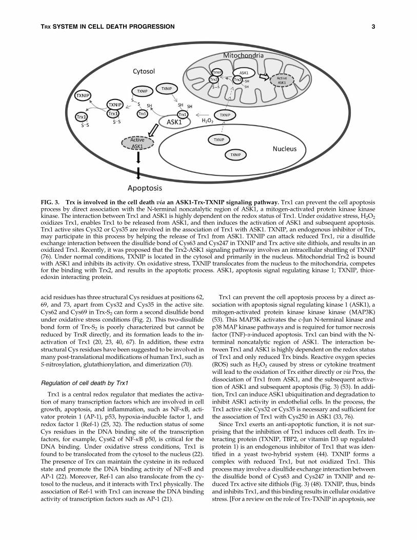
Trx1 can prevent the cell apoptosis process by a direct as-sociation with apoptosis signal regulating kinase 1 (ASK1), amitogen-activated protein kinase kinase kinase (MAP3K)(53). This MAP3K activates the c-Jun N-terminal kinase andp38 MAP kinase pathways and is required for tumor necrosisfactor (TNF)-a-induced apoptosis. Trx1 can bind with the N-terminal noncatalytic region of ASK1. The interaction be-tween Trx1 and ASK1 is highly dependent on the redox statusof Trx1 and only reduced Trx binds. Reactive oxygen species(ROS) such as H2O2 caused by stress or cytokine treatmentwill lead to the oxidation of Trx either directly or via Prxs, thedissociation of Trx1 from ASK1, and the subsequent activa-tion of ASK1 and subsequent apoptosis (Fig. 3) (53). In addi-tion, Trx1 can induce ASK1 ubiquitination and degradation toinhibit ASK1 activity in endothelial cells. In the process, theTrx1 active site Cys32 or Cys35 is necessary and sufficient forthe association of Trx1 with Cys250 in ASK1 (33, 76).
Since Trx1 exerts an anti-apoptotic function, it is not sur-prising that the inhibition of Trx1 induces cell death. Trx in-teracting protein (TXNIP, TBP2, or vitamin D3 up regulatedprotein 1) is an endogenous inhibitor of Trx1 that was iden-tified in a yeast two-hybrid system (44). TXNIP forms acomplex with reduced Trx1, but not oxidized Trx1. Thisprocess may involve a disulfide exchange interaction betweenthe disulfide bond of Cys63 and Cys247 in TXNIP and re-duced Trx active site dithiols (Fig. 3) (48). TXNIP, thus, bindsand inhibits Trx1, and this binding results in cellular oxidativestress. [For a review on the role of Trx-TXNIP in apoptosis, see
FIG. 3. Trx is involved in the cell death via an ASK1-Trx-TXNIP signaling pathway. Trx1 can prevent the cell apoptosisprocess by direct association with the N-terminal noncatalytic region of ASK1, a mitogen-activated protein kinase kinasekinase. The interaction between Trx1 and ASK1 is highly dependent on the redox status of Trx1. Under oxidative stress, H2O2
oxidizes Trx1, enables Trx1 to be released from ASK1, and then induces the activation of ASK1 and subsequent apoptosis.Trx1 active sites Cys32 or Cys35 are involved in the association of Trx1 with ASK1. TXNIP, an endogenous inhibitor of Trx,may participate in this process by helping the release of Trx1 from ASK1. TXNIP can attack reduced Trx1, via a disulfideexchange interaction between the disulfide bond of Cys63 and Cys247 in TXNIP and Trx active site dithiols, and results in anoxidized Trx1. Recently, it was proposed that the Trx2-ASK1 signaling pathway involves an intracellular shuttling of TXNIP(76). Under normal conditions, TXNIP is located in the cytosol and primarily in the nucleus. Mitochondrial Trx2 is boundwith ASK1 and inhibits its activity. On oxidative stress, TXNIP translocates from the nucleus to the mitochondria, competesfor the binding with Trx2, and results in the apoptotic process. ASK1, apoptosis signal regulating kinase 1; TXNIP, thior-edoxin interacting protein.
TRX SYSTEM IN CELL DEATH PROGRESSION 3
the reference (72)]. Indeed, the overexpression of TXNIP madefibroblasts, cardiomyocytes, and pancreatic b-cells more sus-ceptible to apoptosis. Glucose-caused b-cell death has beenshown to be due to the stimulation of the overexpression ofTXNIP by glucose (10). However, it should be pointed out thatTXNIP is a member of the a-arrestin protein superfamily, andit does not only act as the inhibitor of Trx, but also emerges asa metabolic regulatory protein (74). TXNIP plays a dual role inapoptosis in redox-dependent (29, 51) and -independentregulatory (10, 12, 73) manners, which is reviewed by Spindelet al. (58). More recently, it is reported that the binding of Trxwith TXNIP can prevent the TXNIP degradation and adipo-cyte differentiation (14, 75).
In agreement with an unknown role of Trx1 in embryo-genesis, probably involving oxidative stress defense andtranscription regulation, homozygotes with a targeted dis-ruption of the mouse TXN gene died shortly after implanta-tion, indicating that Trx1 is essential for early differentiationand morphogenesis of the mouse embryo (41).
Regulation of cell death by Trx2
Mitochondria are considered a major site for ROS pro-duction. ROS level is a determining factor for cell death. Thelow level of ROS promotes the cells into apoptosis, while thehigh level of ROS causes cell necrosis. Along with Prx3, theTrx2 system is a major player that controls ROS levels in mi-tochondria and, therefore, plays a key role in regulating cellapoptosis. The deficiency of Trx2 resulted in the elevation ofcellular ROS level, cytochrome c release from mitochondria,and the activation of caspase 3 and 9 in chicken DT40 (61).Very interestingly, the transfection of hTrx2 or redox-inactivehTrx2CS can rescue the Trx2-deficeint DT40 from cell death.This may be due to the fact that Trx2 maintains the Bcl-xLprotein level and controls the mitochondrial outer membranepermeabilization in a redox-active, site-independent mecha-nism (64). Trx2 can also bind and inhibit mitochondrial-located ASK1 apoptotic activity. Cys30 in the N-terminaldomain of TXNIP is critical for the binding with Trx2 (76).Recently, it has been proposed that the Trx2-ASK1 signalingpathway also involves an intracellular shuttling of TXNIP.Under normal conditions, TXNIP is located in the cytosol andalso primarily in the nucleus. Mitochondrial Trx2 is boundwith ASK1 and inhibits its protein kinase activity. On oxida-tive stress, TXNIP translocates from the nucleus to the mito-chondria, competes for the binding to Trx2, and results in theapoptotic process (Fig. 3) (55). Another possible mechanism ofTrx2 protection against cell death is via the regulation ofp66Shc, a lifespan regulator (42). p66Shc has been revealed to bea disulfide substrate of Trxs (18). Thus, the p66Shc tetramerthat is able to cause cell death can be reduced by the mito-chondrial Trx system into the inactive form p66Shc dimer (18).
Mitochondrial Trx2 is essential for the normal developmentof the mouse embryo. Homozymous mutants embryos withthe silencing TXN2 gene showed massive increased apoptosisat 10.5 days postcoitus and died in the Theiler stage 15/16(45). The timing of the embryonic lethality coincides with thematuration of the mitochondria. The embryonic fibroblastsfrom Trx2-null embryos were not viable. The heterozygousmice are fertile, but theTrx2 decrease mice showed increasedoxidative damage to nuclear DNA, lipid, and proteins in theliver (49).
The role of Trx2 in the regulation of apoptosis has beendemonstrated by the protection of Trx2 against oxidant-induced cell death. The liver from the Trx2 + / - mice has anincreased apoptosis compared with wild type (WT) mice ondiquat treatment (49). WT Trx2 overexpression, but not aC93S Trx2 mutant protein, can significantly inhibit TNF-a-induced apoptosis in HeLa cells (19). The redox statue of Trx2is a key factor for many oxidant-induced cell death processes,and Trx2 get oxidized after the treatment of peroxides anddiamide (11). Most recently, we found that some cationic tri-phenylmethanes such as brilliant green and gentian violet, thelong time-used antifungal and antibacterial agents, accumu-late in the mitochondria and cause the oxidation and degra-dation of Trx2. The disruption of the mitochondrial Trxsystem resulted in a subsequent release of cytochrome c andapoptosis inducing factor (AIF) from mitochondria into thecytosol. Very interestingly, HeLa cells were more sensitive tobrilliant green than to fibroblasts. In HeLa cells, Trx2 down-regulation by small interfering RNA (siRNA) resulted in anincreased sensitivity, whereas for normal fibroblasts, the Trx2or Trx1 down-regulation had no effects (77).
Besides Trx1 and Trx2, the Trx family contains many otherthiol-disulfide oxidoreductases with a CXXC active site suchas Grxs, an endoplasmic reticulum-localized transmembraneTrx-related protein, and a macrophage migration inhibitoryfactor. These proteins have also been indicated as playingcritical roles in the regulation of apoptosis, which have beensummarized in reviews (32, 62, 72).
Role of TrxRs in Mammalian Cell Death Progression
Structure and reaction mechanism of mammalian TrxR
Three TrxRs, cytosolic TrxR1, mitochondrial TrxR2, andtestis-specific Trx glutathione reductase (GR) have beenfound in mammalian cells, corresponding to the localizationof three types of Trxs (31, 40, 59). All the three mammalianTrxRs are selenoenzymes, with a selenocysteine (Sec, U)residue in their C-terminus (4, 37). Sec is known to be the 21stgenetically translated amino acid that is encoded by a UGAcodon, which, in most circumstances, acts as stop codon. TheSec incorporation into the selenoprotein polypeptide chainrequires a complex machinery containing the Sec insertionsequence (SECIS) element, tRNA[Ser]Sec, SECIS binding pro-tein 2 (SBP2), and other components in mammalian cells andthe availability of selenium (46). The Sec incorporation effi-ciency affects the activity of selenoprotein TrxR (56). Thedeficiency of Sec synthesis machinery can also change theexisting form of TrxR (39). For example, selenium deficiencycauses the Sec residue to be replaced by Cys in the rat liverTrxR (39). On the other hand, the Trx system can reducedisulfide bonds and/or glutathione (GSH) mixed disulfidesin oxidized SBP2, and participate in the regulation ofSBP2 compartmental location and selenoprotein synthesisefficiency (47).
TrxRs from mammalian cells possess different properties ascompared with the TrxR from E. coli, yeast, or plants (Fig. 2)(69). The TrxR from mammalian cells and higher eukaryotesare large homodimmeric flavoproteins with 55 kDa or largersubunits, instead of the bacterial TrxRs, which are smaller di-meric flavoproteins with 33 kDa subunits (69). MammalianTrxRs have a very broad range of substrates, including sele-nite, which could be reduced to selenide for selenoprotein
4 LU AND HOLMGREN
synthesis (34, 36). In contrast, TrxRs in prokaryotes, yeast, andplants have a narrow substrate specificity (36).
In contrast to the single active site with a CXXC motif in thedimeric bacterial TrxR, mammalian TrxRs are a head to taildimer and have an N-terminal active site disulfide CVNVGCas well as a conserved C-terminal sequence Gly-Cys-Sec-Gly(78). The structures of the mammalian TrxRs have been solvedby X-ray crystallography (7, 13, 17, 54). The overall structureof TrxR is similar to that of GR with an identical N-terminalactive site, NADPH and flavin adenine dinucleotide (FAD)binding domains (Fig. 2). However, mammalian TrxR1 un-iquely contains a 16-residue C-terminal tail. Mechanisms ofthe action of mammalian TrxR have been proposed thatelectrons from NADPH are transferred to the N-terminal re-dox-active dithiols via FAD, then to the selenenylsulfide of theother subunit, and, subsequently, to the substrates (Fig. 4) (13,78, 79). Crystal structures of oxidized and NADPH-reduced mouse TrxR2 have shown a similar overall structureas that of rat TrxR1 (7).
Role of TrxRs in mammalian cell death progression
To elucidate the roles of TrxR in vivo, several TrxR knock-out models have been studied (8, 15, 27). Mice with a ubiq-uitous inactivation of TrxR2 were dead at embryonic day 13(15). TrxR2-deficient embryologic fibroblasts grew slowerthan their WT counterparts and were highly sensitive to bu-thionine sulfoximine treatment to block GSH synthesis (15).The inactivation of TrxR1 also led to early embryonic lethalityaround E9.5 (8) or E10.5 (27). Surprisingly, mice with a heart-specific inactivation of TrxR1 developed normally and ap-peared healthy (27), and the mice with TrxR-null hepatocyte
also survived for more than 1 year (52, 60). These results in-dicate that TrxR1 and TrxR2 are critical in embryo develop-ment, but they are not essential for cell proliferations. Thenonessential role of TrxR1 in cell proliferation is consistentwith the observation that the down-regulation of TrxR1 bysiRNA did not change Trx1 redox status and caused celltoxicity (66, 77). The mechanistic details are unknown as yet.
One reason that TrxR is not essential for cell proliferationmay be due to the compensatory effects of another disulfidereductase system, GSH and Grx system, and this cellular re-sponse process may be regulated by the Nrf2 pathway (60).The decrease in TrxR1 activity resulting in Nrf2 activation wasalso observed under other conditions such as selenium defi-ciency (Fig. 5) (9, 39). Grxs are Trx-fold proteins with a CPYCor CGFS active site motif. In the GSH-Grx system, electronstransfer from NADPH to GR, then to GSH, and, subsequently,to Grx. The functions of Trx and GSH-Grx systems are par-tially overlapping (35). In particular, when TrxR is absent, Trxmay get the electrons from the GSH-Grx system to maintainits reduced status (Y. Du, J. Lu, A. Holmgren, unpublishedresults).
Regulation of Activity in Trx Systemand Its Medical Applications
Mammalian TrxR as an anticancer target
Though TrxR1 is not essential for cell proliferation, TrxRemerges as a new target for anticancer drug development,because many malignant cells appear to be more dependenton an efficient Trx system. TrxR and Trx are overexpressed inaggressive cancers (5). The correlation between the Trx system
FIG. 4. Inhibition of TrxR can convert TrxR to be a source for ROS production. As a unique Sec-containing flavoprotein,TrxR is a very suitable target for drug development. The C-terminal active site Sec makes the enzyme highly reactive withelectronphilic agents due to its low pKa value and easily accessible location. The inhibition of TrxR does not only decrease theactivity of TrxR but also makes TrxR become an ROS production site and results in the oxidation of Trx. ROS, reactive oxygenspecies.
TRX SYSTEM IN CELL DEATH PROGRESSION 5
and cancer hallmarks has been reviewed (5). Moreover,mouse lung carcinoma cells with TrxR stable-knockdown ex-hibited a similar cell morphology and anchorage-independentgrowth property as normal cells (71). The cells transfected withthe siTrxR1 construct grew in a monolayer, whereas the con-trol cancer cells grew in a multilayer and loosely attached tothe culture dish. Growth of the TrxR1-knockdown cells in softagar was inhibited compared with control cells (71). Growingunanchored in soft agar is a characteristic of many malignantcells (71). When these TrxR-knockdown cells were injected intomice, the tumor progression and metastasis were dramaticallyreduced (71). The requirement of protein disulfide reductasefor tumor growth was originally reported by Apffel et al. (2),even before the identification of the enzymes to be Trx andTrxR by Holmgren (23). These results strongly suggest thatTrxR is critical for cancer cell growth in vivo. In addition, amain potential substrate of the Trx system, RNR, is also a well-known anticancer target. The blockage of the Trx system ac-tivity will decrease RNR activity (43). More importantly, theinhibition of TrxR does not only decrease the activity of TrxRbut also results in the elevation of ROS production and theoxidation of Trx (Fig. 5).
As a unique Sec-containing flavoprotein, TrxR is verysuitable as a target for drug development. The C-terminalactive site Sec makes the enzyme highly reactive with elec-trophilic agents due to its low pKa value and easily accessiblelocation. Indeed, many TrxR inhibitors selectively exhibit theinhibition of TrxR against GR (3). This is also verified in vivo(65). Moreover, the inhibition of TrxR may result in a modifiedTrxR, which induces rapid cell death (1). Indeed, many clin-ically used anticancer compounds (3, 63), including alkylatingand platinum-containing drugs, arsenic trioxide (35), and
chemoprevention agents such as flavonoids (38) and curcu-min (16), have been found to possess TrxR inhibitory effects.However, since TrxR is involved in a wide range of cell ac-tivities, the inhibition of TrxR may also cause some side effectsin normal cells. Thus, further investigation that elucidates thedifferent roles of TrxR along with Trx in normal and cancercells and the reaction mechanism of TrxR inhibitors may fa-cilitate and enlarge the therapeutic windows and selectivity intreatment.
Principles for some TrxR inhibition reactions
We have classified TrxR inhibitors as four types (36) (Fig. 6).The first type is metal- or metalloid-containing compounds,including gold, platinum-, mercury-, arsenic-containingcompounds, some other organometallic complexes, and so on.‘‘Hard and soft acids and bases’’ theory provides a usefulprinciple for understanding the reaction mechanism betweenthese compounds with TrxR (28). Since Se2 - is a softer basethan S2 - , it reacts preferentially with soft bases such asgold(I), platinum(II), mercury(II), and arsenic(III). This prin-ciple may also explain why Sec-containing TrxR shows apriority to react with arsenic compounds than Cys-containingproteins. Though the metal- or metalloid-containing com-pounds have a high priority to attack the C-terminal Sec, theN-terminal CVNVGC active site in TrxR is also involved inthe reaction (35). Zinc or lead compounds only show theirinhibitory effects in the absence of EDTA in vitro. In cells orin vivo, they do not display the selective inhibition of TrxR asarsenic and mercury compounds do. The reason may be thatarsenic and mercury compounds are softer acids and, thus,have a higher affinity with TrxR. For this type of inhibitors,
FIG. 5. Compensation ofGSH-Grx system under theconditions of the deficiencyof TrxR1. The activity ofTrxR1 can be decreased undervarious conditions such as theinhibition by inhibitors, sele-nium deficiency, Sec synthesismachinery such as tRNA[Ser]Sec,SECIS binding protein 2 mu-tations, and siRNA treat-ments. The decrease in TrxR1activity always results in theNrf2 pathway activation andcompensatory effects from theGSH-Grx system. Grx, glu-taredoxin; GSH, glutathione;SECIS, Sec insertion se-quence; siRNA, small inter-fering RNA.
6 LU AND HOLMGREN
one interesting observation is that some inhibitor has the ac-tivity to cross-link TrxR with Trx or other small proteins (50).
The second type of TrxR inhibitors are Michael acceptors (a,b-unsaturated carbonyl compounds), including flavonoids,quinols, quinines, and so on. The Michael acceptor curcuminhas an a, b-unsaturated ketone structure that is equilibratedwith its enol form. The latter is a highly active electrophilicagent that can have a Michael conjugation with the selenide ofSec in TrxR (16). The inhibition of TrxR by the flavonoidsmyricetin, quercetin, quinols, and quinones may involve adirect reaction between the TrxR and semiquinone (38). Thismay also indicate that many quinones act as ‘‘suicide inhibi-tors.’’ They can be reduced by TrxR, and, at the same time, thereduced product semiquinones inhibit TrxR.
The third type is sulfur, selenium, or telluride-containingcompounds. The inhibition of TrxR by 1,2-[bis(1,2-benziso-selenazolone-3(2H)-ketone)]ethane (BBSKE) is reversible,which is different from most of the other inhibitors. The fourthtype is alkylation agents. Dinitrochlorobenzene is an aromaticalkylation agent. This compound can have a covalent bonding
with C-terminal Sec and also converts the antioxidant enzymeinto a pro-oxidant (Fig. 4).
Concluding Remarks
In summary, the mammalian Trx system is obviously a keyplayer in cell death for a large number of cellular activitiesthey are involved in. The redox status of Trx is a determiningfactor for the cell death, which is controlled by TrxR activityand the cellular ROS level. Mammalian TrxR is a suitableanticancer target due to its unique structural properties.Various types of compounds can exert inhibitory effects onTrxR. The challenges are how to design and synthesize thecompounds to possess higher selectivity related to the inhib-itory efficiency and ROS production capacity via TrxR.
Acknowledgments
The authors acknowledge the support from the SwedishResearch Council Medicine (3529), the Swedish Cancer
FIG. 6. Classification of TrxR inhibitors and general principles for the inhibition reaction. Numerous compounds exhibitinhibitory effects on mammalian TrxR. We have classified TrxR inhibitors as four types and list some general principles tounderstand the inhibition mechanism in the right panel. ‘‘Hard and soft acids and bases’’ theory is a good principle thatexplains the reaction between type I metal- or metalloid- containing compounds and TrxR, because R-Se - is a soft base andreacts preferentially with soft bases, including gold, platinum-, mercury-, and arsenic-containing compounds. The reaction oftype II Michael acceptors with TrxR is closely linked to ROS production. The inhibition of TrxR with many flavonoidsmyricetin, quercetin, quinols, and quinones may involve a direct reaction between the TrxR and semiquinone. Some quinonesmay act as ‘‘suicide inhibitors.’’ They can be reduced by TrxR, and, at the same time, the reduced product semiquinonesinhibit TrxR. Type III inhibitors are sulfur, selenium, or telluride-containing compounds. BBSKE, a compound structurallysimilar to the bridge combination of two ebselen compounds, is a reversible inhibitor on TrxR. The type IV compound DNCBis an aromatic alkylation agent. This compound can form a covalent bond with C-terminal Sec and convert the antioxidantenzyme into a pro-oxidant. BBSKE, 1,2-[bis(1,2-benzisoselenazolone-3(2H)-ketone)]ethane; DNCB, dinitrochlorobenzene.
TRX SYSTEM IN CELL DEATH PROGRESSION 7
Society (961), the K.A. Wallenberg Foundation, Ake WibergStiftelse, and the Karolinska Institutet.
References
1. Anestal K and Arner ES. Rapid induction of cell death byselenium-compromised thioredoxin reductase 1 but not bythe fully active enzyme containing selenocysteine. J BiolChem 278: 15966–15972, 2003.
2. Apffel CA, Walker JE, and Nathanson IT. Tumor growthand disulfide reduction: possible dependence on protein-disulfide reductase. J Natl Cancer Inst 51: 575–583, 1973.
3. Arner ES. Focus on mammalian thioredoxin reductases—important selenoproteins with versatile functions. BiochimBiophys Acta 1790: 495–526, 2009.
4. Arner ES and Holmgren A. Physiological functions ofthioredoxin and thioredoxin reductase. Eur J Biochem 267:6102–6109, 2000.
5. Arner ES and Holmgren A. The thioredoxin system in can-cer. Semin Cancer Biol 16: 420–426, 2006.
6. Avval FZ and Holmgren A. Molecular mechanisms ofthioredoxin and glutaredoxin as hydrogen donors forMammalian s phase ribonucleotide reductase. J Biol Chem284: 8233–8240, 2009.
7. Biterova EI, Turanov AA, Gladyshev VN, and Barycki JJ.Crystal structures of oxidized and reduced mitochondrialthioredoxin reductase provide molecular details of the re-action mechanism. Proc Natl Acad Sci U S A 102: 15018–15023, 2005.
8. Bondareva AA, Capecchi MR, Iverson SV, Li Y, Lopez NI,Lucas O, Merrill GF, Prigge JR, Siders AM, Wakamiya M,Wallin SL, and Schmidt EE. Effects of thioredoxin reductase-1deletion on embryogenesis and transcriptome. Free Radic BiolMed 43: 911–923, 2007.
9. Burk RF, Hill KE, Nakayama A, Mostert V, Levander XA,Motley AK, Johnson DA, Johnson JA, Freeman ML, andAustin LM. Selenium deficiency activates mouse liver Nrf2-ARE but vitamin E deficiency does not. Free Radic Biol Med44: 1617–1623, 2008.
10. Chen J, Hui ST, Couto FM, Mungrue IN, Davis DB, AttieAD, Lusis AJ, Davis RA, and Shalev A. Thioredoxin-interacting protein deficiency induces Akt/Bcl-xL signalingand pancreatic beta-cell mass and protects against diabetes.FASEB J 22: 3581–3594, 2008.
11. Chen Y, Cai J, and Jones DP. Mitochondrial thioredoxin inregulation of oxidant-induced cell death. FEBS Lett 580:6596–6602, 2006.
12. Chen Z, Lopez-Ramos DA, Yoshihara E, Maeda Y, MasutaniH, Sugie K, Maeda M, and Yodoi J. Thioredoxin-bindingprotein-2 (TBP-2/VDUP1/TXNIP) regulates T-cell sensitiv-ity to glucocorticoid during HTLV-I-induced transforma-tion. Leukemia 25: 440–448, 2011.
13. Cheng Q, Sandalova T, Lindqvist Y, and Arner ES. Crystalstructure and catalysis of the selenoprotein thioredoxin re-ductase 1. J Biol Chem 284: 3998–4008, 2009.
14. Chutkow WA and Lee RT. Thioredoxin regulates adipo-genesis through thioredoxin-interacting protein (Txnip)protein stability. J Biol Chem 286: 29139–29145, 2011.
15. Conrad M, Jakupoglu C, Moreno SG, Lippl S, Banjac A,Schneider M, Beck H, Hatzopoulos AK, Just U, Sinowatz F,Schmahl W, Chien KR, Wurst W, Bornkamm GW, andBrielmeier M. Essential role for mitochondrial thioredoxinreductase in hematopoiesis, heart development, and heartfunction. Mol Cell Biol 24: 9414–9423, 2004.
16. Fang J, Lu J, and Holmgren A. Thioredoxin reductase isirreversibly modified by curcumin: A novel molecularmechanism for its anticancer activity J Biol Chem 280: 25284–25290, 2005.
17. Fritz-Wolf K, Kehr S, Stumpf M, Rahlfs S, and Becker K.Crystal structure of the human thioredoxin reductase-thior-edoxin complex. Nat Commun 2: 383, 2011.
18. Gertz M, Fischer F, Wolters D, and Steegborn C. Activationof the lifespan regulator p66Shc through reversible disulfidebond formation. Proc Natl Acad Sci U S A 105: 5705–5709,2008.
19. Hansen JM, Zhang H, and Jones DP. Mitochondrialthioredoxin-2 has a key role in determining tumor necrosisfactor-alpha-induced reactive oxygen species generation, NF-kappaB activation, and apoptosis. Toxicol Sci 91: 643–650, 2006.
20. Hashemy SI and Holmgren A. Regulation of the catalyticactivity and structure of human thioredoxin 1 via oxidationand S-nitrosylation of cysteine residues. J Biol Chem 283:21890–21898, 2008.
21. Hirota K, Matsui M, Iwata S, Nishiyama A, Mori K, andYodoi J. AP-1 transcriptional activity is regulated by a directassociation between thioredoxin and Ref-1. Proc Natl AcadSci U S A 94: 3633–3638, 1997.
22. Hirota K, Murata M, Sachi Y, Nakamura H, Takeuchi J, MoriK, and Yodoi J. Distinct roles of thioredoxin in the cytoplasmand in the nucleus. A two-step mechanism of redox regu-lation of transcription factor NF-kappaB. J Biol Chem 274:27891–27897, 1999.
23. Holmgren A. Bovine thioredoxin system. Purification ofthioredoxin reductase from calf liver and thymus andstudies of its function in disulfide reduction. J Biol Chem 252:4600–4606, 1977.
24. Holmgren A. Thioredoxin. Annu Rev Biochem 54: 237–271,1985.
25. Holmgren A and Lu J. Thioredoxin and thioredoxin reduc-tase: current research with special reference to human dis-ease. Biochem Biophys Res Commun 396: 120–124, 2010.
26. Holmgren A and Sengupta R. The use of thiols by ribonu-cleotide reductase. Free Radic Biol Med 49: 1617–1628, 2010.
27. Jakupoglu C, Przemeck GK, Schneider M, Moreno SG, MayrN, Hatzopoulos AK, de Angelis MH, Wurst W, BornkammGW, Brielmeier M, and Conrad M. Cytoplasmic thioredoxinreductase is essential for embryogenesis but dispensable forcardiac development. Mol Cell Biol 25: 1980–1988, 2005.
28. Jungwirth U, Kowol CR, Keppler BK, Hartinger CG, BergerW, and Heffeter P. Anticancer activity of metal complexes:involvement of redox processes. Antioxid Redox Signal 15:1085–1127, 2011.
29. Junn E, Han SH, Im JY, Yang Y, Cho EW, Um HD, Kim DK,Lee KW, Han PL, Rhee SG, and Choi I. Vitamin D3 up-regulated protein 1 mediates oxidative stress via suppres-sing the thioredoxin function. J Immunol 164: 6287–6295,2000.
30. Kang SW, Rhee SG, Chang TS, Jeong W, and Choi MH. 2-Cys peroxiredoxin function in intracellular signal transduc-tion: therapeutic implications. Trends Mol Med 11: 571–578,2005.
31. Lee SR, Kim JR, Kwon KS, Yoon HW, Levine RL, GinsburgA, and Rhee SG. Molecular cloning and characterization of amitochondrial selenocysteine-containing thioredoxin reduc-tase from rat liver. J Biol Chem 274: 4722–4734, 1999.
32. Lillig CH and Holmgren A. Thioredoxin and relatedmolecules-from biology to health and disease. Antioxid RedoxSignal 9: 25–47, 2007.
8 LU AND HOLMGREN
33. Liu Y and Min W. Thioredoxin promotes ASK1 ubiquitina-tion and degradation to inhibit ASK1-mediated apoptosis ina redox activity-independent manner. Circ Res 90: 1259–1266, 2002.
34. Lu J, Berndt C, and Holmgren A. Metabolism of seleniumcompounds catalyzed by the mammalian selenoproteinthioredoxin reductase. Biochim Biophys Acta 1790: 1513–1519,2009.
35. Lu J, Chew EH, and Holmgren A. Targeting thioredoxinreductase is a basis for cancer therapy by arsenic trioxide.Proc Natl Acad Sci U S A 104: 12288–12293, 2007.
36. Lu J and Holmgren A. Selenoprotein and the thioredoxinsystem. In: Selenium: Its Molecular Biology and Role in HumanHealth, edited by Hatfield DL, Berry MJ, and Gladyshev VN.New York: Springer Verlag, 2011, pp. 153–165.
37. Lu J and Holmgren A. Selenoproteins. J Biol Chem 284: 723–727, 2009.
38. Lu J, Papp LV, Fang J, Rodriguez-Nieto S, Zhivotovsky B,and Holmgren A. Inhibition of Mammalian thioredoxin re-ductase by some flavonoids: implications for myricetin andquercetin anticancer activity. Cancer Res 66: 4410–4418, 2006.
39. Lu J, Zhong L, Lonn ME, Burk RF, Hill KE, and Holmgren A.Penultimate selenocysteine residue replaced by cysteine inthioredoxin reductase from selenium-deficient rat liver.FASEB J 23: 2394–2402, 2009.
40. Luthman M and Holmgren A. Rat liver thioredoxin andthioredoxin reductase: purification and characterization.Biochemistry 21: 6628–6633, 1982.
41. Matsui M, Oshima M, Oshima H, Takaku K, Maruyama T,Yodoi J, and Taketo MM. Early embryonic lethality causedby targeted disruption of the mouse thioredoxin gene. DevBiol 178: 179–185, 1996.
42. Migliaccio E, Giorgio M, Mele S, Pelicci G, Reboldi P, Pan-dolfi PP, Lanfrancone L, and Pelicci PG. The p66shc adaptorprotein controls oxidative stress response and life span inmammals. Nature 402: 309–313, 1999.
43. Muniyappa H, Song S, Mathews CK, and Das KC. Reactiveoxygen species-independent oxidation of thioredoxin inhypoxia: inactivation of ribonucleotide reductase and redox-mediated checkpoint control. J Biol Chem 284: 17069–17081,2009.
44. Nishiyama A, Matsui M, Iwata S, Hirota K, Masutani H,Nakamura H, Takagi Y, Sono H, Gon Y, and Yodoi J.Identification of thioredoxin-binding protein-2/vitamin D(3)up-regulated protein 1 as a negative regulator of thioredoxinfunction and expression. J Biol Chem 274: 21645–21650, 1999.
45. Nonn L, Berggren M, and Powis G. Increased expression ofmitochondrial peroxiredoxin-3 (thioredoxin peroxidase-2)protects cancer cells against hypoxia and drug-induced hy-drogen peroxide-dependent apoptosis. Mol Cancer Res 1:682–689, 2003.
46. Papp LV, Lu J, Holmgren A, and Khanna KK. From sele-nium to selenoproteins: synthesis, identity, and their role inhuman health. Antioxid Redox Signal 9: 775–806, 2007.
47. Papp LV, Lu J, Striebel F, Kennedy D, Holmgren A, andKhanna KK. The redox state of SECIS binding protein 2controls its localization and selenocysteine incorporationfunction. Mol Cell Biol 26: 4895–4910, 2006.
48. Patwari P, Higgins LJ, Chutkow WA, Yoshioka J, and LeeRT. The interaction of thioredoxin with Txnip. Evidence forformation of a mixed disulfide by disulfide exchange. J BiolChem 281: 21884–21891, 2006.
49. Perez VI, Lew CM, Cortez LA, Webb CR, Rodriguez M,Liu Y, Qi W, Li Y, Chaudhuri A, Van Remmen H, Ri-
chardson A, and Ikeno Y. Thioredoxin 2 haploinsuffi-ciency in mice results in impaired mitochondrial functionand increased oxidative stress. Free Radic Biol Med 44: 882–892, 2008.
50. Prast-Nielsen S, Cebula M, Pader I, and Arner ES. Noblemetal targeting of thioredoxin reductase—covalent com-plexes with thioredoxin and thioredoxin-related protein of14 kDa triggered by cisplatin. Free Radic Biol Med 49: 1765–1778, 2010.
51. Rani S, Mehta JP, Barron N, Doolan P, Jeppesen PB, ClynesM, and O’Driscoll L. Decreasing Txnip mRNA and proteinlevels in pancreatic MIN6 cells reduces reactive oxygenspecies and restores glucose regulated insulin secretion. CellPhysiol Biochem 25: 667–674, 2010.
52. Rollins MF, van der Heide DM, Weisend CM, Kundert JA,Comstock KM, Suvorova ES, Capecchi MR, Merrill GF, andSchmidt EE. Hepatocytes lacking thioredoxin reductase 1have normal replicative potential during development andregeneration. J Cell Sci 123: 2402–2412, 2010.
53. Saitoh M, Nishitoh H, Fujii M, Takeda K, Tobiume K,Sawada Y, Kawabata M, Miyazono K, and Ichijo H.Mammalian thioredoxin is a direct inhibitor of apoptosissignal-regulating kinase (ASK) 1. EMBO J 17: 2596–2606,1998.
54. Sandalova T, Zhong L, Lindqvist Y, Holmgren A, andSchneider G. Three-dimensional structure of a mammalianthioredoxin reductase: implications for mechanism andevolution of a selenocysteine-dependent enzyme. Proc NatlAcad Sci U S A 98: 9533–9538, 2001.
55. Saxena G, Chen J, and Shalev A. Intracellular shuttling andmitochondrial function of thioredoxin-interacting protein. JBiol Chem 285: 3997–4005, 2010.
56. Schoenmakers E, Agostini M, Mitchell C, Schoenmakers N,Papp L, Rajanayagam O, Padidela R, Ceron-Gutierrez L,Doffinger R, Prevosto C, Luan J, Montano S, Lu J, CastanetM, Clemons N, Groeneveld M, Castets P, Karbaschi M,Aitken S, Dixon A, Williams J, Campi I, Blount M, Burton H,Muntoni F, O’Donovan D, Dean A, Warren A, Brierley C,Baguley D, Guicheney P, Fitzgerald R, Coles A, Gaston H,Todd P, Holmgren A, Khanna KK, Cooke M, Semple R,Halsall D, Wareham N, Schwabe J, Grasso L, Beck-Peccoz P,Ogunko A, Dattani M, Gurnell M, and Chatterjee K. Muta-tions in the selenocysteine insertion sequence-binding pro-tein 2 gene lead to a multisystem selenoprotein deficiencydisorder in humans. J Clin Invest 120: 4220–4235, 2010.
57. Sengupta R and Holmgren A. The role of thioredoxin in theregulation of cellular processes by S-nitrosylation. BiochimBiophys Acta 1820: 689–700, 2012.
58. Spindel ON, World C, and Berk BC. Thioredoxin interactingprotein: redox dependent and independent regulatorymechanisms. Antioxid Redox Signal 16: 587–596, 2012.
59. Sun QA, Wu Y, Zappacosta F, Jeang KT, Lee BJ, Hatfield DL,and Gladyshev VN. Redox regulation of cell signaling byselenocysteine in mammalian thioredoxin reductases. J BiolChem 274: 24522–24530, 1999.
60. Suvorova ES, Lucas O, Weisend CM, Rollins MF, Merrill GF,Capecchi MR, and Schmidt EE. Cytoprotective Nrf2 path-way is induced in chronically txnrd 1-deficient hepatocytes.PLoS One 4: e6158, 2009.
61. Tanaka T, Hosoi F, Yamaguchi-Iwai Y, Nakamura H, Ma-sutani H, Ueda S, Nishiyama A, Takeda S, Wada H, SpyrouG, and Yodoi J. Thioredoxin-2 (TRX-2) is an essential generegulating mitochondria-dependent apoptosis. EMBO J 21:1695–1703, 2002.
TRX SYSTEM IN CELL DEATH PROGRESSION 9
62. Thiele M and Bernhagen J. Link between macrophage mi-gration inhibitory factor and cellular redox regulation. An-tioxid Redox Signal 7: 1234–1248, 2005.
63. Urig S and Becker K. On the potential of thioredoxin re-ductase inhibitors for cancer therapy. Semin Cancer Biol 16:452–465, 2006.
64. Wang D, Masutani H, Oka S, Tanaka T, Yamaguchi-Iwai Y,Nakamura H, and Yodoi J. Control of mitochondrial outermembrane permeabilization and Bcl-xL levels by thior-edoxin 2 in DT40 cells. J Biol Chem 281: 7384–7391, 2006.
65. Wang X, Zhang J, and Xu T. Cyclophosphamide as a potentinhibitor of tumor thioredoxin reductase in vivo. Toxicol ApplPharmacol 218: 88–95, 2007.
66. Watson WH, Heilman JM, Hughes LL, and Spielberger JC.Thioredoxin reductase-1 knock down does not result inthioredoxin-1 oxidation. Biochem Biophys Res Commun 368:832–836, 2008.
67. Watson WH, Pohl J, Montfort WR, Stuchlik O, Reed MS,Powis G, and Jones DP. Redox potential of human thior-edoxin 1 and identification of a second dithiol/disulfidemotif. J Biol Chem 278: 33408–33415, 2003.
68. Weissbach H, Etienne F, Hoshi T, Heinemann SH, LowtherWT, Matthews B, St John G, Nathan C, and Brot N. Peptidemethionine sulfoxide reductase: structure, mechanism ofaction, and biological function. Arch Biochem Biophys 397:172–178, 2002.
69. Williams CH, Arscott LD, Muller S, Lennon BW, LudwigML, Wang PF, Veine DM, Becker K, and Schirmer RH.Thioredoxin reductase two modes of catalysis have evolved.Eur J Biochem 267: 6110–6117, 2000.
70. Wu C, Parrott AM, Fu C, Liu T, Marino SM, Gladyshev VN,Jain MR, Baykal AT, Li Q, Oka S, Sadoshima J, Beuve A,Simmons WJ, and Li H. Thioredoxin 1-mediated post-translational modifications: reduction, transnitrosylation,denitrosylation, and related proteomics methodologies. An-tioxid Redox Signal 15: 2565–2604, 2011.
71. Yoo MH, Xu XM, Carlson BA, Gladyshev VN, and HatfieldDL. Thioredoxin reductase 1 deficiency reverses tumorphenotype and tumorigenicity of lung carcinoma cells. J BiolChem 281: 13005–13008, 2006.
72. Yoshihara E, Chen Z, Matsuo Y, Masutani H, and Yodoi J.Thiol redox transitions by thioredoxin and thioredoxin-binding protein-2 in cell signaling. Methods Enzymol 474: 67–82, 2010.
73. Yoshihara E, Fujimoto S, Inagaki N, Okawa K, Masaki S,Yodoi J, and Masutani H. Disruption of TBP-2 amelioratesinsulin sensitivity and secretion without affecting obesity.Nat Commun 1: 127, 2010.
74. Yoshioka J, Chutkow WA, Lee S, Kim JB, Yan J, Tian R,Lindsey ML, Feener EP, Seidman CE, Seidman JG, and LeeRT. Deletion of thioredoxin-interacting protein in mice impairsmitochondrial function but protects the myocardium fromischemia-reperfusion injury. J Clin Invest 122: 267–279, 2012.
75. Zhang P, Wang C, Gao K, Wang D, Mao J, An J, Xu C, WuD, Yu H, Liu JO, and Yu L. The ubiquitin ligase itch regu-lates apoptosis by targeting thioredoxin-interacting proteinfor ubiquitin-dependent degradation. J Biol Chem 285: 8869–8879, 2010.
76. Zhang R, Al-Lamki R, Bai L, Streb JW, Miano JM, Bradley J,and Min W. Thioredoxin-2 inhibits mitochondria-locatedASK1-mediated apoptosis in a JNK-independent manner.Circ Res 94: 1483–1491, 2004.
77. Zhang X, Zheng Y, Fried LE, Du Y, Montano SJ, Sohn A,Lefkove B, Holmgren L, Arbiser JL, Holmgren A, and Lu J.Disruption of the mitochondrial thioredoxin system as a celldeath mechanism of cationic triphenylmethanes. Free RadicBiol Med 50: 811–820, 2011.
78. Zhong L, Arner ES, and Holmgren A. Structure and mech-anism of mammalian thioredoxin reductase: the active site isa redox-active selenolthiol/selenenylsulfide formed from theconserved cysteine-selenocysteine sequence. Proc Natl AcadSci U S A 97: 5854–5859, 2000.
79. Zhong L and Holmgren A. Essential role of selenium in thecatalytic activities of mammalian thioredoxin reductase re-vealed by characterization of recombinant enzymes withselenocysteine mutations. J Biol Chem 275: 18121–18128,2000.
Address correspondence to:Prof. Arne Holmgren
Division of BiochemistryDepartment of Medical Biochemistry and Biophysics