with tall stature, syndactyly and generalized sclerosis of the skeleton, especially the skull bones
and mandible. Some patients suffer from neurological complications such as cranial nerve
palsies due to the sclerosis of the skull base or cephalgia caused by increased intracranial
pressure. In 2011, we demonstrated that mutations in another Wnt-signaling coreceptor,
namely LRP4, can also cause sclerosteosis [21]. LRP4, has been shown to facilitate sclerostin
inhibitory action and therefore partial loss of function mutations in this gene result in a similar
phenotype as the mutations in SOST. A related, yet milder condition, is Van Buchem Disease
(VBD). Patients suffering from this rare, autosomal recessive disorder display sclerosis of the
skeleton, most prominent in the skull bones, and show progressive enlargement of the
mandible. The disease is caused by a 52kb deletion of a regulatory element localized 35kb
downstream from the SOST gene, resulting in decreased production of sclerostin [22]. Recently,
mutations in SOST gene have also been described as causative for craniodiaphyseal dysplasia, a
severe disorder marked by typical facial distortion termed “leontiasis ossea”. These mutations
have been shown to largely impair sclerostin secretion [23].
Osteopathia striata is another sclerosing bone disorder, however with X-linked dominant mode
of inheritance. This disease usually results in fetal or neonatal death in males, while females
display longitudinal striations in the submetaphyseal regions of long tubular bones, pelvis and
scapula. Clinical findings include cleft palate, hearing loss and macrocephaly. Causative
mutations have been found in the WTX gene encoding a Wnt-signaling inhibitory protein
capable of binding β-catenin [24]. The phenotypic variability amongst affected females is most
likely due to non-random X-inactivation.
Individuals with Craniometaphyseal dysplasia usually show a peculiar face with hypertelorism
and a thick bony wedge over the bridge of the nose and glabella. Narrowing of the nasal
passages may result in mouth breathing. Frequently, signs of cranial nerve impingement are
seen with hearing loss, impaired vision or facial paresis. Mutations in the ANKH gene encoding a
membrane transporter of pyrophosphate have been shown to cause the milder and more
common autosomal dominant form of the disease [25, 26]. Pyrophosphate is believed to inhibit
mineralization of the bone matrix; therefore mutations in the gene might impair the transporter
role of ANKH. Moreover, ANKH has also been shown to stimulate osteoblastic maturation and
differentiation. Recently, mutations in the GJA1 gene, encoding connexin 43, have been
identified in patients suffering from the autosomal recessive form of the disease [27].
Camurati-Engelmann disease is a rare, autosomal dominant condition characterized by
muscular weakness and leg pain in affected individuals. Moreover, cortical thickening of the
long bones and hyperostosis of the skull base is observed. Camurati-Engelmann disease is
caused by activating mutations in TGFβ1. Normally, TGFβ1 is stored in the bone in an inactive
form due to its binding with latency-associated protein (LAP) [28, 29]. Resorbing osteoclast
releases the complex from the bone tissue initiating the migration of mesenchymal stem cells
and their differentiation towards osteoblasts. With mutations disrupting the binding between
TGFβ1 protein and LAP, this controlling process is disabled which may lead to pathologically up-
regulated bone formation.
Mutations in LEMD3, a nuclear membrane protein that antagonizes both the TGFβ and BMP
signaling pathways, have been identified as causative for Osteopoikilosis [30, 31]. This
autosomal dominant skeletal dysplasia is largely asymptomatic with radiological features
including small, focal lesions at one or multiple skeletal sites. If the bone phenotype is
accompanied by connective tissue nevi or juvenile elastoma, the condition is referred to as the-
Buschke-Ollendorff syndrome.
Another disease caused by enhanced bone formation is Raine syndrome. This rare, severe
syndrome usually results in death within the first weeks of life (mainly due to choanal
atresia/stenosis). Surviving patients suffer from generalized increase in BMD, especially
prominent at skull bones and severe facial distortion. Mutations in FAM20C gene encoding for a
Golgi caseins kinase protein have been identified in patients with this disorder [32]. The protein
has been shown to be crucial in the differentiation process of osteoblasts [33].
2.3 SCLEROSING BONE DYSPLASIAS WITH INCREASED BONE TURNOVER
As bone resorption remains tightly coupled with bone formation some disorders display
elevated levels of both processes. Such is the case in Paget’s disease of bone (PDB) where
defective, numerous osteoclasts are accompanied by elevated osteoblastic activity. As a result
of that, disorganized and weak bone tissue is produced. The typical age of disease onset situates
within the 5th or 6th decade of life. Patients suffer from focal lesions affecting one or more
skeletal sites, bone pain, increased incidence of fractures, bone deformities and elevated risk of
developing osteosarcoma [34]. So far, mutations in SQSTM1 (sequestosome 1) and VCP (valosin
containing protein) have been discovered in PDB patients suggesting the possible involvement
of autophagy in the pathogenesis of the disease [35-37].
Another example of disease with disturbed bone turnover is Juvenile Paget’s disease
(Osteoectasia with hyperphosphatasia) marked by severe malformations of the skeleton with
“bowing bones”, short stature and kyphoscoliosis. Inactivating mutations in TNFRS11B, coding
for osteoprotegerin (OPG) have been identified in this disease [38]. Activating mutations in
TNFRSF11A coding for RANK have been identified in a rare autosomal dominant disorder named
Familial expansile osteolysis resulting in the same pathogenic mechanism [39]. First hallmarks
of the disease include hearing impairment and premature loss of dentition. Focal lesions, severe
bone deformities, bone pain and frequent fractures appear early in life, usually between 15 and
45 years of age. In addition to that mutations in the gene encoding RANK have been found to be
causative for Expansile skeletal hyperphosphatasia [40]. The disease is characterized by a
progressive, generalized hyperostosis, early onset deafness and loss of dentition. Extreme bone
pain usually occurs around adolescence affecting mainly the hands [41].
2.4 SCLEROSING BONE DYSPLASIAS WITH UNKNOWN GENETIC CAUSE
Although many genes involved in the development of sclerosing bone disorders are already
discovered as demonstrated above, there are also a number of patients diagnosed with
sclerosing bone disorders with unknown genetic cause. This group includes both patients with a
clear-cut diagnosis but without a mutation in the causative genes as well as patients with
disorders for which the responsible gene is yet to be determined. As examples of the former,
there are still several cases diagnosed with sclerosteosis, high bone mass phenotype, endosteal
hyperostosis or different forms of osteopetrosis without mutations in the known genes (Table 1)
[42-44]. This can be due to locus heterogeneity or to misdiagnosis, as described recently for
some cases of osteopetrosis by Pangrazio and colleagues [44]. On the other hand there are still
several sclerosing bone disorders for which no causative genes are known despite some
attempts to identify mutations.Fortunately, novel technologies like next generation sequencing
will help with the identification of the causative genes in the yet molecularly unsolved bone
dysplasias.
Next generation sequencing technologies have already proven to be successful in the gene
discovery of many skeletal dysplasias which is nicely reviewed by Lazarus et al [45].
Hyperostosis cranialis interna is a rare autosomal dominant disorder which is characterized by
intracranial hyperostosis and osteosclerosis of the skull. Linkage analysis in one Dutch family
demonstrated recently that the causative gene is located in a region on chromosome 8p21
encompassing 64 genes, however the causative mutation is still to be identified [46]. X-linked
calvarial hyperostosis is a very rare sclerosing bone disorders only affecting the skull. Only one
family is described so far by Pagon and colleagues in 1986 but the causative gene is yet to be
identified [47, 48]. Another sclerosing bone dysplasia with unknown cause is melorheostosis. It
is characterized by asymmetric hyperostotic lesions in the cortex of tubular bones. The lesions
usually affect one limb and besides the bone also other adjacent tissues can be affected [30].
Melorheostosis is, albeit rarely seen in families with osteopoikilosis and consequently, it was
suggested that germline or somatic mutations in LEMD3 can be the cause for this disorder,
however, several studies were unable to confirm this [30, 31, 49]. Finally, identification of the
genetic cause of above described disorders and several other unidentified disorders such as for
example Pyle disease and osteomesopyknosis will increase the insights in bone biology greatly
which is interesting for the development of novel agents for treatment of common bone
disorders such as osteoporosis [50, 51].
3 TARGETS FOR OSTEOPOROSIS TREATMENT
As mentioned before, osteoporosis is a common disease with a high socioeconomic impact.
Nowadays, bisphosphonates are widely used for osteoporosis treatment. Bisphosphonates can
bind to bone and after internalization by the osteoclasts, they are able to prevent further bone
resorption and bone loss. However, prevention of bone loss is shown to be insufficient for the
prevention of osteoporosis related fractures. Genetic studies identifying the cause of
monogenic sclerosing bone dysplasias have not only provided major insights into the bone
biology, but also have highlighted novel pathways and sites of potential pharmacological
intervention. Over the years a list of such findings has been translated into therapeutic
strategies for management of osteoporosis (Table 2).
3.1 Inhibition of bone resorption
Identification of the genetic cause of osteopetrosis increased the insights into
osteoclastogenesis and osteoclast function. In this way the RANK-RANKL-OPG pathway has been
described as an important regulator of osteoclast formation and function. Binding between
RANK and RANKL leads to osteoclast activation and is regulated by osteoprotegerin (OPG), an
inhibitor of the pathway secreted by osteoblasts (Figure 1). Denosumab is a monoclonal
humanized RANKL antibody mimicking the action of OPG and in this way preventing bone loss.
Initially denosumab was approved by the FDA in 2010 for the treatment of postmenopausal
osteoporosis. More recently, it is also approved for treatment in men with high risk of fracture
[52]. Finally, a combined treatment with denosumab and teriparatide was recently evaluated in
a two-year randomized trial in osteoporotic women. This combined therapy seems promising
and showed a significant increase in spine, femoral neck and hip BMD in comparison to the use
of a single therapeutic agent [53].
CLCN7 is another target for osteoporosis treatment that is identified through the study of
causative genes for osteopetrosis. CLCN7 is a chloride channel present in the osteoclasts and
important for acidification of the resorption lacunae. In 2004, NS3736 was identified as possible
drug for osteoporosis treatment by inhibiting the osteoclastic chloride channel encoded by
CLCN7. In ovariectomized rats, it was shown that NS3736 inhibits bone resorption without
affecting bone formation [54]. However, further studies are needed to evaluate efficacy and
safety of this small molecule. In addition to mutations in CLCN7, mutations in TCIRG1 are also
shown to be causative for autosomal recessive osteopetrosis. Furthermore, TCIRG1 encodes a
subunit of the osteoclast specific vacuolar H+-ATPase which is important for acidification of the
resorption lacunae. Several inhibitors of the V-ATPase activity are described for example
Bafilomycin A1, Concanamycin A, SB242784, FR167356 and FR202126, however, the available
experimental data regarding treatment of osteoporosis for all components is limited and more
research is needed regarding specificity, efficacy and safety [55].
Pycnodysostosis is another sclerosing bone disorder caused by defects in osteoclast function.
Nonsense mutations in CTSK, a lysosomal protease released by the osteoclast, are shown to be
causative for the increase in bone mass seen in these patients [15]. The role of cathepsin K was
also confirmed by the osteopetrotic phenotype of the ctsk knockout mouse [56]. Based on these
data, cathepsin K was considered as an interesting target for osteoporosis treatment. As a
consequence, several inhibitors were developed but clinical trials for most agents are stopped
as a result of adverse reactions or lack of selectivity. The most promising inhibitors which are
still under study are Odanacatib, ONO-5334 and MIV-711 [57]. Clinical studies investigating the
effect of Odanacatib on BMD are most advanced and have reached phase III. Results of the
phase II clinical trials show that Odanacatib reduces bone resorption by blocking osteoclast
function without affecting differentiation or survival. Finally, Odanacatib does not affect bone
formation indicating that bone resorption and formation are uncoupled [58]. Although
Odanacatib treatment looks promising, more studies are needed to determine its effect on
fracture risk and safety [59].
3.2 Increasing bone formation
The importance of Wnt signaling in the regulation of bone formation was highlighted by genetic
studies unraveling the genetic cause of the high bone mass phenotype, sclerosteosis and Van
Buchem disease. Loss of function of sclerostin causes both sclerosteosis and Van Buchem
disease. Furthermore, sclerostin is almost exclusively expressed in the osteocytes and sost
knockout mice have an increased bone mass. These findings point to sclerostin as a promising
drug target for osteoporosis. Several pharmaceutical companies are developing sclerostin
antibodies (Romosozumab, Blosozumab and BPS804) as treatment for osteoporosis. Most
advanced are the studies of Romosozumab (Amgen), a humanized monoclonal antibody
targeting sclerostin which entered phase III of clinical trials. In a recent phase II study, it was
shown that monthly subcutaneous injection of sclerostin results in an increased BMD at several
sites in post-menopausal women with low bone mass. Based on bone turnover markers the
study demonstrated that Romosozumab effects bone formation rapidly and clear, however, the
effect is transient. This is in contrast with the effect on bone resorption which is moderate but
continuous during the period of treatment [60].
Besides a function of sclerostin antibodies in the treatment of postmenopausal osteoporosis,
the efficacy of these antibodies is also tested in several animal models with monogenic
osteoporosis. Both in a model for osteoporosis pseudoglioma (lrp5-/- mouse) and in a model for
osteogenesis imperfecta (Brtl/+ mouse) inhibition of sclerostin can improve bone mass and
decrease fractures [61, 62]. The effect of complete deletion of sclerostin in the OPPG mouse
was also studied in an lrp5/sost double knockout mouse model. These double knockout mice
have larger and stronger bones than lrp5-/- mice, indicating that sclerostin acts also through
LRP5 independent pathways to increase bone mass.
In addition to sclerostin, there are several other modulators of the canonical Wnt signaling
pathway. Recently, it was shown that both in patients and mice lacking sclerostin, expression of
dickkopf1 (DKK1), another inhibitor of the pathway, is upregulated [63, 64]. Complete deletion
of dkk1 in mice is lethal, but a heterozygous dkk1+/- mouse model has an increased bone mass
[65]. These data indicated that DKK1 is an interesting target for the treatment of bone disease.
Fully human monoclonal DKK1-neutralizing antibodies are currently under study in several
animal models for OVX-induced osteoporosis, multiple myeloma and erosive rheumatoid
arthritis with promising results. However, more studies are needed. Especially for the
management of multiple myeloma, DKK1-antibody treatment (BHQ880, Novartis) looks
promising and clinical trials are ongoing (clinicaltrials.gov).
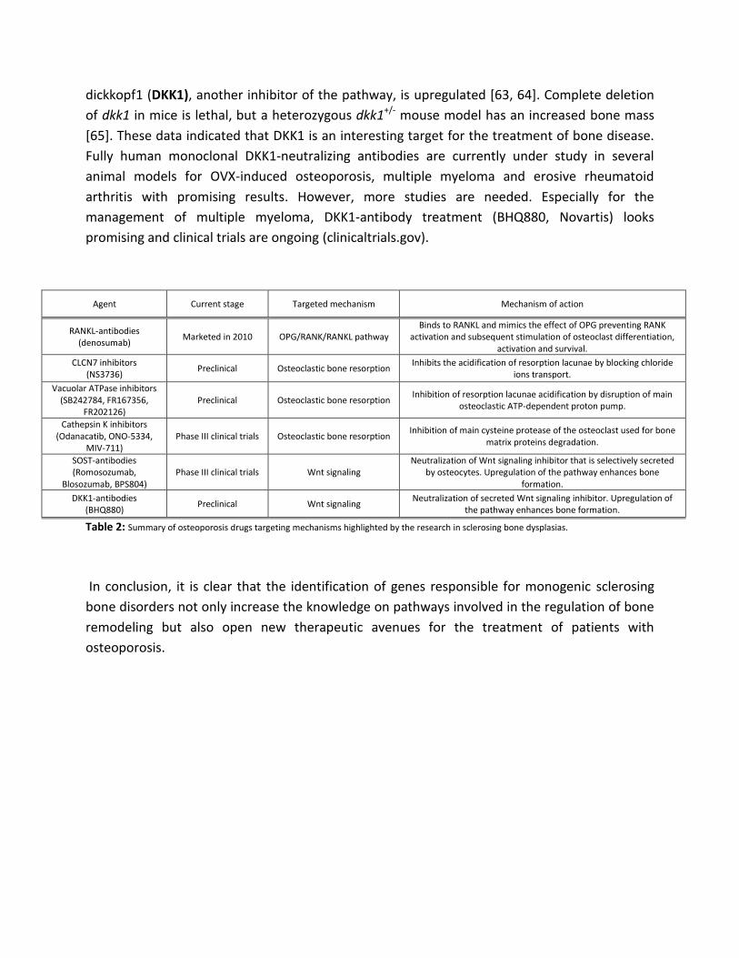
Agent Current stage Targeted mechanism Mechanism of action
RANKL-antibodies (denosumab)
Marketed in 2010 OPG/RANK/RANKL pathway Binds to RANKL and mimics the effect of OPG preventing RANK
activation and subsequent stimulation of osteoclast differentiation, activation and survival.
CLCN7 inhibitors (NS3736)
Preclinical Osteoclastic bone resorption Inhibits the acidification of resorption lacunae by blocking chloride
ions transport.
Vacuolar ATPase inhibitors (SB242784, FR167356,
FR202126) Preclinical Osteoclastic bone resorption
Inhibition of resorption lacunae acidification by disruption of main osteoclastic ATP-dependent proton pump.
Cathepsin K inhibitors (Odanacatib, ONO-5334,
MIV-711) Phase III clinical trials Osteoclastic bone resorption
Inhibition of main cysteine protease of the osteoclast used for bone matrix proteins degradation.
SOST-antibodies (Romosozumab,
Blosozumab, BPS804) Phase III clinical trials Wnt signaling
Neutralization of Wnt signaling inhibitor that is selectively secreted by osteocytes. Upregulation of the pathway enhances bone
formation.
DKK1-antibodies (BHQ880)
Preclinical Wnt signaling Neutralization of secreted Wnt signaling inhibitor. Upregulation of
the pathway enhances bone formation.
Table 2: Summary of osteoporosis drugs targeting mechanisms highlighted by the research in sclerosing bone dysplasias.
In conclusion, it is clear that the identification of genes responsible for monogenic sclerosing
bone disorders not only increase the knowledge on pathways involved in the regulation of bone
remodeling but also open new therapeutic avenues for the treatment of patients with
osteoporosis.
Figure 1: Overview of pathways and mechanisms involved in the pathogenesis of sclerosing bone dysplasias. Gene names were discussed in
the text.
Acknowledgments
Research relevant for this review was supported by a grant from the University of Antwerp
(TOP-BOF) and two research grants (G.0065.10N and G.0197.12N) from the
Fonds Wetenschappelijk Onderzoek-Vlaanderen (FWO) to W. Van Hul. I. Fijałkowski holds a pre-
doctoral specialization scholarship from the “Institute for the Promotion of Innovation through
Science and Technology in Flanders (IWT-Vlaanderen)”. E. Boudin holds a post-doctoral
fellowship of the FWO (Fund for Scientific Research) Vlaanderen.
References
* of importance
** of major importance
1. Rachner, T.D., S. Khosla, and L.C. Hofbauer, Osteoporosis: now and the future. Lancet, 2011. 377(9773): p. 1276-87.
2. Ferrari, S., Human genetics of osteoporosis. Best Pract Res Clin Endocrinol Metab, 2008. 22(5): p. 723-35. 3. Kornak, U., et al., Mutations in the a3 subunit of the vacuolar H(+)-ATPase cause infantile malignant
osteopetrosis. Hum Mol Genet, 2000. 9(13): p. 2059-63. 4.* Sobacchi, C., et al., Osteopetrosis: genetics, treatment and new insights into osteoclast function. Nat Rev
Endocrinol, 2013. 9(9): p. 522-36. 5. Kornak, U., et al., Loss of the ClC-7 chloride channel leads to osteopetrosis in mice and man. Cell, 2001.
104(2): p. 205-15. 6. Chalhoub, N., et al., Grey-lethal mutation induces severe malignant autosomal recessive osteopetrosis in
mouse and human. Nat Med, 2003. 9(4): p. 399-406. 7. Sly, W.S., et al., Carbonic anhydrase II deficiency in 12 families with the autosomal recessive syndrome of
osteopetrosis with renal tubular acidosis and cerebral calcification. N Engl J Med, 1985. 313(3): p. 139-45. 8. Guerrini, M.M., et al., Human osteoclast-poor osteopetrosis with hypogammaglobulinemia due to
TNFRSF11A (RANK) mutations. Am J Hum Genet, 2008. 83(1): p. 64-76. 9. Sobacchi, C., et al., Osteoclast-poor human osteopetrosis due to mutations in the gene encoding RANKL.
Nat Genet, 2007. 39(8): p. 960-2. 10. Smahi, A., et al., The NF-kappaB signalling pathway in human diseases: from incontinentia pigmenti to
ectodermal dysplasias and immune-deficiency syndromes. Hum Mol Genet, 2002. 11(20): p. 2371-5. 11. Van Wesenbeeck, L., et al., Involvement of PLEKHM1 in osteoclastic vesicular transport and osteopetrosis
in incisors absent rats and humans. J Clin Invest, 2007. 117(4): p. 919-30. 12. Aker, M., et al., An SNX10 mutation causes malignant osteopetrosis of infancy. J Med Genet, 2012. 49(4):
p. 221-6. 13. Cleiren, E., et al., Albers-Schonberg disease (autosomal dominant osteopetrosis, type II) results from
mutations in the ClCN7 chloride channel gene. Hum Mol Genet, 2001. 10(25): p. 2861-7. 14. Bollerslev, J., et al., Autosomal dominant osteopetrosis revisited: lessons from recent studies. Eur J
Endocrinol, 2013. 169(2): p. R39-57. 15. Gelb, B.D., et al., Pycnodysostosis, a lysosomal disease caused by cathepsin K deficiency. Science, 1996.
273(5279): p. 1236-8. 16. Little, R.D., et al., A mutation in the LDL receptor-related protein 5 gene results in the autosomal dominant
high-bone-mass trait. Am J Hum Genet, 2002. 70(1): p. 11-9. 17. Boyden, L.M., et al., High bone density due to a mutation in LDL-receptor-related protein 5. N Engl J Med,
2002. 346(20): p. 1513-21. 18. Ai, M., et al., Clinical and molecular findings in osteoporosis-pseudoglioma syndrome. Am J Hum Genet,
2005. 77(5): p. 741-53. 19. Balemans, W., et al., Increased bone density in sclerosteosis is due to the deficiency of a novel secreted
protein (SOST). Hum Mol Genet, 2001. 10(5): p. 537-43. 20. Brunkow, M.E., et al., Bone dysplasia sclerosteosis results from loss of the SOST gene product, a novel
cystine knot-containing protein. Am J Hum Genet, 2001. 68(3): p. 577-89. 21. Leupin, O., et al., Bone overgrowth-associated mutations in the LRP4 gene impair sclerostin facilitator
function. J Biol Chem, 2011. 286(22): p. 19489-500. 22. Balemans, W., et al., Identification of a 52 kb deletion downstream of the SOST gene in patients with van
Buchem disease. J Med Genet, 2002. 39(2): p. 91-7. 23. Kim, S.J., et al., Identification of signal peptide domain SOST mutations in autosomal dominant
craniodiaphyseal dysplasia. Hum Genet, 2011. 129(5): p. 497-502.
24. Jenkins, Z.A., et al., Germline mutations in WTX cause a sclerosing skeletal dysplasia but do not predispose to tumorigenesis. Nat Genet, 2009. 41(1): p. 95-100.
25. Nurnberg, P., et al., Heterozygous mutations in ANKH, the human ortholog of the mouse progressive ankylosis gene, result in craniometaphyseal dysplasia. Nat Genet, 2001. 28(1): p. 37-41.
26. Reichenberger, E., et al., Autosomal dominant craniometaphyseal dysplasia is caused by mutations in the transmembrane protein ANK. Am J Hum Genet, 2001. 68(6): p. 1321-6.
27. Hu, Y., et al., A novel autosomal recessive GJA1 missense mutation linked to Craniometaphyseal dysplasia. PLoS One, 2013. 8(8): p. e73576.
28. Janssens, K., et al., Mutations in the gene encoding the latency-associated peptide of TGF-beta 1 cause Camurati-Engelmann disease. Nat Genet, 2000. 26(3): p. 273-5.
29. Kinoshita, A., et al., Domain-specific mutations in TGFB1 result in Camurati-Engelmann disease. Nat Genet, 2000. 26(1): p. 19-20.
30. Hellemans, J., et al., Loss-of-function mutations in LEMD3 result in osteopoikilosis, Buschke-Ollendorff syndrome and melorheostosis. Nat Genet, 2004. 36(11): p. 1213-8.
31. Mumm, S., et al., Deactivating germline mutations in LEMD3 cause osteopoikilosis and Buschke-Ollendorff syndrome, but not sporadic melorheostosis. J Bone Miner Res, 2007. 22(2): p. 243-50.
32. Ababneh, F.K., et al., Hereditary deletion of the entire FAM20C gene in a patient with Raine syndrome. Am J Med Genet A, 2013. 161A(12): p. 3155-60.
33. Wang, X., et al., Inactivation of a novel FGF23 regulator, FAM20C, leads to hypophosphatemic rickets in mice. PLoS Genet, 2012. 8(5): p. e1002708.
34. Singer, F.R. and B.G. Mills, Evidence for a viral etiology of Paget's disease of bone. Clin Orthop Relat Res, 1983(178): p. 245-51.
35. Laurin, N., et al., Recurrent mutation of the gene encoding sequestosome 1 (SQSTM1/p62) in Paget disease of bone. Am J Hum Genet, 2002. 70(6): p. 1582-8.
36. Hocking, L.J., et al., Domain-specific mutations in sequestosome 1 (SQSTM1) cause familial and sporadic Paget's disease. Hum Mol Genet, 2002. 11(22): p. 2735-9.
37. Johnson, J.O., et al., Exome sequencing reveals VCP mutations as a cause of familial ALS. Neuron, 2010. 68(5): p. 857-64.
38. Whyte, M.P., et al., Osteoprotegerin deficiency and juvenile Paget's disease. N Engl J Med, 2002. 347(3): p. 175-84.
39. Hughes, A.E., et al., Mutations in TNFRSF11A, affecting the signal peptide of RANK, cause familial expansile osteolysis. Nat Genet, 2000. 24(1): p. 45-8.
40. Whyte, M.P. and A.E. Hughes, Expansile skeletal hyperphosphatasia is caused by a 15-base pair tandem duplication in TNFRSF11A encoding RANK and is allelic to familial expansile osteolysis. J Bone Miner Res, 2002. 17(1): p. 26-9.
41. Whyte, M.P., et al., Expansile skeletal hyperphosphatasia: a new familial metabolic bone disease. J Bone Miner Res, 2000. 15(12): p. 2330-44.
42. Boudin, E., et al., No mutations in the serotonin related TPH1 and HTR1B genes in patients with monogenic sclerosing bone disorders. Bone, 2013. 55(1): p. 52-6.
43. Boudin, E., et al., Mutations in sFRP1 or sFRP4 are not a common cause of craniotubular hyperostosis. Bone, 2013. 52(1): p. 292-5.
44. Pangrazio, A., et al., Exome sequencing identifies CTSK mutations in patients originally diagnosed as intermediate osteopetrosis. Bone, 2014. 59: p. 122-6.
45.** Lazarus, S., A. Zankl, and E.L. Duncan, Next-generation sequencing: a frameshift in skeletal dysplasia gene discovery. Osteoporos Int, 2014. 25(2): p. 407-22.
46. Borra, V.M., et al., Localization of the gene for hyperostosis cranialis interna to chromosome 8p21 with analysis of three candidate genes. Calcif Tissue Int, 2013. 93(1): p. 93-100.
47. Borra, V.M., et al., Localization of the gene for X-linked calvarial hyperostosis to chromosome Xq27.3-Xqter. Bone, 2014. 58: p. 67-71.
48. Pagon, R.A., J.B. Beckwith, and B.H. Ward, Calvarial hyperostosis: a benign X-linked recessive disorder. Clin Genet, 1986. 29(1): p. 73-8.
49. Zhang, Y., et al., Novel and recurrent germline LEMD3 mutations causing Buschke-Ollendorff syndrome and osteopoikilosis but not isolated melorheostosis. Clin Genet, 2009. 75(6): p. 556-61.
50. Kasapkara, C.S., et al., An extremely rare case: osteosclerotic metaphyseal dysplasia. Genet Couns, 2013. 24(1): p. 69-74.
51. Yao, A.L. and P.M. Camacho, Osteomesopyknosis: A Case Report and Review of Sclerosing Bone Disorders. Endocr Pract, 2014: p. 1-14.
52. Das, S. and J.C. Crockett, Osteoporosis - a current view of pharmacological prevention and treatment. Drug Des Devel Ther, 2013. 7: p. 435-48.
53. Leder, B.Z., et al., Two Years of Denosumab and Teriparatide Administration in Postmenopausal Women with Osteoporosis (The DATA Extension Study): a Randomized Controlled Trial. J Clin Endocrinol Metab, 2014: p. jc20134440.
54. Schaller, S., et al., The chloride channel inhibitor NS3736 [corrected] prevents bone resorption in ovariectomized rats without changing bone formation. J Bone Miner Res, 2004. 19(7): p. 1144-53.
55. Qin, A., et al., V-ATPases in osteoclasts: structure, function and potential inhibitors of bone resorption. Int J Biochem Cell Biol, 2012. 44(9): p. 1422-35.
56. Saftig, P., et al., Impaired osteoclastic bone resorption leads to osteopetrosis in cathepsin-K-deficient mice. Proc Natl Acad Sci U S A, 1998. 95(23): p. 13453-8.
57. Duong, L.T. Therapeutic inhibition of cathepsin K-reducing bone resorption while maintaining bone formation. Bonekey Reports, 2012. DOI: doi: 10.1038/bonekey.2012.67.
58. Langdahl, B., et al., Odanacatib in the treatment of postmenopausal women with low bone mineral density: five years of continued therapy in a phase 2 study. J Bone Miner Res, 2012. 27(11): p. 2251-8.
59. Sims, N.A. and K.W. Ng, Implications of osteoblast-osteoclast interactions in the management of osteoporosis by antiresorptive agents denosumab and odanacatib. Curr Osteoporos Rep, 2014. 12(1): p. 98-106.
60.** McClung, M.R., et al., Romosozumab in postmenopausal women with low bone mineral density. N Engl J Med, 2014. 370(5): p. 412-20.
61. Kedlaya, R., et al., Sclerostin inhibition reverses skeletal fragility in an Lrp5-deficient mouse model of OPPG syndrome. Sci Transl Med, 2013. 5(211): p. 211ra158.
62.* Sinder, B.P., et al., Sclerostin antibody improves skeletal parameters in a Brtl/+ mouse model of osteogenesis imperfecta. J Bone Miner Res, 2013. 28(1): p. 73-80.
63.* Chang, M.K., et al., Reversing LRP5-dependent osteoporosis and SOST deficiency-induced sclerosing bone disorders by altering WNT signaling activity. J Bone Miner Res, 2014. 29(1): p. 29-42.
64. van Lierop, A.H., et al., Serum Dickkopf 1 levels in sclerostin deficiency. J Clin Endocrinol Metab, 2014. 99(2): p. E252-6.
65. Morvan, F., et al., Deletion of a single allele of the Dkk1 gene leads to an increase in bone formation and bone mass. J Bone Miner Res, 2006. 21(6): p. 934-45.


![Eveline pupeter fellner emporia london-july2010 [kompatibilitätsmodus]](https://static.documents.pub/doc/80x56/5a6ec83b7f8b9ad9638b4fdf/eveline-pupeter-fellner-emporia-london-july2010-kompatibilitaetsmodus.jpg)


