160

This page intentionally left blank
Anatomy of Flowering Plants
Understanding plant anatomy is not only fundamental
to the study of plant systematics and palaeobotany,
but is also an essential part of evolutionary biology,
physiology, ecology, and the rapidly expanding science
of developmental genetics. In the third edition of
her successful textbook, Paula Rudall provides a
comprehensive yet succinct introduction to the anatomy
of flowering plants. Thoroughly revised and updated
throughout, the book covers all aspects of comparative
plant structure and development, arranged in a series of
chapters on the stem, root, leaf, flower, seed and fruit.
Internal structures are described using magnification aids
from the simple hand-lens to the electron microscope.
Numerous references to recent topical literature are
included, and new illustrations reflect a wide range of
flowering plant species. The phylogenetic context of plant
names has also been updated as a result of improved
understanding of the relationships among flowering
plants. This clearly written text is ideal for students
studying a wide range of courses in botany and plant
science, and is also an excellent resource for professional
and amateur horticulturists.
Paula Rudall is Head of Micromorphology(Plant
Anatomy and Palynology) at the Royal Botanic Gardens,
Kew. She has published more than 150 peer-reviewed
papers, using comparative floral and pollen morphology,
anatomy and embryology to explore evolution across seed
plants.

Anatomy ofFlowering PlantsAn Introduction toStructure and Development
PAULA J. RUDALL

LC Control No.: 2006100134
Type of Material: Book (Print, Microform, Electronic, etc.)
Personal Name: Rudall, Paula. » More like this
Main Title: Anatomy of flowering plants : an introduction to structure and development / Paula J. Rudall.
Edition Information: 3rd ed.
Published/Created: Cambridge ; New York : Cambridge University Press, 2007.
Description: xii, 145 p. : ill. ; 21 cm.
ISBN: 9780521692458 (pbk.) 0521692458 (pbk.)
Notes: Includes bibliographical references (p. [128]-137) and index.
Subjects: Angiosperms--Anatomy. » More like this Plant anatomy. » More like this
LC Classification: QK641 .R84 2007
Dewey Class No.: 580 22
Quality Code: pcc
Links: Publisher description: http://www.loc.gov/catdir/enhancements/fy0617/2006100134-d.html Table of contents only: http://www.loc.gov/catdir/enhancements/fy0617/2006100134-t.html Contributor biographical information: http://www.loc.gov/catdir/enhancements/fy0729/2006100134-b.html

CAMBRIDGE UNIVERSITY PRESS
Cambridge, New York, Melbourne, Madrid, Cape Town, Singapore, São Paulo
Cambridge University PressThe Edinburgh Building, Cambridge CB2 8RU, UK
First published in print format
ISBN-13 978-0-521-69245-8
ISBN-13 978-0-511-29453-2
© Paula J. Rudall 2007
2006
Information on this title: www.cambridge.org/9780521692458
This publication is in copyright. Subject to statutory exception and to the provision of relevant collective licensing agreements, no reproduction of any part may take place without the written permission of Cambridge University Press.
ISBN-10 0-511-29453-0
ISBN-10 0-521-69245-8
Cambridge University Press has no responsibility for the persistence or accuracy of urls for external or third-party internet websites referred to in this publication, and does not guarantee that any content on such websites is, or will remain, accurate or appropriate.
Published in the United States of America by Cambridge University Press, New York
www.cambridge.org
paperback
eBook (EBL)
eBook (EBL)
paperback

Contents
Preface ix
Taxonomic overview xi
1 Organs, Cells and Tissues 1
1.1 Organs 1
1.2 Cells 2
1.3 Cell Inclusions 5
1.4 Secretory Ducts and Laticifers 7
1.5 Transfer Cells 9
1.6 Tissues 9
1.6.1 Parenchyma 10
1.6.2 Aerenchyma 10
1.6.3 Collenchyma 10
1.6.4 Sclerenchyma 11
1.7 Epidermis 13
1.7.1 Stomata 13
1.7.2 Trichomes 15
1.8 Ground Tissue 17
1.9 Vascular Tissue 18
1.9.1 Xylem 18
1.9.2 Phloem 19
1.10 Meristems 21
1.10.1 Apical Meristems 21
1.10.2 Lateral Meristems 22
1.10.3 Meristemoids and Asymmetric
Cell Division 22
2 Stem 23
2.1 Shoot Apex 23
2.2 Primary Stem Structure 24
2.3 Primary Vascular System 26
2.4 Nodal Vasculature 27
2.5 Vascular Cambium 29
2.6 Secondary Xylem 31
2.7 Secondary Phloem 35
2.8 Primary and Secondary Thickening
Meristems 36
2.9 Periderm 40
3 Root 43
3.1 Primary Root Structure 43
3.2 Root Apex 43
3.3 Root Cap 45
3.4 Root Epidermis and Hypodermis 46
3.5 Root Cortex and Endodermis 48
3.6 Pericycle and Vascular Cylinder 49
3.7 Initiation of Lateral and Adventitious
Roots 50
3.8 Secondary Growth in Roots 51
3.9 Roots Associated with
Micro-Organisms 53
3.10 Haustoria of Parasitic Angiosperms 54
4 Leaf 57
4.1 Leaf Morphology and Anatomy 57
4.2 Leaf Development 60
4.3 Leaf Epidermis 61
4.3.1 Pavement Epidermal Cells 61
4.3.2 Stomata 62
vi Contents

4.3.3 Trichomes and Papillae 63
4.3.4 Cuticle and Wax 66
4.4 Extrafloral Nectaries 66
4.5 Mesophyll 68
4.6 Sclerenchyma and Idioblasts 69
4.7 Leaf Vasculature 70
4.8 Bundle Sheath and Kranz Anatomy 72
5 Flower 75
5.1 Floral Organs 75
5.2 Floral Vasculature 77
5.3 Perianth 79
5.4 Androecium 81
5.5 Pollen 84
5.6 Gynoecium 87
5.6.1 Stigma and Style 87
5.6.2 Ovary 89
5.7 Ovule 90
5.8 Embryo Sac 93
5.9 Pollen-Tube Growth 94
5.10 Floral Secretory Structures 96
6 Seed and fruit 99
6.1 Seed Coat 99
6.2 Pericarp 101
6.3 Grass Caryopsis 102
6.4 Endosperm 104
6.5 Perisperm 106
6.6 Embryo 107
6.7 Seedling 109
Glossary 111
References 128
Index 139
Contents vii

Preface
In the twenty-first century, plant anatomy remains highly relevant
to systematics, paleobotany, and the relatively new science of
developmental genetics, which interfaces disciplines and utilizes
a combination of techniques to examine gene expression in
growing tissues. Modern students need to consider information
from an increasingly wide range of sources, most notably inte-
grating morphological and molecular data. The third, thoroughly
revised, edition of this book presents an introduction to plant
anatomy for students of botany and related disciplines.
Although the simple optical lens has been used for centuries
to examine plant structure, detailed studies of plant anatomy
originated with the invention of the compound microscope in the
seventeenth century. Nehemiah Grew (1641�1712) and Marcello
Malpighi (1628�1694), physicians working independently in
England and Italy respectively, were early pioneers of the
microscopical examination of plant cells and tissues. Their
prescient work formed the foundation that eventually led to the
development of our understanding of cell structure and cell
division27. Other early outstanding figures included Robert Brown
(1773�1858), who discovered the nucleus, and the plant
embryologist Wilhelm Hofmeister (1824�1877), who first
described the alternation of generations in the life cycle of land
plants. In the nineteenth and twentieth centuries plant anatomy
became an important element of studies of both physiology and
systematic biology, and an integral aspect of research in the
developing field of anatomical paleobotany, led by such luminaries
as Dukinfield Henry Scott (1854�1934). The physiologist
Gottlieb Haberlandt (1854�1945) utilized anatomical observa-
tions in his ground-breaking work on photosynthetic carbon
metabolism. One of the most notable plant anatomists of the
twentieth century was Katherine Esau (1898�1997), recognized
particularly for her work on the structure and development of
phloem and her influential textbooks on plant anatomy30. Other
important textbooks include works on paleobotany, morphology,
anatomy and embryology13,34,68,106.
The invention of the transmission electron microscope (TEM)
in the mid twentieth century allowed greater magnification than
any optical microscope, and hence revitalized studies in cell ultra-
structure and pollen morphology98. The subsequent invention of
the scanning electron microscope (SEM) provided greater image
clarity and much greater depth of focus than light microscopes,
and thus further increased accessibility of minute structures,
including seeds, pollen grains and organ primordia28,98. More
recent innovations, including fluorescence microscopy, differen-
tial interference contrast (DIC) microscopy and confocal imaging,
have allowed enhanced visualization of tissue structure. Others,
including nuclear magnetic resonance (NMR) imaging and
high-resolution X-ray computed tomography (HRCT) facilitate
enhanced visualization of three-dimensional objects.
x Preface

Taxonomic Overview
In textbooks published before 1990, extant angiosperms were
consistently subdivided into two major groups � dicotyledons
(dicots) and monocotyledons (monocots), based partly on the
number of cotyledons in the seedling. This dichotomy was long
considered to represent a fundamental divergence at the base of
the angiosperm evolutionary tree. Other features marked this
distinction, including the absence of a vascular cambium and
presence of parallel leaf venation in monocots. However, the
expansion of molecular phylogenetics through the early 1990s
indicated that some species that were formerly classified as
primitive dicots do not belong to either category, though the
monophyly of monocots was confirmed2,3,103. Thus, although the
dicot/monocot distinction remains useful for generalized descrip-
tions of angiosperm groups, current evidence suggests that it does
not represent a wholly natural classification. It is now widely
accepted that several relatively species-poor angiosperm lineages
(here termed early-divergent angiosperms or magnoliids) evolved
before the divergence of the two major lineages that led to the
monocots and the remaining dicots (now termed eudicots, or
sometimes tricolpates).
Early-divergent angiosperms (including magnoliids) are a small
but highly diverse assemblage of taxonomically isolated lineages
that probably represent the surviving extant members of their
respective clades, accounting for only about 1% of extant species.
They possess some morphological features in common with both

monocots and eudicots, and include the New Caledonian shrub
Amborella, the water lilies (Nymphaeaceae), woody families such as
Magnoliaceae and Lauraceae, and herbaceous or climbing families
such as Piperaceae and Aristolochiaceae. Monocots account for
approximately a quarter of all flowering plants species. They
dominate significant parts of world ecosystems, and are of
immense economic importance, including the staple grass food
crops (wheat, barley, rice and maize) and other important food
plants such as onions, palms, yams, bananas and gingers. Eudicots
represent about 75% of extant angiosperm species, and encompass
a wide range of morphological diversity, especially in the two
largest subclades, Rosidae (rosid eudicots) and Asteridae (asterid
eudicots).
xii Taxonomic overview
1
Organs, cells and tissues
1.1 OrgansPlants consist of several organs, which in their turn are composed
of tissues. Broadly, vegetative organs support plant growth, and
reproductive organs enable sexual reproduction. The three main
types of vegetative organ are the root, stem and leaf. Roots
typically occur underground, and extract moisture and nutrients
from the soil, though there are many examples of plants with
aerial roots. The stem and leaves together comprise the shoot
(Fig. 1.1). Stems occur both above and below ground. Some stems
are modified into underground perennating or storage organs
such as corms or rhizomes. Leaves typically occur above ground
level, though some underground stems possess reduced scale
leaves, and underground bulbs possess swollen leaves or leaf bases.
Primary organs and tissues develop initially from the shoot
and root apical meristems and from cell divisions in meristems
closely adjacent to them, such as the primary thickening meristem.
Secondary tissues such as secondary xylem (wood) develop from
lateral meristems such as the vascular cambium. Organs such as
adventitious roots develop from differentiated cells that have
retained meristematic capacity. At the onset of flowering, the shoot
apical meristem undergoes structural modification from a veg-
etative to a reproductive apex and subsequently produces flowers
(chapter 5). Flowers are borne on an inflorescence, either in
groups or as solitary structures. A group of inflorescences borne
on a single plant is termed a synflorescence121 (Fig. 1.2).

1.2 CellsPlant cells typically have a cell wall containing a living protoplast
(Fig. 1.3). The layer that contacts the walls of adjacent cells is
termed the middle lamella. Following cessation of growth, many
cells develop a secondary cell wall which is deposited on the inside
surface of the primary wall. Both primary and secondary walls
consist of cellulose microfibrils embedded in a matrix and ori-
ented in different directions. Secondary cell walls consist mostly of
cellulose, but primary walls commonly contain a high proportion
of hemicelluloses in the gel-like matrix, affording a greater degree
of plasticity to the wall of the growing cell. The secondary wall
can also contain deposits of lignin (in sclerenchymatous cells) or
suberin (in many periderm cells), and often appears lamellated.
Thin areas of the primary wall, which usually correspond with
thin areas of the walls of neighbouring cells, are primary pit fields,
and usually have protoplasmic strands (plasmodesmata) passing
through them, connecting the protoplasts of neighbouring cells36.
The connected living protoplasts are collectively termed the sym-
plast. Primary pit fields often remain as thin areas of the wall even
after a secondary wall has been deposited, and are then termed
Figure 1.1 Hyptis ditassoides (Lamiaceae), transverse section of vegetative bud
near apex, showing three successive pairs of leaf primordia surrounding
central stem. Scale ¼ 100 mm.
2 Organs, cells and tissues

pits, or pit-pairs if there are two pits connecting adjacent cells. Pits
may be simple, as in most parenchyma cells, or bordered, as in
tracheary elements. In simple pits the pit cavity is of more or less
uniform width, whereas in bordered pits the secondary wall
Figure 1.2 Salvia involucrata (Lamiaceae), dissected developing synflorescence
showing flower clusters, each consisting of three flowers enclosed within
a bract; younger stages towards apex. b ¼ bract. Scale ¼ 500 mm.
Cells 3

arches over the pit cavity so that the opening to the cavity is rela-
tively narrow. Through a light microscope the outer rim of the
primary pit field appears as a border around the pit opening.
The cell protoplast is contained within a plasma membrane.
It consists of cytoplasm that encloses bodies such as the nucleus,
plastids and mitochondria, and also non-protoplasmic contents
such as oil, starch or crystals. The nucleus, which is bounded by
a nuclear membrane, often contains one or more recognizable
bodies (nucleoli) together with the chromatin in the nuclear
sap. During cell division the chromatin becomes organized into
chromosomes. Most cells possess a single nucleus, but examples
of multinucleate cells (coenocytes) include the non-articulated
laticifers found in many plant families (chapter 1.4). Such cells
elongate and penetrate established tissues by intrusive tip growth,
Figure 1.3 Diagram of a generalised plant cell illustrating details of
protoplasmic contents.
4 Organs, cells and tissues
in which the cell apices secrete enzymes that dissolve the middle
lamellae of neighbouring cells; bifurcation occurs when they
encounter an obstacle36.
Mitochondria and plastids are surrounded by double mem-
branes. Plastids are larger than mitochondria, and are classified
into different types depending on their specialized role.
For example, chloroplasts are plastids that contain chlorophyll
within a system of lamellae that are stacked to form grana; this
is the site of photosynthesis. Chloroplasts occur in all green cells,
but are most abundant in the leaf mesophyll, which is the primary
photosynthetic tissue (chapter 4.5). Membranes occur widely
throughout the cytoplasm, sometimes bounding a series of cav-
ities. For example, the endoplasmic reticulum is a continuous
membrane-bound system of flattened sacs and tubules, sometimes
coated with ribosomal particles. Dictyosomes are systems of sacs
associated with secretory activity. Vacuoles are cavities in the
cytoplasm; they are usually colourless and contain a watery fluid.
Their size and shape varies in different cell types, and also changes
during the life of a cell.
1.3 Cell InclusionsMany cells possess non-protoplasmic contents such as oils,
mucilage (slime), tannins, starch granules, calcium oxalate crystals
and silica bodies. Both oil and mucilage are produced in secretory
idioblasts which are often larger than adjacent parenchymatous
cells. Tannins are phenol derivatives which are common in plant
cells; they are amorphous, and appear yellow, red or brown in
colour in cells of sectioned material. Cystoliths are cellulose bodies
encrusted with calcium carbonate that occur in epidermal cells
in some species (Fig. 4.4); the body is attached to the cell wall by
a silicified stalk36.
Starch is especially common in storage tissues such as
endosperm or in parenchyma adjacent to a nectary. Starch granules
Cell inclusions 5

are formed in plastids (amyloplasts). They often appear layered
due to the successive deposition of concentric rings, and may
possess characteristic shapes. For example, in species of Euphorbia,
starch grains in laticifers are elongated and sometimes rod-shaped
or bone-shaped compared with the more rounded starch grains of
neighbouring parenchyma cells (Fig. 1.4)70.
Calcium oxalate crystals (Figs 1.5, 1.13) are borne in crystal
idioblasts that can occur in almost every part of the plant, includ-
ing both vegetative and reproductive organs82. They are often
present near veins, possibly due to transport of calcium through
the xylem, and are sometimes associated with air space formation;
some aquatic plants possess calcium oxalate crystals projecting into
air spaces. Crystals form within vacuoles of actively growing cells
and are usually associated with membrane chambers, lamellae,
mucilage and fibrillar material. Crystal sand is relatively amor-
phous and represents fragmented non-nucleated crystalline
particles. Druses (cluster crystals) are aggregated crystalline struc-
tures that have precipitated around a nucleation site. Raphides are
bundles of needle-like crystals that are borne in the same cell; they
occur commonly in monocots. In the monocot family Araceae,
Figure 1.4 Monadenium ellenbeckii
(Euphorbiaceae). Elongated I-shaped
starch grains in laticifer (L); ovoid
starch grains present in adjacent
parenchyma cells. Scale¼ 20 mm.
6 Organs, cells and tissues
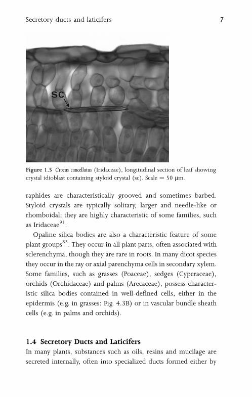
raphides are characteristically grooved and sometimes barbed.
Styloid crystals are typically solitary, larger and needle-like or
rhomboidal; they are highly characteristic of some families, such
as Iridaceae91.
Opaline silica bodies are also a characteristic feature of some
plant groups83. They occur in all plant parts, often associated with
sclerenchyma, though they are rare in roots. In many dicot species
they occur in the ray or axial parenchyma cells in secondary xylem.
Some families, such as grasses (Poaceae), sedges (Cyperaceae),
orchids (Orchidaceae) and palms (Arecaceae), possess character-
istic silica bodies contained in well-defined cells, either in the
epidermis (e.g. in grasses: Fig. 4.3B) or in vascular bundle sheath
cells (e.g. in palms and orchids).
1.4 Secretory Ducts and LaticifersIn many plants, substances such as oils, resins and mucilage are
secreted internally, often into specialized ducts formed either by
Figure 1.5 Crocus cancellatus (Iridaceae), longitudinal section of leaf showing
crystal idioblast containing styloid crystal (sc). Scale ¼ 50 mm.
Secretory ducts and laticifers 7

cell wall separation (schizogenous ducts) or cell wall degradation
(lysigenous ducts), or a combination of the two processes (schizo-
lysigenous ducts)13. Some angiosperms, especially eudicots such
as Euphorbia and Ficus, produce latex from specialized cells (latici-
fers) that permeate their tissues (Figs 1.4, 1.6). In Euphorbia, the
laticifers are derived from a small group of initial cells in the
cotyledonary node of the embryo; these cells are coenocytes, since
they undergo repeated nuclear divisions without corresponding
wall formation. They grow intrusively between cells of surround-
ing tissues, and often branch and eventually ramify throughout
the entire plant31,71,87,90. Coenocytic laticifers are termed
non-articulated laticifers. By contrast, laticifers of a few species
(e.g. Hevea brasiliensis, the source of commercial rubber) undergo
cell-wall formation, and thus consist of linked chains of cells; these
are termed articulated laticifers. Laticifers of the opium poppy
Figure 1.6 Euphorbia eyassiana (Euphorbiaceae), longitudinal section of stem
showing branched non-articulated laticifers in parenchyma. Scale ¼ 50 mm.
8 Organs, cells and tissues
(Papaver somniferum) are always associated with vascular bundles122;
the alkaloids produced in the latex of these cells are the source of
narcotic analgesics such as morphine.
1.5 Transfer CellsTransfer cells occur at the interface between tissues; they are
specialized cells that facilitate transport (absorption or secretion)
of soluble substances across tissue boundaries. For example, they
can occur at the junction of the megagametophyte and mega-
sporophyte, in companion cells in phloem tissue (especially
at the node of a stem), in root nodules, in the haustoria of parasitic
plants, and in the epidermis of water plants80. Several cells
of the embryo sac and seed, including synergids, antipodals
and specialized endosperm cells, have been identified as transfer
cells in different species. Transfer cells are typically characterized
by numerous cell-wall ingrowths protruding into their protoplasts
or those of adjacent cells; these ingrowths are sometimes visible
using light microscopy. Secretory cells, such as those of glandular
hairs and nectaries, also frequently possess wall ingrowths.
The plasma membrane of the transfer cell follows the contour
of the wall ingrowths, thus increasing the surface area.
1.6 TissuesSimple tissues, such as parenchyma, collenchyma and scleren-
chyma, consist of a single cell type, though they may be
interspersed with other, isolated, cell types (idioblasts). Complex
tissues consist of multiple cell types, and can be divided into three
main groups: dermal tissue (epidermis), ground tissue and
vascular (conducting) tissue, each distributed throughout the
plant body, and often continuous between the various organs.
Complex tissues often include elements of several different simple
tissue types; for example, secondary xylem includes not only
vascular tissue, but also parenchyma and sclerenchyma.
Tissues 9

1.6.1 ParenchymaParenchyma cells are typically thin-walled and often polyhedral
or otherwise variously shaped, sometimes lobed. Cells with living
contents that do not fit readily into other categories are often
termed parenchyma cells. They are the least specialized cells of the
mature plant body and often resemble enlarged meristematic cells.
Parenchyma cells may occur in primary or secondary tissues.
Relatively specialized types of parenchyma include certain
secretory tissues and chlorenchyma, which contains chloroplasts
for photosynthesis. Parenchymatous cells may be tightly packed
or may be interspersed with intercellular air spaces.
Callus tissue is a cellular proliferation that is often produced
at the site of a wound by divisions in parenchyma cells that have
retained the ability to divide at maturity. A single isolated callus
cell can be used to artificially grow a new plant using tissue culture
methods.
1.6.2 AerenchymaAerenchyma is a specialized parenchymatous tissue that often
occurs in aquatic plants (hydrophytes). It possesses a regular, well-
developed system of large intercellular air spaces (Fig. 1.7) that
facilitates internal diffusion of gases. In leaves, stems and roots
of some water plants (e.g. Hydrocharis), aerenchyma is associated
with a system of transverse septa or diaphragms that provide
mechanical resistance to compression. These septa are uniseriate
layers of parenchyma cells that are thicker-walled than neigh-
bouring aerenchyma cells.
1.6.3 CollenchymaCollenchyma consists of groups of axially elongated, tightly-
packed cells with unevenly thickened walls. This tissue has
a strengthening function and often occurs in the angles of young
stems, or in the midribs of leaves, normally in primary ground
tissue. Collenchyma cells differ from fibres in that they often retain
10 Organs, cells and tissues

their contents at maturity and do not generally have lignified walls,
though they may ultimately become lignified.
1.6.4 SclerenchymaSclerenchyma, also a supporting or protective tissue, consists
of cells with thickened, often lignified, walls, which usually lack
contents at maturity. Sclerenchyma cells occur in primary or
secondary tissue, either in groups or individually as idioblasts
interspersed in other tissue types. They are categorized as either
fibres or sclereids, though transitional forms occur.
Fibres are long narrow cells that are elongated along the long
axis of the organ concerned; they normally occur in groups. Bast
Figure 1.7 Cyperus papyrus (Cyperaceae), longitudinal section of leaf showing
aerenchyma. a ¼ air space. Scale ¼ 100 mm.
Tissues 11
fibres are extraxylary cortical fibres which can be of economic use,
as in flax and hemp.
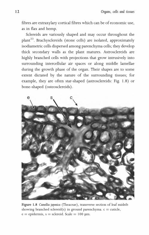
Sclereids are variously shaped and may occur throughout the
plant33. Brachysclereids (stone cells) are isolated, approximately
isodiametric cells dispersed among parenchyma cells; they develop
thick secondary walls as the plant matures. Astrosclereids are
highly branched cells with projections that grow intrusively into
surrounding intercellular air spaces or along middle lamellae
during the growth phase of the organ. Their shapes are to some
extent dictated by the nature of the surrounding tissues; for
example, they are often star-shaped (astrosclereids: Fig. 1.8) or
bone-shaped (osteosclereids).
Figure 1.8 Camellia japonica (Theaceae), transverse section of leaf midrib
showing branched sclereid(s) in ground parenchyma. c ¼ cuticle,
e ¼ epidermis, s ¼ sclereid. Scale ¼ 100 mm.
12 Organs, cells and tissues
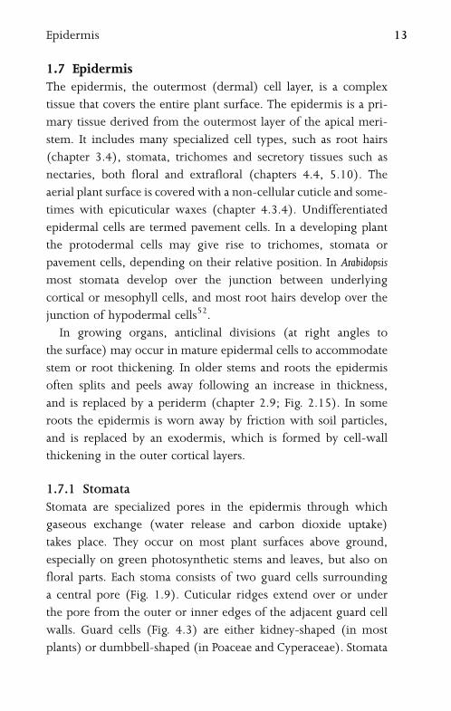
1.7 EpidermisThe epidermis, the outermost (dermal) cell layer, is a complex
tissue that covers the entire plant surface. The epidermis is a pri-
mary tissue derived from the outermost layer of the apical meri-
stem. It includes many specialized cell types, such as root hairs
(chapter 3.4), stomata, trichomes and secretory tissues such as
nectaries, both floral and extrafloral (chapters 4.4, 5.10). The
aerial plant surface is covered with a non-cellular cuticle and some-
times with epicuticular waxes (chapter 4.3.4). Undifferentiated
epidermal cells are termed pavement cells. In a developing plant
the protodermal cells may give rise to trichomes, stomata or
pavement cells, depending on their relative position. In Arabidopsis
most stomata develop over the junction between underlying
cortical or mesophyll cells, and most root hairs develop over the
junction of hypodermal cells52.
In growing organs, anticlinal divisions (at right angles to
the surface) may occur in mature epidermal cells to accommodate
stem or root thickening. In older stems and roots the epidermis
often splits and peels away following an increase in thickness,
and is replaced by a periderm (chapter 2.9; Fig. 2.15). In some
roots the epidermis is worn away by friction with soil particles,
and is replaced by an exodermis, which is formed by cell-wall
thickening in the outer cortical layers.
1.7.1 StomataStomata are specialized pores in the epidermis through which
gaseous exchange (water release and carbon dioxide uptake)
takes place. They occur on most plant surfaces above ground,
especially on green photosynthetic stems and leaves, but also on
floral parts. Each stoma consists of two guard cells surrounding
a central pore (Fig. 1.9). Cuticular ridges extend over or under
the pore from the outer or inner edges of the adjacent guard cell
walls. Guard cells (Fig. 4.3) are either kidney-shaped (in most
plants) or dumbbell-shaped (in Poaceae and Cyperaceae). Stomata
Epidermis 13

may be sunken or raised, and are often associated with a sub-
stomatal cavity in the mesophyll, which is caused by differential
expansion between the guard cell mother cell and the developing
underlying mesophyll cells42.
The epidermal cells immediately adjacent to the guard cells
are termed subsidiary cells if they differ morphologically from
surrounding epidermal cells. Classifications of stomatal types are
based either on the arrangement of mature subsidiary cells, or on
their patterns of development. Types of mature stomata include
anomocytic, anisocytic, diacytic and paracytic124. Anomocytic
stomata lack subsidiary cells entirely; anisocytic stomata possess
three unequal subsidiary cells; diacytic stomata possess one or
more pairs of subsidiary cells with their common walls at right
angles to the guard cells; and paracytic stomata possess one or
Figure 1.9 Arabidopsis thaliana (Brassicaceae), SEM abaxial leaf surface,
showing a single stomatal pore. Scale ¼ 10 mm.
14 Organs, cells and tissues

more subsidiary cells at either side of the guard cells. However,
different developmental pathways may lead to similar stomatal
types, so this classification could group types that are non-
homologous.
Ontogenetic stomatal types include agenous, mesogenous and
perigenous85,112. During development, a protodermal cell under-
goes an unequal mitotic division to produce a larger daughter cell
and a meristemoid (guard cell mother cell). In the agenous devel-
opmental type the meristemoids give rise directly to the guard
cells, and there are no subsidiary cells. In the perigenous type, the
meristemoid gives rise directly to the guard cells, and subsidiary
cells are formed from neighbouring cells, often by oblique divi-
sions. In the mesogenous developmental type the guard cells and
subsidiary cells have a common origin; the meristemoid under-
goes a further mitotic division into two cells, of which one further
subdivides to form the guard cells, and the other usually forms one
or more subsidiary cells. In mesoperigenous stomatal complexes
the guard cells and subsidiary cells are of mixed origin. Subsidiary
cells derived from the meristemoid are termed mesogene cells,
whereas those derived from neighbouring cells are termed
perigene cells, though in some cases mesogene cells are not
distinct from surrounding epidermal cells at maturity.
1.7.2 TrichomesTrichomes are epidermal outgrowths that occur on all parts
of the plant surface (Fig. 1.10). They vary widely in both form
and function, and include unicellular or multicellular, branched
or unbranched forms, and also scales, glandular (secretory) hairs,
hooked hairs and stinging hairs. Papillae are generally smaller than
trichomes and unicellular, though the distinction is not always
clear. In cases where there are several small outgrowths on each
epidermal cell, these outgrowths are termed papillae, but where
there is only one unicellular outgrowth per cell, the distinction
is dependent on size.
Epidermis 15

Glandular trichomes usually possess a unicellular or multi-
cellular stalk and a secretory head with one to several cells.
Secreted substances such as volatile oils collect between the
secretory cells and a raised cuticle, which later breaks to release
them. There are many different types of glandular hair, and they
secrete a variety of substances, including essential oils and salt;
some carnivorous plants’ digestive juices contain proteolytic
enzymes31. Leaf glandular hairs of Cannabis sativa secrete a resinous
substance containing the mild hallucinogen tetrahydrocannabinol.
Glandular hairs of Drosophyllum and Drosera secrete both sticky
mucilage and proteolytic enzymes. The stinging hairs of Urtica
dioica (stinging nettle) are rigid, hollow structures that contain
a poisonous substance (Fig. 1.11). The spherical tip of the hair
is readily broken off in contact with an outside body, and the
remaining sharp point may then penetrate the skin and release
Figure 1.10 Salvia involucrata (Lamiaceae), trichomes on petal surface.
n ¼ nonglandular trichome, g1 ¼ glandular trichome with unicellular head,
g4 ¼ glandular trichome with four-celled head. Scale ¼ 50 mm.
16 Organs, cells and tissues
the fluid. Other examples of specialized hair types include
water-absorptive leaf scales in many Bromeliaceae, and salt-
secreting glands of species of Avicennia60.
1.8 Ground TissueGround tissue, sometimes termed packing tissue, forms the bulk
of primary plant tissue and occupies the areas that are not taken
up by vascular tissue or cavities. It has a mechanical function,
and may be concerned with storage or photosynthesis. Ground
tissue typically consists of parenchyma, sclerenchyma or collen-
chyma, and is often interspersed with idioblasts and secretory
cells or canals. Ground tissue is initially formed at the apical
meristems but may be supplemented by intercalary growth,
and in monocots by tissues differentiated from primary and
secondary thickening meristems. In dicots the ground tissue of
secondary xylem (wood), formed by the vascular cambium,
consists of fibres and axial parenchyma (chapter 2.6). In older
stems the central area of ground tissue (pith) often breaks down,
leaving a cavity (Fig. 2.2).
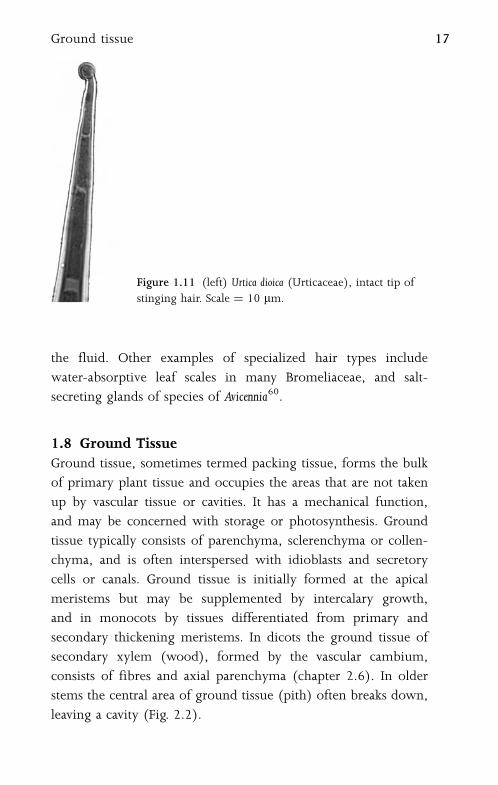
Figure 1.11 (left) Urtica dioica (Urticaceae), intact tip of
stinging hair. Scale ¼ 10 mm.
Ground tissue 17
1.9 Vascular TissueVascular tissue consists of xylem and phloem, and may be primary
or secondary in origin. Primary vascular tissue is derived from
procambium, itself produced by the apical meristems, and also
by the primary thickening meristem in stems of monocots
(chapter 2.8). Secondary vascular tissue is derived from the
vascular cambium in dicots, and from the secondary thickening
meristem in a few monocots (Fig. 2.13). Both xylem and phloem
are complex tissues, composed of many different cell types. Xylem
is primarily concerned with water transport and phloem with
food transport. Distribution of vascular tissue varies considerably
between different organs and taxa.
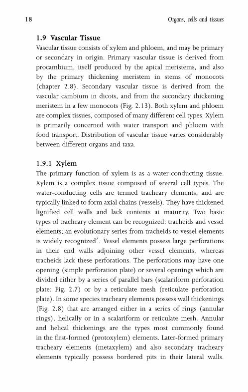
1.9.1 XylemThe primary function of xylem is as a water-conducting tissue.
Xylem is a complex tissue composed of several cell types. The
water-conducting cells are termed tracheary elements, and are
typically linked to form axial chains (vessels). They have thickened
lignified cell walls and lack contents at maturity. Two basic
types of tracheary element can be recognized: tracheids and vessel
elements; an evolutionary series from tracheids to vessel elements
is widely recognized7. Vessel elements possess large perforations
in their end walls adjoining other vessel elements, whereas
tracheids lack these perforations. The perforations may have one
opening (simple perforation plate) or several openings which are
divided either by a series of parallel bars (scalariform perforation
plate: Fig. 2.7) or by a reticulate mesh (reticulate perforation
plate). In some species tracheary elements possess wall thickenings
(Fig. 2.8) that are arranged either in a series of rings (annular
rings), helically or in a scalariform or reticulate mesh. Annular
and helical thickenings are the types most commonly found
in the first-formed (protoxylem) elements. Later-formed primary
tracheary elements (metaxylem) and also secondary tracheary
elements typically possess bordered pits in their lateral walls.
18 Organs, cells and tissues

These pits vary considerably in size, shape and arrangement;
they may be oval, polygonal or elongated (scalariform pitting),
organized in transverse rows (opposite pitting) or in a tightly
packed arrangement (alternate pitting).
1.9.2 PhloemPhloem has complex roles in translocation and messaging within
the plant. Primary phloem is formed by the apical meristem and
secondary phloem by the vascular cambium. Phloem may develop
precociously in regions that require a copious supply of nutrients,
such as developing sporogenous tissue.
Phloem is a complex tissue that consists of conducting cells
(sieve elements) and associated specialized parenchyma cells
(companion cells) (Figs. 1.12; 1.13); these two closely inter-
dependent cell types are produced from a common parent cell but
develop differently. Angiosperm sieve elements lack nuclei and
most organelles at maturity, but retain plastids and phloem-specific
Figure 1.12 Lilium tigrinum (Liliaceae), transverse section of stem vascular
bundle. bs ¼ bundle sheath, c ¼ companion cell, mx ¼ metaxylem vessel,
px ¼ protoxylem vessel, s ¼ sieve tube element. Scale ¼ 100 mm.
Vascular tissue 19

proteins (P-proteins) which occur in several morphological
forms (amorphous, filamentous, tubular and crystalline) that
are often highly characteristic for particular plant families, and thus
of systematic and evolutionary value14,116. Sieve-element plastids
are classified according to their inclusions: starch (S-type plastids),
protein (P-type plastids), or both. By contrast, companion cells
are densely cytoplasmic, retaining nuclei and many active
mitochondria.
Sieve elements are linked axially to form sieve tubes. The two
basic types of sieve element, sieve cells and sieve-tube elements,
are differentiated by their pore structure; most angiosperms
exclusively possess sieve-tube elements. The walls of sieve ele-
ments are thin and possess characteristic regions (sieve areas) that
connect adjacent sieve elements; sieve areas consist of groups
Figure 1.13 Crocus cancellatus (Iridaceae), transverse section of leaf vascular
bundle. bs ¼ bundle sheath, c ¼ crystal, v ¼metaxylem vessel, p ¼ phloem.
Scale ¼ 50 mm.
20 Organs, cells and tissues

of pores and associated callose. In sieve cells the sieve areas
are distributed throughout the cell wall, but in sieve-tube elements
they are mainly localized on the adjoining end walls, forming
sieve plates that link two axially linked elements of a sieve vessel.
Sieve plates can be simple or compound.
1.10 MeristemsMeristematic tissue consists of thin-walled, tightly packed
living cells which undergo frequent divisions. Meristematic cells
undergo cell division and wall formation followed by differential
cell expansion. After nuclear division there is progressive deposi-
tion of membranes in the cytoplasm into a cell plate that is located
in the equatorial zone between the two daughter nuclei108. The
cell plate extends to join the cell walls, thus depositing a new
wall. Most of the plant body is differentiated at the meristems
in well-defined zones, though cells in other regions may also
occasionally divide. There are some remarkable examples of fully-
differentiated cells giving rise to entire plantlets, notably on leaves
of Crassulaceae, such as Kalanchoe102.
1.10.1 Apical MeristemsApical meristems are located at the shoot apex (Fig. 2.1), where
primary stem, leaves and flowers differentiate, and at the root apex
(Figs 3.1, 3.2), where primary root tissue is produced. Subsequent
elongation of the shoot axis may occur by random cell divisions
and growth throughout the youngest internodes. This region
of diffuse cell division is termed an uninterrupted meristem, and
is continuous with the apical meristem. However, in some plant
stems, particularly in grasses, most cell divisions contributing
to stem elongation occur in a limited region, usually at the base
of the internode, which is then termed an intercalary meristem.
Both intercalary and uninterrupted meristems represent growth
in regions of already differentiated tissues.
Meristems 21
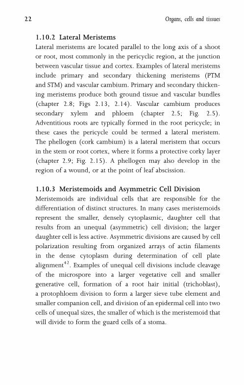
1.10.2 Lateral MeristemsLateral meristems are located parallel to the long axis of a shoot
or root, most commonly in the pericyclic region, at the junction
between vascular tissue and cortex. Examples of lateral meristems
include primary and secondary thickening meristems (PTM
and STM) and vascular cambium. Primary and secondary thicken-
ing meristems produce both ground tissue and vascular bundles
(chapter 2.8; Figs 2.13, 2.14). Vascular cambium produces
secondary xylem and phloem (chapter 2.5; Fig. 2.5).
Adventitious roots are typically formed in the root pericycle; in
these cases the pericycle could be termed a lateral meristem.
The phellogen (cork cambium) is a lateral meristem that occurs
in the stem or root cortex, where it forms a protective corky layer
(chapter 2.9; Fig. 2.15). A phellogen may also develop in the
region of a wound, or at the point of leaf abscission.
1.10.3 Meristemoids and Asymmetric Cell DivisionMeristemoids are individual cells that are responsible for the
differentiation of distinct structures. In many cases meristemoids
represent the smaller, densely cytoplasmic, daughter cell that
results from an unequal (asymmetric) cell division; the larger
daughter cell is less active. Asymmetric divisions are caused by cell
polarization resulting from organized arrays of actin filaments
in the dense cytoplasm during determination of cell plate
alignment42. Examples of unequal cell divisions include cleavage
of the microspore into a larger vegetative cell and smaller
generative cell, formation of a root hair initial (trichoblast),
a protophloem division to form a larger sieve tube element and
smaller companion cell, and division of an epidermal cell into two
cells of unequal sizes, the smaller of which is the meristemoid that
will divide to form the guard cells of a stoma.
22 Organs, cells and tissues

2
Stem
2.1 Shoot ApexThe vegetative shoot apex contributes to extension growth of the
shoot and initiates leaf primordia. Most shoot apices are inde-
terminate, though some (e.g. shoot thorns) become determinate.
The vegetative shoot apical meristem is typically dome-shaped
and partitioned by distinct zones of activity (Fig. 2.1). In many
species, the outermost two (sometimes more) cell layers (L1 and
L2, collectively termed the tunica) are maintained predominantly
by anticlinal cell divisions. The corpus (L3), in which cell
divisions are randomly oriented, is the region proximal to the
tunica. Thus, the outer layers contribute to surface growth and the
inner layers to an increase in volume, though there is often slight
intergradation between the two layers20.
The central regions of both tunica and corpus are sometimes
larger and more highly vacuolated than those on either side. The
central region underlying the corpus layer is a rib meristem; this
gives rise to files of cells that later become the pith. This central
region is surrounded by a peripheral flank meristem that produces
the procambium, cortical region and leaf primordia.
Reproductive shoot apices are complex examples of deter-
minate growth. During the transition to the flowering
phase (termed floral transition), the shoot apex commonly
undergoes profound morphological change, though the tunica/
corpus structure is maintained77. In general, at floral transition
there is an overall increase in mitotic activity at the shoot apex,
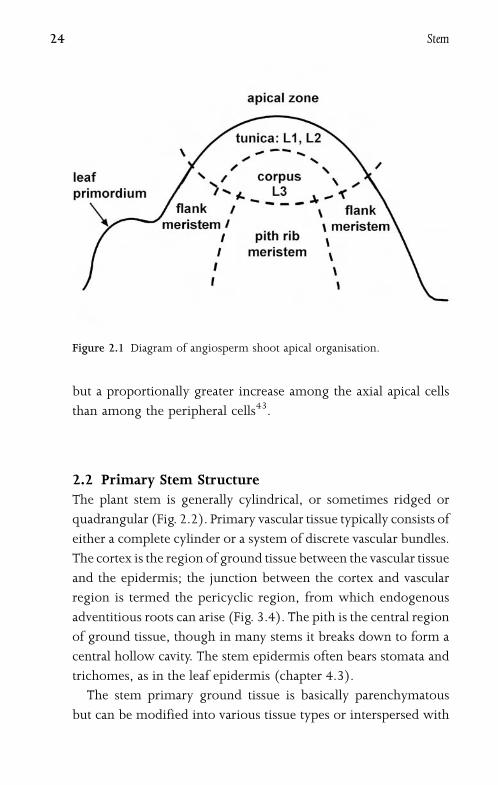
but a proportionally greater increase among the axial apical cells
than among the peripheral cells43.
2.2 Primary Stem StructureThe plant stem is generally cylindrical, or sometimes ridged or
quadrangular (Fig. 2.2). Primary vascular tissue typically consists of
either a complete cylinder or a system of discrete vascular bundles.
The cortex is the region of ground tissue between the vascular tissue
and the epidermis; the junction between the cortex and vascular
region is termed the pericyclic region, from which endogenous
adventitious roots can arise (Fig. 3.4). The pith is the central region
of ground tissue, though in many stems it breaks down to form a
central hollow cavity. The stem epidermis often bears stomata and
trichomes, as in the leaf epidermis (chapter 4.3).
The stem primary ground tissue is basically parenchymatous
but can be modified into various tissue types or interspersed with
Figure 2.1 Diagram of angiosperm shoot apical organisation.
24 Stem
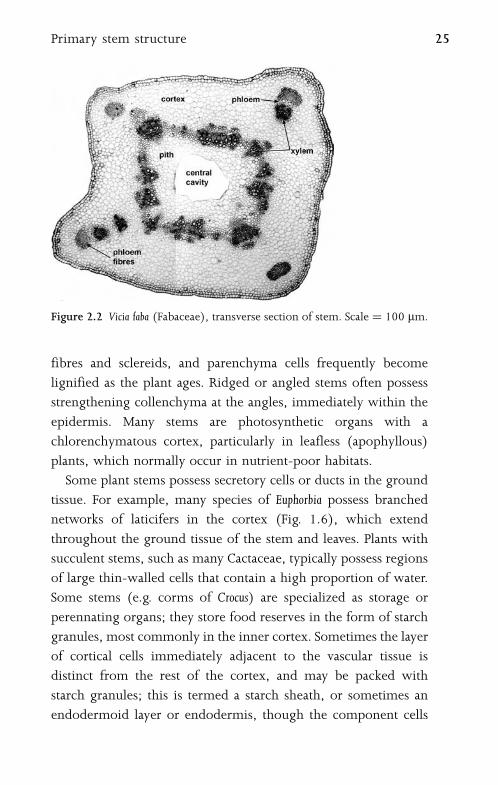
fibres and sclereids, and parenchyma cells frequently become
lignified as the plant ages. Ridged or angled stems often possess
strengthening collenchyma at the angles, immediately within the
epidermis. Many stems are photosynthetic organs with a
chlorenchymatous cortex, particularly in leafless (apophyllous)
plants, which normally occur in nutrient-poor habitats.
Some plant stems possess secretory cells or ducts in the ground
tissue. For example, many species of Euphorbia possess branched
networks of laticifers in the cortex (Fig. 1.6), which extend
throughout the ground tissue of the stem and leaves. Plants with
succulent stems, such as many Cactaceae, typically possess regions
of large thin-walled cells that contain a high proportion of water.
Some stems (e.g. corms of Crocus) are specialized as storage or
perennating organs; they store food reserves in the form of starch
granules, most commonly in the inner cortex. Sometimes the layer
of cortical cells immediately adjacent to the vascular tissue is
distinct from the rest of the cortex, and may be packed with
starch granules; this is termed a starch sheath, or sometimes an
endodermoid layer or endodermis, though the component cells
Figure 2.2 Vicia faba (Fabaceae), transverse section of stem. Scale ¼ 100 mm.
Primary stem structure 25
usually lack the Casparian thickenings that are typically found in
the root endodermis (chapter 3.5).
2.3 Primary Vascular SystemThe primary vascular system is mostly derived from the pro-
cambium near the shoot apex. Primary vascular bundles possess
both xylem and phloem, arranged either adjacent to each other
(in collateral vascular bundles: Fig. 1.12), or with strands of
phloem on both sides of the xylem (bicollateral vascular bundles),
or with xylem surrounding the phloem (amphivasal vascular
bundles). In woody angiosperms, internodal stem vasculature is
typically arranged either in a continuous cylinder, or in a cylinder
of separate or fused collateral bundles, with the phloem external
to the xylem (Fig. 2.2). In some stems the bundles may be
bicollateral; for example in species of Cucurbita internal phloem
is present in addition to the external phloem. The vascular
cambium, which produces secondary vascular tissue in woody
species, is initially situated between the xylem and phloem within
vascular bundles, but eventually extends between the vascular
bundles to form a complete vascular cylinder. Some stems also
possess cortical or medullary (pith) bundles, which can be
associated with leaf vasculature.
In monocots, which lack a vascular cambium, the stem vascu-
lar bundles are typically scattered throughout the central ground
tissue (Fig. 2.3), or sometimes arranged in two or more distinct
rings. Vascular bundles may be collateral, bicollateral or amphi-
vasal. Cortex and pith are frequently indistinct from each other,
though the cortex may be defined by an endodermoid layer, or
a distinct ring of vascular bundles, or in some stems, particularly
inflorescence axes, by a cylinder of sclerenchyma that encloses the
majority of vascular bundles. The monocot vascular system is often
extremely complex128. Each major bundle, when traced on an
upward course from any point in the stem, branches or forms
26 Stem

bridges with other bundles at several points before passing into a
leaf. One of its major branches then continues a similar upward
course towards the apex. Some palms possess literally thousands of
vascular bundles in a single transverse section of the stem,
though in most other monocots the number is much smaller.
2.4 Nodal VasculatureAt regions of leaf insertion on the stem (nodes), the vasculature
of the leaf and stem are connected. Openings (termed lacunae or
leaf gaps) occur in the stem vascular cylinder beneath their
point of contact (Fig. 2.4). In eudicots and magnoliids, nodal
anatomy is often characteristic of taxonomic groups, particularly
the number and arrangement of leaf traces and leaf gaps.
Figure 2.3 Monocot stem anatomy: Lilium tigrinum (Liliaceae), transverse
section of inflorescence axis, showing cortex (c), surrounding central
region with numerous distinct vascular bundles. sc ¼ sclerenchymatous
layer. Scale ¼ 100 mm.
Nodal vasculature 27

Nodes may be unilacunar, trilacunar or multilacunar, depending
on the number of leaf gaps in the stem vascular cylinder. This
feature is most obvious in stems in which there is otherwise
a continuous vascular cylinder, especially where a limited amount
of secondary thickening has taken place; as a result, nodal anatomy
has been studied far more extensively in woody than herbaceous
plants54. Sometimes the number of leaf gaps per node varies
within a species or individual, usually increasing with increased
plant size and age67.
Figure 2.4 Prunus lusitanica (Rosaceae), transverse section of stem at node,
showing connection of petiole vasculature to main vascular cylinder of stem.
lg ¼ leaf gap, lt ¼ leaf trace. Scale ¼ 100 mm.
28 Stem
The number of leaf traces departing from each gap is also
generally characteristic of a species, but may vary within a plant,
especially in species with unilacunar and trilacunar nodes.
For example, in Clerodendrum two traces typically depart from a
single gap, and in Prunus a single trace departs from each of three
gaps in the central vascular cylinder (Fig. 2.4). In Quercus up to
five traces depart through a trilacunar node. Normally, leaf
trace bundles are initiated acropetally from the stem procambial
system near the shoot apex, to serve developing primordia67.
However, in some species (e.g. Populus deltoides) subsidiary vascular
bundles are initiated at the base of each developing primordium,
and grow basipetally to meet the stem procambial trace.
Nodal vasculature is further complicated by the axillary bud
vascular traces, which are connected to the main stem vascu-
lature immediately above the leaf gaps. In most species two
traces diverge to supply each bud or branch.
In large woody trees, the junction of the trunk and its
branches is characterized by a complex arrangement of secondary
vascular tissue, which typically forms a collar around the base of
the branch99. This branch collar is enveloped by a trunk collar,
which links the vascular tissue of the trunk above and below the
branch. There is no direct connection of xylem from the trunk
above a branch into the branch xylem, as the tissues are oriented
perpendicular to each other. If a branch dies, a protection zone
forms around its base to prevent spread of infection into the trunk,
and the branch is often shed.
2.5 Vascular CambiumIncrease in height, achieved by growth at the apical meristem,
is inevitably followed by at least some degree of increase in stem
thickness. This is achieved by different types of meristems in
different species. In woody eudicots and most magnoliids (but not
monocots), secondary vascular tissue (both xylem and phloem)
Vascular cambium 29

is produced by the vascular cambium (Fig. 2.5), which usually
becomes active at a short distance behind the stem apex. The
vascular cambium is initiated between xylem and phloem within
vascular bundles, but soon consists of an unbroken cylinder of
meristematic cells. It typically generates secondary xylem (wood)
at its inner edge and secondary phloem at its outer edge, though
plants with anomalous secondary growth do not always follow
this pattern. The amount of secondary vascular tissue produced is
extremely variable, depending on the habit of the plant. Vascular
cambium is absent in monocots and some herbaceous eudicots
(e.g. Ranunculus) and magnoliids (e.g. Saururus).
The vascular cambium is a single cell layer (uniseriate) or
several cell layers (multiseriate) if xylem and phloem mother
cells are included55. It is a complex tissue consisting of both
Figure 2.5 Prunus communis (Rosaceae). Transverse section of stem in region
of vascular cambium, with secondary phloem (above) and secondary xylem
(below). cc ¼ companion cell, r ¼ ray, s ¼ sieve element, vc ¼ vascular
cambium, ve ¼ vessel element.
30 Stem

fusiform initials and ray initials, which form the axial and radial
systems respectively. Both fusiform and ray initials are vacuolate
(unlike most meristematic tissue) and plastid-rich. Fusiform
initials are axially elongated cells with tapering ends. They
divide periclinally to form the axial elements of secondary tissues:
tracheary elements, fibres and axial parenchyma in secondary
xylem, and sieve elements, companion cells and fibres in second-
ary phloem. Ray initials are isodiametric cells that divide peri-
clinally to form ray parenchyma cells in both xylem and phloem.
Fusiform initials sometimes give rise to new ray initials as the stem
increases in circumference and new rays are formed.
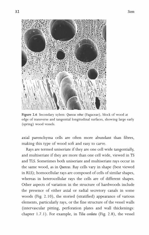
2.6 Secondary XylemSecondary xylem (wood) varies considerably between species.
The texture and density of a particular type of wood depend
on the size, shape and arrangement of its constituent cells73.
Wood is composed of a matrix of cells (Fig. 2.6), some arranged
parallel to the long axis (fibres, vessels and chains of axial
parenchyma cells), and others (ray parenchyma cells) forming
the wood rays that extend radially from the vascular cambium
towards the pith. The precise cellular arrangement in wood is
often characteristic of species or genera. To observe their structure,
woods are sectioned transversely (transverse section: TS) and in
two longitudinal planes: along the rays (radial longitudinal
section: RLS) and perpendicular to the rays (tangential longi-
tudinal section: TLS). In some woods the vessels are solitary when
viewed in transverse section (Figs 2.6, 2.10), but in other woods
they are arranged in clusters or radial chains (Fig. 2.9). Axial
parenchyma cells may be independent of the vessels (apotracheal)
or associated with them (paratracheal), and sometimes occur in
regular tangential bands. The relative abundance of axial paren-
chyma varies in different species, from sparse (or even completely
absent) to rare cases such as Ochroma pyramidale (balsa), in which
Secondary xylem 31
axial parenchyma cells are often more abundant than fibres,
making this type of wood soft and easy to carve.
Rays are termed uniseriate if they are one cell wide tangentially,
and multiseriate if they are more than one cell wide, viewed in TS
and TLS. Sometimes both uniseriate and multiseriate rays occur in
the same wood, as in Quercus. Ray cells vary in shape (best viewed
in RLS); homocellular rays are composed of cells of similar shapes,
whereas in heterocellular rays the cells are of different shapes.
Other aspects of variation in the structure of hardwoods include
the presence of either axial or radial secretory canals in some
woods (Fig. 2.10), the storied (stratified) appearance of various
elements, particularly rays, or the fine structure of the vessel walls
(intervascular pitting, perforation plates and wall thickenings:
chapter 1.7.1). For example, in Tilia cordata (Fig. 2.8), the vessel
Figure 2.6 Secondary xylem: Quercus robur (Fagaceae), block of wood at
edge of transverse and tangential longitudinal surfaces, showing large early
(spring) wood vessels.
32 Stem

element walls are helically thickened, and in many Fabaceae the pit
apertures are surrounded by numerous warty protuberances,
termed vesturing19. Perforated ray cells, an unusual feature of some
woods, are ray cells that link two vessel elements and themselves
resemble and function as vessel elements, with perforation plates
corresponding to those of the adjacent vessel elements. However,
like other ray cells, perforated ray cells are formed from ray initials
rather than from fusiform initials, like vessel elements.
In many woody temperate plants cambial activity is seasonal
(usually annual), which results in the formation of growth rings.
The secondary xylem formed in the early part of the season (early
wood or spring wood) is generally less dense and consists of
thinner-walled cells than the xylem formed later in the growing
season (late wood or summer wood). In ring-porous woods
the vessels are considerably larger in early wood than in late
wood (Fig. 2.11). In diffuse porous woods the main distinction
between early and late wood is in size and wall thickness of the
fibres (Fig. 2.9). As woody plants age and their trunks increase in
Figure 2.7 Betula utilis (Betulaceae). Wood in (A) tangential longitudinal
section (TLS) and (B) radial longitudinal section (RLS). b ¼ bar of
scalariform perforation plate, r ¼ ray. Scale ¼ 100 mm.
Secondary xylem 33
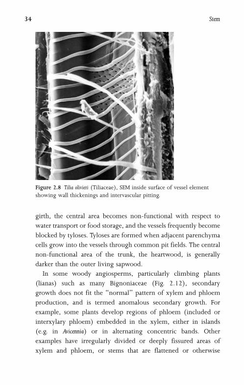
girth, the central area becomes non-functional with respect to
water transport or food storage, and the vessels frequently become
blocked by tyloses. Tyloses are formed when adjacent parenchyma
cells grow into the vessels through common pit fields. The central
non-functional area of the trunk, the heartwood, is generally
darker than the outer living sapwood.
In some woody angiosperms, particularly climbing plants
(lianas) such as many Bignoniaceae (Fig. 2.12), secondary
growth does not fit the ‘‘normal’’ pattern of xylem and phloem
production, and is termed anomalous secondary growth. For
example, some plants develop regions of phloem (included or
interxylary phloem) embedded in the xylem, either in islands
(e.g. in Avicennia) or in alternating concentric bands. Other
examples have irregularly divided or deeply fissured areas of
xylem and phloem, or stems that are flattened or otherwise
Figure 2.8 Tilia olivieri (Tiliaceae), SEM inside surface of vessel element
showing wall thickenings and intervascular pitting.
34 Stem
irregularly shaped73. Such anomalous forms are achieved either
by the formation of new vascular cambia in unusual positions or
by the unusual behaviour of the existing cambium in producing
phloem instead of xylem at certain points.
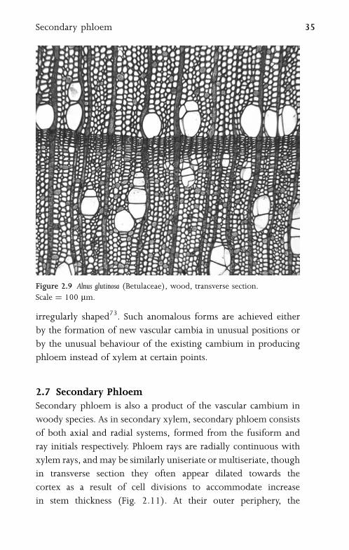
2.7 Secondary PhloemSecondary phloem is also a product of the vascular cambium in
woody species. As in secondary xylem, secondary phloem consists
of both axial and radial systems, formed from the fusiform and
ray initials respectively. Phloem rays are radially continuous with
xylem rays, and may be similarly uniseriate or multiseriate, though
in transverse section they often appear dilated towards the
cortex as a result of cell divisions to accommodate increase
in stem thickness (Fig. 2.11). At their outer periphery, the
Figure 2.9 Alnus glutinosa (Betulaceae), wood, transverse section.
Scale ¼ 100 mm.
Secondary phloem 35

parenchymatous ray cells are often difficult to distinguish from
cortical cells. Older ray cells sometimes become lignified to form
sclereids. The axial system of the phloem consists of sieve elements
and companion cells, as in primary phloem (chapter 1.9.2). It also
typically includes fibres, sclereids and axial parenchyma cells.
In some species fibres are formed in groups at regular intervals,
resulting in characteristic tangential bands of fibres alternating
with groups of sieve elements and parenchyma cells.
2.8 Primary and Secondary Thickening MeristemsIn monocots, which lack a vascular cambium, increase in stem
diameter is typically relatively limited. However, most monocots
Figure 2.10 Shorea resina-nigra (Dipterocarpaceae), wood, transverse
section showing vessels (v) and axial secretory canals (sc), r ¼ ray.
Scale ¼ 100 mm.
36 Stem
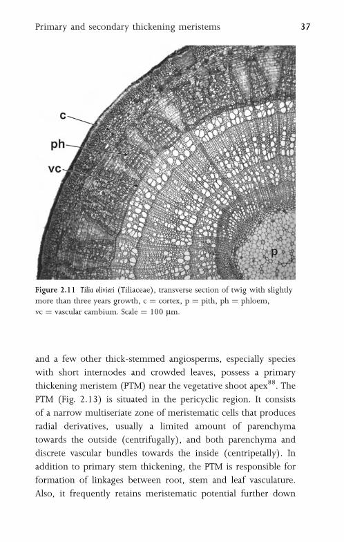
and a few other thick-stemmed angiosperms, especially species
with short internodes and crowded leaves, possess a primary
thickening meristem (PTM) near the vegetative shoot apex88. The
PTM (Fig. 2.13) is situated in the pericyclic region. It consists
of a narrow multiseriate zone of meristematic cells that produces
radial derivatives, usually a limited amount of parenchyma
towards the outside (centrifugally), and both parenchyma and
discrete vascular bundles towards the inside (centripetally). In
addition to primary stem thickening, the PTM is responsible for
formation of linkages between root, stem and leaf vasculature.
Also, it frequently retains meristematic potential further down
Figure 2.11 Tilia olivieri (Tiliaceae), transverse section of twig with slightly
more than three years growth, c ¼ cortex, p ¼ pith, ph ¼ phloem,
vc ¼ vascular cambium. Scale ¼ 100 mm.
Primary and secondary thickening meristems 37

the stem and is the site of adventitious root production in some
species.
The PTM normally ceases activity at a short distance behind
the apex, and subsequent stem thickening is limited. Tree-forming
palms possess an extensive PTM that forms a large sunken apex;
considerable further stem thickening occurs by subsequent
division and enlargement of ground parenchyma cells. This is
termed diffuse secondary growth.
In some woody monocots in the order Asparagales (e.g. Agave,
Aloe, Cordyline, Yucca) further increase in stem thickness is achieved
by means of a secondary thickening meristem (STM) (Fig. 2.14).
The STM is essentially similar to the PTM in that it is located in
the pericyclic region and produces radial derivatives. However,
it is active further from the primary apex and produces second-
ary vascular bundles that are often amphivasal and radially
elongated. In some species (e.g. Nolina recurvata, Cordyline terminalis)
Figure 2.12 Tynanthus elegans (Bignoniaceae). Transverse section of woody
stem showing anomalous secondary growth: xylem region with four
deep fissures of phloem. Scale ¼ 1 mm.
38 Stem
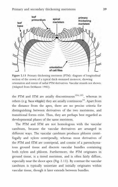
the PTM and STM are axially discontinuous104,105, whereas in
others (e.g. Yucca whipplei) they are axially continuous26. Apart from
the distance from the apex, there are no precise criteria for
distinguishing between derivatives of the two meristems, and
transitional forms exist. Thus, they are perhaps best regarded as
developmental phases of the same meristem.
The PTM and STM are not homologous with the vascular
cambium, because the vascular derivatives are arranged in
different ways. The vascular cambium produces phloem centri-
fugally and xylem centripetally, whereas most derivatives of
the PTM and STM are centripetal, and consist of a parenchyma-
tous ground tissue and discrete vascular bundles containing
both xylem and phloem. Furthermore, the PTM originates in
ground tissue, is a tiered meristem, and is often fairly diffuse,
especially near the shoot apex (Fig. 2.13). By contrast the vascular
cambium is typically uniseriate and initially originates within
vascular tissue, though it later extends between bundles.
Figure 2.13 Primary thickening meristem (PTM): diagram of longitudinal
section of the crown of a typical thick-stemmed monocot, showing
orientation and extent of radial PTM derivatives. Vascular strands not shown.
(Adapted from DeMason 1983).
Primary and secondary thickening meristems 39

2.9 PeridermPeriderm is a protective tissue of corky (suberinized) cells that
is produced either as a response to wounding or in the outer
layers of the cortex of a stem or root that has increased in
thickness. The periderm consists of up to three layers: phellogen,
phellem and phelloderm. The phellogen is a uniseriate meri-
stematic layer of thin-walled cells that produces phellem to the
outside, and (in some cases) phelloderm to the inside. The
phellem cells constitute the corky tissue. They are tightly-packed
cells that lack contents at maturity. They possess deposits of
suberin and sometimes lignin in their walls, and form an
impervious layer to prevent water loss and protect against injury.
Phelloderm cells are non-suberinized and parenchymatous, and
contribute to the secondary cortex.
Figure 2.14 Secondary thickening in monocots: Dracaena indivisa
(Ruscaceae), transverse section of stem showing secondary thickening
meristem (STM) and radial internal vascular derivatives. Scale ¼ 100 mm(left hand image).
40 Stem
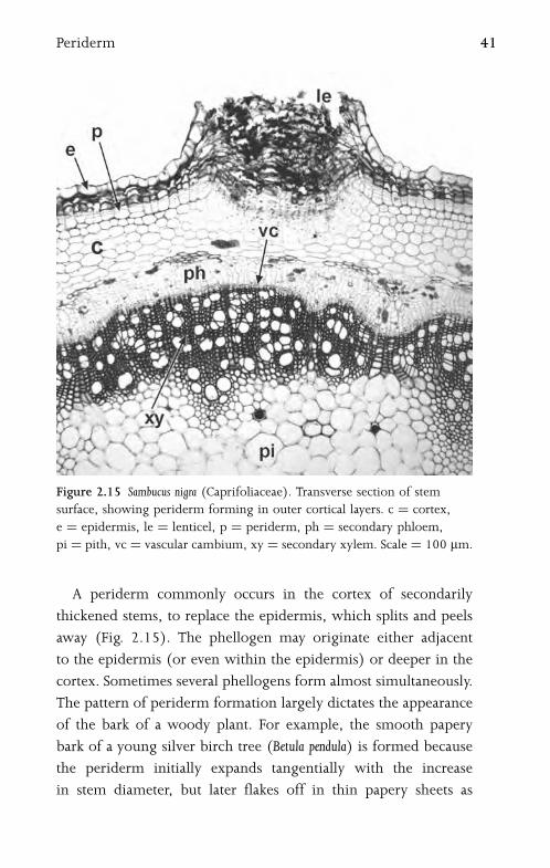
A periderm commonly occurs in the cortex of secondarily
thickened stems, to replace the epidermis, which splits and peels
away (Fig. 2.15). The phellogen may originate either adjacent
to the epidermis (or even within the epidermis) or deeper in the
cortex. Sometimes several phellogens form almost simultaneously.
The pattern of periderm formation largely dictates the appearance
of the bark of a woody plant. For example, the smooth papery
bark of a young silver birch tree (Betula pendula) is formed because
the periderm initially expands tangentially with the increase
in stem diameter, but later flakes off in thin papery sheets as
Figure 2.15 Sambucus nigra (Caprifoliaceae). Transverse section of stem
surface, showing periderm forming in outer cortical layers. c ¼ cortex,
e ¼ epidermis, le ¼ lenticel, p ¼ periderm, ph ¼ secondary phloem,
pi ¼ pith, vc ¼ vascular cambium, xy ¼ secondary xylem. Scale ¼ 100 mm.
Periderm 41
abscission bands of thin-walled cells are formed. In the trunk of
cork oak (Quercus suber), the initial phellogen may continue activity
indefinitely, and produces seasonal growth rings. In the commer-
cial process it is removed after about 20 years to make way
for a second, more vigorous phellogen, which produces the
commercial cork.
Many species possess lenticels in the bark (Fig. 2.15); these are
areas of loose cells in the periderm, which are often initially
formed beneath stomata in the epidermis, and are thought to be
similarly concerned with gaseous exchange.
42 Stem
3
Root
3.1 Primary Root StructureThe seedling radicle ultimately becomes the primary root
(tap root), which frequently develops side branches (lateral
roots). In monocots the seedling radicle commonly dies at an
early stage; the stem-borne (adventitious) roots of the mature
plant originate from differentiated cells (Fig. 3.4). Adventitious
roots can be branched or unbranched. Although roots can
originate from various organs, their basic primary structure
retains a characteristic root groundplan that is different from
that of the stem. Each root possesses clearly-defined concentric
tissue regions: dermal tissue (epidermis), ground tissue (cortex,
including the endodermis) and central vascular tissue surrounded
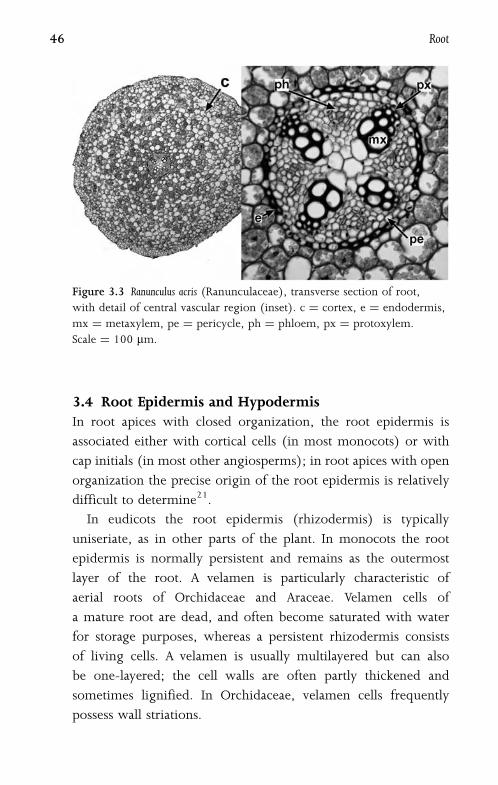
by a pericycle (Fig. 3.3).
3.2 Root ApexRoot apices possess a terminal protective root cap and a proximal
root apical meristem8,32 (Fig. 3.1). The quiescent centre is
a group of relatively inactive cells at the very centre and tip of
the root apical meristem. The cells of the quiescent centre divide
infrequently; their role is obscure, but they maintain initial
cells in an undifferentiated state. These cells, together with the
root cap initials, are derived from the uppermost cell of the
suspensor (hypophysis) in the embryo18 (Fig. 6.7). Cell division
activity occurs in the cells surrounding the quiescent centre.

In Arabidopsis thaliana the initial cells lie in clearly defined regions
relative to the quiescent centre, the pericycle and vascular initials
proximal to it (on the shoot side), the root cap and epidermis
initials distal to it (on the root cap side) and the cortical and
endodermal initials radial to it. However, in other species
(e.g. Vicia faba) there is an undifferentiated initiating region
common to all root tissues102. The active region is termed the
promeristem.
The junction between the root cap and the root apical meristem
is either clearly defined by a distinct cell boundary (termed closed
organization, as in Zea mays and Arabidopsis thaliana), or ill-defined
(termed open structure, as in Vicia faba: Fig. 3.2), though inter-
mediates exist (e.g. in Daucus carota)11,21. In open meristems the
boundary between the cap and the rest of the root is unstable.
Figure 3.1 Diagram of root apical organization in Zea mays (Poaceae),
a species with closed structure. Arrows indicate direction of displacement
of cell derivatives. (Adapted from Feldman 1984).
44 Root

3.3 Root CapThe root cap is composed of several layers of parenchymatous
cells. The cells of the root cap are initially derived from the
apical meristem. However, ontogenetic studies in maize (Zea mays),
a species with ‘‘closed’’ root apical structure (Fig. 3.1), have
shown that the cap initials become established and independent
from the apical meristem at an early stage in seedling develop-
ment8. The cap meristematic cells, located adjacent (distal) to the
quiescent centre, produce derivatives that are eventually displaced
towards the outside of the root cap, and subsequently sloughed
off, contributing to the external slime that allows the root to push
through the soil. Cells are generated and lost in the root cap at
approximately the same rate.
Figure 3.2 Vicia faba (Fabaceae), longitudinal section of root apex, showing
open apical structure. rc ¼ root cap. Scale ¼ 100 mm.
Root cap 45
3.4 Root Epidermis and HypodermisIn root apices with closed organization, the root epidermis is
associated either with cortical cells (in most monocots) or with
cap initials (in most other angiosperms); in root apices with open
organization the precise origin of the root epidermis is relatively
difficult to determine21.
In eudicots the root epidermis (rhizodermis) is typically
uniseriate, as in other parts of the plant. In monocots the root
epidermis is normally persistent and remains as the outermost
layer of the root. A velamen is particularly characteristic of
aerial roots of Orchidaceae and Araceae. Velamen cells of
a mature root are dead, and often become saturated with water
for storage purposes, whereas a persistent rhizodermis consists
of living cells. A velamen is usually multilayered but can also
be one-layered; the cell walls are often partly thickened and
sometimes lignified. In Orchidaceae, velamen cells frequently
possess wall striations.
Figure 3.3 Ranunculus acris (Ranunculaceae), transverse section of root,
with detail of central vascular region (inset). c ¼ cortex, e ¼ endodermis,
mx ¼ metaxylem, pe ¼ pericycle, ph ¼ phloem, px ¼ protoxylem.
Scale ¼ 100 mm.
46 Root

Most angiosperms possess absorptive root hairs in under-
ground roots, usually confined to a region about a centimetre
from the root apex, beyond the meristematic region, but in an
area where cells are still enlarging. In general, root hairs persist
for only a limited period before withering. This region of the
root is the most active in absorption of water, and the root
hairs serve to present a greater surface area for this purpose. Root
hairs are formed from epidermal cells by apical intrusive growth.
In some plants only specialized root epidermal cells (trichoblasts)
are capable of root hair production. Trichoblasts are formed in
Figure 3.4 Ligustrum vulgare (Oleaceae), transverse section of stem with
adventitious roots. Scale ¼ 100 mm.
Root epidermis and hypodermis 47
meristematic epidermal cells that overlie the junction between
two cortical cells18. Thus, in many species the root epidermis is
dimorphic and clearly differentiated into short cells (trichoblasts)
and long cells (sometimes termed atrichoblasts), as in Arabidopsis
thaliana. Some other species (including many monocots such as
species of Asparagales and Araceae) instead possess a dimorphic
hypodermal layer immediately below the root epidermis63; this is
normally interpreted as the outermost cortical (exodermal) layer
but may represent the innermost layer of a multilayered persistent
rhizodermis. The hypodermal short cells resemble trichoblasts,
and are probably transfusion cells.
3.5 Root Cortex and EndodermisThe cortex is the region between the pericycle and the epidermis,
including the innermost layer, the endodermis. In underground
roots the rhizodermis becomes worn away, and is replaced as
an outer layer either by a periderm that forms in the cortex
(in most woody eudicots and magnoliids) or by a suberinized or
lignified exodermis (in some monocots), which is sometimes
multilayered.
Apart from these specialized layers, most cortical cells are
parenchymatous and often perform an important storage function.
In some plants, such as Daucus carota (carrot), the tap root is
a modified swollen storage organ with awide cortex. In most roots
the bulk of the cortical cells are formed by sequential periclinal
divisions, the innermost cells (later the endodermis) being the
last formed.
Many plants with underground stems (corms, bulbs or
rhizomes), particularly bulbous or cormous monocots such as
Crocus, Freesia and Hyacinthus, periodically produce contractile roots
which draw the stem deeper into the soil57. These roots grow
downwards, and then shorten vertically and expand radially. They
48 Root

are recognizable by their wrinkled surface, and characteristically
possess two or three clearly distinct concentric regions of cortical
parenchyma, distinguishable by cell size, including a region of
collapsed outer cortical cells interspersed with occasional thicker-
walled cells. In some species the process of root contraction is
initiated by active cell enlargement in the inner cortex, followed
by collapse of outer cortical cells and subsequent surface folding.
In other species the collapse of outer cortical cells results from
the difference between atmospheric pressure and relatively low
xylem pressure (due to transpiration), causing centripetal loss
of turgidity.
The endodermis is a uniseriate cylinder of cortical cells sur-
rounding the central vascular region, adjacent to the pericycle.
Endodermal cells are typically characterized by deposition of
a band of suberin or lignin in their primary walls, termed a
Casparian strip, which forms a barrier against non-selective passage
of water through the endodermis. Older endodermal cells often
possess thick lamellated secondary walls, in most cases on the inner
periclinal wall, so that the Casparian strip is not apparent. The
secondary wall is often lignified, and therefore serves as a second
effective barrier to water loss. Occasional endodermal cells
(passage cells) can remain thin-walled, probably for selective
passage of water between the cortex and vascular region.
3.6 Pericycle and Vascular CylinderThe vascular tissue in the centre of the root is surrounded by
a single layer (or rarely, more layers) of thin-walled cells, termed
the pericycle (Fig. 3.3). Both the pericycle and vascular tissue are
derived from cells on the proximal (shoot) side of the quiescent
centre. The pericycle is potentially meristematic in younger roots,
as it is the site of lateral root initiation, but in older roots it can
become lignified.
Pericycle and vascular cylinder 49
The primary vascular tissue consists of several strands of phloem
alternating with the rays of a central area of xylem that appears
star-shaped in transverse section. In a mature root, the protoxylem
elements, which were the first-formed and are the narrowest in
diameter, are located at the tips of the rays, nearest to the pericycle.
The metaxylem elements are larger and located closer to the centre
of the root. Both xylem and phloem are exarch in the root
(i.e. they mature centripetally). Similarly, the protophloem is
located close to the pericycle, in contrast with the metaphloem,
which is situated closer to the centre of the root.
Roots possess two, three, four or more protoxylem poles (rays),
inwhich case they are said to be diarch, triarch, tetrarch or polyarch
respectively. There is often variation in the number of xylem
poles, sometimes even within the same plant, depending on the
diameter of the root. Most commonly, roots possess relatively
few xylem poles (usually two, three or four) and the central region
is occupied by a group or ring of xylem vessels. However,
some monocots (e.g. Iris) possess polyarch roots, and the centre
of the root is parenchymatous, sometimes becoming lignified
in older roots.
3.7 Initiation of Lateral and Adventitious RootsLateral roots are branches of the tap root. They are initiated
in relatively mature tissues some distance from the apex, often in
acropetal sequence; the most recently-formed lateral roots are
usually those nearest to the root apical meristem. In angiosperms,
lateral roots have a deep-seated (endogenous) origin. Root forma-
tion is usually initiated in groups of ‘‘founder cells’’ in the peri-
cycle, often adjacent to the xylem poles. The position of lateral
root initiation in the pericycle is usually at a point adjacent to
a protoxylem pole, unless the root is diarch, in which case initia-
tion is sometimes opposite a phloem pole. However, in monocots
lateral root initiation can be opposite either protoxylem or phloem
50 Root

poles, though in roots with a large number of vascular poles it
is often difficult to determine the precise site of initiation72.
The founder cells undergo a series of periclinal and anticlinal
divisions to form a lateral root primordium. In many species
some subsequent cell divisions occur in the endodermis, so that
ultimately both the pericycle and the endodermis contribute to
the tissues of the lateral root. The growing lateral root pushes its
way through the cortex and epidermis of the parent root, either by
mechanical or enzymatic action.
Adventitious roots are formed in other parts of the plant,
primarily stem tissue. They have various sites of origin, from
deep-seated (endogenous) (Fig. 3.4), to (more rarely) exogenous,
arising from superficial tissues such as the epidermis (e.g. in
surface-rooting Begonia leaves). In most monocots adventitious
roots arise from cell divisions in the pericyclic region of the stem;
the primary thickening meristem contributes to adventitious root
formation (chapter 2.8). Adventitious roots are often formed at
nodes on the stem, which is why in horticulture cuttings are most
commonly taken from just below a node. Adventitious roots may
also form from callus tissue at the site of a wound.
3.8 Secondary Growth in RootsIn some woody eudicots the thickening and strengthening of the
root system is important in supporting the trunk. Most dicot roots
possess at least a small amount of secondary thickening (Fig. 3.5),
with the exception of a few herbaceous species such as Ranunculus
(Fig. 3.3). In contrast, secondary growth in roots is extremely rare
in monocots, even among arborescent or woody species that
possess a secondary thickening meristem (chapter 2.8). A notable
exception is Dracaena, in which a limited region of secondary tissue
is formed111.
As in the stem, secondary vascular tissues of the root are
produced by a vascular cambium. This initially develops in the
Secondary growth in roots 51
Figure 3.5 Ulmus sp. (Ulmaceae), transverse sections of roots with (top)
recent secondary growth and (bottom) more extensive secondary thickening.
pe ¼ periderm, ph ¼ phloem, vc ¼ vascular cambium, xy ¼ xylem.
52 Root

regions between the primary xylem and phloem, then in deriv-
atives of cell divisions in the pericycle next to the xylem poles.
Since cambial activity proceeds in this sequence, the xylem
cylinder soon appears circular in transverse section (Fig. 3.5).
Further pericyclic cell divisions result in a secondary cortex, and
in many cases a periderm forms, particularly where secondary
growth is extensive. The epidermis splits and is sloughed off
together with the primary cortex and endodermis. Root secondary
xylem usually resembles that of the stem in the same plant, but
may differ in several respects. For example, in Quercus robur stem
wood is ring porous, with earlywood vessels markedly larger than
latewood vessels, but root wood is diffuse porous, with vessels of
relatively consistent sizes across each growth ring. As with trunk
wood, root wood of individual taxa often exhibits identifiable
characteristics24.
3.9 Roots Associated with Micro-OrganismsMany vascular plants form symbiotic relationships with soil
microorganisms. In legumes, nitrogen-fixing bacteria invade the
root cortex through root hairs, and stimulate meristematic activity
in the cortex (and sometimes also in the pericycle) to form a root
nodule, which often becomes elongated to resemble a short
lateral root. Other soil micro-organisms may induce the formation
of modified lateral roots. For example, in many woody angio-
sperms, invading filamentous bacteria promote the development
of short, swollen lateral roots, and in some temperate woody forest
species, especially in the families Fagaceae and Betulaceae,
ectomycorrhizal fungi form a mantle over stunted lateral roots.
By contrast, the more common endomycorrhizal fungi, which
invade the cells of the host root, often have little influence on
root morphology (Fig. 3.6).
Roots associated with micro-organisms 53
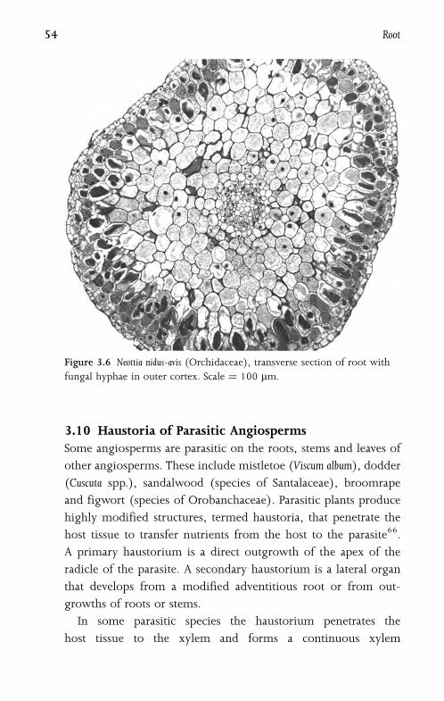
3.10 Haustoria of Parasitic AngiospermsSome angiosperms are parasitic on the roots, stems and leaves of
other angiosperms. These include mistletoe (Viscum album), dodder
(Cuscuta spp.), sandalwood (species of Santalaceae), broomrape
and figwort (species of Orobanchaceae). Parasitic plants produce
highly modified structures, termed haustoria, that penetrate the
host tissue to transfer nutrients from the host to the parasite66.
A primary haustorium is a direct outgrowth of the apex of the
radicle of the parasite. A secondary haustorium is a lateral organ
that develops from a modified adventitious root or from out-
growths of roots or stems.
In some parasitic species the haustorium penetrates the
host tissue to the xylem and forms a continuous xylem
Figure 3.6 Neottia nidus-avis (Orchidaceae), transverse section of root with
fungal hyphae in outer cortex. Scale ¼ 100 mm.
54 Root
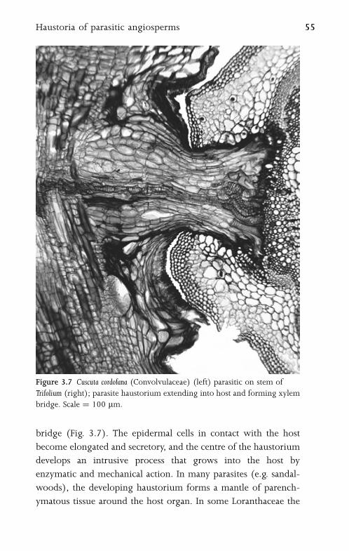
bridge (Fig. 3.7). The epidermal cells in contact with the host
become elongated and secretory, and the centre of the haustorium
develops an intrusive process that grows into the host by
enzymatic and mechanical action. In many parasites (e.g. sandal-
woods), the developing haustorium forms a mantle of parench-
ymatous tissue around the host organ. In some Loranthaceae the
Figure 3.7 Cuscuta cordofana (Convolvulaceae) (left) parasitic on stem of
Trifolium (right); parasite haustorium extending into host and forming xylem
bridge. Scale ¼ 100 mm.
Haustoria of parasitic angiosperms 55
haustorium does not invade the host tissues after forming a
mantle, but instead induces the host tissue to form a placenta-like
outgrowth of vascular tissue to supply nutrients to the parasite.
When the parasite dies, a woody outgrowth of convoluted host
tissue remains as a vestige known as a ‘‘woodrose’’.
56 Root
4
Leaf
4.1 Leaf Morphology and AnatomyAngiosperm leaves display much morphological and anatomical
diversity. Mature leaves of monocots are typically narrow and
consist of a linear lamina with parallel venation and a leaf base
that ensheathes the stem. This contrasts with the typical leaf
of eudicots and magnoliids, which has a well-defined petiole
and elliptical blade (lamina) with reticulate venation. However,
exceptions and transitional forms are common; for example,
leaves of some monocots (e.g. Dioscorea and Smilax) are petiolate and
net-veined, and leaves of some eudicots (e.g. some Apiaceae)
are linear. Some species possess compound leaves in which
individual leaflets are borne either on a central stem-like axis
(pinnate leaves; e.g. tomato, Solanum lycopersicum) or radiate from
a single point at the distal end of the petiole (palmate leaves;
e.g. Arisaema).
Some species that grow in dry (xeric) or seasonally dry habitats,
or otherwise nutrient-deficient habitats, possess specialized xero-
morphic features, including sunken stomata to minimize water
loss and well-developed sclerenchyma to provide mechanical
support and minimize tissue collapse. Other xeromorphic features
include the presence of a hypodermis or thick epidermis and thick
cuticle which diminish the intensity of light that reaches
photosynthetic tissue. Well-developed palisade tissue is also some-
times correlated with high light intensity. Some xeromorphic
species possess thick, sometimes even succulent, leaves; others
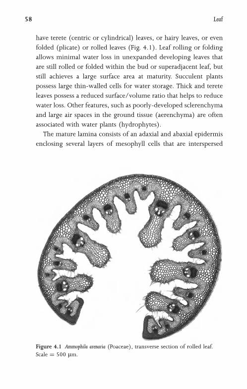
have terete (centric or cylindrical) leaves, or hairy leaves, or even
folded (plicate) or rolled leaves (Fig. 4.1). Leaf rolling or folding
allows minimal water loss in unexpanded developing leaves that
are still rolled or folded within the bud or superadjacent leaf, but
still achieves a large surface area at maturity. Succulent plants
possess large thin-walled cells for water storage. Thick and terete
leaves possess a reduced surface/volume ratio that helps to reduce
water loss. Other features, such as poorly-developed sclerenchyma
and large air spaces in the ground tissue (aerenchyma) are often
associated with water plants (hydrophytes).
The mature lamina consists of an adaxial and abaxial epidermis
enclosing several layers of mesophyll cells that are interspersed
Figure 4.1 Ammophila arenaria (Poaceae), transverse section of rolled leaf.
Scale ¼ 500 mm.
58 Leaf

with a network of vascular bundles (Fig. 4.2). Each tissue may
be variously differentiated into specialized cell types, though the
degree of differentiation varies considerably among taxa. Leaves
are most commonly bifacial (dorsiventral), in which case the
upper and lower surfaces differ morphologically; for example,
in relative numbers of stomata and trichomes. However,
some species possess isobilateral or unifacial leaves, in which the
epidermis and mesophyll are similar on both surfaces. In unifacial-
leafed monocots the leaf base is bifacial and the lamina is unifacial
and either flattened (e.g. in Acorus and most Iridaceae) or rounded
(terete) (e.g. in some Allium species).
Stipules are (often leaf-like) foliar appendages that occur at leaf
base in some angiosperms, especially eudicots. Ligules are out-
growths of the abaxial epidermis that occur in the region between
the leaf sheath and lamina in grasses and several other mono-
cot groups94; they are derived from a cross zone in the leaf
Figure 4.2 Ficus pretoriae (Moraceae), transverse section of leaf. bs ¼ bundle
sheath, bse ¼ bundle sheath extension, e ¼ epidermis, pm ¼ palisade
mesophyll, sm ¼ spongy mesophyll, st ¼ stomatal pore. Scale ¼ 100 mm.
Leaf morphology and anatomy 59
primordium. Squamules are small hair-like structures associated
with the axils of foliage leaves and sometimes floral pedicels,
especially in Alismatales (e.g. Potamogeton) and Brassicaceae
(e.g. Arabis); in some species they secrete mucilage.
4.2 Leaf DevelopmentLeaves are initiated from groups of founder cells close to the
stem apex. These undergo periclinal divisions either in the outer-
most cell layers or in the layers immediately below them, to form
small conical projections (leaf primordia)46,107. In monocots
the leaf primordium rapidly develops into a bifacial hood-like
structure, and the base of the primordium partially or wholly
encircles the stem, forming a leaf sheath94. Within a simple
leaf, most subsequent meristematic activity occurs in a highly
plastic transition zone between the precursor tip and sheath46.
One or more adaxial cross (transverse) meristems in the transi-
tion zone give rise to many other structures such as ligules and
stipules.
The adaxial marginal cells divide rapidly to form a flattened
leaf blade. This marginal growth is suppressed in the region that
later becomes the petiole, and in many monocots it often occurs
at the same time as apical growth. Marginal growth is subsequently
replaced by cell divisions across the whole leaf blade; by this
stage the approximate number of cell layers has been established,
and the whole lamina functions as a plate meristem. Cell divisions
are mainly anticlinal, resulting in regular layers of cells that
are disrupted only by the differentiation and maturation of the
vascular bundles.
Rates of growth and cell division sometimes vary in different
parts of the leaf. Individual leaflets of a compound leaf
may be produced either acropetally or basipetally. For example,
in the tomato (Solanum lycopersicum), the uppermost (distal)
leaflets are initiated first, followed by middle and lower leaflets
60 Leaf

(i.e. a basipetal sequence)56. Smaller intermediate leaflets are
formed later, in a more chaotic sequence. In some monocots,
meristematic activity governing leaf elongation is restricted to a
region at the base of the leaf, the basal rib meristem; this results
in axial files of cells that increase in maturity towards the
distal end of the leaf. The unifacial leaves of some monocots
(e.g. Acorus), which possess a bifacial sheathing leaf base and
a unifacial upper blade, result from suppressed marginal growth
and the presence of an adaxial (ventral) meristem in the transition
zone61,62.
4.3 Leaf EpidermisThe leaf epidermis is a complex tissue that usually consists of a
single layer of cells, though in a few species (e.g. Ficus and Peperomia)
the epidermis proliferates to form several cell layers (a multiple
epidermis). At maturity it is difficult to distinguish a multiple
epidermis from a hypodermis. The specialized elements of the
leaf epidermis are essentially the same as those of the stem:
stomata, trichomes, papillae, surface sculpturing, epicuticular wax
and variously differentiated pavement epidermal cells.
4.3.1 Pavement Epidermal CellsIn surface view, pavement epidermal cells can be elongated or
more or less isodiametric, and their anticlinal walls can be straight
or undulating (Fig. 4.3A). Anticlinal cell walls are often more
sinuous on the abaxial than the adaxial surface of the same leaf.
Cells that lie over veins are often elongated in the direction of the
veins. In linear leaves of the type found in many monocots, epi-
dermal cells are typically elongated parallel to the long axis of the
leaf (Fig. 4.3B).
Epidermal cells frequently vary in size and wall thickness in
different parts of the same leaf. In some Poaceae (e.g. Zea mays),
enlarged cells (termed bulliform cells) occur in restricted regions
Leaf epidermis 61
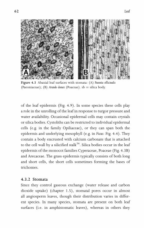
of the leaf epidermis (Fig. 4.9). In some species these cells play
a role in the unrolling of the leaf in response to turgor pressure and
water availability. Occasional epidermal cells may contain crystals
or silica bodies. Cystoliths can be restricted to individual epidermal
cells (e.g. in the family Opiliaceae), or they can span both the
epidermis and underlying mesophyll (e.g. in Ficus: Fig. 4.4). They
contain a body encrusted with calcium carbonate that is attached
to the cell wall by a silicified stalk36. Silica bodies occur in the leaf
epidermis of the monocot families Cyperaceae, Poaceae (Fig. 4.3B)
and Arecaceae. The grass epidermis typically consists of both long
and short cells, the short cells sometimes forming the bases of
trichomes.
4.3.2 StomataSince they control gaseous exchange (water release and carbon
dioxide uptake) (chapter 1.5), stomatal pores occur in almost
all angiosperm leaves, though their distribution varies in differ-
ent species. In many species, stomata are present on both leaf
surfaces (i.e. in amphistomatic leaves), whereas in others they
Figure 4.3 Abaxial leaf surfaces with stomata: (A) Paeonia officinalis
(Paeoniaceae); (B) Arundo donax (Poaceae). sb ¼ silica body.
62 Leaf
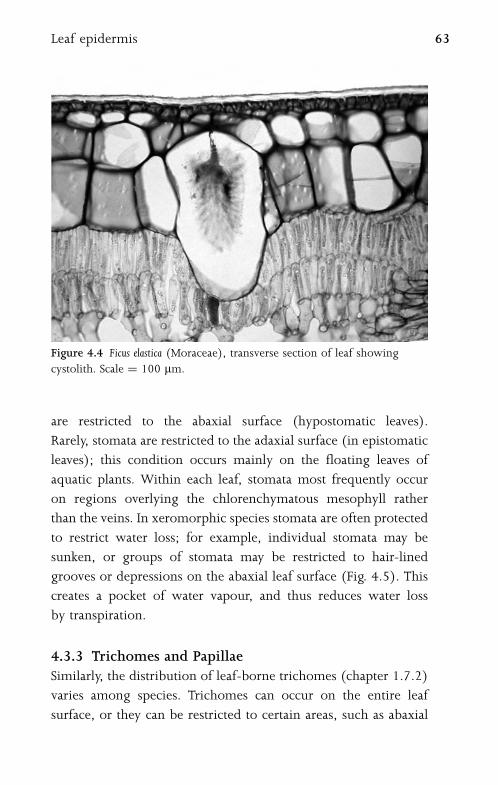
are restricted to the abaxial surface (hypostomatic leaves).
Rarely, stomata are restricted to the adaxial surface (in epistomatic
leaves); this condition occurs mainly on the floating leaves of
aquatic plants. Within each leaf, stomata most frequently occur
on regions overlying the chlorenchymatous mesophyll rather
than the veins. In xeromorphic species stomata are often protected
to restrict water loss; for example, individual stomata may be
sunken, or groups of stomata may be restricted to hair-lined
grooves or depressions on the abaxial leaf surface (Fig. 4.5). This
creates a pocket of water vapour, and thus reduces water loss
by transpiration.
4.3.3 Trichomes and PapillaeSimilarly, the distribution of leaf-borne trichomes (chapter 1.7.2)
varies among species. Trichomes can occur on the entire leaf
surface, or they can be restricted to certain areas, such as abaxial
Figure 4.4 Ficus elastica (Moraceae), transverse section of leaf showing
cystolith. Scale ¼ 100 mm.
Leaf epidermis 63
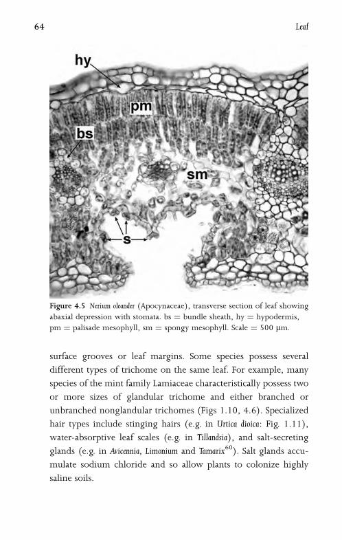
surface grooves or leaf margins. Some species possess several
different types of trichome on the same leaf. For example, many
species of the mint family Lamiaceae characteristically possess two
or more sizes of glandular trichome and either branched or
unbranched nonglandular trichomes (Figs 1.10, 4.6). Specialized
hair types include stinging hairs (e.g. in Urtica dioica: Fig. 1.11),
water-absorptive leaf scales (e.g. in Tillandsia), and salt-secreting
glands (e.g. in Avicennia, Limonium and Tamarix60). Salt glands accu-
mulate sodium chloride and so allow plants to colonize highly
saline soils.
Figure 4.5 Nerium oleander (Apocynaceae), transverse section of leaf showing
abaxial depression with stomata. bs ¼ bundle sheath, hy ¼ hypodermis,
pm ¼ palisade mesophyll, sm ¼ spongy mesophyll. Scale ¼ 500 mm.
64 Leaf
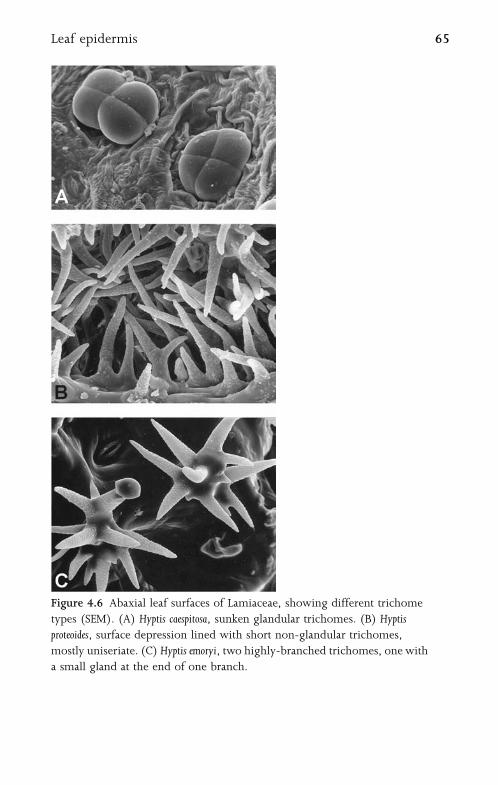
Figure 4.6 Abaxial leaf surfaces of Lamiaceae, showing different trichome
types (SEM). (A) Hyptis caespitosa, sunken glandular trichomes. (B) Hyptis
proteoides, surface depression lined with short non-glandular trichomes,
mostly uniseriate. (C) Hyptis emoryi, two highly-branched trichomes, one with
a small gland at the end of one branch.
Leaf epidermis 65
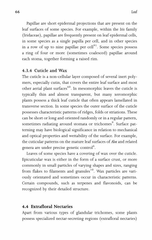
Papillae are short epidermal projections that are present on the
leaf surfaces of some species. For example, within the Iris family
(Iridaceae), papillae are frequently present on leaf epidermal cells,
in some species as a single papilla per cell, and in other species
in a row of up to nine papillae per cell91. Some species possess
a ring of four or more (sometimes coalesced) papillae around
each stoma, together forming a raised rim.
4.3.4 Cuticle and WaxThe cuticle is a non-cellular layer composed of several inert poly-
mers, especially cutin, that covers the entire leaf surface and most
other aerial plant surfaces60. In mesomorphic leaves the cuticle is
typically thin and almost transparent, but many xeromorphic
plants possess a thick leaf cuticle that often appears lamellated in
transverse section. In some species the outer surface of the cuticle
possesses characteristic patterns of ridges, folds or striations. These
can be short or long and oriented randomly or in a regular pattern,
sometimes radiating around stomata or trichomes9. Surface pat-
terning may have biological significance in relation to mechanical
and optical properties and wettability of the surface. For example,
the cuticular patterns on the mature leaf surfaces of Aloe and related
genera are under precise genetic control6.
Leaves of some species have a covering of wax over the cuticle.
Epicuticular wax is either in the form of a surface crust, or more
commonly in small particles of varying shapes and sizes, ranging
from flakes to filaments and granules10. Wax particles are vari-
ously orientated and sometimes occur in characteristic patterns.
Certain compounds, such as terpenes and flavonoids, can be
recognized by their detailed structure.
4.4 Extrafloral NectariesApart from various types of glandular trichomes, some plants
possess specialized nectar-secreting regions (extrafloral nectaries)
66 Leaf

on the leaf or petiole (Fig. 4.7). Although in some cases the
function of the extrafloral nectaries remains in doubt, and may be
related to regulation of surplus sugars, most are believed to
have a role in insect�plant relations. For example, some extra-
floral nectaries attract ants (e.g. in Acacia), which protect the plant
against potential insect herbivores. Extrafloral nectaries are often
found over veins, or in the angles of principal veins, or at the
proximal or distal ends of the petiole. As with floral nectaries
(chapter 5.10), they consist of groups of glandular trichomes
(e.g. in some Hibiscus species) or regions of anticlinally elongated
secretory epidermal cells, often associated with underlying
vascular tissue. Extrafloral nectaries are sometimes situated in
specialized pockets (domatia), or alternatively occur in pits or
raised regions (e.g. in many Euphorbiaceae). Pearl glands, or pearl
bodies, occur on leaves of some tropical eudicots (e.g. Fabaceae)
and magnoliids (e.g. Piperaceae); these are globular trichomes
that are specialized to attract ants, which then protect the plant
Figure 4.7 Omphalea diandra (Euphorbiaceae). Extrafloral nectary on abaxial
leaf surface (SEM).
Extrafloral nectaries 67
from herbivory. They secrete substances rich in carbohydrates,
lipids and proteins76.
4.5 MesophyllChlorophyll is contained in chloroplasts in the mesophyll,
which is the primary photosynthetic tissue of the leaf. In many
plant species the mesophyll is divided into distinct regions
(termed palisade and spongy tissues), though in others it is rela-
tively undifferentiated and homogeneous throughout the leaf.
Palisade mesophyll is typically adaxial. Palisade cells are anticlinally
elongated and possess relatively few intercellular air spaces. By
contrast, spongy mesophyll is typically located on the abaxial
side of the leaf, and consists of variously-shaped cells with many
air spaces between them. Both palisade and spongy tissues can
be up to several cell layers thick. Occasionally there is intergrada-
tion between the two tissues.
Many tropical grasses, and also some other taxa that are only
distantly related (both monocots and dicots), possess a ring
of mesophyll cells radiating from the vascular bundles (Kranz
anatomy; chapter 4.8). This structure is commonly associated with
the C4 pathway of photosynthesis (Fig. 4.11). In thick leaves,
particularly those of some monocots, the central cells are often
large, undifferentiated and non-photosynthetic. In the thick
‘‘keel’’ or ‘‘midrib’’ of Crocus leaves, a region of large parenchy-
matous cells with their walls often broken down to form a cavity
causes the characteristic white stripe along the centre of the
leaf (Fig. 4.8)91.
In some xeromorphic plants (e.g. Ilex and Ficus: Fig. 4.4)
subdermal layers immediately within the adaxial (or, more
rarely, the abaxial) epidermis are modified into a hypodermis.
This consists of one or more layers of non-photosynthetic cells that
are usually slightly larger and thicker-walled than the adjacent
68 Leaf

mesophyll cells and in transverse section frequently resemble
epidermal pavement cells. Hypodermal cells become lignified
in some older leaves.
4.6 Sclerenchyma and IdioblastsMesophyll is often interspersed with sclerenchyma, particularly
at leaf margins and extending as girders from the vascular bundles
to the epidermis. Fibres are typically found in groups associated
with the vascular bundles or leaf margins, but sclereids are
normally isolated in the mesophyll. For example, star-shaped
astrosclereids occur in leaves of Nymphaea and petioles of Camellia
(Fig. 1.8). Osteosclereids are characteristic of species with centric
leaves, such as Hakea. In some species sclereids are associated with
veinlet endings.
Other types of idioblast may also be interspersed in the
mesophyll. For example, secretory myrosin cells are often found
in the leaves of many Brassicaceae, and laticifers occur in leaves of
angiosperms, especially those of some eudicots such as Euphorbia
and Ficus (chapter 1.4).
Figure 4.8 Crocus cancellatus (Iridaceae), transverse section of leaf.
Scale ¼ 100 mm.
Sclerenchyma and idioblasts 69
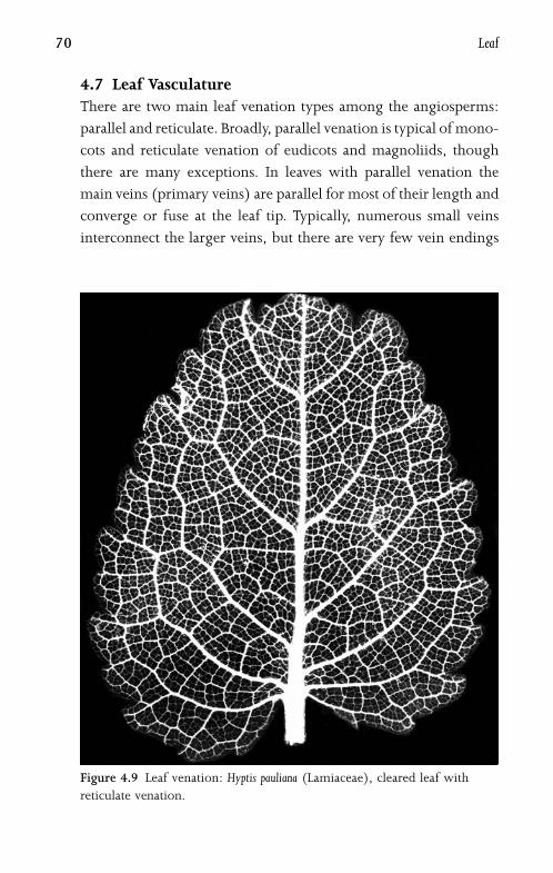
4.7 Leaf VasculatureThere are two main leaf venation types among the angiosperms:
parallel and reticulate. Broadly, parallel venation is typical of mono-
cots and reticulate venation of eudicots and magnoliids, though
there are many exceptions. In leaves with parallel venation the
main veins (primary veins) are parallel for most of their length and
converge or fuse at the leaf tip. Typically, numerous small veins
interconnect the larger veins, but there are very few vein endings
Figure 4.9 Leaf venation: Hyptis pauliana (Lamiaceae), cleared leaf with
reticulate venation.
70 Leaf
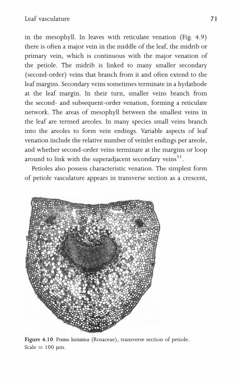
in the mesophyll. In leaves with reticulate venation (Fig. 4.9)
there is often a major vein in the middle of the leaf, the midrib or
primary vein, which is continuous with the major venation of
the petiole. The midrib is linked to many smaller secondary
(second-order) veins that branch from it and often extend to the
leaf margins. Secondary veins sometimes terminate in a hydathode
at the leaf margin. In their turn, smaller veins branch from
the second- and subsequent-order venation, forming a reticulate
network. The areas of mesophyll between the smallest veins in
the leaf are termed areoles. In many species small veins branch
into the areoles to form vein endings. Variable aspects of leaf
venation include the relative number of veinlet endings per areole,
and whether second-order veins terminate at the margins or loop
around to link with the superadjacent secondary veins51.
Petioles also possess characteristic venation. The simplest form
of petiole vasculature appears in transverse section as a crescent,
Figure 4.10 Prunus lusitanica (Rosaceae), transverse section of petiole.
Scale ¼ 100 mm.
Leaf vasculature 71

with xylem on the adaxial side and phloem on the abaxial side
(Fig. 4.10). Some species possess additional bundles outside
the main vascular crescent, which may itself be inrolled at the
ends, or in a ring, or divided into separate bundles. Classification
of the various forms of petiole vasculature depends on how it is
linked to the stem vasculature at the node. One or more vascular
traces may depart from each gap in the stem vascular cylinder
(chapter 2.3). The number and pattern of vascular bundles
sometimes vary along the length of the petiole. Midrib vasculature,
which is continuous with that of the petiole, is subject to similar
variation.
In transverse sections of the lamina, vascular bundles are usually
arranged in a single row. However, in some species with very
thick leaves, such as Agave, there are two or more rows of vascular
bundles. Lamina bundles are usually collateral, with adaxial xylem
and abaxial phloem, but orientation can vary, and in some cases
bundles are bicollateral or even amphivasal. In the isobilateral
leaves of some monocots there are two rows of vascular bundles
with opposite orientation to each other, the xylem poles being
oriented towards the leaf centre. Centric leaves possess a ring of
vascular bundles.
Leaf vasculature develops acropetally from the primordial pro-
cambial strand at its base67. The central trace develops first, and
ultimately becomes the midvein. The xylem conducting system
of the leaf blade often consists entirely of tracheids, usually with
helical or annular thickenings, though in some leaves both vessel
elements and xylem parenchyma are also present. The smallest
vascular bundles often consist of only one or two rows of xylem
tracheids and a few files of phloem sieve tube elements.
4.8 Bundle Sheath and Kranz AnatomyMost minor vascular bundles in angiosperm leaves are surrounded
by a bundle sheath which extends even to the very smallest veins
72 Leaf
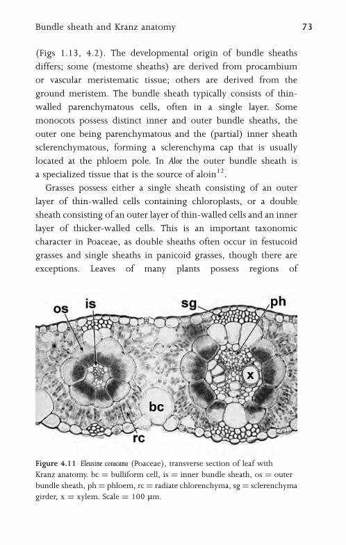
(Figs 1.13, 4.2). The developmental origin of bundle sheaths
differs; some (mestome sheaths) are derived from procambium
or vascular meristematic tissue; others are derived from the
ground meristem. The bundle sheath typically consists of thin-
walled parenchymatous cells, often in a single layer. Some
monocots possess distinct inner and outer bundle sheaths, the
outer one being parenchymatous and the (partial) inner sheath
sclerenchymatous, forming a sclerenchyma cap that is usually
located at the phloem pole. In Aloe the outer bundle sheath is
a specialized tissue that is the source of aloin12.
Grasses possess either a single sheath consisting of an outer
layer of thin-walled cells containing chloroplasts, or a double
sheath consisting of an outer layer of thin-walled cells and an inner
layer of thicker-walled cells. This is an important taxonomic
character in Poaceae, as double sheaths often occur in festucoid
grasses and single sheaths in panicoid grasses, though there are
exceptions. Leaves of many plants possess regions of
Figure 4.11 Eleusine coracana (Poaceae), transverse section of leaf with
Kranz anatomy. bc ¼ bulliform cell, is ¼ inner bundle sheath, os ¼ outer
bundle sheath, ph¼ phloem, rc¼ radiate chlorenchyma, sg¼ sclerenchyma
girder, x ¼ xylem. Scale ¼ 100 mm.
Bundle sheath and Kranz anatomy 73

sclerenchyma or parenchyma that extend from the vascular bundle
sheaths towards either or both epidermises. These bundle sheath
extensions, which are termed girders if they reach the epidermis,
afford mechanical support to the leaf and are a xeromorphic
feature (Fig. 4.1).
Most plants from warm temperate areas that possess the C4pathway of photosynthesis display a modified leaf anatomy that
is termed Kranz anatomy (Fig. 4.11). Kranz anatomy occurs in
a few groups of both dicots and monocots97. It is characterized
by elongated mesophyll cells that radiate from a single layer of
large parenchymatous bundle-sheath cells containing starch and
enlarged chloroplasts. This normally forms a second bundle sheath
layer, though in some grasses the primary vascular bundle sheath
is itself recruited for this purpose. C4 plants concentrate CO2 by
photosynthetic carbon assimilation in the radiating mesophyll
cells, and subsequent photosynthetic carbon reduction in the
bundle sheath cells.
74 Leaf

5
Flower
5.1 Floral OrgansFlowers are complex structures that consist of several organ types
borne on a central axis (the receptacle). In many species each
flower is subtended by a modified leaf-like structure termed
a bract (Fig. 1.2), though bracts are absent from some other
species. Within each flower, the organs are arranged in distinct
bands (whorls) or in a spiral pattern (Figs 5.1, 5.2). The degree of
fusion of individual floral organs within each flower is normally
characteristic of a species (i.e. genetically determined). Fusion
between similar organ types borne in the same whorl is termed
connation. Fusion between different organ types borne in adjacent
whorls is termed adnation.
The outer two types of floral organs (collectively the perianth)
are modified leaf-like structures, termed sepals (collectively the
calyx, or sometimes the first whorl) and petals (collectively the
corolla, or the second whorl). In many monocots and magnoliids
the perianth organs are morphologically indistinguishable from
each other, and are collectively termed tepals, rather than
differentiated into sepals and petals. Enclosed within the perianth
are the stamens, which are collectively termed the androecium,
or sometimes the third whorl, though they are often borne
in two or more distinct whorls. The carpels (collectively the
gynoecium, sometimes termed the fourth whorl) are borne in
the centre of the flower, and normally terminate the floral axis.

This general pattern varies considerably among angiosperm
groups, and many species possess unisexual flowers. The grass
flower is typically subtended by a bract-like structure, the
palea, that surrounds three (or, more commonly, two) reduced
structures termed lodicules, which are normally interpreted as
homologous with a single perianth whorl (probably the inner
tepals) of other monocots4,22,95.
Figure 5.1 Arabidopsis thaliana (Brassicaceae), SEM dissected flower. a¼ anther,
n ¼ nectary, p ¼ petal, st ¼ style. Scale ¼ 100 mm.
76 Flower

Floral organ primordia are typically initiated from the outside
inwards, in centripetal (acropetal) sequence towards the floral
apex (Fig. 5.3). For example, in Drimys the innermost stamens
are the last to be initiated in the developing bud, though they are
the largest and the first to dehisce in the open flower113. However,
in some species certain floral organs, either within a single whorl
or in more than one whorl, are initiated in a different sequence or
in groups (fascicles) (Fig. 5.4). For example, the stamens of some
polyandrous palms are initiated centrifugally115.
5.2 Floral VasculatureIn the majority of flowers, each organ is served by a single
vascular strand that diverges from the central vascular cylinder
in the receptacle and subsequently branches (Figs 5.5, 5.6).
Perianth traces are normally highly branched, often forming a
Figure 5.2 Taraxacum officinale (Asteraceae), transverse section of
inflorescence, showing individual florets. Scale ¼ 100 mm.
Floral vasculature 77

vascular network. Most stamens typically bear a single vascular
strand, but some families characteristically possess three or four
stamen traces. Others, such as Araceae35, possess diverse and often
branching stamen vasculature. Many species possess two carpellary
vascular bundles: the ventral carpellary trace, which diverges into
the ovules, and the dorsal carpellary trace, which passes up the
style into the stigma (Fig. 5.7). The number of vascular bundles in
the style of a syncarpous gynoecium is often an indicator of the
number of carpels present, though in some species bundles are
branched or fused.
Figure 5.3 Clematis argentilucida (Ranunculaceae), SEM dissected flower bud.
fa ¼ floral apex, p ¼ developing petal, s ¼ stamen primordium.
Scale ¼ 100 mm.
78 Flower

5.3 PerianthIn their simplest form, perianth parts are essentially leaf-like in
morphology, though there are many modifications. Sepals are
typically green and photosynthetic; it is common to find stomata
and trichomes on sepal surfaces. In insect-pollinated plants the
petals are typically the largest and showiest part of the flower,
though in wind-pollinated plants the petals are sometimes reduced
or even absent. Flower colour is largely controlled by the
chemistry of the diverse pigments present. Anthocyanins, beta-
lains and ultraviolet-absorbing flavonoids are largely confined to
the epidermal cells of the petal, whereas other pigments, such
Figure 5.4 Hypericum empetrifolium (Clusiaceae), SEM dissected developing
flower bud. g¼ gynoecium, p¼ petal, sf¼ stamen fascicle. Scale¼ 100 mm.
Perianth 79
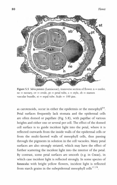
as carotenoids, occur in either the epidermis or the mesophyll64.
Petal surfaces frequently lack stomata and the epidermal cells
are often domed or papillate (Fig. 5.8), with papillae of various
heights and either one or several per cell. The effect of the domed
cell surface is to guide incident light into the petal, where it is
reflected outwards from the inside walls of the epidermal cells or
from the multi-faceted walls of mesophyll cells, thus passing
through the pigments in solution in the cell vacuoles. Many petal
surfaces are also strongly striated, which may have the effect of
further scattering the incident light into the interior of the petal.
By contrast, some petal surfaces are smooth (e.g. in Crocus), in
which case incident light is reflected strongly. In some species of
Ranunculus with bright yellow flowers, incident light is reflected
from starch grains in the subepidermal mesophyll cells17,78.
Figure 5.5 Salvia pratensis (Lamiaceae), transverse section of flower. n¼ nutlet,
ne ¼ nectary, ov ¼ ovule, pt ¼ petal tube, s ¼ style, sb ¼ stamen
vascular bundle, st ¼ sepal tube. Scale ¼ 100 mm.
80 Flower
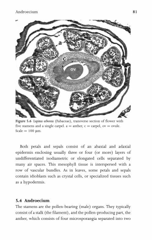
Both petals and sepals consist of an abaxial and adaxial
epidermis enclosing usually three or four (or more) layers of
undifferentiated isodiametric or elongated cells separated by
many air spaces. This mesophyll tissue is interspersed with a
row of vascular bundles. As in leaves, some petals and sepals
contain idioblasts such as crystal cells, or specialized tissues such
as a hypodermis.
5.4 AndroeciumThe stamens are the pollen-bearing (male) organs. They typically
consist of a stalk (the filament), and the pollen-producing part, the
anther, which consists of four microsporangia separated into two
Figure 5.6 Lupinus arboreus (Fabaceae), transverse section of flower with
five stamens and a single carpel. a ¼ anther, c ¼ carpel, ov ¼ ovule.
Scale ¼ 100 mm.
Androecium 81
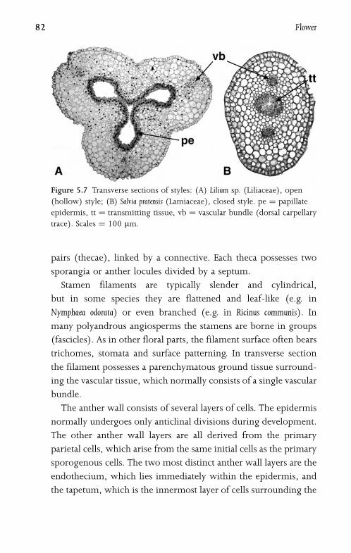
pairs (thecae), linked by a connective. Each theca possesses two
sporangia or anther locules divided by a septum.
Stamen filaments are typically slender and cylindrical,
but in some species they are flattened and leaf-like (e.g. in
Nymphaea odorata) or even branched (e.g. in Ricinus communis). In
many polyandrous angiosperms the stamens are borne in groups
(fascicles). As in other floral parts, the filament surface often bears
trichomes, stomata and surface patterning. In transverse section
the filament possesses a parenchymatous ground tissue surround-
ing the vascular tissue, which normally consists of a single vascular
bundle.
The anther wall consists of several layers of cells. The epidermis
normally undergoes only anticlinal divisions during development.
The other anther wall layers are all derived from the primary
parietal cells, which arise from the same initial cells as the primary
sporogenous cells. The two most distinct anther wall layers are the
endothecium, which lies immediately within the epidermis, and
the tapetum, which is the innermost layer of cells surrounding the
Figure 5.7 Transverse sections of styles: (A) Lilium sp. (Liliaceae), open
(hollow) style; (B) Salvia pratensis (Lamiaceae), closed style. pe ¼ papillate
epidermis, tt ¼ transmitting tissue, vb ¼ vascular bundle (dorsal carpellary
trace). Scales ¼ 100 mm.
82 Flower

anther locule (Fig. 5.9). Intervening layers usually consist of thin-
walled cells that are often crushed and destroyed at anthesis.
Endothecial cells typically develop fibrous wall thickenings which
contribute to the anther dehiscence mechanism.
The tapetum is a specialized cell layer that functions as a source
of nutrients for developing pollen grains. Tapetal cells are secretory
and contain dense cytoplasm. They produce exine precursors,
proteins and lipids that form the pollen coat. In many species
a layer of tapetal cells remains intact around the anther locule; this
type of tapetum is termed the secretory type (or cellular, glandular
or parietal). In some other species the tapetal cells degenerate and
their protoplasts fuse to form a multinucleate tapetal plasmodium
(a periplasmodium) in the anther locule; this type of tapetum is
termed the plasmodial type (or amoeboid, invasive, periplasmo-
dial or syncytial type). Transitional types also occur in some
species, especially in magnoliids38.
Figure 5.8 Arabidopsis thaliana (Brassicaceae), SEM petal surface.
Scale ¼ 100 mm.
Androecium 83

5.5 PollenIn the developing anther the primary sporogenous cells, which
are derived from the same initials as the primary parietal cells,
give rise either directly or by successive mitotic divisions to
the microspore parent cells (microsporocytes). These in turn
each undergo two meiotic divisions (microsporogenesis), either
successively or almost simultaneously, to form a tetrad of haploid
microspores. In the successive microsporogenesis type, callose
cell walls are formed after both meiosis I and meiosis II. In the
simultaneous type, cytokinesis does not occur until both meiotic
nuclear divisions are complete. Microsporogenesis is simultaneous
in most eudicots, whereas both successive and simultaneous types
occur in monocots and magnoliids39,41.
Following meiosis, the tetrad normally fragments into indivi-
dual microspores, though in some families (e.g. Winteraceae) the
microspores typically remain together as permanent tetrads. Prior
to anthesis, each microspore undergoes an unequal (asymmetric)
Figure 5.9 Lilium martagon (Liliaceae), transverse section of anther wall.
e ¼ epidermis, en ¼ endothecium, ml ¼ middle layer, m ¼ microspores,
t ¼ tapetum. Scale ¼ 100 mm.
84 Flower

mitosis to form a larger vegetative cell and a smaller generative cell
enclosed within the pollen grain wall. The generative (spermato-
genous) nucleus later undergoes a further mitosis (spermatogen-
esis) to form two sperm cells, either in the pollen grain or in the
pollen tube. The microgametophyte is therefore either bicellular
or tricellular. Spermatogenesis is itself asymmetric in some
angiosperms (e.g. Plumbago and Zea), in which dimorphic sperm
are produced that can preferentially fertilize either the egg nucleus
or the polar nuclei in the embryo sac114.
Pollen grains are radially or bilaterally symmetrical bodies
that represent units of dispersal from the anther to the stigma
(Fig. 5.10). They vary considerably in size and shape among
species29. The pollen-grain wall consists of two distinct domains:
a hard outer exine, which is composed mainly of sporopollenin
(a carotenoid polymer), and a relatively soft inner intine com-
posed of polysaccharides. The exine is itself a layered structure,
often differentiated into an outer sculptured ectexine (sexine)
and an inner non-sculptured endexine (nexine), though endexine
is normally well-developed only in eudicots with tricolpate
pollen41.
Apertures are often present in the pollen-grain wall, though
pollen grains of some species lack a clearly defined aperture
(termed inaperturate pollen37). Apertures represent specialized
regions in which the outer layer (the exine) is reduced or absent,
and the underlying layer (the intine) is thickened. They range in
shape from elongated furrows to roughly circular pores. Some
apertures possess a lid-like operculum40. Apertures that lie along
the distal face of the pollen grain (normally the face that was
directed outwards in the tetrad) are termed sulci. Sulcate pollen
grains are characteristic of monocots and magnoliids. Apertures
that lie along the equatorial face of the pollen grain, as defined
during the tetrad phase, are termed colpi. Pollen with three
equatorial apertures (tricolpate pollen) characterizes the eudicots
(sometimes termed the tricolpates). In many eudicots, microspore
Pollen 85

tetrads are tetrahedral with three apertures (colpi) arranged
equidistantly around the equator of the microspore. Colpi are
typically elongated and slit-like, but can occasionally be reduced to
pores, and in some eudicot species the number can increase to
four, five, six, or more. In spiraperturate pollen grains (e.g. some
Crocus species) the aperture spirals around the grain.
Pollen grains dehydrate after contact with the air, and the
exine contracts. Subsequently, rehydration and exine expansion
occurs on the stigmatic surface. Dry and hydrated pollen grains of
the same species can appear very different in size, shape and even
surface features. There are many different patterns of exine sculp-
turing. For example, the exine may be reticulate or areolate, or
it may possess surface holes (puncta), granules, warts or spines84.
Figure 5.10 Taraxacum officinale (Asteraceae), SEM pollen grain. Scale¼ 10 mm.
86 Flower
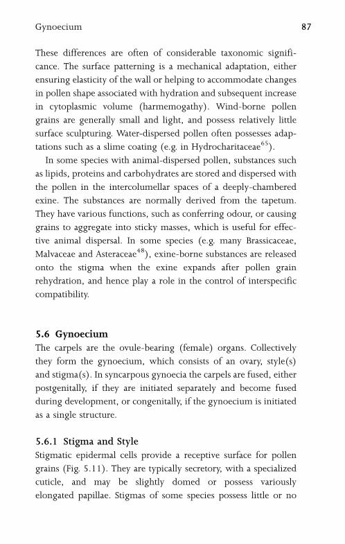
These differences are often of considerable taxonomic signifi-
cance. The surface patterning is a mechanical adaptation, either
ensuring elasticity of the wall or helping to accommodate changes
in pollen shape associated with hydration and subsequent increase
in cytoplasmic volume (harmemogathy). Wind-borne pollen
grains are generally small and light, and possess relatively little
surface sculpturing. Water-dispersed pollen often possesses adap-
tations such as a slime coating (e.g. in Hydrocharitaceae65).
In some species with animal-dispersed pollen, substances such
as lipids, proteins and carbohydrates are stored and dispersed with
the pollen in the intercolumellar spaces of a deeply-chambered
exine. The substances are normally derived from the tapetum.
They have various functions, such as conferring odour, or causing
grains to aggregate into sticky masses, which is useful for effec-
tive animal dispersal. In some species (e.g. many Brassicaceae,
Malvaceae and Asteraceae48), exine-borne substances are released
onto the stigma when the exine expands after pollen grain
rehydration, and hence play a role in the control of interspecific
compatibility.
5.6 GynoeciumThe carpels are the ovule-bearing (female) organs. Collectively
they form the gynoecium, which consists of an ovary, style(s)
and stigma(s). In syncarpous gynoecia the carpels are fused, either
postgenitally, if they are initiated separately and become fused
during development, or congenitally, if the gynoecium is initiated
as a single structure.
5.6.1 Stigma and StyleStigmatic epidermal cells provide a receptive surface for pollen
grains (Fig. 5.11). They are typically secretory, with a specialized
cuticle, and may be slightly domed or possess variously
elongated papillae. Stigmas of some species possess little or no
Gynoecium 87

surface secretions (termed dry stigmas), whereas ‘‘wet’’ stigmas
exude copious surface secretions that play a role in pollen recogni-
tion and germination49. The stigmatic cuticle is often stratified
in transverse section, with a lamellated outer layer and reticulate
inner layers. Some Crocus species possess a chambered cuticle,
and in some Euphorbia species the cuticle is fenestrated47.
Rarely, the stigma is borne directly on the top of the ovary,
but most commonly it is borne on a style. The ground tissue of
the style is parenchymatous, and it typically possesses two or
Figure 5.11 Arabidopsis thaliana (Brassicaceae), SEM stigma. Scale ¼ 100 mm.
88 Flower
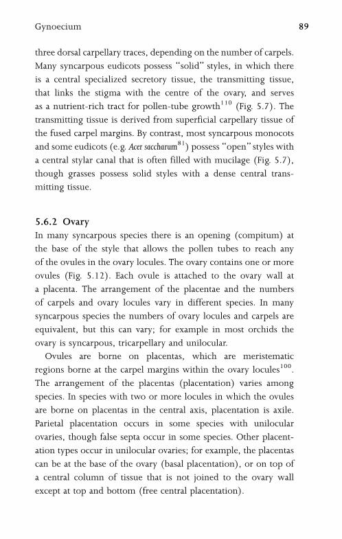
three dorsal carpellary traces, depending on the number of carpels.
Many syncarpous eudicots possess ‘‘solid’’ styles, in which there
is a central specialized secretory tissue, the transmitting tissue,
that links the stigma with the centre of the ovary, and serves
as a nutrient-rich tract for pollen-tube growth110 (Fig. 5.7). The
transmitting tissue is derived from superficial carpellary tissue of
the fused carpel margins. By contrast, most syncarpous monocots
and some eudicots (e.g. Acer saccharum81) possess ‘‘open’’ styles with
a central stylar canal that is often filled with mucilage (Fig. 5.7),
though grasses possess solid styles with a dense central trans-
mitting tissue.
5.6.2 OvaryIn many syncarpous species there is an opening (compitum) at
the base of the style that allows the pollen tubes to reach any
of the ovules in the ovary locules. The ovary contains one or more
ovules (Fig. 5.12). Each ovule is attached to the ovary wall at
a placenta. The arrangement of the placentae and the numbers
of carpels and ovary locules vary in different species. In many
syncarpous species the numbers of ovary locules and carpels are
equivalent, but this can vary; for example in most orchids the
ovary is syncarpous, tricarpellary and unilocular.
Ovules are borne on placentas, which are meristematic
regions borne at the carpel margins within the ovary locules100.
The arrangement of the placentas (placentation) varies among
species. In species with two or more locules in which the ovules
are borne on placentas in the central axis, placentation is axile.
Parietal placentation occurs in some species with unilocular
ovaries, though false septa occur in some species. Other placent-
ation types occur in unilocular ovaries; for example, the placentas
can be at the base of the ovary (basal placentation), or on top of
a central column of tissue that is not joined to the ovary wall
except at top and bottom (free central placentation).
Gynoecium 89
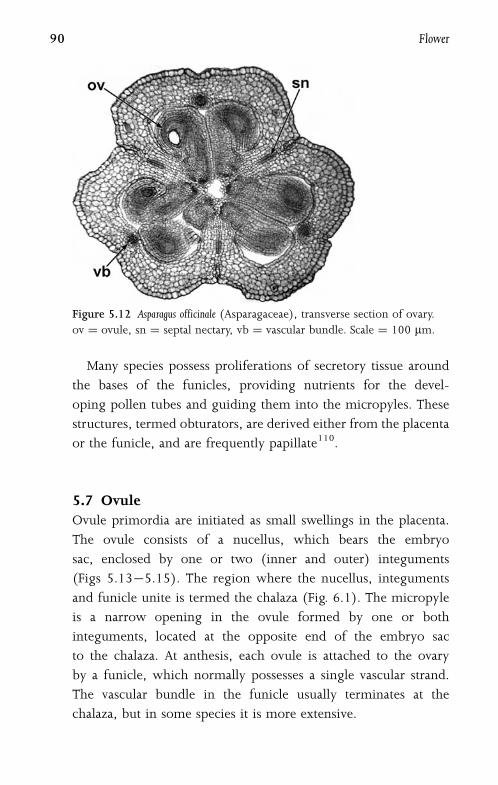
Many species possess proliferations of secretory tissue around
the bases of the funicles, providing nutrients for the devel-
oping pollen tubes and guiding them into the micropyles. These
structures, termed obturators, are derived either from the placenta
or the funicle, and are frequently papillate110.
5.7 OvuleOvule primordia are initiated as small swellings in the placenta.
The ovule consists of a nucellus, which bears the embryo
sac, enclosed by one or two (inner and outer) integuments
(Figs 5.13�5.15). The region where the nucellus, integuments
and funicle unite is termed the chalaza (Fig. 6.1). The micropyle
is a narrow opening in the ovule formed by one or both
integuments, located at the opposite end of the embryo sac
to the chalaza. At anthesis, each ovule is attached to the ovary
by a funicle, which normally possesses a single vascular strand.
The vascular bundle in the funicle usually terminates at the
chalaza, but in some species it is more extensive.
Figure 5.12 Asparagus officinale (Asparagaceae), transverse section of ovary.
ov ¼ ovule, sn ¼ septal nectary, vb ¼ vascular bundle. Scale ¼ 100 mm.
90 Flower
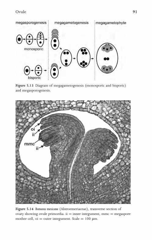
Figure 5.13 Diagram of megagametogenesis (monosporic and bisporic)
and megasporogenesis.
Figure 5.14 Bomarea mexicana (Alstroemeriaceae), transverse section of
ovary showing ovule primordia. ii ¼ inner integument, mmc ¼ megaspore
mother cell, oi ¼ outer integument. Scale ¼ 100 mm.
Ovule 91
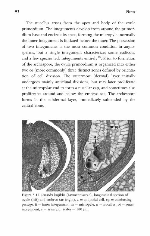
The nucellus arises from the apex and body of the ovule
primordium. The integuments develop from around the primor-
dium base and encircle its apex, forming the micropyle; normally
the inner integument is initiated before the outer. The possession
of two integuments is the most common condition in angio-
sperms, but a single integument characterizes some eudicots,
and a few species lack integuments entirely16. Prior to formation
of the archespore, the ovule primordium is organized into either
two or (more commonly) three distinct zones defined by orienta-
tion of cell division. The outermost (dermal) layer initially
undergoes mainly anticlinal divisions, but may later proliferate
at the micropylar end to form a nucellar cap, and sometimes also
proliferates around and below the embryo sac. The archespore
forms in the subdermal layer, immediately subtended by the
central zone.
Figure 5.15 Lomandra longifolia (Laxmanniaceae), longitudinal section of
ovule (left) and embryo sac (right). a ¼ antipodal cell, cp ¼ conducting
passage, ii ¼ inner integument, m ¼ micropyle, n ¼ nucellus, oi ¼ outer
integument, s ¼ synergid. Scales ¼ 100 mm.
92 Flower
In some species the nucellus proliferates in the central zone
to form a hypostase, which is a chalazal region of often refractive
and suberinized or lignified tissue. For example, a large hypostase
is present in Acorus and Crocus. Sometimes other specialized
structures are formed in the central zone of the nucellus, such
as a postament or conducting passage (Fig. 5.15); these often
persist after fertilization. The dermal nucellar cells surrounding
the embryo sac often break down before (or shortly after) fertili-
zation, in which case the innermost epidermal layer of the
inner integument is sometimes differentiated to form an endo-
thelium or integumentary tapetum (Fig. 6.6). Endothelium
cells are frequently enlarged and densely cytoplasmic, and
sometimes endopolyploid; in some species they have a secretory
function.
5.8 Embryo SacWithin the nucellus, a single (normally hypodermal) cell becomes
a primary sporogenous cell (archesporial cell, or archespore).
The archespore rarely consists of more than one cell, though it
can be multicellular in a few species (e.g. Brassica campestris86), in
which one cell produces the megagametophyte. In turn, the
archesporial cell either gives rise directly to the megaspore mother
cell (megasporocyte) or undergoes mitosis to form a primary
parietal cell and a megasporocyte. The megasporocyte then under-
goes two meiotic divisions (megasporogenesis) to form a tetrad
of four megaspores, which are usually either in a linear or
T-shaped arrangement. In the majority of angiosperm flowers, one
megaspore (most commonly the chalazal one) gives rise
to the mature embryo sac by further mitotic divisions, and the
other three megaspores degenerate (Fig. 5.13). This type of
development is termed monosporic. However, in relatively few
angiosperms, two or four megaspores play a role in embryo
Embryo sac 93
sac formation; these types are termed bisporic or tetrasporic
respectively69,125,126. Degenerated megaspores are often sur-
rounded by persistent callose68.
In most angiosperms the mature embryo sac (megagameto-
phyte) possesses eight nuclei arranged in seven cells (Fig. 5.15),
though types with four and sixteen or more nuclei have also
been recorded. The most common type is monosporic and
eight-nucleate; this is sometimes termed the Polygonum type of
embryo sac development. At the binucleate stage, the two nuclei
migrate to the micropylar and chalazal poles and subsequently
divide. Of the two micropylar nuclei, the one closest to the
micropyle divides to form the synergids, and the other divides
to form the egg cell and one of the polar nuclei. The two
chalazal nuclei each divide so that one forms two antipodal
cells and the other forms an antipodal and a polar nucleus.
The two polar nuclei migrate to the centre and fuse to form
a diploid fusion nucleus. Cellularization follows, so that the
mature megagametophyte consists of three antipodal cells at
the chalazal end, a central cell with a fusion nucleus, and two
synergids plus an egg cell at the micropylar end.
The synergids and the egg cell are so tightly pressed together
that they are collectively termed the egg apparatus. The syner-
gids play a role in directing the pollen tube into the embryo
sac; they are calcium-rich and normally possess a series of wall
thickenings, the filiform apparatus, which extends into the micro-
pyle (Fig. 5.15). In many species the antipodals degenerate at an
early stage, but in others they persist, and sometimes undergo
cell division (e.g. in many grasses) or endoreduplication.
5.9 Pollen-Tube GrowthPollen tubes emerge from a pollen grain that has landed on
a conspecific receptive stigma, normally via an aperture in the
exine. The pollen-tube wall is initially formed from the innermost
94 Flower
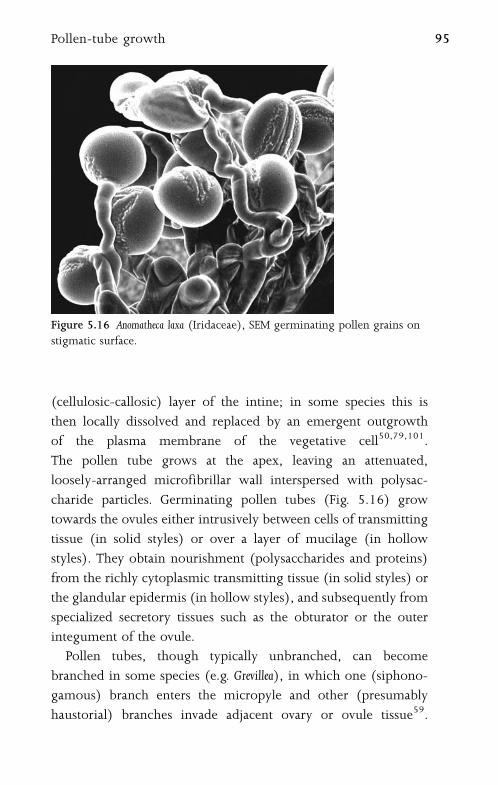
(cellulosic-callosic) layer of the intine; in some species this is
then locally dissolved and replaced by an emergent outgrowth
of the plasma membrane of the vegetative cell50,79,101.
The pollen tube grows at the apex, leaving an attenuated,
loosely-arranged microfibrillar wall interspersed with polysac-
charide particles. Germinating pollen tubes (Fig. 5.16) grow
towards the ovules either intrusively between cells of transmitting
tissue (in solid styles) or over a layer of mucilage (in hollow
styles). They obtain nourishment (polysaccharides and proteins)
from the richly cytoplasmic transmitting tissue (in solid styles) or
the glandular epidermis (in hollow styles), and subsequently from
specialized secretory tissues such as the obturator or the outer
integument of the ovule.
Pollen tubes, though typically unbranched, can become
branched in some species (e.g. Grevillea), in which one (siphono-
gamous) branch enters the micropyle and other (presumably
haustorial) branches invade adjacent ovary or ovule tissue59.
Figure 5.16 Anomatheca laxa (Iridaceae), SEM germinating pollen grains on
stigmatic surface.
Pollen-tube growth 95
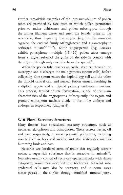
Further remarkable examples of the intrusive abilities of pollen
tubes are provided by rare cases in which pollen germinates
prior to anther dehiscence and pollen tubes grow through
the anther filament tissue and enter the female tissue at the
receptacle, thus bypassing the stigma (e.g. in the monocot
Sagittaria, the eudicot family Malpighiaceae and a gametophytic
Arabidopsis mutant1,58,120). Some angiosperms (e.g. Lavatera)
exhibit polysiphony: multiple (15�20) pollen tubes emerge
from a single region of the grain on the side in contact with
the stigma, though only one tube bears the sperm75.
When the pollen tube reaches an ovule, it enters through the
micropyle and discharges the male gametes (sperm cells) before
collapsing. One sperm enters the haploid egg cell and the other
the diploid central cell, and nuclear fusion ensues, resulting in
a diploid zygote and a triploid primary endosperm nucleus.
This process, termed double fertilization, is one of the main
characteristics of the angiosperms. Subsequently, the zygote and
primary endosperm nucleus divide to form the embryo and
endosperm respectively (chapter 6).
5.10 Floral Secretory StructuresMany flowers bear specialized secretory structures, such as
nectaries, elaiophores and osmophores. These secrete nectar, oil
and scent respectively, to attract potential pollinators, including
insects such as bees and moths, and also vertebrates such as
humming birds and bats.
Nectaries are localized areas of tissue that regularly secrete
nectar, a sugar-rich substance that is attractive to animals31.
Nectaries usually consist of secretory epidermal cells with dense
cytoplasm, sometimes modified into trichomes. Adjacent sub-
epidermal cells may also be secretory, and in some cases
nectar passes to the surface through modified stomatal pores.
96 Flower
Vascular tissue close to the nectary often consists mainly or
entirely of phloem, which transports sugars to the secretory
region.
Nectaries may occur on any floral organ, or they may represent
an entire modified organ, or even a novel structure. Most species
of the mint family (Lamiaceae) possess an enlarged nectariferous
disc surrounding the base of the ovary (Fig. 5.5), which is
derived from developing ovary tissue. In most Brassicaceae
(e.g. Arabidopsis thaliana) the nectary is located at the base of the
stamen filament (Fig. 5.1). Septal nectaries are characteristic of
many monocots; these occur at the unfused carpel margins in
monocots with postgenitally fused ovaries (Fig. 5.12). Nectar
produced from septal nectaries is exuded from secretary epidermal
cells and emerges from small pores or slits on the surface of
the gynoecium25, 93.
Flowers of some insect-pollinated species lack nectaries, but
possess other secretory structures that attract potential pollinators.
For example, some flowers bear oil-secreting glands, termed
elaiophores (Fig. 5.17), which are morphologically similar to
some types of nectary118. Osmophores119 are modified floral
Figure 5.17 Tigridia meleagris (Iridaceae), elaiophores on tepal surface
(A) SEM, (B) LM. Scale ¼ 100 mm.
Floral secretory structures 97
structures that produce volatile secretions (scents); in some
orchids the odours are pheromone-like substances. Osmophores
typically possess a relatively thick, domed or papillate epidermis
with densely cytoplasmic contents. In Platanthera bifolia the epi-
dermis of the labellum secretes a nocturnal scent, and in
Ophrys species osmophores on the labellum consist of areas of
dome-shaped, papillate, dark-staining epidermal cells. Flowers of
Narcissus emit pollinator-specific volatiles that are probably derived
from the colourful corona5.
98 Flower

6
Seed and fruit
6.1 Seed CoatThe seed coat prevents destruction of the seed by dehydration
or predation. In bitegmic seeds the testa is derived from the outer
integument, and the inner integument forms the tegmen15
(Fig. 6.1). In unitegmic seeds the term ‘‘testa’’ applies to the
entire seed coat. Seed coats are multilayered tissues; they generally
Figure 6.1 Seed organization: Diagram of a generalized campylotropous
bitegmic dicotyledonous seed with perisperm. (Adapted from Boesewinkel
and Bouman 1984.)

include a hard, protective mechanical layer that is formed from
all or part of the testa or tegmen23. In exotestal seed coats the
mechanical layer is derived from the outer epidermis of the outer
integument, whereas in endotegmic seed coats it is derived from
the inner epidermis of the inner integument. In some species the
mechanical layer consists of one or more rows of elongated,
palisade-like cells, such as the macrosclereids in the exotesta
of many Fabaceae.
Seed coat surfaces exhibit a variety of cellular patterns,
often with characteristic papillate or striate surface sculpturing9
(Fig. 6.2). Some seeds possess epidermal trichomes; for example,
the seed coat hairs of Gossypium (cotton) are an important source
of textile fibres.
Seed-coat vasculature usually consists of a single bundle passing
from the raphe to the chalaza, but this can vary in extent and degree
of branching. Many seed coats possess specialized structures that
are related to dispersal15. For example, some wind-dispersed seeds
possess wings, and some animal-dispersed seeds are fleshy.
Figure 6.2 Seed surface: Silene nutans (Caryophyllaceae), entire seed (left)
and detail (right) showing papillate epidermal cells with sinuous anticlinal
walls.
100 Seed and fruit

The fleshy part of the seed coat, termed the sarcotesta, is most
commonly formed from part of the outer integument. Arils are
fleshy outgrowths of the funicle. Some plants, especially parasitic
or mycoheterotrophic plants such as Monotropa or orchids, produce
large numbers of highly reduced ‘‘dust seeds’’ from each ovary;
these minute seeds can be blown over long distances. The seed coat
of many orchids lacks vasculature entirely (Fig. 6.3).
6.2 PericarpIn fruits that are derived from a single ovary, the fruit wall,
termed the pericarp, is typically derived from the ovary wall.
The pericarp displays a similar range of variation to the seed coat,
depending particularly on whether it is dry or fleshy and dehiscent
or indehiscent. It is typically divided into three layers � the outer
exocarp, central mesocarp and inner endocarp � though in some
fruits the three layers are not readily distinguished. At least one
layer of the fruit wall often consists of thick-walled lignified cells
(Fig. 6.4), though in some fleshy fruits (berries), such as those of
Vitis vinifera (grape), the entire endocarp consists of thin-walled
succulent cells. In other fleshy fruits (drupes), such as those
Figure 6.3 Dust seed: Cypripedium calceolus (Orchidaceae), light micrograph
of entire seed, showing thin testa surrounding globular embryo.
Scale ¼ 100 mm.
Pericarp 101
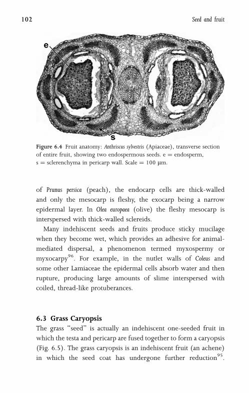
of Prunus persica (peach), the endocarp cells are thick-walled
and only the mesocarp is fleshy, the exocarp being a narrow
epidermal layer. In Olea europaea (olive) the fleshy mesocarp is
interspersed with thick-walled sclereids.
Many indehiscent seeds and fruits produce sticky mucilage
when they become wet, which provides an adhesive for animal-
mediated dispersal, a phenomenon termed myxospermy or
myxocarpy96. For example, in the nutlet walls of Coleus and
some other Lamiaceae the epidermal cells absorb water and then
rupture, producing large amounts of slime interspersed with
coiled, thread-like protuberances.
6.3 Grass CaryopsisThe grass ‘‘seed’’ is actually an indehiscent one-seeded fruit in
which the testa and pericarp are fused together to form a caryopsis
(Fig. 6.5). The grass caryopsis is an indehiscent fruit (an achene)
in which the seed coat has undergone further reduction95.
Figure 6.4 Fruit anatomy: Anthriscus sylvestris (Apiaceae), transverse section
of entire fruit, showing two endospermous seeds. e ¼ endosperm,
s ¼ sclerenchyma in pericarp wall. Scale ¼ 100 mm.
102 Seed and fruit
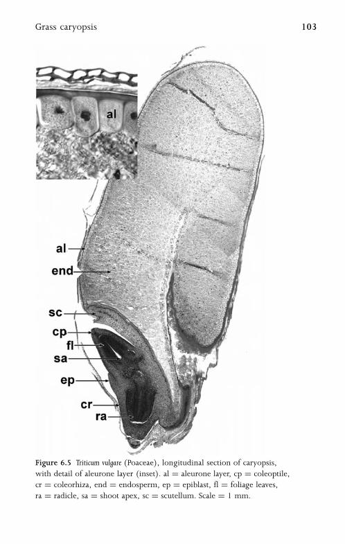
Figure 6.5 Triticum vulgare (Poaceae), longitudinal section of caryopsis,
with detail of aleurone layer (inset). al ¼ aleurone layer, cp ¼ coleoptile,
cr ¼ coleorhiza, end ¼ endosperm, ep ¼ epiblast, fl ¼ foliage leaves,
ra ¼ radicle, sa ¼ shoot apex, sc ¼ scutellum. Scale ¼ 1 mm.
Grass caryopsis 103

After fertilization the pericarp consists of a few cell layers, and
the integuments disintegrate completely, leaving only a hyaline
membrane covered with a cuticle, derived from the outer layer
of the inner integument. Grass seeds also possess highly diffe-
rentiated embryos with a unique highly characteristic prominent
outgrowth of the embryo, termed the scutellum, which is
normally considered to be a modified cotyledon. Some grasses
possess an outgrowth opposite the scutellum, termed the epiblast,
which has been variously interpreted as a second cotyledon or
an outgrowth of the first cotyledon or of the coleorhiza.
Grass embryos are well-differentiated within the seed, prior to
germination. They characteristically possess a sheath (coleoptile)
surrounding the epicotyl and plumule, and a well-developed
radicle also surrounded by a sheath (the coleorhiza). In some
grasses (and a few other angiosperms) the outermost layer of the
endosperm, termed the aleurone layer, is a specialized tissue of
enlarged cells containing protein bodies and large nuclei.
6.4 EndospermEndosperm forms a food-storage tissue in the seed. It not only
promotes growth and longevity for the seed, but can also facilitate
dispersal, as an attractant to animals. Mature endosperm typically
consists of tightly-packed cells that contain food-reserve materials
such as starch grains or protein bodies. Endosperm is typically
a triploid tissue formed by fusion of one sperm cell with two
female polar nuclei. It is present in most angiosperm seeds but in
greatly contrasting amounts; for example, endosperm formation
is negligible in orchid seeds but extensive in grass seeds, in which
it forms an important economic crop.
Early endosperm development is traditionally classified into
three types, termed nuclear, cellular and helobial, based on the
timing and degree of cell wall formation, though transitional
forms exist117. Nuclear endosperm is the most common type,
104 Seed and fruit
and occurs in many eudicots (e.g. Arabidopsis thaliana). Nuclear
endosperm possesses both a syncytial (free-nucleate) phase
and a cellular phase. Early cell divisions are not followed by
cell wall formation, and the nuclei are initially free in the
cytoplasm of the embryo sac, usually surrounding a central
vacuole. Cell walls eventually form in most endosperm tissues, but
sometimes the nuclei at the chalazal end remain free; for example,
the liquid ‘‘milk’’ of the coconut palm (Cocos nucifera) is a syncy-
tium that contains many free endosperm nuclei in addition to oil
droplets and protein granules.
In the cellular type of endosperm formation, which occurs
in some eudicots (e.g. Acanthaceae), even the earliest nuclear
divisions are followed by cell-wall formation. In the helobial
endosperm type, which is restricted to some monocots, the
primary endosperm nucleus undergoes division to form two
unequal chambers, normally a small chalazal chamber and a large
micropylar chamber. The nucleus of the micropylar chamber
migrates to the top of the embryo sac, and its initial divisions are
not accompanied by cell wall formation, though cell walls are
formed with later mitoses. The chalazal chamber has far fewer
nuclear divisions, and its nuclei remain free in the cytoplasm; it
typically has a haustorial role.
Endosperm haustoria may develop in all three types of endo-
sperm. Haustoria assist nutrient absorption and sometimes invade
adjacent tissues. For example, most species of the mint family
Lamiaceae possess both chalazal and micropylar haustoria
(Fig. 6.6), which may be either free-nucleate or cellular, some-
times even amoeboid89. In these species, the first division of the
primary endosperm nucleus is longitudinal, followed by formation
of a transverse wall. The chalazal nucleus forms a small chalazal
haustorium close to the antipodals, and the micropylar nucleus
divides further to form a micropylar haustorium and a central
cellular endosperm. The micropylar haustorium transfers nutrients
from the integument to the embryo and cellular endosperm.
Endosperm 105

The chalazal haustorium transfers nutrients from the vascular
bundle to the endosperm.
6.5 PerispermIn some plants, especially monocots, parts of the nucellus enlarge
or proliferate after fertilization, and have a role as a regulating
or storage tissue for the developing embryo. Seed storage tissues
derived from the nucellus are termed perisperm92. In some
Figure 6.6 Prunella grandiflora (Lamiaceae), longitudinal section of fertilized
seed showing endosperm with haustoria. ch ¼ chalazal haustorium,
e ¼ endosperm, gpe ¼ globular proembryo, it ¼ integumentary tapetum,
mh ¼ micropylar haustorium. Scale ¼ 100 mm.
106 Seed and fruit
monocots (e.g. some members of the order Poales) endosperm is
entirely absent from the mature seed, and perisperm represents
the primary storage tissue95. Seeds of Yucca possess perisperm that
contains membrane-bound protein and oil bodies within the cells,
together with reserve carbohydrates in the thick cell walls53. Some
members of the monocot order Zingiberales (gingers, bananas
and their relatives) possess perisperm, but this is often entirely
compressed in the mature seed, with only the cell walls remaining
(e.g. Musa44). In other members of Zingiberales (e.g. Canna45),
mitotic activity during ovule development causes the chalaza
region of the nucellus to become massive; this region is then
sometimes termed a pachychalaza. In Acorus the perisperm is
dermal in origin, formed from nucellar epidermal cells that
elongate and become filled with transparent proteinaceous cell
contents.
6.6 EmbryoIn a normal angiosperm reproductive system the embryo develops
from the diploid fertilized egg cell (zygote). Following fertil-
ization, the zygote often undergoes a change in volume, either
shrinkage or enlargement, before cell division commences. The
initial cell division is usually transverse and sometimes asym-
metric, to form a small apical and larger basal cell123. The pattern
of subsequent cell division varies among species, and has been
classified into several types74. Most embryos eventually differ-
entiate into an undifferentiated globular mass of cells (the
proembryo) attached to the embryo sac wall by a stalk (the sus-
pensor) (Fig. 6.7). In Arabidopsis the apical cell gives rise to the
proembryo, which ultimately forms the bulk of the embryo, and
the basal cell produces the suspensor and the hypophysis, which
is the precursor to the root cortex initials and the central region
of the root cap. The proembryo can be massive (e.g. Degeneria),
or small, as in Capsella, in which it consists of only eight cells102.
Embryo 107
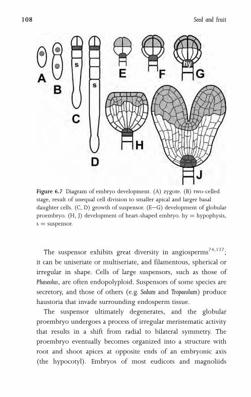
The suspensor exhibits great diversity in angiosperms74,127;
it can be uniseriate or multiseriate, and filamentous, spherical or
irregular in shape. Cells of large suspensors, such as those of
Phaseolus, are often endopolyploid. Suspensors of some species are
secretory, and those of others (e.g. Sedum and Tropaeolum) produce
haustoria that invade surrounding endosperm tissue.
The suspensor ultimately degenerates, and the globular
proembryo undergoes a process of irregular meristematic activity
that results in a shift from radial to bilateral symmetry. The
proembryo eventually becomes organized into a structure with
root and shoot apices at opposite ends of an embryonic axis
(the hypocotyl). Embryos of most eudicots and magnoliids
Figure 6.7 Diagram of embryo development. (A) zygote. (B) two-celled
stage, result of unequal cell division to smaller apical and larger basal
daughter cells. (C, D) growth of suspensor. (E�G) development of globular
proembryo. (H, J) development of heart-shaped embryo. hy ¼ hypophysis,
s ¼ suspensor.
108 Seed and fruit
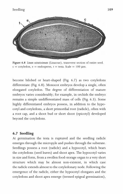
become bilobed or heart-shaped (Fig. 6.7) as two cotyledons
differentiate (Fig. 6.8). Monocot embryos develop a single, often
elongated cotyledon. The degree of differentiation of mature
embryos varies considerably; for example, in orchids the embryo
remains a simple undifferentiated mass of cells (Fig. 6.3). Some
highly differentiated embryos possess, in addition to the hypo-
cotyl and cotyledons, a short primordial root (radicle), often with
a root cap, and a shoot bud or short shoot (epicotyl) developed
beyond the cotyledons.
6.7 SeedlingAt germination the testa is ruptured and the seedling radicle
emerges through the micropyle and pushes through the substrate.
Seedlings possess a root (radicle) and a hypocotyl, which bears
the cotyledons (seed leaves) and shoot apex. The hypocotyl varies
in size and form, from a swollen food-storage organ to a very short
structure which may be almost non-existent, in which case
the radicle extends almost to the cotyledonary node. Following the
emergence of the radicle, either the hypocotyl elongates and the
cotyledons and shoot apex emerge (termed epigeal germination),
Figure 6.8 Linum usitatissimum (Linaceae), transverse section of entire seed.
c ¼ cotyledon, e ¼ endosperm, t ¼ testa. Scale ¼ 100 mm.
Seedling 109

or the cotyledons remain enclosed in the testa and the internode
above them (the epicotyl) elongates, pushing the shoot apex
upwards (termed hypogeal germination). Epigeal germination is
the most common type in angiosperms; the cotyledons are borne
above ground, and are usually photosynthetic. By contrast, some
larger-seeded eudicots such as legumes (e.g. Vicia faba) are
hypogeal, and possess fleshy, swollen cotyledons.
In monocots the radicle withers at an early stage, and
subsequent roots are shoot-borne (adventitious); they are each
initially surrounded by a sheath (coleorhiza), which develops
from outer cortical tissue by cell elongation. The cotyledons
are usually morphologically different from the first foliage leaves;
they typically possess simpler vasculature which often consists of
a single vascular bundle. In monocot seedlings the cotyledon
typically includes three parts109: a basal sheath, a ligule or ligular
sheath, and a limb, though the relative differentiation of each
region varies; for example, in Tigridia seedlings both the hypocotyl
and the basal sheath are extremely reduced.
110 Seed and fruit

Glossary
abaxial: away from axis (the abaxial leaf epidermis is usually the
lower one). cf. adaxial.
abscission layer: well-defined region of tissue separation, e.g. for
abscission of leaf from stem.
acropetal: towards the apex (cf. basipetal).
adaxial: towards the axis (the adaxial leaf surface is usually the
upper one). cf. abaxial.
adventitious roots: stem or leaf-borne roots.
aerenchyma: specialized parenchymatous tissue normally
associated with aquatic plants, with a regular, well-developed
system of intercellular air spaces.
aleurone layer: specialized outermost cell layer of endosperm.
amphistomatic leaf: one with stomata present on both surfaces.
amphivasal vascular bundle: one with xylem surrounding
phloem.
amyloplast: plastid containing starch.
androecium: collective term for stamens.
anisocytic: one of the classification types of mature stomata;
with three unequal subsidiary cells.
anomalous secondary growth: secondary growth that does not
fit the ‘‘normal’’ pattern of xylem and phloem production;
e.g. xylem with included phloem.
anomocytic: one of the classification types of mature stomata;
subsidiary cells absent.
anther: part of stamen bearing pollen (in anther locules).
anthesis: opening (dehiscence) of anther to release pollen;
sometimes applied to opening of the flower bud.
anticlinal: perpendicular to the plant surface.
antipodal cell: part of megagametophyte; one of a group of
(typically three) cells at chalazal end of mature embryo sac.
aperture (of pollen grain): thin or modified region of pollen
exine, through which the pollen tube grows at germination.
apocarpous gynoecium: one in which carpels are not fused.
apoplast: cell areas not bounded by plasmalemma (i.e. cell wall,
middle lamella, intercellular spaces).
apotracheal parenchyma (in secondary xylem): axial parenchyma
not associated with vessels.
archesporial cell (archespore): primary sporogenous cell
(or tissue).
areole: region of mesophyll between smallest veins in leaf.
aril (in seeds): fleshy outgrowth of funicle.
articulated laticifer: one composed of several cells.
astrosclereid: star-shaped or highly branched sclereid.
bark: part of woody stem outside secondary xylem; i.e. including
vascular cambium, phloem, cortex and periderm, though
sometimes applied only to periderm and outer cortex.
basipetal: towards the base; i.e. away from apex (cf. acropetal).
bast fibre: extraxylary fibre in stem; i.e. cortical or phloem fibre.
bicollateral vascular bundle: one with phloem on both sides of
xylem.
bifacial (dorsiventral) leaf: one with both adaxial and abaxial
surfaces, usually morphologically different from each other.
bitegmic seed: one with two seed coat layers, derived from two
integuments.
brachysclereid (stone cell): more or less isodiametric sclereid.
bulliform cells (usually in grass leaves): groups of epidermal
cells that are markedly larger than neighbouring epidermal
cells.
bundle sheath: layer of cells surrounding leaf vascular bundles.
112 Glossary

callose (b�1,3 glucan): product of plasma membrane that is
primarily a component of the cell wall; acts as a permeability
barrier or sealant in developing tissues such as microspore
tetrads or pollen tubes, or in response to wounding or
pathogens.
callus tissue: undifferentiated mass of thin-walled cells; usually
wound tissue.
cambium: meristematic band of cells; e.g. cork cambium or
vascular cambium.
Casparian strip (Casparian thickening): band of suberin
deposited in primary cell walls of root endodermis.
cellulose: primary carbohydrate component of plant cell walls;
a large linear polymer composed of D-glucose units with
b�1,4 linkages.
centric (terete) leaf: one that is cylindrical, or circular in
transverse section.
centrifugal: outwards (from the inside) (cf. centripetal).
centripetal: inwards (from the outside) (cf. centrifugal).
chalaza: region of ovule or seed where nucellus and integuments
merge, opposite micropyle.
chlorenchyma: photosynthetic tissue; specialized parenchyma
containing chloroplasts.
chlorophyll (chlorophyll a or chlorophyll b): complex
magnesium porphyrin compounds forming green
photosynthetic pigment contained within chloroplasts.
chloroplast: plastid containing chlorophyll, the site of
photosynthesis.
coenocyte: multinucleate cell; i.e. one in which cell division has
occurred without cell wall formation (e.g. non-articulated
laticifer).
coleoptile (in seedlings): parenchymatous sheath enclosing
plumule.
coleorhiza (in monocot seedlings, especially grasses):
parenchymatous sheath covering primary root.
Glossary 113

collateral vascular bundle: one with xylem and phloem adjacent
to each other.
collenchyma: strengthening tissue, consisting of groups of
axially elongated, tightly-packed cells with unevenly thickened
walls.
colpus (pl. colpi): aperture in pollen grain wall, aligned
equatorially during the tetrad stage.
companion cell: parenchymatous cell associated with sieve tube
element in phloem.
compitum: opening in transmitting tissue of ovary, near
micropyle.
cork: suberinized tissue (periderm).
cortex: region in stems and roots between epidermis and central
vascular region.
cotyledon: first leaf of the embryo.
cuticle: non-cellular layer of a fatty substance (cutin) that is
resistant to enzymes, covering surface of epidermis.
cystolith: calcareous body found in epidermal cell, or in leaf
mesophyll.
cytokinesis: cytoplasmic cleavage following nuclear division.
diacytic: one of the classification types of mature stomata; with
one or more pairs of subsidiary cells with their common
walls at right angles to the guard cells.
diarch root: one with two protoxylem poles.
dictyosome (Golgi body): cell organelle associated with secretory
activity.
distal: situated away from the centre of a body or its point of
attachment; sometimes terminal on axis (cf. proximal).
domatia: specialized pockets or tufts of hairs on some leaf
surfaces, providing shelter for small insects; sometimes
associated with extrafloral nectaries.
dorsiventral (bifacial) leaf: one with surfaces morphologically
different from each other.
druse: cluster crystal, or compound crystal.
114 Glossary

ectomycorrhizal: (fungal mycelium on roots) associated with root
surface (cf. endomycorrhizal).
ectexine (sexine): outer, sculptured part of exine in pollen
grain wall.
egg apparatus (in mature embryo sac): egg cell and two
synergids.
egg cell: part of megagametophyte; haploid cell at micropylar
end of mature embryo sac that will fuse with sperm to
form a zygote.
elaiophore: oil-secreting trichome or tissue in flower.
embryo sac: megagametophyte.
embryogenesis: embryo development. Somatic embryogenesis
is the induction of an embryo-like structure in cell suspension
cultures and on the surface of callus cultures.
endexine (nexine): inner layer of exine in pollen grain wall.
endocarp: inner layer of fruit wall (pericarp).
endodermis: innermost cell layer of cortex (mainly in roots);
initially often with Casparian strip, later often thick walled.
endogenous: of deep-seated (internal) origin (cf. exogenous).
endomycorrhizal: (fungal mycelium on roots) invading tissues
and cells (cf. ectomycorrhizal).
endoplasmic reticulum (ER): continuous membrane-bound
system of flattened sacs and tubules permeating cell cytoplasm,
sometimes coated with ribosomal particles.
endosperm: seed storage tissue, formed by fusion of one sperm
cell with two polar nuclei (i.e. usually triploid).
endotegmic seed coat: one with thickened, mechanical layer
derived from inner epidermis of inner integument.
endothecium: anther wall layer immediately within epidermis;
often possessing characteristic thickenings.
epiblast: in grass embryos, outgrowth opposite scutellum.
epicotyl: seedling axis above cotyledons.
epidermis: outermost layer of cells, covering entire primary plant
surface.
Glossary 115
epigeal germination: seedling germination type in which
cotyledons are green and borne above ground.
epigynous flower: one with inferior ovary (i.e. the ovary is
attached to the receptacle above the level of insertion of
the stamens and perianth parts).
epistomatic (leaves): possessing stomata on adaxial surface only.
epithem: tissue (often loosely packed parenchyma) in hydathode
between epidermis and vascular tissue.
equatorial (pollen grain aperture): located at or crossing a line
midway between the two poles of a microspore or pollen grain.
exarch root: one that matures centripetally.
exine: outer coat of pollen grain, often differentiated into outer
ectexine and inner endexine.
exocarp: outermost layer of pericarp.
exodermis: outer few cell layers of root cortex that have become
thicker walled and lignified.
exogenous: of superficial (external) origin (cf. endogenous).
exotestal seed coat: one with mechanical layer formed from
outer epidermis of outer integument.
fibre (fiber): axially elongated, thick-walled cell, usually
occurring as part of a group, lacking contents at maturity,
and with simple pits.
fibre-tracheid: cell type that is transitional between a fibre and
a tracheid, possessing bordered pits.
filament (in flower): stalk of stamen.
filiform apparatus: wall thickenings in synergid cells of mature
embryo sac.
funicle (funiculus): stalk attaching ovule to placenta in ovary.
fusiform: elongated with pointed ends.
generative cell: part of microgametophyte; divides (usually
within pollen tube) to form two sperm cells.
girder (bundle sheath extension): in leaves, a group of cells
(parenchymatous or sclerenchymatous) linking a vascular
bundle sheath with either or both epidermises.
116 Glossary

graft: union (by cell differentiation) of tissues of two different
individuals so that one (the scion) can survive on the other
(the stock).
ground tissue: unspecialized parenchymatous tissue enclosed
within epidermis.
growth ring: (in secondary xylem) a distinct growth
increment caused by differential rates of growth during
a growing season.
guard cell: one of a pair of stomatal cells that surround a pore.
guttation: secretion (usually passive) of water droplets, often at
hydathodes.
gynoecium (in flower): collective term for carpels; including
ovary, style(s) and stigma(s).
hair (trichome): epidermal appendage.
haustorium (pl. haustoria): cellular process that penetrates
adjacent tissues and plays a role in nutrient transport. For
example, haustoria of parasitic plants are modified roots;
endosperm haustoria are specialized absorptive cells.
hemicellulose: carbohydrate constituent of plant cell walls;
heterogeneous group of polysaccharides.
heterocellular ray: one composed of cells of different shapes
and sizes.
hilum: scar on seed indicating point of attachment of funicle
to ovary wall.
histogenesis: tissue differentiation.
homocellular ray: one composed of cells of similar shape
and size.
hydathode: region of secretion of water droplets (usually on
leaf margin).
hydrophyte: water plant, sometimes displaying specialized
features (e.g. aerenchyma).
hypocotyl: seedling axis bearing cotyledons and shoot apex.
hypodermis (often applied to leaves): distinct cell layer(s)
immediately within epidermis.
Glossary 117

hypogeal germination: type in which cotyledons remain enclosed
in seed coat after radicle has emerged.
hypogynous flower: one with superior ovary; i.e. ovary attached
to the receptacle above level of insertion of stamens and
perianth parts.
hypophysis: uppermost cell of the suspensor.
hypostase: proliferation of nucellus at chalazal end of embryo sac.
hypostomatic (leaves): possessing stomata on abaxial surface
only.
idioblast: cell that differs from cells of surrounding tissue.
included phloem: region of phloem embedded in secondary
xylem.
inaperturate (microspore or pollen grain): one lacking a clearly
defined aperture on the surface, so that the pollen tube can
potentially emerge at any point.
integument: either one or two structures (inner and outer
integuments) ensheathing the embryo sac, around the
nucellus.
intercalary growth (intercalary meristem): diffuse cell division
separate from apical meristem or other well-defined
meristems.
internode: region between two nodes on stem.
intine: inner layer of pollen grain wall.
isobilateral leaf: one with both surfaces similar, or with palisade
tissue on both sides.
Kranz anatomy: (in some plants with C4 photosynthesis)
distinctive leaf anatomy with mesophyll cells radiating from
bundle sheaths.
lateral roots: branches of tap root, of endogenous origin.
latex: complex fluid contained within laticifers, consisting
of a suspension of fine particles; used to form commercial
rubber.
laticifer: latex-secreting cell.
lenticel: region of loose cells in periderm (bark).
118 Glossary
lignin: strengthening material deposited with cellulose in plant
cell walls, giving rigidity; a high polymer composed of several
different types of phenyl-propane units.
ligule: outgrowth of abaxial epidermis in region between sheath
and petiole (or between sheath and lamina if petiole absent);
derived from cross zone in leaf primordium.
lysigenous (ducts or cavities): developing by cellular breakdown
(lysis). (cf. schizogenous).
macrosclereid: elongated sclereid often located in seed coat.
megagametophyte (female gametophyte): mature embryo sac,
most commonly consisting of seven cells and eight nuclei
(two synergid cells, an egg cell, three antipodal cells, two polar
nuclei).
megaspore: female haploid cell resulting from meiosis; usually
one of four (or two), of which only one is functional.
megasporogenesis: process of megaspore formation from
megaspore mother cell (megasporocyte).
megasporocyte (megaspore mother cell): a diploid cell that
will give rise to (usually) four haploid megaspores following
meiosis.
meiosis (I and II): two successive divisions of a diploid nucleus
to form a haploid gamete.
meristem: region of cell division and tissue differentiation
(e.g. apical meristem, intercalary meristem, lateral meristem,
vascular cambium, primary and secondary thickening
meristems).
meristemoid: isolated meristematic cell, usually the smaller cell
resulting from an asymmetric division (e.g. guard cell mother
cell).
mesocarp: middle layer of pericarp.
mesogene cell: stomatal subsidiary cell derived from
meristemoid.
mesomorphic: displaying no xeromorphic or hydromorphic
characteristics.
Glossary 119
mesophyll: ground tissue of leaf; mainly consisting of
parenchyma or chlorenchyma; often differentiated into
palisade and spongy mesophyll.
mesophyte: plant growing in conditions of relatively continuous
moisture and/or other nutrients.
metaxylem: primary xylem formed after protoxylem.
microfibril: thread-like component of cell wall, primarily
cellulose.
microgametophyte (male gametophyte): cellular component of
mature pollen grain, in angiosperms consisting of vegetative
cell and generative cell.
micropyle: opening at one end of ovule, usually surrounded by
integuments.
microsporangium: pollen sac, contained within anther.
microspore: individual haploid cell that will give rise to
microgametophyte. Undergoes unequal mitotic division to
form vegetative and generative cells.
microsporocyte: a diploid cell that will give rise to four haploid
microspores following meiosis.
microsporogenesis: developmental process leading to production
of four haploid microspores from a diploid microsporocyte
by meiosis and cytokinesis.
middle lamella: layer between walls of neighbouring cells.
mitochondrion (pl. mitochondria): cytoplasmic organelle.
mitosis: cell division to form two cells of equivalent
chromosome composition to parent cell; involving four
main stages: prophase, metaphase, anaphase and telophase.
mucilage (slime): strongly hydrophilic polymer containing
polysaccharides.
multiseriate: consisting of more than one layer or row of
cells.
nectary (floral or extrafloral): localized cell or cells that secrete
a sugary liquid (nectar).
node: part of stem where leaves are attached.
120 Glossary

non-articulated laticifer: one composed of a single multinucleate
coenocytic cell.
nucellus: ovule cell layer(s) immediately surrounding
megagametophyte.
obturator: proliferation of (usually) ovary tissue near micropyle;
secretes substances that guide growing pollen tubes into
micropyle.
ontogeny: development; differentiation and growth.
organogenesis: development of organs.
osmophore: scent-producing gland in flower.
osteosclereid: bone-shaped sclereid.
papilla (pl. papillae): epidermal appendage; small unicellular
trichome.
paracytic: classification type of mature stomata, with one or
more subsidiary cells present at either side of the guard cells.
paratracheal parenchyma: axial parenchyma associated with
vessels (in secondary xylem).
parenchyma: tissue composed of unspecialized thin-walled cells
with living contents.
passage cell: endodermal cell that remains thin walled compared
with neighbouring endodermal cells.
pearl gland (pearl body): secretory leaf emergence or trichome
that provides food (carbohydrates, lipids and proteins)
for ants.
perforated ray cell: (in secondary xylem) ray cell linking two
vessel elements, and itself resembling and functioning as
a vessel element.
perforation plate: opening in end wall of vessel element;
may be simple (a single opening) or scalariform (with bars),
or more rarely, reticulate (mesh-like) or foraminate (with
pores).
perianth: outer sterile part of flower, consisting of whorls of sepals
(calyx) and petals (corolla), or sometimes undifferentiated
tepals.
Glossary 121

pericarp: fruit wall.
periclinal: parallel with plant surface.
pericycle (in roots): layer of thin-walled cells within the
endodermis.
periderm: cork tissue.
perigene cell: stomatal subsidiary cell derived from cell adjacent to
meristemoid, not from the meristemoid itself.
periplasmodium: coalescent mass in anther locule, formed from
protoplasts of tapetal cells.
perisperm: food-storage tissue in the seed, derived from part of
nucellus.
phellem: external derivatives of phellogen.
phelloderm: internal derivatives of phellogen: cork cambium,
or cork meristem.
phloem: tissue that transports food in the form of assimilates;
either primary (produced by apical meristem) or secondary
(produced by vascular cambium).
photosynthesis: process driven by light energy, in which carbon
dioxide is reduced to carbohydrate form, with concomitant
oxygen release.
phyllotaxis: pattern of arrangement of organs on an axis,
e.g. leaves on a stem, flowers on an inflorescence.
pith: central parenchymatous region of stems, often breaking
down to form a cavity.
pit: thin area of the primary and secondary cell wall, often
corresponding with a pit in adjacent cells (forming a pit-pair).
placenta: region of attachment of ovules on ovary wall.
placentation: arrangement of placentae and locules in ovary
(e.g. axile, basal, free central, parietal).
plasma membrane (plasmalemma): cell membrane (within cell
wall) that encloses protoplast.
plasmodesmata: protoplasmic strands passing through primary
pit fields between adjacent cells, and connecting their
protoplasts.
122 Glossary
plastid: cell organelle contained within cytoplasm, often with
specialized function (e.g. chloroplast, amyloplast).
polar nucleus: one of a pair of nuclei of the mature
megagametophyte, often located in a central position.
pollen grain: microgametophyte enclosed by sporopollenin.
pollen tube: tube that emerges from intine of germinating pollen
grain.
polyarch root: one with several (more than four) protoxylem
poles.
primordium: organ at an early stage of differentiation.
procambium: primary tissue located near (shoot or root) apex
that gives rises to primary vascular tissue.
proembryo: early (globular) stage of embryo development, prior
to differentiation of cotyledons and hypocotyl.
promeristem (in root apices): region of greatest mitotic activity.
protoplast: living part of cell, surrounded by a plasma
membrane.
protoxylem: first-formed primary xylem.
proximal: situated closer to the centre of a body or its point of
attachment (cf. distal).
quiescent centre: region of cells at root apex possessing little or no
cell division activity.
radicle: first-formed root of seedling.
raphe: stalk attaching seed to ovary (directly derived from the
funicle).
raphide: fine, needle-like crystal of calcium oxalate, one of
a group of several raphides formed within a single cell.
ray (in secondary xylem): tissue of radially oriented cells, usually
parenchymatous, derived from ray initials.
root cap: protective covering of cells over root apex.
root hair: water-absorbing hair on root epidermis.
sarcotesta: fleshy part of seed coat.
scale (peltate hair): specialized trichome, consisting of a fused
disc of cells attached to the epidermis by a stalk.
Glossary 123
schizogenous (ducts or cavities): developing from intracellular
spaces by separation of cell walls. (cf. lysigenous).
schizo-lysigenous (ducts or cavities): developing by a combina-
tion of cell separation and degradation.
sclereid: thick-walled sclerenchymatous cell that is either
isodiametric (brachysclereid or stone cell), or variously shaped
(e.g. astrosclereid, osteosclereid, macrosclereid).
sclerenchyma: strengthening tissue, consisting of cells with
thickened lignified walls, usually lacking contents at maturity.
(cf. fibre, sclereid).
scutellum: specialized structure in grass embryos, normally
interpreted as a modified cotyledon.
sieve tube element: conducting cell in phloem; possessing sieve
areas and sieve plates in walls.
sporopollenin: highly resistant complex polymer that forms outer
wall of pollen grain.
squamules: appendages (often glandular) that occur in leaf
axils.
stigma: secretory region on gynoecium that is receptive to pollen
grains.
stipules: appendages at base of leaf sheath, often paired and
sometimes leafy.
stoma (pl. stomata): epidermal cell complex that facilitates
gaseous exchange, usually present on aerial parts of plant;
consisting of two guard cells surrounding a pore.
stone cell: isodiametric sclereid.
storied (in secondary xylem, referring to vessels, rays or
axial parenchyma): with stratified structure, occurring
in rows.
styloid: elongated prismatic crystal of calcium oxalate.
suberin: lipophilic substance (similar to cutin) deposited in walls
of some cell types, e.g. in cork cells.
subsidiary cells: epidermal cells adjacent to stomata that differ
from other pavement epidermal cells.
124 Glossary
sulcus (pl. sulci): aperture in pollen grain wall, located on its distal
face.
suspensor: row of cells attaching globular proembryo to wall
of embryo sac.
symplast: connected living protoplasts of adjacent cells.
syncarpous gynoecium: one in which carpels are (at least
partially) fused.
syncytium: cytoplasmic region enclosed by a single plasma
membrane and bearing several nuclei.
synergid: part of megagametophyte; one of a pair of cells located
at micropylar end of mature embryo sac.
tannins: a group of phenol derivatives.
tap root: main central root, formed directly from seedling
radicle.
tapetum: layer of nutritive tissue between microsporocytes and
wall of anther locule.
tegmen: inner layer of seed coat, formed from inner integument.
testa: seed coat; or in bitegmic seeds, outer layer of seed coat,
formed from outer integument.
tetrad: group of four microspores or megaspores, the daughter
cells of a single microsporocyte or megasporocyte.
tetrarch (root): one with four protoxylem poles.
tracheid: xylem water-conducting cell, usually possessing
bordered pits but lacking perforation plates.
transfer cell: specialized plant cell that facilitates transport of
soluble substances across tissue boundaries.
transmitting tissue (stigmatoid tissue): secretory tissue of style
through which pollen tubes grow, from stigma to ovule.
triarch root: one with three protoxylem poles.
trichoblast: root epidermal cell that gives rise to a root hair.
trichome (hair): epidermal outgrowth.
tricolpate (microspore or pollen grain): one with three equatorial
apertures (colpi).
tunica-corpus: regions of central shoot apical organization.
Glossary 125
tylose: (in secondary xylem) outgrowth of wall of axial
parenchyma cell into a vessel element through a pit; eventually
blocking water passage through vessel.
unifacial leaf: one with both surfaces similar, sometimes derived
from a single (usually abaxial) surface.
uninterrupted meristem: region of diffuse cell divisions that
is continuous with the apical meristem; producing extension
growth of the axis.
uniseriate: consisting of a single layer or row of cells.
vacuole: cavity.
vascular bundle (vascular trace): axial strand of vascular tissue.
vascular cambium: meristem that prcoduces secondary vascular
tissue.
vascular tissue: conducting tissue (phloem and xylem).
vegetative cell: one of two cells of microgametophyte.
velamen: outer dermal layer on aerial roots of some tropical
epiphytes such as Orchidaceae and Araceae.
venation: arrangement of vascular bundles in leaf (e.g. parallel or
reticulate venation).
vessel element: water-conducting cell of xylem, possessing
bordered pits on lateral walls and perforation plates on end
walls. Groups of axially linked vessel elements form a vessel.
vestured pitting (in secondary xylem): bordered pits surrounded
by numerous warty protuberances.
wax: fatty substance often deposited on surface of cuticle
(epicuticular wax).
whorl (in flower): band or ring of organs encircling floral axis;
sometimes applied to a region of a similar organ type,
e.g. stamen whorl.
wood: secondary xylem.
xeromorphic: showing characteristics that are associated with dry
or nutrient-poor environments.
126 Glossary

xerophyte: plant that grows in a dry (xeric) or nutrient-poor
environment.
xylem: complex water-transporting tissue consisting of several
different cell types.
zygote: cell formed by fusion of egg and sperm cells; eventually
divides to form the proembryo.
Glossary 127
References
1. Anderson, W. R. 1980. Cryptic self-fertilization in the
Malpighiaceae. Science 207: 892�893.
2. APG. 1998. An ordinal classification for the families of flowering
plants. Annals of the Missouri Botanical Garden 85: 531�553.
3. APG. 2003. An update of the Angiosperm Phylogeny Group
classification for the orders and families of flowering plants:
APG II. Botanical Journal of the Linnean Society 141: 399�436.
4. Arber, A. 1934. The Gramineae. Cambridge: Cambridge University
Press.
5. Arber, A. 1937. Studies in flower structure III. On the ‘corona’
and androecium in certain Amaryllidaceae. Annals of Botany 1:
293�304.
6. Baijnath, H. and D. F. Cutler. 1993. A contribution to the anatomy
and surface details of leaves of the genus Bulbine (Asphodelaceae) in
Africa. South African Journal of Botany 59: 109�115.
7. Bailey, I.W. 1953. Evolution of the tracheary tissue of land plants.
American Journal of Botany 40: 4�8.
8. Barlow, P.W. 1975. The root cap. In: The development and form of roots.
J. G. Torrey and D. F. Clarkson (eds). London: Academic Press,
pp. 21�54.
9. Barthlott, W. 1981. Epidermal and seed surface characters of
plants: systematic applicability and some evolutionary aspects.
Nordic Journal of Botany 1: 345�355.
10. Barthlott, W. and E. Wollenweber. 1981. Zur Feinstruktur, Chemie
und taxonomischen Signifikanz epicuticularer Wachse und
ahnlicher Sekrete. Tropische und Subtropische Pflanzenwelt 32: 1�67.

11. Baum, S. B., J. G. Dubrovsky and T. L. Rost. 2002. Apical
organization and maturation of the cortex and vascular cylinder
in Arabidopsis thaliana (Brassicaceae) roots. American Journal of Botany
89: 908�920.
12. Beaumont, J., D. F. Cutler, T. Reynolds and J. G. Vaughan. 1985.
The secretory tissue of aloes and their allies. Israel Journal of Botany
34: 265�282.
13. Beck, C. R. 2005. An Introduction to Plant Structure and Development.
Cambridge: Cambridge University Press.
14. Behnke, H. D. 1991. Distribution and evolution of forms
and types of sieve tube plastids in the dicotyledons. Aliso 3:
167�182.
15. Boesewinkel, F. D. and F. Bouman. 1984. The seed: structure.
In: Embryology of Angiosperms. B.M. Johri (ed.). Berlin:
Springer-Verlag, 567�610.
16. Bouman, F. 1984. The ovule. In: Embryology of Angiosperms.
B. M. Johri (ed.). Berlin: Springer-Verlag, 123�157.
17. Brett, D.W. and A. P. Sommerard. 1986. Ultrastructural
development of plastids in the epidermis and starch layer of
glossy Ranunculus petals. Annals of Botany 58: 903�910.
18. Casson, S. A. and K. Lindsey. 2003. Genes and signalling in root
development. Tansley Review. New Phyologist 158: 11�38.
19. Choat, B., S. Jansen, M. Zwieniecki, E. Smets and N.M. Holbrook.
2004. Changes in pit membrane porosity due to deflection
and stretching: the role of vestured pits. Journal of Experimental
Botany 55: 1569�1575.
20. Clowes, F. A. L. 1961. Apical meristems. Oxford: Blackwell.
21. Clowes, F. A. L. 1994. Origin of the epidermis in root meristems.
New Phytologist 127: 335�347.
22. Cocucci, A. E. and A.M. Anton. 1988. The grass flower:
suggestions on its origin and evolution. Flora 181: 353�362.
23. Corner, E. J. H. 1976. The Seeds of Dicotyledons. Cambridge:
Cambridge University Press.
24. Cutler, D. F., P. J. Rudall, P. E. Gasson and R.M.O. Gale. 1987.
Root Identification Manual of Trees and Shrubs. London: Chapman
and Hall.
References 129

25. Daumann, E. 1970. Das Blutennektarium der Monocotyledon
unter besonderer Berucksichtigung seiner systematischen und
phylogenetischen Bedeutung. Feddes Repertorium 80: 463�590.
26. DeMason, D. A. 1983. The primary thickening meristem:
definition and function in monocotyledons. American Journal of
Botany 70: 955�962.
27. Eames, A. J. and L. H. Macdaniels. 1925. An Introduction to Plant
Anatomy. London: McGraw-Hill.
28. Endress, P. K. 2001. The flowers in extant basal angiosperms and
inferences on ancestral flowers. International Journal of Plant Sciences
162: 1111�1140.
29. Erdtman, G. 1966. Pollen Morphology and Plant Taxonomy. New York:
Hafner Publishing Company.
30. Esau, K. 1977. Anatomy of Seed Plants. Canada: John Wiley and Sons.
31. Fahn, A. 1979. Secretory Tissues in Plants. London: Academic Press.
32. Feldman, L. J. 1984. The development and dynamics of the root
apical meristem. American Journal of Botany 71: 1308�1314.
33. Foster, A. S. 1956. Plant idioblasts; remarkable examples of cell
specialisation. Protoplasma 46: 184�193.
34. Foster, A. S. and E.M. Gifford. 1989. Comparative Morphology of Vascular
Plants. New York: W.H. Freeman & Co.
35. French, J. C. 1986. Patterns of stamen vasculature in the Araceae.
American Journal of Botany 73: 434�449.
36. Frey-Wyssling, A. 1976. The Plant Cell Wall. Third edition. Berlin:
Gebruder Borntraeger.
37. Furness, C. A. and P. J. Rudall. 1999. Inaperturate pollen in
monocotyledons. International Journal of Plant Sciences 160: 395�414.
38. Furness, C. A. and P. J. Rudall. 2001. The tapetum in basal
angiosperms: early diversity. International Journal of Plant Sciences 162:
375�392.
39. Furness, C. A., P. J. Rudall and F. B. Sampson. 2002. Evolution
of microsporogenesis in angiosperms. International Journal of Plant
Sciences 163: 235�260.
40. Furness, C. A. and P. J. Rudall. 2003. Apertures with lids:
distribution and significance of operculate pollen in monocots.
International Journal of Plant Sciences 164: 835�854.
130 References

41. Furness, C. A. and P. J. Rudall. 2004. Pollen aperture evolution �
a crucial factor for eudicot success? Trends in Plant Science 9:
1360�1385.
42. Galatis, B. and P. Apostolakos. 2004. The role of the cytoskeleton
in morphogenesis and function of stomatal complexes. Tansley
Review. New Phytologist 161: 613�639.
43. Gifford, E.M. and G. E. Corson. 1971. The shoot apex in seed
plants. Botanical Review 37: 147�229.
44. Graven, P., C. G. De Koster, J. J. Boon and F. Bouman. 1996.
Structure and macromolecular composition of the seed coat of
the Musaceae. Annals of Botany 77: 105�122.
45. Grootjen, C. J. and F. Bouman. 1988. Seed structure in Cannaceae:
taxonomic and ecological implications. Annals of Botany 61:
363�371.
46. Hagemann, W. 1973. The organization of shoot development.
Revista Biologia (Lisbon) 9: 43�67.
47. Heslop-Harrison, H. and Y. Heslop-Harrison. 1982. The
specialised cuticles of the receptive surfaces of angiosperm
stigmas. In: The Plant Cuticle. D. F. Cutler, K. L. Alvin and
C. E. Price (eds). London: Academic Press, pp. 99�119.
48. Heslop-Harrison, J. 1976. The adaptive significance of the
exine. In: The Evolutionary Significance of the Exine. I. K. Ferguson and
I. Muller (eds). London: Academic Press, pp. 27�37.
49. Heslop-Harrison, J. and K. R. Shivanna. 1977. The receptive
surface of the angiosperm stigma. Annals of Botany 41:
1233�1258.
50. Heslop-Harrison, J. 1987. Pollen germination and pollen-tube
growth. International Reviews in Cytology 107: 1�78.
51. Hickey, L. J. 1973. A classification of the architecture of
dicotyledonous leaves. American Journal of Botany 60: 17�33.
52. Holroyd, G. H., A.M. Hetherington and J. E. Gray. 2002. A role
for the cuticular waxes in the environmental control of stomatal
development. New Phytologist 153: 433�439.
53. Horner, H. T. and H. J. Arnott. 1966. Histochemical and
ultrastructural study of pre- and post-germinated Yucca seeds.
Botanical Gazette 127: 48�64.
References 131
54. Howard, R. A. 1974. The stem�node�Ieaf continuum of the
Dicotyledonae. Journal of the Arnold Arboretum 55: 125�181.
55. Iqbal, M. 1995. Structure and behaviour of vascular cambium
and the mechanism and control of cambial growth. In:
The Cambial Derivatives, M. Iqbal (ed.). Berlin: Gebruder Borntraeger,
pp. 1�67.
56. Janssen, B. J., L. Lund and N. Sinha. 1998. Overexpression of
a homeobox gene, LET6, reveals indeterminate features in the
tomato compound leaf. Plant Physiology 117: 771�786.
57. Jernstedt, J. A. 1984. Root contraction in hyacinth. I. Effects of
IAA on differential cell expansion. American Journal of Botany 71:
1080�1089.
58. Johnson, S. A. and S. McCormick. 2001. Pollen germinates
precociously in the anthers of raring-to-go, an Arabidopsis
gametophytic mutant. Plant Physiology 126: 685�695.
59. Johri, B.M. 1992. Haustorial role of pollen tubes. Annals of Botany
70: 471�475.
60. Juniper, B. E. and C. E. Jeffree. 1983. Plant Surfaces. London: Edward
Arnold.
61. Kaplan, D. R. 1973. The Monocotyledons: their evolution and
comparative biology. VII. The problem of leaf morphology and
evolution in the Monocotyledons. Quarterly Review of Biology 48:
437�451.
62. Kaplan, D. R. 1975. Comparative developmental evaluation of
the morphology of unifacial leaves in the monocotyledons.
Botanische Jahrbucher 95: 1�105.
63. Kauff, F., P. J. Rudall and J. G. Conran. 2000. Systematic root
anatomy of Asparagales and other monocotyledons. Plant Systematics
and Evolution 223: 139�154.
64. Kay, Q. O. N., H. S. Daoud and C. H. Stirton. 1981. Pigment
distribution, light reflection and cell structure in petals. Botanical
Journal of the Linnean Society 83: 57�84.
65. Knox, R. B. 1984. The pollen grain. In: Embryology of Angiosperms,
B. M. Johri (ed.). Berlin: Springer-Verlag, pp. 197�271.
66. Kuijt, J. 1969. The biology of parasitic flowering plants. Berkeley:
University of California Press.
132 References
67. Larsen, P. R. 1984. The role of subsidiary trace bundles in stem and leaf
development of the Dicotyledonae. In: Contemporary Problems in Plant Anatomy
R. A. White and W. C. Dickison (eds.). London: Academic Press,
109�143.
68. Lersten, N. R. 2004. Flowering Plant Embryology. Oxford: Blackwell.
69. Maheshwari, P. 1950. An Introduction to the Embryology of the Angiosperms.
New York: McGraw-Hill.
70. Mahlberg, P. 1975. Evolution of the laticifer in Euphorbia as
interpreted from starch grain morphology. American Journal of Botany
62: 577�583.
71. Mahlberg, P. 1993. Laticifers � an historical perspective. Botanical
Review 59: 1�23.
72. McCully, M. E. 1975. The development of lateral roots. In: The
Development and Function of Roots. J. G. Torrey and D. T. Clarkson (eds.).
London: Academic Press, pp. 105�124.
73. Metcalfe, C. R. and L. Chalk. 1983. Anatomy of the Dicotyledons. II.
Second edition. Oxford: Clarendon Press.
74. Natesh, S. and M. A. Rau. 1984. The embryo. In: Embryology of
Angiosperms. B.M. Johri (ed.). Berlin: Springer-Verlag, pp. 377�443.
75. Nepi, M. and E. Pacini. 1999. What may be the significance
of polysiphony in Lavatera arborea? In: Anther and Pollen: from
Biology to Biotechnology. C. Clement and J. C. Audran (eds.). Berlin:
Springer-Verlag, pp. 13�20.
76. O’Dowd, D. J. 1982. Pearl bodies as ant food: an ecological role
for some leaf emergences of tropical plants. Biotropica 14: 40�49.
77. Ormense, S., A. Havelange, G. Bernier and C. van der Schoot.
2002. The shoot apical meritem of Sinapis alba L. expands its
central symplastic field during the floral transition. Planta 215:
67�78.
78. Parkin, J. 1928. The glossy petal of Ranunculus. Annals of Botany 42:
739�755.
79. Parre, E. and A. Geitmann. 2005. More than a leak sealant: the
mechanical properties of callose in pollen tubes. Plant Physiology
137: 274�286.
80. Pate, J. S. and B. E. S. Gunning. 1972. Transfer cells. Annual Reviews in
Plant Physiology 23: 173�196.
References 133

81. Peck, C. J. and N. R. Lersten. 1991. Gynoecial ontogeny and
morphology, and pollen tube pathway in black maple, Acer
saccharum ssp. nigrum (Aceraceae). American Journal of Botany 87:
247�259.
82. Prychid, C. J. and P. J. Rudall. 1999. Calcium oxalate crystals in
monocotyledons: structure and systematics. Annals of Botany 84:
725�739.
83. Prychid, C. J., P. J. Rudall and M. Gregory. 2003. Systematics
and biology of silica bodies in monocotyledons. Botanical Review
69: 377�440.
84. Punt, W., S. Blackmore, S. Nilsson and A. Le Thomas. 1994. Glossary
of Pollen and Spore Terminology. Utrecht: LPP Foundation.
85. Rasmussen, H. 1983. Stomatal development in families of Liliales.
Botanische Jahrbucher 104: 261�287.
86. Reiser, L. and R. L. Fischer. 1993. The ovule and the embryo sac.
The Plant Cell 5: 1291�1301.
87. Rudall, P. J. 1987. Laticifers in Euphorbiaceae � a conspectus.
Botanical Journal of the Linnean Society 94: 143�163.
88. Rudall, P. J. 1991. Lateral meristems and stem thickening growth
in monocotyledons. Botanical Review 57: 150�163.
89. Rudall, P. J. and L. Clark. 1992. The megagametophyte in Labiatae.
In: Advances in Labiate Science, R.M. Harley and T. R. Reynolds (eds.).
London: Royal Botanic Gardens, Kew, pp. 65�84.
90. Rudall, P. J. 1994. Laticifers in Crotonoideae (Euphorbiaceae):
homology and evolution. Annals of the Missouri Botanical Garden 81:
270�282.
91. Rudall, P. J. 1995. Anatomy of the Monocotyledons. VIII. Iridaceae.
Oxford: Oxford University Press.
92. Rudall, P. J. 1997. The nucellus and chalaza in monocotyledons:
structure and systematics. Botanical Review 63: 140�184.
93. Rudall, P. J. 2002. Homologies of inferior ovaries and septal
nectaries in monocotyledons. International Journal of Plant Sciences
163: 261�276.
94. Rudall, P. J. and M. Buzgo. 2002. Evolutionary history of the
monocot leaf. In: Developmental Genetics and Plant Evolution, Q. C. B.
134 References
Cronk, R.M. Bateman and J. A. Hawkins (eds.). London: Taylor
and Francis, pp. 432�458.
95. Rudall, P. J., W. Stuppy, J. Cunniff, E. A. Kellogg and B. G. Briggs.
2005. Evolution of reproductive structures in grasses (Poaceae)
inferred by sister-group comparison with their putative closest
living relatives, Ecdeiocoleaceae. American Journal of Botany 92:
1432�1443.
96. Ryding, O. 1992. The distribution and evolution of myxocarpy
in Lamiaceae. In: Advances in Labiate Science. R.M. Harley and
T. R. Reynolds (eds.). London: Royal Botanic Gardens, Kew,
pp. 85�96.
97. Sage, R. F. 2004. The evolution of C4 photosynthesis. Tansley
Review. New Phytologist 161: 341�370.
98. Sampson, F. B. 2000. Pollen diversity in some modern Magnoliids.
International Journal of Plant Sciences 161: S193�S210.
99. Shigo, A. L. 1985. How tree branches are attached to trunks.
Canadian Journal of Botany 63: 1391�1401.
100. Skinner, D. J., T. A. Hill and C. S. Gasser. 2004. Regulation of
ovule development. Plant Cell 16: S32�45.
101. Steer, M. and J. Steer. 1989. Pollen tube tip growth. New Phytologist
111: 323�358.
102. Steeves, T. A. and I.M. Sussex. 1989. Patterns in Plant Development.
Cambridge: Cambridge University Press.
103. Stevens, P. F. (2006). Angiosperm phylogeny website. Version 6.
http://www.mobot.org/MOBOT/Research/APweb/
104. Stevenson, D.W. 1980. Radial growth in Beaucarnea recurvata. American
Journal of Botany 67: 476�489.
105. Stevenson, D.W. and J. B. Fisher. 1980. The developmental
relationship between primary and secondary thickening growth
in Cordyline (Agavaceae). Botanical Gazette 141: 264�268.
106. Stewart, W.N. and G.W. Rothwell. 1993. Paleobotany and the Evolution
of Plants, Second edition. Cambridge: Cambridge University Press.
107. Sylvester, A.W., L. Smith and M. Freeling. 1996. Acquisition of
identity in the developing leaf. Annual Reviews of Cell and Developmental
Biology 12: 257�304.
References 135
108. Sylvester, A.W. 2000. Division decisions and the spatial regulation
of cytokinesis. Current Opinion in Plant Biology 3: 58�66.
109. Tillich, H. J. 1995. Seedlings and systematics in monocotyledons.
In: Monocotyledons: Systematics and Evolution, P. J. Rudall, P. J. Cribb,
D. F. Cutler, C. J. Humphries (eds.). Royal Botanic Gardens,
London/Kew, pp. 303�352.
110. Tilton, V. R. and H. T. Horner. 1980. Stigma, style and obturator
of Ornithogalum caudatum (Liliaceae) and their function in the
reproductive process. American Journal of Botany 67: 1113�1131.
111. Tomlinson, P. B. and M.H. Zimmerman. 1969. Vascular anatomy
of monocotyledons with secondary growth � an introduction.
Journal of the Arnold Arboretum 50: 159�179.
112. Tomlinson, P. B. 1974. Development of the stomatal complex as
a taxonomic character in Monocotyledons. Taxon 23: 109�128.
113. Tucker, S. C. 1972. The role of ontogenetic evidence in floral
morphology. In: Advances in Plant Morphology, Y. S. Murty, et al. (eds.).
Meerut: Sarita Prakashan, pp. 359�369.
114. Twell, D. 2002. The developmental biology of pollen. In: Plant
Reproduction. Annual Plant Reviews 6. S. D. O’Neill and J. A. Roberts
(eds.). Sheffield: Sheffield Academic Press, pp. 86�153.
115. Uhl, N.W. and H. E. Moore. 1977. Centrifugal stamen initiation
in phytelephantoid palms. American Journal of Botany 64:
1152�1161.
116. Van Bel, A. J. E., K. Ehlers and M. Knoblauch. 2002. Sieve elements
caught in the act. Trends in Plant Science 7: 126�132.
117. Vijayaraghavan, M. R. and K. Prabhakar. 1984. The endosperm. In:
Embryology of Angiosperms, B. M. Johri (ed.). Berlin: Springer-Verlag,
pp. 321�376.
118. Vogel, S. 1974. Olbumen und Olsammelnde Bienen. Tropische und
Subtropische Pflanzenwelt 7: 285�577.
119. Vogel, S. 1990. The role of Scent Glands in Pollination: on the Structure and
Function of Osmophores. New Delhi: Amerind.
120. Wang, X. F., Y. B. Tao and Y. T. Lu. 2002. Pollen tubes enter
neighbouring ovules by way of receptacle tissue, resulting in
increased fruit-set in Sagittaria potamogetifolia. Annals of Botany 89:
791�796.
136 References
121. Weberling, F. 1989. Morphology of Flowers and Inflorescences. Cambridge:
Cambridge University Press.
122. Weld, M., J. Ziegler and T.M. Kutchan. 2004. The roles of latex
and the vascular bundle in morphine biosynthesis in the opium
poppy, Papaver somniferum. Proceedings of the National Academy of Sciences
101: 13957�13962.
123. West, M. A. L. and J. J. Harada. 1993. Embryogenesis in higher
plants: an overview. The Plant Cell 5: 1361�1369.
124. Wilkinson, H. P. 1979. The plant surface. In: Anatomy of the
Dicotyledons. C. R. Metcalfe and L. Chalk (eds.). Oxford: Clarendon,
pp. 97�165.
125. Willemse, M. T.M. and J. L. Van Went. 1984. The female
gametophyte. In: Embryology of Angiosperms. B. M. Johri (ed.).
Berlin: Springer-Verlag, pp. 159�196.
126. Yadegari, R. and G. N. Drews. 2004. Female Gametophyte
Development. Plant Cell 16: S133�141.
127. Yeung, E. C. and D.W. Meinke. 1993. Embryogenesis in
angiosperms: development of the suspensor. The Plant Cell 5:
1361�1369.
128. Zimmerman, M.H. and P. B. Tomlinson. 1972. The vascular
system of monocotyledonous stems. Botanical Gazette 133:
141�155.
References 137

Subject index
abscission, 22, 111adventitious root, 22, 24, 43, 50, 51, 111aerenchyma, 10, 58, 111aleurone layer, 103, 104, 111amyloplast, 6, 111, 123androecium, 75, 81, 111anomalous secondary growth, 30, 34,
111anther, 81, 82, 85, 111anthocyanin, 79antipodal cell, 9, 92, 94, 105, 112aperture, 85, 112apical meristem, 13, 18, 21, 23, 24archesporial cell (archespore), 92, 93, 112areole, 44, 71, 112aril, 101, 112asymmetric (unequal) mitosis, 22, 84, 119axial parenchyma, 17, 31, 32
bark, 42, 112bordered pits, 18bract, 75bulliform cells, 61, 73, 112bundle sheath, 72, 73, 74, 112
C4 photosynthesis, 74calcium oxalate crystals, 5, 6, 7, 20callose, 21, 84, 113callus, 10, 51, 113calyx, 75carpel, 75, 87, 89caryopsis, 102, 103Casparian thickenings, 26, 49, 113cell, 2, 4cell inclusions, 5
cell wall, 2, 4, 54cellulose, 2, 113chalaza, 90, 105, 113chlorenchyma, 10, 17, 25, 113chloroplast, 5, 10, 68, 113, 123coenocyte, 4, 8, 113coleoptile, 104, 113coleorhiza, 104, 110, 113collenchyma, 9, 10, 114colpus (colpi), 85, 86, 114companion cell, 19, 20, 22, 31, 114compitum, 89, 114compound leaf, 60contractile root, 48cork, 42, 114corolla, 75cortex, 24, 25, 26, 43, 48, 49, 114cotyledon, 104, 109, 110, 114, 124crystals, 5, 6, 7, 20, 62, 124crystal sand, 6cuticle, 13, 66, 87, 88, 114cystolith, 5, 62, 63, 66, 114cytokinesis, 84
dictyosome, 4, 5, 114domatia, 67, 114double fertilisation, 96druse (cluster crystal), 6, 114dust seeds, 101
ectexine, 85, 115ectomycorrhizal fungi, 53, 115endomycorrhizal fungi, 53, 115egg cell, 94, 96, 107, 115elaiophore, 97, 98, 115
embryo sac, 90, 92–94, 107, 115embryogenesis, 107, 115endexine, 85, 115endocarp, 101, 102, 115endodermis, 25, 26, 43, 48, 49, 115endoplasmic reticulum, 4, 5, 115endosperm, 9, 96, 97, 103–106, 115endothecium, 82, 115endothelium, 93epiblast, 103, 104, 115epicotyl, 104, 109, 110, 115epicuticular wax, 13, 61, 66, 126epidermis, 9, 13, 24, 25, 43, 61, 81, 115epigeal germination, 109, 110, 116epithem, 116exine, 83, 85, 86, 94, 116exocarp, 101, 116exodermis, 13, 48, 116extrafloral nectary, 66, 67
fascicle, 82fibre, 11, 12, 31, 32, 36, 69, 112, 116,
124fibre-tracheid, 116filament, 81, 96, 116filiform apparatus, 94, 116funicle, 90, 101, 116, 123fusiform initial, 31
generative cell, 85, 116glandular trichomes, 16, 64–66, 97growth ring, 33, 117guard cells, 13–15, 117, 124gynoecium, 75, 78, 87, 117, 125
haustorium (haustoria), 9, 54, 55, 105,106, 108, 117
hilum, 99, 117hydathode, 71, 117hydrophyte, 10, 58, 117hypocotyl, 108, 109, 110, 117hypodermis, 13, 61, 64, 68, 81, 117hypogeal germination, 110, 118hypophysis, 107, 108, 118hypostase, 93, 118
idioblast, 9, 11, 17, 69, 118inaperturate pollen, 85, 118integument, 90, 91, 92, 100, 104, 118intercalary growth, 17, 118intercalary meristem, 21, 118
intine, 85, 95, 118
Kranz anatomy, 68, 72, 73, 74, 118
lateral meristem, 22, 126lateral root, 43, 50, 118laticifer, 4, 6, 7, 8, 25, 69, 112, 113, 118,
121lenticel, 41, 42, 118lignin, 2, 40, 49, 119ligule, 59, 60, 119lodicule, 76lysigenous duct, 8, 119
megagametophyte, 94, 119megaspore, 93, 119megasporocyte, 93, 119megasporogenesis, 93, 119meristem, 21, 22, 23, 29, 36–40, 43–45,
53, 119meristemoid, 15, 22, 119mesocarp, 101, 119mesogene cells, 15, 119mesophyll, 59, 63, 64, 68, 69, 71,
120mestome sheath, 73metaphloem, 50metaxylem, 18, 50, 120microfibrils, 2, 120micropyle, 90, 92, 95, 109, 120microspore, 84, 120, 125microsporocyte, 84, 120microsporogenesis, 84, 120middle lamella, 2, 4, 120mitochondria, 4, 5, 20, 120mucilage, 5, 16, 60, 89, 102, 120multiple epidermis, 61myrosin cells, 69myxocarpy, 102myxospermy, 102
nectary, 13, 67, 97, 98, 120node, 27, 28, 29, 72, 120nucellus, 90, 92, 93, 121nucleus, 4
obturator, 90, 121operculum, 85osmophore, 96–98, 121ovary, 89, 90ovule, 78, 89, 90–96, 101
140 Subject index
palea, 76papilla (papillae), 15, 61, 63, 66, 80, 87,
121parenchyma, 9, 10, 17, 19, 25, 34, 36, 49,
72, 74, 121passage cell, 49, 121pavement cells, 13, 61pearl gland, 67, 121perforated ray cell, 44, 121perforation plate, 18, 32, 33, 121perianth, 75, 76, 77, 79, 121pericarp, 101, 102, 104, 122pericycle, 22, 43, 44, 48, 49, 50, 122periderm, 40, 41, 42, 48, 53, 122perigene cell, 15, 122periplasmodium, 83, 122perisperm, 106, 107, 122petal, 75, 79, 80, 81petiole, 71, 72phellem, 40, 122phelloderm, 40, 122phellogen, 22, 40, 41, 42phloem, 18–20, 22, 26, 29, 30, 34–36,
38, 39, 50, 72, 118, 122pit, 3, 4, 19, 34, 122, 126pith, 17, 24, 26, 122placenta, 89, 122plasma membrane, 4, 122plasmodesmata, 2, 4, 122plastid, 4, 5, 6, 19, 123plumule, 104polar nucleus, 94, 104, 123pollen grain, 85, 123pollen tube, 94–96, 118, 123polysiphony, 96postament, 93primary thickening meristem, 1, 17, 18,
21, 22, 36–39, 51, 119procambium, 18, 23, 26, 123proembryo, 107, 108, 123promeristem, 44, 123protophloem, 50protoplast, 2, 4, 123protoxylem, 18, 50, 123
quiescent centre, 43, 44, 49, 123
radicle, 43, 104, 109, 110, 123raphe, 100, 123raphides, 6, 123ray, 31–33, 117, 123, 124
ray cell, 32, 33, 121receptacle, 77rhizodermis, 46, 48root apex, 21, 43–45root cap, 43–45, 109, 123root hair, 13, 47, 48, 123
salt-secreting gland, 16, 64sarcotesta, 101, 123schizogenous duct, 8, 119, 124schizo-lysigenous duct, 8, 124sclereid (including astrosclereid,
brachysclereid, macrosclereid,osteosclereid, stone cell), 11, 12, 25,36, 69, 100, 102, 112, 119, 121, 124
sclerenchyma, 9, 11, 17, 26, 58, 69, 74,124
scutellum, 102–104, 124secondary phloem, 30, 35, 36secondary thickening meristem, 17, 22,
36, 38, 39, 119secondary xylem, 17, 30–35, 51–53seed coat, 99–101sepal, 75, 79, 81septal nectary, 97shoot apical meristem (shoot apex), 1, 21,
23, 24sieve area, 20, 21, 124sieve cell, 21sieve element, 19, 20, 36, 124sieve-element plastid, 20sieve plate, 21, 124sieve-tube element, 20, 21, 22, 72, 124silica body, 5, 7,62sperm cell, 96sporopollenin, 85, 124squamule, 60, 124stamen, 77, 79, 81, 97starch, 5, 6, 25stigma, 78, 85, 87, 88, 124stinging hair, 16, 17, 64stipule, 59, 60, 124stomata, 13–15, 22, 24, 59, 61–63, 79,
80, 82, 124style, 82, 87, 88, 89styloid crystal, 7, 124suberin, 2, 40, 49, 124subsidiary cell, 14, 15, 124sulcus (sulci), 85, 125suspensor, 107, 108, 125synergid, 9, 92, 94, 125
Subject index 141

tannins, 5, 125tap root, 43, 48, 100, 125tapetum, 82, 83, 84, 87, 125tegmen, 99, 125testa, 99, 100, 109, 125tetrad, 84, 86, 125tetrarch root, 125thecae, 82, 125tracheids, 18, 19, 125transfer cells, 9, 125transmitting tissue, 82, 89, 95, 125trichoblast, 22, 47, 48, 125trichome, 13, 15, 16, 24, 59, 61, 63–67,
79, 82, 117, 123, 125tunica-corpus, 23, 24, 125tylose, 34, 126
unifacial leaf, 126uninterrupted meristem, 21, 126
vascular cambium, 17, 18, 19, 22, 26,29–31, 35, 36, 39, 51, 113, 119, 126
vegetative cell, 85, 126velamen, 46, 126venation, 70, 71, 126vessel element, 18, 33, 126
wood, 17, 30–35, 126
xeromorphy, 57, 68, 126xylem, 9, 18, 22, 26, 29, 30–35, 39,
50–53, 72, 124, 126, 127
zygote, 96, 107, 127
142 Subject index
Taxonomic index
Acacia (Fabaceae) 67Acanthaceae 105Acer (Sapindaceae) 89Acorus (Acoraceae) 59, 61, 93, 107Agave (Agavaceae) 38, 72Allium (Alliaceae) 59Alnus (Betulaceae) 35Aloe (Asphodelaceae) 38, 66, 73Alstroemeriaceae 91Ammophila (Poaceae) 58Anomatheca (Iridaceae) 95Anthriscus (Apiaceae) 102Apiaceae 57, 102Apocynaceae 64Arabidopsis (Brassicaceae) 13, 14, 44, 48,
76, 83, 88, 96, 97, 105, 107Arabis (Brassicaceae) 60Araceae 6, 46, 48, 78Arisaema (Araceae) 57Arundo (Poaceae) 62Asparagaceae 90Asparagus (Asparagaceae) 90Avicennia (Avicenniaceae) 17, 34, 64, 77
balsa (Ochroma pyramidale) 31banana (Musa) 107Nolina (Ruscaceae) 38Begonia (Begoniaceae) 51Betula (Betulaceae) 33, 41Betulaceae 33, 35, 53Bignoniaceae 34, 38Bomarea (Alstroemeriaceae) 91Brassicaceae 14, 60, 69, 76, 83, 87, 93, 97Bromeliaceae 17broomrape (Orobanchaceae) 54
Cactaceae 25Camellia (Theaceae) 12, 69Canna (Cannaceae) 107Cannabis (Cannabaceae) 16Caprifoliaceae 41Capsella (Brassicaceae) 107Clematis (Ranunculaceae) 78Clerodendrum (Lamiaceae) 29Cocos (Arecaceae) 105Coleus (Lamiaceae) 102Convallariaceae 105Convolvulaceae 55Cordyline (Laxmanniaceae) 38Crassulaceae (Saxifragales) 21Crocus (Iridaceae) 7, 20, 25, 48, 68, 69, 80,
86, 88, 93Cucurbita (Cucurbitaceae) 26Cuscuta (Convolvulaceae) 54, 55Cyperaceae 7, 11, 13, 62Cyperus (Cyperaceae) 11Cypripedium (Orchidaceae) 101
Daucus (Apiaceae) 44, 48Degeneria (Degeneriaceae) 107Dioscorea (Dioscoreaceae) 57Dipterocarpaceae 36Dracaena (Ruscaceae) 40, 51Drimys (Winteraceae) 77Drosera (Droseraceae) 16Drosophyllum (Drosophyllaceae) 16
Eleusine (Poaceae) 73Euphorbia (Euphorbiaceae) 6, 8, 25, 69,
88Euphorbiaceae 6, 67

Fabaceae 25, 33, 45, 67, 81, 100Fagaceae 53Ficus (Moraceae) 8, 59, 61, 62, 63, 68, 69flax (Linum usitatissimum) 12Freesia (Iridaceae) 48
ginger (Zingiber) 107grape (Vitis vinifera) 101Grevillea (Proteaceae) 95Gossypium (cotton) 100grass (Poaceae) 7, 13, 44, 58, 61, 62, 68,
73, 74, 76, 89, 94, 102, 103, 104, 115
Hakea (Proteaceae) 69Hevea (Euphorbiaceae) 8Hibiscus (Malvaceae) 67Hyacinthus (Iridaceae) 48Hydrocharis (Hydrocharitaceae) 10, 87Hypericum (Clusiaceae) 79Hyptis (Lamiaceae) 2, 65, 70
Ilex (Aquifoliaceae) 68Iridaceae 7, 20, 59, 66, 69, 96, 98Iris (Iridaceae) 66
Kalanchoe (Crassulaceae) 21
Lamiaceae 2, 3, 16, 64, 65, 70, 80, 82, 97,102, 105, 106
Lavatera 96Ligustrum (Oleaceae) 47Liliaceae 19, 27, 82, 84Lilium (Liliaceae) 19, 27, 82, 84Limonium (Plumbaginaceae) 64Linum (Linaceae) 109Lomandra (Laxmanniaceae) 92Loranthaceae 55Lupinus (Fabaceae) 81
Malpighiaceae 96Malvaceae 87Monadenium (Euphorbiaceae) 6Monotropa (Ericaceae) 101Moraceae 59
Narcissus (Amaryllidaceae) 98Neottia (Orchidaceae) 54Nerium (Apocynaceae) 64Nolina 38Nymphaea (Nymphaeaceae) 69, 82
Ochroma (Bombacoideae - Malvaceae) 31Olea (Oleaceae) 102
Oleaceae 47Omphalea (Euphorbiaceae) 67Ophrys (Orchidaceae) 98opium poppy (Papaver somniferum) 8, 98Orchidaceae 7, 46, 54, 57, 89, 101,
109Orobanchaceae 54
Paeonia (Paeoniaceae) 62palm (Arecaceae) 7, 27, 38, 62, 105Papaver (Papaveraceae) 9Peperomia (Piperaceae) 61Phaseolus (Fabaceae) 108Piperaceae 67Platanthera (Orchidaceae) 98Plumbago (Plumbaginaceae) 85Poaceae 7, 13, 44, 58, 61, 62, 73, 103Populus (Salicaceae) 29Potamogeton (Potamogetonaceae) 60Prunella (Lamiaceae) 106Prunus (Rosaceae) 28, 29, 30, 71, 102
Quercus (Fagaceae) 29, 32, 42, 53
Ranunculaceae 46, 78Ranunculus (Ranunculaceae) 30, 46, 51, 80Ricinus (Euphorbiaceae) 82Rosaceae 28, 30, 71
Sagittaria (Sagittariaceae) 96Salvia (Lamiaceae) 3, 16, 80, 82Sambucus (Caprifoliaceae) 41Santalaceae 54, 55Saururus (Saururaceae) 30sedge (Cyperaceae) 7Sedum (Crassulaceae) 108Shorea (Dipterocarpaceae) 36Silene (Caryophyllaceae) 100Smilax (Smilacaceae) 57Solanum (Solanaceae) 57, 60
Tamarix (Tamaricaceae) 64Taraxacum (Asteraceae) 77, 86Tigridia (Iridaceae) 97, 110Tilia (Malvaceae) 32, 34, 37Tillandsia (Bromeliaceae) 64Trifolium(Fabaceae) 55Triticum (Poaceae) 103Tropaeolum (Tropaeolaceae) 108
Ulmaceae 52Ulmus (Ulmaceae) 52
144 Taxonomic index

Urtica (Urticaceae) 16, 17, 64Urticaceae 17, 92
Vicia (Fabaceae) 25, 44, 45, 110Viscum (Santalaceae) 54
Winteraceae 84
Yucca (Agavaceae) 38, 39, 107
Zea (Poaceae) 44, 45, 61, 85
Taxonomic index 145


