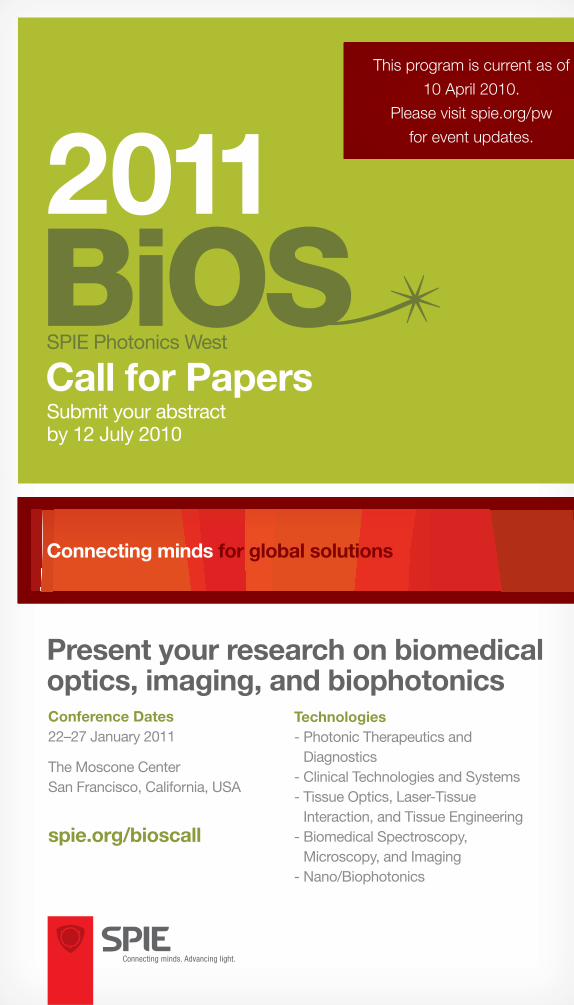
Present your research on biomedical optics, imaging, and biophotonics Submit your abstract by 12 July 2010 2011 Call for Papers SPIE Photonics West Technologies - Photonic Therapeutics and Diagnostics - Clinical Technologies and Systems - Tissue Optics, Laser-Tissue Interaction, and Tissue Engineering - Biomedical Spectroscopy, Microscopy, and Imaging - Nano/Biophotonics Conference Dates 22–27 January 2011 The Moscone Center San Francisco, California, USA spie.org/bioscall Connecting minds for global solutions This program is current as of 10 April 2010. Please visit spie.org/pw for event updates.
Transcript
Present your research on biomedical optics, imaging, and biophotonics
Submit your abstractby 12 July 2010
2011
Call for PapersSPIE Photonics West
Technologies- Photonic Therapeutics and
Diagnostics- Clinical Technologies and Systems- Tissue Optics, Laser-Tissue
Interaction, and Tissue Engineering- Biomedical Spectroscopy,
Microscopy, and Imaging- Nano/Biophotonics
Conference Dates22–27 January 2011
The Moscone CenterSan Francisco, California, USA
spie.org/bioscall
Connecting minds for global solutions
This program is current as of
10 April 2010.
Please visit spie.org/pw
for event updates.
BiOS CALL Cover.indd 1 5/28/10 4:13 PM
Present and publish your groundbreaking work to an audience looking for new solutions
Present your research at the world’s largest international biomedical optics conferenceBiOS attracts an audience of industry and academic leaders looking for the developments that will launch new applications and technologies for diagnostics and therapeutics. Receive immediate feedback, collaborate and broaden your professional network, and accelerate your research.
SPIE will advance your research globallyYour research will reach far beyond the conference room—all work from Photonics West will be published in the SPIE Digital Library. Promote yourself, your ideas, and your organization to millions of key researchers from around the world.
2011Call for PapersSubmit your abstractby 12 July 2010
spie.org/bioscall
Conferences and Courses: 22–27 January 2011 Photonics West Exhibition: 25–27 January 2011BiOS Exhibition: 22–23 January 2011The Moscone Center | San Francisco, California, USA
essential photonics eventSubmit your abstract today!
Plan now to participate!BiOS is the world’s largest international biomedical
optics conference, encompassing clinical, translational, and fundamental research and
development in the fi eld of biomedical optics. It provides a premier technical forum for reporting and learning about the latest research and development,
as well as for launching new applications and technologies. BiOS is part of SPIE Photonics West,
the largest laser, electro-optics, and imaging event in North America, with over 5,000 technical
attendees from over 50 countries. Special events include hot topics presentations, the international
biomedical optics exhibit, and industry leading working groups and panels.
Present your research at BiOS 2011.
R. Rox Anderson M.D., Wellman Ctr. for Photomedicine, Massachusetts General Hospital (USA) and Harvard School of Medicine (USA)
James Fujimoto, Massachusetts Institute of Technology (USA)
Symposium Chairs
“SPIE is the most effective scientifi c society for exchange of technical information between people from industry, academia and medical.”
–Dr. Gregory Altshuler, Senior Vice President of Research,
Palomar Medical Technologies, Inc.
SPIE Photonics West 2011 Call for Papers · spie.org/bioscall2
CONTENTS
Photonic Therapeutics and DiagnosticsProgram Chair: Brian Jet-Fei Wong, Beckman Laser Institute and Medical Clinic, Univ. of California, Irvine (USA)
BO100 Photonics in Dermatology and Plastic Surgery (Kollias/Choi/Zeng) . . . . . . . . . . 5
BO207 Optical Tomography and Spectroscopy of Tissue IX (Tromberg/Yodh/Tamura/Sevick-Muraca/Alfano ) . . . . . . . . . . . . . 19
Tissue Optics, Laser-Tissue Inter-action, and Tissue EngineeringProgram Chairs: Steven L. Jacques, Oregon Health & Science Univ. (USA) and William P. Roach, U.S. Air Force (USA)
BO300 Optical Interactions with Tissue and Cells XXII (Jansen/Thomas) . . . . . 20
BO301 Dynamics and Fluctuations in Biomedical Photonics VI (Tuchin/Duncan/Larin/Leahy/Wang) . . . 21
BO302 Photons Plus Ultrasound: Imaging and Sensing 2011 (Oraevsky/Wang) . . 22
Biomedical Spectroscopy, Microscopy, and ImagingProgram Chairs: Ammasi Periasamy, Univ. of Virginia (USA) and Daniel L. Farkas, Cedars-Sinai Medical Ctr. (USA)
BO400 Imaging, Manipulation, and Analysis of Biomolecules, Cells, and Tissues IX(Farkas/Nicolau/Leif) . . . . . . . . . . . . . . . 27
BO401 Multiphoton Microscopy in in the Biomedical Sciences XI (König/So/Periasamy) . . . . . . . . . . . . . . 28
BO402 Three-Dimensional and Multidimensional Microscopy: Image Acquisition and Processing XVIII(Brown/Wilson/Cogswell/Conchello) . . . 29
Photonic Therapeutics and DiagnosticsProgram Chair: Brian Jet-Fei Wong, Beckman Laser Institute and Medical Clinic, Univ. of California, Irvine (USA)
Photonics in Dermatology and Plastic Surgery (BO100)Conference Chairs: Nikiforos Kollias, Johnson & Johnson CPPW (USA); Bernard Choi, Beckman Laser Institute and Medical Clinic (USA); Haishan Zeng, The BC Cancer Agency Research Ctr. (Canada)Program Committee: Anthony J. Durkin, Beckman Laser Institute and Medical Clinic (USA); Iltefat Hamzavi, Henry Ford Hospital (USA); Jessica C. Ramella-Roman, The Catholic Univ. of America (USA)The research and development of highly selective lasers has forever transformed the clinical practice of dermatology and plastic surgery by allowing vas-cular lesions, pigmented lesions, tattoos, and hair to be removed without scarring. These important examples of selective photothermal injury continue to be refi ned and extended. The potential for laser or non-laser applications in skin diagnosis, imaging, and treatment for burns and other conditions such as psoriasis, acne, and vitiligo far exceeds their present use. A detailed understanding of skin optics, photo-thermal, photoacoustic, and photobiological pro-cesses is emerging. Innovative schemes for delivery and control of laser irradiation, including robotics, can potentially improve therapy. Optical spectros-copy, microscopy, and imaging techniques hold signifi cant diagnostic promise in dermatology, and submissions in these areas are especially welcome. New laser therapeutics, including burn treatment, wound healing, drug delivery and photodynamic therapy of infl ammatory skin conditions and cancer, will also be topics of interest for this session. Laser/tissue interaction, therapeutics, and diagnostics relating to light and skin, as well as competing technologies in the same scope, are also invited. Contributions from all medical, dental, and vet-erinary specialties, military-related applications, and basic sciences contributions are encouraged. Presentations that focus on unmet clinical needs in dermatology and plastic surgery are also welcomed.
Urology: Diagnostics, Therapeutics, Robotics, Minimally Invasive, and Photodynamic Therapy (BO101)Conference Chairs: Hyun Wook Kang, American Medical Systems Holdings, Inc. (USA); Bodo E. Knudsen, The Ohio State Univ. (USA)Program Committee: Nathaniel M. Fried, The Univ. of North Carolina at Charlotte (USA); Matthew T. Gettman, Mayo Clinic (USA); Patrice Jichlinski, Ctr. Hospitalier Univ. Vaudois (Switzerland); Ed Koullick, American Medical Systems Holdings, Inc. (USA); Unyime O. Nseyo, North Florida Foundation for Research and Education, Inc. (USA); Rudolf M. Verdaasdonk, Vrije Univ. Medical Ctr. (Netherlands)This era of high technology in urology includes many new and routine uses of lasers. In addition, the use of light for both photodiagnostic purposes and photodynamic therapy continues to move from the research lab to clinical use. Additional energy forms, including radio frequency and microwaves, are also being applied for tissue destruction. Papers are solicited on the following and related topics: • All Urologic Diagnostic and Therapeutic
Modalities including Minimally Invasive Surgical Techniques (MIST)
• Lasers for Benign and Neoplastic Tissue Coagulation and Vaporization - laser prostatectomy (contact,
interstitial, and free-beam delivery) - Nd:YAG, Ho:YAG, KTP, diode
or other laser sources - laser therapy of transitional cell carcinoma
of bladder, ureter, and renal pelvis - laser therapy of renal and adrenal lesions - laser therapy of male genital
and urethral tumors.• Laser Treatment of Ureteropelvic Junction,
Ureter, and Urethral Strictures • Laser Applications for Tissue Welding and
Approximation • Laser Lithotripsy
- optimum parameters for fragmentation systems
- feedback applications during lithotripsy - instrumentation - clinical results.
• Light Penetration in Urologic Tissues - photodynamic therapy (PDT) - photodynamic diagnosis (PDD) - light as a diagnostic tool - optical biopsy - chromophores.
• Other Energy Forms for Tissue Coagulation and Ablation - microwaves (including TUMT) - radio frequency (including dry and
wet interstitial RF procedures) - ultrasound.
• Robotics and Their Application to Urologic Surgery
• Unmet Clinical Needs in Urology
SPIE Photonics West 2011 Call for Papers · spie.org/bioscall6
Photon ic Therapeut ics and D iagnost ics
Advanced Technology and Instrumentation in Otolaryngology: Lasers, Optics, Radio Frequency, and Related Technology (BO102)Conference Chairs: Brian J. Wong, Beckman Laser Institute and Medical Clinic (USA); Justus F. R. Ilgner, Univ. Hospital Aachen (Germany)Program Committee: James A. Burns, Massachusetts General Hospital (USA); Udayan K. Shah, Nemours/Alfred I. duPont Hospital for Children (USA); Henricus J. C. M. Sterenborg, Erasmus MC (Netherlands)Otolaryngology and head and neck surgery con-tinues to be a rich fi eld for the application of new technologies. Precise focused beams and advanced energy delivery systems provide the foundation for the development of innovative microsurgical techniques. Optical diagnostics including elastic scattering spectroscopy, differential path-length spectroscopy, fl uorescence spectroscopy and infra-red spectroscopy can enhance tissue differentiation and identifi cation. Interferometric and stroboscopic optical techniques (i.e. optical coherence tomogra-phy) are used to monitor motion of the vocal folds and the tympanic membrane. Progress in these and other areas is facilitated with interactions among clinicians, scientists, engineers, and researchers. This conference covers the use of lasers and opti-cal technology in otolaryngology and head and neck surgery from the early phases of development to clinical practice. This conference provides an infor-mative and crucial face-to-face interaction between the basic scientist and the clinician. Papers from physicians, scientists, engineers, and manufacturers are solicited in the following medical subspecialty areas:• imaging of the vocal cords and airway • cochlear imaging • femtosecond laser surgery applications • CO2 laser ablation • middle ear surgery/Stapes surgery • endoscopic cancer resection • RF surgical applications • plasma-mediated ablation • optical diagnostic techniques • laryngology and speech science • head and neck surgery • photodynamic therapy• facial plastics and reconstructive surgery • endoscopic nasal and sinus surgery • image-guided surgery • novel surgical approaches • imaging techniques • unmet clinical needs • surgery for sleep apnea • airway simulation • telemedicine applications • lasers in robotics.
Diagnostic and Therapeutic Applications of Light in Cardiology (BO103)Conference Chairs: Kenton W. Gregory, Oregon Medical Laser Ctr. (USA); Guillermo J. Tearney, Wellman Ctr. for Photomedicine (USA); Laura Marcu, Univ. of California, Davis (USA)During the last decade there have been signifi cant scientifi c developments in the diagnosis and treat-ment of cardiovascular diseases using lasers and optical technology. Recent progress in vulnerable plaque detection has motivated the development of a variety of new optical techniques for intra-coronary diagnosis. The scientific and medical communities have additionally continued to search for new technologies and to improve on existing technologies that can be applied to areas such as cardiovascular recanalization or angioplasty, welding of vascular anastomoses, ablation of arrhythmogenic foci, transmyocardial revascularization, etc. The interdisciplinary research and collaboration among physicians, scientists, engineers, and manufacturers is paramount to maintaining and strengthening the fi eld of biomedical cardiovascular laser applications. This conference intends to bring together re-searchers and industry partners to present and dis-cuss the important advances in laser and diagnostic applications in cardiovascular medicine. The intent is to stimulate interactions, which will contribute to future progress in developing effective clinical cardiovascular diagnostic and therapeutic systems. Topics will include, but are not limited to, the following:• new laser sources for transmyocardial laser
revascularization• mechanisms of laser revascularization• ablation of arrhythmogenic foci and bypass
pathways• excimer laser applications• ablative and nonablative applications• catheter design for laser therapy of arrhythmias• evaluation of available laser angioplasty devices• new fi bers and catheter designs for laser
angioplasty• new lead extraction laser catheters• optical methods of assessing cardiovascular
structure, biomechanics and function• vascular welding• vascular reactivity to laser energy• approaches to control and assess the extent of
laser injury• new applications for laser thrombolysis• spectroscopy• fl uorescence spectroscopy• photodynamic therapy• photodynamic therapy for intimal hyperplasia• MEMS for cardiovascular diagnostics• optical coherence tomography• Raman, fl uorescence, and absorption
spectroscopy• angioscopy• thermography• optical studies of the cardiovascular system in
Post-Meeting Manuscript Due Date: 15 December 2010
Please Note: Submissions imply the intent of at least one author to register, attend the conference, present the paper as scheduled, and submit a full-length manuscript for publication in the conference proceedings.
Optical Techniques in Neurosurgery, Brain Imaging, and Neurobiology (BO104)Conference Chairs: Henry Hirschberg, Beckman Laser Institute and Medical Clinic (USA); Steen J. Madsen, Univ. of Nevada, Las Vegas (USA)Program Committee: Herbert Stepp, Ludwig-Maximilians-Univ. München (Germany); Victor X. D. Yang, Ryerson Univ. (Canada)
Recent technological advances have opened excit-ing opportunities for lasers and optical techniques in neurosurgery and brain imaging. Therapeutic applications include: tumor debulking/removal via thermal or photochemical interactions, and spinal decompression and discectomy. Optical techniques have also shown promise in a number of diagnostic applications, including fl uorescence-guided tumor resection, imaging of cortical function and intra-vascular examinations. Of special interest is the use of optical or other imaging modalities in the detection and therapy of neuro-degenerative diseases such as Alzheimer’s or Parkinson’s. The purpose of this conference is to provide a forum for clinicians, scientists, engineers and manu-facturers to report on current developments and to discuss future opportunities for optical techniques in neurosurgery and brain imaging.
Contributed papers are solicited concerning, but not limited to, the following areas: • laser surgery in the brain and spine • fl uorescence-guided resection • optical spectroscopy of normal brain and
neoplastic tissue • optical techniques in the diagnosis and
treatment of neuro-degenerative diseases • functional imaging of the brain using optical
techniques • photodynamic therapy in neurosurgery • optical instrumentation and devices, including
microscopes and endoscopes • optical localization and registration techniques
for neuronavigation • fMRI/PET • techniques for in vivo microscopy • microscopic imaging and advanced microscopic
techniques • virtual reality • real-time functional imaging in the OR • co-registration (optical and other imaging
modalities) • unmet clinical needs.
SPIE Photonics West 2011 Call for Papers · spie.org/bioscall8
Photon ic Therapeut ics and D iagnost ics
Ophthalmic Technologies XXI (BO106)Conference Chairs: Fabrice Manns, Univ. of Miami (USA); Per G. Söderberg, Uppsala Univ. (Sweden); Arthur Ho, Institute for Eye Research Ltd. (Australia)Program Committee: Rafat R. Ansari, NASA Glenn Research Ctr. (USA); Michael Belkin, Tel Aviv Univ. (Israel); Ralf Brinkmann, Univ. zu Lübeck (Germany); Wolfgang Drexler, Medizinische Univ. Wien (Austria); Daniel X. Hammer, Physical Sciences Inc. (USA); Karen M. Joos, Vanderbilt Univ. (USA); Katsuhiko Kobayashi, Topcon Corp. (Japan); Kirill V. Larin, Univ. of Houston (USA); Ezra I. Maguen, American Eye Institute (USA); Donald T. Miller, Indiana Univ. (USA); Daniel V. Palanker, Stanford Univ. School of Medicine (USA); Jean-Marie A. Parel, Bascom Palmer Eye Institute (USA); Roberto Pini, Istituto di Fisica Applicata Nello Carrara (Italy); Luigi L. Rovati, Univ. degli Studi di Modena (Italy); Georg Schuele, OptiMedica Corp. (USA); Jerry Sebag, The Univ. of Southern California (USA); William B. Telfair, IRIDEX Corp. (USA); Valery V. Tuchin, N.G. Chernyshevsky Saratov State Univ. (Russian Federation)
PASCAL ROL AWARD 2011 Outstanding extended abstracts submitted to the Ophthalmic Technologies XXI conference will be nominated for the Pascal Rol Award for Best Paper in Ophthalmic Technologies. The award and prize will be presented after the last scientifi c session of the conference to recognize the best paper and presentation. The 2010 recipient of the Pascal Rol Award was Dr. Daniel X. Hammer and his colleagues from Physical Sciences Inc. (see www.pascalrolfoundation.org).
SPECIAL PRESENTATION Unmet Ophthalmic Technology Needs
This presentation series was established to pro-mote the exchange of ideas between clinicians with a technological need and engineers inter-ested in solving problems in ophthalmology. The invited lecture is sponsored by the Pascal Rol Foundation (www.pascalrolfoundation.org). The topic and keynote speaker will be announced in the preliminary program.
You are invited to submit papers to Ophthalmic Tech-nologies XXI - the premier international meeting on therapeutic and diagnostic technology in the fi eld of ophthalmology, which brings together engineers and scientists developing the next innovations, and clini-cians and practitioners extending the technology. Some recent topics covered include: • ophthalmic diagnostics• ophthalmic applications of OCT • retinal prosthesis and bionic vision• wavefront sensing and wavefront-guided surgery • laser surgical systems • artifi cial cornea and keratoprostheses • optoacoustic monitoring • selective retinal photocoagulation • adaptive optics• dynamic light scattering • Raman spectroscopy • optics of the eye and vision correction• ocular biometrics• ocular diagnostics of neural diseases• new devices and techniques• virtual reality in ophthalmology• optics and laser applications in ophthalmic drug
delivery.Papers on any other relevant topics are welcomed.
NOTE ON DUPLICATE ABSTRACTS Authors that submit an abstract on a similar topic for presentation at another BiOS conference may be invited to present a short communication that should focus on ophthalmic issues.
EXTENDED ABSTRACT REQUIRED In addition to the 250-word text abstract that will be published in the Abstract Digest, and the short 100-word abstract that will be posted online, au-thors must submit a structured two-page extended abstract by the Abstract Due Date. The two-page extended abstract should be a stand-alone dodu-ment including author names and affi liations, text, fi gures and tables, and suffi cient data to permit re-view. All extended abstracts will be peer-reviewed by the Program Committee to determine acceptance. The best extended abstracts will be nominated for the Pascal Rol Award. The extended abstracts will be used only for the purpose of peer review and will not be published.
Optical Methods for Tumor Treatment and Detection: Mechanisms and Techniques in Photodynamic Therapy XX (BO107)Conference Chair: David H. Kessel, Wayne State Univ. (USA)Cochair: Tayyaba Hasan, Wellman Ctr. for Photomedicine (USA)Photodynamic therapy (PDT) has been approved by health agencies in several countries for treat-ment of neoplasia in a variety of sites, and is being examined with regard to other pathologic condi-tions including actinic keratosis, atherosclerosis, and age related macular degeneration (AMD). PDT can be used to target different subcellular sites for photodamage, e.g., the endoplasmic reticulum, ly-sosomes, mitochondria, and the plasma membrane. Photodamage can elicit cell death by activation of apoptosis, circumventing many common modes of drug resistance. This conference will emphasize drug development, mechanisms, clinical applications, instrumentation for light delivery and dosimetry determinations along with new information on pho-todynamic mechanisms. Abstracts are encouraged dealing with these topics:• drug development and characterization• clinical protocols and outcomes• mechanisms of phototoxicity• techniques for light delivery and dosimetry • tissue optics.
Lasers in Dentistry XVII (BO105)Conference Chairs: Peter Rechmann, Univ. of California, San Francisco (USA); Daniel Fried, Univ. of California, San Francisco (USA)Program Committee: Gregory B. Altshuler, Palomar Medical Technologies, Inc. (USA); Tatjána Dostálová, Charles Univ. in Prague (Czech Republic); John D. Featherstone, Univ. of California, San Francisco (USA); David M. Harris, Bio-Medical Consultants, Inc. (USA); Harvey A. Wigdor, Advocate Illinois Masonic Medical Ctr. (USA)Laser applications for dental hard tissue are a clinical reality. Several exciting future applications are being developed and will be featured at this conference. An entire session will be devoted to lesion detec-tion by optical methods. This is one of the areas of rapidly expanding interest in dental research and applications to clinical practice, especially in relation to dental caries. Soft tissue clinical applications con-tinue to be expanded. This conference will provide a forum for presentation of both basic and applied research in laser dentistry. Presentations of clinical studies are especially welcome. Manuscripts will be reviewed prior to publication. Papers are solicited in all dental laser and biomedi-cal optics dental application areas including, but not limited to, the following: • optical methods for lesion detection, especially
dental caries• early caries detection• optical coherence tomography in dentistry• dental holography, 3D imaging• decay removal with lasers• decay prevention with lasers• laser endodontics• laser applications in periodontology• lasers and dental implants• laser photopolymerization• laser hard-tissue and soft tissue surgery• CO2 laser use in dentistry• Nd:YAG laser use in dentistry• Er:YAG/ErCr:YSGG use in dentistry• Ho:YAG laser use on dental tissues• excimer laser treatment of dental tissues• other wavelengths for hard or soft tissue use• clinical trials of lasers for dental applications• hard-tissue ablation and plasma production• laser-tissue interactions relevant to dentistry• wavelength and energy dependence of dental
laser applications• unmet clinical needs.
SPIE Photonics West 2011 Call for Papers · spie.org/bioscall10
Photon ic Therapeut ics and D iagnost ics
Optics in Bone Biology and Diagnostics (BO109)Conference Chair: Andreas Mandelis, Univ. of Toronto (Canada)With the new millennium and the rapid penetration of lasers, optics, and imaging techniques in the fi eld of tissue diagnostics, a rapid growth in bone-related biophotonics and applications has emerged. While x-ray bone diagnostics and imaging are well established photonic techniques due to their deep sub-dermal penetration, bone research has been associated more with endoscopic technologies for which light waves face serious penetration barriers. However, we are witnessing an explosion in optical probes and diagnostics of bones and bone disease in the last 4-7 years. This conference emphasizes optical applications to bone biology and diagnostics. It targets interdis-ciplinary research reports which bring out the inter-actions of bone with photons and can be used as a forum for the cross-fertilization and advancement of the science and technologies associated with bone optics and photonics. Technical and scientifi c papers related to the interactions and studies of bones with photons with energies spanning the entire spectral range from γ-rays to microwave and millimeter waves are solicited. These include: • mineral density, mechanical strength, strain,
optical and other bone property measurements using optical absorption, refl ection, transmission and scattering techniques.
• optics in bone biology and biomedicine• γ- and x-ray imaging and bone densitometry• general bone radiography• bone applications of ultrafast laser techniques• advanced optical techniques for the study of
bones and osteopathologic processes: optical coherence tomography, terahertz, photoacoustic and photothermal detection and spectroscopy, diffuse photon techniques
• bone tissue ablation with lasers• bone spectroscopies, microscopies and
diagnostic instrumentation• in-vitro and in-vivo imaging and tomographies• fl uorescence imaging and emission tomography• transdermal optics• optics in bone endoscopy and surgery• bone optical engineering and clinical systems • non-linear effects in bone-photon interactions.
Mechanisms for Low-Light Therapy VI (BO108)Conference Chairs: Michael R. Hamblin, Massachusetts General Hospital (USA); Ronald W. Waynant, U.S. Food and Drug Administration (USA); Juanita Anders, Uniformed Services Univ. of the Health Sciences (USA)Low levels of visible light (frequently red or near-infrared) can have signifi cant therapeutic effects on multiple classes of diseases, injuries and medical disorders. In particular it is effective for wound healing and pain control as well as reduction of infl ammation and swelling. It is believed that the primary cellular chromophore that absorbs low levels of red and near-infrared light is cytochrome c oxidase, which is located in mitochondria. This absorption of energy may lead to increase in ATP synthesis and release of reactive oxygen species from the electron transport chain that can subsequently activate transcription factors and lead to cell proliferation and migration. Despite many reports of positive fi ndings from ex-periments conducted in vitro, in animal models and in randomized controlled clinical trials, LLLT remains controversial. This likely is due to two main reasons; fi rstly the biochemical mechanisms underlying the positive effects are incompletely understood, and secondly the complexity of rationally choosing amongst a large number of illumination parameters such as wavelength, fl uence, power density, pulse structure and treatment timing has led to the pub-lication of a number of negative studies as well as many positive ones. This conference covers a field that is rapidly achieving a general level of acceptance in the medi-cal and biomedical communities and will cover all of the important areas of LLLT research.
Contributed papers are solicited in the following areas (among others):• mechanistic studies and cellular chromophores• development of light sources for LLLT (LED
photomodulation; pulsed IR light therapy)• study of LLLT dosimetry• in vitro research in mammalian cells• in vitro research in micro-organisms in culture • stimulation of wound healing and scar reduction
in animal models• nerve regeneration and neural stimulation• prevention of ischemia-induced tissue death and
regeneration.
Well-controlled clinical trials in the following areas:• stimulation of wound healing such as non-
healing ulcers• treatments for stroke and degenerative brain
disease• pain reduction in post-surgical and neuralgia
patients• dental applications• dermatology applications• reduction of pain and infl ammation in arthritis
and other orthopedic conditions• macular degeneration prevention• reduction of edema.
Frontiers in Biological Detection: From Nanosensors to Systems (BO110)Conference Chairs: Benjamin L. Miller, Univ. of Rochester (USA); Philippe M. Fauchet, Univ. of Rochester (USA)Program Committee: Holger Becker, microfl uidic ChipShop GmbH (Germany); Xudong Fan, Univ. of Michigan (USA); Jiri Homola, Institute of Photonics and Electronics of the ASCR, v.v.i. (Czech Republic); Hongrui Jiang, Univ. of Wisconsin-Madison (USA); Laura M. Lechuga, Ctr. d’Investigacions en Nanociència i Nanotecnologia (Spain); Frances S. Ligler, U.S. Naval Research Lab. (USA); Daniel V. Lim, Univ. of South Florida (USA); Christopher Myatt, Precision Photonics Corp. (USA); Elric W. Saaski, Research International, Inc. (USA); Michael J. Sailor, Univ. of California, San Diego (USA); Christopher C. Striemer, Adarza Biosystems Inc. (USA); Sharon M. Weiss, Vanderbilt Univ. (USA)
Cal l fo r Papers
Submit your abstract today!
spie.org/bioscall
Detection of biological materials, from DNA strands to whole pathogens, is increasingly becoming a concern throughout society, not only in diagnostic laboratories in hospitals but also for on-site uses by health care providers or soldiers. From monitoring incidence of drug-resistant bacteria in hospitals and detecting harmful pathogens for homeland security to ensuring that our food is safe and our water clean, new, simple, inexpensive, sensitive, and fast methods of identifying biological molecules and pathogens are a pressing need. Optical solutions promise to provide many of these advantages and as a result many platforms for optical detection are being demonstrated in the laboratory. The deployment of bio-detection systems however requires that stringent specifi cations be met, for example in terms of sensitivity, false-positive and false-negative assessments, automated sample pro-cessing and analysis, system design and integration, and low cost. This conference seeks to gather scientists, engineers and users active in biological detec-tion. Contributions are sought in all areas, from novel optical detection platforms to nanosensors to system integration and commercialization. The symposium will also feature a joint session with the Microfl uidics, BioMEMS and Medical Microsystems conference (MF104).
Topics of interest include but are not limited to: • new sensing platforms • ultrasensitive (single pathogen) detection
methods • utilization of nanomaterials and new optically
responsive materials for pathogen detection • miniaturized optic components such as
microring resonators, photonic crystals, integrated optical waveguides, and nanoparticles
• label-free vs. tagged detection systems • organic and inorganic platforms • probe design • strategies to eliminate non-specifi c binding • integration of optics and microfl uidics • systems demonstrations • new applications for environmental, medical,
and food testing.
SPIE Photonics West 2011 Call for Papers · spie.org/bioscall12
Clinical Technologies and SystemsProgram Chairs: Tuan Vo-Dinh, Duke Univ. (USA) and Anita Mahadevan-Jansen, Vanderbilt Univ. (USA)
Optical Coherence Tomography and Coherence Domain Optical Methods in Biomedicine XIV (BO200)Conference Chairs: James G. Fujimoto, Massachusetts Institute of Technology (USA); Joseph A. Izatt, Duke Univ. (USA); Valery V. Tuchin, N.G. Chernyshevsky Saratov State Univ. (Russian Federation)Program Committee: Peter E. Andersen, Technical Univ. of Denmark (Denmark); Stephen A. Boppart, Univ. of Illinois at Urbana-Champaign (USA); Zhongping Chen, Beckman Laser Institute and Medical Clinic (USA); Johannes F. de Boer, Vrije Univ. Amsterdam (Netherlands); Wolfgang Drexler, Medizinische Univ. Wien (Austria); Christoph K. Hitzenberger, Medizinische Univ. Wien (Austria); Rainer A. Leitgeb, Medizinische Univ. Wien (Austria); Xingde Li, The Johns Hopkins Univ. (USA); Adrian G. Podoleanu, Univ. of Kent (United Kingdom); Andrew M. Rollins, Case Western Reserve Univ. (USA); Natalia M. Shakhova, Institute of Applied Physics (Russian Federation); Guillermo J. Tearney, Wellman Ctr. for Photomedicine (USA); Ruikang K. Wang, Oregon Health & Science Univ. (USA); Maciej Wojtkowski, Nicolaus Copernicus Univ. (Poland); Yoshiaki Yasuno, Univ. of Tsukuba (Japan)
IMPORTANT NOTE: PEER REVIEWAll submissions will be peer reviewed by the full program committee. In order to facilitate the evalu-ation of your submission, please submit both an abstract and summary by the Abstract and Summary Due Dates. The summary should be three pages in length and should have suffi cient technical and sci-entifi c information to permit review of the paper. To accommodate the peer review process, abstracts/summaries will not be accepted after 12 July 2010. Optical coherence tomography and other optical methods and instruments based on coherent light interactions with inhomogeneous tissues are very promising for noninvasive medical diagnostics and for monitoring a wide spectrum of human patholo-gies. The focus of this conference will be on dis-cussion of the physical and mathematical basis of coherence domain methods, and their applications in biomedical science and clinical practice. Directions of research and development in areas such as opti-cal coherence tomography (OCT), low-coherence interferometry, speckle and speckle interferometry measurement and imaging technologies, polarized light diagnostic methods, coherent light microscopy, and coherence technologies for fl ow and functional imaging will be considered. Applications of coher-ence domain optical methods for studies of living tis-sue and clinical applications will also be discussed.
Papers are solicited on the following and related topics:• optical coherence tomography (OCT) systems,
theory, • image processing techniques in OCT • spectral/Fourier domain and swept source OCT • optical coherence microscopy (OCM) • full-fi eld OCT• molecular, spectroscopic and functional OCT • Doppler and polarization-sensitive OCT • phase-contrast techniques • novel light sources for OCT • imaging devices and probes for OCT • low-coherence interferometry and topography • white-light interferometry• novel contrast mechanisms• clinical applications of OCT
- ophthalmic- cardiovascular- cancer imaging- endoscopic
Advanced Biomedical and Clinical Diagnostic Systems IX (BO201)Conference Chairs: Anita Mahadevan-Jansen, Vanderbilt Univ. (USA); Tuan Vo-Dinh, Duke Univ. (USA); Warren S. Grundfest, Univ. of California, Los Angeles (USA)Program Committee: Maurice C. Aalders, Jr., Univ. van Amsterdam (Netherlands); Jennifer K. Barton, The Univ. of Arizona (USA); Stephen A. Boppart, Univ. of Illinois at Urbana-Champaign (USA); Laura Marcu, Univ. of California, Davis (USA); Mary-Ann Mycek, Univ. of Michigan (USA); Jianan Y. Qu, Hong Kong Univ. of Science and Technology (Hong Kong, China); Urs Utzinger, The Univ. of Arizona (USA); Georges A. Wagnieres, Ecole Polytechnique Fédérale de Lausanne (Switzerland)
As surgical systems and clinical diagnostics adapt to new methods, instrumentation and assay technol-ogy, the pace of system innovations continues to accelerate. Many technologies originally developed for other applications, e.g., defense, energy, and aerospace, have found applications in the medical industry/environment. This conference provides a forum for scientists, engineers, manufacturers, and clinical providers to present the most recent advances in instrumentation and methods of diag-nostic and therapeutic guidance systems for clinical applications. The conference goal is to provide an interdisciplinary forum for state-of-the-art methods in instrumentation research and development of biomedical technologies, approaching the transla-tion from research and development prototypes to user application. The emphasis will be on clinical translation and implementation. The conference will address the interests of researchers, applied scientists, engineers, and clinicians whose goal is to implement clinical systems with extended, improved performance capabilities. Papers are sought that describe the methodolo-gies, instrumentation systems and analysis of bio-medical optics technologies for clinical implementa-tion. Topics will span the areas of instrumentation, system engineering, assay automation, delivery technology, and data management for biomedical diagnostics and surgical assistance in the clinical setting. Examples include stereotactic systems developed for brain surgery, fl exible micronaviga-tion devices engineered for medical laser ablation treatments, real-time monitoring system of critical function, online sensing of biological assays. Oral and poster sessions are planned in the fol-lowing subject areas:
Clinical diagnostic systems• minimally invasive diagnostics systems based
on Fluorescence, Raman scattering, diffuse refl ectance
• early screening applications • clinical monitoring of early pathological or
physiological states • multi-modality diagnostic systems (optics
combined with acoustic, ultrasound, MRI, PET, X-ray)
• component/system design, engineering, and performance of biomedical instruments
• microinstrumentation and miniaturized clinical systems
• integrated instrument systems • high throughput systems.
Advanced sensors for medicine• clinical applications of optical biosensors
(enzyme, antibody, gene probe-based systems) • physical sensors and chemical in vivo sensors
• biochip technologies • bio-electrics and nanosecond pulsing
technology • sensors for burn diagnostics • advances in assay automation and delivery
technology.
Medical imaging techniques and devices • IR, OCT and other optical imaging systems • advanced endoscopy techniques and devices • smart catheters • laser radar sensing and imaging • RF radar imaging • terahertz techniques and systems (imaging,
spectroscopy, and modeling) • optical methods of assessing structure and
function • trauma and critical care • telemedicine, telesurgery • virtual reality technologies.
SPIE Photonics West 2011 Call for Papers · spie.org/bioscall14
Design and Quality for Biomedical Technologies IV (BO202)Conference Chairs: Ramesh Raghavachari, U.S. Food and Drug Administration (USA); Rongguang Liang, Carestream Health, Inc. (USA)Cochair: T. Joshua Pfefer, U.S. Food and Drug Administration (USA)Program Committee: Anthony J. Durkin, Beckman Laser Institute and Medical Clinic (USA); Kazuhiro Gono, Olympus Medical Systems Corp. (Japan); Jeeseong Hwang, National Institute of Standards and Technology (USA); Stephen P. Morgan, The Univ. of Nottingham (United Kingdom); Jannick P. Rolland-Thompson, Univ. of Rochester (USA); Eric J. Seibel, Univ. of Washington (USA); Tomasz S. Tkaczyk, Rice Univ. (USA); Rudolf M. Verdaasdonk, Vrije Univ. Medical Ctr. (Netherlands)
Rapid advances in optical technologies and com-putational power have brought about a revolution in biomedical diagnostics and therapeutics. However, these advances necessitate parallel progresses in techniques used for development and evaluation. This conference will focus on four key areas that are critical to the design and production of safe, ef-fective, and commercially-viable biomedical devices and technologies:1. the devices used in biomedical researches and
clinical applications 2. the design and analysis of systems and
components which require unique solutions for biomedical applications
3. the evaluation of quality and safety of the biomedical imaging technologies and device
4. the establishment of device reliability, including failure and degradation of performance.
This conference provides a unique forum for sci-entists and engineers from academia, industry and government to discuss issues that are relevant to all biomedical imaging modalities. Interactions between these parties should facilitate the development of biomedical devices and evaluation methods that will benefi t the fi eld of medicine. Submissions pertain-ing to optical diagnostics and therapeutics for all fi elds of medicine as well as optical evaluation of pharmaceuticals and biotechnology products are solicited for this conference.
I. Devices • optical and biophotonic devices & instruments
for researches and clinical applications • biosensor, noninvasive photonics medical
sensors • biomedical instrumentation • fi ber based biomedical devices • image guided biopsy, surgical and therapeutic
device and technologies • cost effective optics and devices • preclinical, in vivo-simulating device and
technologies.
II. Design• optical and system design for biomedical
imaging systems, including fl uorescence, optical coherence tomography, polarization, confocal, multispectral, multiphoton, spectroscopic, and multimodal imaging systems
• optics for biomedical imaging technologies and devices
• illumination and detection geometry for imaging and spectroscopic systems
• fi ber optic imaging systems • novel optical sensing and detection • micro-optics and MEMS based optical systems • sources, detectors and other components • maximum permissible light exposure levels in
vivo • development, validation and application of
computer-aided design tools.
III. Quality• Quality by Design (QbD) • device calibration and intercomparison • phantoms and test methods • metrology and standardization • development/evaluation of novel measurement
tools • computer-aided diagnosis algorithms • critical metrics for assessing quality • quality, compliance and regulatory issues related
to biomedical devices • statistical approaches for designing, evaluating,
and validating medical device, databases, and technologies
• patient and user safety; photothermal, biochemical, etc.
IV. Reliability• physics, analysis, failure mechanisms and
testing for failure • aging, dormancy and component degradation • computational and analytical modeling • determination of factors of safety • reusability of new optical devices.
V. Biomedical imaging technologies• hyperspectral, spectroscopic techniques • image enhancing techniques • CMOS technologies for biomedical applications • multi-modal techniques, including hybrid
devices with non-optical (e.g., MRI, ultrasound) components
• small animal imaging • molecular imaging • digital Imaging and telemedicine • high resolution optical imaging.
A special regulatory session is planned for this conference.
Multimodal Biomedical Imaging VI (BO203)Conference Chairs: Fred S. Azar, Becton, Dickinson and Co. (USA); Xavier Intes, Rensselaer Polytechnic Institute (USA)Program Committee: Nicholas Ayache, INRIA Sophia Antipolis - Méditerranée (France); David A. Boas, Massachusetts General Hospital (USA); Britton Chance, Univ. of Pennsylvania (USA); Yu Chen, Univ. of Maryland, College Park (USA); Sergio Fantini, Tufts Univ. (USA); Keyvan Farahani, National Cancer Institute (USA); Gultekin Gulsen, Univ. of California, Irvine (USA); Dimitris N. Metaxas, Rutgers, The State Univ. of New Jersey (USA); Nassir Navab, Technische Univ. München (Germany); Tim Nielsen, Philips Research (Germany); Vasilis Ntziachristos, Helmholtz Zentrum München GmbH (Germany); Brian W. Pogue, Dartmouth Hitchcock Medical Ctr. (USA); Siavash Yazdanfar, GE Global Research (USA); Arjun G. Yodh, Univ. of Pennsylvania (USA)
Data generated by novel imaging technologies such as optical tomography are complex to analyze, due to the inherent scattering of light through anatomical systems. Cross validation and direct comparison with established methods in other imaging modali-ties are especially challenging. There is critical need for new computational techniques to provide rapid, accurate and cost-effective means for quantifi cation and characterization of such data, either indepen-dently or integrated with other modalities. These computational methods will enable faster accep-tance of novel imaging modalities into viable clinical and/or pre-clinical systems. The applications are diverse and range from imaging at the cellular level to the whole body while incorporating molecular, functional and anatomical information. The conference objectives are to provide a forum:1. To review and share recent developments in
novel multimodal imaging techniques, 2. to report development of novel computational
methods, and 3. to bring together the optical imaging and image
analysis communities.
Topics include, but are not limited to:• multimodal imaging integrating structural,
molecular and functional information • 2D, 3D, 4D, tomographic and / or multi-spectral
techniques applied to optical imaging (e.g. visualization, segmentation, registration)
• detection and diagnostic analysis techniques which may provide better quantitative and/or diagnostic insight into clinical & pre-clinical imaging (e.g. methods for quantitative measurements, computer-assisted diagnosis)
• imaging analysis and/or image processing techniques used to combine optical imaging with other imaging modalities (e.g. MR, X-Ray, PET)
• image analysis, computational methods and reconstruction approaches which may help bring optical imaging into the clinic (visual rendering of complex data set, novel algorithms for assisted optical reconstruction)
• clinical evaluation of these new technologies (Physiological and functional interpretation of image data, visual perception and observer performances, validation of quantitative assessment of optical signatures in-vivo).
Publish with SPIE and advance your research globally.
SPIEDigitalLibrary.org
SPIE Photonics West 2011 Call for Papers · spie.org/bioscall16
Endoscopic Microscopy VI (BO204)Conference Chairs: Guillermo J. Tearney, Wellman Ctr. for Photomedicine (USA); Thomas D. Wang, Univ. of Michigan (USA)Program Committee: David L. Dickensheets, Montana State Univ. (USA); Arthur F. Gmitro, The Univ. of Arizona (USA); Ralf Kiesslich, Johannes Gutenberg Univ. Mainz (Germany); Francois Lacombe, Mauna Kea Technologies (France); Stephen Lam, The BC Cancer Agency Research Ctr. (Canada); Hiroshi Mashimo, VA Boston Healthcare System (USA); Kenzi Murakami, Olympus Corp. (Japan); Norman S. Nishioka, Massachusetts General Hospital (USA); Wibool Piyawattanametha, Stanford Univ. (Thailand); Mark J. Schnitzer, Stanford Univ. School of Medicine (USA); Peter T. C. So, Massachusetts Institute of Technology (USA); Melissa J. Suter, Massachusetts General Hospital (USA)
Papers for this session focus on the development of high resolution imaging systems that are endoscope compatible and include novel techniques for early detection, screening, diagnosis, intervention, and treatment of disease in either pre-clinical models or human subjects. In general, remote detection is based on the use of an optical fi ber to transmit light between the instrument and the tissue, and involves the collection of information about molecular and cellular processes without physical excision of tissue. These methods of in vivo optical imaging extends across a wide range of resolution, from molecular, sub-cellular and cellular to tissue and organ levels. The creation of an image may require a variety of different scanning mechanisms. Endoscopic techniques provide unique advantages over other (e.g. whole body) imaging modalities for evaluating disease present over the epithelial surface of hollow organs. The direct proximity of collection optics to regions of disease can result in unparalleled achievement of resolution, unraveling of molecular mechanisms, and use of fl uorescence contrast agents. Examples of applications of endoscopic microscopy to medicine include the detection of pre-malignant le-sions, identifi cation of disease below the tissue surface, assessment of depth of tumor invasion, localization of cancer margins, evaluation of effectiveness of pharma-cological therapy, and reduction in number of physical biopsies and frequency of surveillance. This conference provides an inter-disciplinary forum for physicians, molecular biologists, chem-ists, biochemists, optical engineers, and instrument designers to report and discuss recent results, im-provements, and new approaches in the emerging fi eld of endoscopic microscopy. Contributed papers are solicited concerning, but not limited to, the following areas:• confocal microendoscopy • multi-photon microendoscopy • endoscopic OCT • endoscopic OCM • fl uorescence endoscopic imaging • video capsule endoscopy • endocytoscopy • high magnifi cation endoscopy • light scattering spectroscopy • Raman spectroscopy • infrared spectroscopy • MEMS scanning mechanisms • micro-actuators • luminal optical contrast agents • endoscopic image processing and analysis.
JOINT SESSION with MF105Miniature Instruments for Endoscopic Microscopy This special joint session is in conjunction with MOEMS-MEMS conference MF105: MOEMS and Miniaturized Systems. Papers are solicited that ad-dress the unique challenges to deliver high-fi delity
microscopic imaging of tissue with a miniaturized instrument platform. Example topics include mecha-nisms for distal beam scanning, focus control and aberration correction using MOEMS devices, MEMS actuators or electrowetting optics; proximal scan-ning based on DMD or other SLM technologies; novel optical assembly and alignment techniques; highly corrected miniature optical systems.
SPECIAL SESSION ON PULMONARY MEDICINEDue to the high prevalence of lung diseases and the lack of available technologies to adequately evaluate the complex processes of the respiratory system, the use of optical techniques for studying, diagnosing, and treating the pulmonary airways has rapidly increased in recent years. While the applica-tion of optical technologies in pulmonary medicine is an emerging trend, scientists in this fi eld are working towards developing new, and advancing existing, optical technologies for disease detection, diagnosis, and treatment, and for guiding and evalu-ating pulmonary interventions. In addition, these techniques are being utilized to study respiratory physiology and pathophysiolgy including airway structure and function relationships such as perfu-sion, ventilation, mechanics and modeling. This session will cover all aspects relating to optical based molecular, structural and functional imaging, diagnosis, and treatment of the lung. The goal is to bring together scientists and clinicians to discuss and progress current state-of-the-art optical diagnostic and therapeutic techniques, and to use the devel-oped optical approaches to challenge and further develop our current understanding of the physiology and pathophysiology of the respiratory system. Topics will include, but are not limited to, the fol-lowing areas:• optical techniques for assessing bronchial and
alveolar lung structure• optical evaluation of pulmonary function:
perfusion, ventilation, mechanics and modeling• optical diagnostics • evaluation of normal and diseased/injured airway• optical detection, evaluation and treatment of
airway pathology• modeling of the conducting airways and alveoli• optical coherence tomography• diffuse optical tomography• confocal and two-photon microscopy• spectroscopy• optical molecular imaging• fl uorescence imaging• optical imaging and activation of inhaled agents• novel bronchoscopy techniques• laser ablation treatment techniques• optical monitoring of therapeutics• photodynamic and ablative therapy• preclinical and clinical optical studies of the lung
and airway• small animal imaging• unmet clinical needs.
Optical Fibers, Sensors, and Devices for Biomedical Diagnostics and Treatment XI (BO205)Conference Chair: Israel Gannot, Tel Aviv Univ. (Israel)Program Committee: James P. Clarkin, Polymicro Technologies, A Subsidiary of Molex Inc. (USA); Ilko K. Ilev, U.S. Food and Drug Administration (USA); Jin Ung Kang, The Johns Hopkins Univ. (USA); Karl-Friedrich Klein, Fachhochschule Giessen-Friedberg (Germany); Pierre Lucas, The Univ. of Arizona (USA); Yuji Matsuura, Tohoku Univ. (Japan)
Optical devices and components, lab on chip, deliv-ery devices, fi bers, waveguides, bundles, detectors, and sensors are essential components needed to perform biological and clinical laboratories and in-vivo medical procedures. This conference will cover various topics in these fi elds. In the medical fi eld there are still pressing de-mands for the following: • new fi bers and waveguides, other delivery
devices, and transendoscopic tools for the x-ray, UV, visible, and IR regions of the spectrum
• invasive and noninvasive optical, biological, and chemical sensors for UV/VIS/NIR/IR fl uorescence, Raman, absorption and evanescent-wave and photothermal spectroscopy, optical, and thermal bundles.- biosensors for the medical
and biological areas- fi ber sensors for physical,
chemical, and immunoassays- waveguide sensors, including
molecular diagnostic arrays• covering an increasing number of laser output
wavelengths (i.e., ultrafast lasers, x-ray lasers, and fi ber lasers)
• Femto, Atto, Zepto and Yocto pulse delivery• low loss waveguides and fi ber bundles for
terahertz, thermal, and optical imaging• theory of waves and signals delivery in fi bers• fi ber damage thresholds for high energy pulse
delivery, nonlinear effects• making the therapeutic and diagnostic systems
more intelligent and safer, i.e., by increasing the number and the role of fi ber based and control devices in the diagnostic and surgical apparatus (this will lead to safer operative procedures for both patients and physicians)
• optical fi ber and waveguides materials (i.e. 1-d and 2-d photonic bandgap crystals) and devices required specifi cally for medical applications
• new surgical and diagnostic procedures based on optical fi bers and waveguides
• fi ber based methods and systems• imaging bundles for the visible and IR• embedded optical sensors• imaging capsules• bio-compatibility of fi ber materials for
endoscopic tools• mechanical properties of fi bers (bending, tensile
strength) for endoscopic applications• new fi bers and catheter designs for laser surgical
applications• sensor and detector arrays• sensors for the smart medical home• tran-endoscopic optical methods such as OCT,
Confocal, Bragg sensing.
The aim of the conference is to bring together groups of researchers, applied scientists, engineers, clinicians, and students belonging to different dis-ciplines who have as a common link the develop-ment and use of optical sensing and fi ber based techniques and methodologies. The conference subject is particularly suitable to obtain the right balance among the various disci-plines. To this aim, the papers should report on both technical and biomedical advances. Contributions focusing on the development of fi bers, endoscopic delivery, and diagnostic systems sensing methods for applications in all biomedical areas are solicited.
Cal l fo r Papers
SPIE Photonics West 2011 Call for Papers · spie.org/bioscall18
Optical Biopsy IX (BO206)Conference Chair: Robert R. Alfano, The City College of New York (USA)Cochair: Stavros G. Demos, Lawrence Livermore National Lab. (USA)Program Committee: Stefan Andersson-Engels, Lund Univ. (Sweden); Christopher H. Contag, Stanford Univ. School of Medicine (USA); Jason M. Eichenholz, Ocean Optics, Inc. (USA); Amir H. Gandjbakhche, National Institutes of Health (USA); Israel Gannot, Tel Aviv Univ. (Israel); Urs Utzinger, The Univ. of Arizona (USA); Wubao Wang, The City College of New York (USA); Siavash Yazdanfar, GE Global Research (USA)
The goal of the conference is to present novel state-of-the-art work in non-invasive spectroscopic methods to detect the onset and progression of disease including pre-malignancy and malignancy and tissue response to external conditions includ-ing therapeutic intervention, unintended injury, and laser energy deposition. The conference will focus on the work investigating the differences in optical signatures of normal and diseased tissues, and on understanding the underlying biochemical and structural changes of tissues and cells responsible for the observed spectroscopic signatures. This conference covers a wide array of well established techniques and novel approaches to diagnose tissues changes including in vivo and ex vivo fl uo-rescence spectroscopy, spectral imaging, Raman spectroscopy and photonic methods to modify the tissue properties or functions implemented in vivo or ex-vivo covering the technology development steps from bench to bedside. Compact smart spectral explorers, multi-spectral imagers and hyper spectral imaging will be highlighted and covered in part with invited speakers.
Topics:• fl uorescence spectroscopy • phosphorescence spectroscopy • fl uorescence imaging • excitation spectroscopy • absorption spectroscopy • THz spectroscopy • Raman spectroscopy • near infrared diagnostic methods • Stokes shift spectroscopy • inelastic light scattering • time resolved techniques • nonlinear optical biopsy mapping • polarization spectral imaging • diffusive refl ectance methods • origin of tissue optical properties • instrumentation for in-vivo optical biopsy • video spectral imaging and mapping of tissue • cell smears spectroscopy • physiological state of tissue • chemo- and molecular targeting agents • spectroscopy with micro-endoscopes • nano particle tagging• STED nano-scale imaging • supercontuuim for medical and biological • stimulated Raman Gain spectroscopy • diabetes noninvasive detection • speckle spectroscopy for diagnoses • time reversal techniques • tissue modifi cation with light pulses • laser tissue welding • dynamics of laser-tissue interactions.
Optical Tomography and Spectroscopy of Tissue IX (BO207)Conference Chairs: Bruce J. Tromberg, Beckman Laser Institute and Medical Clinic (USA); Arjun G. Yodh, Univ. of Pennsylvania (USA); Mamoru Tamura, Hokkaido Univ. (Japan); Eva M. Sevick-Muraca, The Univ. of Texas Health Science Ctr. at Houston (USA); Robert R. Alfano, New York State Ctr. for Advanced Technology in Photonics Applications (USA)Program Committee: Samuel Achilefu, Washington Univ. in St. Louis (USA); David A. Boas, Massachusetts General Hospital (USA); Sergio Fantini, Tufts Univ. (USA); Marco Ferrari, Univ. degli Studi dell’Aquila (Italy); Amir H. Gandjbakhche, National Institutes of Health (USA); Jeremy C. Hebden, Univ. College London (United Kingdom); Andreas H. Hielscher, Columbia Univ. (USA); Brian W. Pogue, Dartmouth Hitchcock Medical Ctr. (USA); Quing Zhu, Univ. of Connecticut (USA)
Medical imaging based on near infrared (NIR) trans-illumination is a powerful and cost-effective approach for characterizing thick tissues. Techno-logical developments using principles of Diffuse Optical Spectroscopy (DOS), Diffuse Optical Imaging (DOI), Diffuse Optical Tomography (DOT), and Dif-fuse Correlation Spectroscopy (DCS) have helped drive signifi cant advances in quantitative, model-based NIR methods. Diffuse optical technologies account for the effects of multiple light scattering in tissue and have been applied to a broad variety of problems in biology and medicine spanning from cancer and wound healing to muscle function and brain imaging. Many of these methods are designed to provide functional diagnostic information about tissue physiology in real or near-real time. Capa-bilities include characterization and localization of endogenous absorption and scattering contrast and the use of exogenous absorption, fl uorescence, and scattering agents for enhancing cellular and molecular specifi city. The transformation of optical contrast elements into validated imaging biomarkers that are predictive of disease and clinical outcome is a topic of major interest and activity. Because of their sensitivity to hemoglobin absorption and tissue scattering, a particularly powerful and unique capability of diffuse optical methods is their ability to measure tissue oxygen utilization and blood fl ow. The combina-tion of intrinsic and extrinsic contrast elements into multi-modality DOI platforms provides functional, dynamic images with capabilities that rival, and in some cases exceed, conventional radiologic imag-ing approaches. Similarly, the combination of diffuse optical methods with established anatomic imaging technologies such as MRI, ultrasound, and x-ray imaging is a powerful strategy that can signifi cantly enhance information content. As a result, diffuse optical methods provide cost-effective solutions for diagnostic imaging and therapeutic guidance as either “stand-alone” or integrated “multi-modality” platforms. This conference emphasizes all aspects of diffuse optical methods in tissues, including novel hardware and instrumentation, modeling and computation, and applications in human subjects and pre-clinical models. Suggested topics include the following:• non-invasive spectroscopy and imaging of
normal brain function, neuro-degeneration, brain trauma, and neuro-pathologies
• non-invasive functional spectroscopy and imaging of skeletal-muscular systems, including joints, bones, and muscle
• non-invasive spectroscopy and imaging of normal breast function, hormonal stimulation, disease risk, and breast pathologies
• spectroscopy and imaging of soft tissues including wounds, injuries, and therapeutic interventions on skin, muscle, and internal organs
• spectroscopy imaging of tissue response to chemo-, radiation-, preventative, and hormonal therapies
• image-guided differential diagnosis, surgical planning, and medical (drug) therapy
• correlation of optical imaging biomarkers with gold standard methods, clinical endpoints, and validation in multi-center settings; novel hardware, probe, and imaging designs including improved signal processing, advances in sources and detectors, novel imaging geometries
• advances in forward/inverse methods and models, methods that account for tissue heterogeneity, and understanding limits of resolution and contrast; fundamental properties of photon density waves (PDW) and methods for control and measurement of PDW propagation
• multi-modality platforms, image co-registration and data visualization; functional origins of endogenous absorption contrast, including interpretation of dynamic signals, vascular reactivity, effects of dynamic perturbations, and cell-vascular coupling phenomena
• origins of tissue scattering contrast including relationship to cellular and extracellular matrix structure, dynamic signals, and in vivo histopathology
• connecting optical contrast elements across spatial scales, from microscopic to macroscopic, and specialized technologies for small animal model imaging
• exogenous absorption, scattering, and fl uorescence contrast agents for enhanced cellular and molecular specifi city
• enhancing speed, bandwidth, and information content of hardware methods and computational models
• development of specialized probes for spectroscopy and imaging in small tissue volumes and minimally-invasive, intra-operative settings
• cost-effectiveness, reducing barriers to access, and miniaturization.
Cal l fo r Papers
SPIE Photonics West 2011 Call for Papers · spie.org/bioscall20
Tissue Optics, Laser-Tissue Interaction, and Tissue EngineeringProgram Chairs: Steven L. Jacques, Oregon Health & Science Univ. (USA) and William P. Roach, U.S. Air Force (USA)
Optical Interactions with Tissue and Cells XXII (BO300)Conference Chairs: E. Duco Jansen, Vanderbilt Univ. (USA); Robert J. Thomas, Air Force Research Lab. (USA)Program Committee: Randolph D. Glickman, The Univ. of Texas Health Science Ctr. at San Antonio (USA); Duncan J. Maitland, The Texas A&M Univ. System (USA); Jessica C. Ramella-Roman, The Catholic Univ. of America (USA); Marissa N. Rylander, Virginia Polytechnic Institute and State Univ. (USA); Alfred Vogel, Univ. zu Lübeck (Germany); Lihong V. Wang, Washington Univ. in St. Louis (USA); Gerald J. Wilmink, Air Force Research Lab. (USA)
The basic mechanisms of laser tissue and cell interactions fall into three categories: photochemi-cal, photomechanical, and photothermal. These mechanisms form a fundamental basis for the fi eld but are now expanded to include the cellular and bio-molecular response to irradiation from lasers and laser systems both in vitro and in vivo. Understand-ing the fundamental mechanisms of interactions between light, tissue and cells is the basis for the development of future biomedical optic technolo-gies that include both therapeutic and diagnostic applications. This conference will focus on papers which exam-ine the fundamental mechanisms of the light-tissue interaction, at both the tissue and cellular levels, and their role in the emerging optical technologies for biomedical applications. The conference seeks papers regarding both theoretical and experimental approaches, including approaches for advanced numerical simulations. The aim of this conference is to provide a forum for those investigating fun-damental physics, biochemistry, and biology in order to seed future engineering approaches. The conference identifi es optical technologies that will be useful in addressing problems of biomaterials, tissue engineering and tissue mechanics.
A preliminary list of session topics:• photochemical and photo-oxidative interactions • photothermal interactions • mechanisms of pulsed laser ablation • ultrafast pulsed laser interactions • optical monitoring of tissue mechanics • tissue optics • local laser treatment and immune responses • low intensity light stimulation • cellular micro-and nanosurgery • laser-induced lysis, microdissection and
catapulting of cells • cellular biomolecular response • novel applications of lasers and light in
biomedicine• optical assessment of tissue
- mechanical properties - heterogeneity - function
• new mechanisms for optical and acoustic imaging
• biological effects of terahertz radiation • laser welding and soldering of tissue • numerical approaches simulating interactions.
Critical DatesAbstract Due Date: 12 July 2010
Post-Meeting Manuscript Due Date: 13 December 2010
Please Note: Submissions imply the intent of at least one author to register, attend the conference, present the paper as scheduled, and submit a full-length manuscript for publication in the conference proceedings.
Dynamics and Fluctuations in Biomedical Photonics VI (BO301)Conference Chairs: Valery V. Tuchin, N.G. Chernyshevsky Saratov State Univ. (Russian Federation); Donald D. Duncan, Portland State Univ. (USA); Kirill V. Larin, Univ. of Houston (USA); Martin J. Leahy, Univ. of Limerick (Ireland); Ruikang K. Wang, Oregon Health & Science Univ. (USA)Program Committee: Vadim S. Anischenko, N.G. Chernyshevsky Saratov State Univ. (Russian Federation); Pierre-Olivier Bagnaninchi, The Univ. of Edinburgh (United Kingdom); Wei R. Chen, Univ. of Central Oklahoma (USA); Joseph P. Culver, Washington Univ. in St. Louis (USA); Miya Ishihara, National Defense Medical College (Japan); Jingying Jiang, Tianjin Univ. (China); Sean J. Kirkpatrick, Michigan Technological Univ. (USA); Jürgen M. Lademann, Charité Universitätsmedizin Berlin (Germany); Hong Liu, The Univ. of Oklahoma (USA); Qingming Luo, Britton Chance Ctr. for Biomedical Photonics (China); Igor Meglinski, Univ. of Otago (New Zealand); Vladislav Y. Toronov, Ryerson Univ. (Canada); Lihong V. Wang, Washington Univ. in St. Louis (USA); Ying Yang, Keele Univ. (United Kingdom); Vladimir P. Zharov, Univ. of Arkansas for Medical Sciences (USA); Dmitry A. Zimnyakov, N.G. Chernyshevsky Saratov State Univ. (Russian Federation)
The conference will be devoted to applications of recent developments in dynamics, coherence and synchronicity in networks, and statistical physics to biomedical photonics. The goal of the confer-ence is to gather essentially fi ve groups of leading researchers: • biophysicists • physicians • biologists and neuroscientists • mathematicians • optical and laser engineers
Along with graduate and undergraduate students to facilitate future progress in the development of optical and laser technologies based on a dynamics approach to biomedical science and clinical applica-tions. This approach will be useful for diagnosis and therapy of diseases such as those of the heart and vasculature, cancer, psoriasis, mental illness, and many others that manifest as a breakdown of the living organism’s auto-regulation systems at the level of molecule, cell, organ, or organism as a whole. This methodology is also intended to promote a deeper understanding of the role of complex dynamics in biological development across all length scales from embryos, to tissues, to organs and systemic functions. This conference contributes to the de-velopment of interdisciplinary fi elds of science and applications such as dynamics and structures of living systems, biomedical optics, and laser medi-cine, and we hope that it will continue to be helpful for scientists, physicians, engineers, and students.
Papers are solicited on photonics technologies, including diffusion, fl uorescence and polarization spectroscopies, OCT, Doppler, speckles, photo-acoustics, and nanophotonics, applied for estima-tion, monitoring, and/or controlling of: • nonlinear dynamics of stochastic systems in
biology and medicine • fractal point processes and cell structure
analysis • brain functioning • nonlinear dynamics of heartbeat, breath, fi brillary
tremor, eyeball tremor, etc. • bio-vibrations, tremor and breath measuring
technologies and instruments • contractile activity of blood and lymph vessels,
fl ow measurement and imaging • microcirculation imaging • circulation in the blood and lymph microvessels • red blood cell and other bio-particle
sedimentation and aggregation processes • cell proliferation • cell drug and dye uptake • intracellular fl ows and contractile activity of cells • molecular agents, intelligent particles and
collective behavior • molecular motor driving • stochastic cluster dynamics of macromolecules • nonlinear diffusion of metabolic and exogenous
agents and nanoparticles in tissues • glucose sensing • fl uctuations and chaos in living systems and
organisms • stochastic resonance and synchronization in
neural science and cardiology • adaptive coherent optical systems for medicine• engineered tissues and organs.
Cal l fo r Papers
SPIE Photonics West 2011 Call for Papers · spie.org/bioscall22
Photons Plus Ultrasound: Imaging and Sensing 2011 (BO302)Conference Chairs: Alexander A. Oraevsky, Fairway Medical Technologies, Inc. (USA); Lihong V. Wang, Washington Univ. in St. Louis (USA)Program Committee: Mark A. Anastasio, Illinois Institute of Technology (USA); Paul C. Beard, Univ. College London (United Kingdom); Claude Boccara, Ecole Supérieure de Physique et de Chimie Industrielles (France); Gerald J. Diebold, Brown Univ. (USA); Charles A. DiMarzio, Northeastern Univ. (USA); Stanislav Y. Emelianov, The Univ. of Texas at Austin (USA); Rinat O. Esenaliev, The Univ. of Texas Medical Branch (USA); Martin Frenz, Univ. Bern (Switzerland); Steven L. Jacques, Oregon Health & Science Univ. (USA); Robert A. Kruger, OptoSonics, Inc. (USA); Pai-Chi Li, National Taiwan Univ. (Taiwan); Andreas Mandelis, Univ. of Toronto (Canada); Vasilis Ntziachristos, Helmholtz Zentrum München GmbH (Germany); Matthew O’Donnell, Univ. of Washington (USA); Günther Paltauf, Karl-Franzens-Univ. Graz (Austria); Wiendelt Steenbergen, Univ. Twente (Netherlands); William M. Whelan, Univ. of Prince Edward Island (Canada); Vladimir P. Zharov, Univ. of Arkansas for Medical Sciences (USA); Quing Zhu, Univ. of Connecticut (USA)
• infrared sensors and thermal sensors • monitoring of health related gases and
environment • detection of physiologically important molecules
in human tissue and body liquids • contrast agents
7. Thermal wave and thermoacoustic imaging• systems and methods • diagnostic and therapeutic applications • thermal waves in biological systems • optical monitoring of thermal processes in cells • combination of electromagnetic energy and
ultrasound
8. Clinical applications of optoacoustic imaging • in oncology and photodynamic therapy • in monitoring digestive system • in dentistry • in ophthalmology • in monitoring trauma and other acute conditions
9. Signal processing and image reconstruction • algorithms for 2D and 3D image reconstruction • fi lters and denoising procedures • wavelets and multiresolution imaging • post-processing methods
10. Novel technologies and applications • ultrasound-modulated optical (acousto-optical)
tomography • hybrid technologies employing combination of
light and ultrasound • combined systems employing ultrasound and
optoacoustic imaging • comparative studies of optoacoustic imaging
versus other imaging modalities • gold, silver, and other strongly absorbing
nanoparticles as contrast agents • thermoacoustic nanothermolysis of cells • novel ultrawide-band acoustic transducers and
arrays • new biomedical applications of photons plus
ultrasound.
BEST PAPER AWARD AND BEST POSTER AWARD
Fairway Medical Technologies of Houston, Texas, will sponsor two Awards for this Confer-ence: Best Paper and Best Poster presented. Authors to compete for the two awards must email a 3-page Summary of their research along with the Abstract to the conference chairs three week before the conference, i.e., by January 1, 2011. The chairs’ email addresses are Alexander A. Oraevsky ([email protected]) and Lihong Wang ([email protected]).
This conference will be dedicated to imaging, spec-troscopy, and monitoring by synergistically combining the high optical or microwave contrast and the high ultrasonic resolution. The hybrid technology can provide anatomical and functional imaging for cancer detection, localization, and characterization. It is also capable of providing high-resolution molecular imaging. The areas of interest include methods involving optically induced acoustic waves and acoustically modulated optical waves and a variety of photothermal and photoacoustic phenomena, covering basic research, instrumentation and applications. A preliminary list of topics includes the following, where optoacoustic and photoacoustic are used synonymously:
1. Large-scale optoacoustic tomography• imaging of human organs: breast, prostate, and
brain • functional imaging of hemoglobin concentration
and oxygen saturation (oxymetry with spatial resolution)
• detection of lymph nodes and image-guided biopsy • monitoring of therapeutic intervention
2. Small animal optoacoustic tomography• imaging of the brain and other organs • imaging with contrast agents • molecular imaging and monitoring • imaging of cancer in animals • whole body three-dimensional small animal imaging
3. Microscopic optoacoustic imaging • characterization of blood vessels • imaging of angiogenesis network • detection of superfi cial and microscopic lesions • contrast-agent-based angiography • measurement of blood perfusion • microscopy of cells and organelles • biosensing of single cells and micro-organisms
4. Optoacoustic tomography with optical and interferometric detection
• novel free-space interferometric devices • novel fi ber-optic interferometric devices • validation in phantoms • validation in tissue
5. Optoacoustic monitoring of therapy • monitoring of thermal therapy by lasers, RF, and
ultrasound • imaging of physiological changes in tissue • imaging with optical and ultrasonic contrast agents • monitoring of laser-induced cavitation • monitoring of drug delivery
6. Optoacoustic spectroscopy and analytic monitoring • measurements of tissue optical properties
T issue Opt ics , Laser-T issue In teract ion, and T issue Eng ineer ing
Biophotonics and Immune Responses VI (BO303)Conference Chair: Wei R. Chen, Univ. of Central Oklahoma (USA)Program Committee: Yuncheng Ge, Beijing Glass Research Institute (China); Sandra O. Gollnick, Roswell Park Cancer Institute (USA); Michael R. Hamblin, Massachusetts General Hospital (USA); Tomas L. M. Hode, ImmunoPhotonics, Inc. (USA); Zheng Huang, Univ. of Colorado at Denver and Health Sciences Ctr. (USA); Mladen Korbelik, The BC Cancer Agency Research Ctr. (Canada); Mark F. Naylor, The Univ. of Oklahoma (USA); Karl-Goran Tranberg, Lund Univ. (Sweden); Xunbin Wei, Fudan Univ. (China); Da Xing, South China Normal Univ. (China); Vladimir P. Zharov, Univ. of Arkansas for Medical Sciences (USA)Immunological responses are crucial in the treat-ment of diseases. Phototherapy (photothermal, photochemical, and photomechanical), often used in conjunction with immunotherapy, has shown promise in stimulating and enhancing host immune systems. Recently, many researchers have started human clinical studies using photo-immunotherapy. This conference will provide a forum for discussion and interaction among people from academia, indus-try, and health professions who are working in this area. It will serve as a bridge between technology development and clinical applications, in the fi eld of phototherapy-related immune activities. This conference will focus on induction, enhance-ment, mechanisms, and detection of immune re-sponses induced by phototherapy and combination modalities. It will specifi cally address the issue of searching for an effective immunological modality for different diseases, ranging from autoimmune diseases to cancer, using optical methods. It will also address the issue of detecting immune activities, using modern technologies such as molecular and cellular imaging, as well as other imaging modalities. It will include research using different combination modalities, novel approaches for stimulations of systemic responses, and innovative methods in monitoring and guiding photo-immunotherapy. Preliminary session topics include: • opto-immune therapies • novel technologies in monitoring immune
activities • novel combination therapy in cancer treatment • new approaches in inducing immune responses • local and systemic effects of low intensity laser
irradiation • local and systemic effects of high intensity laser
irradiation • sensing and sensors for immune activities • molecular imaging for immune responses • in vitro cellular and biomolecular responses • in vivo cellular and biomolecular responses • photodynamic therapy and immune responses • localized thermal effect on immune systems • immunological mechanism of laser therapies • combination of laser therapy and
immunoadjuvants • laser induced cell proliferation and apoptosis • detections of molecular interaction in immune
responses • clinical trials of photo-immunotherapy • gene expression and signaling in the immune
system.
Optics in Tissue Engineering & Regenerative Medicine V (BO304)Conference Chair: Sean J. Kirkpatrick, Michigan Technological Univ. (USA)Program Committee: Stephen A. Boppart, Univ. of Illinois at Urbana-Champaign (USA); Irene Georgakoudi, Tufts Univ. (USA); Miya Ishihara, National Defense Medical College (Japan); Stephen P. Morgan, The Univ. of Nottingham (United Kingdom); Ying Yang, Keele Univ. (United Kingdom)Real time monitoring of structures, biomechanics, and biochemical contents of engineered tissues during the growth cycle has been identifi ed as an urgent need in tissue engineering. Equally in demand is the development and application of non/minimally invasive monitoring techniques to monitor the en-gineered tissue after implantation, since a clinical failure can complicate the treatment procedure that in turn makes the regulation and clinical assessment of engineered tissues slow and expensive. The purpose of this conference is to identify techniques related to optics/electro-magnetic waves for the monitoring of tissue engineering processes, such as in vivo repair or in vitro bioreactors. This conference is designed to offer a unique forum for interchanging ideas between tissue engineers and physicists, thus addressing current clinical problems, and forging collaborations between the disciplines as well as stimulating novel optical applications. Abstracts are solicited on the following and related topics:• tissue optics related to tissue engineering and
regenerative medicine • functional imaging and optical manipulation of
living cells and tissues • fl uorescence and luminescence lifetime imaging
in cells and tissues • high spatial resolution methods for cellular
imaging (AFM, near-fi eld, etc) • speckle and other metrology techniques • assessment of tissue mechanical properties
(optical, ultrasonic, hybrid, etc) • optics in tissue engineering and regenerative
medicine • novel instrumentation, applications and
analysis.
Cal l fo r Papers
SPIE Photonics West 2011 Call for Papers · spie.org/bioscall24
Design and Performance Validation of Phantoms Used in Conjunction with Optical Measurement of Tissue III (BO305)Conference Chair: Robert J. Nordstrom, National Institutes of Health (USA)Program Committee: Anant Agrawal, U.S. Food and Drug Administration (USA); Gerald T. Fraser, National Institute of Standards and Technology (USA); William W. Mantulin, Beckman Laser Institute and Medical Clinic (USA); Brian W. Pogue, Dartmouth Hitchcock Medical Ctr. (USA); Scott A. Prahl, Providence St. Vincent Medical Ctr. (USA); Lihong V. Wang, Washington Univ. in St. Louis (USA)
As optical technologies in medical detection and diagnosis progress toward clinical utility, the use of phantoms is becoming increasingly important. While information regarding the fabrication and validation of specifi c phantom devices can be found in the literature, there is as yet no effort to bring a level of standardization and NIST traceability to this problem. As optical imaging technologies mature, the FDA may require standardized phantom measurements for areas such as the use of imaging as a biomarker to monitor drug response. This conference provides a forum for scientists and engineers from academia, industry and govern-ment to discuss the issues affecting the direction and relevance of phantoms for optical devices and methods in medicine.
The conference will focus on these important areas of phantom development:• Phantoms in optical coherence tomography • Phantoms for fl uorescence spectroscopy • Phantoms for tissue refl ectance and scattering
measurements • Novel phantom design such as dynamic
phantoms.
As a part of each of these areas, papers are invited that describe design and validation procedures, quality control and testing, shape and boundary issues, and disease conditions being simulated. Submissions dealing with simulation of benign or disease-free tissue conditions as well as the simu-lation of a variety of disease conditions including cancer precursors such as breast calcifi cations and epithelial lesions are encouraged.
T issue Opt ics , Laser-T issue In teract ion, and T issue Eng ineer ing
Access last year’s papers for this year’s innovations.
Energy-based Treatment of Tissue and Assessment VI (BO306)Conference Chair: Thomas P. Ryan, Freefall Consulting (USA)Program Committee: James E. Coad, West Virginia Univ. (USA); Chris J. Diederich, Univ. of California, San Francisco (USA); P. Jack Hoopes, Dartmouth Hitchcock Medical Ctr. (USA); Paul R. Stauffer, Duke Univ. (USA); Sharon L. Thomsen, Consultant (USA)
Modeling and heat transfer in tissue• 2D, 3D, and 4D models of tissue effects
Assessment of damage or tissue changes during or following treatment optical• acoustic • electromagnetic • elastography • MR, CT, PET.
Assessment of temperature during treatment• CT, MR, ultrasound, microwave.
Clinical applications• applications of devices on the market • new technologies, applications, or sites • surgical, radiological and clinical applications • animal and clinical studies • pathological assessment • automated histomorphometry • nanotechnologies.
Guidance of therapy• diagnostic ultrasound, MR, CT • navigation systems • robotic assistance.
This is a comprehensive session encompassing tissue treatment, monitoring, assessment, and guidance, and marks the eighth occurrence of this two-day conference. The conference will gather work covering a range of energy-based delivery systems including thermal (laser, RF, conductive, microwave, cryo, and ultrasound) and non-thermal techniques. These delivery systems are for the treatment of tissue, in both malignant and nonmalignant states, and in some cases, to enhance or replace a surgi-cal procedure. Low-level energy systems facilitate healing or ablate tissue with non-thermal techniques. All anatomical sites are open for discussion. The conference will bring together engineers, physi-cists, and other scientists as device designers and modelers, as well as clinicians, including surgeons, radiologists and pathologists who are practitioners of the art and science in delivery and evaluation of energy-based treatment. Companies with Medical Devices or Imaging mo-dalities currently on the market or in development are also welcome to present modeling, performance and clinical effects, including new sites and applica-tions. Coverage of anatomical sites such as skin, liver, kidney, lung, heart, blood vessels, prostate, uterus, breast, eye, brain, hard and soft connective tissues including bone, cartilage and spine, and other sites are invited to participate. Robotics as-sistance and Image Guided devices and techniques will be covered. The conference will have sessions that include:
Therapeutic energy delivery• microwaves • lasers • radiofrequency (RF) • cryotherapy • conductive sources • therapeutic ultrasound • energy delivery + adjuvant therapy or conductive
solutions.
Low-level energy delivery (non-thermal) for healing, ablation, and tissue/cellular modifi cation• electroporation, mechanical ultrasound • electrotherapy, bone and soft-tissue healing • blood brain barrier disruption • drug delivery control • clot busting/stroke treatment.
Cal l fo r Papers
SPIE Photonics West 2011 Call for Papers · spie.org/bioscall26
Photons and Neurons III (BO307)Conference Chairs: Anita Mahadevan-Jansen, Vanderbilt Univ. (USA); E. Duco Jansen, Vanderbilt Univ. (USA)
Neurons are the basic building blocks of the nervous system and study of neuronal structure, function and behavior forms the basis of understanding and manipulating neural activity. Conventional methods in the research laboratory as well as in the clinic rely on electrical methods for stimulating, modulat-ing and recording neural activity. The past decade has seen the rise of optical methods in this domain. This conference aims to bring together biomedical engineers, neuroscientists, optical physicists as well as clinicians involved in stimulating, modulating, manipulating and detecting neural activity at a mo-lecular, cellular and tissue level using optical means. Direct stimulation of neural cells and tissues using (laser) light has seen tremendous interest in the past few years. The use of infrared light to stimulate neural activity in the peripheral and central nervous system is only the beginning of this fi eld. The conference will bring together researchers focused on the applica-tion of light-based technologies for stimulating neural tissue and cells using different wavelengths from the UV through the infrared so that discussion on the mechanism of the light induced neural activation can be initiated. Researchers who have developed novel optically inducible ion channels such as the -opsins for neural targeting, present an additional dimension to this area of research and opportunities for cross-fertilization of research ideas. Different methods of optical imaging have been developed in recent years for the detection of neural activity. This fi eld began with the development of near infrared spectroscopy (NIRS) and progressed to the use of a variety of different techniques and technologies towards the goal of better temporal and spatial resolution and enhanced sensitivity in a variety of organ systems. By creating a common forum for neural stimulation researchers and optical detection scientists we can move forward towards an all-optical approach that takes maximum advantage of the spatial selectivity, high sensitivity and resolu-tion of this methodology. This call solicits abstracts in biomedical appli-cations of optical methods for the induction and detection of neural activity. We encourage submis-sions of existing as well as new discoveries in the implementation of such techniques. This conference will encompass methodologies that probe neural sig-nals at tissue, cell, sub-cellular and molecular levels as well as technologies that initiate neural activity in cells, cranial and peripheral nerves, brain as well as other excitable tissues. Abstracts may be focused on the development of such optical modalities as well as their applications in neuroscience and medicine.
Some methods of interest (amongst others) include: • infrared neural stimulation (INS) • optical activation of neurons (genetically
targeted light activated ion channels) • other methods for neural activation • near infrared spectroscopy (NIRS) and imaging
(OIS) • fl uorescence imaging • other methods include DP-OCT, birefringence
imaging, etc. • novel applications of optical methods for
neurons.
Studies may be conducted in cells, animal models, human tissues in vitro, ex vivo as well as in vivo. Applications include research in: • fundamental understanding of brain function • optical neural prosthetics • nerve conduction • therapeutic interventions • feedback control • diagnosis of nerve dysfunction • pain control and modulation • traumatic brain injury • neural regeneration • artifi cial neurons.
T issue Opt ics , Laser-T issue In teract ion, and T issue Eng ineer ing
Biomedical Spectroscopy, Microscopy, and ImagingProgram Chairs: Ammasi Periasamy, Univ. of Virginia (USA) and Daniel L. Farkas, Cedars-Sinai Medical Ctr. (USA)
Imaging, Manipulation, and Analysis of Biomolecules, Cells, and Tissues IX (BO400)Conference Chairs: Daniel L. Farkas, Cedars-Sinai Medical Ctr. (USA); Dan V. Nicolau, Univ. of Liverpool (United Kingdom); Robert C. Leif, Newport Instruments (USA)Cochairs: James F. Leary, Purdue Univ. (USA); Ramesh Raghavachari, U.S. Food and Drug Administration (USA); J. Paul Robinson, Purdue Univ. (USA); Attila Tarnok, Univ. Leipzig (Germany)Program Committee: Vincenza Andrisano, Univ. degli Studi di Bologna (Italy); Christopher H. Contag, Stanford Univ. School of Medicine (USA); Ewa M. Goldys, Macquarie Univ. (Australia); Charles P. Lin, Wellman Ctr. for Photomedicine (USA); Andreas G. Nowatzyk, Cedars-Sinai Medical Ctr. (USA); Markus Sauer, Univ. Bielefeld (Germany); Takahisa Taguchi, National Institute of Advanced Industrial Science and Technology (Japan)
This interdisciplinary conference addresses the knowledge continuum from molecular structure and fundamental mechanisms to biological, energy production, and medical applications, focusing on new approaches to imaging, manipulation, and analysis of biomolecules, cells and tissues, both in vitro and in vivo. The conference will report on the latest developments in functional and multispectral imaging, optical manipulation, analysis, algorithms, quantifi cation, separation, sorting, and standards for cells (cytomics) and tissues (histomics). The principal aim is to further improve the interdisciplinary dialog between those who design and implement critical technologies and the primary users who study im-portant problems that drive developments, in order to advance translational research. Reports of original research contributions are solicited on the following topics:
Functional imaging of biomolecules, live cells and tissues:• light microscopy of living cells and tissues (2D,
3D, 4D) • spectral imaging of multiple cellular and tissue
components • new and automated methods for monitoring
biological structure and physiology • microscopic imaging of electric potentials and
events • mesoscopic (microscopic resolution in vivo)
tissue imaging • multimodality tissue imaging in vivo.
Biophotonic techniques for regenerative medicine:• stem cell characterization in vitro • stem cell imaging and tracking in vivo • in vivo studies of immunologic events • imaging methods in organ transplantation • functional monitoring of tissue engineering
constructs • imaging of tissue oxygenation and
vascularization.
Optical manipulation of cells and tissues:• cell micromanipulation using optical trapping • cellular effects of localized energy deposition
into micro- and nano-absorbers • cells and biomolecules in micro- and
nanoconfi ned spaces • scanning probe microscopy of cells and surface-
immobilized biomolecules.
Spectral imaging (microscopic and macroscopic):• spectral pathology • small animal imaging • clinical applications • bioenergy applications.
Multiparameter measurements:• tools and approaches for combining optical and
other measurements • tools and approaches for combining several
optical imaging methods • advanced registration and visualization.
Microarrays for biomolecules, cells and tissues:• printing technologies • readout methods, including image analysis and
quantifi cation • applications of microarrays in diagnostics and
drug discovery.
Advanced quantitation in cells (cytomics) and tissues (histomics):• fl uorescence and phosphorescence lifetime imaging• intrumentation and technology• probes, including new dyes• high throughput cytometry• in situ diagnostic applications • new technologies for multispectral and
multiparameter imaging, including acquisition, segmentation and analysis methods
• new components for cytometry instrumentation, including ultraminiature and nano- systems
• clinical & research applications of cytometry, with emphasis on new and unusual approaches.
Rare event detection:• new methods for cell separation including
high-speed, optical and magnetic-paramagnetic sorting
• circulating stem, fetal, cancer, and other rare cells • digital imaging for quantitative pathology • mutant selection.
Image and data processing, quantifi cation, standards, and display methods:• cell-based high throughput and high content
screening
clustering algorithms • analytical quantifi cation, including new methods
for multiparameter cell and tissue analysis and data manipulation
• automated 3D image processing, including tracking of tissue section surfaces, image segmentation, and fl uorometry/densitometry
• software standards and regulatory requirements for spectroscopy, fl ow cytometry, and imaging
• image formats, databasing and retrieval • advanced image registration and display
methods, including co-display of multimodality image sets.
Monitoring of pilot and industrial cell and tissue growth and production, for: • biomedical products applications • tissue engineering • energy applications.
SPIE Photonics West 2011 Call for Papers · spie.org/bioscall28
Multiphoton Microscopy in in the Biomedical Sciences XI (BO401)Conference Chairs: Ammasi Periasamy, Univ. of Virginia (USA); Peter T. C. So, Massachusetts Institute of Technology (USA); Karsten König, Saarland Univ. (Germany)Program Committee: Wolfgang Becker, Becker & Hickl GmbH (Germany); Keith M. Berland, Emory Univ. (USA); Guy C. Cox, The Univ. of Sydney (Australia); Alberto Diaspro, Univ. degli Studi di Genova (Italy); Chen-Yuan Dong, National Taiwan Univ. (Taiwan); Dennis Donley, Olympus America, Inc. (USA); Kevin W. Eliceiri, Univ. of Wisconsin-Madison (USA); Scott E. Fraser, California Institute of Technology (USA); Paul M. French, Imperial College London (United Kingdom); Hans C. Gerritsen, Utrecht Univ. (Netherlands); Min Gu, Swinburne Univ. of Technology (Australia); Stefan W. Hell, Max-Planck-Institut für biophysikalische Chemie (Germany); Brian A. Herman, The Univ. of Texas Health Science Ctr. at San Antonio (USA); Satoshi Kawata, Osaka Univ. (Japan); Arnd K. Krueger, Newport Spectra-Physics (USA); Joseph R. Lakowicz, Univ. of Maryland School of Medicine (USA); Stephen M. McDonald, Coherent, Inc. (USA); Simon C. Watkins, Univ. of Pittsburgh (USA); Paul W. Wiseman, McGill Univ. (Canada); Sunney X. Xie, Harvard Univ. (USA); Bernhard Zimmermann, Carl Zeiss Jena GmbH (Germany); Warren R. Zipfel, Cornell Univ. (USA)
Modern biology research on molecular, cellular, and organism levels requires the precise measurement of cellular or sub-cellular activity in two or three dimen-sions. Many available fl uorescence microscope tech-niques are yielding limited in vivo information about the organizations and dynamics of complex cellular and tissue structures. The multiphoton (one- or two- or three- or- more photons) excitation fl uorescence imaging microscopy process will provide a unique, state-of-the-art, minimally invasive imaging system for thick tissue (deeper) and cellular imaging, and can effectively utilize UV absorption fl uorophores. This multidisciplinary conference will be a forum for scientists from various fi elds of research, including both commercial and academic sectors, to discuss the system development and advantages of multi-photon microscopy in the various fi elds of biomedi-cal sciences and clinical imaging. Topics include: • multiphoton (one- or two- or three- or more
photons) microscopy theory and system development
• multiphoton (MP) in wide-fi eld and laser scanning confocal microscopy
• infrared lasers for MP system (femtosecond vs. picosecond vs. CW)
• tissue engineering and spectral imaging • fl uorescence resonance energy transfer imaging
(FRET) • fl uorescence lifetime imaging (frequency and
time domain) (FLIM) • fl uorescence correlation spectroscopy and
Raman Spectroscopy (CARS) • fl uorescence recovery after photobleaching
(FRAP) and Photoactivation • deep tissue and cellular imaging • calcium and pH imaging • developmental, neurobiology, plant biology and
other biological applications • photodynamic therapy (PDT) and clinical imaging • inducing localized chemical reactions such as
probe uncaging • photo-thermal, -chemical and -mechanical
effects of IR radiation • laser safety and other related applications.
STUDENT POSTER SESSION COMPETITION
Graduate Students and postdoctoral fellows are welcome to participate in the poster session competition of the conference on Multiphoton Microscopy in the Biomedical Sciences. There is a cash award for the winner(s). The partici-pants should mention that their submission is for the “Students Poster Session Competition” (SPSC-MP). The participants should follow the rules and regulations of the SPIE organization for submission of their abstract and manuscript.
Biomedica l Spect roscopy, Microscopy, and Imag ing
Three-Dimensional and Multidimensional Microscopy: Image Acquisition and Processing XVIII (BO402)Conference Chairs: Jose-Angel Conchello, Harvard Univ. Ctr. for Brain Science (USA); Carol J. Cogswell, Univ. of Colorado at Boulder (USA); Tony Wilson, Univ. of Oxford (United Kingdom); Thomas G. Brown, Univ. of Rochester Medical Ctr. (USA)Program Committee: G. J. Brakenhoff, Univ. van Amsterdam (Netherlands); Charles A. DiMarzio, Northeastern Univ. (USA); Mats G. L. Gustafsson, Howard Hughes Medical Institute, Jenelia Farm Research Ctr. (USA); Raimund J. Ober, The Univ. of Texas at Dallas (USA); Chrysanthe Preza, The Univ. of Memphis (USA)
Papers are invited in the following and related areas: • instrumentation and methods for microscopy in
2, 3, or more dimensions• innovations in optical modes for microscopy
(such as interference, fl uorescence, or polarizing) in refl ection or transmission
detection and imaging confocal microscopy• new modes of multiphoton fl uorescence
excitation microscopy• probe microscopy (atomic force microscopy,
near-fi eld scanning optical microscopy)• time-resolved image acquisition systems• image processing and analysis• image reconstruction in 2, 3, or more dimensions• deconvolution and image estimation in 2, 3, or
more dimensions• computational models• super-resolution• wavefront manipulation techniques for
correcting aberrations, extending depth of fi eld, etc.
• spectral imaging• specimens and procedures for testing and
evaluating new instruments and algorithms• spatio-temporal reconstruction of living cells and
tissues• applications in materials science• 3D image visualization techniques, including
volume rendering, animation, stereoscopic and holographic displays.
This conference will explore the rapidly developing fi eld of multidimensional microscopy, including light microscopy as well as probe microscopy. Consider-ation will be given to the characteristics of the overall system design, as well as to the topics of image for-mation, image recording, deconvolution and image restoration in two, three or more dimensions, image classifi cation, and digital methods of producing and displaying the resulting reconstruction. Recent inno-vations in multidimensional microscopy are having a large impact in the biological and medical fi elds, as well as in materials science and the semiconductor industry. Many microscopes are now fully integrated systems, including computer hardware and software. It is hoped that the broad range of relevant topics being presented at this conference will serve to encourage interaction among instrumentation en-gineers, computer image analysts, and researchers in the various fi elds of application. We especially encourage submission of articles on novel optical and digital techniques for imag-ing or detecting object features approaching the nanometer-scale range. In addition, we encourage submission of articles on the growing research area of full-fi eld quantita-tive phase imaging, including hardware, models, multi-view tomography, algorithms and applications.
SPIE Photonics West 2011 Call for Papers · spie.org/bioscall30
Biomedica l Spect roscopy, Microscopy, and Imag ing
Single Molecule Spectroscopy and Imaging IV (BO403)Conference Chairs: Jörg Enderlein, Eberhard Karls Univ. Tübingen (Germany); Zygmunt K. Gryczynski, Univ. of North Texas Health Science Ctr. at Fort Worth (USA); Rainer Erdmann, PicoQuant GmbH (Germany)The focus of this meeting are all fi elds of optical single molecule spectroscopy and imaging, ranging from fundamental physics, technical and method-ological questions, towards applications in chemi-cal, biological and biomedical research as well as medical diagnostics. It provides a state-of-the-art interdisciplinary forum for information exchange on new technological developments, advanced applica-tions, and fundamental questions of single molecule detection and imaging and imaging. Ultrasensitive spectroscopic techniques have become an important tool in fundamental biologi-cal and biomedical research, allowing study of the function and interaction of individual biomolecules. Improving and extending the existing arsenal of techniques for studying specifi c biophysical and biochemical questions on a single molecule level is of paramount interest for the biology community. A topic of particular interest has become the application of single-molecule imaging to achieve sub-diffraction superresolution in fluorescence microscopy. It has opened previously unknown op-portunities to image live cells in the optical far fi eld with unprecedented optical resolution. Fascinating alternative optical methods are stimulated-depletion optical (STED) microscopy, single molecule local-ization microscopy (PALM, STORM) or non-linear structured illumination microscopy. The conference shall also provide an interdisciplinary platform for these new and exciting developments in fl uores-cence imaging. The need for ultrasensitive and specific bio-medical diagnostics requires development of opti-cal and photonic detection/sensing technologies capable of reaching the single biomolecule level. The technical challenges to rapidly and specifi cally detecting chemical and biological agents at minimal concentration levels are enormous and largely yet to be realized. All spectroscopic techniques (optical spectros-copy, fl uorescence spectroscopy, elastic scatter-ing, Raman scattering, IR spectroscopy, terahertz spectroscopy) as well as the chemical and biological sciences themselves, are potentially critical compo-nents for a multidisciplinary approach to ultrasensi-tive sensing and diagnostics.
Contributed papers are solicited concerning, but not limited to, the following areas:• techniques and methods of SM detection • techniques and methods of SM spectroscopy
(such as FCS, FLCS, FLIM, FRET) • techniques of single molecule manipulation • superresolution fl uorescence imaging (STED,
PALM, STORM and related techniques) • multi-modal SM detection such as combining
AFM with confocal microscopy • fundamental aspects of SM spectroscopy • biophysical applications of SM spectroscopy • SM imaging and tracking in cells and tissues • ultrasensitive biomedical diagnostics • high-througput screening applications • chemical and biochemical sensing • photonic materials for ultrasensitive optical
detection • plasmonics and metal enhanced fl uorescence
for ultrasensitive optical detection • microfl uidic and capillary devices • lab-on-a-chip sensing platforms • biochip systems.
PICOQUANT YOUNG INVESTIGATOR AWARD
Young scientists (age 35 or below) are encour-aged to participate in this best paper competi-tion which offers a cash award. Participants must be both the primary author and presenter of an accepted abstract to be eligible. Please note in your abstract submission to this confer-ence “Young Investigator best paper competi-tion BO403” to be considered. This award is sponsored by PicoQuant GmbH Berlin.
Optical Diagnostics and Sensing XI: Toward Point-of-Care Diagnostics (BO404)Conference Chair: Gerard L. Coté, Texas A&M Univ. (USA)Program Committee: Rafat R. Ansari, NASA Glenn Research Ctr. (USA); Werner Gellermann, The Univ. of Utah (USA); Yuri I. Gurfi nkel, Central Clinical Hospital (Russian Federation); Jürgen M. Lademann, Charité Universitätsmedizin Berlin (Germany); Michael J. McShane, Texas A&M Univ. (USA); Kenith E. Meissner, Texas A&M Univ. (USA); Risto A. Myllylä, Univ. of Oulu (Finland); Gert E. Nilsson, Univ. Hospital Linköping (Sweden); Jeffery S. Reynolds, Bayer Healthcare LLC (USA); Kexin Xu, Tianjin Univ. (China); Shaoqun Zeng, Britton Chance Ctr. for Biomedical Photonics, Huazhong Univ. of Science and Technology (China); Dmitry A. Zimnyakov, N.G. Chernyshevsky Saratov State Univ. (Russian Federation)
The focus of this conference will be on optical meth-ods for the diagnostics and sensing of all types of biological fl uids: blood, lymph, saliva, mucus, gastric juice, urine, aqueous humor, semen, etc. both in vitro and in vivo. Emphasis will be placed on point-of-care diagnos-tics. The optical properties of these fl uids will be dis-cussed. Elastic, quasi-elastic and inelastic (Raman) light scattering, Doppler fl owmetry, spectrophotom-etry, polarimetry, diffraction, holography, speckle, fl uorescence, imaging, and related spectroscopic and microscopic techniques will be considered. Studies of biological fl uid components on cellular and macromolecular levels, as well as nondestruc-tive measurements of gas and analyte content, will be presented. Theoretical and model studies, as well as clinical applications of the developed opti cal methods and instrumentation, will be outlined.
The suggested topics include, but are not limited to, the following areas: • point-of-care diagnostic and sensing systems • glucose monitoring approaches • analyte monitoring in vivo and in vitro • biological fl uid spectroscopy and imaging • local fl ow velocity measurement • blood microcirculation and tissue perfusion
monitoring • blood cell and macromolecular interaction and
and sedimentation imaging • fl uid viscosity measurement • effects of physical and chemical factors on fl uid
composition, rheological, and other properties • disease diagnostic potential of optical
techniques.
• Present a paper and participate in the conference
• Receive feedback from your peers• Hear the latest research• Network with your colleagues• Publish your work.
Submit your abstract today!
spie.org/bioscall
SPIE Photonics West 2011 Call for Papers · spie.org/bioscall32
Biomedica l Spect roscopy, Microscopy, and Imag ing
Biomedical Applications of Light Scattering V (BO405)Conference Chairs: Adam P. Wax, Duke Univ. (USA); Vadim Backman, Northwestern Univ. (USA)Program Committee: Irving J. Bigio, Boston Univ. (USA); Stephen A. Boppart, Univ. of Illinois at Urbana-Champaign (USA); Bernard Choi, Beckman Laser Institute and Medical Clinic (USA); Steven L. Jacques, Oregon Health & Science Univ. (USA); Lev T. Perelman, Harvard Medical School (USA); Brian W. Pogue, Dartmouth Hitchcock Medical Ctr. (USA); Bruce J. Tromberg, Beckman Laser Institute and Medical Clinic (USA)
Light scattering techniques are under development as biomedical diagnostics for their ability to accu-rately and precisely determine structures within bio-logical samples. Clinical applications have included detecting pre-cancerous and cancerous tissue states both in vivo and with ex vivo biopsy samples, characterizing the mechanical properties of cells and tissues for identifying disease and for assess-ing the presence and concentration of biochemicals for diagnostic purposes. The development of these clinical modalities has relied upon implementing light scattering instruments, creating models of light scat-tering by normal and diseased tissues and devising new analysis methods. In addition, light scattering has also been employed in pre-clinical cell biology studies which seek to detect structural changes of sub-cellular components such as mitochondria and cell nuclei. Improved understanding of the relation-ship between light scattering signals and underlying morphological changes has relied upon these pre-clinical studies in developing new clinical modalities. The increasing use of light scattering techniques for biomedical applications has led to a signifi cant presence at Photonics West in recent years, with in-vited and contributed talks appearing across several conferences. This conference seeks to consolidate this presence with a focused conference on biomedi-cal applications of light scattering. Papers are solicited on (but not restricted to) the following areas:• clinical applications of light scattering methods
Nano/BiophotonicsProgram Chairs: Paras Prasad, SUNY/Buffalo (USA) and Dan V. Nicolau, The Univ. of Liverpool (UK)
Nanoscale Imaging, Sensing, and Actuation for Biomedical Applications VII (BO500)Conference Chairs: Alexander N. Cartwright, Univ. at Buffalo (USA); Dan V. Nicolau, Univ. of Liverpool (United Kingdom)
Novel applications and/or solutions to technological problems involving the use of nanostructures and nanostructured materials for biological applications, photonic studies of nanoscale interactions in biology and medicine, and the use of biological materials for the development of nanophotonic devices and applications are the focus of both industrial and academic research. The interdisciplinary nature of this emerging fi eld requires a seamless transfer of knowledge between physics, chemistry, biology, medicine and engineering. Integrative research efforts have included, for example, (a) nanotechnology (fabrication and ap-plication) as a tool in developing new, and improving existing, optical imaging techniques for real-time imaging of cells and cellular processes, (b) develop-ing the next generation of biosensors and improving sensing applications, (c) using nanotechnology and optical engineering for targeting and treatment of disease, (d) applying computer and information technologies in the development of new models and data analysis for understanding cellular mechanisms, (e) developing new photonic devices and systems that are hybrids of traditional polymeric and semi-conductor materials with biological materials; and (f) very large scale and/or very sensitive detection down to single molecule level for drug discovery and diagnostics applications, such as nanoarrays. The objective of this conference is to bring to-gether scientists and researchers interested in the latest advances in the advancement of materials and methods that combine nanophotonics with biology. More specifi cally, this conference is to discuss the development of processing, characterization, and simulation of bioinspired and bioderived nanopho-tonic structures, nanoscale interactions in biological systems, functionalized nanoparticles for biological applications and use of nanostructures for high throughput analysis (nanoarrays). The Conference will focus on three streams of contributions: • nanoscale imaging • nanospectroscopy • nanoscale sensing and actuation for biomedical
applications.
Papers from industry, government, academia, and other research organizations are solicited on the following and related topics:
Fundamentals (materials and tools) • studies of cellular and membrane biophysics
using nanophotonics (nanoparticles & nano-optics)
• characterization of nanoscale interactions in biological systems, e.g, near-fi eld microscopy, scanning force microscopy
• nanocomposites of inorganic/organic hybrids for biophotonics
• biological templates for fabrication of nanophotonic devices
• bio-inspired and bio-derived nanstructured materials
• localized fl uorescence spectroscopy using nanotechnology
• modeling and simulations of bio/nanophotonics • manipulation of biomolecules and cells at the
nano-level.
Technology (devices) • nanoscale device design and processing for
biological applications • biological sensors based on nanophotonic
structures (e.g. photonic bandgap materials for smart dust)
• multifunctional nanoparticles (targeting, imaging, and treatment)
• hybrid bionanodevices, e.g. molecular motors and nanofl uidics.
Applications • optical imaging using nanophotonics principles • in vitro and in vivo applications of nanophotonics • biomedical instrumentation development.
SPIE Photonics West 2011 Call for Papers · spie.org/bioscall34
Colloidal Quantum Dots/Nanocrystals for Biomedical Applications V (BO501)Conference Chairs: Wolfgang J. Parak, Philipps-Univ. Marburg (Germany); Thomas M. Jovin, Max-Planck-Institut für biophysikalische Chemie (Germany); Kenji Yamamoto, International Medical Ctr. of Japan (Japan); Marek Osinski, The Univ. of New Mexico (USA)Program Committee: Antigoni Alexandrou, Ecole Polytechnique (France); Moungi G. Bawendi, Massachusetts Institute of Technology (USA); Maxime Dahan, Lab. Kastler Brossel (France); Jesus M. de la Fuente, Univ. de Zaragoza (Spain); Niko Hildebrandt, Fraunhofer-Institut für Angewandte Polymerforschung (Germany); Jennifer A. Hollingsworth, Los Alamos National Lab. (USA); Hedi Mattoussi, The Florida State Univ. (USA); Paul Mulvaney, The Univ. of Melbourne (Australia); Jay L. Nadeau, McGill Univ. (Canada); Shuming Nie, Emory Univ. (USA); Tania Q. Vu, Oregon Health & Science Univ. (USA); Horst weller, Univ. Hamburg (Germany); Michael S. Wong, Rice Univ. (USA)
Nano/Biophoton ics
Ongoing rapid progress in the synthesis of a variety of biofunctionalized colloidal nanocrystals with fas-cinating electronic, magnetic, and optical properties not associated with bulk materials symbolizes a fundamental breakthrough in physics and chemistry of condensed matter, which signifi cantly extends our knowledge about the nature of materials and our abilities to manipulate their properties. Inorganic nanostructures that interface with biological systems are attracting an increasingly widespread interest in biology and medicine. Quantum dot intravascular probes can be used in a remarkable number of bio-medical applications, such as highly specifi c markers for cellular microscopy, fl ow cytometry, DNA and protein chips, immunoassays for diagnostics, his-tology, cancer detection, in situ hybridization, PCR DNA detection, biochemical and cell-based drug screening, single molecule studies, and correlation spectroscopy. There are abundant opportunities for improved or completely novel probes and seemingly endless new applications. This conference will consider biomedical appli-cations of colloidal nanocrystals, as well as recent advances in new materials and methods of synthe-sis, coating, and bioconjugation. Its objective is to provide a widely interdisciplinary forum for practic-ing clinicians, biomedical scientists, development engineers, physicists, and chemists specializing in different fi elds to benefi t from each other’s expert knowledge and to create trend-setting interdisciplin-ary links that will accelerate progress in this fi eld. Previously unpublished experimental and theo-retical papers are solicited on the following and related topics: • synthesis of colloidal nanocrystals such as
II-VI, I-VII, III-V, and group-IV semiconductor quantum dots; ternary compounds; core-shell nanocrystals; nano-onions; nanoshells; metal nanocrystals; magnetic nanocrystals; shape and size control; assembly of nanocrystals to bigger particles
• bioconjugation and biolabeling; bioconjugate chemistry; dendron ligands; thiol and oligonucleotide coatings; phospholipid micelles; biotin/avidin; sticky polymers; targeting peptides; target specifi city
• measurement techniques; microscopy (AFM, SFM, STM, TEM, HRTEM, SNOM); XRD; spectroscopy (FTIR, EELS, ICP, DFS); spectroscopy of single quantum dots; multiphoton spectroscopy; frequency upconversion; magnetic sensing and imaging; plasmon spectroscopy
• physics and characterization of colloidal nanocrystals; electronic structure, band alignment; dielectric screening; optical, electronic, and magnetic properties; excitons and biexcitons; quantum effi ciency; intraband transitions; spin dynamics; blinking mechanisms, surface-enhanced Raman spectroscopy; plasmons
• theoretical and experimental studies of interactions with surrounding ambient, including dynamics and electronic structures
• numerical modeling; multiscale modeling; density functional modeling; molecular dynamics; Brownian dynamics; quantum Monte Carlo simulations
• biocompatibility; development of non-toxic nanocrystals; intracellular behavior; long-term effects
• biological applications of colloidal nanocrystals; in vitro and in vivo imaging; biology at molecular level; receptor-ligand interactions; protein folding/unfolding; DNA conjugation, sequencing, and assembly; cell motility; gene expression mutation, etc.
• medical applications of colloidal nanocrystals; immuno-fl uorescent assays; applications in neuroscience; drug delivery and screening; cancer diagnostics and therapy; screening; cancer diagnostics and therapy; biomechanics; etc.
OCEAN OPTICS YOUNGINVESTIGATOR AWARD
This award will be given for the best paper presented by a leading author who is either a graduate student or has graduated within less than fi ve years of the paper submission date. The award consists of a $1,000 cash prize to the Young Investigator and $1,000 Ocean Optics equipment credit to the laboratory where the work was performed. To be eligible, manuscripts of self-nominating authors must be received by the due date.
Reporters, Markers, Dyes, Nanoparticles, and Molecular Probes for Biomedical Applications (BO502)Conference Chairs: Samuel Achilefu, Washington Univ. in St. Louis (USA); Ramesh Raghavachari, U.S. Food and Drug Administration (USA)
Chemical and genetic sensors, reporters, and molecular probes are the cardinal elements in molecular imaging and analyses of normal and pathophysiological processes at the cellular and molecular levels. Diverse molecular designs with unique optical signatures have been developed and are currently used to sense the presence and activity of molecular targets that characterize spe-cifi c biological processes. These have been used to probe human diseases, explore the mechanisms of pathogenesis, monitor drug effi cacy, discriminate healthy from diseased tissues, and assess therapeu-tic outcomes. These applications deploy molecular probes over a range of scales from microscopy and subcelluar resolution to optical tomography of entire organisms. Development and use of these probes typically involve multidisciplinary teams that have assembled to address specifi c biological questions. The objective of this Conference is to provide a forum to present and collate signifi cant and ex-citing studies involving chemically or biologically developed optical molecular probes that have been used in biomedical research. Papers appropriate for this solicitation include, but not limited to stud-ies performed with dyes, bioluminescent enzymes, photoproteins such as GFP, activatable probes, receptor-, tissue-, or function-specifi c probes, non-specifi c contrast effectors, nanoparticles, quantum dots, and multimodal molecular probes, monitoring molecular and physiological processes in cell and living organisms, clinical use of imaging agents, and applications of biomarkers and sensors in medicine and biology. This Conference will target state-of-the-art studies where these probes have been used, in vitro or in vivo, and encompasses a wide variety of applica-tions. To accommodate the multidisciplinary nature of the Conference, papers are requested from a va-riety of subject areas, including the following topics:
Design, synthesis, development, or analyses of near infrared and other fl uorescent dyes• novel dyes (fl uorescent, absorption, and
Design, synthesis, development, and analyses of nanoparticles• caged complexes or chelated optical markers • quantum dots, micro- and nano-particles.
Development of bioluminescence and fl uorescent proteins, intrinsic probes, and molecular reporters• function-specifi c chemical and biological
sensors • molecular beacons.
Markers for membrane potential, electrolytes, and pH regulations• structure-dependent spectroscopy • intracellular and extracellular pH measurements • functional analysis of biological processes in
cells and animals.
In vitro and in vivo applications of contrast agents and molecular probes• genomics and proteomics, including gene
expression biological assays, including immunoassays, cell internalization, receptor binding, LRET, FRET and FISH studies
• physiologic function monitoring, including molecular and cellular events, pH, electrolytes, metabolites, minerals, and membrane potential
• in vivo organ function monitoring • molecular, cellular, and tissue imaging • site-specifi c delivery mechanisms and
endoscopy methods • multi-color and multi-modality imaging systems
and upconversion markers • real time monitoring of disease progression or
regression • tandem diagnostic and therapeutic interventions • molecular ruler design and application • monitoring treatment response • contrast agents for imaging applications.
Time-resolved tissue spectroscopy for medical diagnosis and imaging• fl uorescence lifetime spectroscopy and imaging • fl uorescence lifetime of endogenous and
exogenous fl uorescent materials and molecules • time-resolved fl uorescence methods based on
endogenous and exogenous molecules.
Biomarkers• new disease biomarkers • imaging disease biomarkers • methods for identify and amplify signals for
imaging biomarkers.
Pathway to clinical translation of imaging probes and nanomaterials• human studies using imaging agents • challenges of human translational studies with
contrast agents and optical probes • regulatory issue related to human studies by
optical methods.
SPIE Photonics West 2011 Call for Papers · spie.org/bioscall36
Plasmonics in Biology and Medicine VIII (BO503)Conference Chairs: Tuan Vo-Dinh, Duke Univ. (USA); Joseph R. Lakowicz, Univ. of Maryland School of Medicine (USA)
Nano/Biophoton ics
The goal of this conference is to provide an inter-disciplinary forum for state-of-the-art methods and instrumentation related the new research area of “plasmonics” and related nanosystems and their ap-plications in biology and medicine. Plasmonics refers to the investigation, development and applications of enhanced electromagnetic properties of metallic nanostructures. The term plasmonics is derived from “plasmons”, which are the quanta associated with longitudinal waves propagating in matter through the collective motion of large numbers of electrons. Incident light irradiating these surfaces excites conduction electrons in the metal, and induces excitation of surface plasmons leading to enormous electromagnetic enhancement. A forum that integrates interdisciplinary research and development is critically needed for scientists, engineers, and clinical providers to present the most recent advances in instrumentation and methods as well as biomedical applications in the new fi eld of plasmonics in biology and medicine.
The focus is on the following topics:• properties of metallic nanostructures • nanophotonics systems • plasmonics-based sensors • surface plasmon resonance (SPR) sensing
systems • bioprobes and nanoprobes • nanosensors • nanoarchitectures and naooptics • fabrications of nanostructured substrates • spectroscopies related to plasmonics • surface-enhanced Raman scattering (SERS) and
biomedical applications • surface enhanced luminescence (SEL) and
biomedical applications • single-molecule spectroscopy • single-molecule manipulation • single-cell analysis • cellomicss using nanoparticle technology • nanosystems for drug delivery • metal nanoparticle contrast agents for medical
BiOS Exhibition: 22–23 January 2011Photonics West Exhibition: 25–27 January 2011
“This is our fi rst year at BiOS and we’ve been very busy! We’ve come to Photonics West for years and decided to exhibit at BiOS to promote our new eye-safe laser glass. We’ve had so much traffi c that our table top exhibit is too small.”
–Marlene Deily, Promotions Manager,
SCHOTT North America
The proven marketplace for your biomedical optics products
There are more prospects at BiOS
for your biophotonics and instrumentation
products than anywhere else. BiOS is your event
for reaching qualifi ed buyers and building profi table
relationships in this hot market segment.
As the world’s largest event encompassing clinical,
translational and fundamental
research and development in the fi eld of
biomedical optics, BiOS sits at the forefront
of these groundbreaking technologies.
Add SPIE Photonics West to your BiOS marketing plan
SPIE Photonics West is known in the photonics
world as the place where the leaders in technology
come to grow revenue and connect with their
buyers. This is the fl agship event for companies in
SPIE Photonics West 2011 Call for Papers · spie.org/bioscall38
VenueThe Moscone Center747 Howard StreetSan Francisco, CA 94103-3118 USASan Francisco’s sophisticated and vibrant city cen-ter, The Moscone Center provides the ideal meeting facility. The Moscone Center covers more than 20 acres on 3 adjacent blocks. It features over 400,000 square feet of exhibition space and up to 63 meeting rooms under one roof, including 3 ballrooms, and nearly 30,000 square feet of lobby space.
RegistrationSPIE Photonics West registration will be available October 2010
All participants, including invited speakers, con-tributed speakers, session chairs, co-chairs, and committee members, must pay a registration fee. Authors and coauthors are accorded a reduced symposium registration fee. Fee information for conferences, courses, a regis-tration form, and technical and general information will be available on the SPIE website.
Hotel InformationOpening of the hotel reservation process for SPIE Photonics West 2011 is scheduled for the beginning of July 2010. SPIE will arrange special discounted hotel rates for SPIE attendees that will be available when housing opens. The website will be kept current with any updates.
Student Travel Contingency GrantsA limited number of SPIE contingency student travel grants will be awarded based on need. Applications must be received no later than 15 November 2010. Eligible applicants must present an accepted paper at this meeting. Offer applies to undergraduate/graduate students who are enrolled full-time and have not yet received their PhD.
Clearance InformationIf government and/or company clearance is required to present and publish your presenta tion, start the process now to ensure that you receive clearance if your paper is accepted.
Important News for All Visitors from Outside the United StatesFind important requirements for visiting the United States on the SPIE Photonics West website. There are new steps that ALL visitors to the United States need to follow.spie.org/bioscall
By submitting an abstract, I agree to the following conditions: An author or coauthor (including keynote, invited, oral, and poster presenters) will:• Register at the reduced author registration rate
(current SPIE Members receive an additional discount on the registration fee).
• Attend the meeting.• Make the presentation as scheduled in the pro-
gram.• Submit a full-length manuscript (6 pages mini-
mum) for publication in the SPIE Digital Library, Proceedings of SPIE, and CD-ROM compilations.
• Obtain funding for their registration fees, travel, and accommodations, independent of SPIE, through their sponsoring organizations.
• Ensure that all clearances, including government and company clearance, have been obtained to present and publish. If you are a DoD contractor in the USA, allow at least 60 days for clearance.
Submit an abstract and summary online at: spie.org/bioscall• Please submit a 250-word text abstract for techni-
cal review purposes that is suitable for publication. SPIE is authorized to circulate your abstract to conference committee members for review and selection purposes.
• Please also submit a 100-word text summary suitable for early release. If accepted, this sum-mary text will be published prior to the meeting in the online or printed programs promoting the conference.
• Only original material should be submitted.• Abstracts should contain enough detail to clearly
convey the approach and the results of the re-search.
• Commercial papers, papers with no new research/development content, and papers where support-ing data or a technical description cannot be given for proprietary reasons will not be accepted for presentation in this conference.
• Please do not submit the same, or similar, abstracts to multiple conferences.
Review, Notifi cation, and Program Placement Information• To ensure a high-quality conference, all submis-
sions will be assessed by the Conference Chair/Editor for technical merit and suitability of content.
• Conference Chair/Editors reserve the right to reject for presentation any paper that does not meet content or presentation expectations.
• The contact author will receive notifi cation of ac-ceptance and presentation details by e-mail no later than 20 September 2010.
• Final placement in an oral or poster session is subject to the Chairs’ discretion.
Proceedings of SPIE and SPIE Digital Library Information• Conference Chair/Editors may require manuscript
revision before approving publication and reserve the right to reject for publication any paper that does not meet acceptable standards for a sci-entific publication. Conference Chair/Editors’ decisions on whether to allow publication of a manuscript is fi nal.
• Manuscript instructions are available from the “Author/Presenter Information” link on the confer-ence website.
• Authors must be authorized to transfer copyright of the manuscript to SPIE, or provide a suitable publication license.
• Only papers presented at the conference and received according to publication guidelines and timelines will be published in the conference Proceedings of SPIE and SPIE Digital Library.
• Published papers are indexed in leading scientifi c databases including INSPEC, Ei Compendex, Chemical Abstracts, International Aerospace Abstracts, ISI Index to Scientifi c and Techni-cal Proceedings and NASA Astrophysical Data System, and are searchable in the SPIE Digital Library. Full manuscripts are available to SPIE Digital Library subscribers worldwide.
Critical DatesAbstract Due Date: 12 July 2010Post-Meeting Manuscript Due Date: 15 December 2010
SPIE is the international society for optics and photonics founded in 1955 to advance light-based
technologies. Serving more than 188,000 constituents from 138 countries, the Society advances emerging
technologies through interdisciplinary information exchange, continuing education, publications, patent
precedent, and career and professional growth.
SPIE International HeadquartersP.O. Box 10, Bellingham, WA 98227-0010 USA Tel: +1 888 504 8171 or +1 360 676 3290, Fax: +1 360 647 1445 [email protected] • SPIE.org
SPIE Photonics West 2011 Call for Papers · spie.org/bioscall40
Research driving technological innovation The world’s largest collection of optics and photonics research
SPIEDigitalLibrary.org
Conference Proceedings
Available in 2–4 weeks
SPIE Journals eBooksAsk your librarian for access
SPIE Journals
– BiOS—Biomedical Optics– OPTO—Integrated Optoelectronics– LASE—Lasers and Applications– MOEMS-MEMS—Micro &
Nanofabrication– Green Photonics—New for 2011
22–27 January 2011 The Moscone CenterSan Francisco, California, USA
spie.org/pw
Gain visibility at the essential photonics and laser conference