28
Thymus & Spleen Are you getting immune to exam blocks yet?? Sarah Murray [email protected] November 30, 2005
| Date post: | 22-Dec-2015 |
| Category: |
Documents |
| View: | 218 times |
| Download: | 2 times |
Thymus & Spleen
Are you getting immune to exam blocks yet??
Sarah [email protected]
November 30, 2005
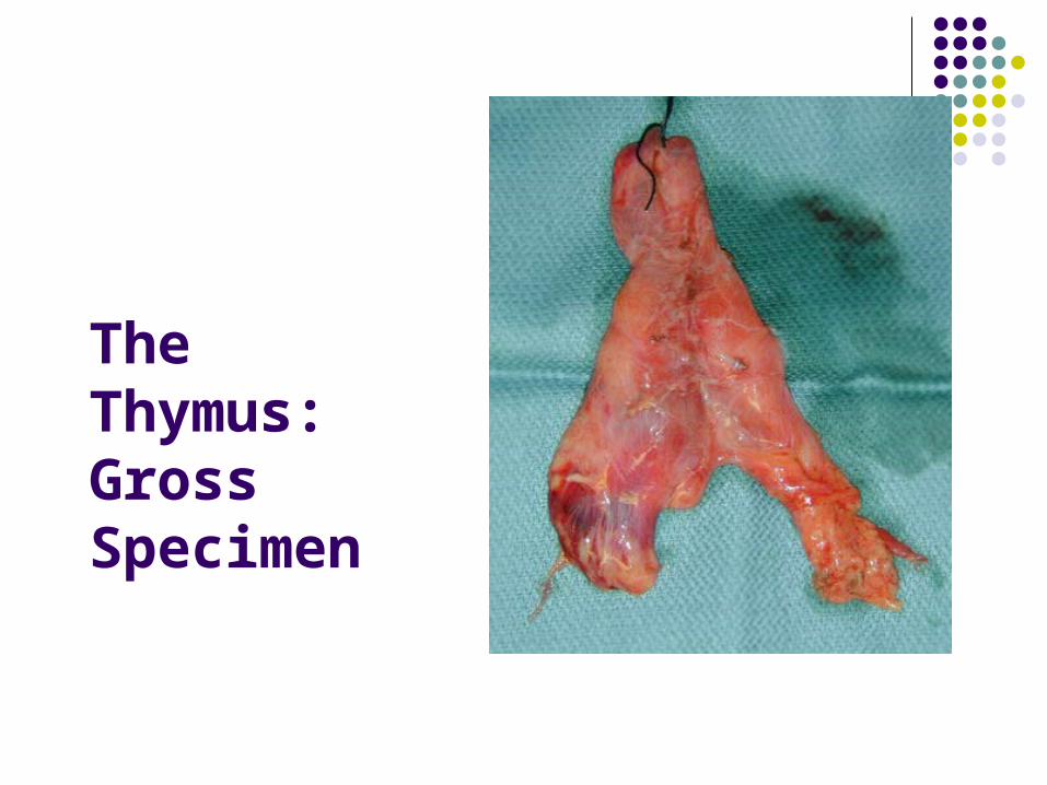
The Thymus:Gross Specimen

Thymic Structure
• Connective tissue capsule, trabeculae.• Both contain blood vessels, efferent lymphatic vessels, and nerves.• Trabeculae demarcate thymic lobules.• Parenchyma is made up of both a cortex and medulla.

Thymus: Cortex and Medulla
The cortex stains darkly basophilic because there lots of small lymphocytes with intensely stained nuclei.
The medulla stains light because it has fewer lymphocytes with more cytoplasm.
Medulla Cortex

Thymus: 3 cell types
Epithelioreticular cells: large, pale, and stellate. (They are not reticular fibers!)
Thymocytes: immature T cells.
Macrophages: phagocytose T cells that react too strongly with self.
Thymocytes Epithelioreticular cells

Hassall’s Corpuscles
Concentrically arranged keratinizing and degenerating epitelioreticular cells and macrophages.
Function is poorly understood (thymic hormones?)
Found in the medulla. Instantly signify the thymus!

Thymus: The Education of T cells
The thymus is the location where thymocytes mature and proliferate.
Thymocytes undergo positive and negative selection.
Schematic: multipotential stem cells enter thymus via postcapillary venule positive selection in cortex negative selection in medulla naïve T cells exit thymus from medulla and enter blood circulation.

Blood-Thymic Barrier Separates developing T
cells from blood (prevents T cells from recognizing foreign proteins as “self”).
Components (from outside inside) Capillary endothelium Endothelial basal lamina Perivascular connective
tissue sheath (and macrophages!)
Basal lamina of epithelioreticular cell
Epithelioreticular sheath

The Adult Thymus
Adult thymus shrinks (involutes).
Adipose tissue replaces thymic tissue.
The medulla and cortex are harder to differentiate because there are fewer lymphocytes.

The Spleen

The Spleen: What is it good for?
1. Filters blood
2. Iron Retrieval
3. RBC reserve
4. Immune Response
5. Fetal Hematopoiesis
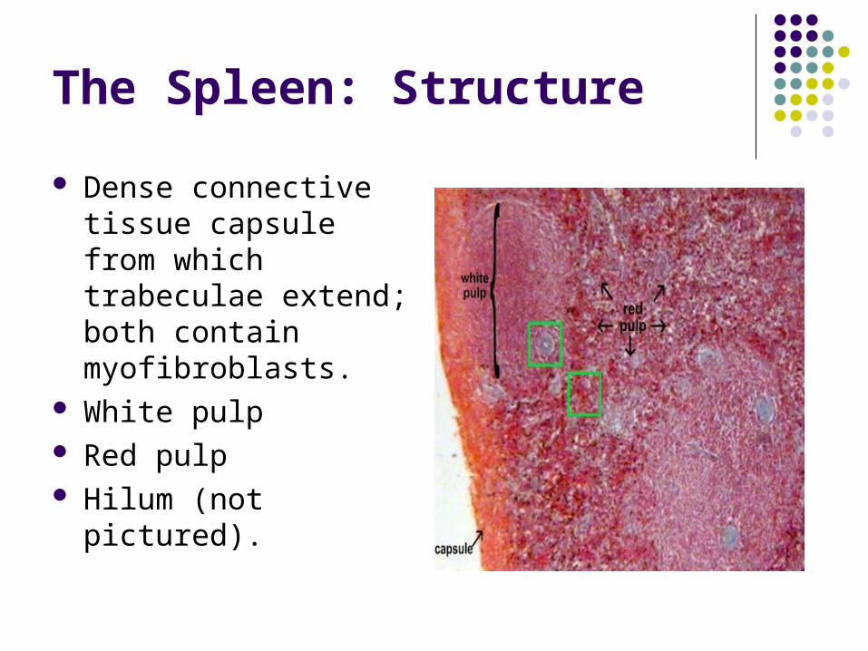
The Spleen: Structure
Dense connective tissue capsule from which trabeculae extend; both contain myofibroblasts.
White pulp Red pulp Hilum (not pictured).

Spleen – Capsule and Trabeculae
*Notice how reticular fibers are evident with silver stain and not H&E.

The Spleen – Vascular Schematic

White Pulp Vasculature
The central artery (branch of splenic artery) is found in the white pulp.
It is surrounded by the PALS, which is T cells.
Lymphatic nodules look like localized expansions of PALS; displace central artery.
Penicilli branch from the central artery into the red pulp.

Red Pulp Vasculature: Leaving the white pulp and
entering the red, penicilli give rise to ellipsoids.
Ellipsoids are capillaries ensheathed by reticular cells and macrophages; their lumens are often occluded in histo sections
Blood is filtered by macrophages through fenestrations in the sinusoids.

Sinusoids
See how the basal lamina is interrupted; evident with both stains.
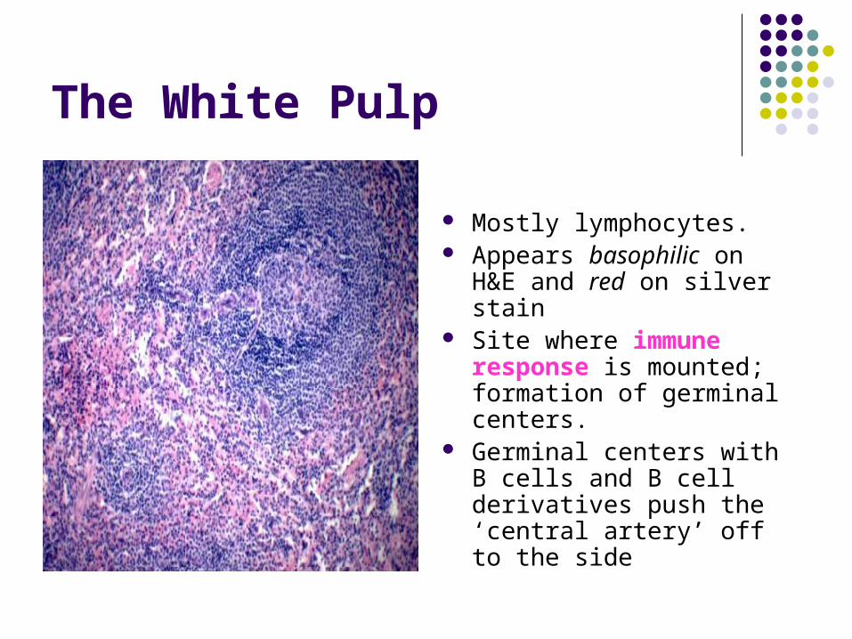
The White Pulp
Mostly lymphocytes. Appears basophilic on
H&E and red on silver stain
Site where immune response is mounted; formation of germinal centers.
Germinal centers with B cells and B cell derivatives push the ‘central artery’ off to the side
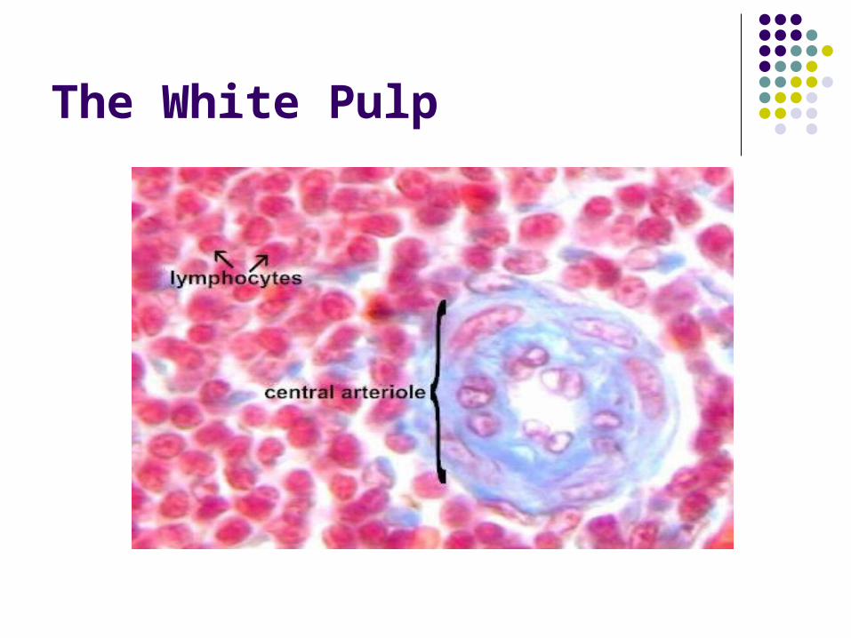
The White Pulp

The Red Pulp
Appears Red on H&E Composed of sinusoids
and Cords of Billroth The cords are the
parenchyma of the red pulp; they are composed of reticular tissue w/ macrophages, red blood cells, and lymphocytes.

Question OneThe function of this organ is
to:A) Secrete antibodies from B
cells into the blood.B) Present antigen to B cells
and filter the blood.C) Guide the maturation of T
cells by positive and negative selection.
D) Present antigen on MHC molecules of mature T cells to epithelioreticular cells.
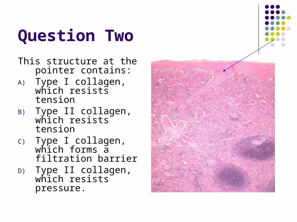
Question Two
This structure at the pointer contains:
A) Type I collagen, which resists tension
B) Type II collagen, which resists tension
C) Type I collagen, which forms a filtration barrier
D) Type II collagen, which resists pressure.

Question Three
The organ shown:
A) Contains blood vessel with a perivascular sheath.
B) Receives lymphocyte precursors via afferent lymphatics.
C) Both A and B.
D) None of the above.
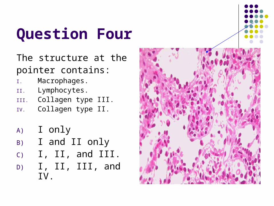
Question Four
The structure at thepointer contains:I. Macrophages.II. Lymphocytes.III. Collagen type III.IV. Collagen type II.
A) I onlyB) I and II onlyC) I, II, and III.D) I, II, III, and IV.

Question Five
Which is the correct order that blood flows through the spleen?
A) Capsular artery, cortex, medulla, pulp veins.
B) Central artery, penicilar arterioles, sinusoids, ellipsoids, pulp veins.
C) Afferent lymphatics, subcapsular sinuses, trabecular sinuses, medullary sinuses, efferent lymphatics.
D) Central artery, penicillar arterioles, ellipsoids, sinusoids, pulp veins.

Question Six
This organ:A) Is encapsulated, capsule
contains reticular fibers.B) Is encapsulated, capsule
contains smooth muscle.C) Is encapsulated, capsule
contains both reticular fibers and smooth muscle.
D) Is encapsulated, capsule contains neither reticular fibers nor smooth muscle.
E) Is not encapsulated.
That’s all, folks…
We hope you got a good education (THYMUS) and that you filtered out
what was important (SPLEEN).

Shameless PlugShameless Plug
** WILDERNESS MEDICINE **
A presentation by Dr. Jay Lemery, Director of Wilderness Medicine Education at Weill Cornell
Medical College.
Thursday, December 16:00 p.m.HSC 305
Dinner will be provided.


