21
Thyroid Cytopathology

Thyroid Cytopathology

ESSENTIALS IN CYTOPATHOLOGY SERIES
Dorothy L. Rosenthal, MD, FIAC, Series Editor
Editorial Board
Syed Z. Ali, MDDouglas P. Clark, MDYener S. Erozan, MD
1. D.P. Clark and W.C. Faquin: Thyroid Cytopathology. 2005 ISBN 0-387-23304-0
2. D. L. Rosenthal and S.S. Raab: Cytologic Detection of Urothelial Lesions. 2005ISBN 0-387-23945-6
3. D.C. Chhieng and E.B. Stelow: Pancreatic Cytopathology. 2007ISBN 978-0-387-68946-3
4. S.Z. Ali and A.V. Parwani: Breast Cytopathology. 2007ISBN 978-0-387-71594-0
5. W.C. Faquin and C.N. Powers: Salivary Gland Cytopathology. 2008ISBN 978-0-387-76622-5
6. Y.S. Erozan and I. Ramzy: Pulmonary Cytopathology. 2009 ISBN 978-0-387-88886-6
7. J.A. Maksem, S.J. Robboy, J.W. Bishop and I. Meiers: Endometrial Cytology with Tissue Correlations. 2009 ISBN 978-0-387-89909-1
8. D.P. Clark and W.C. Faquin: Thyroid Cytopathology, Second Edition. 2010 ISBN 978-1-4419-5952-2
Douglas P. ClarkDepartment of Pathology, The Johns Hopkins Medical Institutions, Baltimore, MD, USA
William C. FaquinDepartment of Pathology, Harvard Medical School, Massachusetts General Hospital, Boston, MA, USA
Thyroid Cytopathology
Second Edition

Douglas P. ClarkDepartment of PathologyThe Johns Hopkins Medical InstitutionsBaltimore, [email protected]
William C. FaquinDepartment of PathologyHarvard Medical SchoolMassachusetts General HospitalBoston, [email protected]
ISBN 978-1-4419-5952-2 e-ISBN 978-1-4419-5954-6DOI 10.1007/978-1-4419-5954-6Springer New York Dordrecht Heidelberg London
Library of Congress Control Number: 2010925490
© Springer Science+Business Media, LLC 2010All rights reserved. This work may not be translated or copied in whole or in part without the written permission of the publisher (Springer Science+Business Media, LLC, 233 Spring Street, New York, NY 10013, USA), except for brief excerpts in connection with reviews or scholarly analysis. Use in connection with any form of information storage and retrieval, electronic adaptation, computer software, or by similar or dissimilar methodology now known or hereafter developed is forbidden.The use in this publication of trade names, trademarks, service marks, and similar terms, even if they are not identified as such, is not to be taken as an expression of opinion as to whether or not they are subject to proprietary rights.While the advice and information in this book are believed to be true and accurate at the date of going to press, neither the authors nor the editors nor the publisher can accept any legal responsibility for any errors or omissions that may be made. The publisher makes no warranty, express or implied, with respect to the material contained herein.
Printed on acid-free paper
Springer is part of Springer Science+Business Media (www.springer.com)

Foreword
The evaluation of thyroid nodules by fine needle aspiration (FNA) is one of the most challenging tasks in all of cytopathology. A cytologist must understand the clinical presentation of thyroid diseases, their defining histopathologic and cytopathologic features, and even the intricacies of patient management. Drs. Clark and Faquin have provided a valuable framework for cytologists learning (and continuing to learn) this exacting discipline. Organ-ized around a practical algorithm, the authors lay out a rational and concise approach toward acquiring the necessary skills for the cytologic diagnosis of thyroid nodules. The first edition, published in 2005, was a very welcome addition to the cytology literature. This new edition, with updated terminology for reporting thyroid FNA results, builds on the success of their approach.
Why are we examining such challenging specimens? Clearly, the clinical need is there. Over 50% of adults have one or more thyroid nodules. Surgical excision of all nodules is certainly neither practical nor desirable. Enter FNA, a minimally invasive cellular sampling method that has proven to be a highly useful screening test for thyroid cancer. Because of it, thousands of patients with a benign diagnosis are spared unnecessary surgery every year, and those with cancer are reliably triaged for appropriate therapy.
The large number of FNAs performed in the US is a tribute to its success as a screening test. In many institutions, the thyroid FNA is the most common FNA specimen. For a relatively new diagnostic test, this is a remarkable state of affairs. Thirty years ago, few thyroid cancers were diagnosed by FNA in the US, and in the 1980s some prominent pathologists still questioned the value
v

vi Foreword
of FNA for thyroid nodules. There is no more debate: FNA has proven its value. In 2009, an estimated 37,200 thyroid cancers will be diagnosed in the United States, and virtually all of them will have been diagnosed directly or triaged for a diagnostic lobectomy by FNA. If approximately nine FNAs are performed for every thyroid cancer that is discovered, then roughly 335,000 thyroid FNAs will have been performed in the US in 2009.
Cytologists must be armed and ready to evaluate these clinically vital specimens. This book, with its practical algorithm, cogent text, and beautiful illustrations, provides the ammunition a cytolo-gist needs to master thyroid FNA interpretation.
Edmund S. Cibas

Series Preface
The subspeciality of cytopathology is 60 years old and has become established as a solid and reliable discipline in medicine. As expected, cytopathology literature has expanded in a remarkably short period of time, from a few textbooks prior to the 1980s to a current and substantial library of texts and journals devoted exclusively to cytomorphology. Essentials in Cytopathology does not presume to replace any of the distinguished textbooks in cytopathology. Instead, the series will publish generously illustrated and user-friendly guides for both pathologists and clinicians.
Building on the amazing success of The Bethesda System for Reporting Cervical Cytology, now in its second edition, the Series will utilize a similar format, including minimal text, tabular criteria, and superb illustrations based on real-life specimens. Essentials in Cytopathology will, at times, deviate from the classic organiza-tion of pathology texts. The logic of decision trees, elimination of unlikely choices, and narrowing of differential diagnosis via a pragmatic approach based on morphologic criteria will be some of the strategies used to illustrate principles and practice in cytopa-thology.
Most of the authors for Essentials in Cytopathology are faculty members in The Johns Hopkins University School of Medicine, Department of Pathology, Division of Cytopathology. They bring to each volume the legacy of John K. Frost and the collective experience of a preeminent cytopathology service. The archives at Hopkins are meticulously cataloged and form the framework for text and illustrations. Authors from other institutions have been selected on the basis of their national reputations, experience, and
vii

viii Series Preface
enthusiasm for cytopathology. They bring to the series comple-mentary viewpoints and enlarge the scope of materials contained in the photographs.
The editor and authors are indebted to our students, past and future, who challenge and motivate us to become the best that we possibly can be. We share that experience with you through these pages, and hope that you will learn from them as we have from those who have come before us. We would be remiss if we did not pay tribute to our professional colleagues, the cytotechnologists, and preparatory technicians who lovingly care for the specimens that our clinical colleagues send to us.
And finally, we cannot emphasize enough throughout these volumes the importance of collaboration with the patient care team. Every specimen comes to us as a question begging an answer. Without input from the clinicians, complete patient history, results of imaging studies, and other ancillary tests, we cannot perform optimally. It is our responsibility to educate our clinicians about their role in our interpretation, and for us to integrate as much information as we can gather into our final diagnosis, even if the answer at first seems obvious.
We hope you will find this series useful and welcome your feed-back as you place these handbooks by your microscopes, and into your book bags.
Baltimore, MD Dorothy L. RosenthalBaltimore, MD Douglas P. ClarkBoston, MA William C. Faquin

Acknowledgments
The authors gratefully acknowledge the advice and encouragement of our colleagues, Dorothy L. Rosenthal, MD, FIAC, Syed Ali, MD, Yener Erozan, MD, Ulrike Hamper, MD, and Edmund S. Cibas, MD, as well as their generous contributions of images. The authors also thank Shirley Long for assistance with manuscript prepara-tion, Tim Phelps for his drawings, and Sharon Blackburn for her computer graphics.
ix

Contents
Foreword ................................................................................ vSeries Preface ......................................................................... viiAcknowledgments .................................................................. ix
1 Introduction and Clinical Aspects ................................. 1
2 How to Perform and Process a Thyroid FNA ............... 9
3 Approach to Thyroid FNA Cytopathology: An Overview ................................................................. 23
4 Inflammatory Lesions and Lymphoma ......................... 33
5 Colloid-Predominant Lesions ....................................... 55
6 Follicular Lesions .......................................................... 69
7 Hurthle Cell Lesions ..................................................... 93
8 Cystic Lesions of the Thyroid ....................................... 109
9 Papillary Thyroid Carcinoma ........................................ 125
10 Medullary Thyroid Carcinoma ..................................... 151
11 Undifferentiated (Anaplastic) Carcinoma and Secondary Tumors .................................................. 167
Index ...................................................................................... 183
xi
1D.P. Clark and W.C. Faquin, Thyroid Cytopathology,Essentials in Cytopathology 8, DOI 10.1007/978-1-4419-5954-6_1,© Springer Science + Business Media, LLC 2010
1Introduction and Clinical Aspects
The second edition of this book incorporates the recent terminology and reporting guidelines for thyroid fine needle aspirations (FNAs), the Bethesda System for Reporting Thyroid Cytopathology (BSRTC) that emerged from a multidisciplinary National Cancer Institute (NCI) Thyroid FNA State of the Science conference (2007). A major challenge in the application of FNA to the diagnosis of thyroid lesions has been the inconsistent use of terminology for reporting results of thyroid FNAs both within laboratories and between different institutions. Not only has this hindered the sharing of information between different institutions, but it has created difficulties for clinicians managing patients with thyroid disease. Throughout this second edition, we will use a simple algorithmic approach, which we have modified to incorporate the BSRTC, to explain how to evaluate thyroid FNAs.
Over the past 3 decades, FNA has developed as the most accurate and cost-effective initial method for guiding the clinical management of patients with thyroid nodules. The purpose of this book is to describe the application of FNA to the assessment of thyroid nodules, with particular emphasis on the key cytologic features that can be used to diagnose FNA specimens based on a simple algorithmic approach.

2 1. Introduction and Clinical Aspects
The clinical application of FNA as a primary diagnostic tool for thyroid nodules is widespread, because thyroid nodules are common. Within the general population, palpable thyroid nodules are present in 4–7% of adults, and subclinical (nonpalpable) nodules are present in up to 70% of individuals. Of these thyroid nodules, 90–95% are benign, and include a wide variety of lesions such as adenomatous nodules, simple thyroid cysts, colloid nodules, follicular adenomas, and inflammatory and developmental conditions, among others.
Benign Causes of Thyroid Nodules
Adenomatous nodule •Colloid nodule •Follicular adenoma •Simple thyroid cyst •Graves disease •Chronic lymphocytic thyroiditis •Focal subacute thyroiditis •Developmental conditions •
The extremely large number of benign thyroid nodules and the small number of admixed malignant ones creates a clinical dilemma: how to manage the many patients with a detectable thyroid enlargement that is most likely benign? FNA has emerged as the most effective method for dealing with this problem. As a screening test for thyroid carcinoma, FNA assists in guiding the clinical management of patients by helping to select those individuals who are more likely to have a malignancy and need surgical management from the larger group of patients with benign nodules that can be managed without surgical intervention.
FNA is now generally accepted by endocrinologists and thyroid surgeons as a safe, cost-effective, and accurate means of evalu-ating a thyroid nodule. Widespread use of FNA has reduced the number of patients requiring thyroid surgery by more than 50%, it has increased the yield of malignancies at thyroidectomy by two to three times, and it has decreased the overall cost of managing a thyroid nodule by more than 25%.

3Accuracy of Thyroid FNA 3
Benefits of Using FNA to Evaluate Thyroid Nodules
Reduces number of patients requiring thyroid surgery by 50% •Increases the yield of thyroid malignancies at thyroidectomy •by two to three timesDecreases the cost of managing thyroid nodules by more than •25%
Incidence and Subtypes of Thyroid Carcinoma
In 2009, it is estimated that there will be over 37,000 new cases of thyroid cancer reported, and more than 1,500 deaths due to thyroid cancer. The rate of new cases of thyroid cancer has been increasing, in part due to the increased detection of small papillary thyroid carcinomas. Overall, thyroid cancer accounts for approxi-mately 2% of the total number of new cancer cases for all anatomic sites and 0.5% of the total number of cancer-related deaths per year. Worldwide, the incidence of thyroid cancer varies from 0.5 to 10 per 100,000 individuals. It is the sixth most common form of cancer in women. Although the majority of thyroid cancers are well-differentiated tumors that have a very favorable prognosis, included within this group of malignancies is one of the most aggressive cancers affecting humans, undifferentiated thyroid carcinoma, with a mean survival of just 2–6 months.
Among the various types of thyroid carcinomas that may be encountered by FNA, the most common is papillary thyroid carcinoma, representing 60–80% of all thyroid malignancies. This incidence is distantly followed by follicular carcinoma (15–25%) and medullary carcinoma (5–10%) (Table 1.1).
Accuracy of Thyroid FNA
Thyroid FNA is widely accepted as an accurate means of evaluating a thyroid nodule, and it is considered by some to be the most sensitive and most specific nonsurgical thyroid cancer test available. For certain tumors, such as papillary thyroid carcinoma, FNA has

4 1. Introduction and Clinical Aspects
even been reported to be superior to frozen section diagnosis. Other modalities for evaluating thyroid nodules such as serum tests, sonography, and scintigraphy have been largely overshad-owed by FNA.
Based on several large studies, the accuracy of thyroid FNA has usually been reported as greater than 95% for satisfactory specimens, with positive predictive values of 89–98% and negative predictive values of 94–99% (Table 1.2). These values, however, are dependent upon several factors including how the indeterminate and suspicious groups of lesions are used in the calculations, the skill of the person performing the FNA, and the expertise of the cytopathologist interpreting the specimen. In addition, the accuracy of a thyroid FNA classified as “Benign” is difficult to assess, since so many patients in this group do not have surgery. The wide range of sensitivities and specificities for thyroid FNA that have been reported reflects the influence of these various factors. False-negative and false-positive FNA diagnoses occur, but in most studies, they are very uncommon, and are usually less than 1%.
Table 1.1. Relative percentage of thyroid malignancies.
Thyroid tumor type Relative percentage (%)
Papillary 60–80Follicular (including Hurthle cell) 15–25Medullary 5–10Undifferentiated 1–10Lymphoma <1Metastasis <1
Table 1.2. Accuracy of thyroid fine needle aspiration (FNA).Statistical measurement Percentage (%)
Accuracy for satisfactory specimens >95False-negative rate 0.7–11False-positive rate 0–7Positive predictive value 89–98Negative predictive value 94–99Sensitivity 43–98Specificity 72–100

5Classification of Follicular-Derived Thyroid Carcinomas 5
The only caveat to these values is that the reported false-negative rates are based only upon those patients who undergo surgical resection of their aspirated nodules, and thus the calculations may be an underestimate; approximately 18% of patients who have an FNA are actually treated surgically.
Classification of Follicular-Derived Thyroid Carcinomas
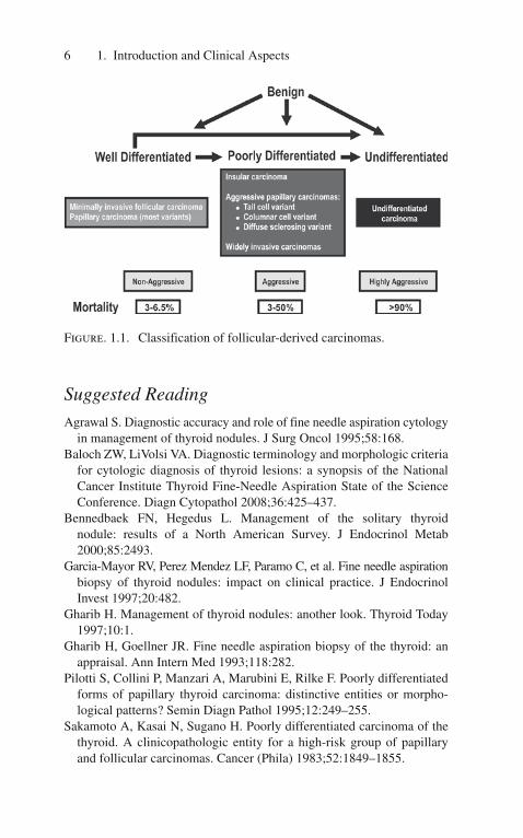
Although the most important clinicopathologic predictors of aggressive clinical behavior for thyroid carcinomas are patient age, tumor size, and tumor stage, cytologic and histologic features that we recognize in daily practice can be used to divide neoplasms of thyroid follicular cells into three general categories that differ in clinical aggressiveness: well-differentiated, poorly differentiated, and undifferentiated carcinoma.
Well-differentiated thyroid carcinomas, representing the major-ity of thyroid cancers, have an excellent overall prognosis with mortalities in the range of 3–6%. In contrast, undifferentiated thyroid carcinoma, at the opposite end of the spectrum, is an extremely aggressive malignancy associated with greater than 90% mortality and a mean survival of only 2–6 months. Poorly differentiated carcinomas, insular carcinoma being the classic example, are characterized by a clinical behavior and mortality rate intermediate between that of the well-differentiated and undif-ferentiated thyroid carcinomas. These three groups of thyroid carcinomas, particularly the poorly differentiated ones, are continuing to be defined by advances in our understanding of their biologic behavior, as well as by their molecular features and their cyto- and histomorphologies. The recent Tourin Proposal (2007) provided a unified series of diagnostic criteria that have further defined the poorly differentiated subset of thyroid carcinomas. Some cases of less-differentiated carcinoma may arise by progression from better-differentiated thyroid carcinomas; however, other cases of poorly differentiated and undifferentiated carcinoma possibly arise de novo because they do not exhibit microscopic evidence of such a progression (Figure 1.1).
6 1. Introduction and Clinical Aspects
Suggested Reading
Agrawal S. Diagnostic accuracy and role of fine needle aspiration cytology in management of thyroid nodules. J Surg Oncol 1995;58:168.
Baloch ZW, LiVolsi VA. Diagnostic terminology and morphologic criteria for cytologic diagnosis of thyroid lesions: a synopsis of the National Cancer Institute Thyroid Fine-Needle Aspiration State of the Science Conference. Diagn Cytopathol 2008;36:425–437.
Bennedbaek FN, Hegedus L. Management of the solitary thyroid nodule: results of a North American Survey. J Endocrinol Metab 2000;85:2493.
Garcia-Mayor RV, Perez Mendez LF, Paramo C, et al. Fine needle aspiration biopsy of thyroid nodules: impact on clinical practice. J Endocrinol Invest 1997;20:482.
Gharib H. Management of thyroid nodules: another look. Thyroid Today 1997;10:1.
Gharib H, Goellner JR. Fine needle aspiration biopsy of the thyroid: an appraisal. Ann Intern Med 1993;118:282.
Pilotti S, Collini P, Manzari A, Marubini E, Rilke F. Poorly differentiated forms of papillary thyroid carcinoma: distinctive entities or morpho-logical patterns? Semin Diagn Pathol 1995;12:249–255.
Sakamoto A, Kasai N, Sugano H. Poorly differentiated carcinoma of the thyroid. A clinicopathologic entity for a high-risk group of papillary and follicular carcinomas. Cancer (Phila) 1983;52:1849–1855.
Figure. 1.1. Classification of follicular-derived carcinomas.
7Classification of Follicular-Derived Thyroid Carcinomas 7
The American Thyroid Association Guidelines Taskforce. Management guidelines for patients with thyroid nodules and differentiated thyroid cancer. Thyroid 2006;16:1–33.
Volante M, Collini P, Nikiforov YE, Sakamoto A, et al. Poorly differentiated thyroid carcinoma: the Tourin Proposal for the use of uniform diagnostic criteria and an algorithmic diagnostic approach. Am J Surg Pathol 2007;31:1256–1264.

9D.P. Clark and W.C. Faquin, Thyroid Cytopathology,Essentials in Cytopathology 8, DOI 10.1007/978-1-4419-5954-6_2,© Springer Science + Business Media, LLC 2010
2How to Perform and Process a Thyroid FNA
Thyroid fine needle aspirations (FNAs) are among the most challenging FNAs to perform because of the anatomic location and the vascularity of the thyroid gland. However, this challenge must be mastered because accurate diagnosis is dependent on a high-quality, well-prepared specimen.
Pre-FNA Evaluation
Historically, patients with a thyroid nodule have received a radio-nuclide scan as well as a thyroid ultrasound examination before an FNA. More recently, it is recognized that for many patients this is neither necessary nor cost-effective. The main purpose of a radionuclide scan is to rule out a hyperfunctioning thyroid nodule, as these are rarely malignant. Because a hyperfunctioning nodule will suppress thyroid-stimulating hormone (TSH) production by the pituitary, a sensitive serum test for TSH levels can be used in place of a radionuclide scan. An abnormally low serum TSH level suggests a hyperfunctioning nodule that can then be evalu-ated clinically before performing an FNA.
Thyroid ultrasound examination is useful in the evaluation of small, difficult to palpate nodules and may give information about cystic areas and calcifications. However, ultrasound does not offer sufficient sensitivity or specificity for malignancy to eliminate
10 2. How to Perform and Process a Thyroid FNA
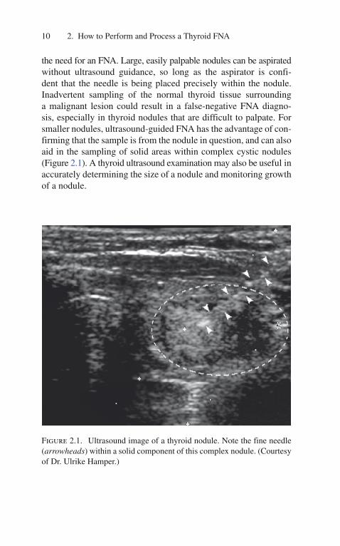
the need for an FNA. Large, easily palpable nodules can be aspirated without ultrasound guidance, so long as the aspirator is confi-dent that the needle is being placed precisely within the nodule. Inadvertent sampling of the normal thyroid tissue surrounding a malignant lesion could result in a false-negative FNA diagno-sis, especially in thyroid nodules that are difficult to palpate. For smaller nodules, ultrasound-guided FNA has the advantage of con-firming that the sample is from the nodule in question, and can also aid in the sampling of solid areas within complex cystic nodules (Figure 2.1). A thyroid ultrasound examination may also be useful in accurately determining the size of a nodule and monitoring growth of a nodule.
Figure 2.1. Ultrasound image of a thyroid nodule. Note the fine needle (arrowheads) within a solid component of this complex nodule. (Courtesy of Dr. Ulrike Hamper.)

History 11
“Incidentaloma”
The term incidentaloma has been coined for any small (less than 1 cm) thyroid nodule that is incidentally discovered during a procedure intended for a different purpose, such as a computed tomography (CT) scan of the cervical spine or an ultrasound study of the carotid arteries. Because the incidence of malignancy in these small lesions is low, physicians should have a high threshold for performing an FNA on these nodules, particularly within a multinodular gland in patients without other indications.
History
For most patients with a thyroid nodule, their clinical history does not contribute significantly to the FNA diagnosis. Features of the clinical history that do raise the suspicion of a thyroid malignancy in patients with a thyroid nodule include male gender, age less than 20 years or greater than 70 years, dysphagia or hoarseness, a his-tory of neck irradiation during childhood or adolescence, a family history of thyroid disease (especially papillary thyroid carcinoma (PTC), medullary carcinoma (MC), or multiple endocrine neopla-sia (MEN)), or a rapid increase in the size of a long-standing goiter. Other useful clinical information includes a history of Hashimoto thyroiditis, a history of Graves disease or 131I therapy, or a history of a nonthyroid malignancy.
Clinical features that raise the suspicion of malignancy in a thyroid nodule are as follows:
History •Male gender —
Age less than 20 or more than 70 years —
History of neck irradiation —
Family history of thyroid disease, especially PTC or MC —
Family or personal history of an MEN syndrome —
Dysphagia or hoarseness —

12 2. How to Perform and Process a Thyroid FNA
Physical examination •Firm, fixed mass —
Nodule size greater than 4 cm —
Cervical lymphadenopathy —
Physical Examination
Physical examination of the thyroid is an art that develops with experience. Often, large nodules can be seen as an asymmetric bulge in the neck, so careful observation is recommended before palpation. Some texts recommend the use of one’s thumbs to examine the thyroid, but we find the first and second fingers to be more sensitive in identifying nodules. Rather than standing behind the patient and reaching around to palpate the thyroid (which can be impractical and unnerving for the patient), we recommend standing to the patient’s right side as the patient sits upright on an examination table. The first and second fingers of the right hand are then used to palpate the thyroid gland (this position may be reversed for left-handed examiners).
To palpate the thyroid, the practitioner should place his first and second fingers firmly and deeply into the angle formed between the trachea and the insertion of the sternocleidomastoid muscle into the sternum (Figure 2.2). While the fingers are press-ing firmly in this region, the patient should be asked to swallow. It is sometimes helpful to hand the patient a glass of water to sip during the examination. Swallowing causes the thyroid to move upward, increasing the sensitivity of palpation because one can often feel the surface contours of a thyroid nodule as it moves superiorly then inferiorly beneath the fingers, which are held in one place. This movement of a nodule also confirms the associa-tion of the nodule with the thyroid gland. A thyroid nodule often feels like a marble sliding beneath one’s fingers. Although larger cystic lesions may feel soft, and cancers can be firm and fixed, the texture of the nodule is not generally predictive of malignancy. To palpate the superior pole of the gland, the fingers should be moved superiorly about 3 cm along the trachea. This entire procedure should then be repeated on the opposite side of the neck; this can be done without changing position relative to the patient,


