Thyroid Nodules: Practical and Molecular Approaches to a Common Problem Bryan R. Haugen, MD University of Colorado, School of Medicine University of Colorado Cancer Center Surgery Grand Rounds May 9, 2011
Transcript
Thyroid Nodules:Practical and Molecular Approaches to a
Common Problem
Bryan R. Haugen, MDUniversity of Colorado, School of Medicine
University of Colorado Cancer CenterSurgery Grand Rounds
May 9, 2011
Disclosure
Research support – Veracyte, Inc
Patient
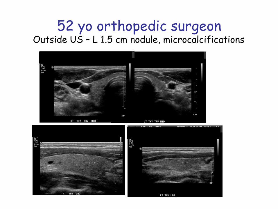
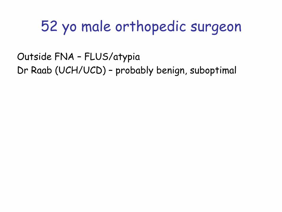
A 52 yo male orthopedic surgeon was self-referred for thyroid nodule and indeterminate FNA biopsy
Member of scoliosis society (800-1000), lots of fluoroscopy, 3 members died of aggressive thyroid cancer
Ultrasound – 1.5 cm L thyroid nodule with microcaclfications
TSH 1.6 mU/LBiopsy – FLUS/atypia
Recommended surgery
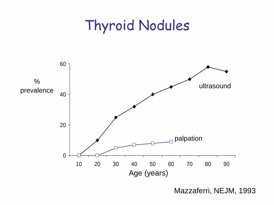
Thyroid Nodules
0
20
40
60
10 20 30 40 50 60 70 80 90
%prevalence
Age (years)
ultrasound
palpation
Mazzaferri, NEJM, 1993
Thyroid Noduleswhat we think we know
• Thyroid nodules are common• Thyroid cancer is not • Biopsy the nodule• Malignant = surgery• Benign = leave it alone• Indeterminate = Damn
What percentage of thyroid nodules are malignant?
A) 2%B) 5%C) 10%D) 15%E) 20%F) 50%
What percentage of thyroid nodules are malignant?
Paper n % malig surgery % maligYassaCancer, 2007
3589 14% 1242 56%
YangCancer, 2007
3207 15% 378 53%
TheoharisThyroid, 2009
4703 16% 1052 46%
“5-10%”??
Definitions
Positive Negative
Positive True positive False positive
Negative False Negative True negativeTest
result
‘gold standard’(histopathology)
PPV
NPV
Sensitivity Specificity
NPV Proportion of patients with a negative test result who are correctly diagnosed
PPV Proportion of patients with a positive test result who are correctly diagnosed
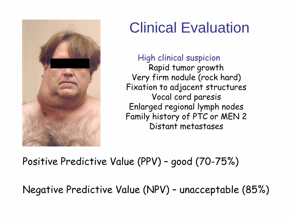
Clinical Evaluation
Positive Predictive Value (PPV) – good (70-75%)
Negative Predictive Value (NPV) – unacceptable (85%)
High clinical suspicionRapid tumor growth
Very firm nodule (rock hard)Fixation to adjacent structures
Vocal cord paresisEnlarged regional lymph nodes
Family history of PTC or MEN 2Distant metastases
Approach to the Patient with Thyroid NodulesTSH
Free T4, Free T3Thyroid antibodies
ThyroglobulinCalcitonin
UltrasoundCT scan
Nuclear Medicine (123I, 99mTc)18FDG-PETCore biopsy
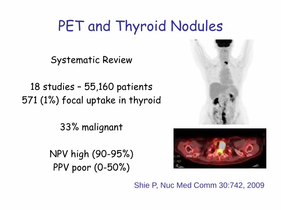
18 studies – 55,160 patients571 (1%) focal uptake in thyroid
33% malignant
NPV high (90-95%)PPV poor (0-50%)
Shie P, Nuc Med Comm 30:742, 2009
Approach to the Patient with Thyroid Nodules
TSH
Ultrasound
Fine-Needle Aspiration Biopsy (FNAB)
Thyroid Ultrasound
• Is the palpable abnormality a thyroid nodule?• Are other nodules present?• Size(s)?• Suspicious features?• > 50% cystic?• Posterior?• Associated abnormal lymph nodes?
R2 Thyroid sonography should be performed in all patients with known or suspected thyroid nodules (A)
Cytology and Molecular Analysis Cancer Probability
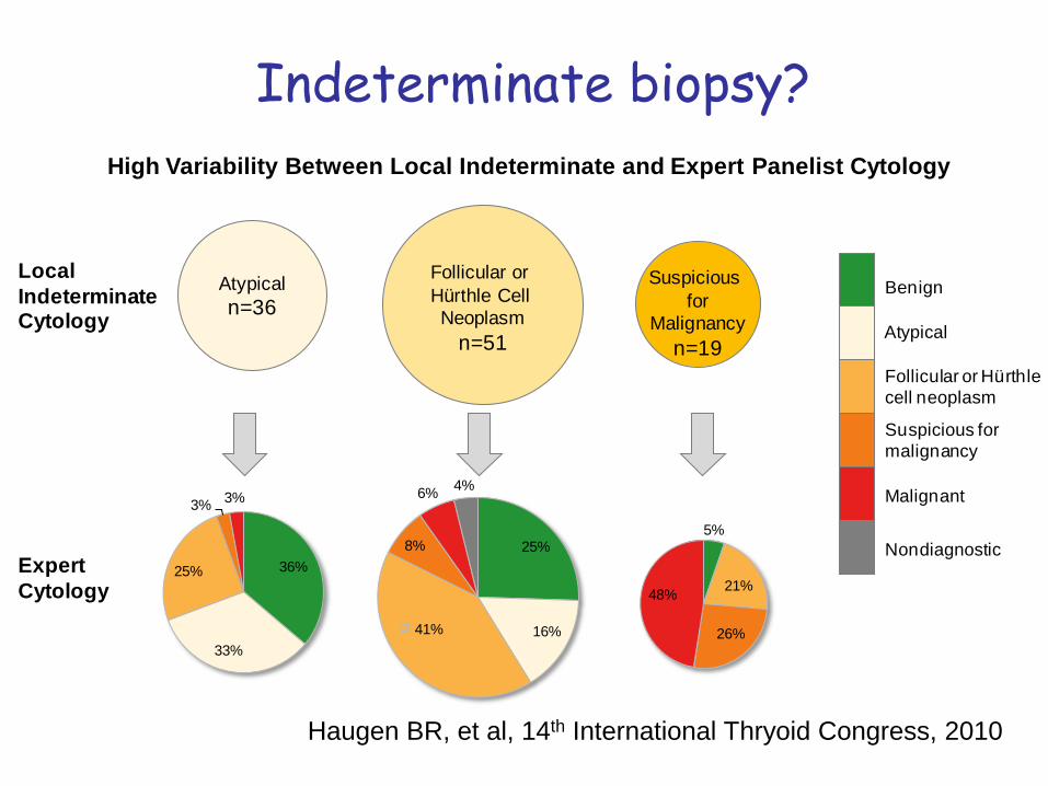
Indeterminate Cytology 40%
Indeterminate Cytology, Pos Mutation 100%
Indeterminate Cytology, Neg Mutation 16%
Negative Cytology 2.1%
Negative Cytology, Neg Mutation 0.9%
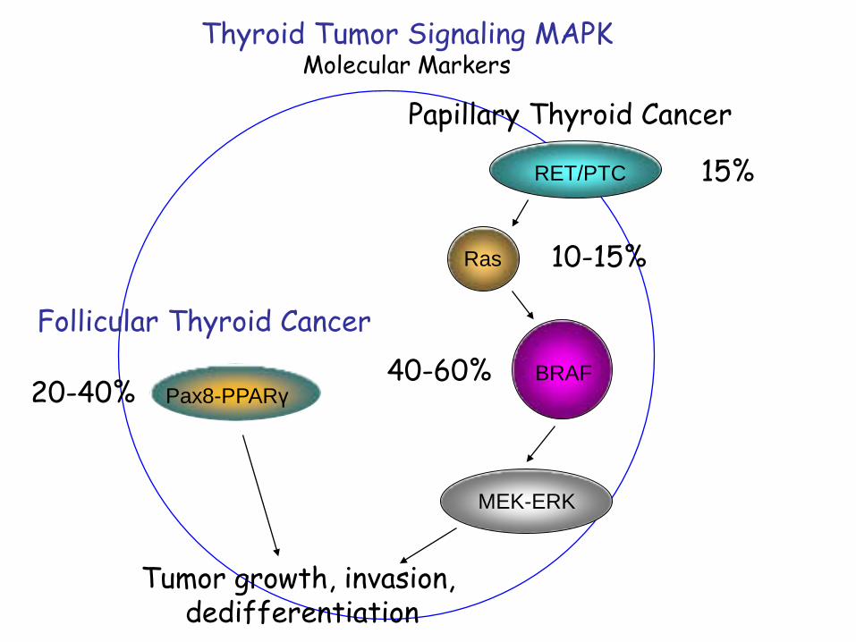
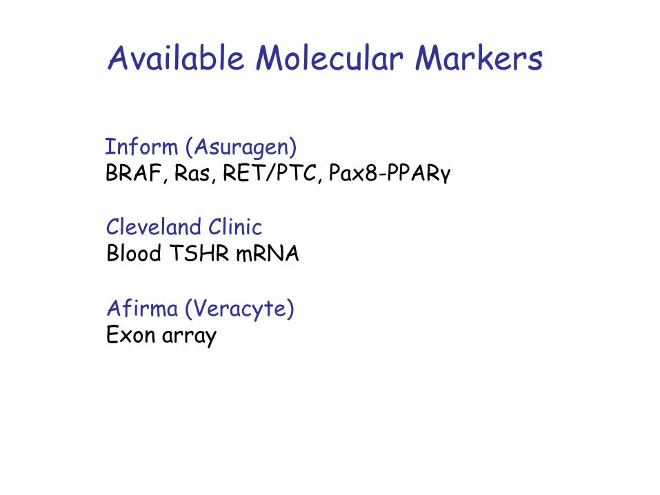
Available Molecular Markers
Inform (Asuragen)BRAF, Ras, RET/PTC, Pax8-PPARγ
Cleveland ClinicBlood TSHR mRNA
Afirma (Veracyte)Exon array
The Evolution of Molecular Analysis:
PAX8: PPARγ TranslocationBRAF V600E
Galectin-3SNP: 9q22.33, and 14q13.3RAS, RET/PTC Oncogenes
Recent Studies: Ongoing Studies:
High POSITIVEPredictive Value
High NEGATIVEPredictive Value
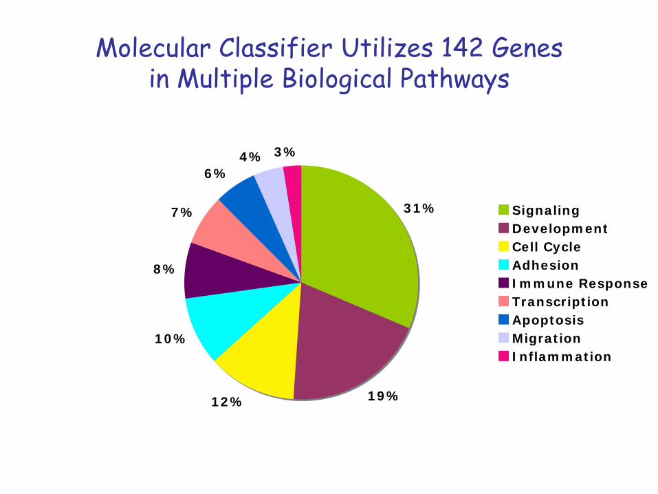
Development of a novel molecular classifier to accurately identify benign thyroid nodules
in patients with indeterminate FNA cytology
Bryan R. Haugen, Zubair Baloch, Darya Chudova, Edmund Cibas, Lyssa Friedman, Giulia C. Kennedy, Richard Kloos, Richard Lanman, Virginia LiVolsi, Susan Mandel, David Steward, Stephen Raab, Juan Rosai, Charles Wang, Eric Wang, Jonathan Wilde, Martha Zeiger,
Erik K. Alexander
14th International Thyroid CongressSept 11-16, 2010
Hypothesis
A molecular classifier can be developed to categorize indeterminate nodules with a high negative predictive value (NPV)
Methods
Train and validate a molecular classifier against the ‘gold standard’ of histopathology by experts (Virginia LiVolsi and Juan Rosai)
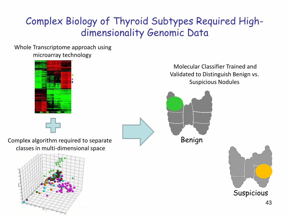
Complex Biology of Thyroid Subtypes Required High-dimensionality Genomic Data
Complex algorithm required to separate classes in multi-dimensional space
Whole Transcriptome approach using microarray technology
Molecular Classifier Trained andValidated to Distinguish Benign vs.
Suspicious Nodules
Benign
Suspicious43
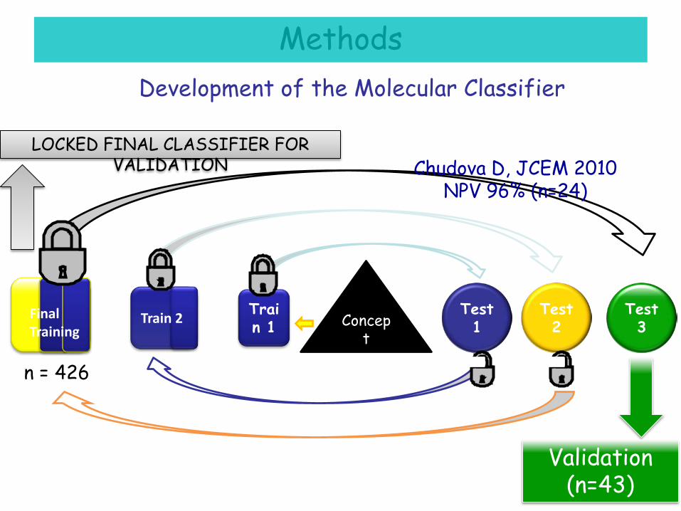
Development of the Molecular Classifier
Concept
Validation (n=43)
Test 1
Test 2
Test 3
Train 1Train 2Final
Training
LOCKED FINAL CLASSIFIER FOR VALIDATION Chudova D, JCEM 2010
Independent Validation of Molecular Classifier by Expert Consensus Histopathologic Diagnosis
Surgical pathology, indeterminate cytology
(n=43)
Sensitivity 95%Specificity 63%PPV 57%NPV 96%
Surgical pathology,all cytology
(N=66)
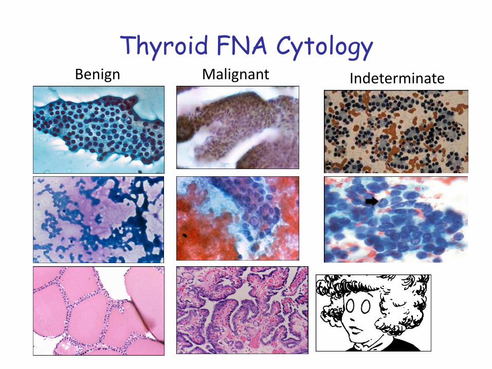
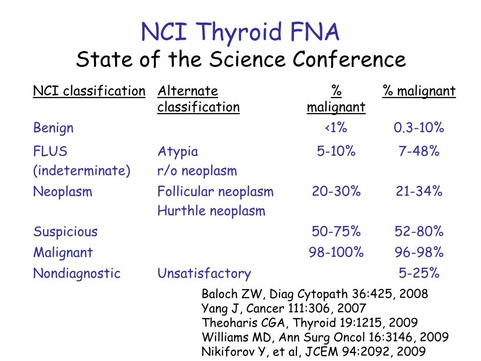
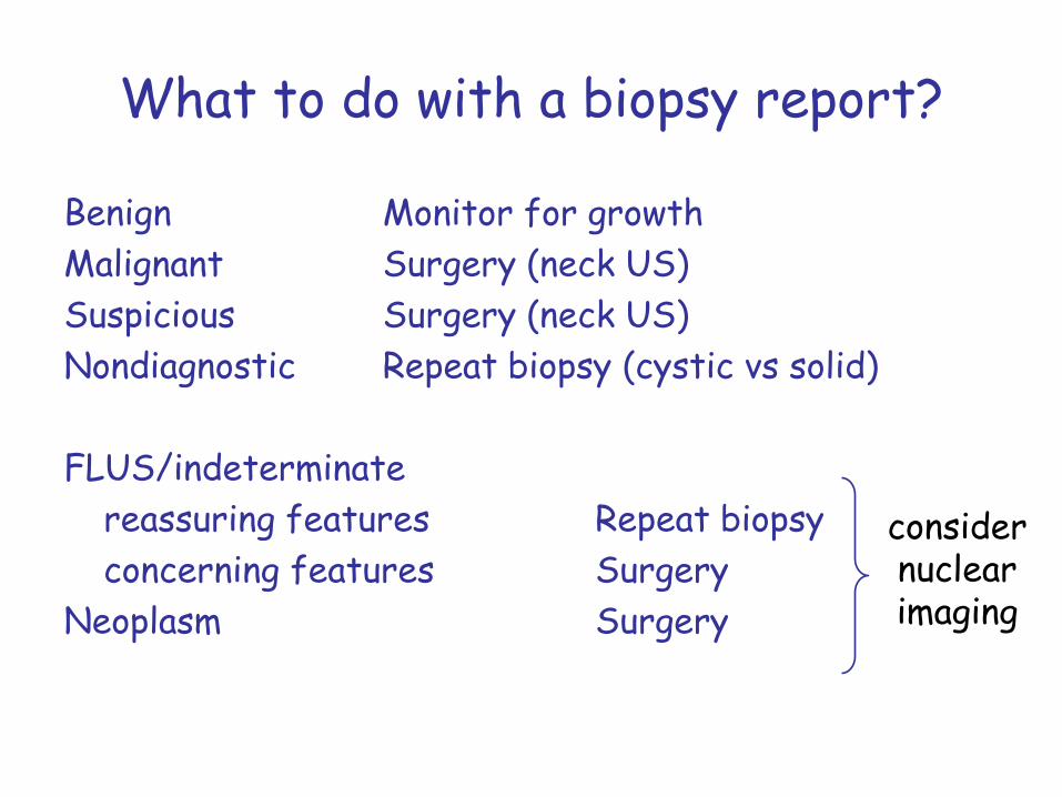
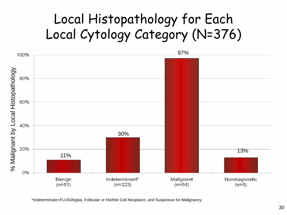
Thyroid FNACytopathology
NCI classification % malignant
Benign 0.3-10%
FLUS 7-48%Neoplasm 21-34%
Suspicious 52-80%
Malignant 96-98%Nondiagnostic 5-25%
Baloch ZW, Diag Cytopath 36:425, 2008Yang J, Cancer 111:306, 2007Theoharis CGA, Thyroid 19:1215, 2009Williams MD, Ann Surg Oncol 16:3146, 2009Nikiforov Y, et al, JCEM 94:2092, 2009
Indeterminate
Thyroid FNACytopathology
NCI classification % malignant
Benign 0.3-10%
FLUS 7-48%Neoplasm 21-34%
Suspicious 52-80%
Malignant 96-98%Nondiagnostic 5-25%
Baloch ZW, Diag Cytopath 36:425, 2008Yang J, Cancer 111:306, 2007Theoharis CGA, Thyroid 19:1215, 2009Williams MD, Ann Surg Oncol 16:3146, 2009Nikiforov Y, et al, JCEM 94:2092, 2009
IndeterminateMolecularclassifier
4%
57%
benign
suspicious
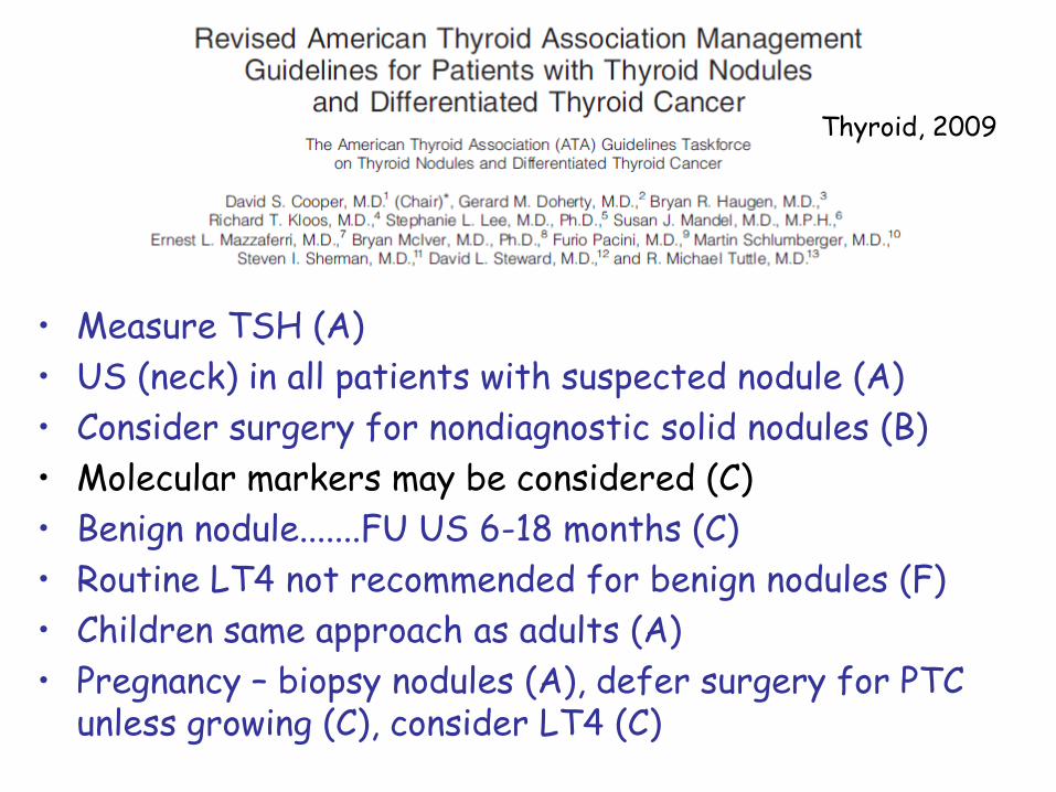
Thyroid, 2009
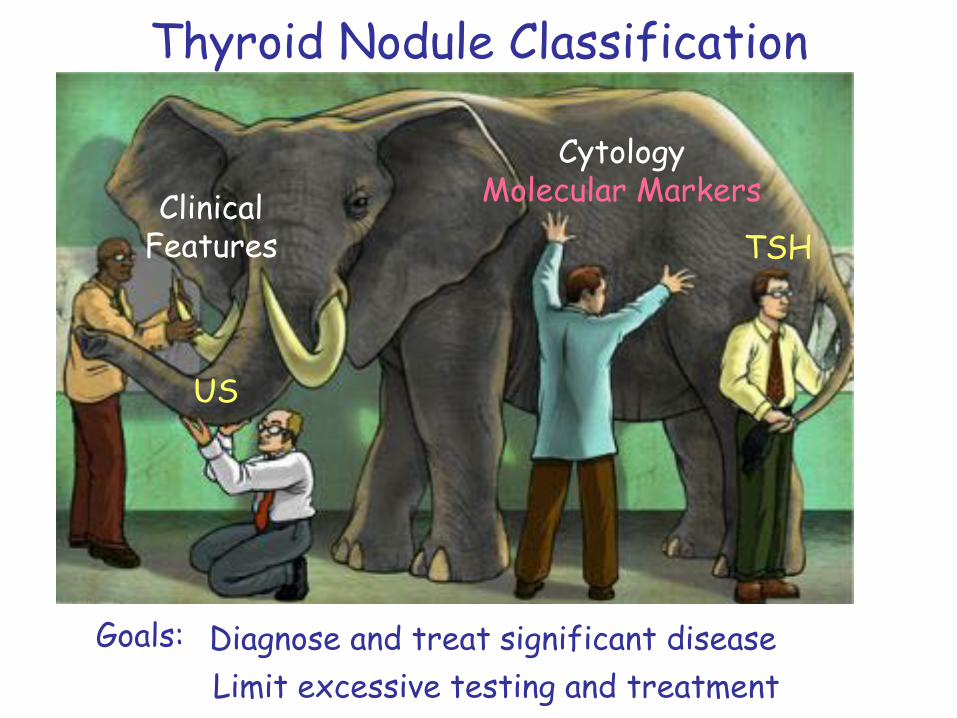
• Measure TSH (A)• US (neck) in all patients with suspected nodule (A)• Consider surgery for nondiagnostic solid nodules (B)• Molecular markers may be considered (C)• Benign nodule.......FU US 6-18 months (C)• Routine LT4 not recommended for benign nodules (F)• Children same approach as adults (A)• Pregnancy – biopsy nodules (A), defer surgery for PTC
unless growing (C), consider LT4 (C)
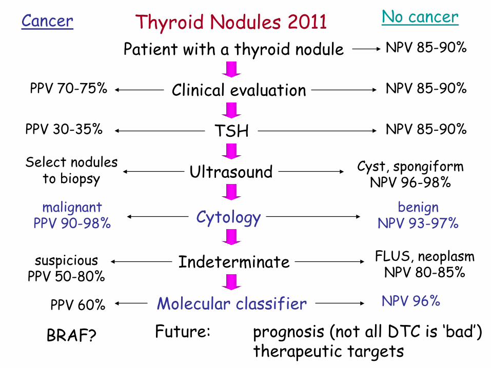
Patient with a thyroid nodule
No cancerCancerNPV 85-90%
Clinical evaluation NPV 85-90%PPV 70-75%
TSH NPV 85-90%PPV 30-35%
Ultrasound Cyst, spongiformNPV 96-98%
Select nodulesto biopsy
Cytology benignNPV 93-97%
malignantPPV 90-98%
Indeterminate FLUS, neoplasmNPV 80-85%
suspiciousPPV 50-80%
Molecular classifier NPV 96%PPV 60%
Future: prognosis (not all DTC is ‘bad’)therapeutic targets