Thyroid hormone concentrations associated withage, sex, reproductive status and apparentreproductive failure in the Amazon river dolphin(Inia geoffrensis)T. R. Robeck1,*, R. S. Amaral2, V. M. F. da Silva3, A. R. Martin4, G. A. Montano1 and J. L. Brown5
1Species Preservation Laboratory, SeaWorld Parks and Entertainment, 2595 Ingraham Rd, San Diego, CA 92109, USA2Federal Institute of Education, Science and Technology of Amazonas—IFAM/CMZL, Av. Cosme Ferreira 8045, Manaus 69086-475, Brazil3Laboratory of Aquatic Mammals, National Institute of Amazonian Research—INPA, Av Andre Araujo 2936, Manaus 69067-375, Brazil4Centre for Remote Environments, University of Dundee, Nethergate, Dundee DD1 4HN, UK5Center for Species Survival, Smithsonian Conservation Biology Institute, 1500 Remount Road, Front Royal, VA 22630, USA
*Corresponding author: Corporate Zoological Department, SeaWorld Parks and Entertainment, 7007 SeaWorld Drive, Orlando 32821, USA.Email: [email protected]
Thyroid hormones play important roles in metabolic and reproductive function; however, they have not been described indetail for any river dolphin species. We characterize these hormones across different age, sex and pregnancy status in theAmazon river dolphin in order to provide baseline normal values for this increasingly threatened species.
This study was conducted to characterize immunoreactive thyroid hormone concentrations in wild Amazon river dolphins,also called boto (Inia geoffrensis) by age group, sex, pregnancy and lactation status, and to determine if thyroid hormoneconcentration differences could be detected between pregnant females with and without successful parturition outcomes.Radioimmunoassays were used to analyse total T3 and total T4 in 182 serum samples collected from 172 botos living in theMamirauá Sustainable Development Reserve, in the Brazilian Amazon from 2003 through 2015. Age significantly affected tT3
and tT4 concentrations in males, with values in immature males and females being significantly lower than those in adult males,whereas no age effects were noted between immature females and adult non-pregnant, non-lactating females. Significantsex differences were noted in tT3 concentrations between immature males and females and in tT4 concentrations betweenadult males and females. These resulted in significant differences in the tT3:tT4 ratio between males and females withinthe immature and adult groups. Lactating and non-pregnant adult females had significantly higher tT3 concentrations thanpregnant females, and this difference was primarily driven by a 12% drop in tT3 concentrations during the last two-thirds ofpregnancy. No differences in thyroid hormone concentrations were detected between females diagnosed as pregnant andlater found to have or not have a live calf. These results are the first to define thyroid hormone reference intervals and normalphysiological variations in a wild population of river dolphins.
Received 29 January 2019; Revised 6 May 2019; Editorial Decision 29 May 2019; Accepted 11 June 2019
Cite as: Robeck TR, Amaral RS, da Silva VMF, Martin AR, Montano GA, Brown JL (2019) Thyroid hormone concentrations associated withage, sex, reproductive status and apparent reproductive failure in the Amazon river dolphin (Inia geoffrensis) . Conserv Physiol 7(1): coz041;doi:10.1093/conphys/coz041.
IntroductionGlobal freshwater habitats are often adjacent to or withindense and expanding human populations and are increasinglybeing degraded due to anthropogenic activity. As a result,freshwater megafauna species, including all species of riverdolphins, are under direct pressures, and if they continueunchecked, it may result in their extinction (He et al., 2017;Huang et al., 2012; Williams et al., 2016). Currently, of anestimated 10 species of fresh water dolphins, six are listed asendangered, critically endangered or functionally extinct, twoare vulnerable and two populations are considered “statusunknown” (Braulik and Smith, 2017; He et al., 2017; Reeveset al., 2011). While it is clear that freshwater dolphins are atextreme risk, what is less understood is how anthropogenicpressures are affecting their survival. In addition, as apexpredators, these species often directly compete with manfor food, and many of the environmental factors that maybe detrimental towards their overall health and well-beingcould also affect human populations. Thus, these species,and cetaceans in general, may represent one of the bestenvironmental sentinels for human populations living nearby(Bossart, 2006; Chen et al., 2017; Lailson-Brito et al., 2008).
Despite the importance of river dolphins for habitat health(Turvey et al., 2012), and as markers of adverse environmen-tal threats, little is known about their physiology, or howvarious species may be affected by deteriorating conditions.The Amazon river dolphin, or boto (Inia geoffrensis), existsin habitats that are currently relatively free from industrialdevelopment and high population density (da Silva et al.,2018). Despite this, recent evidence suggests this speciesis in decline due to anthropogenic pressures created fromdirect harvesting of the animals for use as bait to catchcommercially valuable catfish, the piracatinga (Calophysusmacropterus), by native fisherman (Mintzer et al., 2013; daSilva et al., 2018). In addition, multiple proposals exist forhydroelectric projects throughout the Amazon river basin thatmay threaten the population through habitat fragmentation(Finer and Jenkins, 2012). These current and future pressuresmay threaten boto sustainability, and therefore, there maybe a limited time period during which physiologic healthmarkers and physiological reference ranges can be definedand used as “normal” controls for future population healthassessments.
Several health assessments of wild cetacean populationshave been published over the last 20 years; the value of whichhas been demonstrated by recent research on wild bottlenosedolphin populations adversely impacted by a regional oilspill (Schwanke et al., 2014). Data from both captive andwild healthy populations have enabled researchers to developmodels identifying the adverse short- and long-term impactsfrom this oil spill (Hall et al., 2018; Lane et al., 2015; Smithet al., 2017). Recently, health assessments of wild cetaceanshave been applied to an increasing number of marine andfreshwater cetacean species (Flower et al., 2015; Nabi et al.,
2018; Smith et al., 2017; Van Bressem et al., 2009). Blood andother biological samples collected from animals during theseassessments provided the opportunity to examine multipleorgan and endocrine systems. For cetaceans, overfishing andclimate change have elevated the importance of monitoringthyroid function via thyroid hormones to detect critical home-ostatic changes in response to these external stressors (Fairet al., 2011; Flower et al., 2015; St. Aubin et al., 1996; Wasseret al., 2017). However, before detecting abnormal function,normal concentrations during varying life history events mustbe defined.
Thyroid hormones have been documented to vary by ageand different physiological conditions, e.g. pregnancy andlactation, in multiple mammalian species, including cetaceans(Fair et al., 2011; Flower et al., 2015; St. Aubin, 2001;St. Aubin et al., 1996; West et al., 2014). Comparativelyhigh hormone concentrations and proportionally large thy-roid glands of marine cetaceans (Fair et al., 2011; Ridgwayand Patton, 1971) have led researchers to speculate thatcold environmental temperatures combined with high heatconductivity of water habitats requires an increased metabolicresponse to maintain core body temperature, which is drivenby enhanced thyroid activity. In addition, changes in thyroidhormone concentrations that parallel seasonal environmentchanges and prey availability have been described for belugas(Delphinapterus leucas) and killer whales (Orcinus orca;Flower et al., 2015; Wasser et al., 2017).
Multiple thyroid pathologies have been identified inmarine cetaceans, including colloid depletion, fibrosis,thyroiditis and squamous cysts (Cowan and Tajima, 2006;Harrison, 1969; Schumacher et al., 1993). Abnormal thyroidgland function during pregnancy either in response to iodinedeficiency or due to primary thyroid pathology has beenknown to be associated with both fetal and placental growthabnormalities that can translate into a number of pathologiesincluding abortion, premature birth, low birth weight andneonatal failure to thrive syndrome (Glinoer, 2007). While noreports of thyroid pathology exist for the boto, or other riverdolphin species, the frequency of its occurrence in marinecetaceans warrants such an investigation.
In addition to direct effects of thyroid pathology, mal-nutrition has been indirectly linked to thyroid dysfunction,and if this deficiency occurs during pregnancy, fetal growthrestriction and other developmental abnormalities may result.Some recent evidence in beef cows demonstrated that changesin the percentage of protein while maintaining normalenergy content can affect fetal thyroid development andprogramming without any changes in circulating maternalthyroid hormone concentrations (Micke et al., 2015). Inhumans, maternal malnutrition has resulted in normal T4,and significantly low T3 concentrations compared withcontrols (Mahajan, et al., 2005). Recent attempts to use fecalthyroid hormone concentrations as an indicator of nutritionalstress and subsequent fetal loss in the killer whale haveprovided some support for this premise (Wasser et al., 2017).
..........................................................................................................................................................Conservation Physiology • Volume 7 2019 Research article
While a direct link between malnutrition, thyroid dysfunctionand fetal or neonatal abnormalities remains elusive, evalua-tion of normal thyroid function in the boto would providebaseline data from which future effects of nutritional stressmay be evaluated.
Currently no significant ex situ populations of this speciesexist whereby thyroid hormone concentrations could bedetermined to establish baseline, and to our knowledge,only one report of thyroid hormone concentrations in wildbotos exists. In that report, which analysed serum fromeight wild animals, mean total thyroxine (tT4) concentrationwas determined without any information on sex, age orreproduction status of the animals that had been sampled(Ridgway et al., 1970). Therefore, the goal of this researchwas to describe both tT4 and total triiodothyronine (tT3)concentrations in wild botos. Specific objectives were to(i) determine if concentrations of thyroid hormones aresignificantly affected by sex, age and female reproductivestatus; (ii) develop thyroid reference ranges for wild botosrelative to sex, maturity and female reproductive status(non-pregnant nonlactating, pregnant and lactating); (iii)determine if thyroid hormone concentrations vary duringtrimesters of gestation; and (iv) determine if thyroid hormoneconcentrations in females diagnosed as pregnant differ
between females who are subsequently observed to have hada live calf or are observed without a calf.
MethodsAnimals and sample collectionsA total of 182 samples was collected from 172 animalswho were temporarily restrained during an annual capture–recapture campaign of Projeto Boto at the Mamirauá Sustain-able Development Reserve, Brazilian Amazon, between 2003and 2015. Once restrained, total length, sex, and reproductivestatus of adult females (non-pregnant, pregnant or lactating)was determined by a combination of visual inspection andtransabdominal ultrasonography (Figure 1). Blood sampleswere collected from the ventral tail fluke using a 19-gaugewinged blood collection set (Figure 1). Whole blood wascollected into BD Vacutainers (Becton Dickenson, FranklinLakes, NJ, USA) containing activated thrombin, allowed toclot for 20 min, and then centrifuged at 1000 g for 15 to30 min. Serum was collected and stored at −20◦C untilanalysis. Evidence indicates that minimal change in thyroidhormone concentrations occurs when stored at −25◦C forup to 23 years (Männistö et al., 2007). For pregnancy deter-
Figure 1: Health assessment on wild Amazon river dolphins (boto, Inia geoffrensis). Images depict blood sampling (a), ultrasound bodyexamination (b), wild boto with calf (c) and freeze branding of animal prior to release (d). Images credited to Projeto Boto/INPA
mination, all examinations were performed using a Sonosite180 Plus ultrasound unit (Pyramid Medical Systems, SãoPaulo, São Paulo, 04181-110, Brazil) in conjunction witha 2- to 5-MHz multi-frequency transducer (Convex Array180 Plus/Elite-C60, Pyramid Medical Systems). Animals weredetermined to be pregnant by visualization of a conceptus(placental membranes, fetus) and uterine fluid (amniotic,chorionic or allantoic fluid). Fetal age was assigned to the first(0 to 112 days), second (113 to 224 days) or third trimester(225 to 333 days) of pregnancy using total length, biparitalor transthoracic measurements, by reference to publishedmeasurements for bottlenose dolphins (Tursiops truncatus;Lacave et al., 2004). Bottlenose dolphin fetal growth rateswere chosen to estimate fetal age in boto due to the similarityin gestation length between the two species (Martin & daSilva, 2018). Based on serum testosterone in males (Amaralet al., manuscript in prep), males were classified as immature(calf or juvenile males < 188 cm total body length) or adult(males ≥188 cm total body length). Females were classifiedbased on shortest size at first pregnancy (Martin & da Silva,2018); therefore, immature females were <180 cm total bodylength and adult females were ≥180 cm total body length.Adult females were further categorized based on physiologicalstate—non-pregnant, pregnant, or lactating.
Thyroid hormone radioimmunoassaysThyroid hormones (total T4 and T3) were measured in serumusing commercial solid phase radioimmunoassay (RIA) kits[Siemens Medical Solutions Diagnostics, Los Angeles, CA:total T4 (tT4) catalog number TKT45; total T3 (tT3) catalognumber TKT35]. All RIAs were conducted in accordancewith the manufacturer’s instructions and were validated foruse in dolphins based on observed parallelism between serialdilutions of pooled serum samples and the respective standardcurves and >90% recovery of respective hormone standardsfrom pooled serum samples. Assay sensitivities were 2.5 ng/mlfor tT4, 0.1 pg/ml for fT4, 0.07 ng/ml for tT3 and 0.02 pg/mlfor fT3. For all assays, intra- and inter-assay coefficientsof variation were <15%. The relative percentage of totalimmunoreactive T3 and T4 were determined by dividing themean value across all groups for each hormone, respectively,by the sum of both mean concentrations (μtT3 + μtT4 = totalimmunoreactive thyroid hormone concentrations).
Statistical analysisData were analysed using Stata® (version 14; StataCorpLP, College Station, TX, USA). For females, to control forrepeated samples from the same animal, and an unequalnumber of sampling among animals, we analysed data using alinear mixed effect restricted maximum likelihood regressionmodel (West et al., 2015) to quantify the relationship betweenthe dependent variable (hormone concentration) and fixedeffect variables, with animal ID as the random interceptvariable with an unstructured covariance. The fixed effectcategorical variables included age group (immature male and
female, adult male and adult non-lactating, non-pregnantfemale), physiological state (pregnant, non-pregnant, non-pregnant lactating) and trimester (i.e. first trimester, secondtrimester, third trimester). Separate analyses were performedfor each fixed effect variables and repeated for each depen-dent variable (i.e. hormone concentrations (tT3, tT4 andT3:T4 ratio).
All final mixed models were checked for normality usingquantile plots of the standard residuals. If quantile–quantile(qnorm) plots of standardized residuals exhibited non-normaldistribution, then data were transformed as indicated by theShapiro–Wilk test (Ladder command, STATA) of raw datauntil model residuals were normalized. Pairwise comparisonsof estimated marginal means were conducted using the mar-gins command with Sidak correction or as paired contrastswithout correction. Unless specified, data are expressed asback transformed marginal means and 95% confidence inter-vals. For all analyses, P < 0.05 was considered significant.
Comparisons of thyroid hormone concentration acrossgestational stages in pregnant females (as determined byultrasonography during gestation) that had live calves versuspregnant females that did not have a live calf were determinedby observations during the next annual field assessment. Sincenot all females were observed each year, only females whichcould be confirmed as having a calf within a 2-year periodafter they were diagnosed as pregnant (this length of time wasused to accommodate for the various stages of gestationwhen they were initially assessed, and this the time intervalto when a calf would have been born) were compared usingunpaired two-tailed t test with unequal variances using theWelch (1947) approximation for degrees of freedom. Dueto the length of time between examinations or observationsof females we could not determine at which point the fetusor calf was lost. Therefore, the calf loss classification wouldinclude females that experienced an abortion, or stillbirth andcalves that died during the perinatal period or that failed tothrive.
ResultsOverall resultsAcross all sex and age groups, mean (±SD, 95% CI) tT3and tT4 were 0.52 ± 0.15 ng/ml (0.49–0.55 ng/ml) and38.6 ± 0.84 ng/ml (37.0–40.3 ng/ml), respectively. Based onmean concentrations, the relative percentage contributiontowards total immunoreactive thyroid concentrations for tT3and tT4 were 1.3% and 98.7%, respectively.
Thyroid hormone reference ranges for thebotoPercentiles (2.5, 25, 50, 75, 97.5) for tT3, tT4 and thetT3:tT4 by age class and for adult female physiological
..........................................................................................................................................................Conservation Physiology • Volume 7 2019 Research article
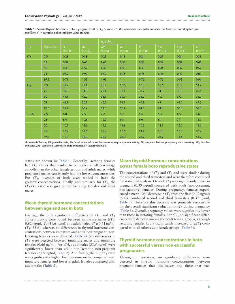
Table 1: Serum thyroid hormone (total T3 ng/ml, total T4, T3:T4 ratio ×1000) reference concentrations for the Amazon river dolphin (Iniageoffrensis) in samples collected from 2003 to 2015
Age class
TH Percentile JF(n = 8)
JM(n = 23)
AM(n = 65)
AF(n = 26)
PF(n = 40)
1st(n = 15)
2nd(n = 21)
LF(n = 14)
tT3 2.5 0.28 0.38 0.25 0.13 0.24 0.27 0.24 0.31
25 0.32 0.43 0.43 0.39 0.33 0.34 0.32 0.45
50 0.46 0.57 0.49 0.50 0.39 0.44 0.37 0.57
75 0.52 0.90 0.59 0.75 0.46 0.46 0.42 0.67
97.5 0.71 1.53 1.05 1.1 0.76 0.76 0.53 0.94
tT4 2.5 31.7 23.1 20.7 16.5 15.8 15.6 20.8 14.7
25 39.3 34.9 29.4 32.1 33.2 31.9 35.8 25.6
50 44.1 42.0 33.1 39.7 38.2 42.7 37.7 36.0
75 46.7 50.9 40.0 51.1 44.5 47 42.6 44.2
97.5 51.2 68.7 51.5 96.7 61.5 61.8 50.3 67.8
T 3:T4 2.5 6.0 7.5 7.2 4.7 5.4 5.4 6.5 4.6
25 8.4 10.6 12.9 9.3 8.0 8.1 7.7 11.7
50 10.2 13.6 15.2 11.9 10.2 11.1 10.0 15.6
75 10.7 17.6 18.2 18.6 14.2 16.8 13.5 26.2
97.5 12.3 32.9 27.7 23.0 24.7 24.7 14.4 44.2
JF: juvenille female, JM: juvenille male, AM: adult male, AF: adult female (nonpregnant, nonlactating), PF: pregnant female (pregnancy with resulting calf ), 1st: firsttrimester, 2nd: combined second and third trimester, LF: lactating female.
status are shown in Table 1. Generally, lactating femaleshad tT3 values that tended to be higher at all percentagecut-offs than the other female groups and adult males, whilepregnant females consistently had the lowest concentrations.For tT4, juveniles of both sexes tended to have thegreatest concentrations. Finally, and similarly for tT3, thetT3:tT4 ratio was greatest for lactating females and adultmales.
Mean thyroid hormone concentrationsbetween age and sex in botoFor age, the only significant differences in tT3 and tT4concentrations were found between immature males (tT3:0.62 ng/ml, tT4: 41.6 ng/ml) and adult males (tT3: 0.51 ng/ml,tT4: 33.6), whereas no differences in thyroid hormone con-centrations between immature and adult non-pregnant, non-lactating females were detected (Table 2). Sex differences intT3 were detected between immature males and immaturefemales (0.66 ng/ml). For tT4, adult males (33.6 ng/ml) weresignificantly lower than adult non-lactating non-pregnantfemales (38.9 ng/ml; Table 2). And finally, the tT3:tT4 ratiowas significantly higher for immature males compared withimmature females and lower in adult females compared withadult males (Table 2).
Mean thyroid hormone concentrationsacross female boto reproductive statesThe concentrations of tT3 and tT4 and were similar duringthe second and third trimesters and were therefore combinedfor statistical analysis. Overall, tT3 was significantly lower inpregnant (0.39 ng/ml) compared with adult (non-pregnant,non-lactating) females. During pregnancy, females experi-enced a mean 12% decrease in tT3 from the first (0.42 ng/ml)to the combined second and third trimesters (0.37 ng/ml,Table 3). Therefore this decrease was primarily responsiblefor the overall significant reduction in tT3 during pregnancy(Table 3). Overall, pregnancy values were significantly lowerthan those in lactating females. For tT4, no significant differ-ences were detected among the adult female groups, althoughlactating females had a significantly increased tT3:tT4 com-pared with all other adult female groups (Table 3).
Thyroid hormone concentrations in botowith successful versus non-successfulpregnanciesThroughout gestation, no significant differences weredetected in thyroid hormone concentrations betweenpregnant females that lost calves and those that suc-
Table 2: Comparisons of marginal mean (95% CI) total T3 (ng/ml), total T4 (ng/ml) and T3:T4 ratio (×1000) between sex within age groups (adultfemale vs adult male), between age groups within the same sex (juvenile vs adult) and in adult females during different physiologic states(pregnant, lactating, or non-lactating non-pregnant adult)
Group tT3 tT4 tT3:tT4
Juvenile male(n = 23)
0.62(0.54–0.71)
41.6(37.7–46.0)
14.5(12.4–16.9)
Juvenile female(n = 8)
0.43(0.34–0.54)
42.6(34.6–52.5)
9.4(7.9–11.1)
Adult male(n = 65)
0.51(0.47–0.55)
33.6(31.7–35.5)
15.2(14.2–16.2)
Adult femalea
(n = 26)0.49(0.43–0.56)
38.9(35.0–43.3)
12.2(10.5–14.2)
Pregnant femaleb
(n = 40)0.39(0.35–0.43)
37.9(34.8–41.4)
10.5(9.4–11.8)
Lactating femalec
(n = 14)0.56(0.47–0.67)
34.7(28.6–42.1)
16.3(12.0–16.9)
Pairwise comparisons of meansd IF < IM; AM < IM; PF < AF and LF AM < IMAM < AF
IF < IM; AF < AMAF & PF < LF
aNon-pregnant, non-lactating adult females.bOnly females with “successful pregnancies” were included in this category. These were females which were diagnosed as pregnant and then were observed to have acalf the following season.cNon-pregnant females.dOnly groups with differences (P < 0.05) were reported.
Table 3: Comparisons of marginal mean (95% CI) total T3 (ng/ml), total T4 (ng/ml) and the T3:T4 ratio (×1000) during the first and combinedsecond and third trimesters of pregnancy with non-pregnant, non-lactating, adult females and non-pregnant, lactating females
Group tT3 tT4 tT3:tT4
Adult female(n = 26)
0.49(0.43–0.56)
38.9(35.0–43.3)
12.2(10.5–14.2)
First trimester(n = 17)
0.42(0.35–0.49)
39.2(34.1–44.9)
11.6(9.5–14.0)
Second and third trimester(n = 23)
0.37(0.32–0.43)
37.3(32.3–42.2)
9.7(8.2–11.5)
Lactating female(n = 14)
0.56(0.47–0.67)
35.0(29.5–41.7)
16.3(13.1–20.4)
Sidak Groups Second and third < AFFirst, second, and third < LF
NSD AF, first, second, and third < LF
Females with confirmed calf loss either during gestation or after delivery were not included in this analysis. NSD: no significant difference.
cessfully gave birth. In addition, no significant differ-ences in thyroid hormone concentrations were foundwhen comparing trimesters between normal and abnormalpregnancies.
DiscussionBoto thyroid hormone reference rangesThis study is the first report of thyroid hormone concentra-tion reference ranges in relation to sex, age and reproductivestatus for any species of river dolphin and provides novelinformation regarding river dolphin physiology, species that
are some of the least understood and most endangered ofthe cetaceans (He et al., 2017). For example, it has beenpostulated that relatively high concentrations of T4 in marinecetaceans (beluga, bottlenose dolphins) compared with non-human terrestrial mammals may be an adaptation to the rel-atively cold climate associated with an aquatic environment(Fair et al., 2011; Flower et al., 2015). The reference range fortT4 in the boto supports this concept, as this cetacean speciesinhabits tropical waters that are considerably warmer thanmost encountered by other marine cetaceans (Sioli, 1984),and their concentrations were approximately half thoseof cold-water cetacean species. Further, tT4 concentrationsappeared to be similar to terrestrial mammals (for review,
..........................................................................................................................................................Conservation Physiology • Volume 7 2019 Research article
see Fair et al., 2011) and the semi tropical marine mammalmanatee (Trichechus manatus, Ortiz et al., 2000).
In addition to adaptive variations, boto reference rangesprovide evidence that thyroid hormone concentrationsdecrease with age in both males and females, similar tobottlenose dolphins (Fair et al., 2011; West et al., 2014)and belugas (Flower et al., 2015). A decrease in thyroidhormones with age has been observed in humans (Kapelariet al., 2008; Lem et al., 2012) and other mammals(Malinowski et al., 1996) and may in part be explained bygrowth hormone, which stimulates peripheral T3 productionin young animals (Lapierre et al., 1990). Defining agespecific changes in thyroid hormone concentrations allowsfor improved sensitivity for the detection of thyroid hormonedeficiencies. When these deficiencies occur in juveniles,they can have profound effects on normal development;and therefore, defining the normal changes with age is animportant component for the development of populationhealth assessment criteria (Dwyer and Stickland, 1992; Lemet al., 2012).
Normal changes or reference ranges within adult groupscan be beneficial for identifying thyroid pathology—thyroiditis, hypo and hyper thyroidism—some of whichcan have profound effects on fetal and neonatal growthand development. While tT4 was relatively consistentamong groups, tT3 and the tT3:tT4 ratio demonstratedlarge variation in concentration ranges, especially betweenpregnant and lactating females. In general, T3:T4 changesare used to reflect the efficiency of thyroid production andpredict clinical disease, such as thyrotoxicosis, euthyroid sicksyndrome, iodine deficiency, follicular cysts, thyroiditis andhyperthyroidism (Mortoglou and Candiloros, 2004). To ourknowledge, most animals in this study were healthy at thetime of sample collection, and combined with a fairly largesample size, these results should provide a robust referenceinterval that will be important for continued monitoring ofindividual animals and potentially provide a useful indicatorfor assessing potential changes in overall population health.
For the boto, the major thyroid hormone in serum is tT4(98.7%) with only ∼ 1.3% represented as tT3. This propor-tional distribution is nearly identical to that observed for wild(98 to 99%; Fair et al., 2011) and captive (99.3%; Westet al., 2014) bottlenose dolphins. This finding is also in linewith the understanding in other mammalian species that T4functions primarily as a prohormone reserve for intracellularconversion to the biologically active form T3 (for review, seeBrent, 2012).
Boto thyroid hormone concentrationcomparisons by age and sexThere were significant age differences in thyroid hormonelevels, but these were not always consistent. For example,T3 and T4 were significantly increased in immature males
compared with mature males but not between immatureand adult females (non-pregnant, non-lactating females). Forsex differences, significant differences in thyroid hormoneconcentrations were observed between immature male andfemale (T3, T3:T4) and adult male and female (T4, T3:T4)boto. These age and sex results differ from a study in wild bot-tlenose dolphins where significant differences in T4 betweensex and age groups were largely due to juvenile males (Fairet al., 2011). Similarly, immature females within both studieshad the lowest T3:T4 ratio. In addition, our results arecomparable with an earlier study in wild bottlenose dolphinwhereby adult females had increased tT4 compared withadult males. However, unlike our findings, they also observedan increase in T3 in adult females compared with adultmales. This observed difference may have been caused by theabsence of controlling for lactating females in their compar-isons with adult males, potentially increasing the overall meanT3 concentration in the female group (St. Aubin et al., 1996).Research with belugas demonstrated a significant decreasewith age in both tT3 and tT4 and significantly higher meanconcentrations for male versus females; however, they didnot directly compare sex differences between age classes. Forthe boto and beluga, the increase in T3 production observedin immature males versus immature females may reflect anincreased or prolonged growth rate for dimorphically largermales whom require longer periods of time to reach sex-ual maturity (Robeck et al., 2005; Martin and da Silva,2006), and therefore, a prolonged growth hormone mediatedincrease in thyroid hormone production.
Effect of reproductive state on thyroidhormone in adult female botoWe observed a significant 20% decrease in mean T3 concen-tration with no change in T4 during pregnancy as comparedwith non-pregnant non-lactating females. While our resultswere similar to a small number (n = 5) of captive beluga(Flower et al., 2015) and to a recent report in mares whencontrolling for season (Fazio et al., 2016a), they contrastwith reported results from captive bottlenose dolphins andmany other mammalian species whereby thyroid hormoneconcentrations are significantly increased during pregnancy(Todini, 2007; West et al., 2014). However, even within bot-tlenose dolphins, conflicting evidence exists, although resultsin wild animals were similar to ours in that tT3 and tT4were reduced in pregnant females when compared to non-pregnant females (Fair et al., 2011). For the captive bottlenosedolphin (West et al., 2014), the significant increase detectedin thyroid hormone during pregnancy compared with non-pregnant animals was primarily due to differences detectedin the first trimester, whereby all thyroid hormones measuredwere increased. One possible explanation for the conflictingresults observed between captive versus wild bottlenose dol-phins and boto is that captive animals had multiple serialsamples collected throughout pregnancy, while only singlepoint samples were “randomly” collected from wild animals.
Therefore, samples may have been clustered towards the endof the first trimester of gestation, a period when thyroid hor-mones are already decreasing towards the second trimester.Therefore, inaccuracy in trimester determination and/or clus-tering of samples in the late first trimester could theoreticallyresult in missing peak concentrations of thyroid hormonesobserved during early pregnancy. For example, when weeklythyroid hormone concentrations were determined in horses,differences between pregnant versus non-pregnant controlswere detected for tT3; however, these were primarily due todiscrete significant increases during Weeks 7 and 12 post-ovulation (Fazio et al., 2016b). If this pattern of secretionholds true for cetaceans, single point sampling from animals,which is typical for wild animal population studies, is morelikely to results in misinterpretation of the thyroid hormonesecretion pattern compared with repeated weekly measure-ments throughout gestation.
Clearly more work is required to understand normalcetacean thyroid function during pregnancy versus non-pregnancy, but what appears consistent between the botoand other cetacean and artiodactyl species (Todini, 2007;Todini et al., 2007) is that thyroid hormone concentrationsdecrease from levels during early gestation to significantlylower concentrations during mid to late pregnancy. Themechanism for this change is believed to be due to acombination of factors that include increased metabolic need,an increase in production of thyroid binding globulin anddirect pituitary stimulation from chorionic gonadotropins(Kennedy et al., 2010). However, while these mechanismshave been advanced as explanations for increases observedduring human pregnancy, they do not appear to apply directlyto cetaceans. For example, the increase in thyroid bindingprotein is believed to be stimulated by early increase inestrogens, an event that does not occur in at least twoodontocete species, bottlenose dolphins (Steinman et al.,2016) and killer whales (Robeck et al., 2017). In addition,no chorionic gonadotropin activity similar to that whichis observed in humans (hCG) and horses (eCG) has beenidentified in cetaceans (Bergfelt et al., 2012). Finally, theprobable inappropriateness of comparing maternal thyroidhormone concentrations during pregnancy between specieswith different placentation should not be overlooked. Forexample, evidence suggests that little if any thyroxine crossesthe placenta in mammals with epitheliochorial placentation(ruminants, cetaceans, etc.) after complete developmentof the maternal-placental barrier (mid to late gestation,Comline et al., 1970; Hopkins and Thorburn, 1971),while significant transfer appears to occur for species withhemochorial placentation (primates). Therefore, species withactive or passive transfer of thyroid hormones or theirtrophic precursors across the maternal-fetal barrier willhave concentrations that reflect a combined homeostaticequilibrium of maternal and fetal requirements during thevarious stages and metabolic requirements of gestation,while in the former group, one would expect maternalconcentrations to reflect metabolic demands for the dam,
largely independent of fetal requirements. As the fetal thyroiddevelops, competition for circulating iodine, which readilydiffuses across all types of placentas, may be the significantfactor that results in decreased maternal circulating thyroidhormone hormones (Yarrington and Pearce, 2011) and maybea partial explanation for the reduced thyroid hormone duringlate-term pregnancy in boto. Measurement of maternal iodineconcentrations during pregnancy may provide evidence tosupport or refute this hypothesis.
The reduction in T3:T4 during late pregnancy providessome evidence to support the concept that iodine competitionacross the placenta, along with increased renal clearance ofiodine may be, in part, the cause of this decreased ratio (Fisher,1996). Because the T3:T4 ratio is believed to represent anindirect measurement of peripheral T4 deiodination to activeT3 concentration (Todini et al., 2007), a reduced ratio duringpregnancy in the boto may reflect a decrease in maternaltT4 which is limited by iodine availability combined with anincreased need for tT3. Accordingly, this phenomenon wasnot observed in captive or wild bottlenose dolphins (Fairet al., 2011; West et al., 2014), whereby their diet whichconsist of marine fish typically have an overabundance ofiodine (Ridgway and Patton, 1971), whereas the fresh waterfish food source on which the boto rely for are a magnitudelower in iodine content (Eckhoff and Maage, 1997; Fordyce,2003). It has been noted that wild pregnant and captive bot-tlenose dolphins had increased thyroid gland mass comparedwith non-pregnant females (Cowan and Tajima, 2006; Kotet al., 2012), and the goiterogenic effects of moderate iodinerestriction during pregnancy in humans are well documented(Glinoer, 2004). In addition to possible iodine deficiency, thechange in T3:T4 could simply indicate the inability of thethyroid to keep up with high metabolic demands of late preg-nancy. Without further evidence, especially maternal iodineand TSH concentrations, and normal values for comparisons,the cause remains speculative.
Similar to lactating wild bottlenose dolphins (Fair et al.,2011), we found a significant increase in tT3, and a non-significant decrease in tT4 (9%) compared with adult non-pregnant non-lactating females, and a corresponding signifi-cant increase in tT3:tT4 during lactation in the boto. Theseresults are also compatible with an observed increase inthyroid size in wild lactating bottlenose dolphins (Cowanand Tajima, 2006) and an observed increase in thyroid vol-ume during lactation in captive Indo-Pacific bottlenose dol-phins (Tursiops aduncus, Kot et al., 2012). Whether theobserved thyroid hypertrophy was a residual effect from theknown goitrogenic effects of pregnancy, as has been observedin humans (Glinoer, 2004), or a true response to increasedemand during lactation for thyroid hormone is unknown.
Thyroid hormones appear to have both species-specificand time-specific alterations during lactation (Polat et al.,2014; Micke et al., 2015; Fazio et al., 2016a). Our resultsmost resemble those of women during the post-partumperiod when a transient decrease in post-partum tT4 occurs
..........................................................................................................................................................Conservation Physiology • Volume 7 2019 Research article
in conjunction with an increased tT3 believed to be causedby the energetic demands of lactation (Weeke et al., 1982).The increase in T3:T4 also supports the concept that tT4is being metabolized to tT3 at a faster rate than canbe compensated for by thyrotrophic hormones or ratelimiting effect of iodine availability. In support, a significantincrease in the T3:T4 ratio paired with increased TSHconcentrations is considered diagnostic for hypothyroidismin humans (Mortoglou and Candiloros, 2004). Withoutmeasures of TSH concentration in conjunction with thyroidhormone concentrations, it will be impossible to classifythese significant changes as hypo- or hyper-thyroidism, butthese changes illustrate and confirm the shifting demand onmaternal thyroid function during these various reproductivestates in the boto.
Thyroid hormone concentrations in botowith successful versus non-successfulpregnanciesPregnant boto females, as diagnosed by ultrasonography,that were not observed with a calf the following year wereassumed to have lost the fetus from an abortion, perinatal lossor had a calf that failed to thrive and died. We did not detectany differences in thyroid hormone concentrations duringpregnancy between females that lost calves verse those withconfirmed normal calves. These results contrast with thoseof captive bottlenose dolphins where a significant decrease inthyroid hormone concentrations during the second and thirrdtrimesters in females that had perinatal loss was observed(West et al., 2014). Our results may simply indicate thatthyroid function was not a factor in the loss of the calvesfrom these females or that the low numbers of females sam-pled would not allow enough sensitivity to detect significantchanges. Given the potentially diverse array of etiologies forperinatal calf loss in wild populations, much larger samplesizes likely are needed to determine exact cause of such losses.
ConclusionsSimilar to many other wildlife species (Kersey and Dehnhard,2014), little is known about thyroid function in any riverdolphin species including the boto, and this study representsthe first significant report. As the enviable loss or change inprey availability continues, and exposure to endocrine dis-rupting contaminants increases, both factors which have beenimplicated as affecting thyroid function (Fair et al., 2011; Hallet al., 2018), the data presented herein will provide a criticalbase of information from which the effects of these changeson the boto can be monitored. In addition, this informationprovides for comparisons with other species of river dolphinsthat are currently experiencing many of these anthropogenicthreats to such a degree they are currently threatened withextinction (He et al., 2017).
AcknowledgementsAll procedures described herein were reviewed and approvedby the National Institute of Amazonian Research (INPA) Ani-mal Care and Use Committee, and this project was approvedby the following Brazilian government agencies: InstitutoBrasileiro do Meio Ambiente e dos Recursos Naturais Ren-ováveis (IBAMA); and Sistema de Autorizacão em Biodiver-sidade (SISBio; permit numbers 2344/9611-AC, 3552/9312-AC, 2002001.002344/96-11, 3462-1 to 13462-5, 13157-1to 13157-4, 50544-1 to 50544-3). Research was conductedunder the NMFS permit 16193 and Brazilian CITES exportpermit number 15BR018682/DF. This study was part ofProjeto Boto, a cooperative agreement between the NationalAmazon Research Institute (INPA/MCTI) and the MamirauáSustainable Development Institute (MSDI-OS/MCTI).We thank Nivia do Carmo of LMA/INPA for help with samplecollection and K. Steinman of SeaWorld and Busch GardensSpecies Preservation Laboratory for help with sample pro-cessing. This is a SeaWorld Technical manuscript contributionnumber 2018-17.
FundingPartial funding of this study was provided by AssociaçãoAmigos do Peixeboi (AMPA)/Petrobras Socioambiental pro-gramme.
ReferencesBergfelt DR, Thompson DL Jr, Brown JL, Presley NA, West KL, Campbell M,
Adams GP (2012) Investigation of an immunoreactive chorionicgonadotropin-like substance in the placenta, serum, and urine ofpregnant bottlenose dolphins (Tursiops truncatus). Aquatic Mammals38: 354–361.
Bossart GD (2006) Marine mammals as sentinel species for oceans andhuman health. Oceanography 19: 134–137.
Braulik GT, Smith BD (2017) Platanista gangetica. The IUCN Red List ofThreatened Species. Version 2017 http://www.iucnredlist.org. (lastaccessed 03 August 2018).
Brent GA (2012) Mechanisms of thyroid hormone action. J Clin Invest 122:2035–3043.
Chen M, Zhuang M, Chou L, Liu J, Shih C, Chen C (2017) Tissue concentra-tions of four Taiwanese toothed cetaceans indicating the silver andcadmium pollution in the western Pacific Ocean. Mar Poll Bull 124:993–1000.
Cowan DF, Tajima Y (2006) The thyroid gland in bottlenose dolphins(Tursiops truncatus) from the Texas coast of the Gulf of Mexico: normalstructure and pathologic changes. J Comp Path 135: 217–225.
Comline RS, Nathanielsz PW, Silver M (1970) Passage of thyroxine acrossthe placenta in the foetal sheep. Proc. 51st Meeting, the EndocrineSociety, pp 126.
da Silva VMF, Freitas CEC, Dias RL, Martin AR (2018) Bothcetaceans in the Brazilian Amazon show sustained, profoundpopulation declines over two decades. PLoS ONE 13, e0191304.doi.org/10.1371/journal.pone.0191304
Dwyer CM, Stickland NC (1992) The effects of maternal undernutritionon maternal and fetal serum insulin-like growth factors, thyroidhormones and cortisol in the guinea pig. J Dev Physiol 18:303–313.
Eckhoff KM, Maage A (1997) Iodone content in fish and other foodproducts from east africa analyzed by ICP-MS. J Food Comp Anal 10:270–282.
Fair PA, Montie E, Bathis L, Reif JS, Bossart GD (2011) Influences of biolog-ical variables and geographic location on circulating concentrationsof thyroid hormones in wild bottlenose dolphins (Tursiops truncatus).Gen Comp Endo 174: 184–194.
Fazio E, Medica P, Cravana C, Bruschetta G, Ferlazzo A (2016a)Seasonal thyroid and lipid profiles in thoroughbred pregnantand nonpregnant mares (Equus caballus). Theriogenology 85:1582–1589.
Fazio E, Medica P, Trifiletti C, Ferlazzo A (2016b) The outcome of thefirst stages of pregnancy on mares bloodstream thyroid hormones.Theriogenology 86: 036–1041.
Finer M, Jenkins CN (2012) Proliferation of hydroelectric dams in theAndean Amazonand implications for Andes–Amazon connectivity.PLoS ONE 7, e35126. doi:10.1371/journal.pone.0035126
Fisher DA (1996) Physiological variation in thyroid hormones: phys-iological and pathophysiological considerations. Clin Chem 42:135–139.
Flower JE et al. (2015) Circulating concentrations of thyroid hormonein beluga whales (Delphinapterus leucas): influence of age, sex andseason. J Zoo Wild Med 46: 456–467.
Fordyce FM (2003) Database of iodine content of food and diets popu-lated with data from published literature. Brit. Geol. Surv. Comm. Rep.CR/03/84N. pp 50.
Glinoer D (2004) The regulation of thyroid function during normal preg-nancy: importance of the iodine nutritional status. Best Pract Res ClinEndo Metab 18: 133–152.
Glinoer D (2007) Clinical and biological consequences of iodine defi-ciency during pregnancy. Endo Dev 10: 62–85.
Hall AJ, McConnell BJ, Schwacke LH, Ylitalo GM, Willams R, Rowles TK(2018) Predicting the effects of polychlorinated biphenyls oncetacean populations through impacts on immunity and calf sur-vival. Environ Poll 233: 407–418.
Harrison RJ (1969) Endocrine organs: hypophysis, thyroid and adrenal.In HT Andersen, ed, The Biology of Marine Mammals. Academic Press,NewYork, pp. 365–372.
He F, Zarfl C, Bremerich V, Henshaw A, Darwall W, Tockner K, Jänig SC(2017) Disappearing giants: a review of threats to freshwatermegafauna. WIREs Water 2017, 4:e1208. doi:10.1002/wat2.1208.
Huang SL, Hao YJ, Mei ZG, Turvey ST, Wang D (2012) Common pattern ofpopulation decline for freshwater cetacean species in deterioratinghabitats. Freshw Biol 57: 1266–1276.
Hopkins PS, Thorburn GD (1971) Placental permeability to maternalthryoxine in the sheep. J Endocrinol 49: 549–550.
Kapelari K, Kirchlechner C, Högler W, Schweitzer K, Virgolini I, Moncayo R(2008) Pediatric reference intervals for thyroid hormone levels frombirth to adulthood: a retrospective study. BMC Endocr Disord. 8: 15.
Kennedy RL, Malabu UH, Jarrod G, Nigam P, Kannan K, Rane A (2010)Thyroid function and pregnancy: before, during and beyond. JObstetrics Gynaecol 30: 774–783.
Kersey DC, Dehnhard M (2014) The use of noninvasive and mini-mally invasive methods in endocrinology for threatened mammalianspecies conservation. Gen Comp endocrinol 203: 296–306.
Kot BCW, Ying MTC, Brook FM, Kinoshita RE, Kane D, Chan WK (2012)Sonographic evaluation of thyroid morphology during differentreproductive events in female Indo-Pacific bottlenose dolphins,Tursiops aduncatus. Mar Mam Sci 28: 733–750.
Lacave G, Eggermont M, Verslycke T, Martin , Brook F, Salbany A,Azevedo , Torres , Roque L, Kinoshita R (2004) Prediction from ultra-sonographic measurements of the expected delivery date in twospecies of bottlenosed dolphin (Tursiops truncatus and Tursiops adun-cus). Vet Rec 154: 228–223.
Lailson-Brito J Jr, Dorneles PR, da Silva VMF, Martin AR, Batos WR,Azevedo-Silva CE, Azevedo AF, Torres JPM, Malm O (2008) Dolphinsas indicators of micropollutant trophic flow in amazon basin. OecolBras 12: 532–541.
Lane SM, Smith CR, Mitchell J, Balmer BC, Barry KP, McDonald T,Mori CS, Rosel PE, Rowles TK, Speakman TR, et al. (2015) Reproductiveoutcome and survival of common bottlenose dolphins sampled inBarataria Bay, Louisiana, USA, following the Deepwater Horizon oilspill. Proc. R. Soc. B. 282, 20151944. doi.org/10.1098/rspb.2015.1944
Lapierre H, Petitclerc D, Pelletier G, Delorme L, Dubreuil P, Morisset J,Gaudreau P, Couture Y, Brazeau P (1990) Dose effect of humangrowth hormone-releasing factor and thyrotropin-releasing factoron hormone concentrations in lactating dairy cows. Dom AnimEndocrinol 7: 485–495.
Lem AJ, de Rijke YB, van Toor H, de Ridder MAJ, Visser TJ,Hokken-Koelega ACS (2012) Serum thyroid hormone levels inhealthy children from birth to adulthood and in short childrenborn small for gestational age. J Clin Endocrinol Metab 97:3170–3178.
Mahajan SD, Aalinked R, Singh S, Shah P, Gupta N, Kochupillai N (2005)Thyroid hormone dysregulation in intrauterine growth retardationassociated with maternal malnutrition and/or anemia. Horm MetabRes 37: 633–640.
Malinowski K, Christensen RA, Hafs HD, Scanes CG (1996) Age andbreed differences in thyroid hormones, insulins-like growth factor(IGF)-I and IGF binding proteins in female horses. J Anim Sci 74:1936–1942.
..........................................................................................................................................................Conservation Physiology • Volume 7 2019 Research article
Männistö T, Surcel H-M, Bloigu A, Ruokonen A, Hartikainen A-L,Järvelin M-R, Pouta A, Vääräsmäki M, Suvanto-Luukkonen E (2007)The effect of freezing, thawing, and short-and long-term storage onserum thyrotropin, thyroid hormones, and thyroid autoantibodies:implications for analyzing samples stored in serum banks. Clin Chem53: 1986–1987.
Martin AR, da Silva VMF (2006) Sexual dimorphism and body scarring inthe boto (Amazon river dolphin) Inia Geoffrensis. Mar Mamm Sci 22:25–33.
Martin AR, da Silva VMF (2018) Reproductive parameters of the Amazonriver dolphin or boto Inia geoffrensis (Cetacea: Iniidae); an evolution-ary outlier bucks no trends. Biol J Linn Soc 123: 666–676.
Micke GC, Sullivan TM, Kennaway DJ, Hernandez-Medrano J (2015)Maternal endocrine adaptation throughout pregnancy to nutri-ent programming of thyroid hormones and development of theirprogeny. Theriogenology 83: 604–615.
Mintzer VJ, Martin AR, da Silva VMF, Barbour AB, Lorenzen K, Frazer TK(2013) Effect of illegal harvest on apparent survival of Amazon riverdolphins (Inia geoffrensis). Biol Conserv 158: 280–286.
Mortoglou A, Candiloros H (2004) The serum triiodthyronine to thyrox-ine (T3/T4) ratio in various thyroid disorders and after levothyroxinereplacement therapy. Hormones 3: 120–126.
Nabi G, Hao Y, Robeck TR, Zheng J, Wang D (2018) Physiological con-sequences of biologic state and habitat dynamics on the criticallyendangered Yangtze finless porpoises (Neophocaena asiaeorientalisssp. Asiaeorientalis) dwelling in the wild and semi-natural environ-ment. Conserv Physiol 6. doi:10.1093/conphys/coy072.
Ortiz RM, MacKenzie DS, Worthy GAJ (2000) Thyroid hormone con-centrations in captive and free-ranging west indian manatees(Trichechus manatus). J Exp Biol 203: 3631–3627.
Polat H, Dellal G, Baritci I, Pehlivan E (2014) Changes of thyroid hormonesin different physiologic periods in white goats. J Anim Plant Sci 24:445–449.
Reeves RR, Jefferson TA, Karczmarski L, Laidre K, O’Corry-Crowe G,Rojas-Bracho L, Secchi ER, Slooten E, Smith BD, Wang JY, Zhou K(2011) Inia geoffrensis. The IUCN Red List of Threatened Species.The IUCN Red List of Threatened Species. Version 2017. http://www.iucnredlist.org. (last accessed 30 May 2018).
Ridgway SH, Simpson JG, Patton GS, Gilmartin BS (1970) Hemato-logic findings in certain small cetaceans. J Amer Vet Med Ass 157:566–575.
Robeck TR, Monfort SL, Calle PP, Dunn JL, Jensen E, Boehm JR, Young S,Clark ST (2005) Reproduction, growth and development in captivebeluga (Delphinapterus leucus). Zoo Biol 24: 29–49.
Robeck TR, Steinman KJ, O’Brien JK (2017) Characterization and lon-gitudinal monitoring of serum androgens and glucocorticoids dur-ing normal pregnancy in the killer whale (Orcinus orca). Gen CompEndocrinol 247: 116–129.
Sioli H (1984) The Amazon and its main affluents: hydrography, morphol-ogy of the river courses, and river types. In H Sioli, ed, The Amazon.Limnology and landscape ecology of a mighty tropical river and itsbasin. First Edition. Springer, Netherlands, pp. 127–165.
St. Aubin DJ (2001) Endocrinology. In L Dierauf, F Gulland, eds, CRChandbook of marine mammal medicine, EdSecond. CRC Press, BocaRaton, Florida, pp. 165–192.
St. Aubin DJ, Ridgway SH, Wells RS (1996) Dolphin thyroid and adrenalhormones: circulating levels in wild and semidomesticated Turiopstruncatus, and influence of sex, age and season. Mar Mamm Sci 12:1–13.
Schwacke LH, Smith CR, Townsend FI, Wells RS, Hart LB, Balmer BC,Collier TK, De Guise S, Fry MM, Guillette LJ Jr, et al. (2014) Health ofcommon bottlenose dolphins (Tursiops truncatus) in Barataria Bay,Louisiana, following the Deepwater Horizon oil spill. Environ Sci Tech-nol 48:93–103.
Schumacher U, Zahler S, Horny H-P, Heidemann G, Skirnisson K,Welsch U (1993) Histological investigations on the thyroid glandsof marine mammals (Phoca vitulina, Phocoena phocoena) andthe possible implications of marine pollution. J Wild Dis 29:103–108.
Smith CR et al. (2017) Slow recovery of Barataria Bay dolphin healthfollowing the Deepwater Horizon oil spill (2013–2014), with evidenceof persistent lung disease and impaired stress response. EndangeredSpecies Res 33: 127–142.
Steinman KJ, Robeck TR, O’Brien JK (2016) Characterization of estrogens,testosterone, and cortisol in normal bottlenose dolphin (Tursiopstruncatus) pregnancy. Gen Comp Endocrinol 226: 102–112.
Todini L (2007) Thyroid hormones in small ruminants: effect ofendongenous, environmental and nutritional factors. Animal 1:997–1008.
Todini L, Malfatti A, Valbonesi A, Trabalza-Marinucci M, Debenedetti A(2007) Plasma total T3 and T4 concentrations in goats at differentphysiologic stages, as affected by energy intake. Small Rumin Res 68:285–290.
Turvey ST, Risley CL, Barrett LA, Hao YJ, Ding W (2012) River dolphins canact as population trend indicators in degraded freshwater systems.PLoS ONE, 7:e37902. doi:10.1371/journal.pone.0037902.
Van Bressem MF, Santos MCO, Oshima JEF (2009) Skin diseases in Guianadolphins (Sotalia guianensis) from the Paranaguá estuary, Brazil: apossible indicator of a compromised marine environment. MarineEnviron Res 67: 63–68.
Wasser SK, Lundin JI, Ayres K, Seely E, Giles D, Balcomb K, Hempelmann J,Parsons K, Booth R (2017) Population growth is limited by nutritionalimpacts on pregnancy success in endangered southern residentkiller whales (Orcinus orca). PLoS One 12: E0179824.
Weeke J, Dybkjaer L, Granlie K, Eskjaer Jensen S, Kjaerulff E, Laurberg P,Magnusson B (1982) A longitudinal study of serum TSH, and total andfree iodothyronines during normal pregnancy. Acta Endocrinologica101: 531–537.
West BT, Welch KB, Galecki AT (2015) Linear mixed models: a practicalguide using statistical software, EdSecond. CRC Press, Boca Raton,pp. 199–248.
Welch BL (1947) The generalization of student’s problem when sev-eral different population variances are involved. Biometrika 34:28–35.
West KL, Ramer J, Brown JL, Sweeney J, Hanahoe EM, Reidarson T,Proudfoot J, Bergfelt DR (2014) Thyroid hormone concentrations
in relation to age, sex, pregnancy, and perinatal loss in bottlenosedolphins (Tursiops truncatus). Gen Comp Endo 197: 73–81.
Williams R, Moore JE, Gomez-Salazar C, Trujillo F, Burt L (2016) Searchingfor trends in river dolphin abundance: designing surveys for loomingthreats, and evidence for opposing trends of two species in theColombian amazon. Biol Conserv 195: 136–145.