Time-resolved x-ray scattering from laser-molten indium antimonide. Nüske, Ralf; v Korff Schmising, C; Jurgilaitis, Andrius; Enquist, Henrik; Allaf Navirian, Hengameh; Sondhauss, Peter; Larsson, Jörgen Published in: Review of Scientific Instruments DOI: 10.1063/1.3290418 2010 Link to publication Citation for published version (APA): Nüske, R., v Korff Schmising, C., Jurgilaitis, A., Enquist, H., Allaf Navirian, H., Sondhauss, P., & Larsson, J. (2010). Time-resolved x-ray scattering from laser-molten indium antimonide. Review of Scientific Instruments, 81(1), [013106]. https://doi.org/10.1063/1.3290418 General rights Copyright and moral rights for the publications made accessible in the public portal are retained by the authors and/or other copyright owners and it is a condition of accessing publications that users recognise and abide by the legal requirements associated with these rights. • Users may download and print one copy of any publication from the public portal for the purpose of private study or research. • You may not further distribute the material or use it for any profit-making activity or commercial gain • You may freely distribute the URL identifying the publication in the public portal Take down policy If you believe that this document breaches copyright please contact us providing details, and we will remove access to the work immediately and investigate your claim.
Transcript
LUND UNIVERSITY
PO Box 117221 00 Lund+46 46-222 00 00
Time-resolved x-ray scattering from laser-molten indium antimonide.
Nüske, Ralf; v Korff Schmising, C; Jurgilaitis, Andrius; Enquist, Henrik; Allaf Navirian,Hengameh; Sondhauss, Peter; Larsson, JörgenPublished in:Review of Scientific Instruments
DOI:10.1063/1.3290418
2010
Link to publication
Citation for published version (APA):Nüske, R., v Korff Schmising, C., Jurgilaitis, A., Enquist, H., Allaf Navirian, H., Sondhauss, P., & Larsson, J.(2010). Time-resolved x-ray scattering from laser-molten indium antimonide. Review of Scientific Instruments,81(1), [013106]. https://doi.org/10.1063/1.3290418
General rightsCopyright and moral rights for the publications made accessible in the public portal are retained by the authorsand/or other copyright owners and it is a condition of accessing publications that users recognise and abide by thelegal requirements associated with these rights.
• Users may download and print one copy of any publication from the public portal for the purpose of private studyor research. • You may not further distribute the material or use it for any profit-making activity or commercial gain • You may freely distribute the URL identifying the publication in the public portalTake down policyIf you believe that this document breaches copyright please contact us providing details, and we will removeaccess to the work immediately and investigate your claim.
Time-resolved x-ray scattering from laser-molten indium antimonideR. Nüske, C. v. Korff Schmising, A. Jurgilaitis, H. Enquist, H. Navirian et al. Citation: Rev. Sci. Instrum. 81, 013106 (2010); doi: 10.1063/1.3290418 View online: http://dx.doi.org/10.1063/1.3290418 View Table of Contents: http://rsi.aip.org/resource/1/RSINAK/v81/i1 Published by the American Institute of Physics. Additional information on Rev. Sci. Instrum.Journal Homepage: http://rsi.aip.org Journal Information: http://rsi.aip.org/about/about_the_journal Top downloads: http://rsi.aip.org/features/most_downloaded Information for Authors: http://rsi.aip.org/authors
Downloaded 29 May 2012 to 130.235.184.47. Redistribution subject to AIP license or copyright; see http://rsi.aip.org/about/rights_and_permissions
Time-resolved x-ray scattering from laser-molten indium antimonideR. Nüske, C. v. Korff Schmising, A. Jurgilaitis, H. Enquist, H. Navirian,P. Sondhauss, and J. LarssonDepartment of Physics, Atomic Physics Division, Lund University, P.O. Box 118, Lund SE-221 00, Sweden
�Received 13 November 2009; accepted 16 December 2009; published online 22 January 2010�
Significant advances have been made in picosecond andsubpicosecond time-resolved x-ray scattering techniques dur-ing the past decade. Experimental efforts have mainly beenfocused on the observation of nonthermal melting1–3 and op-tical and acoustic phonon motion4–8 in crystalline solids,while x-ray studies of transient states of disordered materi-als, e.g., liquids, have remained a greater experimental chal-lenge. In contrast to strong Bragg reflections from crystallinematerial, the liquid state exhibits only weak x-ray scatteringamplitudes due to the short correlation length of the disor-dered structure. The broad features of the liquid structurefactor give insight into nearest-neighbor distances and occu-pation numbers, i.e., directly encode the local structure.Time-resolved liquid x-ray scattering experiments have al-lowed precise measurements of the reaction pathway of mol-ecules in solution,9–12 and have shed light on the dynamicstructural changes of liquid water.13 The emergence of theliquid phase of InSb after laser-driven nonthermal meltingwas recently captured directly with femtosecond resolutionat the SPPS at the SLAC National Accelerator Laboratory.14
This study focused on potential voids and ablated materialwhich occur in the scattering pattern at low momentumtransfer vectors. It was found that the liquid is formed within1 ps. Simultaneously, Bragg peaks, indicative of an orderedlattice, disappeared.
In this letter we demonstrate the possibility of carryingout laser-pump/x-ray probe experiments using a two-dimensional �2D� detector at a synchrotron radiation facilitywith 10 ns bunch spacing and a uniform filling pattern, andreport on laser-molten InSb and its subsequent regrowth witha temporal resolution of 400 ps.
II. SETUP FOR TIME-RESOLVED X-RAY SCATTERING
The time-resolved, liquid scattering experiment was car-ried out at beam line D611 at the MAX-laboratory synchro-tron radiation facility in Lund, Sweden. X rays from a bend-ing magnet of the 1.5 GeV MAX-II storage ring are focused
by a toroidal gold-coated mirror and reduced in aperture by aset of slits. The small x-ray incident angle ��S=0.9�0.05°�leads to an elongated x-ray footprint of 0.1�3.0 mm2. Thex-ray divergence is 3�0.7 m rad2 �horizontal·vertical�, anda multilayer monochromator allows the energy to be set toEx ray=7.5 keV with a bandwidth of �Ex ray /Ex ray=10−2. Asingle x-ray probe pulse at the sample contains about 700photons, and has a duration of approximately 300 ps.
A Ti:Al2O3-based femtosecond laser system, operatingat a repetition rate of 4.25 kHz, with a 790 nm center wave-length, 4.5 W average power and 45 fs pulse duration, wasused for excitation. The optical pump beam was focused bytwo cylindrical lenses to a spot size of 0.3�4.0 mm2 ��fullwidth at half maximum �FWHM�� which, assuming a Gauss-ian beam shape, yielded a fluence of 45 mJ /cm2 incident onthe sample. The laser pulses are synchronized to a particularelectron bunch in the storage ring with a jitter below 10 ps.
A. Detection system
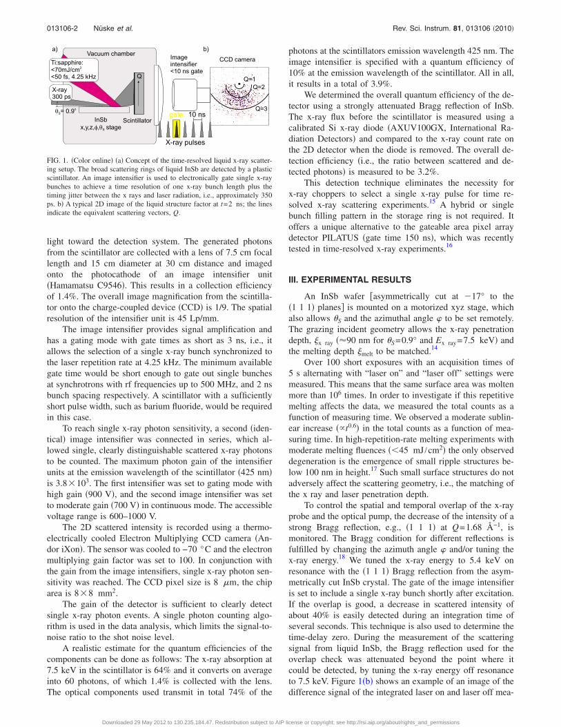
To study the diffuse scattering of the liquid phase ofInSb a single-photon-counting detection system was de-signed and set up as shown in Fig. 1. The scattered x rays areconverted into visible radiation �center wavelength �=425 nm� in a 2-mm-thick plastic scintillator �Saint Gobain,BC-408�. The scintillator features a 2.1 ns decay time �1 /e�,and a 0.9 ns rise time �10%–90%�, which corresponds to atotal 2.5 ns pulse width �FWHM�. The conversion efficiencywas measured and found to be 60 photons per single 7.5 keVx-ray photon. The spatial resolution of the scintillator forx-ray detection is limited due to the deviations from normalincidence for x rays onto the scintillator and its thickness.This effect sets a lower limit for the Q resolution of thedetector. In our case, the spatial resolution is 0.27 mm closeto the beamstop, and 2.8 mm at the outer edge of the scin-tillator. This corresponds to 0.02–0.20 Å−1. The Q reso-lution in our experiment is limited to 0.4 Å−1 at Q=3 Å−1
due to the size of the x-ray spot. A thin aluminum coating��500 nm� on the front of the scintillator blocks any straylight from the pump beam and reflects the generated visible
REVIEW OF SCIENTIFIC INSTRUMENTS 81, 013106 �2010�
light toward the detection system. The generated photonsfrom the scintillator are collected with a lens of 7.5 cm focallength and 15 cm diameter at 30 cm distance and imagedonto the photocathode of an image intensifier unit�Hamamatsu C9546�. This results in a collection efficiencyof 1.4%. The overall image magnification from the scintilla-tor onto the charge-coupled device �CCD� is 1/9. The spatialresolution of the intensifier unit is 45 Lp/mm.
The image intensifier provides signal amplification andhas a gating mode with gate times as short as 3 ns, i.e., itallows the selection of a single x-ray bunch synchronized tothe laser repetition rate at 4.25 kHz. The minimum availablegate time would be short enough to gate out single bunchesat synchrotrons with rf frequencies up to 500 MHz, and 2 nsbunch spacing respectively. A scintillator with a sufficientlyshort pulse width, such as barium fluoride, would be requiredin this case.
To reach single x-ray photon sensitivity, a second �iden-tical� image intensifier was connected in series, which al-lowed single, clearly distinguishable scattered x-ray photonsto be counted. The maximum photon gain of the intensifierunits at the emission wavelength of the scintillator �425 nm�is 3.8�103. The first intensifier was set to gating mode withhigh gain �900 V�, and the second image intensifier was setto moderate gain �700 V� in continuous mode. The accessiblevoltage range is 600–1000 V.
The 2D scattered intensity is recorded using a thermo-electrically cooled Electron Multiplying CCD camera �An-dor iXon�. The sensor was cooled to −70 °C and the electronmultiplying gain factor was set to 100. In conjunction withthe gain from the image intensifiers, single x-ray photon sen-sitivity was reached. The CCD pixel size is 8 �m, the chiparea is 8�8 mm2.
The gain of the detector is sufficient to clearly detectsingle x-ray photon events. A single photon counting algo-rithm is used in the data analysis, which limits the signal-to-noise ratio to the shot noise level.
A realistic estimate for the quantum efficiencies of thecomponents can be done as follows: The x-ray absorption at7.5 keV in the scintillator is 64% and it converts on averageinto 60 photons, of which 1.4% is collected with the lens.The optical components used transmit in total 74% of the
photons at the scintillators emission wavelength 425 nm. Theimage intensifier is specified with a quantum efficiency of10% at the emission wavelength of the scintillator. All in all,it results in a total of 3.9%.
We determined the overall quantum efficiency of the de-tector using a strongly attenuated Bragg reflection of InSb.The x-ray flux before the scintillator is measured using acalibrated Si x-ray diode �AXUV100GX, International Ra-diation Detectors� and compared to the x-ray count rate onthe 2D detector when the diode is removed. The overall de-tection efficiency �i.e., the ratio between scattered and de-tected photons� is measured to be 3.2%.
This detection technique eliminates the necessity forx-ray choppers to select a single x-ray pulse for time re-solved x-ray scattering experiments.15 A hybrid or singlebunch filling pattern in the storage ring is not required. Itoffers a unique alternative to the gateable area pixel arraydetector PILATUS �gate time 150 ns�, which was recentlytested in time-resolved x-ray experiments.16
III. EXPERIMENTAL RESULTS
An InSb wafer �asymmetrically cut at �17° to the�1 1 1� planes� is mounted on a motorized xyz stage, whichalso allows �S and the azimuthal angle to be set remotely.The grazing incident geometry allows the x-ray penetrationdepth, x ray ��90 nm for �S=0.9° and Ex ray=7.5 keV� andthe melting depth melt to be matched.14
Over 100 short exposures with an acquisition times of5 s alternating with “laser on” and “laser off” settings weremeasured. This means that the same surface area was moltenmore than 106 times. In order to investigate if this repetitivemelting affects the data, we measured the total counts as afunction of measuring time. We observed a moderate sublin-ear increase ��t0.6� in the total counts as a function of mea-suring time. In high-repetition-rate melting experiments withmoderate melting fluences ��45 mJ /cm2� the only observeddegeneration is the emergence of small ripple structures be-low 100 nm in height.17 Such small surface structures do notadversely affect the scattering geometry, i.e., the matching ofthe x ray and laser penetration depth.
To control the spatial and temporal overlap of the x-rayprobe and the optical pump, the decrease of the intensity of astrong Bragg reflection, e.g., �1 1 1� at Q=1.68 Å−1, ismonitored. The Bragg condition for different reflections isfulfilled by changing the azimuth angle and/or tuning thex-ray energy.18 We tuned the x-ray energy to 5.4 keV onresonance with the �1 1 1� Bragg reflection from the asym-metrically cut InSb crystal. The gate of the image intensifieris set to include a single x-ray bunch shortly after excitation.If the overlap is good, a decrease in scattered intensity ofabout 40% is easily detected during an integration time ofseveral seconds. This technique is also used to determine thetime-delay zero. During the measurement of the scatteringsignal from liquid InSb, the Bragg reflection used for theoverlap check was attenuated beyond the point where itcould be detected, by tuning the x-ray energy off resonanceto 7.5 keV. Figure 1�b� shows an example of an image of thedifference signal of the integrated laser on and laser off mea-
FIG. 1. �Color online� �a� Concept of the time-resolved liquid x-ray scatter-ing setup. The broad scattering rings of liquid InSb are detected by a plasticscintillator. An image intensifier is used to electronically gate single x-raybunches to achieve a time resolution of one x-ray bunch length plus thetiming jitter between the x rays and laser radiation, i.e., approximately 350ps. b� A typical 2D image of the liquid structure factor at t=2 ns; the linesindicate the equivalent scattering vectors, Q.
013106-2 Nüske et al. Rev. Sci. Instrum. 81, 013106 �2010�
Downloaded 29 May 2012 to 130.235.184.47. Redistribution subject to AIP license or copyright; see http://rsi.aip.org/about/rights_and_permissions
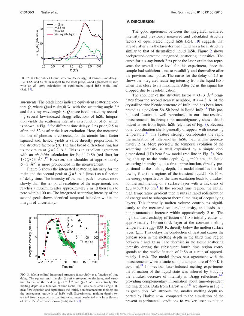
surements. The black lines indicate equivalent scattering vec-tors Q, where Q=4� · sin��� /�, with the scattering angle 2�and the x-ray wavelength �. Q space is calibrated by record-ing several low-indexed Bragg reflections of InSb. Integra-tion yields the scattering intensity as a function of Q, whichis shown in Fig. 2 for different time delays: 2 ns prior, 2.5 nsafter, and 52 ns after the laser excitation. Here, the measurednumber of photons is corrected for the atomic form factorsquared and, hence, yields a value directly proportional tothe structure factor S�Q�. The first broad diffraction ring hasits maximum at Q=2.2 Å−1. This is in excellent agreementwith an ab initio calculation for liquid InSb �red line� for1�Q�3 Å−1.19 However, the shoulder at approximatelyQ=3 Å−1 is more pronounced in the measurement.
Figure 3 shows the integrated scattering intensity for themain and the second peak at Q=3 Å−1 �inset� as a functionof delay time. The intensity of the main peak increases moreslowly than the temporal resolution of the experiment, andreaches a maximum after approximately 2 ns. It then falls tozero within 100 ns. The integrated scattering intensity of thesecond peak shows identical temporal behavior within themargin of uncertainty.
IV. DISCUSSION
The good agreement between the integrated, scatteredintensity and previously measured and calculated structurefactors of equilibrated liquid InSb �Ref. 19� suggests thatalready after 2 ns the laser-formed liquid has a local structuresimilar to that of thermalized liquid InSb. Figure 2 showsbackground-corrected integrated, scattering intensities. Thecurve for a x-ray bunch 2 ns prior the laser excitation repre-sents the overall noise level for this experiment, since thesample had sufficient time to resolidify and thermalize afterthe previous laser pulse. The curve for the delay of 2.5 nsshows the integrated scattering intensity from the liquid InSbwhen it is close to its maximum. After 52 ns the signal hasdropped due to resolidification.
The shoulder of the structure factor at Q=3 Å−1 origi-nates from the second nearest neighbor, at r=4.3 Å, of thecrystalline zinc blende structure of InSb, and has been inter-preted as a covalent Sb–Sb bond in liquid InSb.19 This pro-nounced feature is well reproduced in our time-resolvedmeasurements; its decay time unambiguously shows that itindeed arises from liquid InSb �cf. inset of Fig. 3�. Becauseouter coordination shells generally disappear with increasingtemperature,20 this feature strongly corroborates the rapidthermalization of laser-molten InSb, i.e., within approxi-mately 2 ns. More precisely, the temporal evolution of thescattering intensity is well explained by a simple one-dimensional �1D� heat flow model �red line in Fig. 3�. Not-ing, that up to the probe depth, x ray�90 nm, the liquidscattering intensity is, to a first approximation, directly pro-portional to the melting depth, the model identifies the fol-lowing four time regions of the transient liquid InSb. First,the energy deposited by the laser excitation leads to ultrafast,nonthermal melting of a surface layer with a thickness ofmelt�50�10 nm.1 In the second time region, the initial,high temperature gradient then results in rapid redistributionof energy and to subsequent thermal melting of deeper lyinglayers. This thermally molten volume contributes signifi-cantly to the measured scattered intensity, and leads to anoninstantaneous increase within approximately 2 ns. Thehigh standard enthalpy of fusion of InSb initially causes anapproximately 130-nm-thick layer at the constant meltingtemperature, Tmelt=800 K, directly below the molten surfacelayer, melt. This delays the conduction of heat and causes theplateau seen in the melting depth in the third time regionbetween 3 and 15 ns. The decrease in the liquid scatteringintensity during the subsequent fourth time region corre-sponds to the resolidification of InSb at a rate of approxi-mately 1 m/s. The model shows best agreement with themeasurements when a static sample temperature of 600 K isassumed.21 In previous laser-induced melting experimentsthe formation of the liquid state was inferred by studyingthe ultrafast decrease of intensity in Bragg reflections,1,21
providing complementary information about time-dependentmelting depths. Data from Harbst et al.21 are shown in Fig. 3as green dots. We attribute the smaller melting depth re-ported by Harbst et al. compared to the simulation of thepresent experimental conditions to weaker laser excitation
FIG. 2. �Color online� Liquid structure factor S�Q� at various time delays:�2, +2.5, and 52 ns in respect to the laser pulse. Good agreement is seenwith an ab initio calculation of equilibrated liquid InSb �solid line��Ref. 19�.
FIG. 3. �Color online� Integrated structure factor S�Q� as a function of timedelay. The squares and triangles �inset� correspond to the integrated struc-ture factors of the peak at Q=2.2 Å−1 and Q=3 Å−1, respectively. Themelting depth as a function of time �solid line� was calculated using a 1Dheat flow equation and reproduces the initial, noninstantaneous melting andthe subsequent regrowth of InSb well. Experimental melting depths ex-tracted from a nonthermal melting experiment conducted at a laser fluenceof 36 mJ /cm2 are also shown �dots� �Ref. 21�.
013106-3 Nüske et al. Rev. Sci. Instrum. 81, 013106 �2010�
Downloaded 29 May 2012 to 130.235.184.47. Redistribution subject to AIP license or copyright; see http://rsi.aip.org/about/rights_and_permissions
�36 mJ /cm2 compared to 45 mJ /cm2 in this study�. Thetemporal characteristics reported by Harbst et al. are in ex-cellent agreement with our data.
V. SUMMARY
In conclusion, we have demonstrated that time-resolvedx-ray scattering experiments on laser-molten disordered InSbcan be carried out at a synchrotron radiation facility with auniform bunch fill pattern. Excellent agreement with previ-ously determined liquid structure factors has been shown.The measured rise and decay times of the liquid scatteringamplitude correspond to continued thermal melting and sub-sequent resolidification, respectively.
ACKNOWLEDGMENTS
The authors would like to thank the Swedish ResearchCouncil �VR�, the Knut and Alice Wallenberg Foundation,the Crafoord Foundation, the Carl Trygger Foundation, andthe European Commission via the Marie Curie Programmefor their financial support.
1 A. Rousse, C. Rischel, S. Fourmaux, I. Uschmann, S. Sebban, G. Grillon,P. Balcou, E. Foster, J. P. Geindre, P. Audebert, J. C. Gauthier, and D.Hulin, Nature �London� 410, 65 �2001�.
2 K. Sokolowski-Tinten, C. Blome, C. Dietrich, A. Tarasevitch, M. H. vonHoegen, D. von der Linde, A. Cavalleri, J. Squier, and M. Kammler, Phys.Rev. Lett. 87, 225701 �2001�.
3 A. M. Lindenberg, J. Larsson, K. Sokolowski-Tinten, K. J. Gaffney, C.Blome, O. Synnergren, J. Sheppard, C. Caleman, A. G. MacPhee, D.Weinstein, D. P. Lowney, T. K. Allison, T. Matthews, R. W. Falcone, A. L.Cavalieri, D. M. Fritz, S. H. Lee, P. H. Bucksbaum, D. A. Reis, J. Rudati,P. H. Fuoss, C. C. Kao, D. P. Siddons, R. Pahl, J. Als-Nielsen, S. Dues-terer, R. Ischebeck, H. Schlarb, H. Schulte-Schrepping, T. Tschentscher, J.Schneider, D. von der Linde, O. Hignette, F. Sette, H. N. Chapman, R. W.Lee, T. N. Hansen, S. Techert, J. S. Wark, M. Bergh, G. Huldt, D. van derSpoel, N. Timneanu, J. Hajdu, R. A. Akre, E. Bong, P. Krejcik, J. Arthur,S. Brennan, K. Luening, and J. B. Hastings, Science 308, 392 �2005�.
4 A. M. Lindenberg, I. Kang, S. L. Johnson, T. Missalla, P. A. Heimann, Z.Chang, J. Larsson, P. H. Bucksbaum, H. C. Kapteyn, H. A. Padmore, R.W. Lee, J. S. Wark, and R. W. Falcone, Phys. Rev. Lett. 84, 111 �2000�.
5 K. Sokolowski-Tinten, C. Blome, J. Blums, A. Cavalleri, C. Dietrich, A.Tarasevitch, I. Uschmann, E. Forster, M. Kammler, M. Horn-von Hoegen,and D. von der Linde, Nature �London� 422, 287 �2003�.
6 M. Bargheer, N. Zhavoronkov, Y. Gritsai, J. C. Woo, D. S. Kim, M.Woerner, and T. Elsaesser, Science 306, 1771 �2004�.
7 P. Beaud, S. L. Johnson, A. Streun, R. Abela, D. Abramsohn, D.Grolimund, F. Krasniqi, T. Schmidt, V. Schlott, and G. Ingold, Phys. Rev.Lett. 99, 174801 �2007�.
8 C. von Korff Schmising, M. Bargheer, M. Kiel, N. Zhavoronkov, M.Woerner, T. Elsaesser, I. Vrejoiu, D. Hesse, and M. Alexe, Phys. Rev. Lett.98, 257601 �2007�.
9 A. Plech, M. Wulff, S. Bratos, F. Mirloup, R. Vuilleumier, F. Schotte, andP. A. Anfinrud, Phys. Rev. Lett. 92, 125505 �2004�.
10 H. Ihee, M. Lorenc, T. K. Kim, Q. Y. Kong, M. Cammarata, J. H. Lee, S.Bratos, and M. Wulff, Science 309, 1223 �2005�.
11 J. Vincent, M. Andersson, M. Eklund, A. B. Wohri, M. Odelius, E. Malm-erberg, Q. Kong, M. Wulff, R. Neutze, and J. Davidsson, J. Chem. Phys.130, 154502 �2009�.
12 M. Christensen, K. Haldrup, K. Bechgaard, R. Feidenhans’l, Q. Kong, M.Cammarata, M. L. Russo, M. Wulff, N. Harrit, and M. M. Nielsen, J. Am.Chem. Soc. 131, 502 �2009�.
13 A. M. Lindenberg, Y. Acremann, D. P. Lowney, P. A. Heimann, T. K.Allison, T. Matthews, and R. W. Falcone, J. Chem. Phys. 122, 204507�2005�.
14 A. M. Lindenberg, S. Engemann, K. J. Gaffney, K. Sokolowski-Tinten, J.Larsson, P. B. Hillyard, D. A. Reis, D. M. Fritz, J. Arthur, R. A. Akre, M.J. George, A. Deb, P. H. Bucksbaum, J. Hajdu, D. A. Meyer, M. Nicoul,C. Blome, Th. Tschentscher, A. L. Cavalieri, R. W. Falcone, S. H. Lee, R.Pahl, J. Rudati, P. H. Fuoss, A. J. Nelson, P. Krejcik, D. P. Siddons, P.Lorazo, and J. B. Hastings, Phys. Rev. Lett. 100, 135502 �2008�.
15 M. Cammarata, L. Eybert, F. Ewald, W. Reichenbach, M. Wulff, P. Anin-rud, F. Schlotte, A. Plech, Q. Kong, M. Lorenc, B. Lindenau, J. Räbiger,and S. Polachowski, Rev. Sci. Instrum. 80, 015101 �2009�.
16 T. Ejdrup, H. T. Lemke, K. Haldrup, T. N. Nielsen, D. A. Arms, D. A.Walko, A. Miceli, E. C. Landahl, E. M. Dufresne, and M. M. Nielsen, J.Synchrotron Radiat. 16, 387 �2009�.
17 H. Navirian, H. Enquist, T. N. Hansen, A. Mikkelsen, P. Sondhauss, A.Srivastava, A. A. Zakharov, and J. Larsson, J. Appl. Phys. 103, 103510�2008�.
18 J. Larsson, O. Synnergren, T. N. Hansen, K. Sokolowski-Tinten, S. Werin,C. Caleman, J. Hajdu, J. Sheppard, J. S. Wark, A. M. Lindenberg, K. J.Gaffney, and J. B. Hastings, Second International Conference on Photo-Induced Phase Transitions: Cooperative, Nonlinear and Functional Prop-erties, Journal of Physics Conference Series, Opportunities and Chal-lenges Using Short-Pulse X-Ray Sources Vol. 21, edited by M. Buron andE. Collet �Institute of Physics, Bristol, 2005�.
19 C. Q. Zhang, Y. H. Wei, and C. F. Zhu, Chem. Phys. Lett. 408, 348 �2005�.20 Q. Wang, C. X. Li, Z. H. Wu, L. W. Wang, X. J. Niu, W. S. Yan, Y. N. Xie,
S. Q. Wei, and K. Q. Lu, J. Chem. Phys. 128, 224501 �2008�.21 M. Harbst, T. N. Hansen, C. Caleman, W. K. Fullagar, P. Jonsson, P.
Sondhauss, O. Synnergren, and J. Larsson, Appl. Phys. A: Mater. Sci.Process. 81, 893 �2005�.
013106-4 Nüske et al. Rev. Sci. Instrum. 81, 013106 �2010�
Downloaded 29 May 2012 to 130.235.184.47. Redistribution subject to AIP license or copyright; see http://rsi.aip.org/about/rights_and_permissions