Tintinalli's Emergency Medicine > Section 3: Resuscitative Problems and Techniques > Chapter 19. Tracheal Intubation and Mechanical Ventilation > Tracheal Intubation and Mechanical Ventilation: Introduction Airway integrity, assurance of oxygenation, ventilation, and prevention of aspiration are the mainstays of emergency airway management. The indications for tracheal intubation in the emergency setting most commonly include correction of hypoxia or hypercarbia, prevention of impending hypoventilation, and ensuring maintenance of a patent airway. Secondary indications include provision of a route for resuscitative medication administration and to permit temporizing paralysis during diagnostic testing. Orotracheal Intubation The most reliable way to ensure a patent airway, provide oxygenation and ventilation, and prevent aspiration is endotracheal intubation. Many unconscious and even conscious patients may be unable to spontaneously clear the airway of secretions, may require mechanical ventilation, may have aspirated, or lack protective airway reflexes.1 Preparation The clinical assessment of oxygenation and ventilation may be unreliable in a chaotic emergency department (ED). Continuous, noninvasive bedside monitoring of arterial oxygen saturation is helpful. Isolated oximetry does not assess the status of alveolar ventilation, whereas capnography does allow estimation of the partial pressure of carbon dioxide (PaCO2) based on the waveform display of the end-tidal partial pressure of carbon dioxide. Capnometry refers to the numerical display. In combination, these
Transcript
Tintinalli's Emergency Medicine > Section 3: Resuscitative Problems and Techniques > Chapter 19. Tracheal Intubation and Mechanical Ventilation >
Tracheal Intubation and Mechanical Ventilation: Introduction
Airway integrity, assurance of oxygenation, ventilation, and prevention of aspiration are the mainstays of emergency airway management. The indications for tracheal intubation in the emergency setting most commonly include correction of hypoxia or hypercarbia, prevention of impending hypoventilation, and ensuring maintenance of a patent airway. Secondary indications include provision of a route for resuscitative medication administration and to permit temporizing paralysis during diagnostic testing.
Orotracheal Intubation
The most reliable way to ensure a patent airway, provide oxygenation and ventilation, and prevent aspiration is endotracheal intubation. Many unconscious and even conscious patients may be unable to spontaneously clear the airway of secretions, may require mechanical ventilation, may have aspirated, or lack protective airway reflexes.1
Preparation
The clinical assessment of oxygenation and ventilation may be unreliable in a chaotic emergency department (ED). Continuous, noninvasive bedside monitoring of arterial oxygen saturation is helpful. Isolated oximetry does not assess the status of alveolar ventilation, whereas capnography does allow estimation of the partial pressure of carbon dioxide (PaCO2) based on the waveform display of the end-tidal partial pressure of carbon dioxide. Capnometry refers to the numerical display. In combination, these noninvasive modalities affect decisions regarding tracheal intubation.
Checking the necessary equipment should be standard procedure for ED clinicians at the beginning of their clinical duties. The following items should be available: oral and nasal airways, different-size orotracheal tubes, an O2 setup that is appropriately connected, a self-inflating ventilation bag, different-size masks, and various sizes of Miller and Macintosh blades with the light checked and the suction attached and tested. When intubation is required, the appropriate-size tube and an additional tube (0.5 to 1 mm in diameter smaller) should be selected, and the cuffs should be checked for air leaks with a 10-mL syringe. Selecting a tube of the proper diameter is essential. The approximate sizes for endotracheal tubes are 8.0 to 8.5 mm inner diameter for an adult male and 7.5 to 8.0 mm inner diameter for an adult female. The second hole at the end of the tube above the bevel is called Murphy eye. This hole permits some uninterrupted airflow if the tip is occluded.
Endotracheal tubes (ETTs) with high-volume, low-pressure cuffs are the best design for adults. When properly inflated, thin-walled cuffs prevent aspiration better than medium-
walled cuffs. The operator should test the light on the laryngoscope and then pick an appropriate-size blade. The straight Miller blade is used to physically lift the epiglottis. The curved Macintosh blade is placed in the vallecula above the epiglottis and is used to indirectly lift the epiglottis off the larynx owing to the traction on the frenulum.
The development of expertise with both blade types is desirable, because they offer different advantages. The curved blade may cause less trauma and be less likely to stimulate an airway reflex, because, when used properly, it does not directly touch the larynx. It also allows more room for adequate visualization during tube placement and is helpful in the obese patient. The straight blade is mechanically easier to insert in many patients who do not have large central incisors. Selecting the proper-size blade greatly facilitates intubation. In adults, the curved Macintosh no. 3 is the most popular, and no. 4 is more useful in large patients. The straight Miller no. 2 or 3 is popular for the same purposes.
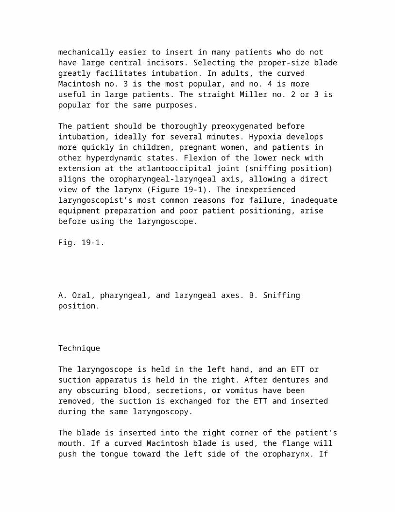
The patient should be thoroughly preoxygenated before intubation, ideally for several minutes. Hypoxia develops more quickly in children, pregnant women, and patients in other hyperdynamic states. Flexion of the lower neck with extension at the atlantooccipital joint (sniffing position) aligns the oropharyngeal-laryngeal axis, allowing a direct view of the larynx (Figure 19-1). The inexperienced laryngoscopist's most common reasons for failure, inadequate equipment preparation and poor patient positioning, arise before using the laryngoscope.
Fig. 19-1.
A. Oral, pharyngeal, and laryngeal axes. B. Sniffing position.
Technique
The laryngoscope is held in the left hand, and an ETT or suction apparatus is held in the right. After dentures and any obscuring blood, secretions, or vomitus have been removed, the suction is exchanged for the ETT and inserted during the same laryngoscopy.
The blade is inserted into the right corner of the patient's mouth. If a curved Macintosh blade is used, the flange will push the tongue toward the left side of the oropharynx. If the blade is inserted directly down the middle, the tongue can force the line of sight posteriorly, which is a common reason for the putative "anterior larynx." After visualization of the arytenoids, the epiglottis is lifted directly with the straight blade or indirectly with the curved blade. The larynx is exposed by pulling the handle in the direction that it points, i.e., 90 degrees to the blade. Cocking the handle back, especially
with the straight blade, risks fracturing central incisors and is ineffective at revealing the cords.
There are a variety of other straight and curved blades available. For example, the Guedel blade is a straight blade with an acute, 72-degree angle to the handle. The Schapira straight blade has a side concavity that helps cradle the large tongue and push it toward the left side of the mouth. The CLM curved laryngoscope blade has a hinged tip, which permits elevation of the epiglottis with minimal force, as the fulcrum is repositioned down within the pharynx.
One technique that avoids the most common error, i.e., overly deep insertion of the blade, is to look for the arytenoid cartilages. If only the posterior commissure is visible, an assistant should apply more pressure on the cricoid (Sellick maneuver) or perform the laryngeal lift. Another option is the "burp" technique. The larynx is manually displaced posteriorly (backward) against the cervical vertebrae, superiorly (upward), and laterally to the right (rightward pressure). To avoid error, the cuff must be seen passing completely through the cords. "Last ditch" attempts at blind passage invite anoxia. The intubator should never be reluctant to abort the attempt if visualization of the larynx is not successful. Whenever feasible, an assistant should apply steady cricoid pressure with the thumb and index finger during the intubation to help prevent aspiration.
With proper technique and practice, semirigid, malleable, blunt-tipped metal, or plastic stylets are not usually necessary for most patients. Nevertheless, a selection of proper-size stylets should be available. The tip of the stylet should not extend beyond the end of the ETT or exit Murphy eye.
One aid to intubation with direct vision is the use of a thin, flexible intubation stylet. This type of stylet can be inserted blindly around the epiglottis into the trachea. The ETT is then threaded over it into the trachea, and the stylet is removed. The Eschmann tracheal tube introducer or stylet, also known as the "gum elastic bougie," is a valuable aid for difficult oral intubations. Another option is to use the tip on the laryngeal tracheal anesthesia kit. With either stylet, orient the tube so that Murphy eye is at the 12-o'clock position.
The tube should never be forced through the vocal cords, which can result in avulsion of the arytenoid cartilages or laceration of the vocal cords. Usually, any difficulty in passing the tube is a result of the tube being too large or too soft and flexible. Directed transoral or translaryngeal anesthesia with lidocaine can help relax the cords. If anesthesia fails, aligning the bevel with the glottic opening may be successful.
The tube should be advanced until the cuff disappears below the cords. Because head motion may move the tip of the tube 1 to 2 cm, correct tube placement is a minimum of about 2 cm above the carina. From the corner of the mouth, this location is approximately 23 cm in men and 21 cm in women. The base of the pilot tube (a tube with the adapter to inflate the cuff) is usually at the level of the teeth. To avoid ischemia of the tracheal mucosa, cuff pressure should be kept below 40 cm H2O. The minimal intracuff pressure
to prevent aspiration is25 cm H2O.2 The operator should secure the tube, being careful not to impede cervical venous return with the umbilical tape or fixator. The use of a modified clove-hitch knot or a commercial fixator is ideal and helps to avoid kinking the pilot tube.
Confirmation of Intubation
Endobronchial or esophageal intubation will result in hypoxia or hypercarbia. There is no clinically reliable substitute for direct visualization of the tube passing through the vocal cords. Hence the adage, "when in doubt, take it out." Nevertheless, there are a number of options to help confirm intratracheal tube positioning. Clinical assessments, including chest and epigastric auscultation, tube condensation, and symmetrical chest wall expansion, are not infallible in the ED. "Breath sounds" from the stomach can be transmitted through the chest after gastric insufflation.
The two basic categories of confirmatory adjuncts are end-tidal CO2 (ETCO2) detectors or monitors and esophageal detection devices. Both have advantages provided that the operator remains cognizant of the sources of interpretation error. Capnometers measure CO2 in the expired air. The most commonly used capnometric devices in the ED are colorimetric, with a pH-sensitive purple filter paper. When in contact with CO2, hydrogen ions are formed, resulting in color changes according to the concentration of CO2. For example, with the Nellcor Easy Cap II, the paper turns yellow after exposure to 2 to 5 percent ETCO2, which is equivalent to 15 to 38 mm Hg PCo2. There is no color change, the filter paper remains purple, with an ETCO2 of less than 0.5 percent, equivalent to less than 4 mm Hg PCo2. Colorimetric capnometers are useful for general readings, as in assessing proper ETT placement, but are not accurate enough when precise determinations are necessary. Capnography displays real-time characteristic CO2 waveforms.
The use of ETCO2 pressure (PETCO2) monitoring can help confirm endotracheal intubation.2 Colorimetric or infrared detection of PETCO2, however, may not occur even with proper ETT placement, during states of low pulmonary perfusion such as cardiac arrest, inadequate chest compressions during cardiopulmonary resuscitation, or massive pulmonary embolism. Another cause of false-negative interpretations is massive obesity. Severe pulmonary edema may obstruct the ETCO2 or PETCO2 monitor with secretions. Alternatively, there may be an initial false-positive detection of CO2 after esophageal intubation if carbonated beverages have been ingested by the patient or for a few minutes after bolus sodium bicarbonate administration. Another cause is gastric distention resulting from bag-valve-mask (BVM) ventilation. A heated humidifier or nebulizer or epinephrine instilled through the ETT also can cause false-positive interpretations.
After intubation and cuff inflation, the capnometer is attached to the ETT. Then a BVM unit is attached to the detector, and the patient is given about six ventilations to wash out residual CO2. The PETCO2 monitor is then checked for color changes. If capnography is available, a persistent positive capnograph formation after clear and direct visualization of tube placement approaches certainty. On rare occasion, misplacement of the
hypopharyngeal glottic tube tip may result in misleadingly normal oximetry and capnography. This error can be recognized by the inadequate depth of tube insertion or inadequate ventilatory volumes or on chest x-ray.
Esophageal detection devices also offer the potential to accurately determine tube location. The various designs depend on their proper function as inline aspirators of the ETT. The device adaptors fit over the 15-mm ETT connector. One advantage of the esophageal detection devices is that accuracy does not depend on adequate cardiac output and pulmonary perfusion. Rather, proper functioning is predicated on the anatomic differences between the esophagus and the trachea. When the ETT is in the esophagus, the soft, non-cartilaginous walls will collapse, and air cannot be aspirated easily.
To perform the syringe aspiration technique, the device should be attached after intubation but before ventilation. The syringe plunger should then be retracted. Resistance to aspiration reflects occlusion from esophageal collapse. If there is no resistance during aspiration, then the tube is assumed to be in the trachea. If a self-inflating bulb is used, the bulb should be compressed and then attached to the ETT. One advantage of the bulb is that it requires one hand.
Complications
The emergency physician should never assume that continued airway patency is assured after ETT insertion.3 Repeated suctioning is necessary to prevent thrombotic or inspissated secretions from obstructing the tube. Endobronchial ball-valve obstruction also can be caused by a clot. The clot can impair ventilation and produce hyperinflation of individual lobes. Cuff displacement or overinflation can result in ball-valve obstruction of the airway. Cuffs inflated in the field during frigid conditions will expand with warming. If tracheal ball-valve obstruction is suspected, the cuff should be deflated. If the tube is blocked, deflation will allow exhalation.
There are many other correctable intubation complications that should be kept in mind. If the ETT cuff leaks after the intubation, the inflation valve should be checked, because it may be defective. One simple remedy is to attach a three-way stopcock to the valve, re-inflate the cuff, and turn off the stopcock. A cuff that seems to be leaking slowly might be sealable. One type of sealant involves instilling an aspirable mixture of normal saline and 2 percent lidocaine jelly, at a 3:1 ratio, into the cuff.
If the ETT needs to be replaced, a tube changer might be considered. There are many commercially available, semirigid catheters that include 15-mm adaptors or connectors to permit ventilation during the tube exchange. These devices have quick-connect adapters that incorporate through-lumen designs to ensure adequate airflow during the procedure.
Although uncommon, morbidity related to emergent endotracheal intubation does occur and may be quite debilitating. Arytenoid cartilage avulsion or displacement, usually on the right, prevents the patient from phonating properly. Intubation of the pyriform sinus
and pharyngeal-esophageal perforation has been reported. Chordal synechiae may develop anteriorly, or commissural stenosis can develop posteriorly.
Subglottic stenosis is the most disastrous sequela. The physician should avoid cuff overinflation and attempt to minimize tube motion in the larynx and trachea. Subglottic stenosis usually occurs in patients with poorly secured tubes who are combative or on ventilators.
Alternative Airway Management Techniques
Nasotracheal Intubation
Nasotracheal intubation (NTI) is an essential psychomotor skill that may be useful in many difficult situations. Operators adept at rapid sequence intubation (RSI) and NTI are in the best position to assess and act on the following prime considerations: What are the potential risks and benefits to having spontaneous respirations preserved rather than ablated? Is there a safe alternative in this patient that may avoid precipitating the need for a potentially unnecessary surgical airway?
Nasal intubation is helpful in situations where laryngoscopy or cricothyrotomy may be difficult and neuromuscular blockade hazardous.4 Severely dyspneic patients with congestive heart failure, chronic obstructive pulmonary disease, or asthma and who are awake often cannot remain supine but can tolerate NTI in the sitting position. It may be impossible to align the oropharyngeal-laryngeal axis in patients with arthritis, masseter spasm, temporomandibular dislocation, or recent oral surgical procedures. Patients with a peculiar body habitus may be difficult to intubate orally. Other considerations for NTI include persistent trismus from seizures, facial trauma, infection, tetanus, or decorticate-decerebrate rigidity. Patients with certain neuromuscular disorders or dystrophies or significant electrolyte abnormalities are not ideal candidates for oral intubation.
To minimize epistaxis, both nares should be sprayed with a topical vasoconstrictor anesthetic. During the brief period for the anesthetic to take effect, a cuffed ETT 0.5 to 1 mm smaller than optimal for oral intubation should be selected. The integrity of the cuff should be verified, and the tube adapter should be checked to ensure a snug fit. Because secretions and blood may be expelled into the air and onto the intubator's face, universal precautions should be observed. An option in addition to a face shield is the use of a protective filtering adapter, such as the Humid-Vent 1, which can be attached to the proximal end of the ETT (Gibeck Respiration, Stockholm, Sweden).
The tube, lubricated with a water-soluble (2 percent lidocaine or K-Y) jelly, is advanced along the nasal floor on the more patent side. Abrasions of the Kiesselbach plexus can be minimized by having the bevel face the septum. Steady, gentle pressure or slow rotation of the tube usually bypasses small obstructions. Passage of the tube is straight back toward the occiput (not upward). If the right side is not passable, the tube should be advanced along the other side before resorting to a smaller tube.
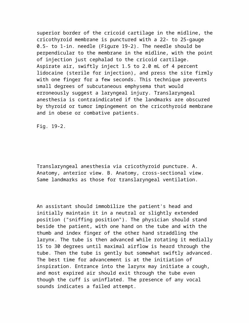
In patients with intact protective airway reflexes, directed transoral or translaryngeal anesthesia often facilitates intubation. Translaryngeal anesthesia, although not widely used in the ED, should be considered when the initial intubation attempt is unsuccessful. After palpating the superior border of the cricoid cartilage in the midline, the cricothyroid membrane is punctured with a 22- to 25-gauge 0.5- to 1-in. needle (Figure 19-2). The needle should be perpendicular to the membrane in the midline, with the point of injection just cephalad to the cricoid cartilage. Aspirate air, swiftly inject 1.5 to 2.0 mL of 4 percent lidocaine (sterile for injection), and press the site firmly with one finger for a few seconds. This technique prevents small degrees of subcutaneous emphysema that would erroneously suggest a laryngeal injury. Translaryngeal anesthesia is contraindicated if the landmarks are obscured by thyroid or tumor impingement on the cricothyroid membrane and in obese or combative patients.
Fig. 19-2.
Translaryngeal anesthesia via cricothyroid puncture. A. Anatomy, anterior view. B. Anatomy, cross-sectional view. Same landmarks as those for translaryngeal ventilation.
An assistant should immobilize the patient's head and initially maintain it in a neutral or slightly extended position ("sniffing position"). The physician should stand beside the patient, with one hand on the tube and with the thumb and index finger of the other hand straddling the larynx. The tube is then advanced while rotating it medially 15 to 30 degrees until maximal airflow is heard through the tube. Then the tube is gently but somewhat swiftly advanced. The best time for advancement is at the initiation of inspiration. Entrance into the larynx may initiate a cough, and most expired air should exit through the tube even though the cuff is uninflated. The presence of any vocal sounds indicates a failed attempt.
The advancement of the tube toward the carina can be observed externally. The normal distance from the external nares to the carina is 32 cm in the adult male and 27 to 28 cm in the adult female. Therefore, before obtaining a chest x-ray, the optimal initial depth of tube placement for NTI in adults, measured at the nares, is 28 cm in men and 26 cm in women. Standard tube confirmation techniques should be performed. Secretions or blood in the tube should be removed before initiating positive-pressure ventilation.
If intubation is unsuccessful, the neck is carefully inspected to determine malposition of the tube. Most commonly, the tube is in the pyriform fossa on the same side as the nostril used. A bulge will be seen and can be palpated laterally. The tube is withdrawn into the retropharynx until breath sounds are heard. The tube is then redirected while the larynx is
manually displaced toward the bulge. If there is no contraindication, flexion and rotation of the neck to the ipsilateral side while the tube is rotated medially often is effective.
The other most common tube misplacement is posteriorly in the esophagus. There are no breath sounds through the tube, and the trachea is slightly elevated. The intubator should attempt redirection after extending the patient's head and performing Sellick maneuver. When cervical spine pathology is suspected, a directional tip-control tube (Endotrol) or a fiberoptic laryngoscope should be considered. Endotrol tubes smaller than 7.5 mm (inner diameter) tend to soften and obstruct and can be difficult to suction. However, the use of these directional-tip tubes often improves the success rate of the first attempt at NTI.
When the tube hangs up on the vocal cords, shrill, turbulent air noises will be heard. The tube can be rotated slightly to realign the bevel with the cords. Alternatively, 2 mL of 4 percent lidocaine (80 mg) can be administered down through the tube onto the cords if transoral or translaryngeal anesthesia was omitted.
Nasal intubation with a fiberoptic laryngoscope may be required when neoplastic lesions, lymphoid tissue, Ludwig angina, peritonsillar abscess, or epiglottitis obstructs the pharynx. The presence of facial trauma does not appear to be a contraindication to NTI.5 Complex nasal and massive midfacial fractures and bleeding disorders are relative contraindications to NTI.
Conversely, oral intubation can impede prompt reduction and stabilization of some maxillary fractures. Because a LeFort I fracture does not extend to the cribriform plate, it is not a contraindication. Fiberoptic guidance or RSI is preferable for LeFort II and III fractures.
The risk of inadvertent intracranial passage of a nasotracheal tube is extremely low, unlike that with nasogastric tube insertion. Very poor technique in the setting of obvious massive head trauma would be required for such an outcome. Severe traumatic nasal or pharyngeal hemorrhage may necessitate orotracheal intubation or cricothyrotomy. Contamination of the spinal fluid is a hazard with some basilar skull fractures.
Serious complications of NTI are rare. In a number of large series, there was no permanent laryngeal damage. Epistaxis will occur with inadequate topical vasoconstriction, excessive tube size, poor technique, or anatomic defects. Excessive force can damage the nasal septum or turbinates.
Frequent suctioning, especially if epistaxis or other upper airway hemorrhage is present, will help to prevent thrombotic occlusion of the tube or a mainstem bronchus. Retropharyngeal lacerations, abscesses, and nasal necrosis have been reported.
Paranasal sinusitis, especially occurring with prolonged NTI or severe cranial trauma, can be an unrecognized source of sepsis. The risk of postintubation sinusitis correlates with the duration of intubation, which often reflects the neurologic insult. In the setting of craniofacial trauma, any subsequent computed tomographic scans should include views
of the paranasal sinuses. Other factors causing sinusitis include the presence of a nasogastric tube, sinus hemorrhage or fracture, and administration of glucocorticoids.
Digital Intubation
Digital intubation is an underused, noninvasive technique for ETT insertion. The performance of this maneuver requires tactile recognition of the epiglottis. Visual landmarks may be impossible to identify with a laryngoscope, because of patient positioning, anatomic disruption, or significant hemorrhage. Tactile digital intubation can avert cricothyrotomy when direct laryngoscopy after neuromuscular blockade has failed. Patients with micrognathia or temporomandibular immobility are poor candidates for this technique.
The patient must be deeply comatose, in cardiac arrest, or in a state of adequate neuromuscular blockade. Before insertion, the well-lubricated ETT should be shaped with a stylet into a J configuration. Then, unless the operator is quite confident, a bite block should be inserted in the opposite side of the mouth. The tongue should be lifted and the mandible pulled forward with the gloved dominant hand. The lubricated middle and index fingers of the gloved nondominant hand are inserted down the middle of the tongue and the cartilaginous epiglottis is palpated with the middle finger.
While the epiglottis is being palpated, the well-lubricated, J-shaped ETT and stylet are inserted and slid along the middle finger. The path from the corner of the mouth opposite the bite block to the epiglottis is the shortest distance. The index finger can help guide the tube from behind. As the larynx is entered, resistance will be encountered. At this point, it is essential to partly withdraw the stylet. Otherwise, the tube will lodge against the anterior wall of the trachea and become difficult to advance.
Transillumination
Transillumination with a lighted stylet can facilitate oral or nasal intubation and help to confirm ETT placement and positioning. This technique is particularly useful when direct laryngoscopy is anatomically impossible. Oral intubation is easiest with a semirigid stylet. Before insertion, the patient's cheek is transilluminated. This serves as a check of the ambient light and will predict the laryngeal light intensity. It may be necessary to dim or shield bright ambient light from the neck. Obese patients who do not transilluminate buccally may not do so laryngeally.
For oral insertion, the lubricated ETT and lighted stylet are inserted after pulling the tongue forward with a 4 x 4 in. gauze pad. The tube initially should be directed into the ipsilateral pyriform fossa to establish the depth of the epiglottis. Then the tube is slightly withdrawn, and the tip is directed toward the midline. The intubator must discriminate between the light emanating from the larynx and the much dimmer light transmitted from the esophagus. Usually the "jack-o-lantern" glow arising from the larynx or trachea is not appreciated when the light source is misplaced in the esophagus. For nasal intubation, the flexible stylet or wand instrument is inserted into a directional-tip ETT (e.g., Endotrol).
After positioning the tube in the retropharynx, very gentle traction is applied to the ring to achieve slight flexion of the tip of the ETT.
Intubating Laryngeal Mask Airway
The laryngeal mask airway (LMA) is a ventilatory device that looks like an ETT with an inflatable silicone mask on the distal end. The LMA is placed blindly, and the mask is inflated over the larynx to provide a supraglottic seal. The technique is quickly mastered, and in most cases the LMA is placed rapidly. LMA's inability to protect against aspiration has limited its role in the ED to a temporizing rescue device.
The intubating LMA (ILMA) addresses this deficiency by providing a conduit to facilitate endotracheal intubation.6 This device also is inserted blindly and uses a rigid handle for insertion, making it an excellent rescue device. Ventilation is achieved in almost all patients, even those with difficult airways or distorted anatomy. Blind intubation through the ILMA with an ETT is over 90 percent successful and improves to almost 100 percent when a lighted stylet or fiberoptic bronchoscope is used to assist.7
The ILMA can be used successfully in cervical spine injuries. Its primary role is as a rescue device, although it has been used as an intubation technique in awake patients with known difficult airways. Aspiration of gastric contents is the most common complication with ILMA and continues to be a risk until successful placement ofthe ETT.
Fiberoptic Assistance
The flexible fiberoptic laryngoscope can be a valuable adjunct when there are anatomic or traumatic limitations that prevent visualization of the vocal cords. Clinical examples include conditions that prevent opening or movement of the mandible, massive tongue swelling (hemorrhage or angioedema), congenital anatomic abnormalities, and cervical spine immobility. These instruments allow visualization of laryngeal structures and can enable difficult intubations, including those around expanding hematomas (Figure 19-3). Patients in need of an immediate airway or those with ongoing hemorrhage or copious secretions are poor candidates.
Fig. 19-3.
A fiberoptic laryngoscope and a Shikani endoscope.
Directed transoral or transnasal and translaryngeal topical anesthesia is essential. The nasal mucosa should be sprayed with a vasoconstrictor. Dual suctioning capability is needed; a suction port should be attached to a suction apparatus for oral blood and
secretions. Tongue extrusion and anterior mandibular displacement are helpful if the oral route is chosen. Fragile equipment is more frequently damaged transorally. The nasal route is also preferred, because the optic tip can enter the glottis at a less acute angle.
For this procedure, the eyepiece is focused, and the flexible shaft is lubricated. The lens at the tip of the laryngoscope is then immersed in warm water to prevent fogging. The intubator should continuously monitor pulse oximetry and ensure that the gag reflex is not present. After attachment of oxygen tubing to the suction port, intermittent insufflation of oxygen at 10 to 15 L per min to keep the optic tip clear should be considered. Insufflation is usually superior to suction for clearing secretions.
The adapter initially is removed from an ETT that is at least 7.0 mm in inner diameter. To prevent barotrauma when high-flow oxygen is insufflated, an ETT of at least 7.5 mm in inner diameter is used. Then the lubricated ETT is slipped over the shaft up to the handle. The distal end of the fiberoptic laryngoscope must extend beyond the end of the ETT. The laryngoscope is held with the left hand, and tip deflection is controlled while advancing it through the cords. The laryngoscope will function as a stylet for the tube. After the laryngoscope is in the trachea, the ETT is advanced and the laryngoscope is removed.
Another option is to insert a nasotracheal tube blindly into the posterior pharynx and stop about 1 to 2 cm proximal to the epiglottis. The scope is then inserted through this hollow conduit, and the fiberoptic tip can be directed into the glottis. The lubricated scope should not pass through Murphy eye. If this occurs, it will be impossible to advance the ETT.
The fiberoptic scope cannot be used as a stylet to guide the ETT into the trachea. The stiffer ETT often will deflect the thin scope tip posteriorly into the esophagus. In addition, the concavity of the ETT is kept anterior toward the 12 o'clock position and places the tube tip and Murphy eye at 3 o'clock (90 degrees to the right). The tip then often hits the right arytenoid cartilage. Rotating the tube 90 degrees counterclockwise aligns the tip with the upper triangular entrance into the trachea.
Indirect Fiberoptic Laryngoscopes
There are many devices that incorporate fiberoptics into a laryngoscope, allowing for indirect visualization of the cords in potentially difficult airways. They are particularly useful when direct visualization of the larynx is impossible due to neck immobility, reduced oral opening, or an anterior larynx. They do not replace the diagnostic capabilities of a flexible fiberoptic scope and have the same visualization restrictions when blood and excessive secretions are present. The Bullard laryngoscope (Circon, ACMI, Stamford, CT), the UpsherScope (Mercury Medical, Clearwater, FL), and the WuScope (Pentax, Fremont, CA) are available. These devices are best used for the anticipated difficult airway as opposed to an emergent rescue device. Only the Bullard model has pediatric sizes.
The Shikani scope (Clarus Medical, Minneapolis, MN) is a device that incorporates the fiberoptics into a malleable stylet.8 Similar to the other devices, the light source is in a portable handle. Setup only requires mounting the ETT. Pediatric sizes are available. The ability to manipulate the angle of the stylet and its brightness allows the use of a blind technique similar to that with the lighted stylet.
Fiberoptic ETTs also are commercially available. Direct line of sight can improve visualization in many difficult intubations. The advantages of direct vision include verification of tube positioning, identification of the right- or left-sided source of pulmonary hemorrhage, and inspection of tracheal injury.
Retrograde Tracheal Intubation
Retrograde tracheal intubation is another option when conventional airway approaches fail. The landmarks are the same as those for cricothyroid puncture (see Figure 19-2). Cervical or mandibular ankylosis and upper airway masses are some of the potential conditions in which retrograde tracheal intubation may help.
The insertion of a retrograde translaryngeal catheter is a less invasive option than cricothyrotomy. This technique can be time consuming and will not be quick enough for apneic patients. The initial angle of the needle should be 30 to 45 degrees cephalad, and a 70- to 75-cm flexible-tip guidewire is advanced through the needle. The wire is then grasped in the oropharynx or nares with Magill forceps. A J wire, which can be slowly twisted once it arrives at the oropharynx, can be easier to locate than a straight guidewire.
The next step is to clasp the guidewire securely with a hemostat at the neck. Then the proximal end of the guidewire is threaded through the Murphy eye on the ETT. This allows more of the ETT to enter the trachea before the guidewire is removed. With both hands, the wire is tightened, like a tightrope, and the tube is advanced. When the ETT will pass no farther, guidewire or catheter is cut flush with the cricothyroid membrane to minimize soft tissue contamination.
Rapid Sequence Intubation
The term induction refers to the production of a deep level of unconsciousness. Rapid sequence induction is the classic anesthesia term pertaining to the induction of anesthesia. In emergency medicine parlance, RSI most commonly involves the combined administration of a sedative and a neuromuscular blocking agent to facilitate tracheal intubation after preoxygenation (Table 19-1).9,10 Tracheal intubation follows laryngoscopy, and cricoid pressure is maintained to prevent aspiration. Sellick maneuver is performed, beginning with the administration of the first RSI agent, and maintained until the cuff is passed through the cords and inflated. The principal contraindication to RSI is any condition preventing mask ventilation or intubation, because this may be the only way to ventilate a patient once the patient is paralyzed.11
Table 19-1 Rapid Sequence Intubation
1. Set up intravenous x2; cardiac monitor, oximetry, and capnography 2. Prepare equipment, suction, difficult-airway cart 3. Explain procedure: document neurologic status 4. Preoxygenate (100%, fraction of inspired oxygen), ideally, for several minutes; no positive pressure ventilation 5. Consider sedation, analgesia, adjunctive lidocaine, and/or atropine 6. Defasciculation agent, if necessary 7. Induce with sedative agent 8. Perform Sellick maneuver 9. Give neuromuscular blocking agent 10. Intubate trachea and release Sellick maneuver 11. Confirm placement
Patient care is optimized if emergency physicians are adept with all methods for managing standard and difficult airways in nonfasting patients. Otherwise, the incidence of cricothyrotomy will exceed the current 1 to 2 percent of patients when RSI is selected and fails.12 The prime goal is to avoid placing the breathing patient in the "can't ventilate, can't intubate" predicament.
Pretreatment Agents
Pretreatment agents attenuate the pathophysiologic responses to laryngoscopy and intubation that may be harmful in certain clinical circumstances (Table 19-2).13 The reflex sympathetic response causes increases in heart rate and blood pressure, which may be harmful in patients with intracranial hemorrhage, myocardial ischemia, and aortic dissection. In children, the vagal response predominates and can result in significant bradycardia, even in the absence of succinylcholine. Patients without cerebral autoregulation can experience a centrally mediated rise in intracranial pressure (ICP). Laryngeal stimulation also can have respiratory effects, including laryngospasm, cough, and bronchospasm.
Table 19-2 Pretreatment Agents Used in Rapid Sequence Intubation
Agent Dose Indications Precautions Lidocaine 1.5 mg/kg IV/topically Elevated ICP May be ineffective Bronchospasm Does not attenuate sympathetic response Fentanyl 3 g/kg IV Elevated ICP Respiratory depression Cardiac ischemia Hypotension
Aortic dissection Chest wall rigidity Atropine 0.02 mg/kg IV Children <5 y Minimal dose 0.10 mg Children <10 y receiving succinylcholine 0.01 mg/kg IV Bradycardia from repeat succinylcholine in adults Defasciculating agents 10% of normal paralyzing dose Elevated ICP Weakness Occasional apnea Haloperidol 5 mg aliquots Combative Dystonia Extreme agitation Rare hypotension Midazolam 0.1 mg/kg IV Sedation Wide therapeutic index Reversible No analgesia Amnesia Apnea
Abbreviation: ICP = intracranial pressure.
To be effective, pretreatment agents are usually given 3 to 5 min before initiation of RSI. Although there is evidence that the adverse effects listed above may be mitigated by use of these agents, it is unclear whether their use improves outcome. Constraints on time or resources may preclude their use in some circumstances, although every effort should be made to use atropine before intubation of children.
Induction Agents
There is no single initial agent of choice for achieving hypnosis and sedation during RSI in the ED. All the commonly used agents offer distinct advantages in specific clinical conditions. Each agent also has significant potential side effects and specific contraindications (Table 19-3).
Table 19-3 Sedative Induction Agents
Agent Dose Induction Duration Benefits Caveats Thiopental 3–5 mg/kg IV 30–60 s 10–30 min ICP BP Methohexital 1 mg/kg IV <1 min 5–7 min ICPShort duration BPSeizuresLaryngospasm Ketamine 1–2 mg/kg IV 1 min 5 min Bronchodilator"Dissociative" amnesia Secretions ICPEmergence phenomenon Etomidate 0.3 mg/kg IV <1 min 10–20 min ICP IOP
Neutral BP Myoclonic excitationVomitingNo analgesia Propofol 0.5–1.5 mg/kg IV 20–40 s 8–15 min AntiemeticAnticonvulsant ICP Apnea BPNo analgesia Fentanyl 3–8 g/kg IV 1–2 min 20–30 min Reversible analgesiaNeutral BP Highly variable doseICP: variable effectsChest wall rigidity
Thiopental is a short-acting barbiturate sedative. An intravenous dose of 3.0 to 5.0 mg/kg will induce unconsciousness in 30 to 60 s and last 10 to 30 min. Hypotension is commonly observed because of myocardial depression and venous dilatation. An ultra-short-acting barbiturate option is methohexital. It is twice as potent as thiopental, with onset of action in 60 s and a duration of action of 5 to 7 min. These cerebroprotective agents should be avoided if systemic hypotension is a problem, as may be the case in the multiple trauma patient. Thiopental and methohexital should be avoided in the setting of left ventricular dysfunction, asthma, or porphyria. Methohexital also can cause laryngospasm. A very rare complication is trismus, or masseter muscle spasm, which also has been reported with fentanyl and propofol, often with rapid bolus administration. Methohexital can reduce the seizure threshold.
Ketamine
Ketamine, a phencyclidine derivative, is a potent bronchodilator to be considered particularly in difficult hypotensive or bronchospastic patients. This agent is indicated for refractory status asthmaticus. Because ketamine increases blood pressure, it is an appropriate choice in hypovolemic patients. It also can increase ICP and thus should be avoided in patients with head injuries. Due to its inotropic and chronotropic cardiac effects, caution is indicated in the elderly. As consciousness returns, the patient may experience "emergence phenomenon" in the form of nightmares, visual hallucinations, and dissociative sensations, although benzodiazepines may attenuate this phenomenon. The dose of ketamine for induction is 1 to 2 mg/kg IV.
Etomidate
Etomidate is a non-barbiturate, non-receptor hypnotic. The advantages of etomidate include protection from myocardial and cerebral ischemia, minimal histamine release, a stable hemodynamic profile, and short duration of action.14 This agent should be considered if patients are hypovolemic or have closed head injury. Myoclonus, nausea, and vomiting do occur, and seizure foci may be stimulated. The incidence of severe etomidate-induced myoclonus can be decreased by pretreatment with diazepam or fentanyl (see Table 19-2). Etomidate lacks analgesic efficacy and does not blunt the sympathetic response to intubation. With one administration as an induction agent, inhibition of adrenocortical function is not a major concern. The dose of etomidate is 0.3 mg/kg IV.
Propofol
Another option is propofol, a highly lipophilic, rapid-acting sedative. During RSI, this agent provides effective hypnosis. Propofol has a more rapid onset of action than does etomidate and a shorter duration of action. Some of the pharmacologic advantages include its anticonvulsant and antiemetic properties and its ability to lower intracranial pressure. A fluid challenge before propofol administration may minimize hypotension. The dose is .5 to 1.5 mg/kg IV.
Opioids
Although not first-line selections, opioids are potent reversible induction agents. Fentanyl has an onset of action of shorter than 2 min. The ideal dose is highly variable (3 to 8 g/kg IV). Fentanyl is popular because of its sedative and analgesic properties. This agent provides a very neutral hemodynamic profile during RSI. Rapid injection of high doses may cause chest wall rigidity. Related compounds, alfentanil (3 to 8 g/kg IV) and remifentanil (1 g/kg IV over 30 to 60 s), are more potent and have a more immediate onset of action.
Paralytic Agents
Depolarizing and nondepolarizing neuromuscular blocking agents facilitate airway management of selected patients in the ED. Depolarizing neuromuscular blocking agents have high affinity for cholinergic receptors of the motor end plate and are resistant to acetylcholinesterase. Initially they produce transient muscle fasciculations, followed by paralysis. This type of blockade is not antagonized, and may be enhanced, by anticholinesterase agents. Succinylcholine, a depolarizing agent, inhibits neuromuscular transmission as long as an adequate concentration remains at one receptor site. However, succinylcholine is rapidly hydrolyzed by plasma cholinesterase. Potential adverse effects are listed in Table 19-4. In contrast, nondepolarizing neuromuscular blocking agents compete with acetylcholine for the cholinergic receptors and usually can be antagonized by anticholinesterase agents. Vecuronium, doxacurium, atracurium, and rocuronium are commonly used nondepolarizing agents (Table 19-5).
Table 19-4 Succinylcholine
Adult dose 1.0–1.5 mg/kg Onset 45–60 s Duration 5–9 min Benefits Rapid onset, short duration Complications Bradyarrhythmias Masseter spasm Increased intragastric, intraocular, and possibly intracranial pressure Malignant hyperthermia Hyperkalemia Prolonged apnea with pseudocholinesterase deficiency Fasciculation-induced musculoskeletal trauma Histamine release Cardiac arrest
Agent Adult Intubating IV Dose Onset Duration Complications Vecuronium (intermediate/long) 0.08–0.15 mg/kg 2–4 min 25–40 min Prolonged recovery time in obese or elderly, or if there is hepatorenal dysfunction 0.15–0.28 mg/kg (high-dose protocol) 60–120 min Rocuronium (intermediate/long) 0.6 mg/kg 1–3 min 30–45 min Tachycardia Doxacurium 0.05–0.08 mg/kg 3.5 min 80–100 min Prolonged block Atracurium (intermediate) 0.4–0.5 mg/kg 2–3 min 25–45 min HypotensionHistamine releaseBronchospasm
In the ED, neuromuscular blockade can facilitate tracheal intubation, improve mechanical ventilation, and help control intracranial hypertension. Paralysis improves oxygenation and decreases peak airway pressures in a variety of disorders, including refractory pulmonary edema and the respiratory distress syndrome. Patients with refractory status asthmaticus, status epilepticus, or tetanic spasms resulting from clostridial infections or a variety of toxins, including strychnine, may improve with blockade. In addition, extremely violent, agitated patients who jeopardize air medical personnel or their own airway security, spinal cord integrity, or fracture stability may require the ultimate pharmacologic restraint (i.e., paralysis).
After documentation of the neurologic examination, including pupil size, pre-sedation with an induction agent is advised unless there are other mitigating circumstances, such as significant head injury or overdose. Neuromuscular blockers (NMBs) are neither anxiolytics nor analgesics. Omission of sedation is a common error in patients who remain aware of their paralysis. The resultant increased sympathetic tone can exacerbate dysrhythmias.
Succinylcholine
When the indication for neuromuscular blockade is tracheal intubation, succinylcholine is the most commonly used agent. It has a more rapid onset (45 to 60 s) and shorter duration of action (average, 5 to 9 min) than do the nondepolarizing agents. After a brief fasciculation, complete relaxation occurs at 60 s, with maximal paralysis at 2 to 3 min. Effective respirations resume in 9 to 12 min.
The dosage of succinylcholine is 1.0 to 1.5 mg/kg IV for adults. Succinylcholine can result in excellent intubation conditions. Succinylcholine is generally preferable to nondepolarizing agents for RSI in the ED. In the event of a failed airway, the duration of BVM ventilation is generally only 10 to 12 min. Giving an induction agent before succinylcholine should be considered to avoid the cognition scenario of the "sentient being in an unresponsive body."
Before injection of succinylcholine, atropine, 0.01 mg/kg IV, may attenuate muscarinic vagal effects, especially in vagotonic adults and adolescents. In infants and small children, atropine pretreatment is essential to avoid bradyarrhythmias and asystole. An additional pretreatment to consider is a subparalytic dose of vecuronium, 0.01 mg/kg, or another nondepolarizing agent of similar duration to prevent the initial muscle fasciculations, which may cause long bone fractures to become displaced. Such fasciculations are most pronounced in muscular adolescents.
Succinylcholine increases intraocular pressure. In addition, the increased intragastric pressure predisposes to aspiration, thus emphasizing the importance of cricoid pressure. Another concern with succinylcholine is its potential to increase ICP. There are no data establishing the clinical relevance of the transient intracranial pressor response to intubation, so it is not contraindicated in patients with head trauma.
Serum potassium will transiently rise an average of 0.5 mEq/L with succinylcholine. Hyperkalemia may be pronounced hours to days after muscle trauma or burns. A clinically significant hyperkalemic response should not be a factor in the immediate aftermath of such injury. Nevertheless, it may be advisable to avoid depolarizing agents in patients with burns, muscle trauma, crush injuries, myopathies, rhabdomyolysis, narrow-angle glaucoma, renal failure, or neurologic disorders. Any patient with "denervated musculature" (e.g., Guillain-Barré syndrome or spinal cord injury) is particularly at risk.
Genetically susceptible individuals may develop acute malignant hyperthermia. Dantrolene sodium should always be available. Patients with an atypical pseudocholinesterase will require prolonged ventilatory support, as will those with burns, cirrhosis, or carcinomas who have low plasma pseudocholinesterase levels. Also, patients recently abusing amphetamines or cocaine may have a prolonged duration of neuromuscular blockade, because cocaine is metabolized by plasma cholinesterase, which reduces the amount of enzyme available for succinylcholine metabolism.15
For the conditions described above, nondepolarizing agents are preferable to succinylcholine. Although the onset of action is delayed, nondepolarizing agents produce fewer adverse cardiovascular and histaminic effects and a longer duration of paralysis.
Nondepolarizing Agents
Pancuronium and d-tubo curare have been largely supplanted by agents with more rapid onset, shorter durations of action, and more favorable hemodynamic profiles. Vecuronium bromide is an intermediate- to long-acting nondepolarizing agent. The usual dose of vecuronium is 0.08 to 0.15 mg/kg IV. Maximal paralysis occurs within 2 to 4 min, with full blockade lasting for 25 to 40 min. One advantage of vecuronium is the lack of hemodynamic alterations. Hypersensitivity reactions are rare, doses are only minimally cumulative, and excretion is biliary. Despite the lack of histamine release, hypotension may occur through two other mechanisms. Blockage of sympathetic ganglia occurs, and venous return is decreased from absent muscle tone and the positive-pressure ventilation.
Rocuronium is an intermediate-duration nondepolarizing agent that is an option for RSI when successful visualization of the trachea is a certainty. The onset of action is more rapid than that with vecuronium. By increasing the dose of rocuronium to 0.9 to 1.2 mg/kg, the onset approximates that of succinylcholine, but also prolongs its duration of action. There are fewer side effects and contraindications with rocuronium than with vecuronium.
Doxacurium chloride is a long-acting nondepolarizing NMB used to facilitate prolonged mechanical ventilation after tracheal intubation. It provides skeletal muscle relaxation, with no dose-related cardiovascular effects.
Atracurium is an agent well suited for patients with hepatic or renal failure. Elimination occurs by ester hydrolysis and Hoffman degradation, a non-enzymatic process. This nondepolarizing agent's elimination half-life is approximately 20 min, as opposed to 65 to 75 min for vecuronium. Recovery time is consistent and unaffected by anticonvulsants. This agent is suitable for intubated patients requiring brief diagnostic or therapeutic procedures. Atracurium also offers advantages when continuous infusion is essential to maintain a precise, required level of neuromuscular blockade. A disadvantage is that histamine release can cause bronchospasm and hypotension. The risk with prolonged infusion is accumulation of laudanosine, a neuroexcitatory metabolic byproduct.
Other nondepolarizing options include cisatracurium and mivacurium. Cisatracurium is an intermediate-duration NMB agent. None of the metabolites have NMB activity, and excretion is independent of hepatorenal function. Mivacurium is the nondepolarizing agent with the shortest duration of action. Histamine release can be minimized by slow infusion.
The reversal of nondepolarizing muscle relaxants is rarely necessary in the ED. Reversal should not be attempted before some sign of motion or spontaneous recovery, because these enzyme inhibitors will have no effect until at least 40 percent of spontaneous recovery has occurred. Reversal is not innocuous and requires atropine 0.01 mg/kg IV, to prevent muscarinic side effects, followed by edrophonium 0.5 to 1.0 mg/kg IV. Edrophonium is an acetylcholinesterase inhibitor with a faster onset of cholinergic and fewer muscarinic side effects than the longer-acting neostigmine. The onset of action is 30 to 60 s, with a duration of 10 to 30 min. This reversal may be shorter than the duration of the muscle relaxant.
The normal sequence in RSI is to induce sedation before administration of the depolarizing NMB agent. If a nondepolarizing agent has been selected, some physicians reverse the sequence of administration by giving the nondepolarizing agent first because of its longer onset of action. Giving a rapid-acting hypnotic agent seconds later results in both medications having a synchronized peak effect.
Difficult Airway
The management of the difficult airway in the ED is, in many ways, more challenging than in the controlled setting of the operating room. The patient generally has not been fasting and is not premedicated. There is rarely time for a leisurely evaluation of the "airway history" and "airway physical examination." The difficult airway constitutes the clinical scenario in which mask ventilation or tracheal intubation is challenging. Approximately 2 to 3 percent of tracheal intubations prove impossible with standard techniques. Difficult mask ventilation is defined as the inability to maintain O2 saturation above 90 percent. Intubation is considered as difficult if more than three attempts are necessary or if conventional laryngoscopy requires more than 10 min. Many emergency physicians prefer to ensure the availability of the appropriate airway equipment by customizing the contents of a portable airway kit (Table 19-6).
Table 19-6 Difficult-Airway Kit
Endotracheal tubes: assorted sizes, designs, tip control, fiberoptic Laryngoscope blades: alternate sizes and designs, fiberoptic (extra bulbs) Laryngoscope handles: extra batteries Stylets: Eschmann bougie, semirigid, hollow, light wand Syringes, fixators, and Magill forceps
The identification of a potentially difficult airway is perhaps more important than the subsequent management and may obviate a rescue airway in the "cannot-intubate, cannot-ventilate" scenario. Before any attempt at airway management, an assessment of potential difficulties with BVM ventilation and intubation must be performed. Once the potential for difficulty is identified, the management will vary with not only the type of airway difficulty but also the operator's experience and availability of alternative devices.16
Potential impediments of BVM ventilation should be considered before proceeding with RSI. The presence of two of the following five factors is predictive of difficult BVM: facial hair, obesity, edentulous patient, advanced age, and snoring. An inability to adequately ventilate with a BVM is usually solved by better positioning, jaw thrust, a tighter seal with two-person bagging, and the use of oral and nasal airways to improve patency. A poor seal due to a beard may be improved with a lubricant. Dentures facilitate BVM ventilation.
Multiple external features are also associated with difficult intubation. These features include facial hair, obesity, a short neck, small or large chin, buckteeth, high arched palate, and any airway deformity due to trauma, tumor, or inflammation. Most studies of airway difficulty use the grade of laryngoscopic view, which is impractical in the ED setting. A simple, systematic, and rapid evaluation of the airway is needed to predict a potentially poor laryngoscopic view before the initiation of neuromuscular blockade.
Clinical examination of the airway anatomy can identify more subtle predictors. The mandibular opening in an adult should be at least 4 cm, or two to three fingerbreadths. The ability of the mandible to accommodate the tongue can be estimated by the distance between the mentum and the hyoid bone, which should be three to four fingerbreadths. A small mandible is more likely to have a tongue that obstructs visualization during laryngoscopy. An unusually large mandible also may impair visualization by elongating the oral axis, referred to above. A high, anterior larynx is possible if the space between the mandible and top of the thyroid cartilage is narrower than two fingerbreadths. The degree to which the tongue obstructs the visualization of the posterior pharynx on mouth
opening has some correlation with the visualization of the glottis. This correlation can be assessed with Mallampati's criteria, with classes III and IV being associated with poor visualization and higher failure rates (up to 5 and 20 percent, respectively) (Figure 19-4).17 Neck immobility also interferes with the ability to align the visual axes by preventing the desired "sniffing position." Neck immobility may be imposed by the presence of a cervical collar. If there is no suspicion of cervical injury, atlanto-occipital extension should be assessed, even in the uncooperative patient.
Fig. 19-4.
Classification of tongue size relative to the size of the oral cavity as described by Mallampati and colleagues.17 Class I: Faucial pillars, soft palate, and uvula can be visualized. Class II: Faucial pillars and soft palate can be visualized, but the uvula is masked by the base of the tongue. Class III: Only the base of the uvula can be visualized. Class IV: None of the three structures can be visualized.
Airway obstruction presents a particular difficulty in airway management. If evidence of obstruction is present, three aspects must be considered: the location of the obstruction, whether it is fixed (e.g., tumor) or mobile (e.g., epiglottis), and how rapidly it is progressing. The location may determine which approach or rescue device can be used. Oral airway obstruction from angioedema of the tongue may limit the physician to nasal techniques or surgical airways that use the cricothyroid membrane. The BVM method is more likely to be successful in mobile as opposed to fixed obstruction. The speed of progression determines whether the management can await transport to another facility, management in the operating room, or immediate management in the ED.
Strategies for Specific Clinical Scenarios
Cerebral Perfusion Issues
Patients with suspected acute intracranial hypertension require aggressive airway management. Direct laryngoscopy can elevate the ICP. Before oral or nasal intubation, pretreatment with intravenous lidocaine may help blunt this deleterious cardiovascular response. Fentanyl also will blunt the hemodynamic changes.
In certain situations, succinylcholine may increase ICP, so the intubator should consider prior use of a defasiculating dose of a nondepolarizing NMB agent. If one is selected, the use of a priming dose will shorten the onset of action. However, a significantly prolonged duration of action may be the result, thereby extending the risks if a difficult airway is encountered. Another consideration is the use of a short-acting sedative induction agent.
Several of these drugs, including thiopental, fentanyl, and etomidate, directly decrease ICP.
Effective oxygenation and ventilation during cerebral resuscitation often require prolonged neuromuscular blockade. Autoregulation of cerebral blood flow (CBF) over a range of perfusion pressures may be impaired. As a result, CBF becomes pressure dependent: CBF = CPP/CVR, where CPP is cerebral perfusion pressure and CVR is cerebral vascular resistance. Autoregulation is usually intact when the CPP ranges between 50 and 150 mm Hg. The CPP equals the mean arterial pressure minus the ICP.
In traumatic brain injury, the goal therefore is to maintain the mean arterial pressure above 90 mm Hg throughout the patient's course; doing so usually will maintain the CPP above 70 mm Hg. Other treatment modalities, such as mannitol administration and hyperventilation, can exacerbate intracranial ischemia. Of note, when cerebral autoregulation is lost, the ability to vasoconstrict with hyperventilation may be maintained. Therefore, prophylactic hyperventilation therapy (PaCo2 <35 mm Hg) should be avoided during the first 24 h after injury.
Conversely, brief hyperventilation therapy should be initiated rapidly in the ED in patients with definite acute signs of intracranial hypertension. If the intracranial hypertension does not respond to adjunctive osmotic diuretics, sedation, neuromuscular blockade, and neurosurgical drainage of CSF, protracted hyperventilation may be necessary.
After blockade, the fraction of inspired oxygen (FIO2) sufficient to maintain a PaO2 of 100 mm Hg is selected. Significant hypoxia can increase reactive oxygen species and contribute to oxidative brain injury. Positive end-expiratory pressure (PEEP) of up to 5 cm H2O may help prevent atelectasis. Higher levels will impair cerebral venous drainage, because of the elevated intrathoracic pressure. Excessively tight ETT straps, tight cervical collars, or Trendelenburg positioning potentiate increased ICP and should be avoided.
Cardiopulmonary Disease
Tracheal intubation and mechanical ventilation can have other significant physiologic consequences. Any conditions that result in the cardiovascular system's reliance on preload to maintain perfusion will predispose to hypotension and cardiac decompensation. Particular caution is needed when intubating previously hypotensive patients who require vasopressor support. In addition, patients with hypercarbia and chronic obstructive pulmonary disease require special consideration. Mechanical ventilation increases the positive intrathoracic pressure, which decreases the preload by decreasing venous return. Hypotension may be anticipated. Additional physiologic considerations include the decreased left ventricular compliance and the fact that most medications useful in RSI decrease the sympathetic tone.
Trauma Airway Management
Airway management of patients with the potential to have an unstable injury of the cervical spine challenges clinical judgment. There is no single best algorithm. Cervical spine radiography without a thorough and reliable neurologic examination does not "clear the neck." From 20 to 30 percent of cervical spine injuries are not appreciated on one cross-table lateral view. In addition, patients with blunt major trauma requiring tracheal intubation have associated unstable cervical spine injuries that range from 1 to 12 percent. Spinal cord injury without radiographic abnormality (SCIWORA) is an important consideration, especially in adolescents and children.
The initial decision is to determine whether immediate airway intervention is really needed. Patients not in urgent need of an airway should be neurologically and radiographically evaluated as thoroughly as is practical given their condition. The need for inline cervical stabilization should not be considered a license for axial inline traction. For example, attempting to visualize C7 radiographically by countertraction of the head and shoulders of the near-hanging patient can cause further distraction injury.
There is a large selection of airway options to consider while attempting to maintain cervical spine immobilization. The selection is far less critical than the timing. Rapid sequence intubation, NTI, transillumination, and fiberoptic laryngoscopy are commonly selected options. Oral intubation appears safe when achieved without hyperdistraction, flexion, or extension of the neck. Maintenance of cervical spine immobilization is the paramount objective, not the approach to secure the airway. Careful endotracheal intubation causes less cervical spine motion than does BVM ventilation.
Visualization of the larynx before cervical spine clearance is difficult, because alignment of the oropharyngeal-laryngeal axis is not possible. One method to move the tip of the tube anteriorly is to use a slightly flexed directional-tip tube (Endotrol) coupled with Sellick maneuver. Another is to use a flexible stylet, such as the Flexiguide, which passes through the tube and has a trigger similar to that of the Endotrol. Another option is to aim the tip of the tube anteriorly with Magill forceps while an assistant advances the tube.
There are several commercially available laryngoscope blade designs that allow vocal cord visualization without manipulation of the neck. Most designs have fiberoptic attachments to the blade, which allow elevation of the tongue to avoid the blood or secretions. Technically, these blades may prove simpler to use than conventional fiberoptic laryngoscopes.
Victims of trauma may be hypotensive due to hemorrhagic shock or maintain a "normal" blood pressure in compensated shock. Many agents used in RSI may cause myocardial depression and vasodilation, potentially worsening hypotension or precipitating decompensation. Barbiturates and benzodiazepines should be avoided in this scenario. Etomidate has a better hemodynamic profile and is the preferred induction agent. Although ketamine will maintain blood pressure, it should be avoided in patients with intracranial injury. If hypotension is due to a clinically suspected tension pneumothorax, a chest tube thoracostomy should be performed before, or simultaneously with airway management.
Suctioning
Many conditions render patients unable to clear their own secretions. Aspiration usually occurs when the tone of the lower esophageal sphincter is insufficient to deal with the increased intragastric pressure and the protective laryngeal airway reflexes are depressed. The common iatrogenic causes include BVM ventilation, the presence of nasogastric tubes, and pharmacologic neuromuscular paralysis. Some common predisposing conditions include trauma, bowel obstruction, obesity, overdose, pregnancy, hiatus hernia, and seizures.
Aspiration after urgent ETT intubation occurs infrequently in ED patients if there is attention to technique. The intubator should consider using large-diameter suction systems or tubing for the removal of particulate matter or clots that are larger than the standard Yankauer tip can handle. The rigid-tip plastic tonsil suction catheter can remove large quantities of oropharyngeal secretions.
When necessary, the patient is placed in a left lateral Trendelenburg position. This position helps get the tongue out of the laryngoscopist's way and facilitates immediate endotracheal suctioning. To suction the nasopharynx and tracheobronchial tree, a well-lubricated, soft, curved-tip catheter is used. Straight catheters usually will pass into the right mainstem bronchus. If a curved-tip catheter is available, turning the head to the right and rotating the catheter often will facilitate passage into the left bronchus. A suction catheter no larger than half the diameter of the tube to be suctioned should be selected to prevent pulmonic collapse from insufficient ventilation during suctioning. The patient should be oxygenated before and after suctioning to avoid transient desaturation. The catheter should be inserted without suctioning and then slowly removed while suctioning with rotation over 10 to 15 s.
Some complications of suctioning include hypoxia, cardiac dysrhythmias, hypotension, pulmonic collapse, and direct mucosal injury. The magnitude of the ICP increase during endotracheal suctioning may be related to the increase in intrathoracic pressure with coughing if lidocaine has been omitted.
Mechanical Ventilatory Support
The knowledge of the pathophysiology of acute respiratory failure and the changes in lung physiology during positive-pressure ventilation will aid in the selection of an appropriate ventilatory modality and of the initial settings. Ventilators are pressure or volume cycled. Volume-cycled ventilators are used routinely in EDs. Other decisions with regard to mechanical ventilatory support in the ED include the rate, mode, FIO2, minute ventilation, use of PEEP, continuous positive airway pressure (CPAP), or bilevel pressure ventilation (BiPAP).
There are three common ventilator methods for providing the tidal volume: controlled mechanical ventilation, assist-control (A/C), and intermittent mandatory ventilation (IMV). The control mode is used for apneic patients. The A/C mode allows the patient to trigger a cycle by inhaling and lowering the air pressure, which can be adjusted by the ventilator's trigger "sensitivity" (1 to 3 cm H2O). The ventilator will provide an untriggered "controlled" breath unless one is triggered during the selected time cycle. A predetermined number of ventilator-generated tidal volumes can be ensured either unsynchronized (IMV) or, more commonly, synchronized to patient effort (SIMV). In the ED, A/C or SIMV is the preferred initial mode, except as noted with an apneic patient.
The initial FIO2 should be guided by the oximetry. Initially, the tidal volume is set at 10 mL/kg ideal body weight, and the rate is adjusted accordingly. Sufficient time should be allowed for expiration. The peak airway pressure is maintained below 35 to 45 cm H2O to prevent barotrauma. The peak airway pressure appears to be related to barotrauma more than to the level of CPAP.
To reclaim lung volumes, PEEP or CPAP should be considered if the decreased pulmonary compliance prevents delivery of an adequate tidal volume or if hypoxemia persists despite 100 percent FIO2. Even low levels (3 to 5 cm H2O) of PEEP/CPAP usually render ventilator "sighs" (1.5 x tidal volume) unnecessary. If hypotension develops, the respiratory rate and PEEP should be adjusted to lower the mean airway pressure.
Selected patients with acute respiratory failure will benefit from noninvasive BiPAP (see Chap. 19).18 Nasal mask or full-face mask BiPAP can decrease length of stay and improve survival. Although pretrial arterial blood gas (ABG) levels do not predict success, aBiPAP trial for stable patients with acute respiratory failure can be followed by intubation and mechanical ventilation if there is inadequate improvement within 30 min.
Extubation
Inherently more relaxing than intubation, extubation nevertheless can be hazardous. This maneuver is rarely indicated in the ED but may be required in some systems that have an observation center. Patients recovering their protective airway reflexes may "fight" the tube yet need to remain intubated. Instillation of 1 to 2 mL of 4 percent lidocaine (sterile for injection) down the ETT will decrease "bucking." The absorption of lidocaine through the airway yields a maximum serum level that is only slightly lower than that from an equivalent intravenous dose.
Before extubation, the physician should consider the impact of metabolic or circulatory abnormalities. The patient should be checked for respiratory insufficiency, and nasogastric decompression is advised. On command, the patient should have an inspiratory capacity of at least 15 mL/kg. Ideally, there should be no intercostal or suprasternal reactions, and the patient's grip should be firm.
After suctioning secretions, the physician should ensure adequate oxygenation and explain the procedure to the patient. Positive-pressure ventilation with a mask will help to exsufflate secretions while the cuff is deflated. At the end of a deep inspiration, the tube should be removed and oxygenation continued by mask to prevent secretory reaccumulation.
The patient should be observed closely for stridor. Postextubation laryngospasm is treated initially with oxygen by positive pressure. If necessary, nebulized racemic epinephrine (0.5 mL of 2.25 percent epinephrine in 4 mL saline) is often helpful.
References
1. Walls RM, Barton ED, McAfee AT: 2392 Emergency department intubations; first report of the ongoing National Emergency Airway Registry study (Near97). Ann Emerg Med 34(4):S14, 1999.
2. Barnhard WN, Cattrell JE, Sirakumarana C, et al: Adjustment of intracuff pressure to prevent aspiration. Anesthesiology 50:313, 1979.
3. Li J, Murphy-Lavoie H, Bugas C, et al: Complications of emergency intubation with or without paralysis. Am J Emerg Med 17:141, 1999. [PMID: 10102312]
4. Roppolo LP, Vilke GM, Chan TC, et al: Nasotracheal intubation in the emergency department, revisited. J Emerg Med 17:791, 1999. [PMID: 10499691]
5. Rosen CL, Wolfe RE, Chew SE, et al: Blind nasotracheal intubation in the presence of facial trauma. J Emerg Med 15:141, 1997. [PMID: 9144052]
6. Levitan RM, Ochroch EA, Stuart S, et al: Use of intubating laryngeal mask airway by medical and non medical personnel. Am J Emerg Med 18:12, 2000. [PMID: 10674524]
7. Ferson DZ, Rosenblatt WH, Johansen MJ, et al: Use of the intubating LMA-Fastrach in 254 patients with difficult-to-manage airways. Anesthesiology 95:1175, 2001. [PMID: 11684987]
8. Agro F, Cataldo R, Carassiti M, et al: The seeing stylet: A new device for tracheal intubation. Resuscitation 44:177, 2000. [PMID: 10825617]
9. Vissers RJ, Barton ED, Sagarin MJ, et al: Success and complication rates of rapid-sequence vs non rapid sequence intubation in 1200 emergency intubations. Acad Emerg Med 5:4, 1998.
10. Dronen S: Rapid-sequence intubation: A safe but ill-defined procedure. Acad Emerg Med 6:1, 1999. [PMID: 9928967]
11. Tayal VS, Riggs RW, Marx JA, et al: Rapid sequence intubation at an emergency medicine residency: Success rate and adverse events during a two-year period. Acad Emerg Med 6:31, 1999. [PMID: 9928974]
12. Sakles JC, Laurin EG, Rantapaa AA, et al: Airway management in the emergency department: A one year study of 610 tracheal intubations. Ann Emerg Med 31:325, 1998. [PMID: 9506489]
13. Sivilotti ML, Ducharme J: Randomized, double-blind study on sedatives and hemodynamics during rapid sequence intubation in the emergency department: The SHRED study. Ann Emerg Med 31:313, 1998. [PMID: 9506488]
14. Smith DC, Bergen JM, Smithline H, et al: A trial of etomidate for rapid sequence intubation in the emergency department. J Emerg Med 18:13, 2000. [PMID: 10645829]
15. Stibolt O, Wachowick-Anderson G: Altered response to IV thiopental and succinylcholine in acute amphetamine abuse. Acta Anaesthesiol Scand 46:609, 2002. [PMID: 12027859]
16. Caplan RA, Benumof JL, Berry FA, et al: Practice guidelines for management of difficult airway: A report by American Society of Anesthesiologists Task Force on Management of the Difficult Airway. Anesthesiology 78:597, 1993.
17. Mallampati SR, Gatt SP, Gugino LD, et al: A clinical sign to predict difficult tracheal intubation: A prospective study. Can Anesth Soc J 32:429, 1985. [PMID: 4027773]
18. Poponick JM, Renston JP, Bennett RP, et al: Use of a ventilatory support system (BiPAP) for acute respiratory failure in the emergency department. Chest 116:166, 1999. [PMID: 10424521]