105
Tips on Evaluation and Diagnosis of Scarring Alopecias Melissa Peck Piliang, MD Dermatology and Anatomic Pathology Cleveland Clinic

Tips on Evaluation and Diagnosis of Scarring Alopecias
Melissa Peck Piliang, MD
Dermatology and Anatomic Pathology
Cleveland Clinic

Disclosures
• I do not have any relevant relationships with industry
• Investigator:• Samumed, Kythera, Incyte, Concert, Allergan
• Advisory Board/Consultant: • Samumed, Castle Biosciences, Proctor and Gamble

Scarring Alopecias
• Evaluation• Labs• Biopsy• Additional tests
• Present diagnostic challenge:• May start subtly• Well described clinical patterns – variations often missed• Age/gender/ethnic variations • Biopsy location key for accurate diagnosis• Mimicker other diagnoses• Mask other diagnoses

CCLE/DLE
+
FAPD
Sperling ,Arch Dermatol, 2000.
Cicatricial Alopecia - Overlap
Predominantly African Americans
Significant Neutrophilic Inflammation
Significant Interface Alteration
FFA
LPP CCSA
FolliculitisDecalvans
DissectingCellulitis
AK
Pseudopelade
of Brocq
+
FFA = Frontal Fibrosing Alopecia
CCSA = Central Centrifugal Scarring Alopecia
FDS = Follicular Degeneration Syndrome
AK = Acne Keloidalis
CCLE/DLE = Chronic Cutaneous Lupus Erythematosus/Discoid Lupus Erythematosus
FDSPseudopelade
+
Modified Sperling, Arch Dermatol 2000
Inflammatory and Scarring Alopecic Disorders

Evaluation

History
• Thorough
• Symptoms• Pain, pruritus, itch
• Scale
• Pustules, oozing
• Course• Rapid onset
• Slow and steady
• Prior treatments

Bacterial and Fungal Culture
• Pustules
• Scale
• Pain
• Drainage
Tinea Capitis
Folliculitis Decalvans with staph colonization

Laboratory Evaluation
• General Health• CBC
• CMP
• Nutritional • FERRITIN
• ZINC
• VITAMIN D
• Hormonal• TSH
• Others (as indicated)• Autoimmune - lupus
• ANA
• AA/LPP/FFA• MICROSOMAL AB
• Androgen Excess• DHEAS
• TESTOSTERONE (FREE & TOTAL)
• SHBG
• HgA1C

Dermoscopy
Lichen Planopilaris
Telangectasias perifollicularWhite patches and lack of follicular orifices
JAAD Dec 2015

Wood’s Light
Wood’s light
White ban frontal hair line
Courtesy Dr. Wilma Bergfeld

Scalp Biopsy
• Two punch specimen• Vertical
• Horizontal
• One punch specimen for DIF
P. Foliaceous

Scalp Biopsy
Lichen Planopilaris
Frontal Fibrosing Alopecia

Scalp Biopsy

CCCA

Folliculitis decalvans (and other inflammatory alopecias)

Frontal Fibrosing Alopecia:Clues

Frontal Fibrosing Alopecia
• Middle-aged, post-menopausal women• Rarely men, but does happen
• Rarely young women
• Preferential involvement of vellus and intermediate hair follicles
• Eyebrows are affected in 50% to 75%
• Less frequently, eyelashes and hairs in the axilla
• Body vellus involvement -> Arms, axilla, pubic, legs• Manifested almost always as non-inflammatory diffuse hair loss


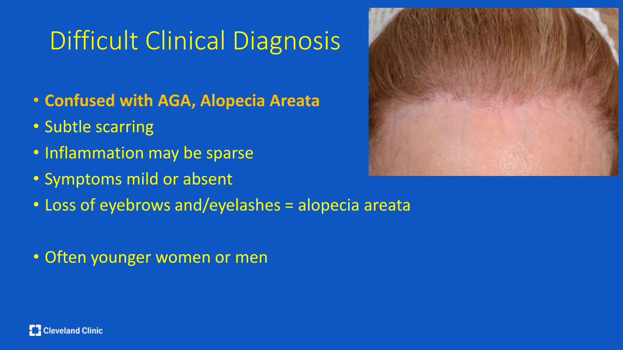
Difficult Clinical Diagnosis
• Confused with AGA, Alopecia Areata
• Subtle scarring
• Inflammation may be sparse
• Symptoms mild or absent
• Loss of eyebrows and/eyelashes = alopecia areata
• Often younger women or men

Clues for FFA?

Atrophy


Hypopigmentation

Subtle Hypopigmentation Wood’s Light

Decrease melanocytes in FFA

Prominent Veins


Kossard S. Arch Derm 1994

Always Lift the Bangs


Young Women


Hypopigmentation

Unusual Distribution


Always Look Behind the Ears


Facial Papules

From: Facial Papules in Frontal Fibrosing Alopecia: Evidence of Vellus Follicle Involvement
Arch Dermatol. 2011;147(12):1424-1427. doi:10.1001/archdermatol.2011.321


JAAD Dec 2015

JAAD Dec 2015
Lichen planus pigmentosis

FFA and LPP Happen in African Americans….

J Clin Aesthet Derm 2016; 9(4):45-51

J Clin Aesthet Derm 2016; 9(4):45-51

Rapid onset loss of eyebrows and scalp hair
Hypopigmentation
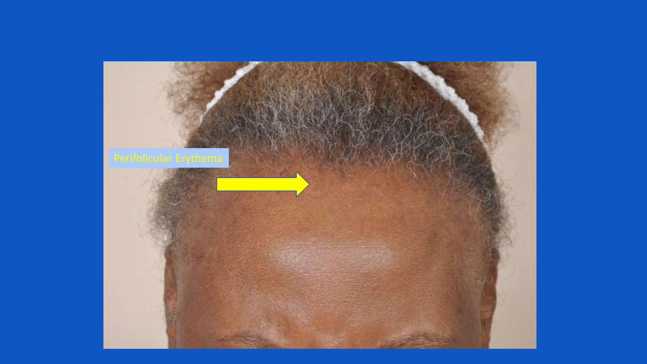
Perifolicular Erythema

Fringe sign

Clues in African Americans
• It is not insidious
• Symptoms
• Rapid loss
• Perifollicular erythema
• Subtle atrophy

Scarring Alopecia Happens in Men….


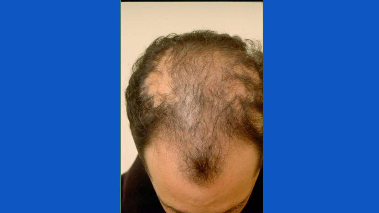
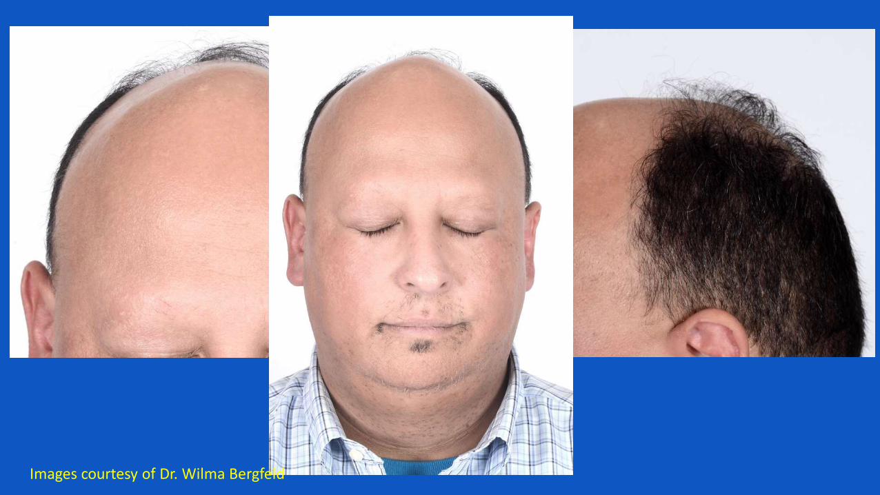
Images courtesy of Dr. Wilma Bergfeld

Images courtesy of Dr. Wilma Bergfeld Facial Papules Dyspigmentation

MimicsA Reminder to Biopsy

Beware of Lichenoid Keratoses
Lupus Erythematosus


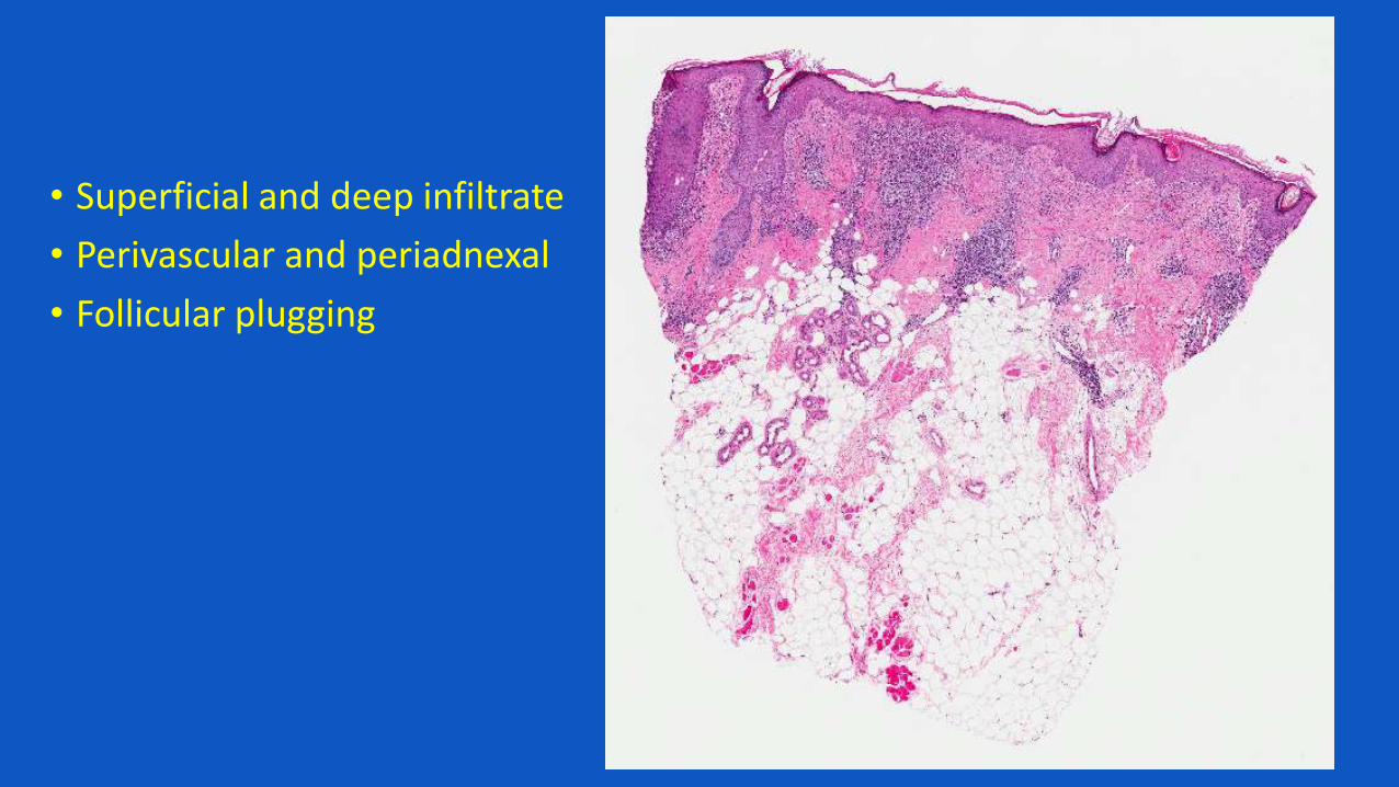
• Superficial and deep infiltrate
• Perivascular and periadnexal
• Follicular plugging

Interface dermatitis-Follicular -Interfollicular


Discoid Lupus Erythematosus
• Mimics AK or SCC – clinical and histology
• Beware or superficial biopsy, recurrent AK/SCC
• Biopsy technique critical• Punch!

• Middle aged man
• Scalp dermatitis and hair loss
• VERY itchy


Treatment
• Methotrexate
• Azathioprine
• Prednisone
• Topical steroids
• Antihistamines


Crusted (Norwegian) Scabies
• All immunosuppressant medications stopped
• Treatment:• Permethrin -> x2, 1 week apart
• Ivermectin -> x2, 2 weeks apart

Outcome
• Pruritus rapidly improve
• LP Pigmentosa faded
• Body and facial itch resolved
• Scalp scale and itch – persisted, but mild
• Alopecia and perifollicular erythema persisted
• Repeat scalp biopsy -> LPP without scabies

Clinical Infectious Diseases. 54(6):882;2012

• When symptoms are out of proportion to findings
• Gets worse with treatment• -> Repeat biopsy!

48 yo with pre diabetes - Hair loss- Scalp pustules - Folliculitis- No pruritus
Frontal Hairline Vertex


Tinea Capitis
• Biopsy and cultures
• Treatment: griseofulvin
• Resolved

Summary
• Clinical Clues• Hypopigmentation• Prominent veins• Loss of eyebrows/eyelashes• Density gradient
• Unusual presentations - Beware• Men• Young women• Parietal scalp/ophiasis -> may spare frontal hair line
• Mimics • Lupus erythematosus• Infections

Thank [email protected]


Always Think About Contact Dermatitis

Biopsy showed:- Spongiotic dermatitis with
eosinophils- Impetiginization- Telogen effluvium
Culture grew MSSA, negative fungus

Clues:- Cyclical recurrence – coincided with hair
coloring- Weeping- Extreme pruritus

Central Scalp Hair LossCCCA or AGA?

CCCA
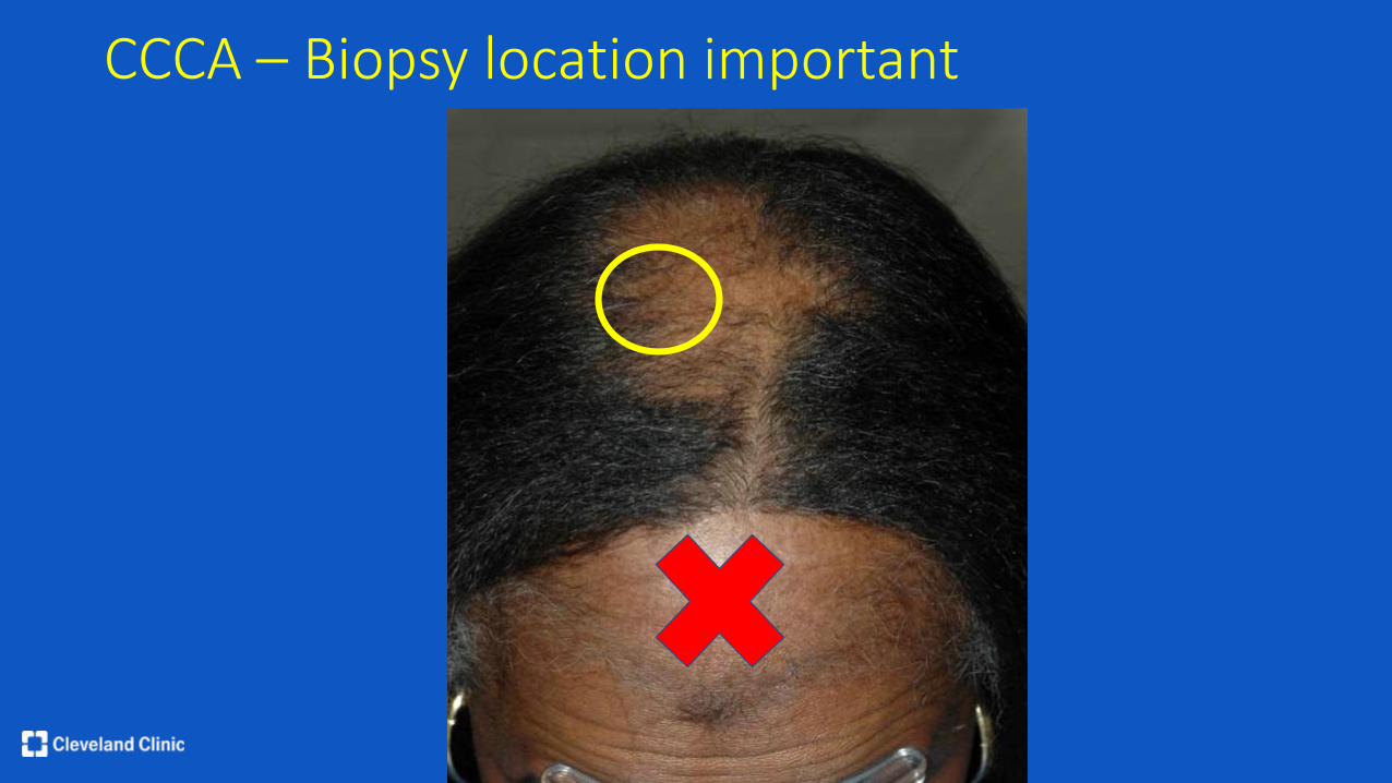
CCCA – Biopsy location important


